for review only - 原泌尿器科病院
TRANSCRIPT
For Review Only(Invited Article) Retrograde Intrarenal Surgery: Past,
Present, and Future
Journal: Investigative and Clinical Urology
Manuscript ID ICU-2020-0526
Manuscript Type: Review Article
Keywords: Current and future endourological topics, Kidney stone, Minimally invasive surgery, Retrograde intrarenal surgery
Areas of specific interest: Endourology/Urolithiasis
https://mc04.manuscriptcentral.com/icurology
Investigative and Clinical Urology

For Review Only
Abstract
With the recent technological advancements in endourology, retrograde intrarenal surgery has
become a more popular procedure for treatment of urolithiasis. Furthermore, since the introduction
of new laser systems and advanced flexible ureteroscopy with miniaturized ureteroscopes, the
treatment indications for retrograde intrarenal surgery have expanded to include not only larger renal
stones of >2 cm but also upper urinary tract urothelial carcinoma, ureteral stricture, and idiopathic
renal hematuria. Clinicians must keep up with these trends and make good use of these technologies
in the rapidly changing field of endourology. Simultaneously, we must consider the risk of various
complications including thermal injury due to laser use, ureteral injury caused by the ureteral access
sheath, and radiation exposure during retrograde intrarenal surgery with fluoroscopic guidance. This
review focuses on the past, present, and future of retrograde intrarenal surgery and provides many
topics and clinical options for urologists to consider.
Key words: Current and future endourological topics; Kidney stone; Minimally invasive surgery;
Retrograde intrarenal surgery
Page 1 of 47
https://mc04.manuscriptcentral.com/icurology
Investigative and Clinical Urology
123456789101112131415161718192021222324252627282930313233343536373839404142434445464748495051525354555657585960
For Review Only
Introduction
Current advancements in endoscopic technology for the upper urinary tract have allowed for the
diagnosis and management of kidney stones, upper urinary tract urothelial carcinoma (UTUC),
ureteral stricture, renal bleeding, and other disorders. In particular, these technological developments
have expanded the treatment options for upper urinary tract stones. Retrograde intrarenal surgery
(RIRS), defined as the use of flexible ureteroscopes (fURSs) and effective lithotripters such as
holmium:yttrium aluminium garnet (holmium:YAG) lasers for intrarenal pelvic diseases, is a useful,
versatile, and minimally invasive procedure for kidney stone management. The current guideline for
management of kidney stones includes RIRS as the first or second recommended procedure in all
categories, even for large stones of >2 cm1)2). In addition, new instruments such as high-power
holmium:YAG lasers, thulium fiber lasers, and single-use ureteroscopes have been introduced for
greater safety, efficiency, and comfort for both patients and surgeons. However, various concerns
have emerged in clinical practice, including complications, cost-effectiveness, and how to use these
new devices simultaneously3). As technological advancements have progressed, the quality of
medical care has changed. This review provides an overview of endourological procedures, RIRS for
the upper urinary tract, key points of surgical techniques including required instruments, and future
trends in this field.
RIRS
Past state of RIRS
Page 2 of 47
https://mc04.manuscriptcentral.com/icurology
Investigative and Clinical Urology
123456789101112131415161718192021222324252627282930313233343536373839404142434445464748495051525354555657585960
For Review Only
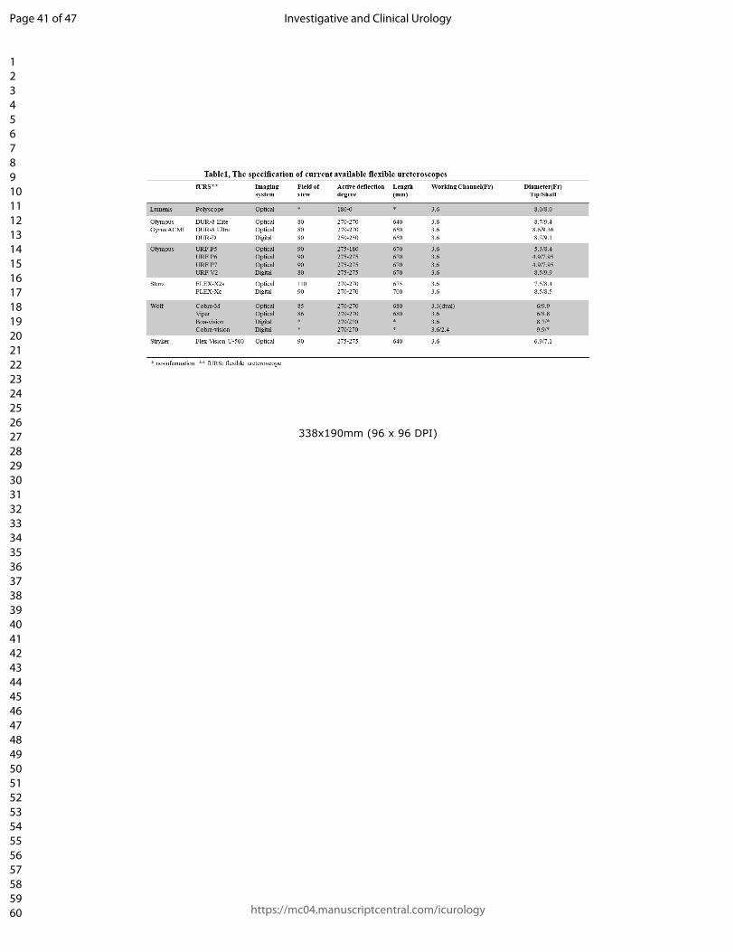
1. History of the fURS
The first fURS, designed by Marshall4) in 1964, was composed of glass fiber that was used to
observe a ureteral stone through a 26-Fr cystoscope. In the early 1970s, Takagi et al.5) and Takayasu
et al.6) first reported the clinical application of a fiberoptic pyeloureteroscope. A few years later,
Bagley et al.7) published their first clinical outcomes of the use of an fURS for diagnosis and
treatment of upper urinary tract disorders. This fURS had a 13-Fr gauge with no working channel or
integrated deflecting function. Therefore, the developments of the fURS during that time were
mainly related to decreasing the diameter of the device and increasing the deflection angle. In 1991,
however, Grasso et al. reported an advanced fURS with a 7.5-Fr tip and an up 120º/down 170º
deflection system. In 1998, they published a clinical study of 492 patients using an fURS with a
larger 3.6-Fr working channel8). Later, in 2001, an fURS with a two-way deflection system
(270º/270º) and stronger durability was introduced to the market, improving access to the
pelvicalyceal system9). With continued progress in technological developments thereafter, the first
digital fURS was manufactured in 2006. This digital fURS provided better image quality and was
much lighter in weight because of the integrated light cable and camera head within the ureteroscope,
which improved the surgeon’s ergonomics. In 2010, Yinghao et al.10) described a newly designed
ureteroscope termed “Sun’s ureteroscope” that had a rigid shaft with a flexible tip. Advancements in
endourological technology have progressed to realize ureteroscopes of much smaller diameter,
stronger durability, and improved image quality. Many fURSs from several companies can now be
Page 3 of 47
https://mc04.manuscriptcentral.com/icurology
Investigative and Clinical Urology
123456789101112131415161718192021222324252627282930313233343536373839404142434445464748495051525354555657585960
For Review Only
utilized in clinical practice (Table 1).
2. Past indications for RIRS
Several decades ago, fURSs were used only for the observation and diagnosis of diseases in the
pelvicalyceal system because of the lack of a useful working channel. Therefore, the indications for
use of fURSs were limited. In 1986, Streem et al.11) first described the use of ureteropyeloscopy for
evaluation of upper tract filling defects. In 1990, Bagley and Rivas12) subsequently reported the
diagnosis and management of upper urinary tract filling defects using an fURS. In 1994, Abdel-
Razzak et al.13) first described the performance of biopsy of upper urinary tract tissues through a
small working channel in an fURS. Furthermore, Bagley and Erhard14) reported the first use of a
holmium:YAG laser for ureteral stones through the working channel in clinical practice in 1995.
Finally in 1998, Bagley15) published the first ureteroscopic laser treatment of upper urinary tract
tumors, which was accomplished using a holmium:YAG laser and neodymium-doped YAG laser.
It has become possible to perform certain procedures through the working channel, such as stone
removal, since Grasso and Bagley8) reported an fURS with a more useful 3.6-Fr working channel. In
addition, successful use of the holmium:YAG laser as a flexible lithotripter expedited the treatment
of upper urinary tract stones in the late 1990s. In 1998, Grasso et al.16) reported the clinical outcomes
of 51 patients with medical comorbidities who underwent RIRS for >2-cm upper urinary tract stones.
They used small-diameter fiberoptic ureteroscopes and a holmium laser lithotripter with a 200-
Page 4 of 47
https://mc04.manuscriptcentral.com/icurology
Investigative and Clinical Urology
123456789101112131415161718192021222324252627282930313233343536373839404142434445464748495051525354555657585960

For Review Only
micron laser fiber. The stone-free rate (SFR) was encouraging at 76% in the first procedure, and the
postoperative complication rate was 6.2%16). Thereafter, many endourologists increasingly utilized
the fURS for treatment of upper urinary stones. Sofer et al.17) reported their experience with 598
patients who underwent ureteroscopy and holmium laser lithotripsy from 1993 to 1999. The average
stone size was 11.3 mm, and 56 patients with intrarenal stones were treated using an fURS. The SFR
among patients with kidney stones was 84% with a low complication rate of 4%17).
Until the 1990s, the definite indications for use of an fURS were unclear with the exception of
evaluating and diagnosing certain upper urinary tract diseases. The main clinical indications for
RIRS seemed to be upper urinary tract stones, especially kidney stones of various sizes. The
advancements of fURSs and the introduction of holmium:YAG lasers to the clinical setting have
promoted progression of urolithiasis treatment18).
Present state of RIRS
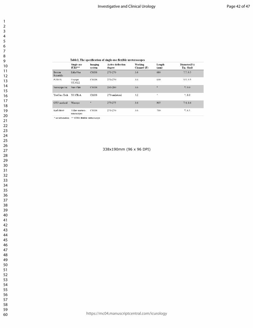
1. Current fURS: Single-use fURS
The fURS has become a mainstay of treatment of nephrolithiasis with increasing indications for
surgical modalities. Most fURSs were manufactured as reusable endoscopes. However, reusable
fURSs have high costs associated with production, maintenance, processing, sterilization, repairs,
and personnel19). Therefore, the cost-effectiveness decreases if an fURS breaks during short
procedures. Doizi et al. conducted an economic analysis of a single-use fURS (LithoVue; Boston
Page 5 of 47
https://mc04.manuscriptcentral.com/icurology
Investigative and Clinical Urology
123456789101112131415161718192021222324252627282930313233343536373839404142434445464748495051525354555657585960

For Review Only
Scientific, Marlborough, MA, USA) and a reusable fURS (URF-V; Olympus, Tokyo, Japan). They
found that the cumulative cost (costs of purchase, maintenance, and repair) of 28 procedures
performed with the reusable fURS was approximately $50,000 (average of $1,786 per case). The
cumulative cost was lower with the single-use fURS (approximately $35,000; average of $1,200 per
case). However, if the price of the single-use fURS were $2,500, the 28 procedures would cost
approximately $70,000. In such a case, the reusable fURS would be more favorable from a financial
standpoint20)21). Although the cost-effectiveness of a single-use fURS depends on the price of the
instrument, the cost-effectiveness of a reusable fURS is also affected by the number of procedures in
which the instrument is used. Martin et al.22) performed a cost assessment between a single-use fURS
(LithoVue) and reusable fURS (Flex-XC; Karl Storz, Tuttlingen, Germany). They found that after 99
ureteroscopic procedures, the cost–benefit analysis favored the reusable fURS over the single-use
fURS and concluded that a single-use fURS may be cost-beneficial at centers with a lower annual
case volume. However, institutions with a high case volume may find reusable fURSs to be more
cost-beneficial22).
A single-use fURS can be very beneficial in patients with large stones, complicated lower pole stones,
anterior lower pole stones, and an anomalous renal anatomy as well as in training of novices, during
which an fURS can be easily damaged23)24). Several single-use fURSs are now available for treatment
of upper urinary tract diseases (Table 2). However, although these single-use fURSs have almost the
same specifications, they have a much thicker tip and shaft than reusable fURSs. Therefore, it is often
Page 6 of 47
https://mc04.manuscriptcentral.com/icurology
Investigative and Clinical Urology
123456789101112131415161718192021222324252627282930313233343536373839404142434445464748495051525354555657585960
For Review Only
difficult to access the upper urinary tract in patients with a narrow ureter and when using a ureteral
access sheath (UAS) smaller than 10 to 12 Fr. In the current era of endourology, the decision to use a
single-use or reusable fURS for treatment of upper urinary tract disease is based on the preoperative
evaluation and intraoperative findings in each case.
2. Current indications for RIRS
The treatment indications for RIRS have been markedly extended with the advancements in
endoscopic technology and lithotripters, such as laser systems. The European Association of Urology
(EAU) guidelines on urolithiasis state that RIRS can generally be applied in patients without specific
contraindications, such as an untreated urinary tract infection (UTI). The guidelines also suggest that
the indications for RIRS include renal stones of <20 mm that are unsuitable for shock wave lithotripsy
(SWL); an unfavorable anatomy for SWL, such as a steep infundibular-pelvic angle, long lower pole
calyx, and narrow infundibulum; lower pole stones of >15 mm not feasible for SWL; the patient’s
preference for kidney stone treatment; and the patient’s social situation (e.g., professions involving
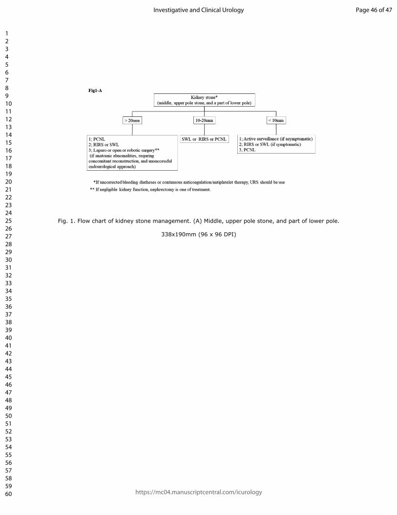
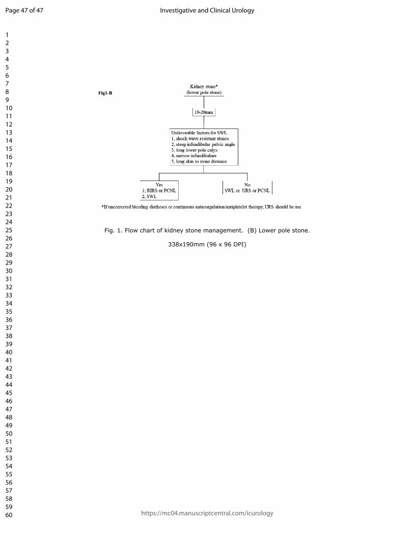
travel, such as a pilot) (Fig. 1A, B)25)26). The other possible indications for RIRS in patients with kidney
stones include radiolucent stones, multiple renal stones unfeasible for SWL, treatment with
anticoagulants, coexistence of renal and ureteral stones, and bleeding disorders26). In general, the first
recommended treatment option for >20-mm kidney stones is percutaneous nephrolithotomy (PCNL).
However, the current surgical techniques of RIRS and laser lithotripsy make it possible to perform
Page 7 of 47
https://mc04.manuscriptcentral.com/icurology
Investigative and Clinical Urology
123456789101112131415161718192021222324252627282930313233343536373839404142434445464748495051525354555657585960

For Review Only
minimally invasive treatment for >20-mm kidney stones. In a recent systematic review and meta-
analysis, the SFR of 20- to 35-mm kidney stones treated by RIRS was 71% to 95%27)28). However,
although it is possible for highly skilled surgeons to successfully perform single procedures for larger
kidney stones, several staged procedures are usually required to achieve a stone-free status. Therefore,
decisions regarding RIRS for larger kidney stones should be made with comprehensive consideration
of various risk factors including the surgeon’s experience, the patient’s comorbidities and preferences,
and the equipment available at the institution29)30).
Favorable indications for single-use fURS in RIRS
A single-use fURS has specific indications in RIRS, including large, hard kidney stones; lower pole
stones with an acute infundibular-pelvic angle; anterior lower pole stones; drug-resistant bacteria in
urine culture; an anomalous renal anatomy; and use by novice trainees. These situations easily induce
damage to the fURS during procedures. Therefore, a single-use fURS would be optimal if the surgical
findings during RIRS allow its use31).
Potential indications for RIRS
With the continued technological developments in endourology, the indications for RIRS have
mainly focused on diseases such as UTUC, ureteral stricture, and ureteropelvic junction stenosis.
One recent topic of interest is ureteroscopic treatment of UTUC by laser ablation using a
Page 8 of 47
https://mc04.manuscriptcentral.com/icurology
Investigative and Clinical Urology
123456789101112131415161718192021222324252627282930313233343536373839404142434445464748495051525354555657585960
For Review Only
holmium:YAG laser or thulium:YAG laser. The EAU guidelines suggest nephron-sparing
management as the primary treatment option not only in patients with low-risk tumors (unifocal, <2
cm in size, low-grade cytology, low-grade fURS-obtained biopsy, and no invasive aspect on
computed tomography urography) but also in patients with kidney deficiency and severe
comorbidities32)33). The role of RIRS in the management of UTUC will be increasingly extended in
the field of endourologic oncology.
3, Surgical steps of RIRS
1) Role of semi-rigid ureteroscope
Semi-rigid ureteroscopes are mainly utilized for the active management of ureteral stones,
direct axial dilation of the distal ureter and ureteral strictures, and the diagnosis of ureteral
tumors. However, semi-rigid ureteroscopes are also used in RIRS to examine the ureteral
stone, check for ureteral relaxation, and assess the extent of the lumen. Selection of an
appropriately sized UAS is very important for negotiation of the renal collecting system30).
Karabulut et al.35) investigated the efficacy of placing the UAS without the obturator over a
semi-rigid ureteroscope under direct vision as the technique of inserting the UAS into the
ureter in RIRS34). This method protects the surgeon and patients from radiation exposure by
shortening the fluoroscopy and operating times35).
Page 9 of 47
https://mc04.manuscriptcentral.com/icurology
Investigative and Clinical Urology
123456789101112131415161718192021222324252627282930313233343536373839404142434445464748495051525354555657585960
For Review Only
2) Safety guide wire
In the first published manual on endourology in 1984, Clayman et al. described the proper
retrograde use of a 0.035- to 0.038-inch wire as a safety guide wire (GW)36). In 1987, Ekman
et al.37) reported the first use of a safety GW in a patient undergoing ureteroscopic stone
removal. During the past three decades, the safety GW has become an indispensable device in
ureteroscopic surgery for ensuring direct access to the collecting system or ureter, decreasing
loss of disorientation in the ureter, avoiding intraoperative complications such as ureteral
injury and perforation, and facilitating insertion of a ureteral stent in cases of failed retrograde
ureteroscopic procedures. However, the use of a safety GW increases the resistance to
passage of the ureteroscope. In particular, the presence of a safety GW interferes with
manipulation of the fURS. Because of current advancements in miniaturized instruments
(e.g., ureteroscope and UAS) and the development of endourological techniques, routine
intraoperative placement of a safety GW might not be needed. Patel et al.38) reported a 2.6%
complication rate in a series of 268 ureteroscopic procedures without a safety GW, with no
perforations or avulsions. Dickstein et al.39) published a series of 305 ureteroscopic
procedures, 270 (89%) of which were uncomplicated even without placement of a safety GW.
However, the remaining 11% of cases required a safety GW because of obstructing ureteral
stones, crushed ureteral stones, and difficult access due to an abnormal anatomy39). Similarly,
a safety GW is not required in our institution when performing RIRS with a UAS because the
Page 10 of 47
https://mc04.manuscriptcentral.com/icurology
Investigative and Clinical Urology
123456789101112131415161718192021222324252627282930313233343536373839404142434445464748495051525354555657585960
For Review Only
placement of a UAS in the upper ureteral portion to access the renal pelvis substitutes for a
safety GW. Therefore, insertion of a UAS in RIRS increases ureter safety intraoperatively.
However, the EAU guideline generally recommends placement of a safety GW in accordance
with best clinical practice in ureteroscopy40). In particular, a safety GW should be placed for
increased ureteral safety in difficult cases, such as an impacted ureteral stone, stricture,
aberrant anatomy, or tortuous ureter, as well as during training of novices.
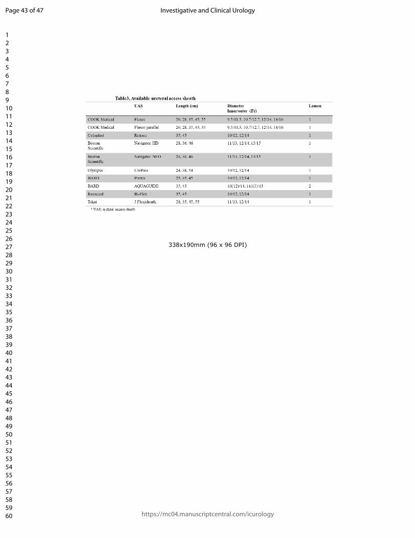
3) UAS
The first UAS was described as a “guide tube” by Takayasu and Aso41) in 1974. They utilized
a UAS to access the proximal ureter with a rigid ureteroscope. The UAS has become an
increasingly popular instrument for treatment of kidney stones and other diseases in the
collecting system during RIRS. A UAS has many advantages, including easy reentry of the
fURS into the collecting system, prevention of increased intrarenal pressure, maintenance of
visualization in the surgical field to facilitate saline irrigation, and use as a possible substitute
for a safety GW26)42). Various UAS sizes ranging from 9.5/11.5 to 14/16 Fr in diameter and
from 28 to 55 cm in length are now available for clinical use (Table 3). However, selection of
the UAS size mostly depends on the surgeons performing the procedure. Ureteral injury may
easily occur if using a UAS larger that the actual ureteral lumen diameter. Traxer and
Thomas43) reported that UAS-related ureteral wall injuries occurred in 46.5% of RIRS
Page 11 of 47
https://mc04.manuscriptcentral.com/icurology
Investigative and Clinical Urology
123456789101112131415161718192021222324252627282930313233343536373839404142434445464748495051525354555657585960

For Review Only
procedures when using a 12- to 14-Fr UAS. They suggested that the ureteral injury severity
determines the grade of injury in terms of the depth of ureteral damage, with a low-grade
injury classified as grade 0 or 1 and a high-grade as grade 2, 3, or 4/5. Grade 2 injuries
involve the ureteral smooth muscle layer (10.1%), and grade 3 injuries involve full-thickness
ureteral perforation (3.3%)43). Generally, the incidence of ureteral injury using a UAS
depends on the relationship between the ureteral diameter and UAS size. Although the
standard UAS size in the United States and Europe seems to be 12 to 14 Fr, the Asian
standard might be 11 to 13 Fr or even smaller because of differences in body size.
Interestingly, one of the current topics in use of a UAS is intrarenal pressure. As mentioned
above, the UAS facilitates the irrigation inflow and outflow of saline. High intrarenal pressure
during procedures may cause urosepsis or a subcapsular renal hematoma. According to some
research, pyelosinus, pyelovenous, and pyelolymphatic backflow of irrigating solution might
occur at intrarenal pressures above 40 cm H2O44). Therefore, keeping the intrarenal pressure
below the limit for intrarenal and pyelosinus backflow might prevent complications during
RIRS. Auge et al.45) reported that a UAS can protect the kidney by reducing the intrarenal
pressure by 57% to 75% during RIRS. Additionally, using a thicker UAS intraoperatively can
decrease the intrarenal pressure46). However, the irrigation inflow and outflow of saline
through a 9.5- to 11.5-Fr UAS is poor. A UAS of this size may result in excessive intrarenal
pressure during RIRS. Therefore, the minimum standard UAS size of 10 to 12 Fr is needed to
Page 12 of 47
https://mc04.manuscriptcentral.com/icurology
Investigative and Clinical Urology
123456789101112131415161718192021222324252627282930313233343536373839404142434445464748495051525354555657585960
For Review Only
acquire acceptable irrigation inflow and outflow of saline and thus maintain good surgical
visualization. In addition, different intrarenal pressures and saline outflow are produced
among the various kinds of available 10- to 12-Fr UASs. Among UASs of this size, the Bi-
Flex (Rocamed, Monaco) and UroPass (Olympus) induce lower intrarenal pressure than the
ReTrace (Coloplast, Humlebæk, Denmark) and Proxis (C.R. Bard, Murray Hill, NJ, USA)
because of their different inner diameters47).
4) Irrigation methods: maintenance of surgical field
In endourological surgery, saline irrigation is mandatory to open and maintain the surgical
field. Visualization of the surgical field is maintained through optimal irrigation of saline. The
irrigation methods used during RIRS have evolved during the past few decades. Lyon et al.48)
first reported the use of an fURS with irrigation connected to the ureteroscopic working
channel and used gravity to maintain the irrigation flow by placing a saline bag 30 cm above
the level of the kidney. A handheld activated syringe-based system was historically used as
the standard method of gravity-induced saline irrigation during RIRS. A foot-activated
syringe-based system is currently available (Peditrol; Wismed, Durban, South Africa)49). In
addition, pressurized irrigant bags and an automatic irrigation pump (AIP) have been
introduced for irrigation during endourological procedures. The view of the surgical field
during RIRS has changed because of increased efficiency of the irrigation flow, which is
Page 13 of 47
https://mc04.manuscriptcentral.com/icurology
Investigative and Clinical Urology
123456789101112131415161718192021222324252627282930313233343536373839404142434445464748495051525354555657585960
For Review Only
influenced by the location and size of the UAS, size of the fURS, and irrigation method.
Irrigation inflow and outflow through the UAS during RIRS is required to open and maintain
optimal renal pelvic distention, good visualization, and low intrarenal pressure. A handheld
activated syringe-based system is commonly used to achieve adequate renal pelvic distention
and a good surgical view. However, a handheld activated syringe-based pump and a foot-
activated syringe-based system may increase the risk of perioperative pyelonephritis and
sepsis secondary to high intrarenal pressure. Therefore, it is crucial to maintain a constant
irrigation flow regardless of the type of instruments in the working channel and ensure an
adequate surgical field to prevent the drastic increases in the intrarenal pressure that might
occur with a handheld activated syringe-based system50). An AIP may help to maintain an
optimal surgical field for easy manipulation of the fURS during RIRS. Lama et al.51) reported
the use of an AIP for irrigation that maintains the same irrigation flow over time in contrast to
gravity irrigation. In addition, Inoue et al.50) recently reported that the irrigation flow from the
tip of the fURS remains almost unchanged by adjusting the pressure control in the AIP system
even when instruments are placed through the working channel of the fURS. Therefore, the
use of an AIP system during RIRS might help to maintain the surgical field and thus
manipulate the fURS with comfort.
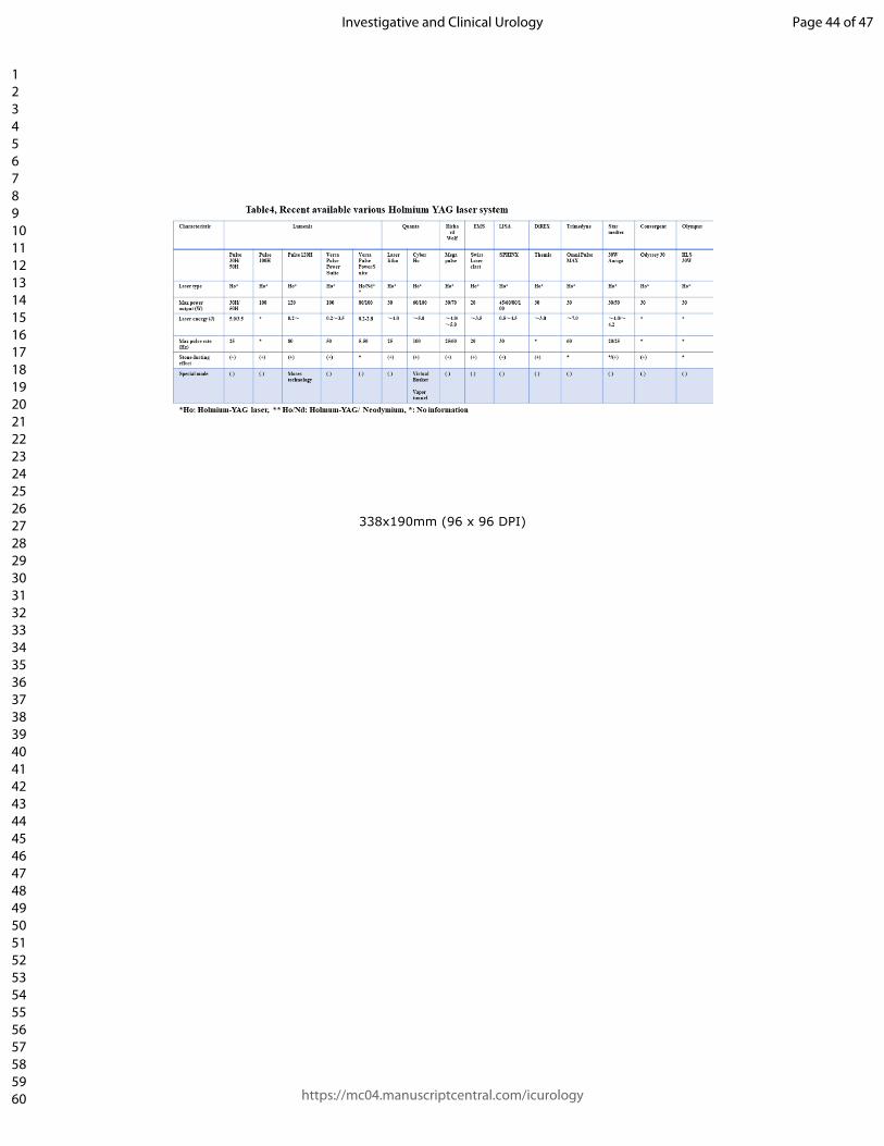
5) Laser instruments and various settings
Page 14 of 47
https://mc04.manuscriptcentral.com/icurology
Investigative and Clinical Urology
123456789101112131415161718192021222324252627282930313233343536373839404142434445464748495051525354555657585960
For Review Only
In RIRS, the holmium:YAG laser system has been the gold standard lithotripsy instrument for
stone management since Denstedt et al.52) first described its use in endourology in their
preliminary report in 1995. Various laser systems with high efficacy and excellent safety
profiles are currently available for stone lithotripsy (Table 4). Traditionally, laser lithotripsy
only allowed for adjustment of the pulse energy and frequency. However, the pulse duration
(width) can now be utilized for stone disintegration. Therefore, stone endourologists can
manipulate these three parameters to perform fragmentation using a lower frequency (5–15
Hz) and higher energy setting (0.6–1.2 J) with a short or long pulse duration or perform
dusting using a high frequency (50–80 Hz) and low energy setting (0.2–0.5 J) with a short or
long pulse duration depending on the particular clinical situation and stone hardness53). The
clinical advantages of a long pulse mode over a short pulse mode are less stone retropulsion,
less fiber degradation, and greater stone dust54). Stone fragmentation involves the creation of
fragments that can be extracted through the UAS with a basket, whereas stone dusting
involves the creation of tiny stone particles of <2 mm that can be spontaneously passed with
no basketing55). However, one currently advocated definition of stone dust (particles of <250
µm) defines dust as particles small enough to meet the following criteria: spontaneous
floating under 40 cm H2O irrigation pressure, mean sedimentation time of <2 s through 10 cm
of saline solution, and full suitability for aspiration through a 3.6-Fr working channel56).
According to data from the Endourological Society worldwide survey in 2014, 26.7% of 414
Page 15 of 47
https://mc04.manuscriptcentral.com/icurology
Investigative and Clinical Urology
123456789101112131415161718192021222324252627282930313233343536373839404142434445464748495051525354555657585960
For Review Only
endourologists from 44 countries actively removed all stone fragments with a basket, whereas
37.4% retrieved only larger fragments but not small fragments. The stone dusting technique
has been increasingly applied in Western countries because of the difficulty of stone
basketing for fragments57). However, Humphreys et al.58) examined whether the SFR is better
with dusting or basketing during RIRS. They concluded that the short-term SFR was higher
with active basket retrieval of fragments (74.3% vs. 58.2%). El-Nahas et al.59) also reported
that the dusting technique had a shorter operation time, whereas the fragmenting technique led
to a significantly higher SFR (78.6% vs. 58.6%). The combination of fragmenting and dusting
may be a more feasible method to break stones. Endourologists choose one of these methods
depending on the situation encountered during surgery (including the stone size, stone
composition, stone location, impaction of stone, stone retropulsion, and surgeon preference)
to improve the effectiveness and outcome of surgery.
High-power holmium:YAG laser therapy with Moses Technology by Lumenis (Clarion
Medical Technologies, Cambridge, Ontario, Canada) has recently become available in clinical
practice. Furthermore, Virtual Basket mode in Cyber-Ho (Quanta System SpA, Samarate,
Italy), which is similar to Moses Technology, has also been introduced. Moses Technology
has improved the stone fragmentation capacity by increasing the energy transmission in water
and reducing stone retropulsion compared with the long pulse mode60). Therefore, Moses
Technology is capable of much less stone retropulsion. In addition, Moses Technology
Page 16 of 47
https://mc04.manuscriptcentral.com/icurology
Investigative and Clinical Urology
123456789101112131415161718192021222324252627282930313233343536373839404142434445464748495051525354555657585960
For Review Only
produces more pronounced disruption of morphological characteristics because it may deliver
a superior laser beam through a vapor channel compared with the conventional
holmium:YAG laser. Higher local temperatures occur during the use of Moses Technology
(direct photothermal effect)61). Therefore, Moses Technology can create a large amount of
tiny stone dust fragments; this is termed the “snow globe effect.” In their in vitro study,
Elhilali et al.62) reported that the Moses mode resulted in a significantly higher stone ablation
volume (160% higher) and less stone movement (50 times less retropulsion) than the regular
mode. Ibrahim et al.63) recently published a randomized clinical trial showing that the Moses
mode was associated with a significantly shorter pulverization time and procedural time than
the regular mode. In addition, there were no significant differences in the success rate at the
end of 3 months (83.3% vs. 88.4%) or intraoperative complications between the Moses mode
group and regular mode group. However, one patient required endoureterotomy for ureteral
stricture in the Moses group63). Thus, close attention should be paid to the risk of thermal
injury and resultant ureteral stricture when using high-power holmium:YAG laser therapy64).
As a cutting-edge instrument in the field of stone lithotripsy, the thulium fiber laser was
launched to disintegrate urinary tract stones. Comparison of the differences between a
holmium laser and thulium fiber laser translate into multiple potential advantages in favor of
the thulium fiber laser, such as a four-fold higher absorption coefficient in water, smaller
operating laser fibers (50- to 150-µm core diameter), lower energy per pulse (as low as 0.025
Page 17 of 47
https://mc04.manuscriptcentral.com/icurology
Investigative and Clinical Urology
123456789101112131415161718192021222324252627282930313233343536373839404142434445464748495051525354555657585960
For Review Only
J), and higher maximal pulse repetition rate (up to 2000 Hz). Comparative in vitro studies
have shown a 1.5- to 4.0-times faster stone ablation rate and much lower stone retropulsion
with the thulium fiber laser than holmium laser65)66). This innovative laser technology is
particularly advantageous for RIRS and may become the next important therapeutic
milestone.
6) Role of preoperative and postoperative ureteral stenting
Preoperative stenting for kidney stone treatment has advantages including a higher SFR, lower
incidence of intraoperative complications (especially ureteral injuries), and greater facilitation of
UAS placement. Preoperative stenting for patients without perioperative infection, severe self-
symptom, anatomical abnormalities, and/or tortuous ureters is not mandatory in most clinical settings
for access to the upper urinary tract because it induces hematuria, pain, urgency, and a risk of febrile
UTI. However, most endourologists have experienced failed access to the upper urinary tract because
of a tight or difficult ureter (8.4%–16.0%)67)68). Once failed access has occurred, staged procedures
are required to achieve passive ureteral dilation 1 to 2 weeks after placing the ureteral stent in the
first ureteroscope.
Postoperative stenting is a quite standard procedure after ureteroscopic surgery not only to prevent
ureteral obstruction due to mucosa edema and ureteral healing but also to avoid ureteral injury,
perforation, residual fragments, bleeding, and UTI. However, the optimal duration of postoperative
Page 18 of 47
https://mc04.manuscriptcentral.com/icurology
Investigative and Clinical Urology
123456789101112131415161718192021222324252627282930313233343536373839404142434445464748495051525354555657585960

For Review Only
ureteral stenting is unknown. The indwelling time preferred by most urologists appears to be 1 to 2
weeks after ureteroscopy. However, routine postoperative stenting is not required if no ureteral
injury is observed under direct ureteroscopic vision at the end of the ureteroscopic surgery, even in
patients who undergo uncomplicated ureteroscopy for impacted ureteral stones69)70). Postoperative
stenting might be associated with higher postoperative morbidity and costs32). Byrne et al. reported
that flank discomfort on postoperative day 1 was significantly less common in patients who did not
undergo stenting; however, there was no significant difference in patient-reported postoperative
hematuria between those who did and did not undergo stenting. With the recent advancements of
smaller instruments for ureteroscopic treatment, the number of patients who do not need
postoperative stenting has increased. However, how to determine which patients do not require
postoperative stenting after ureteroscopic surgery remains unclear.
4. Surgeon’s safety from radiation exposure
Extended low-dose radiation exposure can greatly affect human health in the long term, resulting in
an increased incidence of malignancies including thyroid cancer, breast cancer, and leukemia71). In the
current urological field, radiation exposure among medical personnel and patients has increased.
Therefore, urologists must be aware of the risk of harmful effects caused by radiation exposure. A
major source of radiation exposure for surgeons and medical staff members is scattered radiation
produced by interaction of the primary radiation beam with the patient’s body and the operating table.
Page 19 of 47
https://mc04.manuscriptcentral.com/icurology
Investigative and Clinical Urology
123456789101112131415161718192021222324252627282930313233343536373839404142434445464748495051525354555657585960

For Review Only
Although the dose limit of medical exposure for patients has not been established, the occupational
radiation exposure dose limit has been defined as 50 mSV per year by the National Council on
Radiation Protection and Measurements72). The International Commission on Radiological Protection
has recommended limiting radiation exposure to levels “as low as reasonably achievable” (ALARA)73).
Medical radiation protection principles should be applied for both the patients and medical staff
members involved in imaging, the latter of which include surgeons, nurses, and medical engineers.
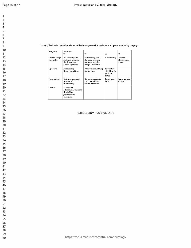
The following are general methods to optimize radiation protection.
① Time: The radiation exposure time should be minimized in terms of both the fluoroscopy time
and the quantity of X-ray photographs acquired.
② Distance: Medical staff members should position themselves as far as possible from the X-ray
source.
③ Shielding: Medical staff members should use adequate shielding materials, such as lead aprons,
lead glasses, and lead radiation-shielding glass.
Shielding for such personnel is usually performed by wearing personal protective clothing. The
standard lead protection protocol requires the use of a 0.35-mm lead apron and thyroid shield by the
operating surgeon and 0.25-mm lead aprons for other personnel74). However, protection from
scattered radiation by protective clothing is incomplete, especially that to the arms, eyes, and brain.
In the endourological field, PCNL using radiologic guidance was initially described by Fernstrom
and Johansson75), who performed this procedure in three patients in 1976. In PCNL, the mean radiation
Page 20 of 47
https://mc04.manuscriptcentral.com/icurology
Investigative and Clinical Urology
123456789101112131415161718192021222324252627282930313233343536373839404142434445464748495051525354555657585960

For Review Only
exposure dose for the surgeon is 12.7 mSV per procedure. This value is higher than the dose of 11.6
mμSV per exposure in flexible ureteroscopy because of the longer fluoroscopic time and close distance
between the radiation source and the surgeon76). The mean fluoroscopy screening time during PCNL
reportedly ranges from 4.5 to 6.04 min (range, 1–12.16 min)77). Furthermore, one study showed that
the mean radiation exposure to the surgeon’s finger and ocular region was 0.28 and 0.125 mSV,
respectively, because of the non-uniform radiation exposure caused by scattered radiation78). Therefore,
the operator’s hands and eyes should also be protected from scattered radiation exposure using gloves
and glasses with lead-threading. Most endourologists generally perform needle puncture for renal
access under fluoroscopy. Therefore, an ultrasound-guided approach is beneficial because it offers
better protection to surgeons from radiation exposure during PCNL than does the fluoroscopic
approach. The surgeon’s radiation dose is lower in ureteroscopy than in PCNL in almost all cases
because ureteroscopy is characterized by a shorter fluoroscopic time and longer distance between the
radiation source and surgeon. Pulsed fluoroscopy was introduced to reduce the radiation dose by
limiting the X-ray exposure time and number of exposures per second. The duration of exposure during
ureteroscopy has been decreased from the original 4.7 min to 0.62 min, and the mean fluoroscopy
screening time during ureteroscopy is reportedly 44.1 s (range, 36.5–51.6 s)79). Kokorowski et al.80)
described the efficacy of a preoperative checklist related to radiation protection. The checklist was
useful for decreasing radiation exposure during procedures. Furthermore, Inoue et al.81) reported that
using protective lead curtains on both sides of the patient table, the operating table end, and the image
Page 21 of 47
https://mc04.manuscriptcentral.com/icurology
Investigative and Clinical Urology
123456789101112131415161718192021222324252627282930313233343536373839404142434445464748495051525354555657585960
For Review Only
intensifier was useful for reducing the surgeon’s radiation exposure during ureteroscopy. The presence
of protective lead curtains caused a 75% to 80% reduction of the scattered radiation dose compared
with the absence of lead curtains. Novel shielding curtains containing bismuth and antimony, which
are also suitable for radiation protection because of their high density and potential weight savings
compared with lead, have also been designed. In modern radiation protection practice, active personal
dosimeters are essential to satisfy the ALARA principle. Most urologists have an insufficient
perception of their own personal radiation protection. A previous study showed that although 84.4%
of urologists who were chronically exposed to ionizing radiation wore lead aprons, only 53.9% wore
a thyroid shield and only 27.9% wore eyeglasses with lead linings. Moreover, only 23.6% of urologists
wore a personal dosimeter82). Awareness of occupational radiation exposure among physicians in the
urological field remains low. Although the risks of harmful effects of occupational radiation exposure
may be relatively low, they should not be ignored (Table 5).
Future state of RIRS
1. Possible indications for RIRS
Various laser systems can be used in RIRS, including a high-power holmium:YAG laser (120 W)
with Moses Technology, thulium fiber laser, thulium:YAG laser, and neodymium-doped YAG laser.
All of these are promising treatment options for several diseases in patients undergoing RIRS. In
addition, a single-use fURS can provide safe and easy access to the kidney anatomy.
Page 22 of 47
https://mc04.manuscriptcentral.com/icurology
Investigative and Clinical Urology
123456789101112131415161718192021222324252627282930313233343536373839404142434445464748495051525354555657585960

For Review Only
The indications for treatment of kidney stones are expected to expand to include larger stones of
>2 cm in future guidelines; less basketing is being performed because of the ability to create large
amounts of stone dust (snow globe effect), and surgical access has improved in patients with a
difficult renal pelvic anatomy, even when the lower pole has an anatomically acute angle. In
addition, for patients with multifocal <3-cm UTUC with low-grade pathological findings and no
invasive aspect on computed tomography urography, retrograde endourological procedures might
become a more common treatment. Furthermore, novel laser systems might help to manage
postoperative ureteral stricture, symptomatic renal cysts, and recurrent ureteropelvic junction
stenosis83)84).
2. New trends in RIRS
1) New fURS with joystick
Usually, fURS manipulation involves torque movement of the hand, back-and-forth
movement of the fURS shaft, and up-and-down movement of the fURS lever. Surgeons must
perform optimal manipulation in a coordinated manner by combinations of these complicated
maneuvers, which may be difficult in some cases. A new fURS with an omni-directional
bending tip using a joystick unit integrated into a handgun-type control unit was recently
introduced. Inoue et al.85) first reported that this novel fURS provided a greater range of reach
along all directions in the lower-pole calyx compared with some usual fURSs in their ex vivo
Page 23 of 47
https://mc04.manuscriptcentral.com/icurology
Investigative and Clinical Urology
123456789101112131415161718192021222324252627282930313233343536373839404142434445464748495051525354555657585960
For Review Only
study. Tambo et al.86) subsequently investigated whether a conventional fURS or novel
joystick fURS is easier to manipulate in their initial constructive validation study. They found
that the novel joystick fURS allowed for much better manipulation by novice trainees and
provided better ergonomics for surgeons. This joystick fURS might have benefits in terms of
ureteroscopic performance86).
2) Thulium versus high-power holmium laser therapy
High-power holmium:YAG lasers have long been available for management of upper urinary
tract stones. Like Moses Technology, the Virtual Basket mode is a special technology that is
quite beneficial in terms of producing tiny particles of stone dust by two forms of ablation: the
photothermal effect and photomechanical effect. In addition, the novel thulium fiber, which is
capable of more quickly producing large amounts of tiny stone dust than the holmium:YAG
laser in vivo, has been introduced to clinical use. Therefore, the stone management strategy
during RIRS has changed from more stone basketing to less stone basketing or no stone
basketing. The differences in the clinical outcomes between the two laser systems is unclear.
However, further refinement of how to use these laser systems will be a key point in the
management of stones, UTUC, and other disorders during RIRS in the coming years.
3) New stone removal devices
Page 24 of 47
https://mc04.manuscriptcentral.com/icurology
Investigative and Clinical Urology
123456789101112131415161718192021222324252627282930313233343536373839404142434445464748495051525354555657585960
For Review Only
Although stone dusting is beneficial, its SFR is still lower than that produced by stone
basketing after RIRS. Therefore, new instruments might be needed to remove the tiny stone
dust particles, such as stone vacuum devices or a novel type of basket. One stone vacuum
device is currently available in clinical practice. Zhu et al.87) compared the efficacy between a
suctioning UAS and traditional UAS. The suctioning UAS had a significantly higher SFR on
postoperative day 1 (82.4% vs. 71.5%), lower incidence of infectious complications (5.5% vs.
13.9%), and shorter operation time (49.7 ± 16.3 vs. 57.0 ± 14.0 min)87). In addition, a new
steerable multi-lumen irrigation/aspiration device (K-VAC; Kalera Medical, San Diego, CA,
USA) was introduced in 2019. This device can be used to access all calyces and navigate
under fluoroscopy to each calyx. The preliminary report showed that it was quite efficient to
remove tiny stone dust fragments and achieve a stone-free status88).
3. Expected trend in RIRS: robotic flexible ureteroscopy
In RIRS, scope manipulation can be technically challenging with a conventional hand-operated
fURS. Therefore, the education to acquire the technical skills of fURS manipulation, such as hands-
on training using a bench model simulator or virtual reality simulator, has recently been expanded89).
However, such education is provided in limited regions and countries. In addition, there are some
another concerns regarding the surgeon’s ergonomics, including radiation exposure, the wearing of a
heavy lead-protector, and the surgeon’s position when operating the fURS. Robotic-assisted fURS
Page 25 of 47
https://mc04.manuscriptcentral.com/icurology
Investigative and Clinical Urology
123456789101112131415161718192021222324252627282930313233343536373839404142434445464748495051525354555657585960
For Review Only
technologies have recently been developed to overcome some of these disadvantages90). The first
robotic fURS (Sensei-Magellan system; Hansen Medical, Mountain View, CA, USA) was reported
in 2011. Desai et al.91) initially attained a 94% technical success rate for stone disintegration and a
complete stone clearance rate of 89% among 18 patients with 5- to 15-mm renal calculi using the
Sensei-Magellan system. However, this robotic fURS was abandoned because difficulties were
encountered in development of the scope design. A few years later, in 2014, Saglam et al.90)
introduced a new robotic fURS system (Roboflex Avicenna; ELMED, Ankara, Turkey). The
Roboflex consisted of a console for operation by the surgeon and a robotic arm for the fURS. The
authors preliminarily reported the clinical efficiency and safety of the Roboflex in 81 consecutive
patients; the clinical outcomes included a short robot docking time of 59.6 s, feasible operation time
of 74 min, and comparable SFR of 96%, all of which were quite acceptable compared with the
conventional hand-operated fURS90). In addition, the Roboflex provided significant advantages in
terms of the surgeon’s ergonomics90)92). Therefore, the system gained CE (Communauté Européenne)
approval for use in Europe in 2013, but Food and Drug Administration approval is still pending.
Although the Roboflex might be optimal in terms of clinical use, it has some limitations included
difficulty of stone removal, hand-operated insertion of the UAS, and difficult adjustment of kidney
movement. However, the newly available high-power holmium:YAG laser and thulium fiber laser
are able to produce large amounts of tiny stone dust particles and may become the next revolutionary
technology in robotic-assisted RIRS93)94).
Page 26 of 47
https://mc04.manuscriptcentral.com/icurology
Investigative and Clinical Urology
123456789101112131415161718192021222324252627282930313233343536373839404142434445464748495051525354555657585960

For Review Only
Conclusion
The endourological technology in RIRS has continued to advance. The single-use fURS, high-
power holmium:YAG laser, and thulium fiber laser may be the next key players in RIRS.
Furthermore, robotic-assisted fURS systems have helped to standardize surgical technical skills and
produce more sustainable surgical outcomes, more comfortable surgeon ergonomics, much less
radiation exposure, and much less surgeon fatigue. Although there are still issues to resolve in RIRS,
endourological procedures are expected to expand the range of treatment indications and become
much less invasive surgical treatment options for patients and surgeons.
Conflicts of interest
We have nothing to disclose.
Page 27 of 47
https://mc04.manuscriptcentral.com/icurology
Investigative and Clinical Urology
123456789101112131415161718192021222324252627282930313233343536373839404142434445464748495051525354555657585960
For Review Only
References
1, Assioms D, Krambeck A, Miller NL, Monga M, Murad MH, Nelson CP, et al. Surgical
management of stones: AUA/Endourology Society Guideline. J Urol. 2016; 196: 1153-1160.
2, Turk C, Knoll T, Petrik A, et al. EUA Guidelines on Urolithiasis: specific stone management of
ureteral stones. 2016. (Cited 8 Nov 2017.) Available from URL: https://uroweb.org/wp-
content/uploads/EAU-Guidelines-Urolithiasis-2016-1. Pdf
3, Hennessey DB, Fojecki G, Papa NP, Lawrentschuk N, Damien B. Single-use disposable digital
flexible ureteroscopes: an ex vivo assessment and cost analysis. BJU int. 2018; 121: 55-61.
4, Marshall VF. Fiber optics in urology. J Urol 1964; 91: 110-114.
5, Takagi T, Go T, Takayasu H, Aso Y. Fiberoptic pyeloureteroscope. Surgery 1971; 70: 661-663.
6, Takayasu H, Aso Y, Takagi T, Go T. Clinical application of fiber-optic pyelouretroscope. Urol Int
1971; 26: 97-104.
7, Bagley DH, Huffman JL, Lyon ES. Flexible ureteropyeloscopy: diagnosis and treatment in the
upper urinary tract. J Urol 1098; 138: 280-285.
8, Grasso M, Bagley D. Small diameter, actively deflectable, flexible ureteropyeloscopy. J Urol
1998; 160: 1648-1653.
9, Ankem MK, Lowry PS, Slovick RW, Munoz Del Rio A, Nakada SY. Clinical utility of dual active
deflection flexible ureteroscope during upper tract ureteropyeloscopy. Urology 2004; 64: 430-434.
10, Yinghao S, Yang B, Gao X. The management of renal caliceal calculi with a newly designed
Page 28 of 47
https://mc04.manuscriptcentral.com/icurology
Investigative and Clinical Urology
123456789101112131415161718192021222324252627282930313233343536373839404142434445464748495051525354555657585960

For Review Only
ureteroscope: a rigid ureteroscope with a deflectable tip. J Endourol 2010; 24: 23-26.
11, Streem SB, Pontes JE, Noviick AC, Montie JE. Ureteropyeloscopy in the evaluation of upper
tract filling defects. J Urol 1986; 136: 383-385.
12, Bagley DH, Rivas D. Upper urinary tract filling defects: flexible ureteroscopic diagnosis. J Urol
1990; 143: 1196-1200.
13, Adbel-Razzak OM, Ehya H, Cubler-Goodman A, Bagley DH. Ureteroscopic biopsy in the upper
urinary tract. Urology 1994; 44: 451-457.
14, Bagley DH, Erhard M. Use of the holmium laser in the upper urinary tract. Tech Urol 1995; 1:
25-30.
15, Bagley DH. Ureteroscopic laser treatment of upper urinary tract tumors. J Clin Laser Med Surg
1998; 16: 55-59.
16, Grasso M, Conlin M, Bagley D. Retrograde ureteropyeloscopic treatment of 2 cm. or greater
upper urinary tract and minor Staghorn calculi. J Urol 1998; 160: 346-351.
17, Sofer M, Watterson JD, Wollin TA, Nott L, Razvi H, Denstedt JD. Holmium; YAG laser
lithotripsy for upper urinary tract calculi in 598 patients. J Urol 2002; 167: 31-34.
Page 29 of 47
https://mc04.manuscriptcentral.com/icurology
Investigative and Clinical Urology
123456789101112131415161718192021222324252627282930313233343536373839404142434445464748495051525354555657585960

For Review Only
18, Cleynenbreugel BV, Kilic O, Akand M. Retrograde intrarenal surgery for renal stones – Part 1.
Turk J Urol 2017; 43: 112-121.
19, Tom WR, Wollin DA, Jiang R, Radvak D, Simmons WN, Preminger GM, Lipkin ME. Next-
generation single-use ureteroscopes: An in vitro comparison. J Endourol 2017; 31: 1301-1307.
20, Scotland KB, Chan J Y.H, Chew BH. Single-use flexible ureteroscopes: How do they compare
with reusable ureteroscopes? J Endourol 2019; 33: 71-78.
21, Hennessey DB, Fojecki GL, Papa NP, Lawrentschuk N, Bolton D. Single-use disposable digital
flexible ureteroscopes: an ex vivo assessment and cost analysis. BJU Int 2018; 121: 55-61.
22, Martin CJ, McAdams SB, Abdul-Muhsin H, Lim VM, Nunez-Nateras R, Tyson MD, Humphreys
MR. The economic implications of a reusable flexible digital ureteroscope: A cost-benefit analysis. J
Urol 2017; 197: 730-735.
23, Leveille R, Kelly EF. Impressive performance: new disposable digital ureteroscope allows for
extreme lower pole access and use of 365μm Holmium laser fiber. J Endourol Case Rep 2016; 2:
114-116.
24, Salvado JA, Cabello JM, Moreno S, Cabello R, Olivaers R, Velasco A. Endoscopic treatment of
lower pole stones; is a disposable ureteroscope preferable? Results of a prospective case-control
study. Cent European J Urol 2019; 72: 280-284.
25, Turk C, Petrik A, Sarica K et al. EAU Guidelines on International Treatment for Urolithiasis. Eur
Urol 2016; 69: 475-482.
Page 30 of 47
https://mc04.manuscriptcentral.com/icurology
Investigative and Clinical Urology
123456789101112131415161718192021222324252627282930313233343536373839404142434445464748495051525354555657585960
For Review Only
26, Inoue T, Okada S, Hamamoto S, Yoshida T, Matsuda T. Current trends and pitfalls in endoscopic
treatment of urolithiasis. Int J Urol 2018; 25: 121-133.
27, Kang SK, Cho KS, Kang DH, Jung HD, Kwon JK, Lee JY. Systematic review and meta-analysis
to compare success rates of retrograde intrarenal surgery versus percutaneous nephrolithotomy for
renal stones > 2cm. Medicine (Baltimore) 2017; 96: e9119.
28, Wilhelm K, Hein S, Adams F, Schlager D, Miernik A, Schoenthaler. Ultra-mini PCNL versus
flexible ureteroscopy: a matched analysis of analgesic consumption and treatment-related patient
satisfaction in patients with renal stones 10-35mm. World J Urol 2015; 33: 2131-2136.
29, Ricchiuti DJ, Smaldone MC, Jacobs BL. Staged retrograde endoscopic lithotripsy as alternative
to PCNL in select patients with large calculi. J Endourol 2007; 21: 1421-1424.
30, Inoue T, Murota T, Okada S, Hamamoto S, Muguruma K, Kinoshita H, Matsuda T; SMART
Study Group. Influence of Pelvicaliceal Anatomy on Stone Clearance After Flexible Ureteroscopy
and Holmium Laser Lithotripsy for Large Renal Stones. J Endourol 2015; 29: 998-1005.
31, Ozimek T, Cordes J, Wiessmeyer JR, Schneider MH, Hupe MC, Gilbert N, Merseburger AS,
Kramer MW. Steep Infundibulopelvic Angle as a New Risk
Factorfor Flexible Ureteroscope Damage and Complicated Postoperative Course. J Endourol 2018;
32: 597-602.
32, Roupret, M, Babjuk M, Burger M, et al. EUA Guidelines on Upper Urinary Tract Urothelial
Carcinoma. Disease management 2019. Available from URL: https://uroweb.org/wp-
Page 31 of 47
https://mc04.manuscriptcentral.com/icurology
Investigative and Clinical Urology
123456789101112131415161718192021222324252627282930313233343536373839404142434445464748495051525354555657585960
For Review Only
content/uploads/EAU-Guidelines-on-Upper-urinary-Tract-Tumours-2019.pdf
33, Kalaitzis C, Zismopoulos A, Glannakopoulos S, Touloupidis S. Ureteroscopic Laser treatment of
Upper urinary tract urothelial cell carcinoma: Can a tumor free status be achieved? Adv Urol 2013;
429585.
34, Aghamir SMK, Salavati A. Endovisually guided zero radiation ureteral access sheath placement
during ureterorenscopy. Minim Invasive Ther Allied Technol 2018; 27: 143-147.
35, Karabulut I, Keskin E, Bedir F, Yilmazel F, Ziypak T, Doluoglu O et al. Rigid ureteroscope
aided insertion of ureteral access sheath in retrograde intrarenal surgery. Urology 2016; 91: 222-225.
36, Dutta R, Vyas A, Landman J, Clayman RV. Death of the safety guidewire. J Endourol 2016; 30:
941-945.
37, Ekman P, Husain I, Sharma ND, Al-Faqih SR. Transurethral ureteroscopy. Safety guide wire as
an aid to a more aggressive approach. Br. J. Urol 1987; 60: 23-27.
38, Patel SR, Mclaren ID, Nakada SY. The ureteroscope as a safety guidewire for
ureteronephroscopy. J Endourol 2012; 26: 351-354.
39, Dickstein RJ, Kreshover JE, Babayan RK, Wang DS. Is a safety wire necessary during routine
flexible ureterscopy? J Endourol 2010; 24: 1589-1592.
40, Turk C, Neisius A, Petrik C. et al. EUA Guidelines on Urolithiasis: Disease Management,
ureteroscopy. 2020. Available from URL: https://uroweb.org/guideline/urolithiasis/#3-4.
41, Takayasu H, Aso Y. Recent development for pyeloureteroscopy: guide tube method for its
Page 32 of 47
https://mc04.manuscriptcentral.com/icurology
Investigative and Clinical Urology
123456789101112131415161718192021222324252627282930313233343536373839404142434445464748495051525354555657585960

For Review Only
introduction into the ureter. J Urol 1974; 112: 176-178.
42, Yong C, Knudsen BE. Ureteroscopy: accessory devices. In: Humphreys M, editor. Ureteroscopy
for stone disease. MUN. Roma, Italy: Minerva Medica; 2016: 55-70.
43, Traxer O, Thomas A. Prospective evaluation and classification of ureteral wall injuries resulting
from insertion of a ureteral access sheath during retro-grade intrarenal surgery. J Urol 2013; 189:
580-584.
44, Rehman J, Monga M, Landman J, Lee D, Felfela T, Conradie MC et al. Characterization of
intrapelvic pressure during ureteropyeloscopy with ureteral access sheaths. Urology 2003; 61: 713-
718.
45, Auge BK, Pietrow PK, Lallas CD, Raj GV, Santa-Cruz RW, Preminger GM et al. Ureteral access
sheath provides protection against elevated renal pressure during routine flexible ureteroscopic stone
manipulation. J Endourol 2004; 18: 33-16.
46, Sener TE, Cloutier J, Villa L, Marson F, Butticè S, Doizi S et al. Can we provide low intrarenal
pressures with good irrigation flow by decreasing the size of ureteral access sheaths? J Endourol
2015; 30: 49-55.
47, Yoshida T, Inoue T, Abe T, Matsuda T. Evaluation of intrapelvic pressure when using small-size
ureteral access sheaths of <10/12F in an Ex Vivo Porcine Kidney Model. J Endourol 2018; 32: 1142-
1147.
48, Lyon ES, Huffman JL, Bagley DH. Ureteroscopy and ureteropyeloscopy. Urology 1984; 23: 29-
Page 33 of 47
https://mc04.manuscriptcentral.com/icurology
Investigative and Clinical Urology
123456789101112131415161718192021222324252627282930313233343536373839404142434445464748495051525354555657585960
For Review Only
36.
49, Blew BD, Dagnone AJ, Pace KT, Honey RJ. Comparison of Peditrol irrigation device and
common methods of irrigation. J Endourol 2005; 19: 562-565.
50, Inoue T, Yamamichi F, Okada S, Hamamoto S, Fujisawa M, SMART Study Group. Change in
irrigation flow through a flexible ureteroscope with various devices in the working channel:
Comparison between an automatic irrigation pump and gravity-based irrigation. Int J Urol 2020; 27:
333-338.
51, Lama DJ, Owyong M, Parkhomenko E, Patel RM, Landman J, Clayman RV. Fluid dynamic
analysis of hand-pump infuser and UROMAT endoscopic automatic system for irrigation through a
flexible ureteroscope. J Endourol 2018; 32: 431-436.
52, Denstedt JD, Razvi HA, Sales JL, Eberwein PM. Preliminary experience with holmium: YAG
laser lithotripsy. J Endourol 1995; 9: 255-258.
53, Schwartz BF, Denstedt JD. A Comprehensive Contemporary Gudide. Ureteroscopy. Springer.
Chapter 8, Ho: YAG laser lithotripsy, 101-112.
54, Wollin DA, Ackerman A, Yang C, Chen T, Simmons WN, Preminger GM et al. Variable pulse
duration from a new holmium; YAG laser: The effect in stone comminution, fiber tip degradation,
and retropulsion in a dusting model. Urology 2017; 103: 47-51.
55, Kronenberg P, Somani B. Advances in lasers for the treatment of stones – a systematic review.
Curr Urol Rep 2018; 19: 45.
Page 34 of 47
https://mc04.manuscriptcentral.com/icurology
Investigative and Clinical Urology
123456789101112131415161718192021222324252627282930313233343536373839404142434445464748495051525354555657585960

For Review Only
56, Keller EX, Coninck VD, Doizi S, Daudon M et al. What is the exact definition of stone dust? An
in vitro evaluation. World J Urol 2020; Apr 8. doi:10.1007/s00345-020-03178-z.
57, Dauw CA, Simeon L. Abdulrahman F, Sanguedolce F, Hollingsworth JM, Roberts WW et al.
Contemporary practice patterns of flexible ureteroscopy for treating renal stones: results of a
worldwide survey. J Endourol 2015; 29: 1221-1230.
58, Humphreys MR, Shah OD, Monga M, Chang TH, Krambeck AE, Sur RL et al. Dusting versus
Basketing during ureeroscopy-Which technique is more efficacious? A prospective multicenter trial
from the EDGD research consortium. J Urol 2018; 199: 1272-1276.
59, El-Nahas AR, Almousawi S, Alqattan Y, Alqadri IM, Al-Shaiji TF, Al-Terki A. Dusting versus
fragmentation for renal stones during flexible ureteroscopy. Arab J Urol 2019; 17: 138-142.
60, Elhilali MM, Badaan S, Ibrahim A, Andonian S et al. Use of the Moses technology to improve
holmium laser lithotripsy outcomes; A preclinical study. J Endourol 2017; 31: 598-604.
61, Keller EX, Coninck Vd, Audouin M, Doizi S, Bazin D, Daudon M, Traxer O. Fragments and
dust after Holmium laser lithotriopsy with or without Moses technology; How are they different? J
Biophotonics 2019; 12: e201800227. Doi: 10.1002/jbio.201800227.
62, Elhilali MM, Badaan S, Ibrahim A et al. Use of the Moses Technology to improve Holmium
laser lithotriopsy outcomes: A Preclinical study. J Endourol 2017; 31: 598-604.
63, Ibrahim A, Elhilali MM, Fahmy N, Carrier S, Andonian S. Double-Blinded Prospective
Randomized Clinical Trial Comparing Regular and Moses Modes of Holmium Laser Lithotripsy. J
Page 35 of 47
https://mc04.manuscriptcentral.com/icurology
Investigative and Clinical Urology
123456789101112131415161718192021222324252627282930313233343536373839404142434445464748495051525354555657585960

For Review Only
Endourol 2020; 34: 624-628.
64, Liang H, Liang L, Yu Y, Chen J, Wang C, Zhu Z, Liang X. Thermal effect of holmium laser during
ureteroscopic lithotripsy. BMC Urol 2020; 20: 69. Doi: 10.1186/s12894-020-00639-w.
65, Taxer O, Keller EX. Thulium fiber laser: the new player for kidney stone treatment? A comparison
with Holmium: YAG laser. World J Urol 2020; 38: 1883-1894.
66, Andreeva V, Vinarov A, Yaroslavsky I, Kovalenko A, Vybornov A, Rapoport L et al. Preclinical
comparison of superpulse thulium fiber laser and a holmium: YAG laser for lithotripsy. World J Urol
2020; 38: 497-503.
67, Cetti RJ, Biers S, Keoghane Sr. The difficult ureter: what is the incidence of pre-stenting? Ann R.
Coll. Surg. Engl. 2011; 93: 31-33.
68, Viers BR, Viers LD, Hull NC, Hanson TJ, Mehta RA, Bergstralh EJ et al. The difficult ureter:
Clinical and radiographic characteristics associated with upper urinary tract access at the time of
ureteroscopic stone treatment. Urology 2015; 86: 878-884.
69, Haleblian G, Kijvikai K, de la Rosette J, Preminger G. Ureteral atenting and urinary stone
management: a systematic review. J Urol 2008; 179: 424-430.
70, Cervik I, Dillioglugil O, Akdas A, Siegel Y. Is stent placement necessary after uncomplicated
ureteroscopy for removal of impacted ureteral stone? J Endourol 2010; 24: 1263-1267.
71, Pukkala E, Kesminiene A, Poliakov S, Ryzhov A, Drozdovitch V, Kovgan L, Kttronen P,
Malakhova IV, Gulak L, Cardis E. Breast cancer in Belarus and Ukraine after the Chernobyl accident.
Page 36 of 47
https://mc04.manuscriptcentral.com/icurology
Investigative and Clinical Urology
123456789101112131415161718192021222324252627282930313233343536373839404142434445464748495051525354555657585960

For Review Only
Int J Cancer. 2006; 119(3): 651-658.
72, Occupational dose limits for adults. Part 20-Standards for protection against radiation, Nuclear
Regulatory Commission Regulations Title 10, Code of federal Regulation. Washington, D. C.: United
States Nuclear Regulatory Commission 1991.
73, Duran A, Hian SK, Miller DL, Le Heron J, Padovani R, Vano E. Recommendations for
occupational radiation protection in interventional cardiology. Cathet Cardiovasc Interv 2013; 82:29-
42.
74, Medical and Dental Guidance Notes: A Good Practice Guide on all Aspects of Ionising Radiation
Protection in the Clinical Environment. York: Institute of Physics and Engineering in Medicine, 2002.
75, Fernstrom I, Johansson B. Percutaneous pyelolithotomy. A new extraction technique. Scand. J
Urol Nephrol 1976; 10: 257-259.
76, Hellawell GQ, Mutch SJ, Thevendran G, Wells E, Morgan RJ. Radiation exposure and the
urologist: What are the risks? J Urol 2005; 174: 948-952.
77, Kumari G, Kumar P, Wadhwa P, Aron M, Gupta NP, Dogra PN. Radiation exposure to the patient
and operating room personnel during percutaneous nephrolithotomy. Int Urol Nephrol 2006; 38: 207-
210.
78, Majidpour HS. Risk of radiation exposure during PCNL. Urol J 2010; 7: 87-89.
79, Elkoushy MA, Shahrour W, Andonian S. Pulsed fluoroscopy in ureteroscopy and perctaneous
nephrolithotomy. Urology 2012; 79:1230-1235.
Page 37 of 47
https://mc04.manuscriptcentral.com/icurology
Investigative and Clinical Urology
123456789101112131415161718192021222324252627282930313233343536373839404142434445464748495051525354555657585960

For Review Only
80, Paul J Kokorowski, Jeanne S Chow, Keith J Strauss, Melanie Pennison, William Tan, Bartley
Cilento, Caleb P Nelson. Prospective systematic intervention to reduce patient exposure to radiation
during pediatric ureteroscopy. J Urol 2013 190(40): 1474-1478.
81, Inoue T, Komemushi A, Murota T, Yoshida T, Taguchi M, Matsuda T, et al. Effect of protective
lead curtains on scattered radiation exposure to the operator during ureteroscopy for stone disease: a
controlled trial. Urology 2017; 109:60-66.
82, Borges CF, Reggio E, Vicentini FC, Reis LO, Carnelli GR, Fregonesi A. How are we protecting
ourselves from radiation exposure? A nationwide survey. Int Urol Nephrol 2015; 47: 271-4.
83, Zewu Z, Hequn C, Yu C, Yang L , Zhongqing Y , Zhiyong C et al. Long-term outcome after
flexible ureteroscopy with holmium laser for simultaneous treatment of a single renal cyst and
ipsilateral renal stones. J Int Med Res 2019; 47: 3601-3612.
84, Geavlete P, Georgescu D, Mirciulescu V, Niţă G. Ureteroscopic laser approach in recurrent
ureteropelvic junction stenosis. Eur Urol 2007; 51: 1542-1548.
85, Inoue T, Okada S, Hamamoto S, Miura H, Matsuzaki J, Tambo M et al. Evaluation of flexible
ureteroscope with an omni-directional bending tip, using a joystick unit (URF-Y0016); an ex-vivo
study. World J Urol 2020; 14. Doi: 10.1007/s00345-020-03151-w.
86, Tambo M, Inoue T, Miura H, Matsuzaki J, Nutahara K , Hamamoto S et al. A novel flexible
ureteroscope with omnidirectional bending tip using joystick-type control unit (URF-Y001): Initial
validation study in bench models. J Endourol 2020; 34: 676-681.
Page 38 of 47
https://mc04.manuscriptcentral.com/icurology
Investigative and Clinical Urology
123456789101112131415161718192021222324252627282930313233343536373839404142434445464748495051525354555657585960

For Review Only
87, Zhu Z, Cui Y, Zeng F, Yang Li 1, Zhiyong Chen 1, Chen Hequn et al. Comparison of suctioning
and traditional ureteral access sheath during flexible ureteroscopy in the treatment of renal stones.
World J Urol 2019; 37: 921-929.
88, Proietti S. New technology in retrograde intrarenal surgery: Unnecessary luxury vs. measurable
benefit. 35 th Annual meeting of European Association Urology Congress – Virtual in 2020.
89, Inoue T, Okada S, Hanamoto S, Matsuda T. New advanced bench model for flexible ureteroscopic
training; The Smart simulator. J Endourol 2018; 32; 22-27.
90, Saglam R, Muslumanoglu AY, Tokatli Z, Caşkurlu T, Sarica K, Taşçi Aİ et al. A new robot for
flexible ureteroscopy: development and early clinical results (IDEAL stage 1-2b). Eur Urol 2014; 66:
1092-1100.
91, Desai MM, Grover R, Aron M, Ganpule A, Joshi S, Desai MR et al. Robotic flexible ureteroscopy
for renal calculi: initial clinical experience. J Urol 2011; 186: 563-568.
92, Geavlete P, Saglam R, Georgescu D, Mulţescu R, Iordache V, Kabakci AS et al. Robotic flexible
ureteroscopy versus classic flexible ureteroscopy in renal stones: the initial Romanian experience.
Chirurgia (Bucur) 2016; 111: 326-329.
93, Brodie A, Vasdev N. The future of robotic surgery. The future of robotic surgery. Ann R Coll Surg
Endl 2018; 100: 4-13.
94, Inoue T, Okada S, Hamamoto S, Yoshida T, Matsuda T. Current trends and pitfalls in endoscopic
treatment of urolithiasis. Int J Urol 2018; 25: 121-133.
Page 39 of 47
https://mc04.manuscriptcentral.com/icurology
Investigative and Clinical Urology
123456789101112131415161718192021222324252627282930313233343536373839404142434445464748495051525354555657585960

For Review Only
Figure legend
Fig. 1. Flow chart of kidney stone management. (A) Middle, upper pole stone, and part of lower pole.
(B) Lower pole stone.
Page 40 of 47
https://mc04.manuscriptcentral.com/icurology
Investigative and Clinical Urology
123456789101112131415161718192021222324252627282930313233343536373839404142434445464748495051525354555657585960
For Review Only
338x190mm (96 x 96 DPI)
Page 41 of 47
https://mc04.manuscriptcentral.com/icurology
Investigative and Clinical Urology
123456789101112131415161718192021222324252627282930313233343536373839404142434445464748495051525354555657585960
For Review Only
338x190mm (96 x 96 DPI)
Page 42 of 47
https://mc04.manuscriptcentral.com/icurology
Investigative and Clinical Urology
123456789101112131415161718192021222324252627282930313233343536373839404142434445464748495051525354555657585960
For Review Only
338x190mm (96 x 96 DPI)
Page 43 of 47
https://mc04.manuscriptcentral.com/icurology
Investigative and Clinical Urology
123456789101112131415161718192021222324252627282930313233343536373839404142434445464748495051525354555657585960
For Review Only
338x190mm (96 x 96 DPI)
Page 44 of 47
https://mc04.manuscriptcentral.com/icurology
Investigative and Clinical Urology
123456789101112131415161718192021222324252627282930313233343536373839404142434445464748495051525354555657585960
For Review Only
338x190mm (96 x 96 DPI)
Page 45 of 47
https://mc04.manuscriptcentral.com/icurology
Investigative and Clinical Urology
123456789101112131415161718192021222324252627282930313233343536373839404142434445464748495051525354555657585960
For Review Only
Fig. 1. Flow chart of kidney stone management. (A) Middle, upper pole stone, and part of lower pole.
338x190mm (96 x 96 DPI)
Page 46 of 47
https://mc04.manuscriptcentral.com/icurology
Investigative and Clinical Urology
123456789101112131415161718192021222324252627282930313233343536373839404142434445464748495051525354555657585960
For Review Only
Fig. 1. Flow chart of kidney stone management. (B) Lower pole stone.
338x190mm (96 x 96 DPI)
Page 47 of 47
https://mc04.manuscriptcentral.com/icurology
Investigative and Clinical Urology
123456789101112131415161718192021222324252627282930313233343536373839404142434445464748495051525354555657585960