folia - uvlf
TRANSCRIPT

F O L I AVETERINARIA
The scientific journal of theUNIVERSITY OF VETERINARY MEDICINE AND PHARMACY IN KOŠICE — Slovakia
ISSN 0015-5748eISSN 2453-7837
1LXIII • 2019

FOLIA VETERINARIA is a scientific journal issued by the University of Veterinary Medicine and Pharmacy in Košice, Komenského 73, 041 81 Košice, Slovakia. The journal is published quaterly in English (numbers 1—4) and distributed worldwide.
The list of Editorial Board of scientific journal Folia Veterinaria:
Editor-in-Chief: Jana Mojžišová
Deputy/Managing Editor: Juraj Pistl
Editorial Board: Aland, A. (Tartu, Estonia), Banhazi, T. (Toowomba, Aus-tralia), Bao, E. (Nanjing, China), Bíreš, J. (Bratislava, Slovakia), Celer, V. (Brno, Czechia), Fablet, Ch. (Ploufragan, France), Faix, Š. (Košice, Slovakia), Faixová, Z. (Košice, Slovakia), Fedoročko, P. (Košice, Slovakia), Gunnarsson, S. (Skara, Sweden), Kolacz, R. (Wrocław, Poland), Könyves, L. (Budapest, Hungary), Nagy, J. (Košice, Slovakia), Novák, M. (Bratislava, Slovakia), Paulsen, P. (Vienna, Austria), Pěchová, A. (Brno, Czechia), Sossidou, E. N. (Thermi Thessa-loniki, Greece), Večerek, V. (Brno, Czechia), Vorlová, V. (Brno, Czechia)
Vargová, M. — technical editor (Košice, Slovakia)
Contact: tel.: +421 915 984 669 e-mail: [email protected]
Electronic Publisher: De Gruyter Poland, Bogumila Zuga 32A 01-811 Warsaw, Poland
ISSN 2453-7837 on-line ISSN 0015-5748 print EV 3485/09
Publisher’s identification number: IČO 00397474
March 2019

FOLIA VETERINARIA
Folia VeterinariaVol. 63, 1, 2019
VYDÁVA UNIVERZITA VETERINÁRSKEHO LEKÁRSTVA A FARMÁCIE V KOŠICIACH
2019
PUBLISHED BYTHE UNIVERSITY OF VETERINARY MEDICINE AND PHARMACY IN KOŠICE
SLOVAKIA

F O L I A V E T E R I N A R I A, 63, 1, 2019
C O N T E N T S
AJADI, R. A., EGBETADE, A. O., AJAYI, O. O., MAKINDE, O. A., OKANDEJI, M. E.:
INTUSUSSCEPTION SECONDARY TO GASTROINTESTINAL FOREIG BODY IN
AN ELEVEN MONTH OLD JUVENILE AFRICAN LION (PANTHERA LEO). CASE REPORT .................................................... 1
GIRETOVÁ, M., MEDVECKÝ, Ľ., PETROVOVÁ, E., ČÍŽKOVÁ, D., MUDROŇOVÁ, D., DANKO, J.:
EFFECTS OF CELL SEEDING METHODS ON CHONDROGENIC DIFFERENTIATION OF RAT
MESENCHYMAL STEM CELLS IN POLYHYDROXYBUTYRATE/CHITOSAN SCAFFOLDS .................................................. 6
DELIA, T. A., DZIKWI-EMENNAA, A. A., KWAGA, J. K. P., KIA, G. S. N., OLUFEMI, O. T., OTOLORIN, G. R.,
ADANU, A. W.: PREVALENCE OF PORCINE ROTAVIRUS ANTIGEN AND ASSOCIATED RISK FACTORS IN
PIG-RAISING COMMUNITIES AND INSTITUTIONAL PIGGERIES IN ZARIA, KADUNA STATE, NIGERIA .................... 17
JANIKOVIČOVÁ, L., DEMČIŠÁKOVÁ, Z., LUPTÁKOVÁ, L., PETROVOVÁ, E.: PRE-INCUBATION
AND ITS EFFECT ON THE DEVELOPMENT AND MALFORMATIONS OF THE CHICK EMBRYO ..................................... 24
ZEMANOVÁ, S., KORYTÁR, Ľ., BENKŐ, Z., PROKEŠ, M., ONDREJKOVÁ, A.: ECOLOGICAL FACTORS
OF TRANSMISSION, PERSISTENCE AND CIRCULATION OF PATHOGENS IN BAT POPULATIONS ................................ 32
ADELAKUN, O. D., AKINSEYE, V. O., ADESOKAN, H. K., CADMUS, S. I. B.: PREVALENCE AND
ECONOMIC LOSSES DUE TO BOVINE TUBERCULOSIS IN CATTLE SLAUGHTERED
AT BODIJA MUNICIPAL ABATTOIR, IBADAN, NIGERIA ................................................................................................................ 41
PIEGEROVÁ, A., KOŠČOVÁ, J., SCHUSTEROVÁ, P., NEMCOVÁ, R., KRYVTSOVA, M.:
IN VITRO INHIBITION OF BIOFILM FORMATION BY STAPHYLOCOCCUS AUREUS UNDER
THE ACTION OF SELECTED PLANT EXTRACTS ............................................................................................................................... 48
SCHIEBER, M.-C., ŠTRKOLCOVÁ, G.: PREVALENCE OF ENDOPARASITES IN CARNIVORES IN
A ZOO AND A WOLVES PARK IN GERMANY ..................................................................................................................................... 54
BYSTRIANSKA, J., PAPAJOVÁ, I., ŠOLTYS, J., SASÁKOVÁ, N.: CONTAMINATION OF SANDPITS
WITH SOIL-TRANSMITTED HELMINTHS EGGS IN AN URBAN ENVIRONMENT ................................................................ 60
DONIČOVÁ, V., LUKAČÍNOVÁ, A., BEŇAČKA, R., NIŠTIAR, F.: EFFECT OF LOW-DOSE
EXPOSURE TO TOXIC HEAVY METALS ON THE REPRODUCTIVE HEALTH OF RATS A
MULTIGENERATIONAL STUDY .............................................................................................................................................................. 64

11
DOI: 10.2478/fv-2019-0001
FOLIA VETERINARIA, 63, 1: 1—5, 2019
ABSTRACT
An eleven month old male, juvenile lion brought for rehabilitation at the Olusegun Obasanjo Presiden-tial Library (OOPL) Wildlife Park was diagnosed with gastrointestinal foreign body and intussusception based on the history of persistent vomiting following ingestion of a cotton towel, and radiographic findings of a radi-opaque gastric foreign body extending from the fundus to the pylorus and a cylindrical soft tissue mid-abdomi-nal mass. The lion was premedicated with an intramus-cular injections of ketamine (5 mg.kg–1) and midazolam (0.25 mg.kg–1), while anaesthesia was induced with an intravenous injection of propofol (2 mg.kg–1). A celiot-omy followed by gastrotomy and subsequent intestinal resection and anastomosis was performed to remove the gastrointestinal foreign bodies and correct the intussus-
ception. The lion recovered well without any complica-tion and was gradually introduced back into the group housing three weeks later following successful alimen-tation process. This report is probably the first case of intussusception in a lion that was associated with a gas-trointestinal foreign body.
Key words: foreign-body; intestine; lion; intussuscep-tion; stomach
INTRODUCTION
An intussusception is an invagination of one segment of the intestine into another and is due to changes in peristal-sis in the intestinal segments [12]. It results in either partial or complete intestinal obstruction with associated clinical
INTUSUSSCEPTION SECONDARY TO GASTROINTESTINAL FOREIGN BODY IN AN ELEVEN MONTH
OLD JUVENILE AFRICAN LION (PANTHERA LEO)CASE REPORT
Ajadi, R. A.1, Egbetade, A. O.1, Ajayi, O. O.2
Makinde, O. A.1, Okandeji, M. E.3
1Department of Veterinary Medicine and Surgery, Federal University of Agriculture2Olusegun Obasanjo Presidential Library Wildlife Park
3Department of Veterinary Anatomy, Federal University of Agriculture, Abeokuta, Ogun StateNigeria

2
Fig. 1. Ventro-dorsal abdominal radiograph of an eleven month old lion showing the gastric foreign body
signs depending on the chronicity, size or location of the intussusception [9]. The majority of intussusceptions are idiopathic [16], however, causes such as: gastrointestinal foreign body, intestinal parasitism, neoplasms and enteritis have been documented [2, 9, 12, 16]. Intussusceptions have been reported in various animal species such as: dogs and domestic cats [6, 9], cattle [8], Maine coons [16], and a Red corn snake [2]. However, there is no record of an intestinal intussusception in a lion in the literature.
A gastrointestinal foreign body is not common in lions. There is only one report of a gastrointestinal foreign bod-ies in a lion and that was associated with the ingestion of a blanket used to provide warmth for the cubs, resulting in gastric outflow obstruction, perforation and subsequent toxaemia [15]. There has been no report of a gastrointes-tinal foreign body resulting in an intussusception in lions either. This report presents the diagnosis and successful management of a gastrointestinal foreign body with an in-tussusception in a lion.
CASE PRESENTATION
An eleven month old juvenile male lion weighing 22 kg was referred to the Veterinary Teaching Hospital, Federal University of Agriculture, Abeokuta, Ogun State, from the Olusegun Obasanjo Presidential Library (OOPL) Wildlife Park, Abeokuta, Ogun State, where the lion was undergo-ing rehabilitation. The Zoo keeper complained that the lion developed a sudden onset of vomiting two days after being suspected to have eaten the cotton towel kept in the house. The lion was weak and lethargic, and appeared severely dehydrated. The examination following anaesthesia with an intramuscular injections of 5 mg.kg–1 ketamine (Ket-amine®, Kepro, Holland) and 0.25 mg.kg–1 of midazolam (Dormicum®, Claris life, India) revealed that ocular and oral mucous membranes were dry and moderately congest-ed, while the rectal temperature was normal (37.2 °C). Also, the heart rate was rapid (165 beats.min–1), while the femo-ral pulse was weak and rapid (172 beats.min–1). A survey abdominal radiograph obtained with a mobile digital X-ray machine (Siemen, Germany) revealed a radio-opaque gastric foreign body (Fig. 1) with a cylindrical soft tissue opaque mid-abdominal mass. In addition, blood obtained from the cephalic vein for a complete blood cell count and determination of plasma concentrations of urea, creatinine
and electrolytes revealed normal packed cell volume (38.0 %) with neutrophilia (neutrophils: 18.4 × 103.l–1) and leukocy-tosis (WBC: 24.9 × 103.l–1). There was mild hypernatre-mia (sodium: 158 mmol.l–1), hyperchloridaemia (chloride: 125 mmol.l–1) and metabolic acidosis (HCO3: 35 mmol.l–1). The values for the level of creatinine (1.7 mg.dl–1), urea (18 mg.dl–1) and potassium (4.0 mmol.l–1) were normal. Based on the findings, a tentative diagnosis of a gastro-intestinal foreign body was made. The lion was therefore scheduled for celiotomy and gastrotomy.
Management and OutcomeThe lion was premedicated with intramuscular in-
jections of 5 mg.kg–1 ketamine and 0.25 mg.kg–1 of mid-azolam and the ventral abdomen was prepared aseptically for surgery. Thereafter, venous access was secured using

3
23 gauge intravenous cannula and a lactated ringers solu-tion was administered at the rate of 5 ml.kg–1.hr–1. Anaes-thesia was induced with 2 mg.kg–1 of 1 % Propofol (Dipri-van, ICI — Zeneca Pharmaceuticals) and maintained with continuous propofol infusion at the rate of 0.2 mg.kg–1.hr–1. The lion was then placed on dorsal recumbency and the limbs secured to the table. A standard celiotomy incision was made extending from the xyphoid cartilage to the pre-pubic tendon. The stomach was exteriorized and an inci-sion was made at the less vascularized area to access the gastric foreign body (Fig. 2). A partially digested cotton towel measuring about 30 centimetre long was removed from the fundus of the stomach. Following the removal of the gastric foreign body, the gastric incision was closed with a double row of a Lembert suture pattern using size 2-0 Polyglactin 910 (Vicryl, Anhui Kangning Ltd, China). Thereafter, the intestine was explored and a portion of the jejuno-ileum with the intussusception (Fig. 3) was exteri-orized and freed. The devitalized portion was removed, and an end to end anastomosis was done with a Lembert suture pattern using size 2-0 braided Polyglactin 910 (Vicryl, An-hui Kangning Ltd, China). Before closure of the laparotomy
incision, the intestinal anastomosis was tested for leakage and obstruction by injecting saline into the anastomosed site. The laparotomy incision was then closed in three lay-ers. The linea alba was closed with simple continuous su-ture pattern using size 0 braided Polyglactin 910 (Vicryl, Anhui Kangning Ltd, China). The subcutaneous layer was closed with a subcurticular pattern using size 0 Polyglactin 910 (Vicryl, Anhui Kangning Ltd, China), while the skin was closed with a horizontal mattress suture pattern using size 1 nylon monofilament (Agary Ltd, China). Follow-ing recovery, 2 mg.kg–1 of tramadol injection (TramadolR, Gland Pharma, India) was administered intramuscularly. The lion was then returned to the transport cage until full recovery. Postoperatively, the lion was treated twice daily with 500 mg of ciprofloxacin (R. K. Laboratories, India) dissolved in drinking water for seven days. Thereafter, meat was gradually introduced to the lion until full alimentation was restored. The lion was introduced back into its housing three weeks after surgery
Fig. 2. Intraoperative picture of an eleven months old lion showing removal of the ingested cotton towel (Red arrow)
Fig. 3. Intraoperative picture of an eleven months old lion showing the intestinal intussusception

4
DISCUSSION
Although gastrointestinal foreign bodies are commonly encountered in domestic cats, they are not common in wild felidae. They may present with a variety of clinical signs de-pending upon the location, the degree and the duration of the obstruction resulting from them [1, 11]. This is prob-ably the first report of an intussusception in a lion resulting from foreign body ingestion. Intestinal obstructions have been reported to result in disturbances of fluid balance, acid-base status and serum electrolyte concentrations due to hypersecretion and sequestration within the gastrointes-tinal tract [4]. In this report, the lion was presented with vomiting, dehydration and metabolic alkalosis resulting either from gastric outflow obstruction or the intussuscep-tion.
Chemical immobilization and anaesthesia is an inte-gral component in conservation, diagnostic and surgical procedures in wild animal species. The risks involved in chemical immobilization and anaesthesia in lions are: loss of thermoregulation, rigidity, depressed respiration, shock, unpredictable recovery, delayed recovery and convulsions [3]. Although inhalational anaesthesia appears to be the safest technique for anaesthesia of the lion, this might not be feasible in field settings, coupled with the difficulty of endotracheal intubation when compared with domestic cats. Ketamine and propofol have been reported to be suit-able for the induction of anaesthesia in lions [3]. In this report, anaesthesia was successfully induced with propofol and maintained also with propofol using a constant rate infusion technique. Many patients with gastrointestinal disorders are dehydrated. The hypotension associated with anaesthesia as well as the distension of the gut occasioned by redistribution of fluid will compound the fluid deficit. This explains why the lactated Ringers solution was admin-istered at 5 ml.kg–1.hr–1 throughout the intra-operative pe-riod.
Majority of obstructive non-linear intestinal foreign bodies compromise the blood supply to the intestinal seg-ment leading to intestinal wall oedema and progressive ne-crosis. These factors contribute to ileus and to an increase in the number of pathogenic intraluminal bacteria result-ing in the breakdown of the mucosal barrier and systemic endotoxemia [5]. This may be responsible for the neutro-philic leukocytosis observed in the lion. The packed cell volume of the lion was normal probably due to the early
detection of the intussusception or may be as a result of the haemo-concentration resulting from the dehydration.
The major challenges with the management of intestinal foreign bodies are early diagnosis of the condition, and risk associated with anaesthesia of patient with compromised electrolyte and acid base status [14]. Early presentation and recognition of the condition in this lion might have been responsible for the favorable outcome recorded compared to the previous record of gastrointestinal foreign body in lion cubs in which the animals were dead prior to interven-tion. Surgical management and wound healing are com-promised by intestinal wall viability, intraluminal bacterial overgrowth, ileus and hypoproteinaemia [13]. There were no complications resulting from wound dehiscence or an-aesthesia in this lion. Enteric wound breakdown and leak-age are the most serious and catastrophic complications of surgery on the gastrointestinal tract [7].
A technique of single enterotomy removal of linear for-eign bodies has been reported and used with good success [7, 16]. However, many chronic foreign objects may not be safely manipulated due to the severe compromise of the lo-cal gastrointestinal segment. This explains the choice of re-section and anastomosis technique over enterotomy. The choice of resection and anastomosis also allows for the re-moval of devitalized intestinal segments occasioned by the intussusception. However, the technique is more time con-suming and with higher risk of leakage. In addition, several techniques have been reported for the anastomosis of the intestinal segments following resection. These include end to end, end to side and side to side [10]. The technique of choice depends on the length of the intestine that is re-sected and the diameter of the two ends. An end to end anastomosis was performed using a Lembert suture pattern because the resected ends were of the same diameter
In conclusion, captive lions are prone to consume in-digestible materials such as blankets out of curiosity, play-fulness, or even nutritional deficiencies resulting in gastro-intestinal complications; thus the use of such materials to provide warmth for young or sick lions should be discour-aged. Prompt diagnosis of gastro-intestinal foreign body in lions followed by appropriate selection of anaesthetic and surgical technique with adequate intensive post-operative follow-up is essential for successful management.

5
REFERENCES
1. Aronson, L. R., Brockman, D. J., Brown, D. C., 2000: Gas-
trointestinal emergencies. Veterinary Clinics of North Ameri-
ca: Small Animal Practice, 30 (3), 555—579.
2. Bercier, M., Zoll, W., Rosenberg, J. F., Giglio, R., McCoy, L.,
Castleman, W. L., et al., 2017: Gastric intussusceptions in a Red
Corn Snake (Pantherophis guttatus) associated with cryptospo-
ridiosis. Case Rep. Vet. Med., May 7, 2017: 4270904. Retrieved
on 13th June, 2018, from: http://doi.org/10.1155/2017/4270904.
eCollection 2017.
3. Bharathidasan, M., William, B. J., Jayaprakash, R., Kan-
nan, T. A., Thirumurugan, R., George, R. S., 2016: Immobi-
lization and anaesthesia in Asiatic lions (Panthera leo persica).
Adv. Ani. Vet. Sci., 4 (3), 134—144.
4. Boag, A. K., Coe, R. J., Martinez, T. A., Hughes, D., 2005:
Acid-base and electrolyte abnormalities in dogs with gastro-
intestinal foreign bodies. J. Vet. Int. Med., 19 (6), 816—821.
5. Boland, L., Lindsay, S., Brunel, L., Podadera, J., Bennett, P.,
2017: Caecocolic intussusception associated with a cae-
cal polyp and concurrent hepatocellular carcinoma in a cat.
J. Fel. Med. Surg., May 4, 2017. Retrieved on 13th June, 2018,
from https://doi.org/10.1177/20551169177066ttps://doi.org/
10.11775.
6. Firmino, M. O., Frade, M. T. S., Alves, R. C., Maia, L. A.,
Olinda, R. G., Ximenes, R. G., et al., 2017: Intestinal intus-
susception secondary to enteritis caused by Pythium insid-
iosum in a bitch: case report. Arquivo Brasileiro de Medicina
Veterinária e Zootecnia (Brazilian Journal of Veterinary and
Animal Science), 69 (3), 623—626. DOI: 10.1590/1678-4162-
9107.
7. Hayes, G., 2009: Gastrointestinal foreign bodies in dogs and
cats: a retrospective study of 208 cases. J. Small Anim. Pract.,
50 (11), 576−583. DOI: 10.1111/j.1748-5827.2009.00783.x.
8. Karapinar, T., Kom, M., 2007: Transrectal ultrasonographic
diagnosis of jejunoileal intussusception in a cow. Irish. Vet. J.,
60 (7), 422—424.
9. Lukanc, B., Pogorevc, E., Kastelic, A., Erjavec, V., 2014:
Retrograde jejunal intussusception in one year—old cat after
treatment with metoclopramide and menbutone. Slov. Vet.
Res., 51 (4), 201—207.
10. Myles, J., Karl, J. S., Sean, J., James, G. G., 2002: Techniques
of bowel resection and anastomosis. J. Gynecol. Oncol., 7 (3),
284—289.
11. Papazoglou, L. G., Patsikas, M. N., Rallis, T., 2003: Intesti-
nal foreign bodies in dogs and cats. Comp. Cont. Edu. Pract.
Vet., 25 (11), 830—843.
12. Queiroz, R. M., Botter, L. A., Gomez, M. P., Oliveria, R. G. G.,
2015: Enteroenteric intussusception in an adult caused by an
ileal angiomyolipoma. Radiol. Bras., 48 (5), 333—340.
13. Ralphs, S. C., Jessen, C. R., Lipowitz, A. J., 2003: Risk fac-
tors for leakage following intestinal anastomosis in dogs and
cats: 115 cases (1991—2000). J. Am. Vet. Medical. Assoc., 223
(1), 73—77.
14. Sajeni, S., Reisinger, W., Mushonga, B., Kandiwa, E.
Habarugira, G., 2017: Foreign body gastrotomy in an adult
captive Cheetah, Acinonyx jubatus. Alexandria Journal of Vet-
erinary Science, 52 (1), 148—152.
15. Squarre, D., Yabe, J., Mumba, C., Mwase, M., Changula, K.,
Mwasinga, W., Munyeme, M., 2015: Toxaemia secondary to
pyloric foreign body obstruction in two African lion (Pan-
thera leo) cubs. Asian. Pac. J. Trop. Biomed., 5 (9), 779—780.
16. Verschoof, J., Thiel, C., Henrich, M., Kramer, M., 2015:
Gastrointestinal intussusception in the Maine Coon: A review
of 19 cases. Vet. Med. Aust., 102 (2015), 29—37.
Received September 13, 2018
Accepted December 14, 2018

66
DOI: 10.2478/fv-2019-0002FOLIA VETERINARIA, 63, 1: 6—16, 2019
ABSTRACT
The aim of our study was to examine the effects of passive and active cell seeding techniques on in vitro chondrogenic differentiation of mesenchymal stem cells (MSC) isolated from rat bone marrow and seeded on porous biopolymer scaffolds based on polyhydroxybu-tyrate/chitosan (PCH) blends. This paper is focused on the distribution of the cells on and in the scaffolds, since it influences the uniformity of the created extracellular matrix (ECM), as well as the homogenity of the distribu-tion of chondrogenic markers in vitro which ultimately affects the quality of the newly created tissue after in vivo implantation. The three types of cell-scaffold constructs were examined by: fluorescence microscopy, SEM, his-tology and quantitative analysis of the glycosaminogly-cans after chondrogenic cultivation. The results demon-strated that the active cells seeded via the centrifugation of the cell suspension onto the scaffold guaranteed an even distribution of cells on the bulk of the scaffold and the uniform secretion of the ECM products by the dif-ferentiated cells.
EFFECTS OF CELL SEEDING METHODS ON CHONDROGENIC DIFFERENTIATION OF RAT MESENCHYMAL STEM CELLS IN
POLYHYDROXYBUTYRATE/CHITOSAN SCAFFOLDS
Giretová, M.1, Medvecký, Ľ.1, Petrovová, E.2
Čížková, D.2, Mudroňová, D.3, Danko, J.2
1Division of Functional and Hybrid SystemsInstitute of Materials Research of the Slovak Academy of Sciences, Watsonova 47, Kosice
2Institute of Anatomy, 3Institute of ImmunologyUniversity of Veterinary Medicine and Pharmacy in Kosice, Komenskeho 73, 041 81 Kosice
Slovakia
Key words: bone marrow; glycosaminoglycans; ECM; mesenchymal stem cells; polyhydroxybutyrate/chitosan scaffold
INTRODUCTION
The healing process of injured cartilage is insufficient due to the fact, that cartilage is an avascular and aneural tissue with a low number of chondrocytes which results in osteoarthritic changes and the production of inferior fibrocartilage. Several techniques designed to restore in-jured articular cartilage like: e. g. autologous chondrocyte implantation, microfracture, mosaikoplasty, are known in medicine [26]. At the present time, much attention is paid to the field of regenerative medicine and cartilage tissue engineering (TE), where with the appropriate cell type, suitable scaffold for cell seeding and biological factors or substances, which control the cell differentiation into de-sired lineages are studied. The key role of the scaffold is to support cell colonization, migration, growth, differentia-tion, and the development and integration of formed tis-

7
sue [27]. Scaffolds for in vitro chondrogenesis has been characterized as biomaterials based on biopolymers; e.g. collagens, polyhydroxyalkanoate, hyaluronate alginate, and polyurethanes [3, 4, 10, 14]. Chitosan is a natural aminop-olysaccharide consisting of sugars close to natural glycos-aminoglycans (GAGs) characteristic for cartilage tissue. Chitosan is formed by the alkaline deacetylation of chi-tin—the second most abundant natural polysaccharide in the world. Another benefit for the utilization of chitosan includes its antimicrobial properties [11]. M a d i h a l l y and M a t t h e w studied animal tissue tolerance to chito-san based implants with the conclusion, that this type of material causes a minimal body response and is consid-ered as biocompatible [19]. These implants were degraded hydrolytically with lysozymes and the rate of degradation was inversely proportional to the degree of crystallinity. Y a m a n e et al. compared the in vitro properties of a hy-brid composite consisting of hyaluronic acid (HA) coated chitosan fibers and the properties of pure chitosan [34]. Cell adhesion, proliferation and aggrecan synthesis were significantly higher in the hybrid composite with HA than in chitosan. SEM observations showed a typical chondro-genic phenotype of cells with a lot of extracellular matrix. Immunohistochemical staining has demonstrated the rich production of collagen type II by chondyrocytes.
C h o et al. demonstrated the ability to differentiate MSC into chondrocytes using an injectable gel based on chitosan-Poly-N-isopropyl acrylamide [15]. MSCs were cultured in vitro and after the injection of the cell-gel com-plex into the animal organism the cartilage tissue forma-tion was revealed.
T a n et al. studied hydrogels consisting of N-succinyl chitosan and aldehyde hyaluronate with encapsulated bo-vine chondrocytes [29]. The hydrogel allowed the survival of chondrocytes and the maintenance of their typical phe-notype. The authors concluded that the composite system has the potential for tissue engineering applications.
C h e n et al. manufactured three-dimensional sub-strates consisting of chondroitin sulphate (CS), dermatan sulfate (DS) and chitosan in various formulations with potential use in cartilage tissue engineering [13]. The ad-dition of CS and DS positively affected the cell morphol-ogy, glycosaminoglycan and collagen production as well as expression of the corresponding genes. W a n g et al. implanted a 3D substrate consisting of poly (3-hydroxybu-tyrate (PHB) and 3-hydroxyhexanoate) seeded with rabbit
chondrocytes into rabbits after 10 days of in vitro culture [33]. The treated defects in rabbits were filled with cartilage tissue with good connection with the subchondral bone. The scaffolds showed higher accumulation of ECM with Type II collagen and GAGs.
Mesenchymal stem cells (MSCs) are used as a cell source for TE and specifically in cartilage regeneration due to their relatively simple availability from multiple tissues (bone marrow, hair follicles, dental pulp, adipose tissue), high proliferation capacity in laboratory conditions and the ability to differentiate among other cell types (osteocyte, adipocyte) including chondrocytes [1, 6, 31]. Their main tasks in chondrogenic differentiation of MSC are affecting and control of the differentiation process from the point of view of enhancing the synthesis of collagen II, aggrecan and GAGs by differentiated cartilage cells. The effective bi-ological active molecules responsible for the in vitro differ-entiation of MSC into the chondrogenic lineage are dexa-methasone and transforming growth factor as supplements in chondrogenic differentiation culture media [8, 28].
For successful tissue regeneration using cartilage tis-sue engineering, it is recommended that the optimal pore size of scaffolds be between 100—300 μm. A critical step involves the cell seeding on to the porous scaffold [22]. The passive seeding technique is based on dropping cells onto the scaffold surface followed by the cell infiltration through the scaffold microstructure. On the other hand, the ac-tive cell seeding utilises a certain external factor, which improves the penetration of cells into the interior of the substrate (rotation, centrifugation, magnetic field) and the results have demonstrated improved cell distribution and ECM formation by differentiated cells by this method [2, 9, 30].
The aim of this study was to examine the effects of pas-sive and active cell seeding techniques on the in vitro chon-drogenic differentiation of mesenchymal stem cells isolated from rat bone marrow and seeded on porous biopolymer scaffolds based on polyhydroxybutyrate/chitosan (PCH) blends.
MATERIALS AND METHODS
Scaffold preparationPorous biopolymer polyhydroxybutyrate/chitosan
(PCH) scaffolds were prepared according to M e d v e c k y

8
et al. [21]. The PCH scaffolds with the PHB:Chit ratio equal to 1 : 1 were prepared by the precipitation of PHB (PHB, GoodFellow, dissolved in propylene carbonate) and chito-san (Chit, SigmaAldrich, dissolved in 1 % acetic acid) mix-ture. After stirring for 10 minutes, acetone was added to the slurry until complete precipitation of the biopolymers occurred. The resulting polymer blends were washed with distilled water, filtered, molded in molds (scaffold type A, B—discs) or microcentrifuge tubes (scaffold type C—cone) and frozen at –20 °C. Finally samples were lyophilized (Il-shin) for 6 hours and sterilized in an autoclave. The mi-crostructure of the scaffold was modified by changing the water content in the suspension.
The microstructure of the scaffolds was observed by the scanning electron microscopy (FE SEM JEOL7000) after the deposition of the conductive carbon layer on the scaf-fold surface. The distribution of the molecular weights of PHB and chitosan in the mixtures was determined by gel permeation chromatography (GPC, Watrex, RI detector). Due to the high porosity of the scaffolds, they were free of closed pores and the true density of the PCH scaffolds was determined by Helium Pycnometer (AccuPyc II, Mi-crometics). The porosity of the scaffolds (%) was calculated from the true density of the blend, mass and dimensions of the individual scaffold.
Isolation and culture of rat MSCs The bone marrow was isolated from the long bones
(femur, tibia) of an adult male Wistar rats (300 g) cadav-ers (for up to 3 hours at 4 °C). The full bone marrow was flushed with ice-cold DMEM LG (Dullbecco’s Modified Es-sential Medium; low glucose, Sigma-Aldrich, UK) + 10 % FBS (fetal bovine serum, Biowest, France) culture medium, homogenized, and centrifuged at 200 g for 10 min. The cell pellet was plated on a 75 cm2 culture flask (SPLLife Sci-ences, Korea) and cultured in 15 ml of DMEM LG with 10 % FBS, and 1 % penicillin-streptomycin-amphotericine (ATB-ATM solution, Sigma-Aldrich, UK); and incubated
at 37 °C in a humidified atmosphere with 5 % CO2. Non-adherent cells were removed by changing the medium after 48 h. The cells were passaged upon reaching 90 % of con-fluence. The subconfluent cell layer from passage 2 were released and cells were used for the determination of the MSC multidifferentiation capacity, confirmation of MSC surface markers (CD29, CD90, CD45) by flow cytometry and in vitro chondrogenesis experiments. The cells were seeded in biopolymeric scaffolds by simple dropping of the cell suspension on to the surface of the scaffold and infil-trated the cells to the porous structure of the scaffold by gentle centrifugation.
Phenotypic characterization of MSC by flow cytometryFor flow cytometric analysis of the cells, direct immu-
nofluorescence staining was used, with a combination of conjugated monoclonal antibodies: CD45/CD29/CD90.1 (eBioscience, USA). The specification of the antibodies used is presented in Table 1.
The flow cytometric analysis was performed on a six color BD FACSCantoTM flow cytometer equipped with blue (488 nm) and red (633 nm) lasers (Becton Dickinson Biosciences, USA). The data were analyzed using the BD FACS DivaTM software. The proportions of cells express-ing analyzed CD markers were expressed in percentages.
Multidifferentiation capacity of MSCThe multidifferentiation ability of isolated adher-
ent cells was confirmed by a commercially purchased kit StemPro Chondrogenesis, Adipogenesis and Osteogenesis Differentiation Kit (Gibco) according to the manufacturer instructions. The cells were cultured in the differentiation media for up to 21 days; the medium was changed three times a week. The differentiated cells were fixed with 4 % formaldehyde and stained—fat vacuoles of adipocytes with Oil Red (Sigma), calcium deposits produced by osteoblasts with Alizarin Red S (Sigma) and GAGs in micromasses of chondrocytic cells with Alcian Blue (Sigma).
Table 1. Specification of the anti-mouse monoclonal antibodies used
Type Fluorochrome Clone Isotype Concentration Volume/105cells
anti-CD45 APC OX1 IgG1, κ 0.2 mg.ml–1 2.5 μl
anti-CD29 R-PE HMb1-1 IgG 0.2 mg.ml–1 5 μl
anti-CD90.1 FITC DX5 IgG2, κ 0.5 mg.ml–1 0.25 μl

9
Cell seeding into the biopolymeric scaffoldsThe MSC were enzymatically released from the culture
flasks, counted and the concentration of the cells in sus-pension was adjusted. The final scaffold characteristics (size and volume), cell concentration as well as method of the cell seeding are described in Table 2. Scaffolds were after seeding transferred separately into wells of 48 nonadherent culture plate (Greiner Bio-One) and incubated (37 °C, 5 % CO2, 95 % humidity) for 2 h. Following the 0.5 ml of com-plete chondrogenic medium (DMEM HG—high glucose 4.5 g.l–1), 1 % ITS + 3.50 µg.ml–1 ascorbic acid, 40 µg.ml–1 proline, 10 ng.ml–1 TGF b1, 1 % HEPES, 1 % NEAA, 1 % ATB-ATM solution (all from Sigma) was added to each well containing the cell-scaffold construct. The medium was changed three times a week.
Morphology and topography of MSC seeded in scaffolds by fluorescence staining
Live/dead staining (fluorescein diacetate/ propidium iodide), acridine orange and DAPI(4’,6-diamidino-2-phe-nylindole) staining were used for the visualisation of the cells focused on their morphology, density and topography on the scaffold surfaces so as to evaluate the cross-sections after 2 and 4 weeks of chondrogenic cultivation.
Fluorescein diacetate is metabolised by live cells to a fluorescent product and stains the living cells green. Prop-idium iodide is permeable by damaged cell membranes and stains the dead cells red (live/dead staining). DAPI stains the cell nuclei blue. Acridin orange stains the cell nuclei yellow-green to orange-red. After washing with PBS, the stained cell-scaffold constructs were observed by a fluores-cence optical microscope Leica DM IL LED, blue filter.
For obtaining quantitative data, which could more pre-cisely characterize the distribution of cells across the sub-strate, the the conical scaffold C was sectioned into 3 parts:
surface, middle and bottom part. The thickness of each part was about 2 milimeters and the cell nuclei on captured im-ages stained with DAPI were counted on areas of 1 mm2.
Histological staining of cell-scaffold constructsAfter 4 weeks of cultivation in complete chondrogenic
media, the constructs were removed, washed and fixed in 4 % paraformaldehyde. The specimens were sectioned (Leica RM 2255) in 5—10 µm slides and stained with alcian blue for 30 minutes (GAGs staining); the cell nuclei were stained with nuclear fast red. Scaffolds C failed to prepare for histological staining due to their disruption in paraffin.
Scanning electron microscopy staining of cells— scaffold constructs
After 2 and 4 weeks of cultivation of the cell-scaffold constructs in chondrogenic media, scaffolds were removed, washed with PBS and fixed in 2.5 % glutaraldehyde in PBS for 24 hours at 4 °C. After dehydratation in an ethanol gradient from 30 to 100 % and freeze drying (Illshin), the specimens were sputter-coated with carbon and observed (JEOL FM SEM JSM-7000F).
Determination of DNA and GAG-s content in cell-scaffold constructs
After 4 weeks of cultivation in chondrogenic medium, scaffolds were removed and washed with PBS and lysed in papain buffer after homogenization (Tissuerupter, Qua-gen) at 60 °C for 24 hours. The aliquots of supernatants were used for GAGs determination by the DMMB meth-odology and DNA estimation by Hoechst 33258. To deter-mine the GAGs content, the 250 μl solution of the dimeth-ylmethylene blue (DMMB) was added to 50 μl of the super-natant and absorbance at 450 and 525 nm was measured by UV VIS spectrophotometer (Shimadzu, UV-1800). The
Table 2. Scaffold characteristics, cell density on scaffolds and methods of cell seeding
Type Composition Cell seedingmethod
Average scaffold porosity*
Scaffold volume(μl)* Cells/scaffold Cells.cm-3 scaf-
fold
A PCH (1 : 1) dropping 85 ± 4.1 50 ± 5 3.5 × 10⁵ 7.0 × 10⁶
B chitosan (100 %) dropping 92 ± 4.8 50 ± 5 3.5 × 10⁵ 7.0 × 10⁶
C PCH (1 : 1) centrifugation (1400 rpm/5 min) 94 ± 3.8 100 ± 15 3.5 × 10⁵ 3.5 × 10⁶
* — mean ± standard deviation

10
chondroitin sulfate from shark cartilage (Sigma) was used as a standard for GAG calibration.
The DNA content in cell lysate from cell constructs was determined using Hoechst 33258. The 20 μl of su-pernatant was added to Hoechst 33258 buffer solution (180 μl) and the DNA content was determined from the calibration curve by fluorimetry (Picofluor, Turner bio-systems). The DNA from a calf thymus (Sigma) was used as a standard.
All of the quantitative measurements were performed on cell-scaffold constructs (n = 3) and then statistically evaluated by ANOVA (Statmost32 statistical programme). The statistical significance of results was determined by one- and two-way ANOVA (P ˂ 0.05).
RESULTS
Scaffold characterizationThe highly porous spongy-like microstructure of the
scaffolds were obtained after lyophilization (Fig. 1). The images document a heterogeneous open microstructure
Fig. 1. SEM images of scaffold microstructures: a) A; b) B; c) C-type
with a high proportion of more regular macropores up to 100 μm size with mutual interconnection via smaller spher-ical pores of < 40 μm. This microstructure allows a faster diffusion of media into the interior of the scaffolds after cell seeding. Also a dense network of fine spherical micropores (diameter < 10 μm) were observable in the pore walls of the scaffolds (Fig. 1b, c). The calculated porosities of the scaf-folds are listed in Table 2 and all of the scaffolds achieved the ≥ 85 % level. The gel permeation chromatography (GPC) analysis showed that the average molecular mass (Mw) of Chit and PHB in the mixtures were 41 kDa and 80 kDa respectively.
Multidifferentiation capacity and phenotype characterization of MSC
The flow cytometric analysis confirmed that > 95 % of the cells expressed CD90, CD29 and around 1.1 % of the cell population expressed CD45. The isolated cells were able to differentiate to adipocytes: red fat vacuoles in adipo-cytes stained with oil red (Fig. 2a); osteoblasts: red colored calcium deposits stained with alizarin red (Fig. 2b); and chondrocytes: blue stained GAGs in micromasses stained
Fig. 2. Multidifferentiation ability of MSC: b) adipogenic differentiation, oil red staining of fat vacuoles; c) osteogenic differentiation, alizarin red staining
of calcium deposits;d) chondrogenic differentiation, alcian blue staining of GAGs

11
Fig. 3. Distribution of cells on scaffolds characterized by fluorescence microscopy:A-type—surface after 2 (a-acridine orange) and 4 weeks (b-live/dead; c- DAPI) of chondrogenic cultivation; cross-sections after 4 weeks of culture (g-acridine orange); B-type—surface after 2 (d-acridine orange) and 4 weeks (e-live/dead; f- DAPI) of chondrogenic cultivation; cross-sections
after 4 weeks of culture (h-live/dead); C-type- cross-sections after 2 (i-DAPI) and 4 weeks (j- live/dead; k- DAPI) of chondrogenic cultivation
with alcian blue (Fig. 2c). These facts confirmed that cells isolated from rat bone marrow were MSC’s.
Morphology and topography of MSC seeded in scaffolds by fluorescence staining
Figs. 3a, b, c, d, e, and f showed cell distributions on A and B scaffolds after 2 and 4 weeks of MSC cultiva-tion in the chondrogenic media. An enormously dense cell population, adhered to the surface was revealed on the sur-
face of the scaffolds. Multiple cell layers with extracellular matrix production were identified using the fluorescence staining techniques. The cross-section of the scaffolds con-firmed the negligible penetration of cells into the inner porous structure, as shown in Fig. 3g and h. A layer of liv-ing cells is clearly visible on the scaffold surfaces. Also the live/dead staining (Figs. 3b, e) did not show the presence of dead cells on the scaffolds, which confirms the low cytotox-icity of the biocomposites.

12
In Figs. 3i, j, and k; a uniform distribution of cells is ob-servable on the surfaces and on the cross-sections of scaf-folds C, but the density of cells was lower than on the A or B samples. The acridine orange staining of scaffolds C was qualitatively unsatisfactory.
The average amount of cells on each assessed part of the scaffold C was 1470 ± 180 cells.mm–2. On the surface part, there were 1320 ± 200 cells.mm–2; on the middle part and bottom parts there were 1180 ± 270 cells.mm–2. Based on these findings it can be stated that cells were randomly distributed and the number of cells in each part of the scaf-fold C was not statistically significantly different (P < 0.05).
Histological staining of cell-scaffold constructs Figs. 4a and b revealed the histological staining of
A and B type scaffolds by Alcian Blue, which demonstrated the presence of GAGs. The arrows indicate cell layers on the surface producing a cartilage-like tissue positively staining dark blue by Alcian blue. The scaffold cross section without seeded cells characterized by porous structure with poly-mer fibers created pore walls is demonstrated in Fig. 4c. Note, the partial staining of biopolymer blends (chitosan) can be visible in Fig. 4, but GAGs were stained more in-tensely blue in the multilayers of cells.
Fig. 4. Histological staining with alcian blue of scaffold A (a) and B (b) after 4 weeks of chondrogenic cultivation and scaffold A without cells (c). Arrows show layer of cells producing cartilage like tissue (positive GAG-s)
Fig. 5. Surface microstructure of scaffolds A (a, b), B (c, d) and cross-section of scaffold C (e, f).Arrows indicate adhered cells) after 2 weeks and 4 weeks of chondrogenic cultivation

13
SEM evaluation of cells—scaffold constructs The SEM images of A and B scaffold surfaces after 2
and 4 weeks of MSC cultivation are shown in Figs. 5a, b, c, and d. The images are consistent with the observations from the fluorescence microscopy and histological stain-ing. The scaffold surfaces are almost completely covered with cell multilayers and clearly indicate the excellent ad-herence of the cells and layers to the scaffold surface. These facts verify a low cytotoxicity of PCH scaffolds.
In the case of the scaffold type C, the SEM showed the microstructure with a cell population adhered and distrib-uted on individual pore walls with partial filling of the scaf-fold pores. The individual cells are relatively difficult to dis-tinguish from the highly porous, plate-like interconnected microstructure of the biopolymers (Figs. 5e and f).
Determination of DNA and GAG-s content in cell-scaffold constructs
The average GAG contents determined by DMMB after 4 weeks of culture of each cell-scaffold type construct are shown in Table 3. We observed statistically significant dif-ferences in GAG contents (P < 0.05) between the individu-al sample types A, B and C with the higher GAG content in the B substrate (220 ± 30 ng) and much lower in the C type substrate (75 ± 10 ng).
Table 3 shows also the amount of DNA in cell-scaffold constructs after chondrogenic cultivation using Hoechst 33258. Similar amounts of DNA were found in the A and C scaffolds, whereas a statistically significant decrease was revealed in scaffold B (P < 0.05).
DISCUSSION
The isolated MSC’s from rat bone marrow represent an adherent population of spindle-shaped fibroblast-like
cells, which were able to differentiate into bone, cartilage and fat cell lineages during cultivation under defined con-ditions. During the in vitro chondrogenic differentiation of MSCs seeded on scaffolds, the supplementation of the culture media with growth factors and other active bio-logical substances is strongly recommended. Certain bio-logically active molecules which bind to the MSC recep-tors affects both the entire differentiation process and the synthesis of extracellular matrix components by the cells. In the chondrogenic differentiation of MSC, glucocorti-coid—dexamethasone and growth factor TGFb1 played crucial roles [28]. The chondrogenic differentiation was mostly carried out in a culture medium without FBS (FBS is a mixture of biological agents like growth factors, hor-mones, etc. which can adversely affect the behavior of the cells in the differentiation process) and FBS was replaced by the supplement containing insulin, transferrin, sel-enite, linoleic acid, oily acid and bovine serum albumin (commercial product ITS + 3), which is used as a serum replacement in serum-free cell cultivation during the chondrogenic differentiation of the stem cells. Also, one of the most used culture media is DMEM HG. DMEM HG contains up to 4.5 g of glucose per liter of medium and enhanced glucose amount is very important in the cultivation of an enormously high number of MSCs be-cause glucose serves as a source of readily available energy for the cells [16, 28, 32]. In our experimental work, the isolation and expansion of MSC took place in DMEM LG medium with the addition of 10 % FBS. However, MSC differentiation to chondrocytes (4 weeks) was performed in a culture medium of DMEM HG with chondrogenic supplements and successful cell differentiation was ex-perimentally demonstrated.
The 3D-porous scaffolds must meet the requirements of biocompatibility, bioresorbability or biodegradability, good mechanical strength, shape, interconnected pores of appro-
Table 3. Measured content of GAGs and DNA in cell-scaffold constructs
Scaffold Seeding of MSC GAG-s content (ng/scaffold) *
DNA content (µg/scaffold)*
A dropping 130 ± 30 0.903 ± 0.1
B dropping 220 ± 30 0.609 ± 0.09
C centrifugation 75 ± 8 0.944 ± 0.1
* — mean ± standard deviation

14
priate size distribution to allow the cells to penetration into the scaffolds as well as allow the flow of nutrients and me-tabolite between the culture medium and the cells [10, 18, 24,]. The above conditions fulfill biomaterials of polymer origin, typically collagen, hyaluronic acid, and chondroi-tin sulphate in the case of cartilage regeneration [7, 12, 14, 25]. A number of authors have developed chitosan-based scaffolds and their composites with other polymers or in-organic compounds (e. g. calcium phosphates). As is well known, chitosan is characterized by favorable properties which predispose its use as a cell scaffold, especially in the regeneration of articular cartilage. Glucosamine groups of chitosan are structurally similar to the GAGs of the carti-lage extracellular matrix and positively affect chondrocyte differentiation. In the living organism, it can be partially enzymatically degraded with lysozymes [5, 20, 23, 35]. The cost of chitosan is several times lower than collagen and hyaluronate which is a prerequisite to its wider availability for patients with traumatic cartilage damage. The porous interconnected structure of the polymer scaffolds provides a large area for the proper distribution, adhesion and cell proliferations as for neovascularization of the scaffold [17, 22]. The pore size of the polymer scaffolds macropores pre-pared in our work was in the range of 50—100 μm with interconnection via micropores of < 30 μm diameter. The open pore spongy-like microstructure significantly improves the nutrients inflow to the deeper parts of the scaffold. A critical step in the cartilage tissue engineering is the cell seeding on the porous scaffold and our results are comparable with results of other research groups [2, 30]. The most appropriate method ensuring the uniform distribution of cells to the inner scaffold pores was active seeding via introducing the cell suspension to the scaffold by centrifugation. In the second method based on drop-ping the cell suspension on to the scaffold surface, the cells didn’t penetrate into the pores and extracellular matrix formation was formed only on the scaffold surface. The cell seeding with very high concentrations for the in vitro chondrogenesis is parallel to cell condensation during em-bryonic development of cartilage in a living organism. For induction of MSC into a chondrogenic lineage, tight con-tacts between cells are desirable and the recommended cell concentrations applied on the scaffold is between 5 and 10 million cells per cm3 of porous scaffold. The much higher cell concentrations as described above were even applied for successful chondrogenesis [2]. In the case of PCH scaf-
folds, the applied concentrations of MSC varied depending on the method of cell seeding. The cell concentrations used in dropping method met the requirement for cell seeding density on the porous cell scaffold contrary to this one in the centrifugation method with the lower cell density. The PCH scaffolds showed the suitable properties for chondro-genic differentiation of MSC´s with production typical of the extracellular matrix component of hyaline-like carti-lage—GAGs.
For comparison in Table 3, the amount of DNA in the cell-scaffold constructs of A and C was approximately the same (0.9 μg DNA/scaffold) contrary to the B type samples where about a 30 % lower amount was found. The dense multilayers of cells were formed after culture on scaffolds A and B (Figs. 3a‒f; 4a, b; 5a—d) and such a cell arrange-ment does not allow for further cell proliferation on scaf-folds because it is not of sufficient size for another adhe-sion and spreading of cells. Simply said, the confluence was achieved. On the other hand, a 3D macroporous mi-crostructure with large areas of pore walls in the scaffold C firstly makes it possible to obtain more homogeneous and uniform distribution of cells in the scaffold volume (Figs. 3i, j, and k; and 5e, and f), but the density of the ad-hered cells in the scaffold was not high enough for suffi-cient mutual cell interconnections during the short-time period after seeding which resulted in lower production of GAGs by the cells on these samples. In the case of scaffolds type A and B, the amount of produced GAGs was around 3 times higher than in the case of substrate C. We con-cluded, that the abundant production of GAGs by cells in the A and B scaffolds was related to the fact, that the cells formed a multilayer on the scaffold surface and the contact between the cells with each other was very tight (parallel to the culture of the chondrogenic pellets or micromasses formed by the very concentrated population of MSC), and this fact supported the differentiation of the cells in multi-layers with good GAGs production. It is possible to assume that the prolonged cultivation of MSC’s can enhance the cell population with significant effects on the production of cartilage markers. Based on recent papers, it can be as-sumed that the integrity of the constructs with active cells evenly distributed therein will be more satisfactory com-pared to scaffolds with active cells adhered to outer scaffold surface only and following overgrown with a differentiated cell layer on its surface and partially ingrowth tissue after implantation. However, it is necessary to consider the cur-

15
rent health condition of the patient with injured cartilage, the cartilage lesion position relative to the anatomy of the entire joint, lesion size, its thickness, and the surgery tech-nique used.
In our future research work, we will focus on increas-ing the initial cell seeding concentration and improving the active cell seeding techniques into porous scaffolds of polymeric origin.
CONCLUSIONS
Two different methods of seeding cells into biopoly-mer scaffolds were compared. The synthesized biopolymer scaffolds were not cytotoxic; the cells grew and differenti-ated on scaffold surfaces and in scaffold pores. The cells seeded by dropping on scaffolds A and B produced higher amounts of GAGs in comparison to cells seeded by cen-trifugation into scaffold C. For chondrogenic cultivation and GAGs production by differentiated cells seeded in suitable porous scaffolds, there is a key prerequisite that a high enough cell seeding concentration and selecting the right cell seeding techniques be utilized. The developed porous PCH scaffold could be utilized in cartilage tissue engineering in veterinary and human medicine due to its satisfactory properties and low cost of input materials for scaffold synthesis.
ACKNOWLEDGEMENTS
This study was supported by the Slovak Grant Agency of the Ministry of Education of the Slovak Republic and the Slo-vak Academy of Sciences, Project No. 2/0047/17.
REFERENCES
1. Barry, F. P., Murphy, J. M., 2004: Review Mesenchymal stem
cells: clinical applications and biological characterization.
Int. J. Biochem. Cell Biol, 36, 568—584. DOI: 10.1016/j.bio-
cel.2003.11.001.
2. Bornes, T. D., Jomha, N. M., Mulet-Sierra, A., Adesida,
A. B., 2016: Optimal seeding densities for in vitro chondro-
genesis of two- and three-dimensional-isolated and expanded
bone marrow-derived mesenchymal stromal stem cells within
a porous collagen scaffold. Tissue Engn. C: Methods, 22, 208—
220. DOI: 10.1089/ten.tec.2015.0365 58.
3. Collins, M. N., Birkinshaw, C., 2013: Hyaluronic acid based
scaffolds for tissue engineering—A review. Carb. Pol., 92 (2),
1262—1279. DOI 10.1016/j.carbpol.2012.10.028.
4. Deng, Y., Lin, X. S., Zheng. Z., Deng. J. G., Chen, J. CH.,
Ma, H., Chen, G. Q., 2003: Poly (hydroxybutyrate-co-hy-
droxyhexanoate) promoted production of extracellular ma-
trix of articular cartilage chondrocytes in vitro. Biomaterials,
24, 4273—4281.
5. Deng, CH. M., He, L. Z., Zhao, M., Yang, D., Liu, Y. 2007:
Biological properties of the chitosan-gelatin sponge wound
dressing. Carb. Pol., 69, 583—589. DOI: 10.1016/j.carbpol.
2007.01.014.
6. Dominici, M., Le Blanc, K., Meller, I., Slaper-Corten-
bach, I., Marini, F. C., Krause, D. S., et al., 2006: Minimal
criteria for defining mesenchymal stromal cells. The Interna-
tional Society for Cellular Therapy position statement. Cyto-
therapy, 8, 315—317.
7. Ehlers, E. M., Fuss, M., Rohwedel, J., Russlies, M., Kueh-
nel, W., Behrens, P., 1999: Development of a biocomposite to
fill out articular cartilage lesions. Light, scanning and trans-
mission electron microscopy of sheep chondrocytes cultured
on a collagen I/III sponge. Ann. Anat., 181 (6), 513—518.
DOI: 10.1016/S0940-9602(99)80055-7.
8. Fernandez Vallone, V. B., Romaniuk, M. A., Choi, H.,
Labovsky, V., Otaeugi, J., Chasseing, N. A., 2013: Review:
Mesenchymal stem cells and their use in therapy: What has
been achieved ? Differentiation, 85 (1—2), 1—10.
9. Godbey, W. T., Hindy, B. S. S., Sherman, M. E., Atala, A.,
2004: A novel use of centrifugal force for cell seeding into po-
rous scaffolds. Biomaterials, 25, 2799—2805. DOI: 10.1016/j.
biomaterials.2003.09.056.
10. Gogolewski, S., 2000: Bioresorbable polymers in trauma and
bone surgery. Injury, 31, S‒D28‒32.
11. Griffon, D. J., Sedighi, M. R., Schaeffer, D. V., Eurell, J. A.,
Johnson, A. L., 2006: Chitosan scaffolds: Interconnective
pore size and cartilage engineering. Acta Biomaterialia, 2 (3),
313—320.
12. Harvanova, D., Rosocha, Bakos, D., Švihla, R., Vasko, G.,
Hornak, S., et al., 2009: Collagen/hyaluronan membrane as
a scaffold for chondrocyte cultivation. Biologia, 64 (5), 1032—
1038. DOI: 10.2478/s11756-009-0171-y.
13. Chen, Y. L., Lee, H. P., Chan, H. Y., Sung, L. Y., Chen,
H. CH., Hu, Y. CH., 2007: Composite chondroitin-6-sulfate/
dermatan sulfate/chitosan scaffolds for cartilage tissue engi-
16
neering. Biomaterials, 28 (14), 2294—2305. DOI: 10.1016/j.
biomaterials.2007.01.027.
14. Chen, G., Akahane, D., Kawazoe, N., Yamamoto, K., Tatei-
shi, T., 2008: Chondrogenic differentiation of mesenchymal
stem cells in a leakproof collagen sponge. Mat. Sci. Engin. C,
28 (1), 195—201. DOI: 10.1016/j.msec.2006.12.009.
15. Cho, J. H., Kim, S. H., Park, K. D., Jung, M. CH., Yang,
W. I., Han, S. W., et al., 2004: Chondrogenic differentiation
of human mesenchymal stem cells using a thermosensitive
poly(N-isopropylacrylamide) and water-soluble chitosan co-
polymer. Biomaterials, 25 (26), 5743—5751. DOI: 10.1016/j.
biomaterials.2004.01.051.
16. Johnstone, B., Hering, T. M., Caplan, A., Goldberg, V. M.,
Yoo, J. U., 1998: In vitro chondrogenesis of bone marrow-
derived mesenchymal progenitor cells. Exp. Cell Res., 238 (1),
265—272. DOI: 10.1006/excr.1997.3858.
17. Kessler, M. W., Grande, D. A, 2008: Review: Tissue engi-
neering and cartilage. Organogenesis, 4 (1), 28—32. https://
doi.org/10.4161/org.6116.
18. Liu, C., Xia, Z., Czernuszka, J. T, 2007: Design and devel-
opment of three-dimensional scaffolds for tissue engineering.
Chem. Eng. Res. Design, 85(A7), 1051—1064.
19. Madihally, S. V., Matthew, H. W. T., 1999: Porous chitosan
scaffolds for tissue engineering. Biomaterials, 20 (12), 1133—
1142. DOI: 10.1016/S0142-9612(99)00011-3.
20. Malafaya, P. B., Silva, G. A., Reis, R. L., 2007: Natural-origin
polymers as carriers and scaffolds for biomolecules and cell
delivery in tissue engineering applications. Adv. Drug Del.
Rev., 59 (4—5), 207—233. DOI:10.1016/j.addr.2007.03.012.
21. Medvecky, L., Giretova, M., Stulajterova, R., 2014: Prop-
erties and in vitro characterization of polyhydroxybutyrate-
chitosan scaffolds prepared by modified precipitation meth-
od. J. Mat. Sci.: Mat. Med., 25 (3), 777—789. DOI:10.1007/
s10856-013-5105-0.
22. Mollon, B., Kandel, R., Chahai, J., Theodoropoulos, J.,
2013: The clinical status of cartilage tissue regeneration in hu-
mans. Osteoarthritis and Cartilage, 21 (12), 1824—1833. DOI:
10.1016/j.joca.2013.08.024.
23. Muzzarelli, R. A. A., 2009: Review: Chitins and chitosans for
the repair of wounded skin, nerve, cartilage and bone. Carb.
Pol., 76 (2), 167—182. DOI: 10.1016/j.carbpol.2008.11.002.
24. Nair, S. L., Laurencin, C. T., 2007: Biodegradable polymers
as biomaterials. Prog. Polym. Sci., 32, 762—798. http://dx.doi.
org/10.1016/j.progpolymsci.2007.05.017.
25. Nehrer, S., Breinan, H. A., Ramappa, A., Young, G., Short-
kroff, S., Louie, L. K., et al., 1997: Matrix collagen type and
pore size influence behaviour of seeded canine chondrocytes.
Biomaterials, 18 (11), 769—776.
26. Pelttari, K., Steck, E., Richter, W., 2008: The use of mesen-
chymal stem cells for chondrogenesis injury, Int. J. Care Inj.,
39S1, S58—S65. DOI:10.1016/j.injury.2008.01.038.
27. Puppi, D., Chiellini, F., Piras, A. M., Chiellini, E., 2010:
Polymeric materials for bone and cartilage repair. Progress in
Polymer Science, 35, 403—444.
28. Solchaga, L. A., Penick, K. J., Welter, J. F., 2011: Chondro-
genic differentiation of bone marrow-derived mesenchymal
stem cells: Tips and tricks. Methods Mol. Biol., 698, 253—278.
DOI: 10.1007/978-1-60761-999-4_20.
29. Tan, H., Chu, C.R., Payne, K. A., Marra, K. G., 2009: In-
jectable in situ forming biodegradable chitosan-hyaluronic
acid based hydrogels for cartilage tissue engineering. Bio-
materials, 30(13), 2499—2506. DOI: 10.1016/j.biomateri-
als.2008.12.080.
30. Tan, L., Ren, Y., Kuijer, R., 2012: A 1-min method for ho-
mogenous cell seeding in porous scaffolds. J. Biomat. Applic.,
877—898. DOI: 10.1177/0885328210389504.
31. Tang, X., Fan, L., Pei, M., Zeng, L., Ge, Z., 2015: Evolving
concepts of chondrogenic differentiation: history, state-of-
the-art and future perspectives. Eur. Cells Mater., 30, 12—27.
32. Vater, C., Kasten, P., Stiehler, M., 2011: Review. Culture
media for the differentiation of mesenchymal stromal cells.
Acta Biomaterialia, 7 (2), 463—477. DOI: 10.1016/j.actbio.
2010.07.037.
33. Wang, Y., Bian, Y. Z., Wu, Q., Chen, G. Q., 2008: Evalua-
tion of three-dimensional scaffolds prepared from poly(3-
hydroxybutyrate-co-3-hydroxyhexanoate) for growth of
allogeneic chondrocytes for cartilage repair in rabbits. Bio-
materials, 29 (19), 2858—2868. DOI: 10.1016/j.biomateri-
als.2008.03.021.
34. Yamane, S., Iwasaki, N., Majima, T., Fukanoshi, T., Masu-
ko, T., Harada, K., et al., 2005: Feasibility of chitosan-based
hyaluronic acid hybrid biomaterial for a novel scaffold in car-
tilage tissue engineering. Biomaterials, 26 (6), 611—619. DOI:
10.1016/j.biomaterials.2004.03.013
35. Yang, B., Li, X. Y., Shi, S., Kong, X. Y., Guo, G., Huang,
M. J., et al., 2010: Preparation and characterization of a novel
chitosan scaffold. Carb. Pol., 80 (3), 860—865.
Received January 18, 2019
Accepted February 22, 2019

1717
DOI: 10.2478/fv-2019-0003
FOLIA VETERINARIA, 63, 1: 17—23, 2019
ABSTRACT
Porcine rotaviruses are potential reservoirs for genet-ic exchange with human rotaviruses. A cross-sectional study was carried out to determine the prevalence of por-cine Rotavirus antigen and associated risk factors in pig-raising communities and institutional piggeries in Zaria, Kaduna State, Nigeria. A total of 376 faecal samples from pigs of all ages were collected from backyard and insti-tutional piggeries by convenience sampling. The faecal samples collected were analysed using commercially available ELISA kit: BioK 343/2, for the antigenic diag-nosis of rotavirus in porcine faeces. The overall preva-lence of rotavirus antigen in pigs was 9.8 % (37/376). Pig-lets (10.4 %) had a higher prevalence than adults (9.1 %), while males (10.1 %) were more infected than females (9.6 %). Breed-specific prevalences revealed 5.9 %, 12 %
PREVALENCE OF PORCINE ROTAVIRUS ANTIGEN AND ASSOCIATED RISK FACTORS IN PIG-RAISING COMMUNITIES AND INSTITUTIONAL PIGGERIES IN ZARIA, KADUNA STATE, NIGERIA
Delia, T. A.1, Dzikwi-Emennaa, A. A.2, Kwaga, J. K. P.1, Kia, G. S. N.1
Olufemi, O. T.2, Otolorin, G. R.2, Adanu, A. W.2
1Department of Veterinary Public Health and Preventive MedicineFaculty of Veterinary Medicine, Ahmadu Bello University, Zaria
Kaduna State 2Department of Veterinary Public Health and Preventive Medicine
Faculty of Veterinary Medicine, University of JosPlateau State
Nigeria
and 15.5 % for local, exotic and cross-breeds, respec-tively. There was a significant association between breed (P < 0.05) (Odds Ratio OR = 2.927; 95 % Confidence In-terval CI on OR = 1.288—6.653) and rotavirus infection. Management system revealed 14 % and 8.2 % prevalence for intensive and semi-intensive systems, respectively. There is evidence of Rotavirus infection (9.8 %) in pigs in Zaria, and the breed is a risk factor. This study provides the first data on the prevalence of rotavirus and risk fac-tors of rotavirus infection among pigs in Zaria and envi-rons, Kaduna state, Nigeria. There is a need to enlighten the public on the zoonotic implication and economic im-pacts of rotavirus infections.
Key words: backyard piggeries; ELISA; institutional piggeries; rotavirus

18
INTRODUCTION
Rotaviruses are important causal agents of diarrhoea in animals, belonging to the family Reoviridae with an eleven-segment double-stranded RNA genome [13, 21]. Presently, based on the VP6 structural protein of the genus Rotavi-rus, ten species designated Rotavirus A–J have been anti-genically identified [4, 14, 17, 21]. Rotaviruses are generally species-specific, but cross-species transmission is possible. Several case studies have indicated the infection of humans by animal rotaviruses [20, 27]. Porcine rotavirus is one of the three major causative agents of viral diarrhoea in swine herds worldwide [6], the others being porcine epidemic di-arrhoea virus (PEDV) and transmissible gastroenteritis vi-rus (TGEV). Porcine rotavirus infects neonates between the first and the second weeks of life, affecting the productivity of the herd as reflected in the economic losses associated with growth retardation, the cost of veterinary treatment and, in some cases, the death of the animal [26]. These fac-tors make it necessary to develop a diagnostic system that permits the specific detection of porcine rotavirus strains in the affected populations [12]. Rotaviruses can also in-fect and cause gastroenteritis in a broad range of animal species resulting in significant economic losses in livestock animals, including young hogs and cattle [13, 20].
Rotavirus A causes acute enteritis in young piglets and a fairly large proportion of neonatal diarrhoea [18, 19]. They have been shown to infect mammals (for example apes), cattle, pigs, sheep, rats, cats, dogs, mice, horses, rab-bits and birds (chickens and turkeys). These are potential reservoirs for genetic exchange with human rotaviruses [10]. There is evidence for interspecies transmission be-tween humans and other animals as well as between vari-ous animal species [15, 20]. There are two major routes to cross the host barriers: direct interspecies transmission and transmission coupled with reassortment [19]. Management failure in pig herds might be pivotal in the development of the disease, as well as infection by bacteria, protozoa and other viruses [11, 19, 35].
Rotavirus infection is endemic in pig herds worldwide [28]. Antigen detection in diarrhoeic pigs have demon-strated rotavirus infection in two-thirds of herds; and se-roprevalence studies in pigs have demonstrated that almost all animals in a herd could be exposed [7, 28]. It therefore becomes important, to screen for the presence of rotavi-rus in asymptomatic infections [29]. Rotavirus infection in
animals can cause great economic loss to the farmer due to the cost of treatment associated with increased morbid-ity and mortality rates. There is a paucity of information on the prevalence of rotavirus in pigs in Zaria, where very few pigs are mainly raised in traditionally restricted pens. These animals live in close proximity to humans, a situa-tion which may result in significant zoonotic implications with rotavirus infections.
The aim of this study was to investigate the prevalence of rotavirus antigen in the faeces and the risk factors associ-ated with rotavirus infection in pigs in Zaria.
MATERIALS AND METHODS
Study areaThis study was carried out in Zaria and environ, com-
prising of Zaria, Sabon Gari and Giwa Local Government Areas (LGAs) in Kaduna State in North-Western Nigeria. Zaria is about 2,800 feet above sea level. It lies on latitude 110 North and 70 42’’ East [23]. It is characterized by a tropical climate with two main seasons; a rainy season (May to October) and a dry/harmattan season (November to April). The monthly mean temperature records show a range from 13.8 to 36.7 °C and a mean annual rainfall of 1092.8 mm [1]. It has an estimated human population of 547,000 and a growth rate of 3.5 % per annum [23]. Hau-sa and Fulani are the main ethnic groups and their major occupation is agriculture. Approximately 40—70 % of the population derive their livelihood from agriculture [1, 24]. Animals reared include: cattle, sheep, goats, poultry and pigs.
Study design and sample collection A cross-sectional study design was used for the study
and convenience sampling method was carried out during sample collection due to the availability of the pigs and con-sent of the farmers. A sample size of 376 was determined for the study using the formula described by T h r u s f i e l d [31]. The study subjects were selected regardless of age, sex or breed. For each study subjects, data was generated on the age, sex, breed and other possible risk factors associated with the subject. Fresh faecal samples (n = 376) were col-lected directly from the rectum of well-restrained pigs. The samples collected were transported to the Viral Zoonoses Laboratory in the Department of Veterinary Public Health

19
and Preventive Medicine, Ahmadu Bello University Zaria, Nigeria, in separate plastic containers placed on ice-packs and stored at 4 °C until analysed. The samples were anal-ysed for the presence of rotavirus antigen using enzyme-linked immunosorbent assay (ELISA) test kits: ELISA kit, BioK 343/2 (Bio-X Diagnostics, Belgium).
Questionnaire surveyA total of three hundred and seventy-six (376) struc-
tured questionnaires were administered to the farm own-ers to obtain information on: age, sex, breed, management system, the presence of other animals on the farm, and other biosecurity measures carried out on the farm. For the purpose of this study, some variables are defined as fol-lows: Intensive; animals that are confined at all times and not allowed to scavenge, Semi-intensive; animals that are allowed to scavenge but come back to their pens to be fed. Adult — pig ≥ 4 months old; piglet — < 4 months old pig.
Data analysisData obtained from the study as well as the question-
naire survey were subjected to statistical tests using Graph-
Pad Prism version 7.03 for Windows (GraphPad Software, La Jolla California, USA) and Microsoft Excel. The fre-quency of occurrence of rotavirus in pigs was determined. Chi-square, Fisher’s exact test and odds ratios were appro-priately calculated where applicable and values of P < 0.05 were considered statistically significant.
RESULTS
This study recorded a 9.8 % prevalence of rotavirus antigen in swine faecal samples obtained from pigs of all ages and from both backyard and institutional piggeries in Zaria and environs. The occurrence was higher in male (10.1 %) pigs than female (9.6 %) pigs (Table 1). It was also slightly higher in piglets (10.4 %) than adult (9.1 %) (Ta-ble 1). The breed prevalence was highest in the crossbred pigs (15.5 %), followed by the exotic (12.0 %) and lowest in the local pigs (5.9 %) (Table 1). There was a statistically significant relationship (P > 0.05) between the prevalence of rotavirus antigen and breed (Table 1).
The prevalence of rotavirus infection and risk factors
Table 1. Prevalence of porcine rotavirus antigen in relation to age, sex, breed and management system of pigs sampled in Zaria, Kaduna State, Nigeria
Variables Number sampled
Number positive [%]
Odds Ratio [OR]
95 %CI on OR P-Value
Age
Pigs < 4 months 201 21 (10.2) 1.16 0.58—2.300.672
Pigs ≥ 4 months 175 16 (9.1)
Sex
Male 168 17 (10.1) 1.06 0.54—2.090.871
Female 208 20 (9.6)
Breed
Local 187 11 (5.9) 2.93 1.28—6.65
0.027*Exotic 92 11 (12.0) 0.99 0.41—2.41
Cross 97 15 (15.5) Ref.
Management system
Intensive 107 15 (14.0) 1.83 0.91—3.6820.086
Semi-Intensive 269 22 (8.2)
Total 376 37 (9.8)
OR — Odds Ratio; CI — Confidence Interval on OR; Ref — Reference Point, * — Significant

20
as the management was not significant (P > 0.05). The in-tensive system of management had a higher prevalence (14.0 %) than the semi-intensive system (8.2 %) of manage-ment (Table 1). There was a high prevalence seen in pigs from institutional farms (12.0 %) compared to those in the backyard farms (9.2 %) (Table 1).
The pigs fed on commercial feed revealed a higher prevalence of Rotavirus infection (11.8 %) than pigs that were fed home-made feed and also scavenged (9.2 %). The source of water also yielded a prevalence of 9.2 % and 11.8 % for borehole and dam respectively (Table 2). There was a slightly higher prevalence rate of 13.8 % in farms with other animal species within the pig premises compared to farms with animals far from the pig premises (9.5 %) (Ta-ble 2).
DISCUSSION
The overall prevalence of 9.8 % of rotavirus antigen in the faecal samples of pigs examined in this study, although seemingly low is of public health significance because of the risk of transmission of infection particularly to han-dlers and pig breeders owing to the fact that rotavirus in-fected pigs may serve as source of infection to humans [32]. Numerous case reports have indicated human infection
Table 2. Association between different risk factors and the prevalence of porcine rotavirus antigen in pigs sampled in Zaria, Kaduna State, Nigeria
Risk factors Number sampled
Number positive [%]
Odds Ratio(OR)
95 % CI on OR P-Value
Source of feed
Home-made/Scavenging 284 26 (9.2) 0.74 0.351—1.5680.433
Commercial 92 11 (11.9)
Source of water
Borehole 284 26 (9.2) 0.74 0.351—1.5680.433
Dam 92 11(11.9)
Other animals
Present 29 4 (13.8) 1.52 0.499—4.640.51*
Absent 347 33 (9.5)
Total 376 37 (9.8)
OR — Odds Ratio; CI — Confidence Interval on Odds ratio; Ref — Reference Point, * — Significant
incriminating animal strains of rotavirus [20, 33]. In this study, rotavirus antigen was isolated in apparently healthy pigs. This supports findings of [3, 9, 22, 29], who reported high viral shedding of rotavirus by asymptomatic pigs. This could be as a result of rotavirus being endemic in pig herds worldwide [28]. The presence of rotavirus antigen in asymptomatic pigs in this study is worthy of note as they could be a significant source of new emerging genotypes.
Some studies have recorded higher prevalences than obtained in this study, such as the findings by A n h et al. [3] in Vietnam, M i d g l e y et al [22] and S t e y e r et al. [29] both in Slovenia who recorded prevalence rates of 24.9 %, 18 %, and 20 %, respectively in mostly asymptomatic pigs. Lower prevalence rates were documented by W e i l e r et al. [34], in Germany and P a r r a et al. [25] in Argen-tina, with findings of 4 % and 3 %, respectively.
The variations in the prevalence of rotavirus infec-tion could be attributed to the study duration, manage-ment system, number and age of pigs sampled and season amongst other factors. There was no significant association (P > 0.05) between the detection of rotavirus antigen in the faeces and the sex of the pigs sampled. This is an indica-tion that both sexes might have fairly equal chances of be-ing infected with the virus. The prevalence rate of rotavirus infection was higher in the pigs < 4 months (10.4 %) than in the pigs ≥ 4 months (9.1 %) (P > 0.05). The < 4 months

21
The Giwa Local Government Area had the highest rate of infection in comparison to other Local Government Areas. Location of these animals had a statistically signifi-cant association with rotavirus infection. Pig farms in this area are raised under the intensive management system. A break in the biosecurity system can lead to transmission of the virus in the farm. The infection of one animal can lead to the transmission of the virus to other animals via contaminated materials in the pen.
CONCLUSIONS
This study demonstrates that rotavirus antigen is preva-lent amongst pigs in the study area. There was an associa-tion between the prevalence of rotavirus antigen with breed and location. This study provides for the first time, data on the prevalence and risk factors of rotavirus infection in pigs in pig-raising communities and institutional farms in Zaria and environs, Kaduna state, Nigeria. The occurrence of ro-tavirus in pigs could lead to economic loss and may pose a zoonotic threat. Improved biosecurity measures and pub-lic enlightenment of the pig farmers on personal hygiene to prevent cross species transmission of rotavirus will aid in its prevention and control.
REFERENCES
1. Agbogu, V. N., Umoh, V. J., Okuofu, C. A., Smith, S. I.,
Ameh, J. B., 2006: Study of the bacteriological and physico-
chemical indicators of pollution of surface waters in Zaria,
Nigeria. African Journal of Biotechnology, 5 (9), 732—737.
2. Amimo, J. O., Junga, J. O., Ogara, W. O., Vlasova, A. N.,
Njahira, M. N., Maina, S., et al., 2015: Detection and genetic
characterization of porcine group A rotaviruses in asymptom-
atic pigs in small holder farms in East Africa: Predominance
of P(8) genotype resembling human strains. Vet. Microbiol.,
175 (2—4), 195—210. DOI: 10.1016/j.vetmic.2014.11.027.
3. Anh, P. H., Carrique-Mas, J., Cuong, N. V., Hoa, N. T., Anh,
N. L., Duy, D. T., et al., 2014: The prevalence and genetic di-
versity of group A rotaviruses on pig farms in the Mekong
Delta Region of Vietnam. J. Vet. Microbiol., 170 (3—4), 258—
265. DOI: doi: 10.1016/j.vetmic.2014.02.030.
4. Bohl, E. H., Kohler, E. M., Saif, L. J., 1978: Rotavirus as a cause
of diarrhoea in pigs. J. Am. Vet. Med. Assoc., 172 (4), 458—463.
here include both the suckling and weanling piglets. High morbidity and mortality from rotavirus infection have been reported in nursing and weanling pigs [5, 30]. A m i m o et al. [2] also reported a prevalence of 78.7 % in asymptom-atic nursing piglets.
The high prevalence of rotavirus antigen detected among the cross breeds and exotic breeds of pigs might be linked to the fact that most of these pigs kept and raised mainly under intensive management system, compared to the local breeds that were kept mostly under the semi-intensive or extensive system. Hence faecal-oral transmis-sion of the virus is enhanced as these animals are in close contact with each other over a long period of time and thus infection of one animal can lead to infection of several ani-mals through contamination of food, water, beddings and even contact with faeces of other infected pigs, and infected aerosols [8]. In addition, most of the intensively managed pig farms in the study area had no strict biosecurity mea-sures in place to prevent cross infection between different pens. This is the likely reason for the high prevalence rate (14.0 %) found in the intensively kept animals in relation to the semi-intensive system. The observation showed that pigs on farms that sourced their water from a dam had an increased rotavirus infection. This agrees with findings of other authors where rotaviruses have been found in surface water samples at high concentrations, indicating that ex-posed water is likely to contribute to rotavirus transmission cycles on farms [3, 16].
The slightly higher prevalence rate of 13.8 % in pig farms with other animal species within the pig premises compared to farms with animals far from the pig premises (9.5 %), gives an indication that there is a possibility of in-terspecies transmission between these animals, as livestock animals kept on the farms may serve as sources of infection to the pigs and vice-versa. This trend of interspecies trans-mission of the virus has been reported previously [15, 20]. The high prevalence seen in pigs from institutional farms (12.0 %) compared to those in the backyard farms (9.2 %) might be due to compromised biosecurity in these farms. There is recirculation and spread of infections through con-taminated boots from one farm into the other by the work-ers. Also, the improper cleaning and disinfection of pens and equipment of the farms lacking adequate biosecurity may result in high prevalence as seen here. The use of one feeder for so many animals can lead to the spread of infec-tion when one of the animals gets infected.

22
5. Bohl, E. H., 1979: Rotaviral diarrhoea in pigs. J. Am. Vet. Med.
Assoc., 174 (6), 613—615.
6. Bohl, E. H., Theil, K. W., Saif, L. J., 1982: Isolation and se-
rotype of porcine rotaviruses and antigenic comparison with
other rotaviruses. J. Clin. Microbiol., 19 (2), 105—111.
7. Carvajal, A., Nistal, P. R., 2011: Infection in pigs due to ro-
tavirus. Available at www.pig333.com/diarrhoea during lacta-
tion. Retrieved July 10, 2016.
8. Collins, P. J., Martella, V., O’Shea, H., 2008: Detection and
characterization of group C rotavirus in asymptomatic pig-
lets in Ireland. J. Clin. Microbiol., 46 (9), 2973—2979. DOI:
10.1128/JCM.00809-08.
9. Collins, P. J., Martella, V., Sleator, R. D., Fanning, S., O’Shea,
H., 2010: Detection and characterization of group A rotavirus
in asymptomatic piglets in Southern Ireland. Arch. Virol., 155
(8), 1247—1259. DOI: 10.1007/s00705-010-0713-1.
10. Dewey, C., Wittum, T. E., Hurd, H. S., Dargatz, D. A., Hill,
G. W., 1995: Herd-and litter-level factors associated with the
incidence of diarrhoea morbidity and mortality in piglets
4—14 days of age. Journal of Swine Health Production, 3 (3),
105—112.
11. Estes, M. K., 1990: Rotaviruses and their replication. In
Fields, B. N.: Virology, 2nd edn., Raven Press, New York,
1329—1352.
12. Estes, M. K., Kapikian, A. Z., 2007: Rotaviruses. In Knipe
D. M., Howley, P. M.: Fields Virology, 5th edn., Lippincott
Williams and Wilkins, USA, 1917—1974.
13. Ha, T. P. M., Kim, H., Saif, L. H., Jeong, Y., Kim, H., Kwon,
H., et al., 2009: Sequence analysis of unusual P(7)G5 bovine
rotavirus strains reveals evidence of interspecies transmis-
sion. J. Clin. Microbiol., 47 (10), 3329—3332. DOI: 10.1128/
JCM.01583-08.
14. He, X., Wei, Y., Cheng, L., Zhang, D., Wang, Z., 2012: Mo-
lecular detection of three gastroenteritis ciruses in urban
surface waters in Beijing and correlation with levels of fae-
cal bacteria. Enviromental Monitoring and Assessment, 184,
5563—5570. DOI: 10.1007/s10661-011-2362-6.
15. Kapikian, A. Z., Hoshino,Y., Chanock, R. M., 2001: Ro-
taviruses. In Knipe, D. M., Howley, M., Griffin, D. E.,
Lamb, R. A., Martin, M. A., Roizman, B., et al.: Fields Vi-
rology, 4th edn., Lippincott Williams, Wilkin, Philadelphia,
1787—1833.
16. Katsuda, K., Kohmoto, M., Kawashima, K., Tsunemitsu,
H., 2006: Frequency of enteropathogen detection in suckling
and weaned pigs with diarrhoea in Japan. J. Vet. Diagn. Invest.,
18, 350—354. DOI: 10.1177/104063870601800405.
17. Martella, V., Bányai, K., Matthijnssens, J., Buonavoglia, C.,
Ciarlet, M., 2010: Zoonotic aspects of rotaviruses. Vet. Micro-
biol., 140, 246—255. DOI: 10.1016/j.vetmic.2009.08.028.
18. Matthijnssens, J., Van Ranst, M., 2012: Genotype constella-
tion and evolution of group A rotaviruses infecting humans.
Current Opinion in Virology. 2: 426—433. DOI: 10.1016/j.co-
viro.2012.04.007.
19. Midgley, S. E., Banyai, K., Buesa, J., Halaihel, N., Hjulsager,
C. K., Jekab, F., et al 2012: Diversity and zoonotic potential of
rotaviruses in swine and cattle across Europe. Vet. Microbiol.,
156 (3—4), 238—245. DOI: 10.1016/j.vetmic.2011.10.027.
20. Ministry of Economic Development, 1996: Kaduna State
Statistical Yearbook, Kaduna, Nigeria. Ministry of Economic
Development, Statistics Division.
21. Mortimore, M. J., 1970: Zaria and its Regions: A Nigerian Sa-
vannah City and its Environment. Ahmadu Bello University,
Zaria.
22. Parra, G., Vidales, G., Gomez, J., Fernandez, F., Parreno,
V., Bok, K., 2008: Phylogenetic analysis of porcine rotavi-
rus in Argentina: increasing evidence of interspecies trans-
mission. Vet. Microbiol., 126, 243—350. DOI:10.1016/j.vet-
mic.2007.06.006.
23. Paul, P., Stevenson, G. W., 1992: Rotavirus and Reovirus. Dis-
eases of Swine. 7th edn., Iowa State University press, Ames,
USA, 331—348.
24. Prabha, S., Verghese, S., 2009: Detection of porcine ro-
tavirus from tissue and fecal specimens. Indian Journal of
Medical Microbiology, 27, 149—152. DOI: 10.4103/0255-
0857.49430.
25. Steele, A. D., Geyer, A., Gerdes, G. H., 2004: Rotavirus infec-
tions. In Coetzer, J. A. W., Tustin, R. C.: Infectious Diseases of
Livestock, Oxford University Press, Cape Town 2, 1256—1264.
26. Steyer, A., Poljšak-Prijatelj, M., Barlič-Maganja, D., Marin,
J., 2008: Human, porcine and bovine rotaviruses in Slove-
nia: evidence of interspecies transmission and genome reas-
sortment. J. Gen. Virol., 89, 1690—1698. DOI: 10.1099/vir.0.
2008/001206-0.
27. Svensmark, B., Askaa, J., Wolstrup, C., Nielsen, K., 1989:
Epidemiological studies of piglet diarrhea in intensively man-
aged Danish sow herds. IV. Pathogenicity of porcine rotavi-
rus. Acta Vet. Scand., 30 (1), 71—76.
28. Thrusfield, M., 1997: Veterinary Epidemiology, 3rd edn.,
Blackwell Science Limited, London, 479 pp.
29. Tonietti, P., Silva, F., Hora, S. A., Gregori, F., 2013: Group
A rotaviruses: an updated review. Journal of Scientific Research
and Reviews, 2, 041—055. DOI: 10.1128/JCM.01175

23
30. Tsugwa, T., Hoshino, Y., 2008: Whole genome sequence and
phylogenetic analysis. reveal human rotavirus G3P [3] strains
Ro1845 and HCR3A are examples of direct virion transmis-
sion of canine/feline rotaviruses in humans. Virology, 380,
344—353. DOI: 10.1016/j.virol.2008.07.041.
31. Weiler, L. H., Ilieff, A., Herbst, W., 2001: Prevalence of en-
teropathogens in suckling and weaned piglets with diarrhoea
in Southern Germany. Journal of Veterinary Medicine, Series B.
48 (2), 151—159. DOI: 10.1111/j.1439-0450.2001.00431.x.
32. Wittum, T. E., Dewey, C. E., Hurd, H. S., Dargatz, D. A.,
Hill, G. W., 1995: Herd-and litter-level factors associated
with the incidence of diarrhoea morbidity and mortality in
piglets 1—3 days of age. Journal of Swine Health and Produc-
tion, 3: 99—104.
33. Bányai, K., Kemenesi, G., Budinski, I., Földes, F., Zana, B.,
Marton, S., et al 2017: Candidate new rotavirus species in
Schreiber’s bats, Serbia. Infect. Genet. Evol., 48, 19—26. DOI:
10.1016/j.meegid.2016.12.002.
34. Estes, M. K., Greenberg, H. B., 2013: Rotaviruses. In Knipe,
P., Howley, P.: Fields Virology, 6th edn., Wolters Kluwer
Health/Lippincott Williams and Wilkins Philadelphia, PA,
USA, 1347—1395.
35. Matthijnssens, J., Van Ranst, M., 2012: Genotype constel-
lation and evolution of group a rotaviruses infecting hu-
mans. Curr. Opin. Virol., 2, 426—433. DOI: 10.1016/j.covi-
ro.2012.04.007.
36. ICTV, International Committee on Taxonomy of Viruses,
2017: Master Species List 2017. Retrieved from https://talk.ic-
tvonline.org/files/master-species-lists/m/msl/7185.
Received December 5, 2018
Accepted February 25, 2019
2424
DOI: 10.2478/fv-2019-0004
FOLIA VETERINARIA, 63, 1: 24—31, 2019
ABSTRACT
This study was conducted to evaluate the effect of eggs stored with and without pre-incubation on chick embry-os with emphasis on: embryo body, heart weight, mal-formations, and mortality. For this study, a total of 120 chick embryos were divided into three groups, based on the length of storage before hatching (3, 7 and 10 days). Observations of the weight of chick embryo bodies, chick embryo hearts, and the level of mortality and ap-pearance of malformations were noted. With an increase in days stored, the chick embryo’s weight decreased. The pre-incubation period had a positive effect on the weight of chick embryo, and chick hearts. Malformations, in-cluding: hydrocephalus, open body cavity and under-developed wings, were observed in all three groups, with the highest proportion seen in the pre-incubated hatch-ing eggs stored for 10 days; this group also displayed the highest level of mortality. Non-pre-incubated eggs showed the most promise with better results in all ex-perimental groups. In conclusion, the research suggests the optimal storage for chick embryos to be 3 days, with
PRE-INCUBATION AND ITS EFFECT ON THE DEVELOPMENT AND MALFORMATIONS OF THE CHICK EMBRYO
Janikovičová, L.1, Demčišáková, Z.2, Luptáková, L.3, Petrovová, E.1
1Institute of Anatomy3Institute of Biology, Zoology and Radiobiology
University of Veterinary Medicine and Pharmacy in Kosice, Komenskeho 73, 04181 Kosice2Department of Biology, Faculty of Humanities and Natural Sciences
University of Presov, 17. November 6600/1, 080 01 PresovSlovakia
lowest levels of mortality, malformations and limited ef-fects on the body and heart weight.
Key words: chick embryo; malformation; pre-incuba-tion; storage
INTRODUCTION
Recent preventative medicine research has focused at-tention on studying the impact of external environmental factors on the growth of the individual, with potential te-ratogens and their effects on organism growth being largely unstudied.
Research into understanding their influence on physi-ological function and abnormal growth of an organism can lead to the identification and clarification of certain patho-logical conditions.
The animal model plays an important role in basic medical research with animal testing done in combination with in vitro methods in accordance with ethical, economic and scientific parameters. Alternative methods in animal

25
testing include: mathematical models, lower organism use and mammals in early stages of development; these reduce unnecessary stress and suffering to the model [13]. Chick embryos have been used as an alternative animal model for many types of experiments. It is also thanks to the fact that with chicken embryos the maternal metabolism does not have any impact. Therefore, we can better understand some of the processes underlying the chick embryo [12]. Chick embryos have also been used for the interpretation of morphogenesis in higher vertebrates as they require less space and storage requirements [4]. Hatching is a biologi-cal process of a fertilised egg, where the embryo develops into an individual. To ensure this process, the correct cli-mate conditions including temperature, humidity and egg rotation are paramount. The success of this process is also impacted by: the biological value of the egg (correlated to storage time), the hatching technique used along with age of hen, egg weight and environmental conditions during the incubation. The time of the storage is mainly important for the biological value of the eggs. The highest hatching rate is observed in eggs stored within a 7 day period; as storage periods longer than 14 days there is a significant decrease in hatchability. Pre-incubating (preheating) the eggs can minimalize the negative effect of long storage, by providing uniform development for the eggs in storage. Preheating is helpful for a better uniformity of the devel-opment of the chicken embryo in the storage eggs, which will be incubated later together [9, 18]. Other factors that affect increased hatchability and chick quality are: genet-ic, and the environment through the storage and incuba-tion (volume and gas exchange) [11]. Post-hatching, chick quality can be influenced by transportation induced stress [1]. With correct storage and pre-incubation, the impact of external factors on chick embryo development can be decreased. Therefore, we decided to evaluate the effect of the eggs storage with and without pre-incubation on chick embryos with emphasise on the embryo body, heart weight, malformations, and mortality.
MATERIALS AND METHODS
For this study, 120 eggs of Lohmann Brown chicken breed were obtained from the farm Parovske Haje (Nitra, Slovakia). These eggs were divided into groups according to their storage duration (3, 7 and 10 days at 15 °C and
75 % relative humidity). Approval was not needed to use 9-day-old chick embryos, as they are not included in the legislation on the protection of animals used for scientific purposes (2010/63/EU).
Each group contained 20 hatching eggs; half pre-incu-bated for 12 hours at room temperature (22—23 °C). All eggs were stored in the incubator under standard condi-tions for 9 days (37.5 ± 0.5 °C, 60 % humidity). On the 9th day, a hole was cut from the broad end of the egg and the embryo removed using a retractor. Foetal membranes were removed from the chick embryo using tweezers. The treat-ment was repeated twice.
Macroscopic changes were focused upon: malforma-tions (open body cavity, beak deformations, upstretched wing/limb, two-headed embryos etc.) and haemorrhages. Subsequently, the bodies of embryos were weighted as well as the hearts of embryos were removed and weighted. Dead embryos were discarded without their use.
Statistical analysis of the data was performed using the programme GraphPad Prism 6.0. A two-pass ANOVA test and multiple comparison tests (Sidak test) were used for analysis. Values of P < 0.05 were deemed statistically sig-nificant.
RESULTS
This study concentrated on the effects of pre-incubation in relation to the incidence of embryonic mortality and malformations as well as its effect on embryonic weight. The results showed that storing hatching eggs although beneficial, repeatedly resulted in problems associated with premature embryonic mortality and variations in morphol-ogy and weight.
Embryonic mortalityEmbryonic mortality was observed in all storage groups
on days 3, 7 and 10. In the group stored for 3 days without pre-incubation, 10 % embryonic mortality was observed. However, in the case of pre-incubation of hatching eggs, mortality was not observed. In the group stored for 7 days without pre-incubation, 0 % embryonic mortality was ob-served; with pre-incubation, 10 % mortality was seen. The highest mortality occurred in eggs with 10 days of storage. In the group stored for 10 days without pre-incubation al-most 32 % mortality was seen; with pre-incubation, 35 %

26
mortality was recorded. Total embryonic mortality after 3 and 7 days of storage was 5 % and after 10 days of stor-age, the embryonic mortality was 33 %. For pre-incubation, total embryonic mortality in eggs without pre-incubation was 14 % and 15 % with pre-incubation. These results show embryonic mortality was not significantly influenced by pre-incubation. Embryonic mortality on the total number
of hatching eggs represented about 14 % on average (Fig. 1, Tables 1 and 2).
Spectrum of malformationsMalformations were observed in all groups. Three-day
storage without pre-incubation had no malformations present; however, with pre-incubation one case of single-
Table 1. Storage of fertilized eggs and effect of pre-incubation
Storage No2 Live Dead Mortality [%] Malformations Weight
[g] SD3
3-day eggs 20 18 2 10.00 0 1.710 0.259
3-day eggs P1 20 20 0 0.00 1 1.835 0.298
7-day eggs 20 20 0 0.00 2 1.620 0.227
7-day egg P 20 18 2 10.00 1 1.731 0.235
10- day egg 19 13 6 31.00 3 1.534 0.226
10-day egg P 20 13 7 35.00 4 1.539 0.195
Total 119 102 17 11
1 — pre-incubation; 2 — number of chicken embryos; 3 — standard deviation
Table 2. Effect of pre-incubation on the development of the chick embryo
Mortality[%]
Number of malformations
Body Weight [g]
Heart weight [g]
Eggs 13.00 3 1.632 18.770
Eggs with pre-incubation 14.00 2 1.744 18.890
Table 3. Effect of pre-incubation on heart weight of the chick embryo
Storage Weight of heart [mg]
SD2 [mg]
3-day eggs 18.0 3.9
3-day eggs pre-incubation 21.0 6.4
7- day eggs 18.6 5.2
7-day eggs pre-incubation 18.9 4.9
10-day eggs 18.1 3.9
10-day eggs pre-incubation 15.01 3.51
1 — significant difference *P ≤ 0.05; 2 — standard deviation

27
Fig. 2. Malformations observed on the embryonic day 9 in relation to the storage day of fertilized eggs A) Haemorrhage (arrows) are present on the body after 3 days of storage; B) Double body of embryo after 3 days of storage and using pre-incubation; C) Double-headed embryo after 7 days of storage and using pre-incubation
Fig. 1. Storage of fertilized eggs and its effect on the mortality
Fig. 3. Storage of fertilized eggs and its effect on the occurrence of malformations

28
headed twins sharing organs and cardiomegaly was dis-covered. Twins weight reached up to 2.58 g and the heart weighed 0.037 g (Table 1).
Seven-day storage without pre-incubation had 2 mal-formed embryos, one with an open body cavity the other with an undeveloped wing; with pre-incubation one under-developed malformed embryo with two heads was found (Table 2).
The highest numbers of malformations were observed in the group of hatching eggs with a 10-day storage. With-out pre-incubation, three embryos with malformations in-
cluding cyanosis, open body cavity, deviations of limb and head were revealed. Hatching eggs with pre-incubation before the incubation contain 4 malformed embryos with malformations (lacked the wings and tail, open body cavity, double-headed embryo, haemorrhagically coloured body, undeveloped beak, and curled limbs (Figs. 2 and 3).
In all experimental groups of hatching eggs, various amounts of haemorrhaging was observed—mainly in the head and pelvic region. It cannot be ruled out that these haemorrhages were a result of removing the embryo from the egg.
Fig. 5. Storage and pre-incubation of fertilized eggs affect the heart weight of the chick embryo
Fig. 4. Storage and pre-incubation of fertilized eggs and the body weight of the chick embryo

29
Monitoring the difference in body weight of chick embryos
The average weight of all embryos without pre-incu-bation reached 1.62 g; with pre-incubation it was 1.70 g. Within the groups divided by days of storage, the weight average decreased with an increase in storage time. The greatest weight was reached by the eggs stored for 3 days, 1.835 ± 0.298 g with pre-incubation; 1.710 ± 0.259 g with-out. The mean average weight of hatching eggs was achieved by embryos with 7 days of storage (1.620 ± 0.227 g, with pre-incubation 1.731 ± 0.235 g). The lowest average weight was seen by the embryos of 10 days of storage, weighing only 1.534 ± 0.226 g without pre-incubation and 1.539 ± 0.195 g with pre-incubation (Table 1, Fig. 4).
Monitoring the difference in heart weight of chick embryos
Similarly, to the body weight correlation, the average weight of the chick hearts decreased in direct correla-tion with the increase in storage time. The lowest weights were observed in the chick embryos stored for 10 days. The average heart weight from the 3 day stored eggs were 18 ± 3.9 mg; with pre-incubation 21 ± 6.4 mg. The average weight from the 7 day stored eggs were 18.6 ± 5.2 mg; with pre-incubation 18.9 ± 4.9 mg. The 10 day stored eggs had the lowest average weight of 18.1 ± 3.9 mg, with pre-incu-bation of only 15 ± 3.5 mg (Table 3, Fig. 5).
In all examined hearts, those pre-incubated reached a higher weight than those that were not pre-incubated. The exceptions to this were those embryos retrieved from eggs stored for 10 days. Overall, we achieved the higher weight of all pre-incubated embryos in comparison to the weight of hatching eggs without pre-incubation.
DISCUSSION
Our results showed that eggs stored 7 days and lon-ger had a decrease in hatching quality; the reason for this negative effect has not been resolved. Suggestion for this reduced hatchability could be due to the decreased viabil-ity of the embryo caused by the higher grade of cell death; another could be optimal time of pre-incubation could dif-fer according to a length of storage (as the storage length correlates to embryo viability) [14]. The reduced heart and embryo weights could be the result of delayed re-initia-
tion of development following longer storage. The storage of hatching eggs seven days or longer often induced cell death, which in turn led to: increased embryo mortality, decreased hatchability and decreased the development of the embryo metabolism. Embryo mortality of eggs stored at 4 days averaged reached 10.7 % increasing to 27.7 % for eggs stored at 14 days [6].
Chicken embryos from the broilers were examined to prove whether storage had any impact on the development of the chick embryo before incubation; results showed that the longer the egg was stored, the lower the embryo weight, which in turn lead to an increased embryonic mortality [5].
Hatching eggs from the broiler Ross 308 were divided into two groups and stored for 4 and 14 days at the same temperature and the humidity. The conclusion of this test-ing was that with the increased time of the storage the mechanisms of the apoptosis in the level of the blastoderm were activated. This may be an attributing factor to the re-duced weight gain observed during incubation [3, 8].
Eggs stored for 14 days saw an increase in apoptosis, re-sulting in a growing number of abnormal embryos or dead embryos. These results clarified the importance of optimal conditions for normal growth and development [2].
Embryos retrieved from eggs stored for 7 and 14 days saw retarded growth after 42 hours, compared to the eggs that were not stored. It is likely not caused by late embryon-ic development but the reduced rate of embryonic growth, which was significantly lower in the first two days of incu-bation of the eggs stored for 14 days (compared to those not stored). No evidence of a relationship between the de-velopmental stages of the embryo and time of incubation was apparent. A positive correlation was seen between the number of dead and malformed embryos and the storage length [10].
This study suggests eggs (from Cobb 500 broilers) pre-incubated 6 hours before storage provides better quality as it reduces the incubation period and level of embryonic mortality. The worst results were seen in eggs preheated for 12 hours then stored for 14 days; these results recommend that this method not to be used in commercial manage-ment [17].
The influence of pre-incubation, broiler breeder age and the impact of storage period on hatching efficiency were all evaluated. Eggs from the broilers Ross 344 x broiler Ross 308 (age of 28, 38 and 53 weeks) were stored follow-

30
ing 10 days in the storage room, others were removed on the fifth day of storage. They were pre-incubated firstly at 26 °C for 2 hours, 37.8 °C for 3 hours and finally at 26 °C for 2 hours. Eggs that were pre-incubated on the 5th day af-ter 11 day storage had increased hatchability; research sug-gests this was due to the eggs coming from young broilers and being in the hypoblast stage at the start of the study [7].
Eggs stored longer than 7 days saw changes to: albu-men characteristics, decreased the quality of incubation, increased the duration of the incubation period and halted embryo development. This study suggests longer storage times, conditions should be modified with the eggs thin pole ventral and the eggs rotated. In addition to these re-quirements, pre-incubation can be used which together should reduce the negative impacts seen in long storage [16].
When the hypoblast is completely formed (during the silent developmental state of an embryo), the embryo seems to be more resistant to the larger storage periods. Within the storage the differences in the albumen viscos-ity, pH of the yolk and albumen are visible. Optimal pH of albumen for the right development is 8.2. It is assumed that the embryo with completely developed hypoblast is able to make a stronger barrier between the embryo and its external environment, and/or it is more capable of produc-ing enough amounts of CO2, which reduce the pH in the microenvironment of the embryo for the optimal 8.2 [15].
Based on our results it can be concluded that the in-creased mortality is linked to increased storage periods of hatching eggs. Each studied group displayed malforma-tions regardless of pre-incubation. Results also showed that pre-incubation increased overall bodyweight along with heart weight. The exceptions to this were the eggs stored for 10 days; to which pre-incubation did not play a sig-nificant role in the embryonic development. This research suggests ensuring good hatchability and chick quality af-ter long periods of storage; the embryo should be in the stage of a completely formed hypoblast. This material may provide insight into enhancing the storage of experimental hatching eggs. It will allow avoiding false-induced malfor-mations or mortality resulting from incorrect storage or pre-incubation of hatching eggs.
ACKNOWLEDGEMENT
This study was carried out within the framework of the VEGA project No. 1/0050/19.
REFERENCES
1. Bergoug, H., Burel, C., Guineberetire, M., Tong, Q.,
Roulston, N., Romanini, C. E. B., et al., 2013: Effect of pre-
incubation and incubation conditions on hatchability, hatch
time and hatch window, and effect of post-hatch handling on
chick quality at placement. World’s Poultry Science Journal, 69,
313—334. DOI: 10.1017/S0043933913000329.
2. Bloom, S. E., Muscarella, D. E., Lee, M. Y., Rachlinski, M.,
1998: Cell death in the avian blastoderm: Resistance to stress-
induced apoptosis and expression of anti-apoptotic genes.
Cell Death Differ., 5, 529—538. DOI: 10.1104/pp.120.2.529.
3. Branum, S. R., Tazawa, H., Burggren, W. W., 2016: Pheno-
typic developmental plasticity induced by preincubation egg
storage in chicken embryos (Gallus gallus domesticus). Physiol.
Rep., 4 (4): pii: e12712. DOI: 10.14814/phy2.12712.
4. Darnell, D. K., Schoenwolf, G. C., 2000: The chick embryo
as a model system for analysing mechanisms of development.
Methods Mol. Biol., 135, 25—29.
5. Fasenko, G. M., 1992: Variability in preincubation, embry-
onic development in domestic fowl, effects of duration of egg
storage period. Poult. Sci., 71 (12), 2129—2132.
6. Fasenko, G. M., 2007: Egg storage and the embryo. Poult.
Sci., 86 (5), 1020—1024.
7. Gucbilmez, M., Özlü, S., Shiranjang, R., Elibol, O., Brake,
J., 2013: Effects of preincubation heating of broiler hatching
eggs during storage, flock age, and length of storage period on
hatchability. Poult. Sci., 92 (12), 3310—3313. DOI: 10.3382/
ps.2013-03133.
8. Hamidu, J. A., Uddin, Z., Li, M., Fasenko, G. M., Guan,
L. L., Barreda, D. R., 2011: Broiler egg storage induces cell
death and influence embryo quality. Poult. Sci., 90 (8), 1749—
1757. DOI: 10.3382/ps.2011-01361.
9. Hrnčár, C., Bujko, J., 2012: Effect of pre-storage incubation
of hatching eggs on hatchability of poultry (In Slovak, English
abstract). Acta fytotechnica et zootechnica, 2, 34—37.
10. Mather, C. M., Laughlin, K. F., 1977: Storage of hatching
eggs: The effect on early embryonic development. Br. Poult.
Sci., 18 (5), 597—603. DOI: 10.1080/00071667708416408.

31
11. Onagbesan, O., Bruggeman, V., De Smit, L., Debonne, M.,
Witters, A., Tona, K., et al., 2007: Gas exchange during stor-
age and incubation of avian eggs: effects on embryogenesis,
hatchability, chick quality and post-hatch growth. World’s
Poultry Science Journal, 63 (4), 557—573. DOI: 10.1017/S00
43933907001614.
12. Petrovová, E., Sedmera, D., Mísek, I., Lesník, F., Luptáková,
L., 2009: Bendiocarbamate toxicity in the chick embryo. Folia
Biol. (Praha), 55, 61—65.
13. Rashidi, H., Sottile, V., 2009: The chick embryo: hatching
a model for contemporary biomedical research. Bioessays, 31
(4), 459—465. DOI: 10.1002/bies.200800168.
14. Reijrink, I. A. M., Berghmans, D., Meijerhof, R., Kemp, B.,
Van den Brand, H., 2010: Influence of egg storage time and
preincubation warming profile on embryonic development,
hatchability, and chick quality. Poult. Sci., 89 (6), 1225—1238.
DOI: 10.3382/ps.2009-00182.
15. Reijrink, I. A. M., Meijerhof, R., Kemp, B., Van Den Brand,
H., 2008: The chicken embryo and its micro environment dur-
ing egg storage and early incubation. World’s Poultry Science
Journal, 64 (4), 581—598. DOI: 10.1017/S0043933908000214.
16. Rocha, J. S. R., Baiáo, N. C., Barbosa, V. M., Pompeu,
M. A., 2013: Negative effect of fertile egg storage on the egg
and the embryo and suggested hatchery management to mi-
nimise such problems. World’s Poultry Science Journal, 69 (1),
35—44. DOI: 10.1017/S0043933913000044.
17. Silva, F. H. A., Faria, D. E., Torres, K. A. A., Faria Filho,
D. E., Coelho, A. A. D., Savino, V. J. M., 2008: Influence
of egg pre-storage heating period and storage length on in-
cubation results. Rev. Bras. Cienc. Avic. (Brasilian Journal of
Poultry Science), 10 (1), 17—22. DOI: 10.1590/S1516-635X
2008000100003.
18. Wineland, M. J., 2013: Almacenamiento de huevos: Mante-
niendo la viabilidad embrionaria de los huevos almacenados.
Revista Plumazos, 43, 16—18.
Received January 25, 2019
Accepted February 28, 2019

3232
DOI: 10.2478/fv-2019-0005
FOLIA VETERINARIA, 63, 1: 32—40, 2019
ABSTRACT
The existence of bats is crucial for all ecosystem units as they fulfil numerous ecological roles. However, they are also considered to be natural reservoirs of a wide range of zoonotic microorganisms, especially viruses. In this review article we briefly summarize current knowl-edge about various ecological factors that facilitate bat pathogen dispersal and about the current approaches to monitoring viral communities present within bat popu-lations. On the basis of the cited papers, we suggest that the increased focus on complex viral populations in bats and their interactions with other populations and the en-vironment is necessary to fully comprehend the relation-ship between emerging infectious diseases, the environ-ment and their toll on human health.
Key words: bats; ecology; reservoir; viruses; zoono-ses
INTRODUCTION
Bats belong to the mammalian order Chiroptera. Many species are ubiquitous; bats can be found on every conti-nent except arctic regions and Antarctica [52]. Members of the order Chiroptera are the only known mammals capable of sustained flight. To this day, approximately 1,232 species of bats have been described [29, 49, 50].
The existence of bats is crucial for all ecosystem units, humans included. Bats fulfil numerous ecological roles [29]: they are important pollinators of fruit-bearing trees and play a role in the process of reforestation [29]. Insectiv-orous species are significant predators of insects, especially crop pests and nuisance insect species [29]. The guano of many bat species is collected due to its high nitrogen con-tent and used as fertilizer [2].
However, bats are also considered to be natural reser-voirs of a wide range of zoonotic microorganisms, espe-cially viruses. These viruses include, but are not limited to, various species of the genus Lyssavirus, henipaviruses, coronaviruses (e. g. SARS-CoV) and filoviruses such as several species of the Ebola virus [7, 34, 39, 51, 54, 56];
ECOLOGICAL FACTORS OF TRANSMISSION, PERSISTENCE AND CIRCULATION OF PATHOGENS IN BAT POPULATIONS
Zemanová, S., Korytár, Ľ., Benkő, Z., Prokeš, M., Ondrejková, A.
Institute of Epizootology and Preventive Veterinary MedicineUniversity of Veterinary Medicine and Pharmacy in Košice, Komenského 73, 041 81 Košice
Slovakia

33
there have been virus isolates from bats identified also as Rhabdoviruses, possibly a Lyssavirus labelled as Kotalahti bat Lyssavirus [38]. Novel viruses in bats are being identi-fied quite often.
Bats are hosts also to a few pathogenic bacterial spe-cies, such as Leptospira spp. and Bartonella spp. [30]; vari-ous E. coli serotypes have also been isolated [1, 8, 11, 37]. In general, bats are not as prominent in carrying dangerous bacteria and transmitting them to humans, as they are in spreading dangerous viruses; however, these bacteria may constitute a potential threat to human health [2] whenever humans are exposed to bat carcasses or excretions.
In this review article we briefly summarize current knowledge about various ecological factors contributing to facilitate bat pathogen dispersal and the current ap-proaches to monitoring viral communities present within bat populations.
The order Chiroptera and its ecological influenceThe order Chiroptera is unique among all other mam-
malian orders. Their anatomy, physiology and behaviour are specifically suited to their unparalleled way of life and their function within the ecosystem of the Earth is condi-tioned by these features. In this article we summarize the factors contributing to the bats’ surprisingly high ability to spread dangerous viral pathogens, while causing massive spillover events.
Colony size and gregarious social behaviourMost bats live in groups with a high number of individu-
als at least for a certain period of time during one year (Figs. 1—2). Some authors describe a correlation between popula-tion size and persistence of pathogens in these populations [3, 41]; this has been observed in many cases. A study from 2012 [40], however, indicates that henipaviruses have been detected in an isolated bat population on an island near Af-rica, counting only a very few individuals. The number of individuals in this population does not reach a population threshold number necessary to keep the viruses circulating [40], nevertheless, it appears to persist in these bats.
Fig. 1. Hibernation of Myotis dasycneme together with M. myotis

34
Fig. 2. A group of Barbastella barbastellus
Fig. 3. A cluster of hibernating Myotis myotis
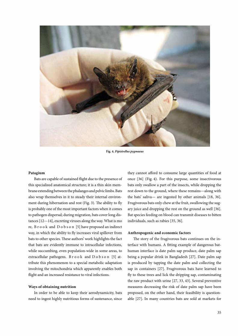
35
Patagium Bats are capable of sustained flight due to the presence of
this specialized anatomical structure; it is a thin skin mem-brane extending between the phalanges and pelvic limbs. Bats also wrap themselves in it to steady their internal environ-ment during hibernation and rest (Fig. 3). The ability to fly is probably one of the most important factors when it comes to pathogen dispersal; during migration, bats cover long dis-tances [12—14], excreting viruses along the way. What is more, B r o o k and D o b s o n [5] have proposed an indirect way, in which the ability to fly increases viral spillover from bats to other species. These authors’ work highlights the fact that bats are evidently immune to intracellular infections, while succumbing, even population-wide in some areas, to extracellular pathogens. B r o o k and D o b s o n [5] at-tribute this phenomenon to a special metabolic adaptation involving the mitochondria which apparently enables both flight and an increased resistance to viral infections.
Ways of obtaining nutrition In order to be able to keep their aerodynamicity, bats
need to ingest highly nutritious forms of sustenance, since
they cannot afford to consume large quantities of food at once [36] (Fig. 4). For this purpose, some insectivorous bats only swallow a part of the insects, while dropping the rest down to the ground, where these remains—along with the bats’ saliva— are ingested by other animals [18, 36]. Frugivorous bats only chew at the fruit, swallowing the sug-ary juice and dropping the rest on the ground as well [36]. Bat species feeding on blood can transmit diseases to bitten individuals, such as rabies [35, 36].
Anthropogenic and economic factors The story of the frugivorous bats continues on the in-
terface with humans. A fitting example of dangerous bat-human interface is date palm sap produce, date palm sap being a popular drink in Bangladesh [27]. Date palm sap is produced by tapping the date palm and collecting the sap in containers [27]. Frugivorous bats have learned to fly to these trees and lick the dripping sap, contaminating the raw product with urine [27, 33, 43]. Several preventive measures decreasing the risk of date palm sap have been proposed, on the other hand, their feasibility is question-able [27]. In many countries bats are sold at markets for
Fig. 4. Pipistrellus pygmaeus

36
meat, which jeopardizes buyers, sellers, hunters and butch-ers alike [25]. Limiting the trade of bat meat is difficult, mainly due to economy in these regions [25, 55]. Bat guano collectors are also exposed to dangerous viruses present in the excretions [53].
Circannual cycle All of the European bat species undergo hibernation,
which is influenced by the outer temperature [44]. There have been findings connecting hibernation to decreased immune function [4, 19], which facilitates the persistence of viral pathogens in infected individuals [4, 5, 19].
Longevity Bats can live for decades [12—14, 21]. The longer a res-
ervoir organism lives and is active, the longer it is able to excrete pathogens; what is more, several authors [5, 47, 48] have connected their longevity with metabolic mechanisms facilitating the persistence of intracellular pathogens.
The importance of ecology in bat-borne pathogen research
W o o d et al. [57] in their work describe the interde-pendence of human health and the health of the ecosystem, and with an emphasis on bats as important reservoirs of emerging infectious diseases, these authors suggest a com-plex approach to studying zoonoses. Their idea is based on a case study involving bats as a source of RNA viruses transmitted to humans. The authors propose a compre-hensive and holistic view of studying zoonoses based on systematic monitoring of the human-wildlife interface, i.e. the interactions between humans and wildlife, taking into account the dynamics of viral and reservoir species populations, but also other possible amplifiers of the dis-eases, such as livestock. They further emphasize the often neglected cultural and political background, which should support the research by spreading the news within the non-scientific public and issuing proper legislation. Preserving nature and reservoir species and protecting their environ-ment while keeping to the proposed approach will, accord-ing to these authors, help minimize spillover of dangerous bat-borne viruses into humans.
Metagenomic analysis of viral populations in batsModern findings in the field of molecular biology en-
able us to analyse samples in bulk in a relatively short time.
Metagenomic analysis facilitates the discovery of novel pathogens; it can be used to obtain genome sequences of complete viral populations from both environmental and biological samples [15, 28], such as mucosal swabs, blood clots etc. The main advantage of metagenomic analysis is the possibility to capture almost all (and often whole) viral sequences present in the analysed sample and thus to study viral populations (viromes) present in the infected organ-ism. Such screening applied to bat guano samples, for in-stance, has the potential to reveal “old known” pathogens that have been the subject of chiropterologic studies in the past, along with completely novel (i. e. never before identi-fied, or possibly new variations of known) viral taxa; these data may be useful for predicting the next potential grave spillover events [31]. There are several particular protocols for metagenomic analysis built on the same bioinformatic principle. With the advent of new generation sequencing (massively parallel sequencing methods), modifications were made to classic molecular-genetic methods in order to discover novel pathogens which had not been cultivated before [15, 22, 23, 26, 28, 32, 42, 46]. Sequence-Indepen-dent Single Primer Amplification [10] was developed by R e y e s and K i m [45]; it allows for fast amplification of a viral genome present in the analysed sample in only a few steps [17]. This method has been modified several times since its first implementation [16, 20, 58] together with the development of new sequencing methods, which simpli-fied the process. Froussard’s modification brought forth the so-called random-PCR, which combines reverse transcrip-tion and PCR using random hexamers labelled with known sequences used during the extension step; thus enough cDNA is produced for further cloning and sequencing [20, 58]. D e p e w et al. [16] modified this protocol so that vi-ruses isolated on a single plaque are identifiable via HiSeq Illumina platform.
The influence of other speciesParasitism is present in many mammalian species, and
bats are no exception (Fig. 5). Even though parasites of bats do not transmit to humans in such a degree as viruses—maybe with the exception of some ticks, e. g. Argas vesper-itlionis [6]—bat parasites may have a direct impact on the ecology of bat and virus populations worldwide [24].
Some of the mentioned viral families have been de-tected also in small rodents [9], which may suggest another element of the viral ecological cycle.

37
CONCLUSIONS
The papers cited in our review focus the attention of the professional public on the importance of interspecies relationships in Earth’s ecosystems with regard not only to animal species, but also in connection with populations of infectious agents and their circulation within the environ-ment. Several authors cited in this review report findings of various animal and insect viruses in the insectivorous
bats; the latter appear as a central meeting point connecting many arthropod, mammalian and other Metazoan species with the human race. It is plausible that by enabling dan-gerous viruses to persist in their populations, bats contrib-ute to the spillover of these pathogens to species (includ-ing, but certainly not limited to, humans) for which they are highly virulent.
Based on the cited papers, we suggest that the increased focus on complex viral populations in bats and their in-
Fig. 5. Rhinolophus hipposideros attacked by tick

38
teractions with other populations and the environment is necessary to fully comprehend the relationship between emerging infectious diseases, the environment and their toll on human health. We assume that this necessity will increase as humans will continue to exploit parts of the world where these viruses have been circulating. Research-ers studying viral populations should do so systematically, with a long-term focus on selected areas. There is currently no known method of exact prediction where or when an-other emerging infectious disease (EID) will appear [31]; however, with enough metagenomic data, it might be pos-sible at least to presume and test which viral taxa are more or less likely to infect mammalian cells [31]. In order to achieve this goal, close cooperation of experts in ecology, zoology, microbiology, molecular biology, immunology and bioinformatics will be necessary; and last, but not least, also the cooperation of state legislation organs and public health officials.
ACKNOWLEDGEMENT
This study was supported by the project APVV-0605-12.
REFERENCES
1. Adesiyun, A. A., Stewart-Johnson, A., Thompson, N. N.,
2009: Isolation of enteric pathogens from bats in Trinidad.
J. Wildl. Dis., 45 (4), 952—961. DOI: 10.7589/0090-3558-
45.4.952.
2. Allocati, N., Petrucci, A. G., Di Giovanni, P., Masulli, M.,
Di Illio, C., De Laurenzi, V., 2016: Bat-man disease transmis-
sion: zoonotic pathogens from wildlife reservoirs to human
populations. In Cell Death Discovery 2, Article No. 16048.
https://www.nature.com/articles/cddiscovery201648.
3. Bartlett, M., 1957: Measles periodicity and community size.
J. Roy. Stat. Soc., 120, 48—70. DOI: 10.2307/2342553.
4. Bouma, H. R., Carey, H. V., Kroese, F. G., 2010: Hiberna-
tion: the immune system at rest ? Journal of Leukocyte Biology,
88 (4), 619—624. DOI: 10.1189/jlb.0310174.
5. Brook, C. E., Dobson, A. P., 2015: Bats as “special” reservoirs
for emerging zoonotic pathogens. Trends Microbiol., 23 (3)
172—180. DOI: 10.1016/j.tim.2014.12.004.
6. Burazerovic, J, Ćakić, S., Mihaljica, D., Sukara, R., Ćirović,
D. Tomanović, Ć, 2015: Ticks (Acari: Argasidae, Ixodidae)
parasitizing bats in the central Balkans. Exp. Appl. Acarol., 66
(2), 281—291. DOI: 10.1007/s10493-015-9891-6.
7. Calisher, C. H., Childs, J. E., Field, H. E., Holmes, K. V.,
Schountz, T., 2006: Bats: important reservoir hosts of
emerging viruses. Clin. Microbiol. Rev., 19 (3), 531—545.
DOI: 10.1128/CMR.00017-06.
8. Chaverri, G., 2006: Aerobic bacterial flora from the digestive
tract of the common vampire bat, Desmodus rotundus (Chi-
roptera: Phyllostomidae). Rev. Biol. Trop., 54 (3), 717—724.
9. Chen, L., Liu, B., Wu, Z., Jin, Q., Yang, J., 2017: DRodVir:
A resource for exploring the virome diversity in rodents. Jour-
nal of Genetics and Genomics, 44 (5), 259—264, https://doi.
org/10.1016/j.jgg.2017.04.004.
10. Chrzastek, K., Lee, D-h., Smith, D., Sharma, P., Suarez,
D. L., Pantin-Jackwood, M., Kapczynski, D. R., 2017: Use of
sequence-independent, single-primer-amplification (SISPA)
for rapid detection, identification, and characterization of
avian RNA viruses. Virology, 509, 159—166, https://doi.org/
10.1016/j.virol.2017.06.019.
11. Costa, D., Poeta, P., Saenz, Y., Vinue, L., Coelho, A. C., Ma-
tos, M., et al., 2008: Mechanisms of antibiotic resistance in
Escherichia coli isolates recovered from wild animals. Microb.
Drug. Resist., 14 (1), 71—77. DOI: 10.1089/mdr.2008.0795.
12. Danko, Š., Kaňuch, P., Krištofík, J., 2012: The common
noctule—Nyctalus noctula. In Krištofík, J., Danko, Š. (Eds.):
Mammals of Slovakia—Spread, Bionomy and Protection (In
Slovak). Publ. house: Veda, Bratislava, 366—373.
13. Danko, Š., Uhrin, M., Krištofík, J., 2012: The particoloured
bat—Vespertilio murinus. In Krištofík, J., Danko, Š. (Eds.):
Mammals of Slovakia—Spread, Bionomy and Protection (In
Slovak). Publ. house: Veda, Bratislava, 405—410.
14. Danko, Š., Kaňuch, P., Uhrin, M., Krištofík, J., Hanzelová,
V., Špakulová, M., 2012: The common pipistrelle—Pipistrel-
lus pipistrellus. In Krištofík, J., Danko, Š. (Eds.): Mammals of
Slovakia—Spread, Bionomy and Protection (In Slovak). Publ.
house: Veda, Bratislava, 378—383.
15. Delwart, E. L., 2007: Viral metagenomics. Rev. Med. Virol., 17
(2), 115—131. DOI: 10.1002/rmv.532.
16. Depew, J., Zhou, B., McCorrison, J. M., Wentworth, D. E.,
Purushe, J. G., Koroleva, G., Fouts, D. E., 2013: Sequencing
viral genomes from a single isolated plaque. Virol. J., 10, 181.
DOI: 10.1186/1743-422X-10-181.
17. Djikeng, A., Halpin, R., Kuzmickas, R., Depasse, J., Feld-
blyum, J., Sengamalay, N., et al., 2008: SpiroViral genome
sequencing by random priming methods. BMC Genomics, 9,
5. DOI: 10.1186/1471-2164-9-5.

39
18. Dobson, A. P., 2005: What links bats to emerging infectious
diseases ? Science, 310 (5748), 628—629. DOI: 10.1126/sci-
ence.1120872.
19. Field, K. A., Sewall, B. J., Prokkola, J. M., Turner, G. G., Ga-
gnon, M. F., Liley, T. M., et al., 2018: Effect of torpor on host
transcriptomic responses to a fungal pathogen in hibernating
bats. Mol. Ecol., 27, 3727—3743. DOI: 10.1111/mec.14827.
20. Froussard, P., 1993: rPCR: a powerful tool for random ampli-
fication of whole RNA sequences. PCR Methods Appl., 2 (3),
185—190.
21. Gaisler, J., Hanák, V., Hanzal, V., Jarský, V., 2003: Results of
bat banding in the Czech and Slovak Republics, 1948—1000
(In Czech with summary in English). Vespertilio, 7, 3—61.
22. Gaynor, A. M., Nissen, M. D., Whiley, D. M., Mackay, I. M.,
Lambert, S. B., Wu, G., et al., 2007: Identification of a novel
polyomavirus from patients with acute respiratory tract in-
fections. PLoS Pathog., 3 (5), e64. DOI: 10.1371/journal.ppat.
0030064.
23. Greninger, A. L., Runckel, C., Chiu, C. Y., Haggerty, T., Par-
sonnet, J., Ganem, D., DeRisi, J. L., 2009: The complete ge-
nome of klassevirus—a novel picornavirus in pediatric stool.
Virol. J., 6, 82. DOI: 10.1186/1743-422X-6-82.
24. Haelewaters, D., Hiller, T., Dick, C. V., 2018: Bats, bat flies,
and fungi: a case of hyperparasitism. Trends in Parasitology,
34 (9). DOI: 10.1016/j.pt.2018.06.006.
25. Kamins, A. O., Restif, O., Ntiamoa-Baidu, Y., Suu-Ire, R.,
Hayman, D. T. S., Cunningham, A. A., et al., 2011: Uncov-
ering the fruit bat bushmeat commodity chain and the true ex-
tent of fruit bat hunting in Ghana, West Africa. Biol. Conserv.,
144 (12), 3000—3008. DOI: 10.1016/j.biocon.2011.09.003.
26. Kapoor, A., Victoria, J., Simmonds, P., Slikas, E., Chieo-
chansin, T., Naeem, A., et al., 2008: A highly prevalent and
genetically diversified Picornaviridae genus in South Asian
children. Proc. Natl. Acad. Sci. U. S. A., 105, (51), 20482—
20487. DOI: 10.1073/pnas.0807979105.
27. Khan, S. U., Gurley, E. S., Hossain, M. J., Nahar, N., Shark-
er, M. A., Luby, S. P., 2012: A randomized controlled trial
of interventions to impede date palm sap contamination by
bats to prevent Nipah virus transmission in Bangladesh. PLoS
One, 7 (8), e42689. DOI: 10.1371/journal.pone.0042689.
28. Kristensen, D. M., Mushegian, A. R., Dolja, V. V., Koonin,
E. V., 2010: New dimensions of the virus world discovered
through metagenomics. Trends Microbiol., 18 (1), 11—19.
DOI: 10.1016/j.tim.2009.11.003.
29. Kunz, T. H., Braun de Torrez, E., Bauer, D., Lobova, T.,
Fleming, T. H., 2011: Ecosystem services provided by bats.
Annals of the New York Academy of Sciences, 1223 (1), 1—38.
DOI: 10.1111/j.1749-6632.2011.06004.x.
30. Lei, B. R., Olival, K. J., 2014: Contrasting patterns in mam-
mal-bacteria coevolution: Bartonella and Leptospira in bats
and rodents. PLoS Negl. Trop. Dis., 8 (3), e2738. DOI: 10.1371/
journal.pntd.0002738.
31. Li, L., Jacobo, V., Chunlin, W., Morris, S. J., Fellers, G. M.,
Kunz, T. H., Delwart, E., 2010: Bat guano virome: Predomi-
nance of dietary viruses from insects and plants plus novel
mammalian viruses. Journal of Virology, 84 (14), 6955—6965.
DOI: 10.1128/JVI.00501-10.
32. Li, L., Kapoor, A., Slikas, B., Bamidele, O. S., Wang, C.,
Shaukat, S., et al., 2010a: Multiple diverse circoviruses infect
farm animals and are commonly found in human and chim-
panzee faeces. J. Virol., 84 (14), 1674—1682. DOI: 10.1128/
JVI.02109-09.
33. Luby, S. P., Hossain, M. J., Gurley, E. S., Ahmed, B. N.,
Banu, S., Khan, S. U., et al., 2009: Recurrent zoonotic trans-
mission of Nipah virus into humans, Bangladesh, 2001—
2007. Emerg. Infect. Dis., 15 (8), 1229—1235. DOI: 10.3201/
eid1508.081237.
34. Mackenzie, J. S., 2005: Emerging zoonotic encephalitis virus-
es: lessons from Southeast Asia and Oceania. J. Neurovirol., 11
(5), 434—440. DOI: 10.1080/13550280591002487.
35. Mehlhorn, H., 2014: Introduction: The world of bats. In
Klimpel, S., Mehlhorn, H. (Eds.): Bats (Chiroptera as Vec-
tors of Diseases and Parasites. Facts and Myths. Parasitology
Research Monographs 5, Springer-Verlag Berlin Heidelberg,
187 pp.
36. Melaun C., Werblow A., Busch M. W., Liston A., Klimpel
S., 2014: Bats as potential reservoir hosts for vector-borne
diseases. In Klimpel, S., Mehlhorn, H. (Eds.): Bats (Chirop-
tera) as Vectors of Diseases and Parasites. Facts and Myths.
Parasitology Research Monographs 5, Springer-Verlag Berlin
Heidelberg, 187 pp.
37. Mühldorfer, K., 2013: Bats and bacterial pathogens: a re-
view. Zoonoses and Public Health, 60 (1), 93—103. DOI:
10.1111/j.1863-2378.2012.01536.x.
38. Nokireki, T., Tammiranta, N., Kokkonen, U. M., Kantala,
T., Gadd, T., 2018: Tentative novel lyssavirus in a bat in Fin-
land. Transbound. Emerg. Dis., 65 (3), 593—596. https://doi.
org/10.1111/tbed.12833.
39. Omatsu, T., Watanabe, S., Akashi, H., Yoshikawa, Y., 2007:
Biological characters of bats in relation to natural reservoir of
emerging viruses. Comp. Immunol. Microbiol. Infect. Dis., 30
(5‒6), 357—374. DOI: 10.1016/j.cimid.2007.05.006.

40
40. Peel, A. J., Baker, K. S., Crameri, G., Barr, J. A., Hayman,
D. T. S., Wright, E., et al., 2012: Henipavirus neutralising
antibodies in an isolated island population of African fruit
bats. PLoS One, 7 (1), e30346. https://doi.org/10.1371/journal.
pone.0030346.
41. Peel, A. J., Pulliam, J. R. C., Luis, A. D., Plowright, R. K.,
O’Shea, T. J., Hayman, D. T. S., et al., 2014: The effect of sea-
sonal birth pulses on pathogen persistence in wild mammal
populations. Proc. R. Soc. B Biol. Sci., 281, 20132962. http://
dx.doi.org/10.1098/rspb.2013.2962.
42. Quan, P. L., Junglen, S., Tashmukhamedova, A., Conlan,
S., Hutchison, S. K., Kurth, A., et al., 2010: Moussa virus:
a new member of the Rhabdoviridae family isolated from Cu-
lex decens mosquitoes in Cote d’Ivoire. Virus Res., 147 (1),
17—24. DOI: 10.1016/j.virusres.2009.09.013.
43. Rahman, M. A., Hossain, M. J., Sultana, S., Homaira, N.,
Khan, S. U., Rahman, M., et al., 2008: Date palm sap linked
to Nipah virus outbreak in Bangladesh. In Institute of Medicine
(US): Improving Food Safety Through a One Health Approach.
Workshop summary, Washington DC, National Academic
Press (US), 2012. A12. https://www.ncbi.nlm.nih.gov/books/
NBK114490/.
44. Reichholf, J., 1996: Mammals (In Slovak). Ikar, Bratislava,
SR. 30—49.
45. Reyes, G. R., Kim, J. P., 1991: Sequence-independent, single-
primer amplification (SISPA) of complex DNA populations.
Mol. Cell. Probes, 5 (6), 473—481.
46. Rosario, K., Duffy, S., Breitbart, M., 2009: Diverse circo-
virus-like genome architectures revealed by environmental
metagenomics. J. Gen. Virol., 90 (Pt 10): 2418—2424. DOI:
10.1099/vir.0.012955-0.
47. Schountz, T., 2014: Immunology of bats and their viruses:
Challenges and opportunities. Viruses, 6 (12), 4880—4901.
DOI:10.3390/v6124880.
48. Schountz, T., Baker, M. L., Butler, J., Munster, V., 2017: Im-
munological control of viral infections in bats and the emer-
gence of viruseshHighly pathogenic to humans. Frontiers
in Immunology. www.frontiersin.org, 8, Article 1098. DOI:
10.3389/fimmu.2017.01098.
49. Schipper, J., Chanson, J. S., Chiozza, F., Cox, N. A., Hoff-
man, M., Katariya, V., et al., 2008: The status of the world’s
land and marine mammals: diversity, threat, and knowledge.
Science 322 (5899), 225—230. DOI: 10.1126/science.1165115.
50. Simmons, N. B., 2010: Personal communication. American
Museum of Natural History, New York. In Elangevan, V., Ku-
mar M.: Diversity, Roost Selection and Ecological Importance
of the Bats of Uttah Pradesh, International Day for Biological
Diversity, 2015, 44—50.
51. van der Poel, W. H., Lina, P. H., Kramps, J. A., 2006: Public
health awareness of emerging zoonotic viruses of bats: Euro-
pean perspective. Vector Borne Zoonotic Dis., 6 (4), 315—324.
DOI: 10.1089/vbz.2006.6.315.
52. Walldorf, V., Mehlhorn, H., 2014: Bats: A glimpse on their
astonishing morphology and lifestyle, Chapter 2. In Klimpel,
S., Mehlhorn, H. (Eds.): Bats (Chiroptera) as Vectors of Dis-
eases and Parasites. Facts and Myths. Parasitology Research
Monographs 5, Springer-Verlag Berlin Heidelberg, 24 pp.
53. Wacharapluesadee, S., Sintunawa, C., Kaewpom, T., Khong-
nomnan, K., Olival, K. J., Epstein, J. H., et al., 2013: Group
C betacoronavirus in bat guano fertilizer, Thailand. Emerg. In-
fect. Dis., 19 (8), 1349—1351. DOI: 10.3201/eid1908.130119.
54. Wibbelt, G., Speck, S., Field, H., 2009: Methods for assessing
diseases in bats, In Kunz, T. H., Parsons, S. (Eds.): Ecological
and Behavioural Methods for the Study of Bats. Johns Hopkins
University Press, Baltimore, MD, 775—794
55. Wilkie, D., Bennett, E., Peres, C., Cunningham, A. A., 2011:
The empty forest revisited. Annals of the New York Academy
of Sciences, 1223, 120—128. DOI: 10.1111/j.1749-6632.2010.
05908.x.
56. Wong, S., Lau, S., Woo, P., Yuen, K. Y., 2007: Bats as a con-
tinuing source of emerging infections in humans. Rev. Med.
Virol., 17 (2), 67—91. DOI: 10.1002/rmv.520.
57. Wood, J. L., Leach, M., Waldman, L., Macgregor, H., Fooks,
A. R., Jones, K. E., et al., 2012: A framework for the study
of zoonotic disease emergence and its drivers: spillover of bat
pathogens as a case study. Philos. Trans. R. Soc. Lond. B Biol.
Sci., 367 (1604), 2881—2892. DOI: 10.1098/rstb.2012.0228.
58. Zou, N., Ditty, S., Li, B., Lo, S. yC., 2003: Random priming PCR
strategy to amplify and clone trace amounts of DNA BioTech-
niques, 35 (4), 758—765. https://doi.org/10.2144/03354st06.
Received January 25, 2019
Accepted March 6, 2019

4141
DOI: 10.2478/fv-2019-0006
FOLIA VETERINARIA, 63, 1: 41—47, 2019
ABSTRACT
Bovine tuberculosis (BTB) is a zoonotic disease re-sponsible for considerable economic losses; with con-sequent negative impact on both public health and the livestock industry. However, the burden of this disease and associated economic losses remain un-investigated among slaughtered cattle in Ibadan, the hub of live-stock activities in south-western Nigeria. A cross sec-tional study was conducted over a three-month period to determine the prevalence and economic losses due to BTB among slaughtered cattle at the Bodija Municipal Abattoir, Ibadan, in south-western Nigeria. Individual slaughtered cattle were purposively inspected for the presence of tuberculous lesions, and representative sam-ple of affected organs and associated lymph nodes from the BTB suspected animals were taken. The suspected le-sions were processed based on Becton Dickinson diges-tion and decontamination procedure and the losses due to BTB were estimated mathematically, using a formu-la. The BTB prevalence of 9.3 % (38/408) was obtained with a significant statistical association (P ≤ 0.05) of the
disease with the breeds of cattle slaughtered. Out of the 46 visceral organs condemned: 29 (63 %) were lungs, 12 (26.1 %) livers, 4 (8.7 %) hearts and 1 (2.2 %) kidney. The total estimated annual economic loss (direct and in-direct) due to BTB was estimated as Nigerian naria NGN 703,980,070 (EUR 1,725,441.4). This study revealed BTB as endemic and a major cause of concern considering its ill-health and economic effects on both humans and the livestock industry. Efforts are therefore required: to con-trol this disease along the beef value chain in order to safeguard human and livestock health; as well as to limit the economic losses associated with its prevalence.
Key words: bovine tuberculosis; economic loss; prev-alence; zoonoses
INTRODUCTION
Bovine tuberculosis (BTB) is a chronic infectious and contagious disease of both domestic and wild animals as well as humans [28]. It is caused by Mycobacterium bo-
PREVALENCE AND ECONOMIC LOSSES DUE TO BOVINE TUBERCULOSIS IN CATTLE SLAUGHTERED AT BODIJA
MUNICIPAL ABATTOIR, IBADAN, NIGERIA
Adelakun, O. D., Akinseye, V. O. Adesokan, H. K., Cadmus, S. I. B.
Department of Veterinary Public Health and Preventive MedicineUniversity of Ibadan, Ibadan, Nigeria

42
vis, a slow growing, non-photochromogenic and acid-fast bacillus. The disease is characterized by the formation of granulomas in tissues and organs, more significantly in the lungs, lymph nodes, intestines, liver and kidneys [32]. The BTB disease is a cause of concern for humans and others animals judging from its negative economic and health implications [6, 10]. Indeed, the disease induces high ani-mal morbidity and mortality that eventually reduces the financial capital and increases production costs [9]. Bovine TB affects cattle health, impacts negatively on profitability and trade and can decimate years of genetic improvement towards desirable production traits [3]. Bovine TB is an important zoonotic disease, known to exist in all parts of the world. It is recognized in 176 countries as an important bovine disease that causes great economic loss [18, 24, 31].
Cattle infected with M. bovis are the main source of infection for other cattle and humans. M. bovis could be excreted through aerosol, sputum, faeces (from both in-testinal lesions and swallowed sputum from pulmonary lesions), milk, urine, vaginal and uterine discharges, and discharges from open peripheral lymph nodes [29]. Do-mestic animals are significant reservoir hosts for human TB, caused by M. bovis [17]. The majority of the cases of human TB, both pulmonary and extra pulmonary in Ni-geria are due to M. tuberculosis [6]. However, due to the endemicity of BTB in the cattle population, people are at risk of exposure to M. bovis from consumption of unpas-teurized milk and milk products as well as the dearth of meat inspection services to identify infected animals in the food chain [5]. Transmission through consumption of milk and unpasteurized dairy products from infected cattle oc-curs mostly among the general public, whereas exposure through airborne infection remains highest among farm-ers, veterinarians, and slaughterhouse workers [25].
The diagnosis of TB in cattle is mainly through tubercu-lin testing, culture, and molecular genotyping [4, 36]. Cul-ture is regarded as the gold standard for confirmation of BTB but most laboratories in the country are ill-equipped. Hence, routine diagnosis of BTB at the abattoir is based on identification of characteristic tuberculous lesions [1, 20]. The disease is a major cause of economic losses due to organ condemnation at the abattoir and slaughter slabs in Nigeria. Available reports showed that BTB causes direct loss of up to 10 % to 18 % decrease in milk and 15 % meat production [37]. Thus, BTB is of paramount importance to cattle breeders and public health authorities because of its
economic and zoonotic implications [19]. This study aims at providing information on the current prevalence and es-timated financial loss due to BTB among slaughtered cattle in Ibadan, located in south-western Nigeria.
MATERIALS AND METHODS
Study area This study was carried out at Bodija Municipal Abat-
toir, a major abattoir in the city of Ibadan, where 250 to 300 heads of cattle are slaughtered on a daily basis. The ab-attoir is a major recipient of animals from different parts of the country especially the north and it is located in the geographical grid of reference longitude 3 °E, latitude 7 °N [14].
Sample sizeThe sampling method was employed to generate data
on cattle presented for slaughter at the abattoir. Because of an expected 50 % prevalence, 408 cattle were examined in this study.
Sampling procedure and laboratory diagnosisA total of 408 cattle were thoroughly examined at post
mortem. Organs such as lungs, liver, heart and kidneys were visually examined, palpated and incised to detect the presence of granulomas; associated lymph-nodes were also palpated and incised, especially the bronchial and hepatic lymph-nodes. Each sample obtained was appropriately kept in sample containers and labelled. Data on breed, sex and body score of each sampled animal were collected and doc-umented. Samples obtained were transported to the Tuber-culosis Laboratory of the Department of Veterinary Public Health and Preventive Medicine, University of Ibadan, Ni-geria for analysis. Suspected lesions were processed based on Becton Dickinson digestion and decontamination pro-cedure [2]. The suspension obtained after the sample pro-cessing was inoculated onto Lowenstein-Jensen slopes with pyruvate and glycerol and incubated at 37 °C for between eight and twelve weeks. Cultures were considered negative if no colony growth was detected after 8 weeks.
Assessment of economic lossesThe loss due to BTB in Bodija Municipal Abat-
toir was estimated mathematically, using a formula set

43
by O g u n r i n a d e and O g u n r i n a d e [27] as modi-fied by G u a d u et al. [16]. The total annual financial loss incurred due to BTB was computed by summing up the di-rect and indirect losses.
A = direct economic losses (annual loss from organs condemnation)B = indirect economic losses (annual loss from carcass weight reduction)
Direct economic lossesThe direct economic loss (A) was calculated based on
condemned edible visceral. There were no documented data on annual slaughter rates at the abattoir, therefore it was estimated based on observations made during the study period; 250 to 300 heads of cattle were slaughtered on a daily basis, six [6] times a week, an average of 275 heads of cattle was estimated, which resulted into a mean slaughter rate of 85,800 head of cattle per annum. The percentage of each condemned organ was taken into consideration and the average market price of healthy lung, liver, heart and kidneys, which are 2000, 3000, 1500 and 700 NGN (Nige-rian naria), respectively.
The economic loss is calculated as follows:A = (MAS × PLu × CLu) + (MAS × PLi × CLi) + + (MAS × PHr × CHr) + (MAS × PKi × CKi)
MAS = mean annual cattle slaughtered at study abattoir PHr = percentage of heart condemnedPLu = percentage of lung condemned CHr = mean cost of a heartCLu = mean cost of a lungPKi = percentage of kidney condemnedPLi = percentage of liver condemnedCKi = mean cost of a kidney CLi = mean cost of a liver
Indirect economic lossesTo calculate the indirect economic losses due to BTB,
a 36.6 % weight loss due to BTB was used as reported by K w a g h e et al. [22] and an average carcass weight of Nigerian cattle is estimated as 292.35 kg according to the report of U m a r et al. [35] in order to obtain percent-age weight of carcass reduction due to BTB. Beef sellers were orally interviewed and an average of NGN 800 was obtained to be the cost a kilogram of beef.
B = MAS × PCW × MCB x PB = MAS × (292.3 KG × 36.6%) × MCB × PMAS = mean annual cattle slaughtered at study abattoir PCW = percentage of carcass weight reductionMCB = mean cost of 1 kg beef in Ibadan city butcheriesP = prevalence rate of bovine tuberculosis at study abattoir
Table 1. Prevalence of bovine tuberculosis in relation to breed, sex and body score
Variables No. examined
No.infected
Prevalence[%] X2 P-value
Breed
Bunaji 158 22 13.9 6.712 0.035
Rahaji 157 9 5.7
Sokoto Ruhali 93 7 7.5
Total 408 38
Sex
Male 60 4 6.7 0.800 0.777
Female 348 34 9.8
Body score
Emaciated 186 17 9.1 0.632 0.427
Moderate 222 21 9.5
P — value is significant at P ≤ 0.05*significant, X2 — chi square

44
Data Analysis The data obtained were coded in Microsoft excel and
subjected to descriptive statistics and chi-square in order to assess the magnitude of the difference of comparable vari-ables using SPSS version 16.0 software. Statistically signifi-cant association between variables is considered to exist if the p-value P ≤ 0.05.
RESULTS
A total of 46 lesions suggestive of tuberculosis were ob-tained from 408 slaughtered cattle examined, out of which only 38 were culture positive for Mycobacterium spp., giving a prevalence of 9.3 % (38/408). The Bunaji breed (22/158; 13.9 %) of cattle had the highest prevalence, fol-lowed by Rahaji (9/157; 5.7 %) and Sokoto Gudali (7/93; 7.5 %). The prevalence for sex were: male (4/60; 6.7 %) and female (34/348; 9.8 %) while the body score was ema-ciated (17/186; 9.1 %) and moderate (21/222; 9.5 %). The chi square test statistic revealed that the prevalence was significantly different (P ≤ 0.05) for breeds, but not for sex and the body score (Table 1). The localization of tubercles (Table 2) was highest in the lungs (29/46, 63 %), followed by liver (12/46; 26.1 %), heart (4/46; 8.7 %) and least in the kidneys (1/46; 2.2 %).
The total annual economic loss due to bovine tubercu-losis in the study abattoir was estimated by summing up losses calculated from organ condemnation (direct losses) and carcass weight reduction (indirect losses). The direct economic losses were calculated to be NGN 21,059,610 (EUR 51,616.7) and the indirect economic losses was cal-culated to be NGN 682,920 460 (EUR 1,673,824.7). A total
annual economic loss (A + B) of NGN 703,980, 070 (EUR 1,725,441.4) was obtained in this study.
*Conversion rate: NGN 408 is equivalent to 1 EUR.
DISCUSSION
From this study, a prevalence of 9.3 % BTB was obtained among slaughtered cattle in Ibadan, in south-western Ni-geria. The fact that BTB due to M. bovis is endemic and despite the global recognition of the disease as an animal and a zoonotic disease, substantial evidence suggests that its impacts on human health has been underestimated [26]. However, consumption of unpasteurized infected milk, a major factor that predisposes humans contracting zoo-notic TB is still popularly practiced in Nigeria, hence the importance of the prevalence obtained in this study. The prevalence of 9.3 % obtained in this study is in line with previous studies on BTB conducted in south-western Nige-ria with an established prevalence of between 8.8 % [6] and 10.5 % [7]. It is also similar to the prevalence of 8.8 % ob-tained by E j e h [12] in Benue State, north-central Nigeria and higher than 6.41 % reported in Borno State, north-east Nigeria by K w a g h e et al. [22]. The difference in preva-lence might be as a result of varying detection abilities of the meat inspectors. The effective detection of BTB tuber-cles at post mortem is dependent on the thoroughness of the examination, training and experience of the meat inspec-tors [15, 23]. Based on this study, the slaughter hall layout and the ratio of meat inspectors to butchers are influential factors, because butchers slaughter and dress carcasses irre-spective of whether meat inspectors get to inspect it or not. A working system can therefore be developed to maintain and improve meat inspection carried out in the country [11, 13, 34].
The lungs recorded the highest prevalence for tubercle localization, which can be attributed to the infection route of entry. Other studies carried out in other parts of the country reveal that lungs are the most affected organs [12, 22]. Pathogenesis studies suggest strongly that the route of transmission of bovine TB is largely via the respiratory system, which requires transmission via infectious aero-sols [8]. The lungs are also regarded as the most exposed organ to different aggressions because of its anatomical and histological peculiarities. This finding therefore sug-
Table 2. Disseminated tubercles of bovine tuberculosis in various organs
Organs Frequency Percentage
Lungs 29 63.0
Liver 12 26.1
Heart 4 8.7
Kidneys 1 2.2
Total 46 100.0

45
gests increasing risk of exposure of livestock workers to BTB due to aerosolization of tubercle bacilli from infected cattle.
The estimated total annual economic loss due to BTB from both direct and indirect losses obtained in this study was estimated to be NGN 703,980,070 (EUR 1,725,441.4) which is huge for a developing country considering the fact that decreased milk production and reproductive rate were not considered. The total annual economic loss obtained in this study is higher compare to the NGN 349,580,199.89 (EUR 856,814.2) reported by K w a g h e et al. [22]. This difference could be due to variation in the prevalence of the disease, mean annual slaughtered cattle and the retail pric-es of organs in different locations. A retrospective study by E j e h et al. [12] to determine the direct economic loss be-tween the years 2008 and 2012, revealed values that ranged from NGN 356,000 (EUR 872.6) to NGN 2,000,000 (EUR 4901.9) which are lower compare to NGN 21,059,610 (EUR 51,616.7) obtained in this study, thereby confirming the en-demicity of BTB as a source of leakage to the buoyancy of the nation’s economy.
Bovine TB has posed a tremendous impact on livestock’s economic growth of Africa, as it affects the continent’s in-volvement in the international trade of animals and animal products. The disease causes severe economic losses in live-stock due to poor production, death and partial condem-nation of organs or total condemnation of carcasses [33]. Many countries, including Nigeria do not implement any control or eradication programs. The BTB is an important economic challenge worldwide, most especially in develop-ing countries and it can cause significant economic losses [37]. The zoonotic nature of BTB, coupled with the fact that it constitutes a significant economic burden to the agricul-tural industries [21] indicates the need to properly strat-egize a means to totally eradicate it. Slaughter houses have been proven to provide the opportunity for detecting dis-eases of both economic and public health importance [30]. It therefore becomes imperative that meat inspection ac-tivities in slaughter houses in Nigeria be scaled up in order to safeguard the health of the public and invariably limit the spread of the disease in both human and other animals with eventual prevention of unwarranted economic losses due to the disease in animals.
CONCLUSIONS
Bovine tuberculosis is still endemic in the cattle popu-lation in Nigeria and causes huge economic losses to the country. The consequent gross shortage of dietary animal proteins, due to condemnation of infected visceral organs and edible meat, as well as indirect weight loss and a poten-tial health hazard to humans as result of its zoonotic nature are over-whelming. There is therefore a need for urgent government intervention in ensuring optimal control mea-sures against BTB from the farm to the slaughter houses. The meat inspection at slaughter houses should also be stepped up by veterinary meat inspectors as this will go a long way to minimize the spread of bovine tuberculosis in the country.
ACKNOWLEDGEMENT
The assistance of the abattoir workers at the Bodija, Mu-nicipal Abattoir, Ibadan, Oyo State, Nigeria, is appreciated.
REFERENCES
1. Aliyu, M. M., Adamu, J. Y., Bilyaminu, Y. A., 2009: Current
prevalence of tuberculosis lesions among slaughtered cattle
in North-eastern States of Nigeria. Revue Elev. Med. Vet. Pays
Trop., 62 (1), 13—16.
2. Becton, D., 1999: BBLTM MycoPrepTM Specimen Digestion/
Decontamination Kit Manual for Processing of Mycobacterial
Specimens. Sparks, MD, USA, BD, 2pp.
3. Boland, F., Kelly, G. E., Good., More, S. J., 2010: Bovine
tuberculosis and milk production in infected dairy herds in
Ireland. Prev. Vet. Med., 93 (2—3), 153—161. DOI: 10.1016/j.
prevetmed.2009.09.021.
4. Brosch, R., Gordon, S. V., Marmiesse, M., Buchrieser,
C., Eiglmeir, K., Garnier, T., et al., 2002: A new evolution-
ary scenario for the Mycobacterium tuberculosis complex.
Proc. Nat. Acad. Sci. USA, 99 (6), 3684—3689. DOI: 10.1073/
pnas.052548299.
5. Cadmus, S. I. B., Alonge, D. O., Adesokan, H. K., 2007:
Meat inspection and cultural isolation of mycobacteria as
predictors of bovine tuberculosis in Ibadan, Nigeria. Tropical
Veterinarian, 25 (3), 101—105.
6. Cadmus, S., Palmer, S., Okker, M., Dale, J., Gover, K.,
Smith, N., et al., 2006: Molecular analysis of human and bo-

46
vine tubercle bacilli from a local setting in Nigeria. J. Clin. Mi-
crobiol., 44 (1), 29—34. DOI: 10.1128/JCM.44.1.29-34.2006.
7. Cadmus, S. I. B., Atsanda, N. N., Oni, S. O., Akang,
E. E. U., 2004: Bovine tuberculosis in one cattle herd in
Ibadan in Nigeria. Vet. Med.-Czech, 49 (11), 406—412. DOI:
10.17221/5730-VETMED.
8. Cassidy, J. P., 2006: The pathogenesis and pathology of bo-
vine tuberculosis with insights from studies of tuberculosis in
humans and laboratory animal models. Vet. Microbiol., 112
(2—4), 151—161. DOI: 10.1016/j.vetmic.2005.11.031.
9. Cosivi, O., Robinson, L. A., Daborn, C. J., Raviglionne,
M. C., Fujikura, T., Cousins, D., et al., 1998: Zoonotic tu-
berculosis due to M. bovis in developing countries. Emerg. Inf.
Dis., 4 (1), 59—69. DOI: 10.3201/eid0401.980108.
10. Cousins, D. V., Corner, L. A., Tolson, J. W., Jones, S. L.,
Wood, P. R., 1998: Eradication of Bovine Tuberculosis from
Australia: Key Management and Technical Aspects. CSL Lim-
ited, Parkville, Australia, 45 pp.
11. Doherr, M. G., Audige, L., 2001: Monitoring and surveil-
lance for rare health-related events: a review from the veteri-
nary perspective. Philos. Trans. R. Soc. B, 356 (1411), 1097—
1106. DOI: 10.1098/rstb.2001.0898.
12. Ejeh, E. F., Raji, M. A., Bello, M., Lawan, F. A., Francis, M. I.,
Kudi, A. C., et al., 2014: Prevalence and direct economic loss-
es from bovine tuberculosis in Makurdi, Nigeria. Veterinary
Medicine International, 6 pp. DOI: 10.1155/2014/904861.
13. Enoe, C., Christensen, G., Andersen, S., Willeberg, P., 2003:
The need for built-in validation of surveillance data so that
changes in diagnostic performance of post mortem meat in-
spection can be detected. Prev. Vet. Med., 57, 117—125.
14. Filani, M. O., 1994: Ibadan Region, Re-Charles Publications in
Conjunction with Connell Publications, Ibadan, Nigeria, 99 pp.
15. Frankena, K., White, P. W., O’Keeffe, J., Costello, E., Mar-
tin, S. W., van Grevenhof, I., et al., 2007: Quantification of
the relative efficiency of factory surveillance in the disclosure
of tuberculosis lesions in attested Irish cattle. Vet. Rec., 161,
679—684. DOI: 10.1136/vr.161.20.679.
16. Guadu, T., Gebremicael, Y., Chanie, M., 2013: Economic
and zoonotic importance of bovine hydatidosis in Shire mu-
nicipal abattoir, north west zone, Tigary region, Ethiopia. Acta
Parasitologica Globalis, 4 (3), 92—98. DOI: 10.5829/idosi.apg.
2013.4.3.7531.
17. Grange, J. M., 2001: Mycobacterium bovis infection in human
beings. Tuberculosis, 81 (1—2), 71—77. DOI:10.1054/tube.
2000.0263.
18. Hines, M. E., Kreeger, J. M., Smith, P. K., 1995: Mycobacte-
rial infections of animals. Pathology and pathogenesis. Labo-
ratory Animal Science, 45 (4), 334—351.
19. Hermandez, J., Baca, D., 1998: Effect of tuberculosis on milk
production in dairy cows. J. Amer. Vet. Med. Assoc., 213 (6),
851—854.
20. Igbokwe, I. O., Madaki, I. Y., Danburan, S., Ameh, J. A.,
Aliyu, M. M., Nwosu, C. O., 2001: Prevalence of pulmonary
tuberculous lesions in cattle slaughtered in abattoirs in north-
eastern Nigeria. Revue Elev. Med. Vet. Pays Trop., 54 (3—4):
191—195.
21. Krebs, J., Independent Scientific Review Group, 1997: Bo-
vine Tuberculosis in Cattle and Badgers. Ministry of Agricul-
ture, Fisheries and Food Publications, London, United King-
dom, 196 pp.
22. Kwaghe, A. V., Ameh, A. J., Ambali, A. G., Kudi, A. C.,
Kachalla, M. G., 2015: Prevalence and economic losses from
bovine tuberculosis in Maiduguri, Borno State, Nigeria. Inter-
national Journal of Life Sciences, 4 (4), 283—287.
23. Lees, V. W., 2004: Learning from outbreaks of bovine tuber-
culosis near Riding Mountain National Park: applications to
a foreign animal disease outbreak. Can. Vet. J., 45 (1), 28—34.
24. Martin, S. W., Dietrich, R. A., Genno, P., Heuschele, W. P.,
Jones, R. L., Koller, M., et al., 1994: Livestock Disease Eradi-
cation: Evaluation of the Cooperative State Federal Bovine
Tuberculosis Eradication Program. National Academy of Sci-
ences, Washington D.C. 1—97.
25. Michel, A. L., Muller, B., Van Helden, P. D., 2010: Mycobac-
terium bovis at animal-human interface; A problem or not ?
Vet. Microbiol., 140 (3—4), 371—381. DOI: 10.1016/j.vetmic.
2009.08.029.
26. Olea-Popelka, F., Muwonge, A., Perera, A., Dean, A. S.,
Mumford, E., Erlacher Vindel, E., et al., 2016: Personal view
zoonotic tuberculosis in human beings caused by Mycobacteri-
um bovis—a call for action. Lancet Infect. Dis., 16 (12), 1305—
1430. http://dx.doi.org/10.1016/ S1473-3099(16)30139-6.
27. Ogunrinade, A., Ogunrinade, B. I., 1980: Economic im-
portance of bovine fascioliasis in Nigeria. Trop. Anim. Health
Prod., 12 (3), 155—160.
28. Radostitis, O. M., Gay, C. C., Blood, D. C., Hinchelift,
K. W., 2002: Veterinary Medicine: A Textbook of Disease of
Cattle, Sheep, Pigs, Goats and Horses, 9th edn., Harcourt Pub-
lisher Ltd., London, 909—918.
29. Radostitis, O. M., Blood, D. C., Gay, C. C., 1994: Veteri-
nary Medicine: A Textbook of the Disease of Cattle, Sheep,
Goats, Pigs and Horses. 8th edn., Bailliere, Tindall, London,
1015—1026.

47
30. Raji, M. A., Salami, S. O., Ameh, J. A., 2010: Pathological
conditions and lesions observed in slaughtered cattle in Zaria
abattoir. Journal of Clinical Pathology and Forensic Medicine,
1 (2), 9—12.
31. Samad, M. A., 2008: Animal Husbandry and Veterinary Sci-
ence. V‒2, 1127, First Published LEP, Pub. No. 11, BAU Cam-
pus, Mymensingh, Bangladesh.
32. Shitaye, J. E., Tsegaye, W., Pavlik, I., 2007: Bovine tu-
berculosis infection in animal and human population in
Ethiopia: a review. Vet. Med.-Czech., 52 (8), 317—332. DOI:
10.17221/1872-VETMED.
33. Suazo, F. M., Escalera, A. M., Torres, R. M., 2003: A review
of M. bovis BCG protection against TB in cattle and other ani-
mal species. Prev. Vet. Med., 58 (1—2), 1—13.
34. Toma, B., Dufour, B., Sanaa, M., Benet, J. J., Moutou, F.,
Louza, A., et al., 1999: Applied Veterinary Epidemiology and
the Control of Disease in Populations. AEEMA, Maisons-Al-
fort, France, 536 pp.
35. Umar, A. S. S., Alamu, J. F., Adeniji, O. B., 2008: Economic
analysis of small scale cow fattening enterprise in Bama local
government area of Borno State, Nigeria. PAT 2008, 4 (1), 110.
36. Warren, R. M., Gey van Pittius, N. C., Barnard, M., Hessel-
ing, A., Engelke, E., de Koch, M. et al., 2006: Differentiation
of Mycobacterium tuberculosis complex by PCR amplifica-
tion of genomic regions of difference. Int. J. Tuberc. Lung Dis.,
10 (7), 818—822.
37. Zinsstag, J., Taleb, M. O., Craig, P. S., 2006: Health of no-
madic pastoralists: New approaches towards equity effective-
ness. Trop. Med. Int. Health, 2 (5), 565—568. DOI: 10.1111/
j.1365-3156.2006.01615.x.
Received November 20, 2018
Accepted March 8, 2019

4848
DOI: 10.2478/fv-2019-0007
FOLIA VETERINARIA, 63, 1: 48—53, 2019
ABSTRACT
In our study we investigated the ability of selected plant extracts to inhibit the formation of biofilms pro-duced by Staphylococcus aureus. In the first phase, we focused on the optimisation of conditions for the correct method of an approach. For optimisation, we standard-ized the culture media and the bacterial culture in order to obtain interpretable results. The TSB (Tryptone Soya Broth) medium was used for the preparation of an in-oculum from the bacterial suspension. For the in vitro tests of antibiofilm activity against the species Staphylo-coccus aureus CCM 3953, we used propylene glycol (PG) plant extracts from sage and rosemary, prepared in three different concentrations of 0.01 %, 0.05 % and 0.1 %. The tests were implemented in microtitre plates using crys-tal violet dye at 0.1 % concentration for visualization of the intensity of a biofilm. The results were obtained, by spectrophotometric measurements at a wavelength of 550 nm. Both rosemary and sage plant extracts had a sig-nificant effect on the formation of a biofilm by S. aureus. The antibiofilm activity was concentration-dependent as
the formation of biofilm was reduced more effectively with increasing concentration of the extracts. The best antibiofilm activity was observed with 0.1 % rosemary extract resulting in 94 % inhibition of the biofilm forma-tion.
Key words: biofilm; inhibition; plant extracts; Staph-ylococcus aureus
INTRODUCTION
Microorganisms living in nature, in many cases survive only through a special form of an association, called bio-films. A biofilm is a community formed by different species of microorganisms reversibly attached to substrates, inter-phases, or to each other, while they are inserted in a ma-trix produced by them [5]. The main skeleton of biofilm is a structure consisting of exopolysaccharides (EPS), pro-teins in the form of enzymes, DNA or RNA, microbial cells and up to 95 % of water. Some pathogenic microorganisms are capable of producing a biofilm as one of a number of
IN VITRO INHIBITION OF BIOFILM FORMATION BY STAPHYLOCOCCUS AUREUS UNDER THE ACTION OF SELECTED PLANT EXTRACTS
Piegerová, A.1, Koščová, J.1, Schusterová, P.1, Nemcová, R.1, Kryvtsova, M.2
1Department of Microbiology and ImmunologyUniversity of Veterinary Medicine and Pharmacy, Komenského 73, 041 81 Košice
Slovakia2Department of Genetics, Plant Physiology and Microbiology, Biological Faculty
Uzhhorod National University, UzhhorodUkraine

49
defence mechanisms, resulting in their increased resistance to antimicrobials, particularly antibiotics [8]. Bacterial spe-cies important from the point of view of biofilm formation include those from the group “ESKAPE” (Enterococcus faecalis, Staphylococcus aureus, Klebsiella pneumoniae, Acinetobacter baumannii, Pseudomonas aeruginosa and Enterobacter spp.). These are bacteria that due to their multiresistance are frequently the agents of nosocomial infections. The infections with involvement of biofilm pro-ducing bacterial strains include for example periodontitis, valvular endocardititis, cystic fibrosis, urinary infections, dental plaques, infections of artificial joints and many oth-ers developing particularly in immunocompromised indi-viduals [15]. Staphylococci belong to the bacteria that live in a natural habitat and are part of a microbiota of skin and mucous membranes of humans and other animals. On the other hand, pathogenic staphylococci may cause local or generalized infections and some of their strains have an ability to produce biofilms on both biotic and abiotic surfaces.
Biofilm formation is a very complex process during which the individually occurring cells form clusters and subsequently grow together. Formation of biofilms by non-motile and motile bacterial species differs. If the conditions are favourable, expression of adhesins on the surface of non-motile bacterial species increases. This results in the increase of cohesion forces among cells and, subsequently in improved adhesiveness to the surface. On the contrary, motile species lose their motility and produce an extracel-lular matrix owing to which they produce aggregates and stay together. The matrix is a dynamic system in which a homeostasis is achieved and the organisation of this sys-
tem allows the cells to use available nutrients. Four basic phases are recognised in the process of biofilm formation. In the first phase, which involves attachment to the surface, microbial cells adhere to the surface by means of fimbria or flagella. For the attachment they can also use physical forces such as van der Waals forces or electrostatic interac-tions [9].
After attachment and stabilization, the process of cell division starts. The initial signal that is controlled geneti-cally, is the production of EPS. Microcolonies consist usu-ally of a large number of the so-called micro-communities. They communicate among themselves in many ways. The coordinated bacterial growth is controlled by the so-called quorum sensing system (QS) utilizing signal proteins and other molecules for stimulation of growth of the micro-colonies [11]. The QS allows bacteria to sense their own density and, subsequently, modify their phenotype [2].
The third phase involves the production of three-di-mensional structure—microcolonies—and their matura-tion. This is related to the increased expression of biofilm formation associated genes. The products of these genes are inevitable for the production of exopolysaccharide, the main biofilm component. Another important part of the maturation of this microcolony is the production of extra-cellular matrix that is initiated immediately after attach-ment of bacteria to the surface.
The last phase is the potential detachment of colonies. There is a sequence of reactions which support the conver-sion of a bacterial colony to the motile form. Saccharolytic enzymes are produced to facilitate detachment of the colo-ny aimed at re-colonization. The cells increase their expres-sion of proteins responsible for the production of flagella
Fig. 1. Biofilm formationSource: http://ib.bioninja.com.au/options/untitled/b3-environmental-protection/biofilms.html

50
so as to ensure motility of the detached bacteria. This de-tachment of the bacterial colony from the surface plays an important role in the spreading of the infections [9].
In staphylococci, the extracellular polysaccharide ma-trix with its principal component poly-N-acetyl-glucos-amine is responsible for biofilm formation. The chemical structure of this polysaccharide corresponds to the struc-ture of the polysaccharide intercellular adhesion (PIA). This was identified as the key component that enables the aggregation of staphylococci cells and their accumulation in the biofilm [17]. The synthesis of the PIA is encoded by the genes of the ica operone [13].
There exist substances, which are able to supress the production of a biofilm, even destroy the already formed biofilm. Metabolites of beneficial bacteria, disinfectants or plants extracts belong to the group of such substances.
Recently, some medicinal plants have increased the at-tention of researchers because of their potential and goal-directed ability to interfere with biofilms and thus facili-tate the penetration of medicines to the bacterial agents. It was confirmed that, for example, lemon grass, oregano and Pimenta racemosa inhibit the formation of biofilms at concentrations ≤ 2 %. Also, rosewood, coriander, tea plant, Mentha × piperita and marjoram are effective at the same concentrations [7]. The components of Humulus lupulus L., such as lupulone, humulone and xanthohumol reduce the number of bacterial cells of Staphylococcus aureus released from biofilms. Extracts from Melaleuca alternifolia con-taining terpineole, terpinene-4-ol, or extracts from Melissa officinalis were more effective against biofilm produced by S. aureus and Escherichia coli than extract from Lavandula angustifolia containing lavender oil [3]. Currently imple-mentation of nanoparticles acting as carriers of previously non-stabile essential oils help to interfere with bacterial biofilms. An example is a cinnamaldehyde obtained from cinnamon. When bound to nanoparticles, this aldehyde exhibits increased stability and thus also increased antib-iofilm activity against methicillin-resistant Staphylococcus aureus (MRSA) strains and other bacteria [16].
The aim of this study was to investigate the antibiofilm activity of propylene glycol extracts of rosemary and sage, tested at various concentrations.
MATERIALS AND METHODS
The experiments were carried out using a reference, biofilm-producing strain Staphylococcus aureus CCM 3953. In order to obtain an inoculum, it was cultivated in meat-peptone broth (Himedia, India) for 18 hours at 37 °C with constant mixing. This way the prepared night culture was subsequently diluted with a modified tryptone soya broth (TSB; Himedia, India) to 1 % inoculum which was then used in an in vitro experiment.
We tested the antibiofilm activity of propylene glycol (PG) extracts prepared from the rosemary leaves (Rosmari-nus officinalis L., PGR extract Certificate No. 310) and sage stems (Salvia officinalis L., PGS extract Certificate No. 309); both extracts were obtained from the company Calendula a. s., Nová Ľubovňa, Slovakia). We prepared the base solu-tions of extracts in dimethyl sulphoxide (DMSO; Sigma-Aldrich, USA) at concentrations of 1 %, 5 % and 10 % and diluted them 10-fold with TBS (Tris-buffered saline) to ob-tain concentrations 0.1 %, 0.5 % and 1 %.
The antibiofilm activity of the PG extracts were tested in standard 96-well microtitration plates (Greiner-Bio-One, Austria) using a modified staining method according to O’ T o o l e [14]. In the first step, we pipetted into indi-vidual wells 180 µl of the bacterial suspension of staphy-lococci (1 % inoculum). To the wells with staphylococci suspension, we added 20 µl aliquots of the 10-fold diluted solutions of extracts (n = 4) to obtain the following effec-tive concentrations of extracts: 0.01 %; 0.05 %; and 0.1 %. We prepared also the respective negative controls by using 20 µl 10 % DMSO (n = 4) instead of the diluted extracts. The plates were then incubated at 37 °C for 24 hours. After the incubation, the supernatant was removed by aspiration and the individual wells with the formed bacterial biofilm were washed with distilled water 3—5 times, as needed. Af-ter thorough but careful washing, the biofilm was stained with 0.1 % crystal violet solution (200 µl per well; 30 min, room temperature). Then the crystal violet solution was as-pirated again and wells were washed thoroughly with dis-tilled water (3—5 times). The following step consisted in a 10-min extraction of the stain using 200 µl of 30 % acetic acid per well. After the action of the acidic solution, the content of each well was mixed with a pipette. The quantity of the stain obtained in this way was determined by mea-surement of the absorbance of each solution at 550 nm em-ploying a spectrophotometer Synergy HT (Biotek, USA).

51
For statistical analysis of the results, a 1-way ANOVA was used. Significant differences between groups were as-sessed by Tukey’s test. The difference between various con-centrations of the plant extracts were determined by the unpaired t-test. P-values < 0.05 were considered significant.
RESULTS AND DISCUSSION
The results of our experiments indicated that the antib-iofilm activity of the PG extracts of rosemary and sage was concentration dependent as shown in Figs. 2 and 3. With
Fig. 3. Inhibition of S. aureus 3953Biofilm by propylene glycol extract of sage (n = 4). PGS—propylene glycol sage extract; NC—negative control in DMSO;
a—significantly different from NC; b—significantly different from 0.01 %; c—significantly different from 0.05 %; ***—P < 0.001
Fig. 2. Inhibition of S. aureus 3953Biofilm by propylene glycol extract of rosemary (n = 4). PGR—propylene glycol rosemary extract; NC—negative control in DMSO;
a—significantly different from NC; b—significantly different from 0.01 %; c—significantly different from 0.05 %; ***—P < 0.001

52
increasing concentration of plant extracts the biofilm for-mation by Staphylococcus aureus decreased. The PG extracts of both plants showed the highest anti-biofilm concentra-tion at a concentration of 0.1 %, followed by concentrations 0.05 % and 0.01 %; however, the antibiofilm action of the sage extract was lower compared to the rosemary extract (P < 0.0001). While the inhibition of biofilm formation by the sage extract ranged from 46 % (0.01 % concentration) to 86 % (0.1 % concentration), the rosemary extract caused 96 % inhibition at 0.1 % concentration and 46 % at 0.01 % concentration.
An antibiofilm activity of PG extract of rosemary was reported also by O l i v e i r a et al. [6]. These authors ob-served that the extract significantly reduced the growth and multiplication of bacteria (CFU per ml) in monomicrobial biofilms of Staphylococcus aureus. The extracts of concen-tration 2 % significantly reduced biofilm within 5 minutes. Better results in comparison with biofilms produced by S. aureus were obtained with the strains Candida albicans and Pseudomonas aeruginosa. The above study used ex-tracts prepared by similar procedure as used in our study. However, the highest inhibition of biofilm formation by PG extract of rosemary was reached with extract with a much lower concentration (0.1 %).
The extract of rosemary at a concentration of 25 μl.ml−1 reduced the biofilm produced by Staphylococcus epidermi-dis by 57.1 %. At lower concentration (12.5 μl.ml−1), rose-mary decreased adhesion of cells by 49.21 % but reduced the formation of biofilm by only 29.05 % [10]. The rose-mary extract tested in this study had an antibiofilm affect similar to that in our experiments but the inhibition ranged from 46 % to 86 %.
In the study by C e y l a n [4], the antibiofilm activ-ity of the rosemary extract against the biofilm produced by various bacteria including S. aureus was tested and the authors reported that the rosemary extract was the most ef-fective specifically against S. aureus produced biofilm. The author used a longer cultivation of the inoculum (48 h) in comparison with our method. The inhibition observed was 60.76 %.
In addition to propylene glycol extract, other types of plant extracts also inhibited the formation of biofilms. Hy-droalcohol extract of rosemary showed anti-adherent ac-tion and decreased the formation of biofilms [12].
A l - B a k r i et al. [1] investigated the antibiofilm ef-fect of ethanol extracts from sage and sage essential oil. Into
individual wells of microtitration plates they added 100 µl of extract and 10 µl of staphylococci inoculum, so the final volume of the well content was smaller than that used in our study which explains the higher anti-biofilm activity reached by these authors. The extract from sage resulted in 86.2 % and 83.4 % inhibition of the production of bio-film by MRSA (methicillin-resistant S. aureus) strain and a 98.3 % inhibition of S. aureus.
The results of several studies are in agreement with our observations that indicated higher anti-biofilm activity of rosemary extract in comparison with sage extract. Also the percentage of inhibition achieved by rosemary extracts was similar or were in the range observed in our study taking into consideration the concentration of the tested extracts. However, standardization of the procedures appears im-portant for reproducibility of results as different conditions of incubation and preparation of relevant inocula applied to microtitration plates influence the investigated activity.
CONCLUSIONS
From the medical point of view, the formation of bio-films by pathogenic microorganisms on both biotic and abiotic surfaces is an undesirable phenomenon with seri-ous implications. The standardization of procedures is im-portant in order to reach creditable interpretable results. Some compounds of natural origin, for example those in certain plant extracts, have the capacity to inhibit the for-mation of such biofilms. Our study confirmed the anti-biofilm concentration-dependent effectiveness of rosemary and sage propylene glycol extracts with the highest activity of rosemary extract at concentration of 0.1 %.
ACKNOWLEDGEMENT
The study was supported by the project APVV-15-0377.
REFERENCES
1. Al-Bakri, A. G., Othman, G., Afifi, F. U., 2010: Determina-
tion of the antibiofilm, antiadhesive, and anti-MRSA activi-
ties of seven Salvia species. Pharmacognosy Magasine, 6 (24),
264—270. DOI: 10.4103/0973-1296.71786.

53
2. Arciola, C. R., Campoccia, D., Ravaioli, S., Montanaro, L.,
2015: Polysaccharide intercellular adhesin in biofilm: struc-
tural and regulatory aspects. Front. Cell Infect. Microbiol., 5, 7,
PMID: 25713785. DOI: 10.3389/fcimb.2015.00007.
3. Budzyńska, A., Wieckowska-Szakiel, M., Sadowska, B., Ka-
lemba, D., Rozalska, B., 2011: Antibiofilm activity of selected
plant essential oils and their major components. Polish Jour-
nal of Microbiology, 60 (1), 35 41.
4. Ceylan, O., 2014: The in vitro antibiofilm activity of Rosmari-
nus officinalis L. essential oil against multiple antibiotic resis-
tant Pseudomonas sp. and Staphylococcus sp. Journal of Food,
Agriculture and Environment, 12 (3—4), 82—86.
5. Charaklis, W. G., Marshall, K. C. (Eds.), 1990: Biofilms.
John Wiley and Sons, New York, 796 pp.
6. de Oliveira, J. R., de Jesus, D., Figueira, F. E., de Oliveira,
F. E., Pacheco Soares, C. P. Camargo, S. E. A., et al., 2017:
Biological activities of Rosmarinus officinalis L. (rosemary)
extract as analysed in microorganisms and cells. Exp. Biol.
Med., 242 (6), 625—634. DOI: 10.1177/1535370216688571.
7. Hammer, K. A., Carson, C. F., Riley, T. V., 1999: Antimicro-
bial activity of essential oils and other plant extracts. J. Appl.
Microbiol., 86 (6), 985—990.
8. Høiby, N., Bjarnsholt, T., Givskov, M., Molin, S., Ciofu, O.,
2010: Antibiotic resistance of bacterial biofilms. Int. J. Antimi-
crob. Agents, 35 (4), 322—332. https//hal.archives-ouvertes.fr/
hal-00567285.
9. Jamal, M., Ahmad, W., Andleeb, S., Jalil, F., Imran, M.,
Nawaz, M. A., et al., 2017: Bacterial biofilm and associated
infections. Journal of the Chinese Medical Association, 81 (1),
7—11. DOI: 10.1016/j.jcma.2017.07.012.
10. Jardak, M., Elloumi-Mseddi, J., Aifa, S., 2017: Chemical
composition, anti-biofilm activity and potential cytotoxic
effect on cancer cells of Rosmarinus officinalis L. essential
oil from Tunisia. Lipids Health Dis., 16, 190. DOI: 10.1186/
s12944-017-0580-9.
11. Kumar, A., Alam, A., Rani, M., Ehtesham, N. Z., Hasnain,
S. E., 2017: Biofilms: Survival and defense strategy for patho-
gens. Int. J. Med. Microb., 307 (8), 481—489. DOI: 10.1016/j.
ijmm.2017.09.016.
12. Nogueira, J., Costa, R., da Cunha, M., Cavalcante, T., 2017:
Antibiofilm activity of natural substances derived from plants.
African Journal of Microbiology Research, 11 (26), 1051—1060.
DOI: 10.5897/AJMR2016.8180.
13. O’Gara, J. P., 2007: ica and beyond: biofilm mechanism and
regulation of Staphylococcus epidermidis and Staphylococcus
aureus. FEMS Microbiol. Lett., 270 (2), 179—88. DOI:10.1111/
j.1574-6968.2007.00688.x.
14. O’Toole G. A., 2011: Microtiter dish biofilm formation as-
say. J. Vis. Exp., 47. http://www.jove.com/details.php?id=2437.
DOI: 10.3791/2437.
15. Rabin, N., Zheng, Y., Opoku-Temeng, C., Du, Y., Bonsu,
E., Sintim, H. O., 2015: Biofilm formation mechanisms and
targets for developing antibiofilm agents. Future Medicinal
Chemistry, 7 (4), 493—512. DOI: 10.4155/fmc.15.6.
16. Ramasamy, M., Lee, J. H., Lee, J., 2017: Direct one-pot syn-
thesis of cinnamaldehyde immobilized on gold nanoparticles
and their antibiofilm properties. Colloids and Surfaces B Bioin-
terfaces, 160, 639—648. DOI: 10.1016/j.colsurfb.2017.10.018.
17. Rohde, H., Frankenberger, S., Zahringer, U., Mack, D.,
2010: Structure, function and contribution of polysaccharide
intercellular adhesin (PIA) to Staphylococcus epidermidis bio-
film formation and pathogenesis of biomaterial-associated
infections. Eur. J. Cell Biol., 89 (1), 103—11. DOI: 10.1016/j.
ejcb.2009.10.005.
Received November 10, 2018
Accepted March 8, 2019

5454
DOI: 10.2478/fv-2019-0008
FOLIA VETERINARIA, 63, 1: 54—59, 2019
ABSTRACT
Endoparasites have the potential to cause significant health problems in humans and other animals. Conse-quently, particularly the endoparasites of a zoonotic na-ture are of great interest to health authorities and scien-tists. This study investigated the prevalence of different intestinal parasite species of carnivores kept in captivity. Altogether 36 pooled samples of faeces obtained from individual animal enclosures from the Neunkircher Zoo and Wolfspark Werner Freund in Germany were exam-ined. The samples were analysed by means of a flotation concentration method with the use of Faust and Kozák-Mágrová solution. Out of a total of 36 samples, 19 were positive for endoparasites (52.78 %). Furthermore, 13 out of 19 positive samples were protozoans (68.42 %).
Key words: carnivores; endoparasites; wolves park; zoo
INTRODUCTION
Zoological gardens and animal parks display wild ani-mals for recreational and educational purposes but also play an important role in the conservation of species, particularly the rare and endangered ones [18]. In their natural habitat, wild animals have large areas available to them. Their exposure to parasitic infections is, therefore, fairly low and they have consequently a low genetic resis-tance against parasitic infections. When groups of these wild animals are kept in confined space and many captive animal species are housed in close proximity to each other, the problem of parasitic infections can become aggravated and pose a serious threat to the animals; occasionally caus-ing sudden local fatalities [16].
The occurrence of parasites in animals housed in zoos varies according to the type of husbandry, parasite pro-phylaxis and type of parasitic treatments. Usually, captive animals in zoos do not show alarming signs of parasitism if deworming is carried out regularly [18]. In addition, cap-tivity animals are often under considerable stress, which
PREVALENCE OF ENDOPARASITES IN CARNIVORES IN A ZOO AND A WOLVES PARK IN GERMANY
Schieber, M.-C., Štrkolcová, G.
Institute of ParasitologyUniversity of Veterinary Medicine and Pharmacy in Košice
Komenského 73, 041 81 KošiceSlovakia

55
further diminishes their resistance to parasitic infections. One should also consider the impact of environmental changes on the zoonotic disease risk, which is mostly the subject of speculation and lacks a coherent framework for understanding environmental drivers of pathogen trans-mission from animal hosts to humans [3].
Endoparasites have the potential to cause a significant health problem in humans and other animals. Particularly endoparasites with a zoonotic nature are of great interest to health authorities and scientists [3, 6].
In the study by K h a t u n et al. [10], 60 % of the ani-mals (72.7 % of carnivores) at Rangpur Recreational Gar-den and Zoo in Bangladesh were found positive for gas-trointestinal parasites. Other authors reported similar [18], higher [5, 17] or lower [14] prevalence, but the prevalence always ranged between 40.4 and 76.6 %. In all animals, ex-cept primates, the prevalence of helminth infections was higher than the prevalence of protozoan infections, similar to other studies [18].
In this study, faecal samples of carnivores from a zoo and a wolves park in Germany were investigated for intes-tinal parasites in order to obtain information on the preva-lence of endoparasites in captive-held carnivores in these facilities. Such an assessment should involve the conse-quences the parasite has on the host itself, its transmission abilities and its role in public health [13]. As environmental factors play an important role in the occurrence of parasites [20], the cleaning and feeding regime of the animals should also be mentioned.
In the zoo, various species of carnivores were kept: yellow-throated martens (Martes flavigula), slender-tailed meerkats (Suricata suricatta), raccoons (Procyon lotor), snow leopards (Uncia uncia), asiatic wild dogs (Cuon alpi-nus) and bush dogs (Speothos venaticus).
The Wolfspark Werner Freund kept 4 Canadian wolves (Canis lupus lycaon), 4 Mongolian wolves (Canis lupus chanco), 3 Swedish wolves (Canis lupus lupus) and 10 arctic wolves (Canis lupus arctos), which were divided into one 4-wolf and two 3-wolf packs. All of the groups had their separate enclosures, spread across 7 hectare of the forest. A pronounced focus of this park is to keep the animals un-der as natural conditions as possible.
An important difference between the animals in the zoo and the wolves park is, that the zoo animals have contact with a higher number of different people (zoo keepers), while the wolves are only in direct contact with two people.
Another difference is that the zoo animals receive an-ti-parasitic treatment every spring and autumn while the wolves are treated with anti-parasitic as puppies and as adults, they will only receive such treatment when spe-cific symptoms indicate a higher infestation with parasites, causing health problems to the animals.
The aim of the study was to get an impression of the current endoparasitic situation in relevant and often-over-looked animals, as those can potentially reflect a zoonotic risk. As a result, a more precise anti-parasitic treatment can be instituted, helping to protect the environment, the ani-mals and counteract resistance development against anti-parasitic products, hence improving the management of the endoparasites.
MATERIALS AND METHODS
In this study, 36 pooled faecal samples of carnivores from the Neunkircher Zoo and the Wolfspark Werner Freund in Merzig in Germany were investigated for intes-tinal parasites.
The feeding and cleaning regimensThe yellow-throated marten was fed a diet composed of
small mammals, birds, insects, nuts and fruits, once a day. The enclosure was cleaned every 2—3 days. The slender-tailed meerkats received three times a day mainly an insec-tivorous diet with an addition of small mammals and some plant material. Their enclosure was cleaned once a day. The raccoons were fed once a day, mainly an insectivorous and plant based diet with fruits and nuts. Occasionally, also fish and eggs might be included in their diet plan. As with the martens, the enclosure was cleaned every 2—3 days. The snow leopards were fed once a day as other solitary mam-mals and birds. They were not fed one day a week. The en-closure was cleaned once a week. The pack of Asiatic wild dogs were fed mainly mammals. Additionally, they also receive some insects and fruits. As with snow leopards, they were fed once a day, while they did not receive any food for one day a week. Their enclosure was cleaned every 1—2 weeks. The bush dogs were fed mammals, particular rodents and birds. As with the previous 2 animals, they did not receive any food one day a week and their enclosure was cleaned every week once.
The wolves were kept as close to their natural condi-

56
tions so they were not fed every day. The time span between their meals depended on the size of the previous meal. They received animals from hunters and forest rangers (mainly deer), farmers (cattle, sheep, goats) and some local private-ly owned animals such as horses. Pigs were not fed.
Collection and processing of samplesThe samples were collected in April 2017 and Novem-
ber 2017. Multiple samples were collected from each indi-vidual enclosure in a plastic bag and stored individually at 4 °C. The pooled samples were evaluated within 24 hours. For analysis of all samples, we used the flotation concentra-tion test with zinc sulphate solution (Faust solution—spe-cific gravity 1.18 g.cm–3) for the presence of protozoan cysts (Giardia duodenalis) and Kozák-Mágrová solution for the presence of protozoan oocysts and helminth eggs [12].
For the flotation concentration method with zinc sul-phate solution we mixed 5—10 grams of a composite fae-cal sample with 20—30 ml of water to make a semi-solid suspension. The suspension was filtered through a plastic sieve into a beaker and the filtrate was transferred to two tubes which were centrifuged for 2 min at 2500 rpm. Sub-sequently, the supernatant was poured off from both tubes. To the remaining pellet on the bottom of one tube we add-ed Faust solution (zinc sulphate solution) up to 1/3 of the tube volume and to the second tube we added Kozák-Má-grová solution. The content of both tubes were mixed and
centrifuged at the same setting as above. Using a parasito-logical loop, we picked up 3 drops from the solution sur-face, transferred them onto a clear glass slide and examined under a light microscope Olympus Model BX41 (Japan). For detection of cysts of Giardia duodenalis the drops were covered with a cover slip and also observed under a light microscope.
RESULTS
Out of a total of 36 samples, 19 were positive for endo-parasites (52.78 %). The results are summarized in Table 1. Thirteen out of 19 positive samples were of protozoan ori-gin (68.42 %).
Out of 6 samples from the yellow-throated martens, 1 was positive for Ancylostoma spp. (Fig. 1). All 6 samples from the slender-tailed meerkats, were positive for Cys-toisospora spp. From the 6 samples obtained from rac-coons, 3 were positive for Cystoisospora spp. (Fig. 2). The 5 samples from the snow leopard, included 3 positive sam-ples for Toxocara cati (Fig. 3) and 1 for Ancylostoma spp. Of the 3 samples from the asiatic wild dog, 2 were positive for Sarcocystis spp. Two samples originated from the bush dog (Speothos venaticus) and both were positive for Sarco-cystis spp. From a total of 8 samples obtained from wolves only 1 sample was positive for Trichuris vulpis.
Table 1. Number of positive samples for each animal and species of endoparasites found
Animal Species Number of samples Number of positive samples
Species of endoparasites
Yellow-throated marten (Martes flavigula) 6 1 Ancylostoma spp.
Slender-tailed meerkat(Suricata suricatta) 6 6 Cystoisospora spp.
Raccoon (Procyon lotor) 6 3 Cystoisospora spp.
Snow leopard (Uncia uncia) 5 3
1Toxocara catiAncylostoma spp.
Asiatic wild dog(Cuon alpinus) 3 2 Sarcocystis spp.
Bush dog (Speothos venaticus) 2 2 Sarcocystis spp.
Wolves(Canis lupus) 8 1 Trichuris vulpis
TOTAL 36 19

57
DISCUSSION
A total of 52.78 % of the samples were positive for one or more endoparasitic species, from which 68.42 % belonged to protozoa. In the study by K h a t u n et al. [10] 72.7 % of the carnivores were found positive for gastrointestinal parasites. Higher (97.3 % and 89.3 %) and lower (50 %) in-fection rates were found by other authors [14, 15] ).
It is interesting to notice that all samples from the meer-
kats were positive for Isospora spp. This can be explained by the fact that in general higher occurrences of this parasite appear in younger animals [8, 11], which correlates with the constitution of the clan, as more than half were only 14 months old at the time of the collection of the samples.
With regard to the zoonotic risk, particularly Ancy-lostoma spp., Toxocara spp. and Sarcocystis spp. should be noticed. Ancylostoma spp. is commonly known as a hook-worm. Pathogenesis arises from mature adults, grasping
Fig. 3. Toxocara cati eggs in a snow leopard. Magn. ×20
Fig. 3. Ancylostoma sp. egg with larva in a yellow-throated marten. Magn. ×100
Fig. 2. Cystoisospora sp. oocyst in a raccoon. Magn. ×100

58
plugs of intestinal mucosa with their toothed buccal cap-sule, to feed from blood (approximately 0.1 ml blood/day) [22]. Pathogenesis is displayed as acute or chronic haem-orrhagic anaemia, particularly in young animals as older animals can often compensate for the anaemia to a certain degree. Yet, after a prolonged period of time, the animal will experience iron deficiency, leading to microcytic hypo-chromic anaemia [22].
Also some species such as Ancylostoma braziliens and Ancylostoma caninum pose a zoonotic risk, causing eosin-ophilic enteritis and cutaneous larva migrans in humans [22]. Humans can be infected via the third stage larvae (fi-lariform), penetrating the skin [19].
Toxocara spp. can be found worldwide in the small intestine of its final host. Humans can become a dead end host, resulting in larva migrans visceralis. Commonly af-fected is the liver, resulting in hepatomegaly and eosino-philia [21]. If larvae escape the circulation, they can end up in other organs, notably the eye, commonly referred to as larva migrans ocularis [21]. The body’s immune system will respond with proliferative inflammation, re-sulting in granuloma formation around the larvae, typi-cally sitting on the retina, rarely on the optic disc. While a total loss of vision is uncommon, partial loss with en-dophthalmitis or granulomatous retinitis is more com-monly reported [21].
Humans can serve as a final host in Sarcocystis hominis and S. suihominis, when consuming undercooked, infected meat. Additionally, humans can also function as intermedi-ate host for S. nescbitti by ingesting faecally contaminated food, water or simply from the environment. Clinical signs might appear as headaches, fever and myalgia [7].
We detected a low occurrence of parasites in the wolves park compared to the zoo. As the number of samples was low, such a trend might be simply due to the chance of parasites shedding irregularly or only in small numbers at the time, and thus they might not have been detected [1]. Yet, it could also be explained by the less confined envi-ronment of the wolves, compared to the zoo [2]. For the zoo animals might be more likely to re-infect themselves after treatment, particularly if the enclosures are not prop-erly cleaned after administration of the drugs. On the other hand, the wolves were in closer contact with wild animals (birds, rodents), which they regularly eat. However, they did not receive regular anti-parasitic treatments, which speaks against such a trend. The decreased use of prophy-
lactic anti-parasitic drugs, which would in the long term result in the development of resistance to antiparasitics [4, 9], might explain the observed trend.
For future study, it would be interesting to investigate the parasitic burden of the zoo animals before and after the treatment in order to investigate the current resistance sta-tus of the parasites on the property.
CONCLUSIONS
Despite the low number of samples, the data can be used to compare the changes in endoparasite burden with regard to species and annual occurrence. This can be ex-pected due to climate change and agricultural influences. Hence, regular epizootiological investigations of captive wild animals will help to increase our insight of not only the prevalence of such parasites but also its transmissi-bility between wild as well as domesticated animals and humans.
ACKNOWLEDGEMENTS
This study was supported by the project VEGA No. 1/ 0536/18 and Neunkircher Zoo and Wolfspark Werner Freund in Merzig.
REFERENCES
1. Al-Sabi, M. N. S., Kapel, Ch. M. O., Johansson, A., Es-
persen, M. C., Koch, J., Willesen, J. L., 2013: A coprological
investigation of gastrointestinal and cardiopulmonary para-
sites in hunting dogs in Denmark. Vet. Parasitol., 196 (3—4),
366—372.
2. Arneberg, P., Skorping, A., Grenfell, B., Read, A. F., 1998:
Host densities as determinants of abundance in parasite com-
munities. Proceedings of the Royal Society of London B: Bio-
logical Sciences, 265 (1043), 1283˙1289. DOI: 10.1098/rspb.
1998—0431.
3. Batchelor, D. J., Tzannes, S., Graham, P. A., Wastling,
J. M., et al., 2008: Detection of endoparasites with zoonot-
ic potential in dogs with gastrointestinal disease in the UK.
Transboundary and Emerging Diseases, 55 (2), 99—104. DOI:
10.1111/j.1865-1682.2007.01005.x.

59
4. Chapman, H. D., 1997: Biochemical, genetic and ap-
plied aspects of drug resistance in Eimeria parasites of the
fowl. Avian Pathology, 26 (2), 221—244. DOI: 10.1080/
03079459708419208.
5. Corden, P., Prados, G. H., Romero, A., Sanchez, M. S., Pon-
tes, M., Osuna, A., Rosales, M. J., 2008: Intestinal parasit-
ism in the animals of the zoological garden ‘‘Peña Escrita’’ (Al-
muñecar, Spain). Vet. Parasitol., 156 (3—4), 302—309. DOI:
10.1016/j.vetpar.2008.05.023 .
6. Estrada-Peña, A., Ostfeld, R. S., Peterson, A. T., et al., 2014:
Effects of environmental change on zoonotic disease risk: an
ecological primer. Trends in Parasitology, 30 (4), 205—214.
DOI: 10.1016/j.pt.2014.02.003.
7. Fayer, R., Esposito, D. H., Dubey, J. P., 2015: Human infec-
tions with Sarcocystis species. Clinical Microbiology Reviews,
28 (2), 295—311. DOI: 10.1128/CMR.00113-14.
8. Harleman, J. H., Meyer, R. C., 1983: Isospora suis infection
in piglets. A review. Veterinary Quarterly, 5 (4), 178—85. DOI:
10.1080/01652176.1983.9693894.
9. Hennessy, D. R., 1994: The disposition of antiparasitic drugs
in relation to the development of resistance by parasites of
livestock. Acta Tropica, 56 (2—3), 125—141. DOI: 10.1016/
0001-706X(94)90059-0.
10. Khatun, M. M., Begum, N., Al Mamun M. A., Mondal,
M. M. H., Shakif-Ul-Azam, M., 2014: Coprological study
of gastrointestinal parasites of captive animals at Rangpur
Recreational Garden and Zoo in Bangladesh. Journal of the
Threatened Taxa, 6, (8), 6142—6147. DOI: 10.11609/JoTT.
o3093.6142-7.
11. Khodakaram-Tafti, A., Mansourian, M., 2008: Pathologic
lesions of naturally occurring coccidiosis in sheep and goats.
Comparative Clinical Pathology, 17 (2), 87—91. DOI: 10.1007/
s00580-008-0719-1.
12. Letková, V., Čisláková, L., et al., 2010: Laboratory Diagnos-
tic Methods in Veterinary Parasitology (In Slovak). University
of Veterinary Medicine and Pharmacy in Košice, Slovakia,
110 pp.
13. Liatis, T. K., Monastiridis, A. A., Panagiotis, B., et al., 2017:
Endoparasites of wild mammals sheltered in wildlife hospitals
and rehabilitation centres in Greece. Frontiers in Veterinary
Science, 4, 220. DOI: 10.3389/fvets.2017.00220.
14. Lim, Y. A. L., Ngui, R., Shukri, J., Rohela, M., Mat, N. H. R.,
2008: Intestinal parasites in various animals at a zoo in Ma-
laysia. Vet. Parasitol., 157 (1—2), 154—159; DOI: 10.1016/j.
vetpar.2008.07.015.
15. Muller-Graf, C. D., 1995: A coprological survey of intestinal
parasites of Wild Lions (Panthera leo) in the Serengeti and
the Ngorongoro crater. J. Parasitol., 81(5), 812—814.
16. Muoria, P. K., Muruthi, P., Rubenstein, D., Oguge,
N. O., Munene, E., 2005: Cross-sectional survey of gastro-in-
testinal parasites of Grevy’s zebras in southern Samburu, Ke-
nya. African Journal of Ecology, 43, 392—395. DOI: 10.1111/
j.1365-2028.2005.00588.x.
17. Opara, M. N., Osuji, C. T., Opara, J. A., 2010: Gastroin-
testinal parasitism in captive animals at the zoological gar-
den, Nekede Owerri, southeast Nigeria. Report and Opinion, 2
(5), 21—28.
18. Parsani, H. R., Momin, R. R., Maradin, M. G., Veer, S.,
2001: A survey of gastrointestinal parasites of captive animals
at Rajkot municipal corporation zoo, Rajkot, Gujarat. Zoos’
Print Journal, 16 (10), 604—606. DOI: 10.11609/JoTT.ZPJ.16.
10.604-6.
19. Prociv, P., Croese, J., 1996: Human enteric infection with
Ancylostoma caninum: hookworms reappraised in the light
of a “new” zoonosis. Acta tropica, 62, 23—44. DOI: 10.1016/
S0001-706X(96)00016-2.
20. Sures, B., 2004: Environmental parasitology: relevancy of
parasites in monitoring environmental pollution. Trends in
Parasitology, 20 (4), 170—177. DOI: 10.1016/j.pt.2004.01.014.
21. Taylor, M. A., Coop, R. L., Wall, R. L., 2007: Veterinary Par-
asitology, 3rd edn., Wiley-Blackwell, 874 pp.
22. Urquhart, G. M., Armour, J., Duncan, J. L., Jennings, F. W.,
Dunnet, A. M., 1996: Veterinary Parasitology, 2nd edn., Ox-
ford U. K., Blackwell Science, 307 pp.
Received January 16, 2019
Accepted March 12, 2019
6060
DOI: 10.2478/fv-2019-0009
FOLIA VETERINARIA, 63, 1: 60—63, 2019
ABSTRACT
The aim of this study was to monitor the occurrence of the propagative stages of intestinal endoparasites in dog excrements collected within the close proximity of sandpits in an urban environment (Košice, Slovakia) and to determine the level of sandpits contamination with soil-transmitted helminths (STHs). A total of 201 dog faecal samples were examined for the presence of helminth eggs with 10.95 % of the samples being posi-tive. In faeces the most prevalent eggs were those of Toxocara canis (7.46 %). The contamination of sand with STH eggs in 84 sandpits was also investigated. Toxocara spp. eggs were found in 21.43 % of the sandpits. The eggs from the family Ancylostomatidae and Toxascaris leoni-na were also present. Taenia type eggs and Trichuris sp. eggs occurred less frequently. In some samples, not only monoinfection but also co-infection with eggs of 2‒3 hel-minth species were detected. In conclusion, the environ-mental contamination of sandpits with STHs eggs might pose a significant threat to the public health.
CONTAMINATION OF SANDPITS WITH SOIL-TRANSMITTED HELMINTHS EGGS IN AN URBAN ENVIRONMENT
Bystrianska, J.1, Papajová, I.1, Šoltys, J.1, Sasáková, N.2
1Institute of Parasitology, Slovak Academy of Sciences, Hlinkova 3, 040 01 Košice 2University of Veterinary Medicine and Pharmacy in Košice, Komenského 73, 041 81 Košice
Slovakia
Key words: dog excrements; sandpits; soil-transmit-ted helminth eggs
INTRODUCTION
Dogs are the most popular pet animal. They play many roles in human society such as: guard dogs, hounds, sheep-dogs, tracker dogs, guide dogs, and are also used in can-istherapy, life-saving actions, transport, and last but not least, for fun and research [2, 8, 14]. On the other hand, intestinal nematodes affecting dogs have a relevant health-risk impact not only for animals but also for human beings. Both dogs and humans are typically infected by ingesting the infective stages, (i. e. larval eggs or larvae) which are present in the environment [15]. Dogs faeces are often in-fected with the cysts of intestinal parasitic protozoa (Ent-amoeba histolytica, Giardia spp.), the eggs of tapeworms (Dipylidium sp., Echinococcus sp.) and parasitic nematodes [11]. Dogs and cats are also major sources of environmen-tal contamination by helminths eggs (e. g. Toxocara spp., Ancylostoma spp.). These helminths are able to cause para-

61
sitic infections also in man [9]. For example, Ascaris lum-bricoides, Trichuris trichiura, Ancylostoma duodenale and Necator americanus (soil-transmitted helminths—STHs) are included in the World Health Organization list of Ne-glected Tropical Diseases [18]. The STHs infections are the most prevalent human infections worldwide [15, 17]. The most vulnerable to helminth infections are children [3, 4,12, 13]. The important source of parasitic infections are children playgrounds with sandpits [9, 10, 11, 16].
The aim of this study was to monitor the occurrence of propagative stages of intestinal endoparasites in dogs ex-crements collected within close sandpits proximity in the city of Košice, and to determine the level of contamination of sandpits with soil-transmitted helminths (STHs).
MATERIALS AND METHODS
Characterization of the locality Košice (48° 43’ 12" N, 21° 15’ 29" E) is the second larg-
est city in the Slovak Republic. It is situated in the valley of the river Hornád in the Košice basin at the eastern reaches of the Slovak Ore Mountains, near the border with Hunga-ry. The population is approximately 240,000. It lies at 206 m above sea level. The climate is moderately warm with aver-age temperatures of 19 °C in July and –3 °C in January. The average annual rainfall is 608 mm.
The town is divided into four districts: Košice I (cover-ing the central and northern parts), Košice II (covering the southwest), Košice III (east), and Košice IV (south), and into 22 boroughs (wards). The survey was carried out from March to December 2018 in Košice III district, borough of the Dargov heroes. The borough consists of the local hous-ing estates, built mainly in the form of panelled blocks of flats. Currently, 26,000 people live in this borough. There are little to no industrial structures or activities there and serves primarily as housing. In the period of investigation there were about 700 dogs registered in the borough.
Dog excrementsA total of 201 faecal samples of unknown dogs were
collected at random from the public areas. After collection, the faecal samples were stored at 4 °C and examined for the presence of propagative stages of endoparasites within 24 hours. A flotation method with the Shaeter’s flotation solution (specific gravity 1.3 g.ml–1) was used for the co-
prological examinations. A total of 3 grams of a faecal sam-ple were centrifuged with 10 ml of water for 5 minutes at 1200 rpm. After pouring off the supernatant, the Shaeter’s flotation solution was poured up to 2/3 of the tube with the sediment, stirred and centrifuged once again. After 5 minutes, the test tube was replenished with the flotation solution until a meniscus formed and the tube was covered with cover glass. The cover glass was removed and put on the mount glass after an hour of egg flotation. The samples were examined under a light microscope (Leica, Germany) with ×20 and ×40 magnification.
Sand samples A total of 84 sand samples were collected from chil-
dren’s sandpits. The sand samples were examined accord-ing to K a z a c o s [5]. To 100 g of pooled sand samples, 100 ml of water and 0.5 ml of the Tween 40 was added and decanted for 10 minutes. Then the sample was passed through a sieve and replenished with 100 ml of water. After an hour of sedimentation, the sand sample was centrifuged and then floated with Sheather’s flotation solution and ex-amined as described above.
RESULTS AND DISCUSSION
Out of 201 dog faecal samples collected around sand-pits in Košice III district, 22 were positive for the presence of the propagative stages of endoparasites, representing the prevalence of 10.94 %. In some samples not only mono-infection but also co-infection with eggs of 2—3 helminth
Table 1. Occurrence of dog endoparasites in excrements collected close to sandpits
Helminth eggs p/n P [%]
Toxascaris leonina 3/201 1.49
Toxocara canis 15/201 7.46
Trichuris vulpis 5/201 2.48
Family Ancylostomatidae 3/201 1.49
Capillaria aerophila 1/201 0.50
n — number of examined samples; p — number of positive samplesP — prevalence

62
species were detected. In the examined samples the eggs of Toxocara canis, Trichuris vulpis, Toxacaris leonina, Capillaria aerophila and eggs from the family Ancylosto-matidae were detected. The occurrence of parasitic species is summarized in Table 1. Our results corresponded with those of S z a b o v á et al. [14], P a p a j o v á et al. [11] and A n t o l o v á et al. [1].
The high prevalence of intestinal helminths in the dog populations indicated a contamination of the environment in which the animals move. Epidemiologically, toxocaria-sis caused by nematodes T. canis and T. cati is considered to be one of the most serious parasitic diseases of humans. Humans became infected usually per os by ingestion of soil with embryonated Toxocara eggs. Toxocariasis manifests it-self in two distinct forms: visceral, larva migrans visceralis, and ocular, larva migrans ocularis [6, 7]. Soil contamina-tion seems to be the most direct indicator of the risk to hu-man populations, mainly to children. For this reason, also STHs contamination of sandpits in Košice III district were also studied. Totally 84 sand samples were collected from the children’s sandpits from areas with frequent move-ment of dogs and examined for the presence of the para-sites. The occurrence of parasitic eggs in the sandpits was as follows: Toxocara spp., Toxascaris leonina, Trichuris spp, Taenia type eggs and eggs from the family Ancylostoma-tidae (Table 2). The sandpits were classified as fenced (53) or unfenced (31). The unfenced sandpits were found to be significantly more contaminated than the fenced sandpits. In unfenced sandpits 43.39 % prevalence of the parasites were recorded, compared with the fenced sandpits where 28.30 % positivity was observed (Table 2). In some samples
not only monoinfection but also co-infection with eggs of 2—3 helminth species were detected. Similar to samples of the soil around sandpits, co-infection with eggs of 2—3 hel-minth species were detected in some of sand samples. Our results correspond with those of the other authors. In the Slovak Republic, S z a b o v á et al. [14] reported eggs of Toxocara spp. in 28.3 % of the sand samples from sandpits in Košice. In previous study P a p a j o v á et al. [11] found STHs eggs in 7.40 % examined sand samples from sandpits in the same town. O n d r i s k a et al. [10] detected Toxo-cara sp. eggs in 27.0 % of the sandpits in Bratislava and in smaller towns in 6.8 % of the sandpits.
CONCLUSIONS
This study demonstrated that the environmental con-tamination of sandpits with STHs eggs might pose a sig-nificant threat to public health. The occurrence of intestinal parasites in dog’s excrements indicate the necessity of the veterinary care for dogs and control of hygienic measures for dog owners.
ACKNOWLEDGEMENT
This study was supported by the Scientific Grant Agency of the Ministry of Education of the Slovak Republic, with support from the Slovak Academy of Sciences, VEGA no. 2/0125/17.
Table 2. Presence of soil-transmitted helminth eggs in fenced and unfenced sandpits
Helminth eggs p/n P (%)Fenced Unfenced
p/n P [%] p/n P [%]
Toxascaris leonina 7/84 8.30 4/53 7.55 3/31 9.68
Toxocara spp. 18/84 21.43 11/53 20.75 7/31 22.58
Taenia type eggs 1/84 1.19 0/53 0 1/31 3.23
Family Ancylostomatidae 9/84 10.71 6/53 11.32 3/31 9.68
Trichuris spp. 1/84 1.19 1/53 1.89 0/31 0
n— number of examined samples; p — number of positive samples; P — prevalence

63
REFERENCES
1. Antolová, D., Reiterová, K., Miterpáková, M., Stanko, M.,
Dubinský, P., 2004: Circulation of Toxocara spp. in suburban
and rural ecosystems in the Slovak Republic. Vet. Parasitol.,
126, 317—324. DOI: 10.1016/j.vetpar.2004.08.005.
2. Beck, A. M., 2000: The human—dog relationship: a tale of
two species. In Macpherson, C. N. L., Meslin, F. X., Wan-
deler, A. I. (Eds.): Dogs, Zoonoses and Public Health. CABI,
Wallingford, 1—16.
3. Despommier, D., 2003: Toxocariasis: clinical aspects, epide-
miology, medical ecology, and molecular aspects. Clin. Micro-
biol. Rev., 6 (2), 265—272.
4. Fisher, M., 2003: Toxocara cati: an underestimated zoonotic
agent. Trends Parasitol., 19 (4), 167—170.
5. Kazacos, K. R., 1983: Improved method for recovering as-
carid and other helminth eggs from soil associated with epi-
zootics and during survey studies. Am. J. Vet. Res., 44 (5),
896—900.
6. Kinčeková, J., Reiterová, K., Dubinský, P., 1998: Relapse of
the manifest course of larval toxocarosis in childhood. Hel-
minthologia, 35 (1), 21—26.
7. Kinčeková, J., Reiterová, K., Juhás, T., 2000: Complications
of ocular larval toxocarosis in 5-year-old child and their sur-
gical treatment. Helminthologia, 37 (1), 19—21.
8. Matter, H. C., Daniels, T. J., 2000: The human—dog rela-
tionship: a tale of two species. In Macpherson, C. N. L., Mes-
lin, F. X., Wandeler, A. I. (Eds.): Dogs, Zoonoses and Public
Health. CABI, Wallingford, 17—62.
9. Moskvina, T. V., Bartkova, A. D., Ermolenko, A. V., 2016:
Geohelminths eggs contamination of sandpits in Vladivo-
stok, Russia. Asian Pacific Journal of Tropical Medicine, 9(12),
1215—1217. DOI: 10.1016/j.apjtm.2016.11.002.
10. Ondriska, F., Mačuhová, K., Melicherová, J., Reiterová, K.,
Valentová, D., Beladičová, V., Halgoš, J., 2013: Toxocariasis
in urban environment of western Slovakia. Helminthologia, 50
(4), 261—268.
11. Papajová, I., Pipiková, J., Papaj, J., Čižmár, A., 2014: Para-
sitic contamination of urban and rural environments in the
Slovak Republic: dog’s excrements as a source. Helmintholo-
gia, 51 (4), 273—280. DOI: https://doi.org/10.2478/s11687-
014-0241-8.
12. Pipiková, J., Papajová, I., Šoltys, J., Schusterová, I., 2017:
Occurrence of the most common helminth infections among
children in the Eastern Slovak Republic. Public Health, 150,
71—76.
13. Rudohradská, P., Halánová, M., Ravaszová, P., Goldová,
M., Valenčáková, A., Halán, M., et al., 2012: Prevalence
of intestinal parasites in children from minority group with
low hygienic standards in Slovakia. Helminthologia, 49, (2),
63—66.
14. Szabová, E., Juriš, P., Miterpáková, M., Antolová, D., Papa-
jová, I., Šefčíková, H., 2007: Prevalence of important zoo-
notic parasites in dog populations from the Slovak Republic.
Helminthologia, 44 (4), 170—176.
15. Traversa, D., di Regalbono, A. F., Di Cesare, A., La Torre,
F., Drake, J., Pietrobelli, M., 2014: Environmental contami-
nation by canine geohelminths. Parasit. Vectors, 7, 67. DOI:
10.1186/1756-3305-7-67.
16. Uga, S., Kataoka, N., 1995: Measures to control Toxocara egg
contamination in sandpits of public parks. Am. J. Trop. Med.
Hyg., 52 (1), 21—24.
17. Weaver, H. J., Hawdon, J. M., 2010: Soil-transmitted hel-
minthiases: implications of climate change and human be-
haviour. Trends Parasitol., 26 (12), 574—581. DOI: 10.1016/j.
pt.2010.06.009.
18. World Health Organization, 2000: Communicable Diseas-
es: Control of Schistosomiasis and Soil Transmitted Helminth
Infections: Report by the Secretariat. Executive Board, 107,
2001. http://www.who.int/iris/handle/10665/78672.
Received January 23, 2019
Accepted March 12, 2019

6464
DOI: 10.2478/fv-2019-0010
FOLIA VETERINARIA, 63, 1: 64—71, 2019
ABSTRACT
The aim of this investigation was to evaluate the ef-fects of the exposure to low doses of lead, mercury and cadmium dissolved in drinking water (200× above maxi-mal permissible dosage) on the reproductive potency of 200 Wistar rats (100 males and 100 females of F1 genera-tion) and their progeny. Ten groups of rats were formed according to their exposure to heavy metals, including one control group without exposure. The females gave births between weeks 13 and 78 of the experiments. Re-production parameters, such as number of litters, total number of newborns, number of newborns per litter, and number of weanlings were assessed weekly. The re-sults demonstrated that the number of litters and new-borns were higher after exposure to mercury and lower after exposure to lead. The number of weanlings and their share from newborns were the highest after expo-sure to cadmium and the lowest after exposure to mer-cury. A sex-specific effect of metals was related to the reproductive success.
Key words: low-level exposure; heavy metals; rat; reproduction effect; reprotoxicity; several generations; sex-specific effect
INTRODUCTION
We live in a toxic world [25, 34]. Every day we are ex-posed to hundreds of toxic metals and chemicals includ-ing mercury, lead, cadmium, aluminium, food additives, pesticides, radiation toxins and many more. Heavy metal poisoning and chemical toxicity result in the accumula-tion of toxins in our tissues and organs, causing nutritional deficiencies [35], hormonal imbalances [26], neurological disorders, and can even lead to autoimmune disorders, cancer [17], and other debilitating chronic conditions [14]. Reproductive hazards from metal exposure is nowadays one of the fastest growing areas of concern in toxicology [12]. Exposure to various heavy metals causes irreversible toxic insult to both male and female reproductive systems [28, 29]. Heavy metals produce cellular impairment of the reproductive system at structural and functional levels.
EFFECT OF LOW-DOSE EXPOSURE TO TOXIC HEAVY METALS ON THE REPRODUCTIVE HEALTH OF RATS
A MULTIGENERATIONAL STUDY
Doničová, V.1, Lukačínová, A.2, Beňačka, R.1, Ništiar, F.1
1Department of Pathological physiology2Department of Physiology, Faculty of Medicine
P. J. Šafárik University, Košice, Trieda SNP 1, 04011 KošiceSlovakia

65
While exposure to toxic (high-level) doses of heavy metals was examined thoroughly, relatively little data is available with regard to chronic exposure, particularly life-long conditions [24, 30]. There are almost no references in the literature regarding the effects of chronic exposure to very low doses of heavy metals involving multigenerational studies [18]. The problematic issue is the contamination of water and food with very low concentrations of these toxic heavy metals [10]. With chronic exposure to low doses, it is difficult to predict the consequences not only for those who are directly exposed, but also for future generations.
This fact motivated us to conduct multigenerational studies of chronic exposure to sub-toxic (low-level) doses of heavy metals such as in the reproductive experiments presented here. In this study we monitored the reproduc-tive success of rats from the first filial generation whose parents were also exposed to low doses of heavy metals by comparing the number of litters, the number of new-borns, and the number of weanlings.
MATERIALS AND METHODS
Animals This investigation was carried out on 200 Wistar rats
(100 males and 100 females of F1 generation) aged four weeks—(F1) generation of rats and their progeny to age of 28 days. Ten groups of rats were formed according to expo-sure to heavy metals, including one control group without exposure. The rats were exposed to low doses of heavy met-als in the drinking water.
After 28 days, the rats were transferred to another study site as representatives of the second filial (F2) generation. The rats were kept in polyethylene cages, one male and one female with free access to water and food in an air-con-ditioned animal house at a temperature of 22 ± 2 °C with steady humidity (50 %) and 12 : 12 h light:dark cycle. The experiments were terminated at 78 weeks. The experiments were done at the Central Animal Laboratory of the Faculty of Medicine, Pavel Jozef Šafarik University in Košice, which is accredited for breeding and testing on laboratory animals in compliance with the relevant legislation. The study was approved by the Ethical Committee of the Faculty of Medi-cine and the State Veterinary and Food Administration of Slovak Republic (No. Ro-7879/04-220/3).
Experimental protocol (Table 1) All animals received standard food for laboratory ani-
mals (Larsen diet, commercially prepared by Velaz Praha, Czech Republic), with the content of heavy metals not exceeding the level of the natural environmental load. The animals were divided to 10 groups (10 females and 10 males, 1 pair in each cage). The first group C (control, n = 20) received pure water only. The second group Pb (n = 20) received drinking water containing basic lead ace-tate (Lachema, Brno, Czech Republic) in a concentration of 100 mmol.l–1 (20.0 mg.l–1 of lead in the drinking water), cor-responding to 200 times the maximum allowable concen-tration (MAC) in water. The third group PbF (n = 20) com-prised 10 males not exposed to Pb and 10 female offspring of parents exposed to lead that received daily 2.35 mg.kg–1 body weight (bw) Pb by gavage, equivalent to the daily dose of their parents. The fourth group PbM (n = 20) comprised 10 females not exposed to Pb and 10 male offspring of par-ents exposed to lead that received daily 2.35 mg.kg–1 bw Pb by gavage, equivalent to the daily dose of their parents. The fifth group Hg (n = 20) received drinking water contain-ing mercuric chloride (Lachema, Brno, Czech Republic) in a concentration of 1 mmol.l–1(0.2 mg.l–1 of mercury in drinking water), corresponding to 200X the MAC in wa-ter. The sixth group HgF (n = 20) comprised 10 males not exposed to Hg and 10 female offspring of parents exposed to mercury that received daily 0.022 mg.kg–1 bw of Hg by gavage, equivalent to the daily dose of their parents. The seventh group HgM (n = 20) comprised 10 females not exposed to Hg and 10 male offspring of parents exposed to mercury that received daily 0.022 mg.kg–1 bw Hg by gavage, equivalent to the daily dose of their parents. The eighth group Cd (n = 20) received drinking water contain-ing cadmium chloride dehydrate (Lachema, Brno, Czech Republic) in a concentration of 20 mmol.l–1 (i. e., 2.0 mg.l–1 of cadmium in drinking water), corresponding to 200X the MAC in water. The ninth group CdF (n = 20) comprised 10 males not exposed to Cd and 10 female offspring of par-ents exposed to cadmium that received daily 0.17 mg.kg–1 bw Cd by gavage, equivalent to a daily dose of their parents. The tenth group (CdM; n = 20) comprised 10 females not exposed to Cd and 10 male offspring of parents exposed to cadmium that received daily 0.17 mg.kg–1 bw Cd by gavage, equivalent to a daily dose of their parents.
All groups were monitored daily and evaluated for the following parameters: animal weight, food intake, and wa-

66
ter intake. Every week toxicological parameters were as-sessed: intake of heavy metal per kg of body weight of a rat, average daily dose (ADD) in the weight of heavy metal/kg bw/d during the experiments. Every week the number of litters, the number of newborns (determined on the date of birth), and the number of weanlings (determined on the 28th day after birth) were assessed.
Statistical methodsThe statistical significance was examined by the Stu-
dent’s t-test or one-way analysis of variance (ANOVA) with the Newman-Keuls post hoc test. The significance was set to P < 0.05.
RESULTS
Our research encountered some problems with the eval-uation of its results. It was difficult to determine whether the exposure was low-, medium-, or high-dose exposures or whether it was acute or chronic exposures. According to L u k a č i n o v á et al. [19], exposure to heavy metals in drinking water in our experiment should be classified as a low-dose exposure, as it is at a level normally found in the environment. Toxicological exposure parameters are pre-sented in Table 2. Exposure assessment and the subsequent health risks are the most important steps in environmental toxicology. The basic unit is ADD (average daily dose). The reproductive period (the period with litters) in this inves-
Table 1. Subdivision of rats into the groups (200 rats—10 females and 10 males/group)
Group Mark Sex Exposure Parents
1 C F, M Control, not exposed Unexposed
2 Pb F, M Received drinking water containing 20.0 mg.l–1 of lead in drinking water Exposed as their offspring
3 PbFF Received daily 2.35 mg.kg–1 bw of lead by gavage,
equivalent to a daily dose of their parentsReceived drinking water containing 20.0 mg/l–1 of lead in drinking water
M Unexposed Unexposed
4 PbM
F Unexposed Unexposed
M Received daily 2.35 mg.kg–1 bw of lead by gavage, equivalent to a daily dose of their parents
Received drinking water containing 20.0 mg/l–1 of lead in drinking water
5 Hg F, M Received drinking water containing 0.2 mg/l–1 of mercury in drinking water Exposed as their offspring
6 HgFF Received daily 0.022 mg.kg–1 bw of mercury by gavage,
equivalent to a daily dose of their parentsReceived drinking water containing 0.2 mg/l–1 of mercury in drinking water
M Unexposed Unexposed
7 HgM
F Unexposed Unexposed
M Received daily 0.022 mg.kg–1 bw of mercury by gavage, equivalent to a daily dose of their parents
Received drinking water containing 0.2 mg/l–1 of mercury in drinking water
8 Cd F, M Received drinking water containing 2.0 mg/l–1 of cadmium in drinking water Exposed as their offspring
9 CdFF Received daily 0.17 mg.kg–1 bw of cadmium by gavage,
equivalent to a daily dose of their parentsReceived drinking water containing 2.0 mg/l–1 of cadmium in drinking water
M Unexposed Unexposed
10 CdM
F Unexposed Unexposed
M Received daily 0.17 mg.kg–1 bw of cadmium by gavage, equivalent to a daily dose of their parents
Received drinking water containing 2.0 mg/l–1 of cadmium in drinking water
F — female; M — male; bw — body weight

67
tigation lasted from week 13 to week 78. Reproductive pa-rameters are shown in Table 3.
The number of litters in the groups exposed to mer-cury and cadmium were higher compared to the control, whereas in the groups exposed to lead, the number of lit-ters were lower. The dynamics of the litters showed that the number of litters in the control group gradually increased up to week 39 and then declined. The number of litters in-creased up to 26 weeks after birth in the Hg, HgF, HgM, and Pb groups, and in this period the number of litters were greater than in the control group. After week 26, the number of litters declined. The reproductive period in the
Pb, PbM, Hg, and HgF groups lasted until week 65. The number of litters in the PbM group peaked on week 13 and then declined. Although the number of litters is itself a little probative, in the context of other reproductive parameters, it is suitable for complex assessment of the reprotoxicity of heavy metals.
The number of newborns was the highest in the groups exposed to mercury and was also significantly higher in the group in which only females were exposed to lead. In the group exposed to cadmium, the number of newborns was similar to that of the control group. Interestingly, in the group with only females exposed to mercury, the number
Table 2. Basic toxicological parameters in week 78 of the trial
Parameter Pb Hg Cd
Total dose received during the experiment in mg.kg–1 bw 1.23 12.9 93.5
% LD50 26.4 34.8 41.6
ADD in mg–1.kg–1.d–1 2.35 0.022 0.17
Pb — exposure to lead (20 mg Pb.l–1 in drinking water or daily 2.35 mg.kg–1 bw of lead by gavage, respectively); Hg — exposure to mercury (0.2 mg Hg.l–1 in drinking water or daily 0.022 mg.kg–1 bw of mercury by gavage, resp.); Cd — exposure to cadmium (2.0 mg Cd.l–1 in drinking water or daily 0.17 mg.kg–1 bw
of cadmium by gavage, respectively); LD50 — 50 % lethal dose; ADD — average daily dose; bw — body weight.
Table 3. Reproductive parameters in the week 78 of the trial
Group Number of litters
Number of newborns
Number of newborns/litter
Number of weanlings % W
C 99 766 7.73 698 91.1
Pb 82* 725* 8.84* 530* 73.1**
PbF 91* 787*, + 8.65 548*, + 69.6**
PbM 82* 735*, x 8.96* 653+, x 88.8*, x
Hg 101 772 7.64 471* 61.0*
HgF 103 773 7.50 482* 62.4*
HgM 108*, +, x 797*, x 7.38 625+, x 78.4**, ++, xx
Cd 95 752 7.92 677 90.0
CdF 101 + 762 7.54 717*, + 94.1*, +
CdM 105*, +, x 768 7,31 687x 89.5xx
Pb — exposure to lead (20 mg Pb.l–1 in drinking water or daily 2.35 mg.kg–1 bw of lead by gavage, respectively); Hg — exposure to mercury (0.2 mg Hg.l–1 in drinking water or daily 0.022 mg kg–1 bw of mercury by gavage, respectively); Cd — exposure to cadmium (2.0 mg Cd.l–1 in drinking water or daily 0.17 mg kg–1 bw of cadmium by gavage, respectively); W — percentage of weanlings from the total number of newborns; bw — body weight; * — sig-nificance P < 0.05 against to C group; ** — significance P < 0.0001 against to C group; + — significance P < 0.05 against to Pb, Hg, and Cd group, respec-tively; ++ — significance P < 0.0001 against to Pb, Hg, and Cd group, respectively; x — significance P < 0.05 between F and M in same exposed groups;
xx — significance P < 0.0001 between F and M in same exposed groups.

68
of newborns was lower but in the group with only males exposed to mercury, the number of newborns was higher. The opposite was seen in the groups exposed to lead, where in the PbF group the number of newborns was higher in compared to the PbM group, where it was lower (Fig. 1).
The number of newborns per litter and the percentage of weanlings from newborns during the reproductive pe-riod is shown in Figs. 2 and 3.
DISCUSSION
The data indicated a difference in the sensitivity to vari-ous toxic heavy metals between the sexes [11, 31, 33]. The higher numbers of litters and newborns have led us to be-lieve that in this case there is some adaptive response [7, 23] to the increased background of toxic heavy metals in
Fig. 1. Number of newborns during the reproductive period (P < 0.05 between PbF, PbM, HgF and HgM)
Fig. 2. The newborns/litter during the reproductive period
Fig. 3. The percentage of weanlings from newborns during the reproductive period (P < 0.05 between F and M)
the environment in environmentally compromised popu-lations. These adaptive responses are well known in bacte-ria [8], plants and lower animals [3, 4, 22]. Other authors also observed an increased number of offspring followed by a high mortality during the first two weeks of their life after chronic exposure to high doses of cadmium [24, 30].
We assume that exposure to low concentrations of heavy metals activate biological mechanisms leading to maintenance of the species (particularly the activation of reproductive function), and the negative effects begin to occur after exceeding a certain exposure (dose) to the heavy metal.
The number of newborns per litter is one of the most commonly used indicators of reprotoxicity [9]. It was sur-prising that the highest number of newborns per litter was observed in the group exposed to lead, even though the number of litters was the lowest. Compared to the control group, only in Pb and PbM groups the difference was sig-nificantly (P < 0.05) higher; the other groups did not differ significantly (Fig. 2). In our opinion, this is related to a hor-metic effects after exposure to low doses of heavy metals [5, 15, 16]. The nature of hormesis was described in detail by C a l a b r e s e [6].
A very important parameter of reproductive toxicity is the number of weanlings (individuals who live up to day 28 of life) and especially the percentage of weanlings from the number of newborns in the litter. At the end of the experi-ment, the lowest numbers of weanlings were in groups ex-posed to mercury, followed by those in groups exposed to lead. The number of weanlings in groups exposed to cad-mium was comparable to that in the control group.

69
The highest percentage of weanlings was found after ex-posure to cadmium, which was comparable to the control group. The lowest percentages of weanling were found after exposure to mercury and lead (P < 0.05). After exposure to mercury and lead in groups with only male exposure, unexposed females took care of the offspring and may ac-count for the significantly higher percentage of weanlings (P < 0.05) compared to groups with only female exposure or with both male and female exposure. The exposed females are less effective at taking care of the offspring (Fig. 3). This phenomenon could involve epigenetic and neurobiological mechanisms [2]. In contrast, after exposure to cadmium, there was a higher percentage of weanlings, which may be the result of the epigenetic phenomena of adaptation [20, 21, 32]. The high percentage of weanlings after exposure to cadmium can be attributed to the fact that low doses of cadmium may have the character of essential elements [1, 13, 27].
CONCLUSIONS
The knowledge obtained during our study allows us to state that the reproductive success after chronic (lifetime) exposure to low doses of toxic heavy metals in drinking wa-ter may involve a number of biological mechanisms:
1. Adaptation to a toxic environmental background re-sults in an increase in the number of litters and newborns (vulnerable populations increase reproductive activity).
2. There exist sex differences in the reproductive suc-cess of exposed individuals as certain toxic metals act more on the reproductive capabilities of males while other affect the females more.
3. Unexposed mothers take better care of their off-spring than the exposed ones.
4. Low doses of certain toxic heavy metals, including mercury and lead, may have hormetic effects.
5. Some heavy metals, including cadmium, may exhib-it essential characteristics related to reproductive success in animals.
6. There is an indication that epigenetic mechanisms are involved in the adaptation to a background with low levels of toxic heavy metals.
Further studies are required to support the above con-clusions. Obtaining detailed knowledge of the relevant processes and their precise mechanisms can significantly
contribute to the reproductive success of vulnerable popu-lations.
ACKNOWLEDGEMENTS
We would like to express our gratitude to all animal quar-ter staff for taking care of our animals and being there for them every day. This work was partially supported by the Ministry of Education grants VEGA 1/8235/01 and VEGA 1/3494/10.
REFFERENCES
1. Anke, M., Dorn, W., Müller, M., Seifert, M., 2005: Recent
progress in exploring the essentiality of the ultratrace element
cadmium to the nutrition of animals and man. Biomed. Res.
Trace Elements, 16 (3), 198—202.
2. Bales, K. L., Saltzman, W., 2016: Fathering in rodents: Neu-
robiological substrates and consequences for offspring. Horm.
Behav., 77, 249—259. DOI:10.1016/j.yhbeh.2015.05.021.
3. Baurand, P. E., Pedrini-Martha, V., de Vaufleury, A., Nieder-
wanger, M., Capelli N., Scheifler, R., et al., 2015: Differential
expression of metallothionein isoforms in terrestrial snail em-
bryos reflects early life stage adaptation to metal stress. PLoS
One, 10 (2), e0116004. DOI: 10.1371/journal.pone. 0116004.
4. Belimov, A. A., Dodd, I. C., Safronova, V. I., Malkov, N. V.,
Davies, W. J., Tikhonovich, I. A., 2015: The cadmium-toler-
ant pea (Pisum sativum L.) mutant SGECdt is more sensitive
to mercury: assessing plant water relations. J. Exp. Bot., 66 (8),
2359—2369. DOI:10.1093/jxb/eru536.
5. Bozhkov, A., Padalko, V., Dlubovskaya, V., Menzianova, N.,
2010: Resistance to heavy metal toxicity in organisms under
chronic exposure. Indian J. Exp. Biol., 48 (7), 679—696.
6. Calabrese, E. J., 2013: Hormetic mechanisms. Crit. Rev. Toxi-
col., 43 (7), 580—606. DOI: 10.3109/10408444.2013.808172.
7. Choi, V. W., Ng, C. Y., Kong, M. K., Cheng, S. H., Yu, K. N.,
2013: Adaptive response to ionising radiation induced by cad-
mium in zebrafish embryos. J. Radiol. Prot., 33 (1), 101—112.
DOI: 10.1088/0952-4746/33/1/101.
8. Dziewit, L., Pyzik, A., Szuplewska, M., Matlakowska, R.,
Mielnicki, S., Wibberg, D., et al., 2015: Diversity and role
of plasmids in adaptation of bacteria inhabiting the Lubin
copper mine in Poland, an environment rich in heavy met-
als. Front. Microbiol., 6, 152. DOI: 10.3389/fmicb.2015.00152.

70
9. Fusani, L., Della Seta, D., Dessì-Fulgheri, F., Farabollini,
F., 2007: Altered reproductive success in rat pairs after envi-
ronmental-like exposure to xenoestrogen. Proc. Biol. Sci., 274
(1618), 1631—1636. DOI: 10.1098/rspb.2007.0064.
10. Goodson, W. H., Lowe, L., Carpenter, D. O., Gilbertson,
M., Manaf Ali, A., Lopez de Cerain Salsamendi, A., et al.,
2015: Assessing the carcinogenic potential of low-dose expo-
sures to chemical mixtures in the environment: the challenge
ahead. Carcinogenesis, 36, Suppl 1, 254—296. DOI: 10.1093/
carcin/bgv039.
11. Hahn, P., Song, Y., Ying, G. S., He, X., Beard, J., Dunaief,
J. L., 2009: Age-dependent and gender-specific changes in
mouse tissue iron by strain. Exp. Gerontol., 44 (9), 594—600.
DOI: 10.1016/j.exger.2009.06.006.
12. Jaishankar, M., Tseten, T., Anbalagan, N., Mathew, B. B.,
Beeregowda, K. N., 2014: Toxicity, mechanism and health ef-
fects of some heavy metals. Interdiscip. Toxicol., 7 (2), 60—72.
DOI: 10.2478/intox-2014-0009.
13. Lafuente, A., Márquez, N., Pérez-Lorenzo, M., Pazo, D., Es-
quifino, A. I., 2001: Cadmium effects on hypothalamic-pitu-
itary-testicular axis in male rats. Exp. Biol. Med. (Maywood),
226 (6), 605—611.
14. Langie, S. A., Koppen, G., Desaulniers, D., Al-Mulla, F., Al-
Temaimi, R., Amedei, A., et al., 2015: Causes of genome in-
stability: the effect of low dose chemical exposures in modern
society. Carcinogenesis, 36, Suppl. 1, 61—88. DOI: 10.1093/
carcin/bgv031.
15. Lefcort, H., Freedman, Z., House, S., Pendleton, M., 2008:
Hormetic effects of heavy metals in aquatic snails: is a little
bit of pollution good ? Ecohealth, 5 (1), 10—17. DOI: 10.1007/
s10393-008-0158-0.
16. López-Diazguerrero, N. E., González Puertos, V. Y., Hernán-
dez-Bautista, R. J., Alarcón-Aguilar, A., Luna-López, A.,
Königsberg Fainstein, M., 2013: Hormesis: What doesn’t
kill you makes you stronger. Gaceta Medica de Mexico, 149
(4), 438—447.
17. Luevano, J., Damodaran, C., 2014: A review of molecular
events of cadmium-induced carcinogenesis. J. Environ. Pathol.
Toxicol. Oncol., 33 (3), 183—194. DOI: 10.1615/JEnviron
PatholToxicolOncol.2014011075.
18. Lukacinova, A., Benacka, R., Sedlakova, E., Lovasova, E.,
Nistiar, F., 2012: Multigenerational lifetime low-dose expo-
sure to heavy metals on selected reproductive parameters in
rats. J. Environ. Sci. Health A Tox. Hazard Subst. Environ. Eng.,
47 (9), 1280—1287. DOI: 10.1080/10934529.2012.672132.
19. Lukačínová, A., Rácz, O., Lovásová, E., Ništiar, F., 2011: Ef-
fect of lifetime low dose exposure to heavy metals on selected
serum proteins of Wistar rats during three subsequent gen-
erations. Ecotoxicol. Environ. Safety, 74 (6), 1747—1755. DOI:
10.1016/j.ecoenv.2011.04.017.
20. Maccari, S., Krugers, H. J., Morley-Fletcher, S., Szyf, M.,
Brunton, P. J., 2014: The consequences of early-life adversity:
neurobiological, behavioural and epigenetic adaptations. J.
Neuroendocrinol., 26 (10), 707—723. DOI: 10.1111/jne.12175.
21. Meaney, M. J., Szyf, M., 2005: Maternal care as a model for
experience-dependent chromatin plasticity ? Trends Neuro-
sci., 28 (9), 456—463. DOI: 10.1016/j.tins.2005.07.006.
22. Messiaen, M., Janssen, C. R., Thas, O., De Schamphelaere,
K. A., 2012: The potential for adaptation in a natural Daphnia
magna population: broad and narrow-sense heritability of net
reproductive rate under Cd stress at two temperatures. Eco-
toxicology, 21 (7), 1899—1910. DOI: 10.1007/s10646-012-
0923-2.
23. Ninkov, M., Popov Aleksandrov, A., Demenesku, J., Mirkov,
I., Mileusnic, D., Petrovic, A., et al., 2015: Toxicity of oral
cadmium intake: Impact on gut immunity. Toxicol. Lett., 237
(2), 89—99. DOI: 10.1016/j.toxlet.2015.06.002.
24. Paksy, K., Varga, B., Lázár, P., 1996: Effect of cadmium on
female fertility, pregnancy and postnatal development in the
rat. Acta Physiol. Hung., 84 (2), 119—130.
25. Pujolar, J. M., Marino, I. A., Milan, M., Coppe, A., Maes,
G. E., Capoccioni, F., et al., 2012: Surviving in a toxic world:
transcriptomics and gene expression profiling in response to
environmental pollution in the critically endangered Euro-
pean eel. BMC Genomics, 13, 507. DOI: 10.1186/1471-2164-
13-507.
26. Rana, S. V., 2014: Perspectives in endocrine toxicity of heavy
metals a review. Biol. Trace Elem. Res., 160 (1), 1—14. DOI:
10.1007/s12011-014-0023-7.
27. Schwarz, K., 1977: Essentiality versus toxicity of metals. In
Brown, S. S. (Ed.): Clinical Chemistry and Chemical Toxicol-
ogy of Metals. Amsterdam, Elsevier, 3—22.
28. Sengupta, P., 2013: Environmental and occupational ex-
posure of metals and their role in male reproductive func-
tions. Drug Chem. Toxicol., 36 (3), 353—368. DOI: 10.3109/
01480545.2012.710631.
29. Sengupta, P., Banerjee, R., Nath, S., Das, S., Banerjee, S.,
2015: Metals and female reproductive toxicity. Human and
Experimental Toxicology, 34, 679‒697. DOI: 10.1177/
0960327114559611.
30. Sutou, S., Yamamoto, K., Sendota, H., Sugiyama, M., 1980:
Toxicity, fertility, teratogenicity, and dominant lethal tests

71
in rats administered cadmium subchronically. II. Fertility,
teratogenicity, and dominant lethal tests. Ecotoxicol. Environ.
Safety, 4 (1), 51—56.
31. Theppeang, K., Glass, T. A., Bandeen-Roche, K., Todd,
A. C., Rohde, C. A., Schwartz, B. S., 2008: Gender and race/
ethnicity differences in lead dose biomarkers. Am. J. Public
Health, 98 (7), 1248—1255. DOI: 10.2105/AJPH.2007.118505.
32. Weaver, I. C., Cervoni, N., Champagne, F. A., D’Alessio,
A. C., Sharma, S., Seckl, J. R., et al., 2004: Epigenetic pro-
gramming by maternal behavior. Nat. Neurosci., 7 (8), 847—
854. DOI: 10.1038/nn1276.
33. Weston, H. I., Weston, D. D., Allen, J. L., Cory-Slechta,
D. A., 2014: Sex-dependent impacts of low-level lead expo-
sure and prenatal stress on impulsive choice behavior and
associated biochemical and neurochemical manifestations.
Neurotoxicology, 44, 169—183. DOI: 10.1016/j.neuro.2016.
03.016.
34. Wolstenholme, A. J., 2012: Evolution. Surviving in a toxic
world. Science, 335 (6068), 545—546. DOI: 10.1126/science.
1218166.
35. Yuswir, N. S., Praveena, S. M., Aris, A. Z., Hashim, Z. 2013:
Bioavailability of heavy metals using in vitro digestion model:
a state of present knowledge. Rev. Environ. Health, 28 (4),
181—187. DOI: 10.1515/reveh-2013-0012.
Received January 23, 2019
Accepted March 14, 2019