focused musculoskeletal u ltrasound course€¦ · focused musculoskeletal u ltrasound course ......
TRANSCRIPT

MSK short course/doc 2018 © SMU Dec 2017 Page - 1 -
FOCUSED MUSCULOSKELETAL U LTRASOUND
COURSE
(in association with the British Institute of Musculoskeletal Medicine)
BIMM
24-25 January 2018
CASE ACCREDITED
Approved by the College of Sports and Exercise Physicians
(ECOSEP)
Endorsed by the European Federation of Societies for Ultrasound in Medicine and Biology
(EFSUMB)

MSK short course/doc 2018 © SMU Dec 2017 Page - 2 -
Table of Contents
Introduction .................................................................................................................... 3 Rationale ........................................................................................................................ 3 Musculoskeletal Ultrasound Programme ....................................................................... 5 Educational Aims ........................................................................................................... 5
Course Flow Diagram - Training Stages ....................................................................... 6 Aim of the Training Programme .................................................................................... 7
Objectives of the Training Programme .......................................................................... 7 Course Design ................................................................................................................ 7 Course Syllabus Summary ............................................................................................. 9 Stage 1 .......................................................................................................................... 10
Provisional Timetable: Stage 1 (2 Day Seminar) .................................................... 14
Logistics Stage 1 (2 day seminar) ............................................................................ 15 Student models ......................................................................................................... 15 Volunteer patients .................................................................................................... 15 Teaching Faculty ...................................................................................................... 16 LOGISTICS ............................................................................................................. 16
Stage 2 .......................................................................................................................... 18
Supervised Ultrasound Practice ............................................................................... 19 Aim .......................................................................................................................... 19 Learning outcomes ................................................................................................... 19
Supervised Clinical Training ................................................................................... 20 Progress and feedback .............................................................................................. 20
Master classes .......................................................................................................... 21 Competency assessment ........................................................................................... 21 Guidelines Stages 1 & 2 ........................................................................................... 22
Stage 3 .......................................................................................................................... 25 Final Assessment ..................................................................................................... 26
Aim .......................................................................................................................... 26
Outcome ................................................................................................................... 26 Final Ultrasound Assessment Procedure ................................................................. 27 Certification of Clinical Competency ...................................................................... 27
Teaching, Learning Strategies & Methods .................................................................. 28 Learning support ...................................................................................................... 29 Admission criteria .................................................................................................... 29 Evaluation of quality & standards in learning & teaching ....................................... 29 Mechanisms for review and evaluation ................................................................... 29
Responsibilities for monitoring and evaluation ....................................................... 29 Mechanisms for gaining student feedback ............................................................... 29 Staff Development Priorities .................................................................................... 29
Course Logistics ....................................................................................................... 30

MSK short course/doc 2018 © SMU Dec 2017 Page - 3 -
Introduction
Diagnostic ultrasound was pioneered by the Glasgow obstetrician Ian Donald. It has
been used in the diagnosis of musculoskeletal injuries since the 1970s; more recent
developments in ultrasound technology with the use of high frequency, high-
resolution transducers have enabled high quality images of soft tissues to be achieved.
High frequency ultrasound provides accurate diagnosis of most common
musculoskeletal conditions. In addition no other modality can match the capacity of
ultrasound to provide relatively comfortable, technically simple, dynamic evaluation
of the soft tissue structures.
There are some limitations in the use of musculoskeletal ultrasound; In particular bone
cannot be imaged because of the sound wave scattering that occurs at the soft
tissue/bone interface. The quality and interpretation of images are primarily dependent
on knowledge, expertise and experience of the operator. A good working knowledge
of joint and soft tissue anatomy, as well as an understanding of the clinical context of
the problem to be investigated, is necessary if a sensible ultrasound report is to be
generated.
The use of musculoskeletal ultrasound in clinical practice is regarded as cost effective
and non-invasive imaging modality in tendon evaluations and bilateral comparisons.
Examinations are quick, easy, with little patient discomfort compared to more invasive
procedures and certainly ultrasound modality is less expensive than other imaging
modalities. Interest in this growing application is resulting in new areas of
investigation.
Rationale
The need for this course has arisen from the desire of sports medicine specialists,
orthopaedic surgeons, rheumatologists, physiotherapists, chiropractors, osteopaths,
podiatrists, general practitioners, radiologists and radiographers to utilise ultrasound in
the assessment of patients with musculoskeletal injuries and those presenting with
acute clinical conditions.

MSK short course/doc 2018 © SMU Dec 2017 Page - 4 -
The course is to be delivered in two parts; Stage 1: 2 day lectures and practical
workshops; Stage 2: Supervised clinical training in hospitals /medical centres
followed by a one day final clinical assessment, leading to an assessment of clinical
competency (see Appendix 1).
To supplement students learning experience there are six additional one day master
classes (one per month) at the SMU, Bournemouth for intensive tutorials and hands-on
experience in small groups with expert tutors (see Appendix 2).

MSK short course/doc 2018 © SMU Dec 2017 Page - 5 -
Musculoskeletal Ultrasound Programme
Programme Name (Title): Musculoskeletal Ultrasound
Primary Purpose: To train Musculoskeletal practitioners
(MSKP’s) in the safe and competent use of
diagnostic ultrasound
Secondary Purpose: To provide relevant and focused ultrasound
expertise in the enhancement of their
clinical judgement
Final Award: Certification of clinical competency in
Musculoskeletal ultrasound RCR Level 1
Awarding Institution or Body: AECC
Teaching Institution: AECC
Programme Accreditation: CASE
Educational Aims
The course is in three stages, with an initial foundation course followed by a
programme of supervised clinical training leading to clinical competency and
accreditation with CASE.
Classroom lectures are designed to highlight ultrasound anatomy and pathology with
knowledge and understanding of the available ultrasound techniques in the
visualisation of relevant musculoskeletal conditions in a clinical setting.

MSK short course/doc 2018 © SMU Dec 2017 Page - 6 -
Course Flow Diagram - Training Stages
STAGE 1 (2 DAY)
STAGE 2 (8-12 MONTHS)
STAGE 3 (1 DAY)
SEMINAR
SUPERVISED CLINICAL TRAINING
(Including six master-classes)
FINAL ASSESSMENT OF COMPETENCY

MSK short course/doc 2018 © SMU Dec 2017 Page - 7 -
Aim of the Training Programme
The aim of the training programme is to train musculoskeletal practitioners (MSKP’s)
in the safe use of diagnostic ultrasound imaging in the competent visualisation and
interpretation of relevant musculoskeletal conditions to RCR (2005) Level 1
guidelines.
Objectives of the Training Programme
The objectives are to train musculoskeletal practitioners:
• in the safe and accurate acquisition of ultrasound images in the examination of
relevant musculoskeletal conditions;
• in making accurate judgement of patient management, based upon the medical
history and the outcomes of the ultrasound examination;
• in competently applying the outcomes of the ultrasound examination in
compiling the overall management of patients presenting with musculoskeletal
conditions.
Course Design
The course is designed by the School of Medical Ultrasound (SMU), AECC
University College, Bournemouth, in association with the BIMM (British Institute of
Musculoskeletal Medicine).
The structure of course and contents were modified using the RCR (2005) Level 1
Guidelines for non-radiologists in Musculoskeletal Ultrasound with the help and
advice from Dr. PP. Raju (Consultant MSK Radiologist, Newcastle), Dr. James Brown
(Consultant Sports Medicine Physician, Leeds) and Dr. John Tanner (Course organiser
and tutor for BIMM MSK modular course).
MSK short course/doc 2018 © SMU Dec 2017 Page - 8 -
MSKP’s, who have satisfactorily completed stage 1 and 2, may present themselves for
final assessment of competency in MSK ultrasound to level one.
The course is delivered in three stages:
• Stage 1 lays down the foundation of ultrasound imaging and seeks to engage
MSKP’s in the safe application of ultrasound for imaging musculoskeletal
conditions.
• Stage 2 is the continuing clinical training element under the supervision of a
competent supervisor with a recognised qualification, approved by SMU,
MSKP’s must sign a clinical agreement form with their supervisor/training
department for the completion of Part 2 of the programme (Appendix)
• Stage 3 addresses MSKP’s “fitness to practice” by undergoing an assessment
of competency in MSK ultrasound.
On successful completion of stage 2, MSKP’s can apply for certification of clinical
competency as laid down by the guidelines. The final assessment of clinical
competency will take place at the SMU, Bournemouth.

MSK short course/doc 2018 © SMU Dec 2017 Page - 9 -
Course Syllabus Summary
Introduction
Physics
Equipment
Quality Assurance
Safety
M2: Ultrasound of the shoulder
M3: Ultrasound of hand/ wrist/ elbow
M4: Ultrasound of foot/ankle/knees
Training Issues
Incorporated at the end of stage 1
Familiarisation with course contents. Structure of
modules and delivery.
Fundamental scientific principles of ultrasound.
Nature of ultrasound. Propagation of ultrasound
through tissues. Generation, interpretation,
storage and display of images.
Equipment components, selection and
manipulation. Types of transducers. Design and
construction. Ergonomics. Cost. Recording
devices and their function.
Definition of QA parameters. QA(equipment and
personnel). QA in hospitals. QA tests.
Evaluation of safety procedure. Operator, patient
and client safety Issues. Healthcare ethics.
Professional role: clinical governance and practice
limitation issues
Location.
Important landmarks.
Size/shape.
Ultrasound anatomy and pathology
Key measurements
Technique
Advancements
Clinical training. Masterclasses. Supervision.
Clinical assessment. Logbooks, case studies and
assessment of clinical proficiency. Fitness to
practice. Accreditation and reaccreditation.
M = Module
M1

MSK short course/doc 2018 © SMU Dec 2017 Page - 10 -
Stage 1
(2 DAY SEMINAR)

MSK short course/doc 2018 © SMU Dec 2017 Page - 11 -
Musculoskeletal Ultrasound (Upper and Lower limb)
Level [Level 1]
AIMS
The knowledge and understanding of musculoskeletal ultrasound practice to provide the basis
on which to build the skills of safe and competent practice. This course aims:
• To develop knowledge and understanding of musculoskeletal ultrasound anatomy and
pathology
• To develop knowledge and understanding of obtaining musculoskeletal ultrasound
images for diagnostic purposes
• To develop skills in the interpretation and assessment of musculoskeletal ultrasound
images
• To develop skills in the dissemination of findings through report writing skills
• To appreciate the role of the professional practitioner as part of a multidisciplinary
team in the management of the patient
INTENDED LEARNING OUTCOMES
Having completed this course the student is expected to demonstrate:
1. Systematic understanding and critical knowledge of musculoskeletal ultrasound
anatomy and pathology;
2. Critical evaluation of the processes in obtaining relevant and accurate musculoskeletal
ultrasound images;
3. Critical knowledge and evaluation of the limitations of the musculoskeletal ultrasound
examination;
4. Critical evaluation of musculoskeletal ultrasound images to inform decisions in
assessing patients and in making referrals;
5. New skills in valid and credible reporting of musculoskeletal ultrasound examination
findings;
6. Critical awareness of professional role in the management of patients.
LEARNING AND TEACHING METHODS
Students will attend formal lectures covering relevant areas. In addition, there will be practical
demonstrations and the opportunity for students to have hands-on experience of the equipment
using models and patients in a clinical setting. Students will be exposed to the clinical use of
ultrasound in small group tutorials at the seminar and master classes. Students will be
expected to adopt a self-directed approach to access relevant material and achieve the learning
outcomes.
TEACHING STRATEGY
Part 1 (Upper limb)
ILO 1covered in Sectional Anatomy of the MSK system and examination of prosected specimens
ILO2 covered in Approach to Scanning, Ultrasound of the Shoulder, Elbow, Wrist and
Hand
ILO3 covered in Ultrasound Potentials and Limitations

MSK short course/doc 2018 © SMU Dec 2017 Page - 12 -
ILO1, 2, 3, 4, 5 and 6 covered in Clinical Workshops/Demos/Interactive Workshops.
Part 2 (Lower Limb)
ILO 1covered in Sectional Anatomy of the MSK system and examination of prosected specimens
ILO 2 covered in Approach to Scanning, Ultrasound of the Knee, Ankle and Foot
ILO 3 covered in Ultrasound Potentials and Limitations
ILO 1, 2, 3, 4, 5 and 6 covered in Clinical Workshops/Demos/Interactive Workshops.
TEACHING HOURS
4 hours examination of relevant prosected body parts. 12 hours Ultrasound Anatomy, Pathology, Technique, Application, Diagnosis, Report-writing
and Patient Management for upper and lower limbs.
26 hours (seminar/masterclasses) of interactive workshops using student models (as
recommended in the BMUS guidelines)* covering anatomy, technique, imaging, optimisation
of machine controls to produce optimum images as well as diagnosis, report writing and
patient management skills.
1 hour Consultant Forum providing students with feedback on their performance in the Clinic
Workshops.
2 hours Discussion and presentations on assignments/case-studies including aims and
objectives, structure of assignments, and assessment criteria.
ILO 1-6 will be assessed through a critical account of assignment/case-studies in the safe use
of musculoskeletal ultrasound in clinical practice supported by empirical evidence. The
following applies:
Module 1: Science and Instrumentation 1500 words x 1 assignment/OSE,
Module 2: Ultrasound of shoulder 2500 words x 1 case study;
Module 3: Ultrasound of elbow/wrist/hand 2500 words x 1 study;
Module 4: Ultrasound of knee/ankle/foot 2500 words x 1 study).
INDICATIVE ASSESSMENT
The written assignment/long-case-studies will demonstrate the student’s knowledge and
understanding of upper and lower limb ultrasound anatomy and pathology and obtaining
accurate and optimum ultrasound images in relevant areas. Students will be expected to make
critical interpretations of these images based on available evidence. Students will include
critical appraisal of the evidence for the role of ultrasound in musculoskeletal conditions.
Guidelines issued to students:
• In the relevant regions, critically evaluate:
o Role of ultrasound in visualising MSK anatomy
o Role of ultrasound in visualising MSK pathology and in making accurate
diagnosis
o Management of patients to include communication skills
o Potentials and limitations of ultrasound in MSK applications
o In all of these, students will be expected to evaluate safety and quality
assurance issues in safe and ethical practice
o Support judgements with research and clinical evidence

MSK short course/doc 2018 © SMU Dec 2017 Page - 13 -
INDICATIVE CONTENT
• Areas will be upper limb and lower limb excluding adult hip and groin. Students will
focus on relevant applications.
• Communication, counselling and report writing skills
• Medical and Ethical issues to take into consideration consent, chaperone, litigation and
complaint procedures
• Professional issues ( including safety of patient, clinical governance, confidentiality,
privacy, Data Protection Act, discrimination, ergonomics of ultrasound equipment and
consideration to RSI)
• Management of a musculoskeletal ultrasound service
INDICATIVE KEY LEARNING RESOURCES
Recommended Text:
Bradley, M., and O'donnell, P., 2009. Atlas of musculoskeletal ultrasound anatomy. 2nd ed.: Cambridge University Press.
Jacobson, J. A., 2012. Fundamentals of musculoskeletal ultrasound. 2nd ed.: Elsevier/Saunders.
McNally, E. G., 2014. Practical musculoskeletal ultrasound. 2nd ed.: Elsevier - Health Sciences Division.
Moore, K. L., Agur, A. M. R., and Dalley, A. F., 2014. Essential clinical anatomy. 5 ed.: Lippincott Williams & Wilkins.
Wakefield, R. J., and D'agostino, M. A., 2010. Essential applications of musculoskeletal ultrasound in rheumatology. Premium ed.: Saunders.
Additional Text
Srivastava, P., 2006. Atlas of musculoskeletal and small parts ultrasound with color flow imaging. 3rd ed.: McGraw-Hill Companies,Incorporated.
Journals British Journal of Sports Medicine Journal of Ultrasound Clinical Ultrasound Journal Journal of Ultrasound in Medicine and Biology Guidelines
American Institute of Ultrasound in Medicine (AIUM): Practice parameters and guidelines.
European Federation of Societies for Ultrasound in Medicine and Biology (EFSUMB).
European Society of Musculoskeletal Radiology (ESSR).
Safety statements BMUS
Standards for the provision of an ultrasound service: The Royal Collage of Radiologists (RCR)

MSK short course/doc 2018 © SMU Dec 2017 Page - 14 -
Provisional Timetable: Stage 1 (2 Day Seminar)
MORNING
0800 - 1300
AFTERNOON
1400 – 1800
DAY 1
M1: Ultrasound physics
Lecture 1
M1: Ultrasound equipment
Lecture 2
Approach to MSK Ultrasound (Consultant
Radiologists overview)
M2: Ultrasound of the Shoulder
Lecture/Demo 3
(Sectional Ultrasound anatomy
Ultrasound pathology
Ultrasound Technique
Potentials and Limitations)
M3: Ultrasound of the Hand/Wrist/Elbow
Lecture/Demo 4
(Sectional Ultrasound anatomy
Ultrasound pathology
Ultrasound Technique)
Potentials and Limitations)
Prosection: 3-D Anatomical modelling Upper/Lower
Limbs
CLINICAL WORKSHOP 1
(Student models and patients)
Summary
Feedback
Consultants overview
DAY2
M1: Ultrasound quality assurance
Lecture 5
M1: Ultrasound safety
Lecture
Approach to MSK Ultrasound (Consultant
Radiologists overview)
M4: Ultrasound of the Knee
Lecture/Demo 7
(Sectional Ultrasound anatomy
Ultrasound pathology
Ultrasound Technique
Potentials and Limitations)
Ultrasound of the Ankle
Lecture/Demo 8
(Sectional Ultrasound anatomy
Ultrasound pathology
Ultrasound Technique
Potentials and Limitations)
Ultrasound of the Foot
Lecture/Demo 9
(Sectional Ultrasound anatomy
Ultrasound pathology
Ultrasound Technique
Potentials and Limitations)
CLINICAL WORKSHOP 2
(Student models and patients)
Consultants Overview
Training Guidelines
Feedback
Discussion

MSK short course/doc 2018 © SMU Dec 2017 Page - 15 -
Logistics Stage 1 (2 day seminar)
1. Number of hours: In total 10 hours of lectures/demonstrations plus 10 hours of
supervised practical and clinical workshops (20 hours over 2 days).
2. Demonstrations: Chiropractic students are used for demonstration of normal
sectional ultrasound anatomy of the MSK system at the end of each lecture.
3. Practical workshops: Delegates will practice on our chiropractic students’
ultrasound sectional anatomy, technique and good practice in small groups with
expert tutors.
Student models
All student models are healthy volunteers who are scanned by the teaching faculty
under the supervision of Dr. Raju (Consultant Radiologist and Course supervisor)
prior to appointments to the workshop. Students are given information on the nature of
the ultrasound training and in any event where suspicion of pathology is evident a full
report by the course supervisor, with recommendations, is available for the models
GP’s action. All students will be expected to sign a consent form in line with the
BMUS (2002) guidelines (see clinical handbook Appendix 3).
Volunteer patients
Patients with demonstrable pathology are invited to attend the seminar in a clinical
setting for purposes of demonstration of good practice, anatomy and pathology under
the supervision of expert tutors. Small group tutorials allow students to appreciate
cross-sectional ultrasound anatomy and pathology with emphasis on good technique
and safe practice. There is also emphasis on report writing skills, importance of second
and higher opinion, clinical governance and effective communications skills.
Prosection
The use of cadaver specimens of upper and lower limbs will be utilised under the
supervision of the senior anatomist at the AECC, to inform students of cross sectional
anatomy in the accurate visualisation of key anatomical landmarks.

MSK short course/doc 2018 © SMU Dec 2017 Page - 16 -
Teaching Faculty: From
MEMBERS
Dr. Budgie Hussain
(Head School of Ultrasound, Bournemouth)
Dr. P.P. J. Raju
(Course Supervisor & Consultant Radiologist, University Hospital of North Tees)
Dr. Marian O’Reilly
(Consultant Radiologist, Kingston Hospital, London)
Dr. James Brown
(Sports Medicine Physician, British Triathlon, Loughborough University )
Dr. John Tanner
(Consultant Sports Medicine Physician, Chichester)
Dr. Thamindu Wedatilake
(Consultant in Sports and Exercise Medicine, Oxford)
Dr. Gina Allen
(Consultant Radiologists, St. Lukes Hospital, Oxford)
Mr. Ron McCullock
(Consultant Podiatric Surgeon, London)
Mr. Jonathan Bailey
(Senior Lecturer Podiatry, Southampton University)
Mr. Matt Southam
(Senior Sonographer, SMU, AECC University College, Bournemouth)
Mr. Warren Foster
(Consultant Sonographer, SMU, Bournemouth)
Dr. Kal Parmar
(Chief Medical Officer, Club Doctor, Leicester Tigers, Sports Medicine &High Performance Centre)
Dr. James Inglebarger
(Consultant Sports Physician, London)
Dr. Tim Swan
(Consultant Sport & Exercise Medicine, Prime Health ) Prof. Pat Collins
(Senior Anatomist, AECC University College, Bournemouth)
Dr. Jane Cook
(Senior Sonographer, SMU, AECC University College, Bournemouth)
Dr. Alf Turner
(Senior Sonographer, SMU, AECC University College, Bournemouth)
Dr. Phil Hume
(Senior Sonographer, SMU, AECC University College, Bournemouth)
Dr. Dave Allen
(Senior Sonographer, SMU, AECC University College, Bournemouth)
Mr. Greg Bailey
(Senior Sonographer, SMU, AECC University College, Bournemouth)
Dr. Kay Pearce
(Senior Sonographer, SMU, AECC University College, Bournemouth)
Mr. John Leddy
(Physiotherapist-Sonographer, Reading)
Mr. Mark Maybury
(Physiotherapist-Sonographer,
Dr. Michael Lanning
(Senior Sonographer, SMU, AECC University College, Bournemouth)
Mr. Shaun Rick
(Senior Sonographer, SMU, AECC University College, Bournemouth)
LOGISTICS
The following companies provide support for practical and clinical workshops:
• Samsung

MSK short course/doc 2018 © SMU Dec 2017 Page - 17 -
Stage 1: Seminar
Two days of lectures and practical workshops on:
A. Science and Instrumentation:
1. Ultrasound physics
2. Ultrasound equipment
3. Ultrasound quality assurance
4. Safety of ultrasound
B. Ultrasound Practice:
5. Ultrasound anatomy and pathology of the musculoskeletal system:
• Ultrasound of the shoulder
• Ultrasound of hand/wrist/elbow
• Ultrasound of foot/ankle/knee
6. Ultrasound techniques in the visualisation of the musculoskeletal system
7. Ultrasound workshops are designed to demonstrate of good clinical practice,
ergonomics, limitations and potentials of MSK ultrasound.

MSK short course/doc 2018 © SMU Dec 2017 Page - 18 -
Stage 2
Supervised Ultrasound Practice

MSK short course/doc 2018 © SMU Dec 2017 Page - 19 -
Supervised Ultrasound Practice
Aim
To develop safe clinical ultrasound skills in the visualisation of the musculoskeletal
system.
Learning outcomes
MSK practitioners will be able to demonstrate the ability to:
• operate equipment controls to produce optimal images of the musculoskeletal
system in longitudinal and transverse sections;
• Adapt scanning technique for patient habitués and pathology;
• Critically evaluate ultrasound images with respect to normal and abnormal
pattern recognition;
• Critically evaluate the limitations of the examination;
• Take accurate measurements in the longitudinal and transverse sections;
• Produce accurate and concise written reports about the examination findings;
• Refer patients to more experienced ultrasound practitioners for a more
definitive examination;
• Carry out basic quality assurance tests on the ultrasound machine;
• Demonstrate knowledge and awareness of ultrasound safety issues.

MSK short course/doc 2018 © SMU Dec 2017 Page - 20 -
Supervised Clinical Training
(A minimum of 8 - 12 month period of supervised training).
After Stage 1 MSK practitioners will use their clinical placements to conduct
ultrasound examinations on patients with a designated supervisor:
• Carry out 120 hours of ultrasound practice (50 hours must be mentored)
• Complete a record of clinical practice (150 patients):
Module 2 (shoulder) 50 cases
Module 3 (hand/wrist/elbow) 50 cases
Module 4 (foot/ankle/knee) 50 cases
• Of the 150 cases, 30 MUST be abnormal cases (10 cases per module)
• Complete ONE pathological long case study (1250 words each) for each of
module
• Complete a case-study in science and instrumentation (1500 words) and a
OSE (Objective Structured Examination)
• Complete the supervised clinical work (minimum 8 months – maximum 1 year
period).
• Complete successfully assessment of competency in upper and lower limb
Progress and feedback
Mock assessments are carried out by the supervisor in the middle of the clinical
training to check MSKP’s progress towards competency. The assessment also gives
the supervisor an opportunity to provide feedback to the course leader, resulting in the
identification of any problems arising earlier on the course on academic and clinician
matters. Progress will be monitored throughout the training programme in order to
ascertain the level and standard of clinical competency achieved.
MSK short course/doc 2018 © SMU Dec 2017 Page - 21 -
Master classes
In addition students attend 6 master-classes (3- upper limb and 3-lower limb); students
would attend at least three of these intensive tutorials and practical workshops. A total
of 15 hours of lectures/demonstrations are carried out covering science and
instrumentation, cross sectional anatomy and pathology, prosection and hands-on
experience on models and patients with demonstrable pathology. A total of 30 hours
of practical supervised training in carried out in small groups. Students would need to
attend at least 3 master-classes.
In addition MSK practitioners will have to complete:
Assignments
• Module 1: Science and instrumentation (1500 word assignment) and an (OSE )
Structured Objective Examination –pass/fail)
• Module 2: Ultrasound shoulder ( 1 x 1250 word assignment)
• Module 3: Ultrasound of hand/wrist/elbow ( 1 x 1250 word assignment)
• Module 4: Ultrasound of foot/ankle ( 1 x 1250 word assignment)
Master-classes
• 6 master-classes (students must attend at least 3)
Competency assessment
• Final assessment will be carried out by the members of the teaching faculty at
SMU clinic, Bournemouth

MSK short course/doc 2018 © SMU Dec 2017 Page - 22 -
Guidelines Stages 1 & 2
MSK short course/doc 2018 © SMU Dec 2017 Page - 23 -
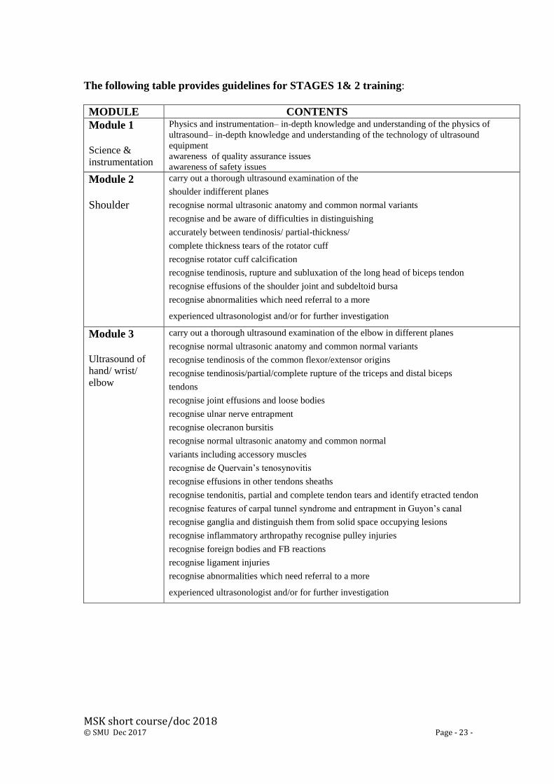
The following table provides guidelines for STAGES 1& 2 training:
MODULE CONTENTS
Module 1
Science &
instrumentation
Physics and instrumentation– in-depth knowledge and understanding of the physics of
ultrasound– in-depth knowledge and understanding of the technology of ultrasound
equipment
awareness of quality assurance issues
awareness of safety issues
Module 2
Shoulder
carry out a thorough ultrasound examination of the
shoulder indifferent planes
recognise normal ultrasonic anatomy and common normal variants
recognise and be aware of difficulties in distinguishing
accurately between tendinosis/ partial-thickness/
complete thickness tears of the rotator cuff
recognise rotator cuff calcification
recognise tendinosis, rupture and subluxation of the long head of biceps tendon
recognise effusions of the shoulder joint and subdeltoid bursa
recognise abnormalities which need referral to a more
experienced ultrasonologist and/or for further investigation
Module 3
Ultrasound of
hand/ wrist/
elbow
carry out a thorough ultrasound examination of the elbow in different planes
recognise normal ultrasonic anatomy and common normal variants
recognise tendinosis of the common flexor/extensor origins
recognise tendinosis/partial/complete rupture of the triceps and distal biceps
tendons
recognise joint effusions and loose bodies
recognise ulnar nerve entrapment
recognise olecranon bursitis
recognise normal ultrasonic anatomy and common normal
variants including accessory muscles
recognise de Quervain’s tenosynovitis
recognise effusions in other tendons sheaths
recognise tendonitis, partial and complete tendon tears and identify etracted tendon
recognise features of carpal tunnel syndrome and entrapment in Guyon’s canal
recognise ganglia and distinguish them from solid space occupying lesions
recognise inflammatory arthropathy recognise pulley injuries
recognise foreign bodies and FB reactions
recognise ligament injuries
recognise abnormalities which need referral to a more
experienced ultrasonologist and/or for further investigation

MSK short course/doc 2018 © SMU Dec 2017 Page - 24 -
Module 4
Ultrasound of
foot/ankle/knee
carry out a thorough ultrasound examination in different planes
recognise normal ultrasonic anatomy and common normal variants
recognise joint effusion
recognise ganglia and bursae, including ruptured Baker’s cyst
recognise meniscal cysts and associated meniscal tears
recognise sprains of collateral ligaments
recognise tendinosis and tears of patellar tendon
recognise abnormalities which need referral to a more
experienced ultrasonologist and/or for further investigation
perform a thorough ultrasound examination of the lower leg in
different planes
recognise normal ultrasonic anatomy and common normal variants
recognise muscle contusions and tears
recognise muscle herniae
recognise normal ultrasonic anatomy and common normal variants including
accessory muscles
recognise tendinosis and tears of achilles, posterior tibial and peroneal tendons
recognise joint effusions and loose bodies
recognise plantar fasciitis
recognise Morton’s neuroma
recognise ganglia and distinguish them from solid space occupying lesions
recognise arthropathy
recognise ankle ligament injuries
recognise foreign bodies and foreign body reactions
recognise abnormalities which need referral to a more
experienced ultrasonologist and/or for further investigation
MSK short course/doc 2018 © SMU Dec 2017 Page - 25 -
Stage 3
Final Ultrasound Assessment

MSK short course/doc 2018 © SMU Dec 2017 Page - 26 -
Final Assessment
Aim
• The assessment of ultrasound competency in musculoskeletal ultrasound of the
upper and lower limb
Outcome
• A demonstration of a safe and competent application of ultrasound imaging in
the visualisation and interpretation of ultrasound anatomy and pathology of the
upper and lower limb

MSK short course/doc 2018 © SMU Dec 2017 Page - 27 -
Final Ultrasound Assessment Procedure
MSK practitioner will:
• scan a minimum of 3 patients in each module to the satisfaction of the assessor
in a clinical situation organised by the course leader;
• demonstrate a satisfactory level of ultrasound competency in the selected
modules;
• demonstrate an ability to operate an ultrasound machine to produce an
optimum image;
• provide a verbal or a written report to the assessor on ultrasound examinations;
• demonstrate an awareness and knowledge of safety and quality assurance
issues.
The ultrasound clinics will consist of student models and suitable patients selected by
the course leader.
(The clinical workshops are supervised by Dr. PP Raju (Consultant Radiologist and
course supervisor), Dr. James Brown (Consultant Sport Medicine Physician),Dr.
Marian O’Reilly (Consultant Radiologist) and Dr. John Tanner (Sports Medicine
Consultant).
Certification of Clinical Competency
MSK practitioners who demonstrate safe practice and are competent in
musculoskeletal ultrasound, and satisfy the EFUSMB (2002)/RCR (2005) Guidelines
and the SMU assessment procedures and guidelines.

MSK short course/doc 2018 © SMU Dec 2017 Page - 28 -
Teaching, Learning Strategies & Methods

MSK short course/doc 2018 © SMU Dec 2017 Page - 29 -
Learning support
• Personal tutor/mentor
• Learning resources (computer room and indicative textbooks/access to journals)
• Access to the SMU tutor by email/telemedicine link and the SMU website
• SMU Moodle
Admission criteria
• Practicing MSKP’s with evidence of satisfactory supervised clinical training site
Evaluation of quality & standards in learning & teaching
• SMU
• Course supervisor
• ADQC
Mechanisms for review and evaluation
• SMU
Responsibilities for monitoring and evaluation
• Course Coordinators
• Student Representatives
• CASE
Mechanisms for gaining student feedback
• Course level student questionnaires
• Dissatisfaction with any aspect of the course allows student to lodge a formal
complaint via the head of the school.
Staff Development Priorities
• Academic staff undertake activities related to research, teaching, learning, student
support and guidance
• Annual staff appraisals match development to needs

MSK short course/doc 2018 © SMU Dec 2017 Page - 30 -
Course Logistics
SMU teaching faculty and the Samsung support the practical and clinical workshops.
These are carried out using consented volunteers and specially selected patients.

MSK short course/doc 2018 © SMU Dec 2017 Page - 31 -