first and only fda approved
TRANSCRIPT
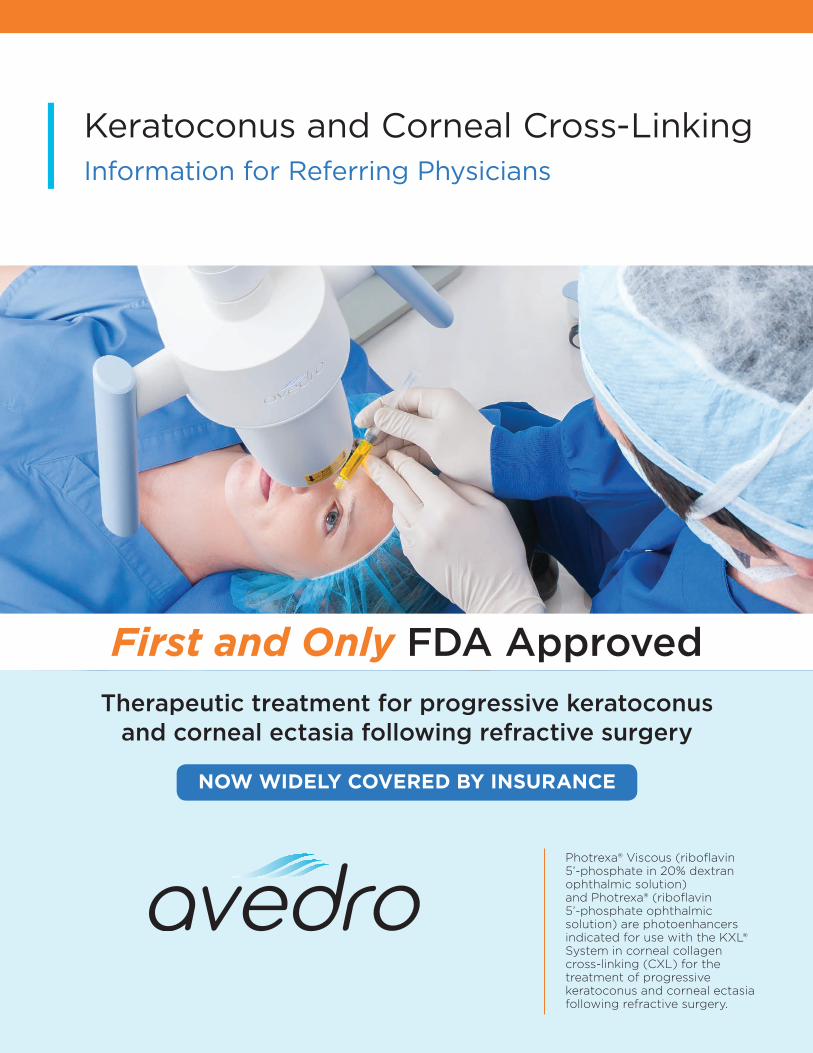
Keratoconus and Corneal Cross-Linking Information for Referring Physicians
Photrexa® Viscous (riboflavin 5’-phosphate in 20% dextran ophthalmic solution) and Photrexa® (riboflavin 5’-phosphate ophthalmic solution) are photoenhancers indicated for use with the KXL® System in corneal collagen cross-linking (CXL) for the treatment of progressive keratoconus and corneal ectasia following refractive surgery.
Therapeutic treatment for progressive keratoconus and corneal ectasia following refractive surgery
First and Only FDA Approved
NOW WIDELY COVERED BY INSURANCE
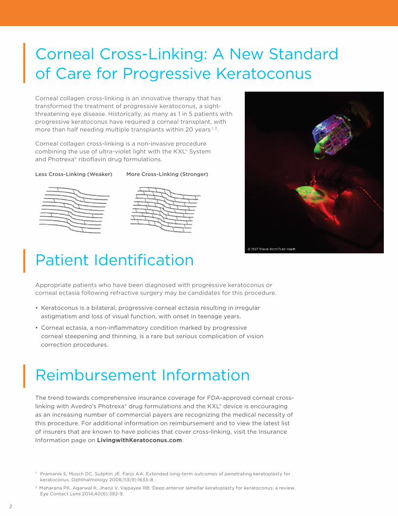
Corneal collagen cross-linking is a non-invasive procedure combining the use of ultra-violet light with the KXL® System and Photrexa® riboflavin drug formulations.
Corneal collagen cross-linking is an innovative therapy that has transformed the treatment of progressive keratoconus, a sight-threatening eye disease. Historically, as many as 1 in 5 patients with progressive keratoconus have required a corneal transplant, with more than half needing multiple transplants within 20 years 1, 2.
Appropriate patients who have been diagnosed with progressive keratoconus or corneal ectasia following refractive surgery may be candidates for this procedure.
Corneal Cross-Linking: A New Standard of Care for Progressive Keratoconus
Patient Identification
Reimbursement Information
• Keratoconus is a bilateral, progressive corneal ectasia resulting in irregular astigmatism and loss of visual function, with onset in teenage years.
• Corneal ectasia, a non-inflammatory condition marked by progressive corneal steepening and thinning, is a rare but serious complication of vision correction procedures.
The trend towards comprehensive insurance coverage for FDA-approved corneal cross-linking with Avedro’s Photrexa® drug formulations and the KXL® device is encouraging as an increasing number of commercial payers are recognizing the medical necessity of this procedure. For additional information on reimbursement and to view the latest list of insurers that are known to have policies that cover cross-linking, visit the Insurance Information page on LivingwithKeratoconus.com.
Less Cross-Linking (Weaker) More Cross-Linking (Stronger)
1 Pramanik S, Musch DC, Sutphin JE, Farjo AA. Extended long-term outcomes of penetrating keratoplasty for keratoconus. Ophthalmology 2006;113(9):1633-8.
2 Maharana PK, Agarwal K, Jhanji V, Vajpayee RB. Deep anterior lamellar keratoplasty for keratoconus: a review. Eye Contact Lens 2014;40(6):382-9.
2
What are the signs I should be looking for?Early signs of keratoconus may include asymmetric refractive error, high or progressive astigmatism, or reduced best corrected visual acuity. The onset of keratoconus often occurs in teenage years or early twenties but can start at any time.
Patient symptoms may include:• Constantly and regularly changing refractive errors• Blurry vision• Increased light sensitivity• Difficultly driving at night• A halo around lights and ghosting (especially at night)• Eye strain• Headaches and general eye pain• Eye irritation, excessive eye rubbing
Keratoconus, especially in the early stages, can be difficult to diagnose and all of the above symptoms could be associated with other eye problems.
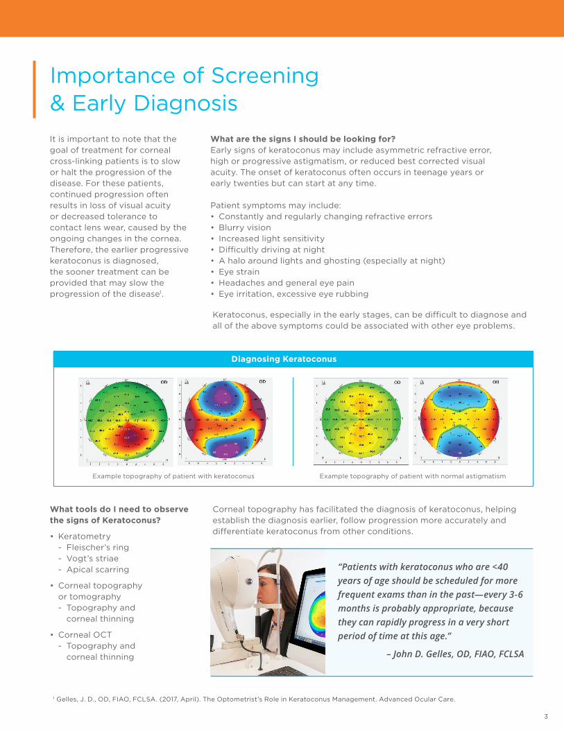
Diagnosing Keratoconus
What tools do I need to observe the signs of Keratoconus?
• Keratometry - Fleischer’s ring
- Vogt’s striae - Apical scarring
• Corneal topography or tomography
- Topography and corneal thinning
• Corneal OCT - Topography and
corneal thinning
Corneal topography has facilitated the diagnosis of keratoconus, helping establish the diagnosis earlier, follow progression more accurately and differentiate keratoconus from other conditions.
Example topography of patient with keratoconus Example topography of patient with normal astigmatism
Importance of Screening & Early Diagnosis
1 Gelles, J. D., OD, FIAO, FCLSA. (2017, April). The Optometrist’s Role in Keratoconus Management. Advanced Ocular Care.
It is important to note that the goal of treatment for corneal cross-linking patients is to slow or halt the progression of the disease. For these patients, continued progression often results in loss of visual acuity or decreased tolerance to contact lens wear, caused by the ongoing changes in the cornea. Therefore, the earlier progressive keratoconus is diagnosed, the sooner treatment can be provided that may slow the progression of the disease1.
“Patients with keratoconus who are <40 years of age should be scheduled for more frequent exams than in the past—every 3-6 months is probably appropriate, because they can rapidly progress in a very short period of time at this age.”
– John D. Gelles, OD, FIAO, FCLSA
3
“Since CXL topography
changes may occur up to
12 months postoperatively,
the contact lens
practitioner must continue
to monitor these patients
to ensure continued good
lens fit and eye health.1”
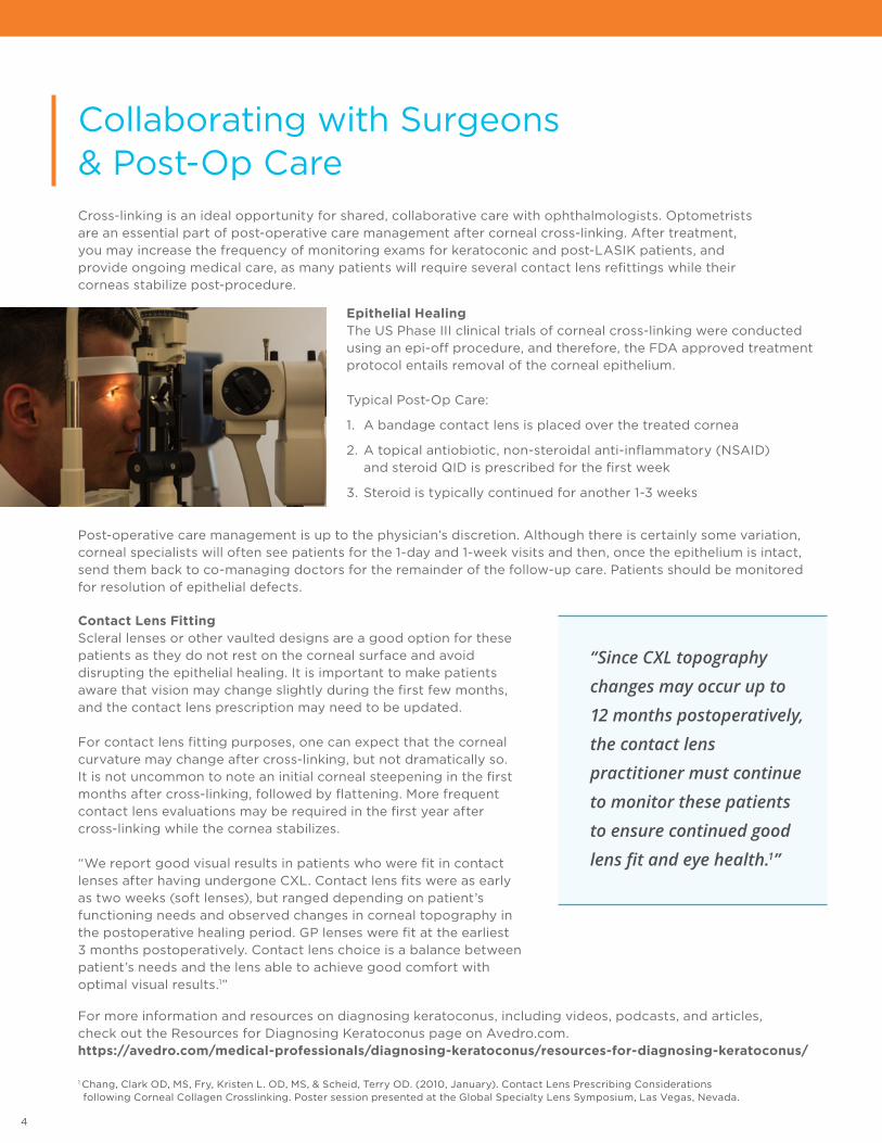
Cross-linking is an ideal opportunity for shared, collaborative care with ophthalmologists. Optometrists are an essential part of post-operative care management after corneal cross-linking. After treatment, you may increase the frequency of monitoring exams for keratoconic and post-LASIK patients, and provide ongoing medical care, as many patients will require several contact lens refittings while their corneas stabilize post-procedure.
Epithelial HealingThe US Phase III clinical trials of corneal cross-linking were conducted using an epi-off procedure, and therefore, the FDA approved treatment protocol entails removal of the corneal epithelium.
Typical Post-Op Care:
1. A bandage contact lens is placed over the treated cornea
2. A topical antiobiotic, non-steroidal anti-inflammatory (NSAID) and steroid QID is prescribed for the first week
3. Steroid is typically continued for another 1-3 weeks
Post-operative care management is up to the physician’s discretion. Although there is certainly some variation, corneal specialists will often see patients for the 1-day and 1-week visits and then, once the epithelium is intact, send them back to co-managing doctors for the remainder of the follow-up care. Patients should be monitored for resolution of epithelial defects.
Contact Lens FittingScleral lenses or other vaulted designs are a good option for these patients as they do not rest on the corneal surface and avoid disrupting the epithelial healing. It is important to make patients aware that vision may change slightly during the first few months, and the contact lens prescription may need to be updated.
For contact lens fitting purposes, one can expect that the corneal curvature may change after cross-linking, but not dramatically so. It is not uncommon to note an initial corneal steepening in the first months after cross-linking, followed by flattening. More frequent contact lens evaluations may be required in the first year after cross-linking while the cornea stabilizes.
“We report good visual results in patients who were fit in contact lenses after having undergone CXL. Contact lens fits were as early as two weeks (soft lenses), but ranged depending on patient’s functioning needs and observed changes in corneal topography in the postoperative healing period. GP lenses were fit at the earliest 3 months postoperatively. Contact lens choice is a balance between patient’s needs and the lens able to achieve good comfort with optimal visual results.1”
For more information and resources on diagnosing keratoconus, including videos, podcasts, and articles, check out the Resources for Diagnosing Keratoconus page on Avedro.com. https://avedro.com/medical-professionals/diagnosing-keratoconus/resources-for-diagnosing-keratoconus/
Collaborating with Surgeons & Post-Op Care
1 Chang, Clark OD, MS, Fry, Kristen L. OD, MS, & Scheid, Terry OD. (2010, January). Contact Lens Prescribing Considerations following Corneal Collagen Crosslinking. Poster session presented at the Global Specialty Lens Symposium, Las Vegas, Nevada.
4
HIGHLIGHTS OF PRESCRIBING INFORMATION
These highlights do not include all the information needed to use PHOTREXA® VISCOUS and PHOTREXA® safely and effectively. See full prescribing information for PHOTREXA VISCOUS and PHOTREXA.
PHOTREXA VISCOUS (riboflavin 5’-phosphate in 20% dextran ophthalmic solution) 0.146% for topical ophthalmic use
PHOTREXA (riboflavin 5’-phosphate ophthalmic solution) 0.146% for topical ophthalmic use
For use with the KXL® System Initial U.S. Approval: 2016
INDICATIONS AND USAGE PHOTREXA VISCOUS and PHOTREXA are photoenhancers indicated for use with the KXL System in corneal collagen cross-linking for the treatment of progressive keratoconus (1.1) and corneal ectasia following refractive surgery (1.2).
DOSAGE AND ADMINISTRATION • Debride the epithelium using standard aseptic technique using
topical anesthesia (2).• Then instill 1 drop of PHOTREXA VISCOUS topically on the eye
every 2 minutes for 30 minutes (2). • After 30 minutes, examine the eye under slit lamp for presence of a
yellow flare in the anterior chamber. If flare is not detected, instill 1 drop of PHOTREXA VISCOUS every 2 minutes for an additional 2 to 3 drops and recheck for yellow flare. Repeat as necessary (2).
• Once flare is observed, perform ultrasound pachymetry. If corneal thickness is less than 400 microns, instill 2 drops of PHOTREXA every 5 to 10 seconds until the corneal thickness increases to at least 400 microns (2).
• Irradiation should not be performed unless this 400 micron threshold is met and the yellow flare is seen (2).
• Irradiate the eye for 30 minutes at 3mW/cm2 using the KXL System as per the instructions in the KXL manual. During irradiation, continue topical instillation of PHOTREXA VISCOUS onto the eye every 2 minutes for the 30 minute irradiation period (2).
• Refer to the KXL Operator’s manual for specific device instructions (2).
DOSAGE FORMS AND STRENGTHS • PHOTREXA VISCOUS in a 3 mL glass syringe containing sterile
1.56 mg/mL riboflavin 5’-phosphate in 20% dextran ophthalmic solution (3.1)
• PHOTREXA in a 3 mL glass syringe containing sterile 1.46 mg/mL riboflavin 5’-phosphate ophthalmic solution (3.2)
CONTRAINDICATIONS None (4)
WARNINGS AND PRECAUTIONS Ulcerative keratitis can occur. Monitor for resolution of epithelial defects (5)
ADVERSE REACTIONS In progressive keratoconus patients, the most common ocular adverse reactions in any CXL-treated eye were corneal opacity (haze), punctate keratitis, corneal striae, corneal epithelium defect, eye pain, reduced visual acuity, and blurred vision (6.1). In corneal ectasia patients, the most common ocular adverse reactions were corneal opacity (haze), corneal epithelium defect, corneal striae, dry eye, eye pain, punctate keratitis, photophobia, reduced visual acuity, and blurred vision (6.1). To report SUSPECTED ADVERSE REACTIONS, contact Avedro at 1-844-528-3376 or FDA at 1-800-FDA-1088 or www.fda.gov/medwatch
See 17 for PATIENT COUNSELING INFORMATION
Revised: 11/2018 FULL PRESCRIBING INFORMATION: CONTENTS*1. INDICATIONS AND USAGE 1.1 Progressive Keratoconus 1.2 Corneal Ectasia following refractive surgery2. DOSAGE AND ADMINISTRATION3. DOSAGE FORMS AND STRENGTHS 3.1. Photrexa Viscous 3.2. Photrexa
4. CONTRAINDICATIONS5. WARNINGS AND PRECAUTIONS6. ADVERSE REACTIONS 6.1. Clinical Trial Experience8. USE IN SPECIFIC POPULATIONS 8.1. Pregnancy 8.2. Lactation 8.4. Pediatric Use 8.5. Geriatric Use11. DESCRIPTION12. CLINICAL PHARMACOLOGY 12.1. Mechanism of Action13. NONCLINICAL TOXICOLOGY 13.1. Carcinogenesis, Mutagenesis, Impairment of Fertility14. CLINICAL STUDIES16. HOW SUPPLIED/STORAGE AND HANDLING17. PATIENT COUNSELING INFORMATION
*Sections or subsections omitted from the full prescribing information are not listed. FULL PRESCRIBING INFORMATION1. INDICATIONS AND USAGEPHOTREXA® VISCOUS and PHOTREXA® are indicated for use in corneal collagen cross-linking in combination with the KXL™ System for the treatment of 1.1 Progressive keratoconus1.2 Corneal ectasia following refractive surgery.2. DOSAGE AND ADMINISTRATIONUsing topical anesthesia, debride the epithelium to a diameter of approximately 9 mm using standard aseptic technique. Post epithelial debridement, instill 1 drop of PHOTREXA VISCOUS topically on the eye every 2 minutes for 30 minutes. At the end of the 30 minute soaking period, examine the eye under the slit lamp for the presence of a yellow flare in the anterior chamber. If the yellow flare is not detected, instill 1 drop of PHOTREXA VISCOUS every 2 minutes for an additional 2 to 3 drops and recheck for the presence of a yellow flare. This process can be repeated as necessary. Once the yellow flare is observed, perform ultrasound pachymetry. If corneal thickness is less than 400 microns, instill 2 drops of PHOTREXA every 5 to 10 seconds until the corneal thickness increases to at least 400 microns. Irradiation should not be performed unless this 400 micron threshold is met and the yellow flare is seen. Irradiate the eye for 30 continuous minutes at 3mW/cm2 at a wavelength of 365 nm, centered over the cornea, using the KXL System as per the instructions in the KXL manual. During irradiation, continue topical instillation of PHOTREXA VISCOUS onto the eye every 2 minutes for the 30 minute irradiation period. For topical ophthalmic use. Do not inject. Single use PHOTREXA VISCOUS and PHOTREXA only. Discard syringe(s) after use. PHOTREXA VISCOUS and PHOTREXA are for use with the KXL System only.PLEASE REFER TO THE KXL OPERATOR’S MANUAL FOR SPECIFIC DEVICE INSTRUCTIONS.3. DOSAGE FORMS AND STRENGTHS3.1 PHOTREXA VISCOUSPHOTREXA VISCOUS in a 3 mL glass syringe containing sterile 1.56 mg/mL riboflavin 5’-phosphate in 20% dextran ophthalmic solution for topical administration.3.2 PHOTREXAPHOTREXA in a 3 mL glass syringe containing sterile 1.46 mg/mL riboflavin 5’-phosphate ophthalmic solution for topical administration.4. CONTRAINDICATIONSNone. 5. WARNINGS AND PRECAUTIONSUlcerative keratitis can occur. Monitor for resolution of epithelial defects. [See Adverse Reactions (6)]. 6. ADVERSE REACTIONSThe following clinically significant adverse reactions are described elsewhere in the labeling: Ulcerative keratitis [Warnings and Precautions (5)]6.1 Clinical Trials Experience:Because clinical trials are conducted under widely varying conditions, adverse reaction rates observed in the clinical trials of a drug cannot be directly compared to rates in the clinical trials of another drug and may not reflect the rates observed in practice. The safety of the corneal collagen cross-linking procedure was evaluated in 3 randomized, parallel-group, open-label, sham-controlled trials; patients were followed up for 12 months. Study 1 enrolled patients with progressive keratoconus or corneal ectasia
5
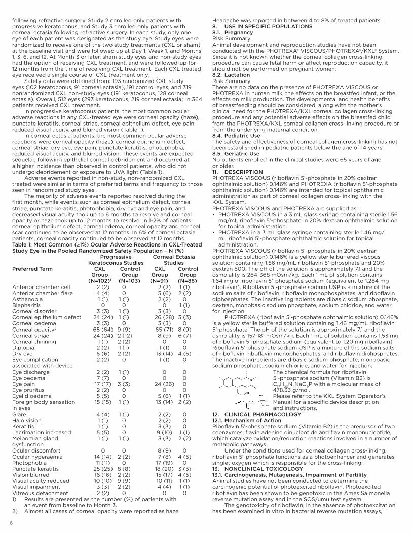
following refractive surgery. Study 2 enrolled only patients with progressive keratoconus, and Study 3 enrolled only patients with corneal ectasia following refractive surgery. In each study, only one eye of each patient was designated as the study eye. Study eyes were randomized to receive one of the two study treatments (CXL or sham) at the baseline visit and were followed up at Day 1, Week 1, and Months 1, 3, 6, and 12. At Month 3 or later, sham study eyes and non-study eyes had the option of receiving CXL treatment, and were followed-up for 12 months from the time of receiving CXL treatment. Each CXL treated eye received a single course of CXL treatment only. Safety data were obtained from: 193 randomized CXL study eyes (102 keratoconus, 91 corneal ectasia), 191 control eyes, and 319 nonrandomized CXL non-study eyes (191 keratoconus, 128 corneal ectasia). Overall, 512 eyes (293 keratoconus, 219 corneal ectasia) in 364 patients received CXL treatment. In progressive keratoconus patients, the most common ocular adverse reactions in any CXL-treated eye were corneal opacity (haze), punctate keratitis, corneal striae, corneal epithelium defect, eye pain, reduced visual acuity, and blurred vision (Table 1). In corneal ectasia patients, the most common ocular adverse reactions were corneal opacity (haze), corneal epithelium defect, corneal striae, dry eye, eye pain, punctate keratitis, photophobia, reduced visual acuity, and blurred vision. These events are expected sequelae following epithelial corneal debridement and occurred at a higher incidence than observed in control patients, who did not undergo debridement or exposure to UVA light (Table 1). Adverse events reported in non-study, non-randomized CXL treated were similar in terms of preferred terms and frequency to those seen in randomized study eyes. The majority of adverse events reported resolved during the first month, while events such as corneal epithelium defect, corneal striae, punctate keratitis, photophobia, dry eye and eye pain, and decreased visual acuity took up to 6 months to resolve and corneal opacity or haze took up to 12 months to resolve. In 1-2% of patients, corneal epithelium defect, corneal edema, corneal opacity and corneal scar continued to be observed at 12 months. In 6% of corneal ectasia patients, corneal opacity continued to be observed at 12 months.Table 1: Most Common (≥1%) Ocular Adverse Reactions in CXL-Treated Study Eye in the Pooled Randomized Safety Population – N (%) Progressive Corneal Ectasia Keratoconus Studies StudiesPreferred Term CXL Control CXL Control Group Group Group Group (N=102)1 (N=103)1 (N=91)1 (N=88)1
Anterior chamber cell 2 (2) 0 2 (2) 1 (1)Anterior chamber flare 4 (4) 0 5 (6) 2 (2)Asthenopia 1 (1) 1 (1) 2 (2) 0Blepharitis 0 0 0 1 (1)Corneal disorder 3 (3) 1 (1) 3 (3) 0Corneal epithelium defect 24 (24) 1 (1) 26 (28) 3 (3)Corneal oedema 3 (3) 0 3 (3) 0Corneal opacity2 65 (64) 9 (9) 65 (71) 8 (9)Corneal striae 24 (24) 12 (12) 8 (9) 6 (7)Corneal thinning 1 (1) 2 (2) 0 0Diplopia 2 (2) 1 (1) 1 (1) 0Dry eye 6 (6) 2 (2) 13 (14) 4 (5)Eye complication 2 (2) 0 1 (1) 0associated with device Eye discharge 2 (2) 1 (1) 0 0Eye oedema 7 (7) 0 0 0Eye pain 17 (17) 3 (3) 24 (26) 0Eye pruritus 2 (2) 0 0 0Eyelid oedema 5 (5) 0 5 (6) 1 (1)Foreign body sensation 15 (15) 1 (1) 13 (14) 2 (2)in eyesGlare 4 (4) 1 (1) 2 (2) 0Halo vision 1 (1) 0 2 (2) 0Keratitis 1 (1) 0 3 (3) 0Lacrimation increased 5 (5) 0 9 (10) 1 (1)Meibomian gland 1 (1) 1 (1) 3 (3) 2 (2)dysfunctionOcular discomfort 0 0 8 (9) 0Ocular hyperaemia 14 (14) 2 (2) 7 (8) 4 (5)Photophobia 11 (11) 0 17 (19) 0Punctate keratitis 25 (25) 8 (8) 18 (20) 3 (3)Vision blurred 16 (16) 2 (2) 15 (17) 4 (5)Visual acuity reduced 10 (10) 9 (9) 10 (11) 1 (1)Visual impairment 3 (3) 2 (2) 4 (4) 1 (1)Vitreous detachment 2 (2) 0 0 01) Results are presented as the number (%) of patients with an event from baseline to Month 3.2) Almost all cases of corneal opacity were reported as haze.
Headache was reported in between 4 to 8% of treated patients.8. USE IN SPECIFIC POPULATIONS8.1. PregnancyRisk SummaryAnimal development and reproduction studies have not been conducted with the PHOTREXA® VISCOUS/PHOTREXA®/KXL® System. Since it is not known whether the corneal collagen cross-linking procedure can cause fetal harm or affect reproduction capacity, it should not be performed on pregnant women. 8.2. LactationRisk SummaryThere are no data on the presence of PHOTREXA VISCOUS or PHOTREXA in human milk, the effects on the breastfed infant, or the effects on milk production. The developmental and health benefits of breastfeeding should be considered, along with the mother’s clinical need for the PHOTREXA/KXL corneal collagen cross-linking procedure and any potential adverse effects on the breastfed child from the PHOTREXA/KXL corneal collagen cross-linking procedure or from the underlying maternal condition. 8.4. Pediatric UseThe safety and effectiveness of corneal collagen cross-linking has not been established in pediatric patients below the age of 14 years.8.5. Geriatric UseNo patients enrolled in the clinical studies were 65 years of age or older.11. DESCRIPTIONPHOTREXA VISCOUS (riboflavin 5’-phosphate in 20% dextran ophthalmic solution) 0.146% and PHOTREXA (riboflavin 5’-phosphate ophthalmic solution) 0.146% are intended for topical ophthalmic administration as part of corneal collagen cross-linking with the KXL System.PHOTREXA VISCOUS and PHOTREXA are supplied as:• PHOTREXA VISCOUS in a 3 mL glass syringe containing sterile 1.56
mg/mL riboflavin 5’-phosphate in 20% dextran ophthalmic solution for topical administration.
• PHOTREXA in a 3 mL glass syringe containing sterile 1.46 mg/mL riboflavin 5’-phosphate ophthalmic solution for topical administration.
PHOTREXA VISCOUS (riboflavin 5’-phosphate in 20% dextran ophthalmic solution) 0.146% is a yellow sterile buffered viscous solution containing 1.56 mg/mL riboflavin 5’-phosphate and 20% dextran 500. The pH of the solution is approximately 7.1 and the osmolality is 284-368 mOsm/kg. Each 1 mL of solution contains 1.64 mg of riboflavin 5’-phosphate sodium (equivalent to 1.284 mg riboflavin). Riboflavin 5’-phosphate sodium USP is a mixture of the sodium salts of riboflavin, riboflavin monophosphates, and riboflavin diphosphates. The inactive ingredients are dibasic sodium phosphate, dextran, monobasic sodium phosphate, sodium chloride, and water for injection. PHOTREXA (riboflavin 5’-phosphate ophthalmic solution) 0.146% is a yellow sterile buffered solution containing 1.46 mg/mL riboflavin 5’-phosphate. The pH of the solution is approximately 7.1 and the osmolality is 157-181 mOsm/kg. Each 1 mL of solution contains 1.53 mg of riboflavin 5’-phosphate sodium (equivalent to 1.20 mg riboflavin). Riboflavin 5’-phosphate sodium USP is a mixture of the sodium salts of riboflavin, riboflavin monophosphates, and riboflavin diphosphates. The inactive ingredients are dibasic sodium phosphate, monobasic sodium phosphate, sodium chloride, and water for injection.
The chemical formula for riboflavin 5’-phosphate sodium (Vitamin B2) is C17H20N4NaO9P with a molecular mass of 478.33 g/mol. Please refer to the KXL System Operator’s Manual for a specific device description and instructions.
12. CLINICAL PHARMACOLOGY12.1. Mechanism of ActionRiboflavin 5’-phosphate sodium (Vitamin B2) is the precursor of two coenzymes, flavin adenine dinucleotide and flavin mononucleotide, which catalyze oxidation/reduction reactions involved in a number of metabolic pathways. Under the conditions used for corneal collagen cross-linking, riboflavin 5‘-phosphate functions as a photoenhancer and generates singlet oxygen which is responsible for the cross-linking.13. NONCLINICAL TOXICOLOGY13.1. Carcinogenesis, Mutagenesis, Impairment of FertilityAnimal studies have not been conducted to determine the carcinogenic potential of photoexcited riboflavin. Photoexcited riboflavin has been shown to be genotoxic in the Ames Salmonella reverse mutation assay and in the SOS/umu test system. The genotoxicity of riboflavin, in the absence of photoexcitation has been examined in vitro in bacterial reverse mutation assays,
6
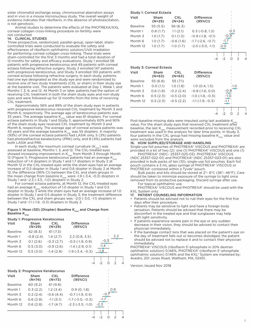
sister chromatid exchange assay, chromosomal aberration assays and in vivo in a mouse micronucleus study. The overall weight of evidence indicates that riboflavin, in the absence of photoexcitation, is not genotoxic. Animal studies to determine the effects of the PHOTREXA/KXL corneal collagen cross-linking procedure on fertility were not conducted.14. CLINICAL STUDIESThree prospective, randomized, parallel-group, open-label, sham-controlled trials were conducted to evaluate the safety and effectiveness of riboflavin ophthalmic solution/UVA irradiation for performing corneal collagen cross-linking. These trials were sham-controlled for the first 3 months and had a total duration of 12 months for safety and efficacy evaluations. Study 1 enrolled 58 patients with progressive keratoconus and 49 patients with corneal ectasia following refractive surgery. Study 2 enrolled 147 patients with progressive keratoconus, and Study 3 enrolled 130 patients with corneal ectasia following refractive surgery. In each study, patients had one eye designated as the study eye and were randomized to receive one of two study treatments (CXL or sham) in their study eye at the baseline visit. The patients were evaluated at Day 1, Week 1, and Months 1, 3, 6, and 12. At Month 3 or later, patients had the option of receiving CXL treatment in both the sham study eyes and non-study eyes and were followed-up for 12 months from the time of receiving CXL treatment. Approximately 56% and 89% of the sham study eyes in patients with progressive keratoconus received CXL treatment by Month 3 and Month 6, respectively. The average age of keratoconus patients was 33 years. The average baseline Kmax value was 61 diopters. For corneal ectasia patients in Study 1 and Study 3, approximately 60% and 90% of the sham study eyes received CXL treatment by Month 3 and Month 6, respectively. The average age of corneal ectasia patients was 43 years and the average baseline Kmax was 55 diopters. A majority (93%) of the corneal ectasia patients had LASIK only, 5 (3%) patients had photorefractive keratectomy (PRK) only, and 8 (4%) patients had both LASIK and PRK. In each study, the maximum corneal curvature (Kmax) was assessed at baseline, Months 1, 3, and 12. The CXL-treated eyes showed increasing improvement in Kmax from Month 3 through Month 12 (Figure 1). Progressive keratoconus patients had an average Kmax reduction of 1.4 diopters in Study 1 and 1.7 diopters in Study 2 at Month 12 in the CXL-treated eyes while the sham eyes had an average increase of 0.5 diopter in Study 1 and 0.6 diopter in Study 2 at Month 12; the difference (95% CI) between the CXL and sham groups in the mean change from baseline Kmax were -1.9 (-3.4, -0.3) diopters in Study 1 and -2.3 (-3.5, -1.0) diopters in Study 2. For corneal ectasia patients, at Month 12, the CXL-treated eyes had an average Kmax reduction of 1.0 diopter in Study 1 and 0.5 diopter in Study 3 while the sham eyes had an average increase of 1.0 diopter in Study 1 and 0.5 diopter in Study 3; the treatment difference between the CXL and sham groups was: -2.0 (-3.0, -1.1) diopters in Study 1 and -1.1 (-1.9, -0.3) diopters in Study 3.
Figure 1: Mean (SD) (Diopter) Baseline Kmax and Change from Baseline Kmax
Post-baseline missing data were imputed using last available Kmax value. For the sham study eyes that received CXL treatment after baseline, the last Kmax measurement recorded prior to receiving CXL treatment was used in the analysis for later time points. In Study 3, four patients in the CXL group had missing baseline Kmax value and were excluded from the analysis. 16. HOW SUPPLIED/STORAGE AND HANDLINGSingle-use foil pouches of PHOTREXA® VISCOUS and PHOTREXA® are provided in a kit of two (2): one (1) PHOTREXA® VISCOUS and one (1) PHOTREXA® (NDC- 25357-025-03). PHOTREXA® VISCOUS (NDC 25357-022-01) and PHOTREXA® (NDC 25357-023-01) are also provided in bulk packs of ten (10), single-use foil pouches. Each foil pouch contains a 3 mL glass syringe of PHOTREXA® VISCOUS or PHOTREXA® contained within a Tyvek® pouch. Bulk packs and kits should be stored at 2°- 8°C (36°- 46°F). Care should be taken to minimize exposure of the syringe to light once removed from its protective packaging. Discard syringe after use. For topical ophthalmic use. PHOTREXA® VISCOUS and PHOTREXA® should be used with the KXL System only.17. PATIENT COUNSELING INFORMATION• Patients should be advised not to rub their eyes for the first five
days after their procedure. • Patients may be sensitive to light and have a foreign body
sensation. Patients should be advised that there may be discomfort in the treated eye and that sunglasses may help with light sensitivity.
• If patients experience severe pain in the eye or any sudden decrease in their vision, they should be advised to contact their physician immediately.
• If the bandage contact lens that was placed on the patient’s eye on the day of treatment falls out or becomes dislodged, the patient should be advised not to replace it and to contact their physician immediately.
PHOTREXA® VISCOUS (riboflavin 5’-phosphate in 20% dextran ophthalmic solution) 0.146%, PHOTREXA® (riboflavin 5’-phosphate ophthalmic solution) 0.146% and the KXL® System are marketed by: Avedro, 201 Jones Road, Waltham, MA, 02451.
Version: Issued Nov 2018
Study 1: Progressive KeratoconusVisit Sham CXL Difference (N=29) (N=29) (95%CI)Baseline 62 (8.3) 61 (7.3)Month 1 -0.8 (2.4) 1.4 (2.7) 2.2 (0.8, 3.5)Month 3 0.1 (2.6) -0.3 (2.7) -0.5 (-1.9, 0.9)Month 6 0.5 (3.0) -0.9 (2.6) -1.4 (-2.9, 0.1)Month 12 0.5 (3.0) -1.4 (2.8) -1.9 (-3.4, -0.3) -1.9
-1.4-0.5
2.2
-4 -2 0 2 4
Study 2: Progressive KeratoconusVisit Sham CXL Difference (N=74) (N=73) (95%CI)Baseline 60 (9.2) 61 (9.8)Month 1 0.3 (2.2) 1.2 (3.4) 0.9 (0, 1.8)Month 3 0.2 (2.4) -0.6 (4.4) -0.7 (-1.9, 0.4)Month 6 0.6 (2.8) -1.1 (5.1) -1.7 (-3.0, -0.3)Month 12 0.6 (2.8) -1.7 (4.7) -2.3 (-3.5, -1.0) -2.3
-1.7-0.7
0.9
-4 -2 0 2 4
Study 1: Corneal EctasiaVisit Sham CXL Difference (N=25) (N=24) (95%CI)Baseline 55 (5.5) 56 (6.3)Month 1 0.8 (1.7) 1.1 (2.1) 0.3 (-0.8, 1.3)Month 3 1.0 (1.7) 0.1 (1.3) -0.9 (-1.8, -0.1)Month 6 1.0 (1.7) -0.6 (1.6) -1.7 (-2.6, -0.7)Month 12 1.0 (1.7) -1.0 (1.7) -2.0 (-3.0, -1.1) -2.0
-1.7-0.9
0.3
-4 -2 0 2 4
Study 3: Corneal EctasiaVisit Sham CXL Difference (N=63) (N=63) (95%CI)Baseline 55 (6.8) 55 (7.1)Month 1 0.0 (1.1) 1.0 (1.8) 1.0 (0.4, 1.5)Month 3 0.6 (1.9) -0.2 (2.4) -0.8 (-1.6, 0.0)Month 6 0.5 (2.3) -0.5 (2.0) -1.0 (-1.8, -0.3)Month 12 0.5 (2.3) -0.5 (2.2) -1.1 (-1.9, -0.3) -1.1
-1.0-0.8
1.0
-4 -2 0 2 4
7

About Avedro, IncWe are committed to advancing the treatment of eye conditions such as keratoconus and corneal ectasia following refractive surgery, through the continued innovation of our corneal cross-linking technology and pharmaceuticals. We develop products that are not only state-of-the-art but uphold the highest levels of quality and safety.
In 2016, the FDA approved Photrexa® Viscous (riboflavin 5’-phosphate in 20% dextran ophthalmic solution) and Photrexa® (riboflavin 5® phosphate ophthalmic solution) and the KXL® system for corneal cross-linking for patients suffering from progressive keratoconus and corneal ectasia following refractive surgery. Cross-linking is a minimally invasive outpatient procedure that combines the use of Photrexa eye drops and ultra-violet (UV) light.
The approval of Photrexa Viscous, Photrexa and the KXL system offers an effective treatment for patients who, until recently, had no therapeutic options to limit the progression of these sight-threatening diseases.
Cross-Linking Procedures Performed Across the Globe
Avedro Procedures Performed
Photrexa Viscous, Photrexa, and the KXL System are available for sale in the United States. No other Avedro products or procedures are available in the United States. MA-00626C
(844) 528-3376 | [email protected] | www.avedro.com