figo staging, cancer datasets and the...
TRANSCRIPT

FIGO staging, cancer
datasets and the
ICCR
Dr Lynn Hirschowitz

1. Cancer staging (including FIGO)
2. Specific points about FIGO staging
3. International Collaboration on Cancer
Reporting (ICCR)
4. New ICCR ovarian/fallopian tube/
primary peritoneal carcinoma dataset

1. General principal for cancer
staging systems
• Stage I: tumour that is strictly confined to the
organ of origin
• Stage II: tumour that has extended locally
beyond the site of origin to involve adjacent
organs or structures
• Stage III: more extensive local involvement or
infiltration of neighbouring organs
• Stage IV: tumour with distant metastases

General principal for all staging
systems
• 4 basic stages divided into substages to
reflect tumour-specific clinical, pathological
or biological prognostic factors within a
given stage.
• Most staging systems have moved from
clinical staging to ‘surgico-pathological’ staging (apart from cervical cancer and
gestational trophoblastic disease).
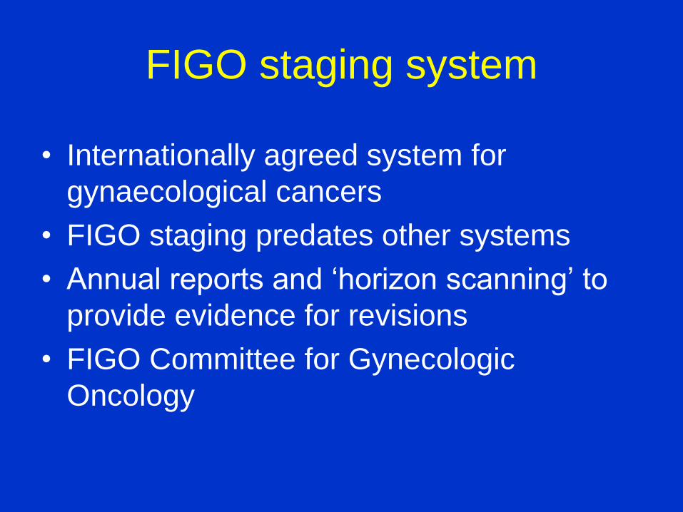
FIGO staging system
• Internationally agreed system for
gynaecological cancers
• FIGO staging predates other systems
• Annual reports and ‘horizon scanning’ to
provide evidence for revisions
• FIGO Committee for Gynecologic
Oncology

Members FIGO Gynaecological
Oncology Committee 2012-2015 • Professor Lynette Denny (Chair), South Africa
• Professor Michael Quinn (Co-Chair), Australia
• Professor Sergio Pecorelli, Italy
• Dr Adriana Bermudez, Argentina
• Dr David Mutch, USA
• Professor Neville Hacker, Australia
• Professor Jaime Prat, Spain
• Professor Elisabeth Åvall Lundqvist, Sweden
• Professor Joanna Cain, USA
• Professor Keiichi Fujiwara, Japan
• Dr Shyam Kishore Shrivastava, India
• Professor Muhieddine A-F Seoud, Lebanon
• Dr Neerja Bhatla, India

FIGO, AJCC, TNM/UICC
• Reciprocal representation
• Collaboration but no agreed co-ordination
of timing of revisions
• TNM staging focus remains ‘anatomic’
• AJCC staging – moves to include ‘non-
anatomic’ data
• FIGO position; dictated by prognosis

On the FIGO radar -
Possible issues:
• Stage III vulval cancer
• LVI in cervical cancer
• Extracapsular invasion and LN metastases
in cervical cancer
• Stage I endometrial cancer

• Poor spread of prognostic groupings with1988
surgical staging system for stage III.
• Importance of lymph node status, number of
positive nodes, size of deposits, extracapsular
extension recognised in 2009 revision.
• Tumour size: node negative IB and II
combined (IB).
2. Specific points about FIGO
staging - vulva

• Clinically staged but FIGO recognises
importance of pre-treatment clinical staging.
• Use of COSD in UK.
• Stage IIA subdivided to take account tumour
size (4 cm or > 4cm).
• Recording of lymph node involvement.
Specific points about FIGO
staging - cervix
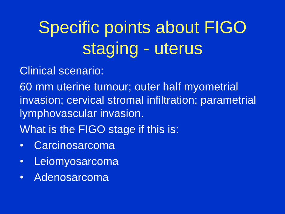
Clinical scenario:
60 mm uterine tumour; outer half myometrial
invasion; cervical stromal infiltration; parametrial
lymphovascular invasion.
What is the FIGO stage if this is:
• Carcinosarcoma
• Leiomyosarcoma
• Adenosarcoma
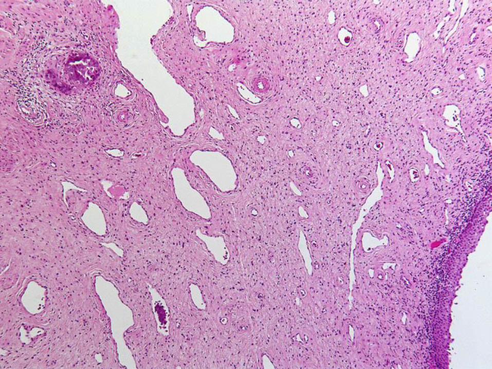
Specific points about FIGO
staging - uterus

Carcinosarcoma (= metaplastic carcinoma)
• Staged in the same way as endometrial
carcinoma
• Lymphovascular invasion without tissue
invasion does not count towards staging
• [Size is a predictor of poor prognosis/
‘peritoneal failure’ for carcinomas]
• Cervical stromal involvement is FIGO stage II
Specific points about FIGO
staging - uterus


Leiomyosarcoma
AJCC 2013

Leiomyosarcoma
• Staged in the same way as endometrial
stromal sarcoma (usually myometrial based)
• Cervical involvement contributes to prognosis
but not to stage
• Lymphovascular invasion without tissue
invasion does not count towards staging
• Tumour size is important for staging
• FIGO stage = IB (>5 cm)
Specific points about FIGO
staging - uterus

Adenosarcoma
AJCC 2013

Adenosarcoma
• Usually endometrial based.
• Stage I = same as 1988 system for endometrial
carcinoma.
• Cervical involvement does not contribute to stage.
• Size is not important for staging.
• Lymphovascular invasion without tissue invasion
does not count towards staging.
• FIGO stage = IC (outer ½ myoinvasion).
Specific points about FIGO
staging - uterus

• Corrigendum published in 2009
• Undifferentiated endometrial/uterine
sarcoma
• Pure heterologous uterine sarcoma
Specific points about FIGO
staging – uterine sarcomas

• Subdivision of stage IC.
• No evidence to support upstaging because of
adhesions.
• Stage IIIA – no evidence that size of nodal
deposits (≤10 mm; >10 mm) is significant.
• Intraperitoneal node involvement = IIIC
• Cytological node involvement of unknown size
= Stage IIIA1(i)
Specific points about FIGO
staging – ovary/FT/PPCa

International Collaboration on Cancer Reporting

International Collaboration on Cancer Reporting
5 founding members
Incorporation September 2014
ICCR Board ICCR Steering Group
• Management issues
• Governance
• Finance
• Publicity
• Membership
• Strategic Alliances
Dataset development
Dataset revision

International Collaboration on Cancer Reporting
Strategic Alliances:
UICC
FIGO
AJCC
EORTC
IARC
Alignment of ICCR dataset development with the IARC
revision of the ‘Blue book’ series

Development of evidence-based ICCR cancer
datasets
• Robust protocols for dataset development.
• Evidentiary support at NHMRC Level III-2 or above.
• Two key dataset components: REQUIRED elements, essential for histological
diagnosis, clinical management, staging, prognosis.
RECOMMENDED elements, non-mandatory, clinically
important; recommended as good practice but not yet
validated or regularly used in patient management.

4. Development of dataset for carcinoma of the
ovary, fallopian tube and primary peritoneal site
• Single dataset for all 3 sites.
• Incorporates 2014 WHO classification of tumours of
the female reproductive organs.
• Incorporates 2013 FIGO staging.
• Includes guidance about site assignment of primary
tumours.
• Includes guidance about chemotherapy response
score/CRS (tumour regression grading).

Russell Vang
Blaise Clarke
Blake Gilks
Colin Stewart
Xavier Matias-Guiu
Ben Davidson Glenn McCluggage
Harry Hollema
Yoshiki Mikami
Jonathan Lederman

♯ Failure to detect the tubal fimbria implies overgrowth by tumour
* Apply criteria as specified in the commentary

Primary peritoneal carcinoma
• Diagnosis only after complete examination of the
fallopian tubes (including the non-fimbrial portions)
• Ovaries must be of normal size or enlarged by a benign
process
• Involvement in extra-ovarian sites > involvement on the
surface of either ovary
• Ovarian tumour involvement must be non-existent,
confined to ovarian surface without stromal invasion or
involve the cortical stroma with tumour size less than 5 x
5 mm.

Chemotherapy Response Score
• Applies to serous carcinomas only.
• Score on a single H&E-stained section.
• Use single block of involved omental tissue with
least response to chemotherapy.
• Assess viable tumour. The presence of fibrosis
may be helpful in marking the site of previous
tumour infiltration.
• 3-tier system: as a guide, >95% of tumour
should be viable for a score of 1, and <5% for a
score of 3.

Score Criterion Tumour Regression Grading
1 Mainly viable tumour with minimal
regression-associated fibro-
inflammatory changes* limited to a few
foci.
No or minimal tumour
response
2 Multifocal or diffuse regression-
associated fibro-inflammatory changes,
with viable tumour ranging from
diffuse sheets, streaks or nodules, to
extensive regression with multifocal
but easily identifiable residual tumour.
Partial tumour response
3 Mainly regression, with few irregularly
scattered individual tumour cells or cell
groups (all measuring less than 2 mm),
or no residual tumour identified.
Complete or near-complete
response
Chemotherapy Response Score (CRS)
* Regression-associated fibro-inflammatory changes: fibrosis associated with macrophages, including foam cells, mixed inflammatory cells and psammoma bodies; to be distinguished from tumour-related inflammation or desmoplasia.
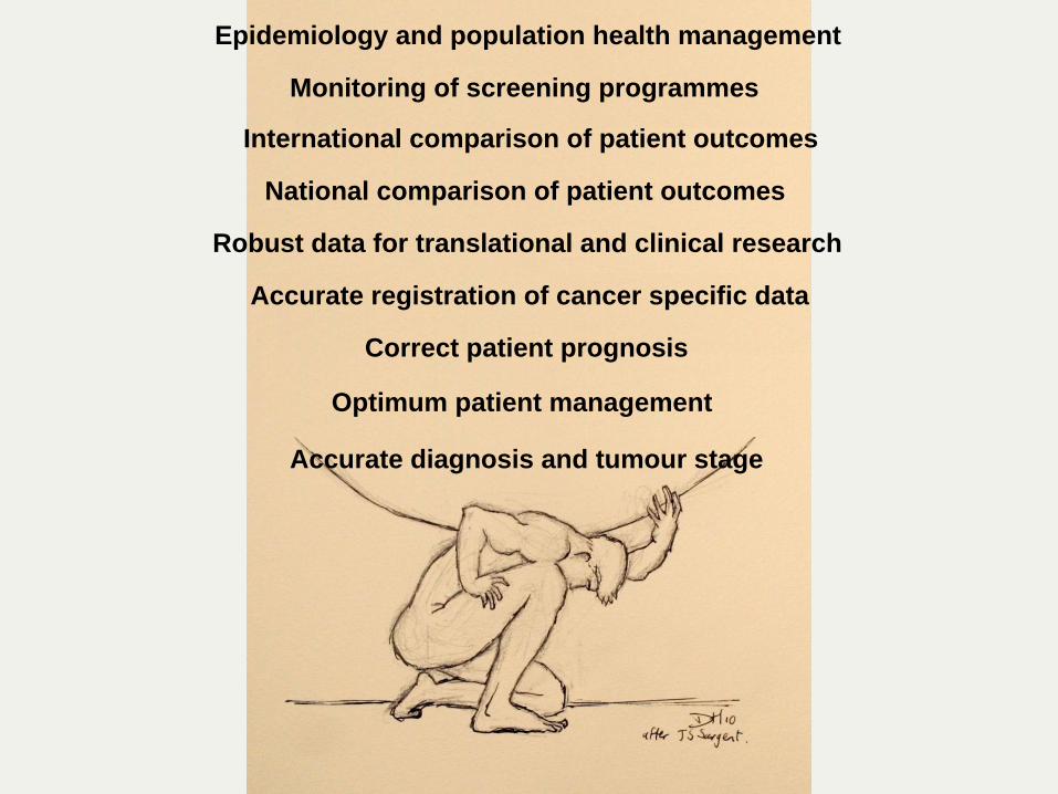
Optimum patient management
Accurate diagnosis and tumour stage
Correct patient prognosis
Accurate registration of cancer specific data
Robust data for translational and clinical research
National comparison of patient outcomes
Epidemiology and population health management
International comparison of patient outcomes
Monitoring of screening programmes