fig. 16-1. building a structural model of dna: scientific inquiry after most biologists became...
TRANSCRIPT
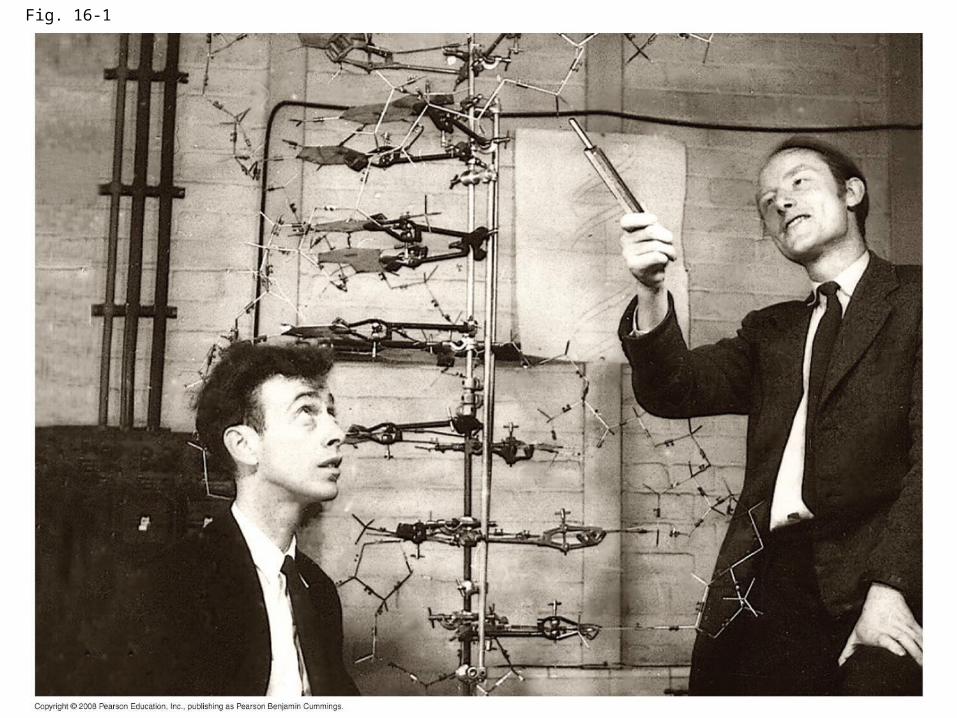
Fig. 16-1

Building a Structural Model of DNA: Scientific Inquiry
• After most biologists became convinced that DNA was the genetic material, the challenge was to determine how its structure accounts for its role
• Maurice Wilkins and Rosalind Franklin were using a technique called X-ray crystallography to study molecular structure
• Franklin produced a picture of the DNA molecule using this technique
Copyright © 2008 Pearson Education Inc., publishing as Pearson Benjamin Cummings

Fig. 16-6a
(a) Rosalind Franklin

Fig. 16-6b
(b) Franklin’s X-ray diffraction photograph of DNA

• Franklin’s X-ray crystallographic images of DNA enabled Watson to deduce that DNA was helical
• The X-ray images also enabled Watson to deduce the width of the helix and the spacing of the nitrogenous bases
• The width suggested that the DNA molecule was made up of two strands, forming a double helix
Copyright © 2008 Pearson Education Inc., publishing as Pearson Benjamin Cummings
Animation: DNA Double HelixAnimation: DNA Double Helix

Fig. 16-7a
Hydrogen bond 3 end
5 end
3.4 nm
0.34 nm
3 end
5 end
(b) Partial chemical structure(a) Key features of DNA structure
1 nm

• Watson and Crick built models of a double helix to conform to the X-rays and chemistry of DNA
• Franklin had concluded that there were two antiparallel sugar-phosphate backbones, with the nitrogenous bases paired in the molecule’s interior
Copyright © 2008 Pearson Education Inc., publishing as Pearson Benjamin Cummings

• At first, Watson and Crick thought the bases paired like with like (A with A, and so on), but such pairings did not result in a uniform width
• Instead, pairing a purine with a pyrimidine resulted in a uniform width consistent with the X-ray
Copyright © 2008 Pearson Education Inc., publishing as Pearson Benjamin Cummings
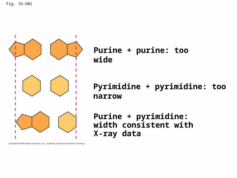
Fig. 16-UN1
Purine + purine: too wide
Pyrimidine + pyrimidine: too narrow
Purine + pyrimidine: width consistent with X-ray data

• Watson and Crick reasoned that the pairing was more specific, dictated by the base structures
• They determined that adenine (A) paired only with thymine (T), and guanine (G) paired only with cytosine (C)
• The Watson-Crick model explains Chargaff’s rules: in any organism the amount of A = T, and the amount of G = C
Copyright © 2008 Pearson Education Inc., publishing as Pearson Benjamin Cummings

Fig. 16-8
Cytosine (C)
Adenine (A) Thymine (T)
Guanine (G)

Concept 16.2: Many proteins work together in DNA replication and repair
• The relationship between structure and function is manifest in the double helix
• Watson and Crick noted that the specific base pairing suggested a possible copying mechanism for genetic material
Copyright © 2008 Pearson Education Inc., publishing as Pearson Benjamin Cummings
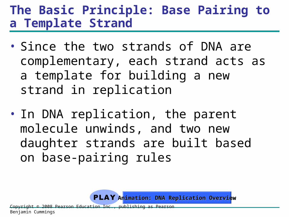
The Basic Principle: Base Pairing to a Template Strand
• Since the two strands of DNA are complementary, each strand acts as a template for building a new strand in replication
• In DNA replication, the parent molecule unwinds, and two new daughter strands are built based on base-pairing rules
Copyright © 2008 Pearson Education Inc., publishing as Pearson Benjamin Cummings
Animation: DNA Replication OverviewAnimation: DNA Replication Overview

Fig. 16-9-1
A T
GC
T A
TA
G C
(a) Parent molecule

Fig. 16-9-2
A T
GC
T A
TA
G C
A T
GC
T A
TA
G C
(a) Parent molecule (b) Separation of strands

Fig. 16-9-3
A T
GC
T A
TA
G C
(a) Parent molecule
A T
GC
T A
TA
G C
(c) “Daughter” DNA molecules, each consisting of one parental strand and one new strand
(b) Separation of strands
A T
GC
T A
TA
G C
A T
GC
T A
TA
G C

DNA Replication: A Closer Look
• The copying of DNA is remarkable in its speed and accuracy
• More than a dozen enzymes and other proteins participate in DNA replication
Copyright © 2008 Pearson Education Inc., publishing as Pearson Benjamin Cummings

Getting Started
• Replication begins at special sites called origins of replication, where the two DNA strands are separated, opening up a replication “bubble”
• A eukaryotic chromosome may have hundreds or even thousands of origins of replication
• Replication proceeds in both directions from each origin, until the entire molecule is copied
Copyright © 2008 Pearson Education Inc., publishing as Pearson Benjamin Cummings
Animation: Origins of ReplicationAnimation: Origins of Replication

• At the end of each replication bubble is a replication fork, a Y-shaped region where new DNA strands are elongating
• Helicases are enzymes that untwist the double helix at the replication forks
• Single-strand binding protein binds to and stabilizes single-stranded DNA until it can be used as a template
• Topoisomerase corrects “overwinding” ahead of replication forks by breaking, swiveling, and rejoining DNA strands
Copyright © 2008 Pearson Education Inc., publishing as Pearson Benjamin Cummings

Fig. 16-13
Topoisomerase
Helicase
PrimaseSingle-strand binding proteins
RNA primer
55
5 3
3
3

• DNA polymerases cannot initiate synthesis of a polynucleotide; they can only add nucleotides to the 3 end
• The initial nucleotide strand is a short RNA primer
Copyright © 2008 Pearson Education Inc., publishing as Pearson Benjamin Cummings

• An enzyme called primase can start an RNA chain from scratch and adds RNA nucleotides one at a time using the parental DNA as a template
• The primer is short (5–10 nucleotides long), and the 3 end serves as the starting point for the new DNA strand
Copyright © 2008 Pearson Education Inc., publishing as Pearson Benjamin Cummings
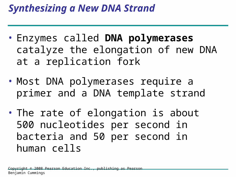
Synthesizing a New DNA Strand
• Enzymes called DNA polymerases catalyze the elongation of new DNA at a replication fork
• Most DNA polymerases require a primer and a DNA template strand
• The rate of elongation is about 500 nucleotides per second in bacteria and 50 per second in human cells
Copyright © 2008 Pearson Education Inc., publishing as Pearson Benjamin Cummings

• Each nucleotide that is added to a growing DNA strand is a nucleoside triphosphate
• dATP supplies adenine to DNA and is similar to the ATP of energy metabolism
• The difference is in their sugars: dATP has deoxyribose while ATP has ribose
• As each monomer of dATP joins the DNA strand, it loses two phosphate groups as a molecule of pyrophosphate
Copyright © 2008 Pearson Education Inc., publishing as Pearson Benjamin Cummings

Fig. 16-14
A
C
T
G
G
G
GC
C C
C
C
A
A
AT
T
T
New strand 5 end
Template strand 3 end 5 end 3 end
3 end
5 end5 end
3 end
Base
Sugar
Phosphate
Nucleoside triphosphate
Pyrophosphate
DNA polymerase

Antiparallel Elongation
• The antiparallel structure of the double helix (two strands oriented in opposite directions) affects replication
• DNA polymerases add nucleotides only to the free 3end of a growing strand; therefore, a new DNA strand can elongate only in the 5to3direction
Copyright © 2008 Pearson Education Inc., publishing as Pearson Benjamin Cummings

• Along one template strand of DNA, the DNA polymerase synthesizes a leading strand continuously, moving toward the replication fork
Copyright © 2008 Pearson Education Inc., publishing as Pearson Benjamin Cummings
Animation: Leading StrandAnimation: Leading Strand

Fig. 16-15a
Overview
Leading strand
Leading strandLagging strand
Lagging strand
Origin of replication
Primer
Overall directions of replication

Fig. 16-15b
Origin of replication
RNA primer
“Sliding clamp”
DNA pol IIIParental DNA
3
5
5
5
5
5
5
3
3
3

• To elongate the other new strand, called the lagging strand, DNA polymerase must work in the direction away from the replication fork
• The lagging strand is synthesized as a series of segments called Okazaki fragments, which are joined together by DNA ligase
Copyright © 2008 Pearson Education Inc., publishing as Pearson Benjamin Cummings
Animation: Lagging StrandAnimation: Lagging Strand
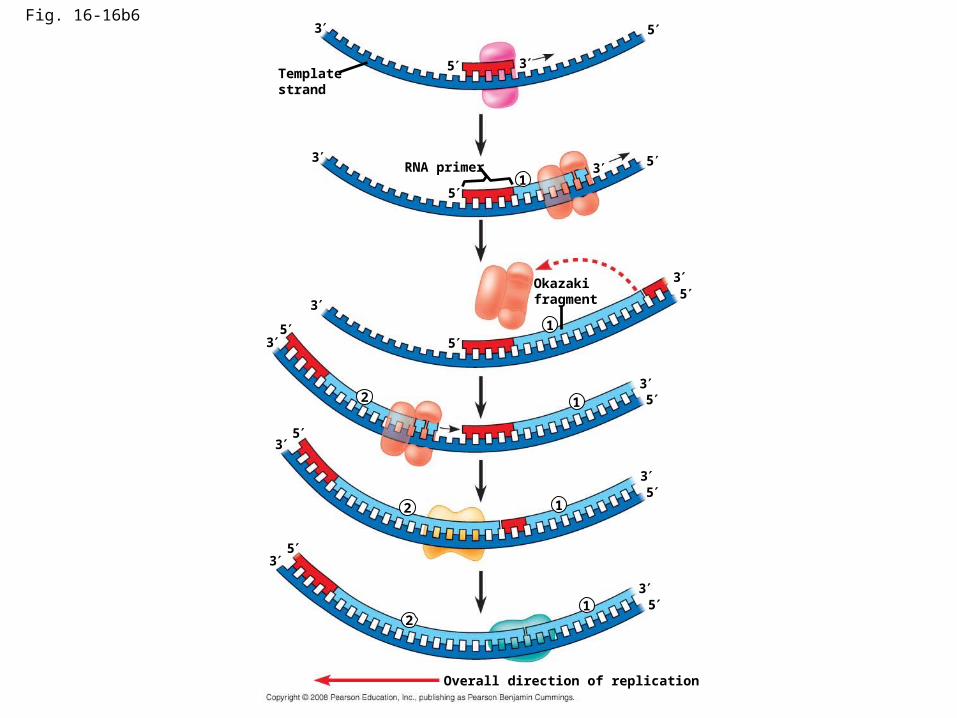
Fig. 16-16b6
Template strand
5
53
3
RNA primer 3 5
5
3
1
1
3
35
5
Okazaki fragment
12
3
3
5
5
12
3
3
5
5
12
5
5
3
3
Overall direction of replication
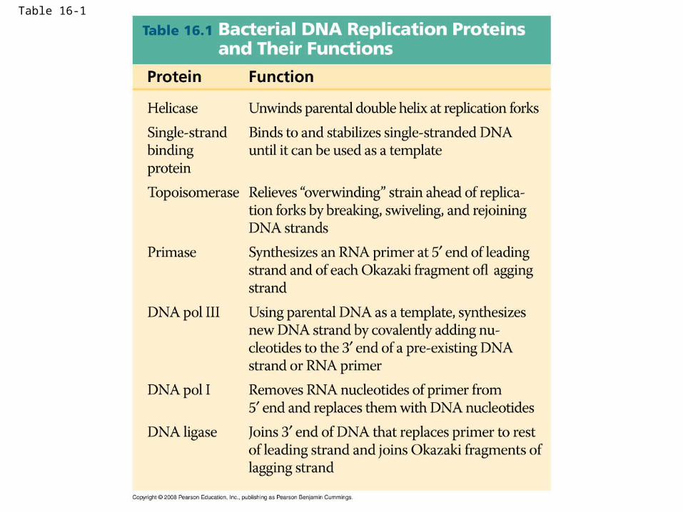
Table 16-1

Fig. 16-UN3
DNA pol III synthesizes leading strand continuously
Parental DNA DNA pol III starts DNA
synthesis at 3 end of primer, continues in 5 3 direction
Lagging strand synthesized in short Okazaki fragments, later joined by DNA ligase
Primase synthesizes a short RNA primer
53
5
5
5
3
3