fetal growth - gp partners australia
TRANSCRIPT

Fetal growth
DR ANTHIA RALLIS
CONSULTANT OBSTETRICIAN GYNAECOLOGIST
VISITING MEDICAL SPECIALIST – WOMEN’S AND CHILDREN’S HOSPITAL

Outline
Placental Function
Influences to fetal growth
SGA/IUGR
LGA/Macrosomia
Measuring fetal growth
Fetal surveillance

Normal Fetal Growth
A fetuses growth is an expression of genetic potential that is not
constrained by internal or external factors
Normal singleton fetal growth is approximately:
5g/day at 14 to 15weeks of gestation
10g/day at 20 weeks of gestation
30 - 35g/day at 32 to 34 weeks of gestation
After 34 weeks of gestation the growth rate decreases.

Utero-placental unit
Adequate maternal circulation
Adequate fetal circulation
Healthy placenta
Anything to interrupt these three can
compromise the fetus

Influences on fetal growth
Maternal factors
Maternal age
Extremes ie. 16 yrs old / 45 yrs old (both IUGR and LGA)
Chronic disease
Hypertension (SGA)
Diabetes (LGA)
Autoimmune (SLE/thrombophilia) (assoc SGA)
Anaemia (SGA)
Weight (maternal weight gain ≥16 kg (OR 10.2)) – paternal BMI ≥30 (OR 3.7)
(LGA)
Smoking/other drug use (SGA)
Socioeconomic status (SGA)

…Influences on fetal growth
Fetal conditions
Multiple gestation
Placental issues
IUGR
Abnormal placentation – Praevia, bilobed, unusual cord insertions
Placental biochemistry – low PappA (SGA)
Genetic - Aneuploidy, genetic syndromes (both SGA and LGA)
Infections - CMV, Syphillis, Rubella, Varicella, Toxo, Tuberculosis, HIV (SGA)
Gender (male more likely macrosomic (OR 2.2)

…Influences on fetal growth
Ethnicity
Parity
Nulliparity → more IUGR
Increasing fetal weight with increasing parity (parity ≥3 (OR 4.8),)
Obstetric conditions/history
Gestational hypertension/pre-eclampsia → increases IUGR
Gestational diabetes → increases macrosomia
Previous IUGR/SGA or Macrosomia
Previous IUFD

Growth trends
Median BW for term babies of same gestational age, is between 0 and
25g higher for male infants and between 5 g and 45 g higher for female
infants, than 10 years ago.
Similar increases in 90th and most 10th percentiles for boys and girls were
also observed.
While these increases may seem small, at a population level they have a
large impact.
A mean increase in BW of 23 g between 1990 and 2005 for male babies in NSW
translated into an 18% increase in those identified as LGA.
For female babies, an increase of 25g translated into a 21% increase in those
identified as LGA.


What’s accounting for this change?
Maternal age has increased
The proportion of women reporting smoking during pregnancy has
reduced
Maternal overweight and obesity has increased
The ethnicity of mothers in Australia has changed (more not born in
Australia)
IVF and assisted fertility increased
More use of prenatal testing
Paternal health more recognised as contributory to fetal health

SGA/IUGR
IUGR/FGR (fetal growth restriction)
FGR is defined by an estimated fetal weight or serial ultrasound evidence of growth restriction or growth arrest (antenatally) or
a birthweight below the 10th percentile using the South Australian birthweight percentiles (at birth)
SGA
BW below the 10 %ile of weight for gestation. Not necessarily IUGR/FGR
Most fetuses with a BW below the 10%ile for gestational age are constitutionally small
Definition RANZCOG / RCOG
SGA: EFW or AC <10th centile
Severe SGA: EFW or AC <3rd centile
Historically defined by population centiles but use of customised charts identifies small babies at higher risk of morbidity and mortality than those identified by population, though this is under debate

SGA/IUGR outcomes
Outcomes among SGA infants is largely due to the high rate of FGR among them (20 % are below the 5%ile.) (RCOG 2002; Walkinshaw and Cochrane 2003)
Reduction in maternal perception of fetal movements → IOL
Prematurity → might be iatrogenic
Meconium stained liquor → lower thresholds in labour
Abnormal heart rate patterns intrapartum → CS
Intrauterine fetal death/stillbirth
Hypoxic ischaemic encephalopathy
Poor neurological development
Delay in cognitive development
Sudden infant death syndrome
In adult life
Type 2 diabetes and hypertension (RCOG 2002)
Good catch up growth in the first few months of life may predict a healthy outcome

SGA/IUGR types
Symmetrical
Head size and trunk are reduced in parallel
Usually represents lower end of normal range for size
May indicate insult that has occurred in the early antenatal period during
general organ growth
Main associated conditions
Chronic maternal illness
Chromosomal / Congenital / Inborn errors of metabolism
Intrauterine infections
Environmental factors – Poor nutrition / BMI <20 or >25 /Age >35 / Daily vigorous
activity

SGA/IUGR types
Asymmetrical
Fetus responds to inadequate nutrition by redistributing blood flow
More to brain, heart and adrenal
Less to liver and kidney
Results in abdominal girth and fat stores reduced more than head → brain
sparing
Associated with later onset pathology
Maternal medical – hypertension, pre-eclampsia / diabetes / anaemia,
pulmonary, cardiovascular or renal disease
Placental – abruption, infarction, praevia, chorionamnionitis
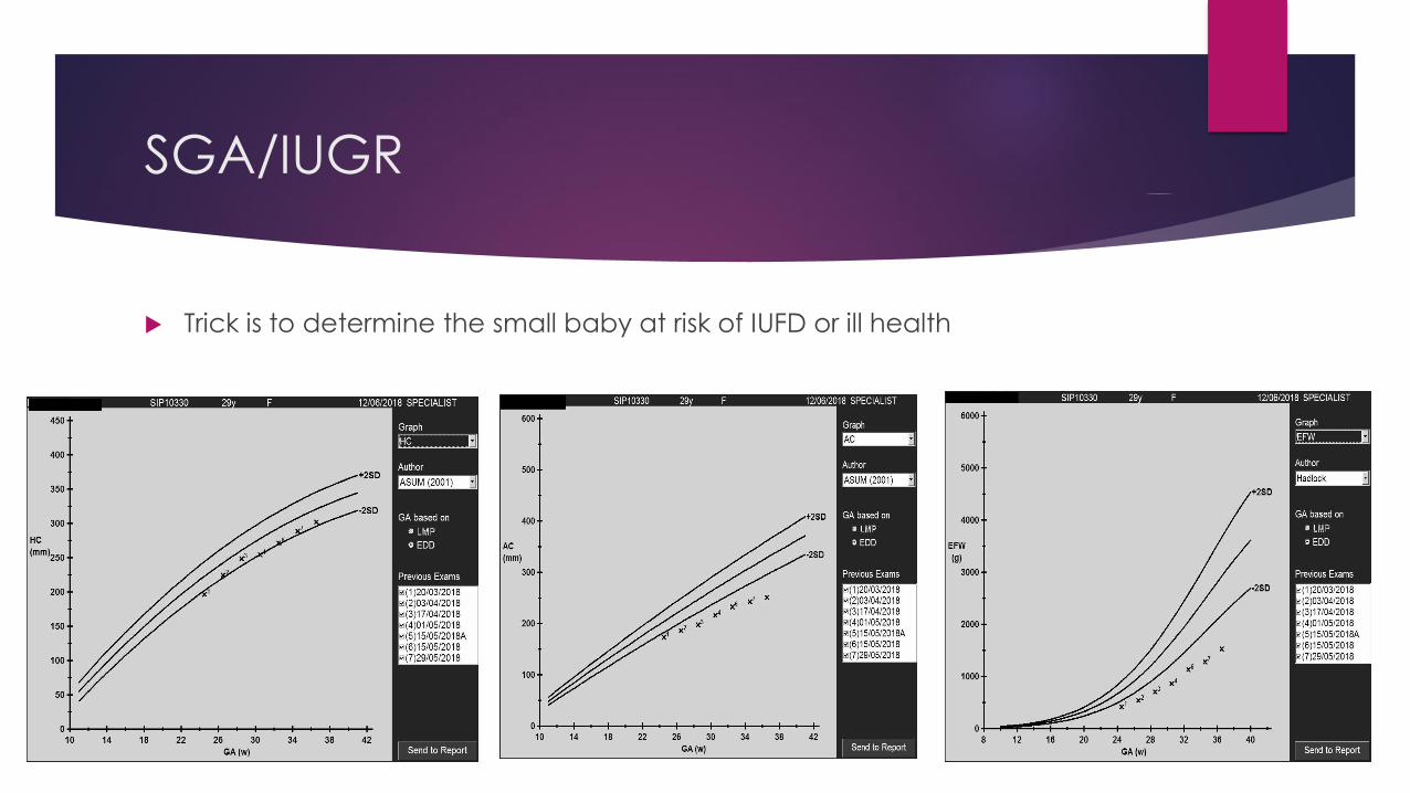
SGA/IUGR
Trick is to determine the small baby at risk of IUFD or ill health

SGA/IUGR
Which baby had the most neonatal concerns?
LGA/Macrosomia
Interchangeable terms
Fetal growth beyond a specific weight
RCOG: weight over 4000g or above 90th centile of weight for gestation
SA PPG: weight over 4000g (or over 4500g!) or above 90th centile for gestation
Australian, non-indigenous population, 90th centile at 40 weeks
Female 4000g
Male 4170g
Considered “different” in diabetics – see shoulder dystocia
Macrosomic infants of Diabetic mothers (>4000g)

LGA/Macrosomia outcomes
Maternal
Meconium stained liquor
Abnormal heart rate patterns
Cephalo-pelvic disproportion
Shoulder dystocia
Clavicular fracture
Brachial plexus injuries and paralysis
Low Apgar score
Genital tract laceration
Caesarean section
Uterine rupture
PPH
LGA/Macrosomia outcomes
Fetal/neonatal
Mostly related to mode of delivery and shoulder dystocia, but
without
Metabolic – low BSL
Low apgar
Non-compliant lungs – more TTN
Birth trauma
Stillbirth (especially in diabetics)
Long term metabolic syndrome

Shoulder dystocia
Most shoulder dystocias occur after a normally progressive labour with
spontaneous delivery or a low pelvic assisted delivery, with babies
weighing less than 4000g – 50–60 %

Shoulder dystocia outcomes
Maternal
As for macrosomia
Fetal/neonatal
Low BSL
Lung compliance
TTN
Asphyxia/Hypoxic ischaemic encephalopathy
Brachial plexus injury – Erb’s palsy
Fractures
Jaundice
Mortality

Macrosomia at 37+ weeks

How do we monitor fetuses?
Antenatal care
Kick charts
Symphysio-fundal height
Cardio Tocogram (CTG)
Ultrasound
EFW, Doppler, AFI
Biophysical profile
Make sure dates are accurate!

Which fetuses need surveillance?
Known abnormalities
Clinical indication
high or low SFH
Early onset pre-eclampsia
Previous severe or early onset IUGR
GDM
Suspected Rhesus disease or
anaemia producing infection
Obesity BMI >40
Decreased fetal movements
Post maturity
Severe prematurity expecting delivery
Trauma or APH
Maternal chronic illness
Sure to have missed some!

Dating
EDC as determined by LMP (allowing for cycle length) and or the
AUA (average US age from scans)
CRL <12 weeks gestation
BPD and CRL 12-14 weeks
Fetal biometry > 15 weeks gestation
Hadlock

Early pregnancy and dating
Gestational Sac
Needs to have a yolk sac and a bright rim (decidual reaction) –otherwise consider pseudosac
Fluid should be echo free
Quant>2000 should see a sac on TV scan
Pregnancy failure
Gestational sac with no fetus, mean sac diameter ≥ 2cm
Fetus ≥ 6mm with no fetal heart watching for > 30 seconds
Often irregular
Dating
CRL between 6 -13 wks
Should see a FH at 5-6 mm

Average ultrasound age
Best assessed by:
CRL at < 12 weeks gestation
Wisser J et al, UOG 1994
Kalish R et al, AJOG 2004
CRL and BPD at 12-14 weeks gestation (such as at NT scan)
Sladkevicus P et al UOG 2005 26: 504-511
BPD/HC/AC/FL at >/= 15 weeks gestation
Chervenak FA et al AJOG 178 (4): 678-687. 1998
With advanced gestation, make sure all four measurements
are registered in the AUA.

Average ultrasound age …continued
Estimated fetal weight from time of viability (> 23 weeks gestation)
There is good data that accurate EFW is reasonable at less
than 3500 grams
Scioscia M, et al Obst & Gynecol Jan 2008
Hadlock 4-5 (in most new ultrasound machines)
EFW within 10% over 80% of the time
EFW within 15% over 90% of the time
Newborn percentile weight charts underestimate the incidence of
low birth weight in preterm infants, thus fetal percentiles should be
noted.
Burkhardt T et al, AJOG 2008

Estimated fetal weight and AFI
Biparietal Diameter and Head circumference
Abdominal circumference
Femur length
AFI
Measure of depth of amniotic fluid in four quadrants of the uterus
Pool of liquor needs to be >1cm wide and have no cord in it
Single deepest pool >8cm → polyhydramnios
Single deepest pool <2cm → oligohydramnios

Biometry
Biparietal Diameter and Head Circumference

Biometry
Abdominal circumference
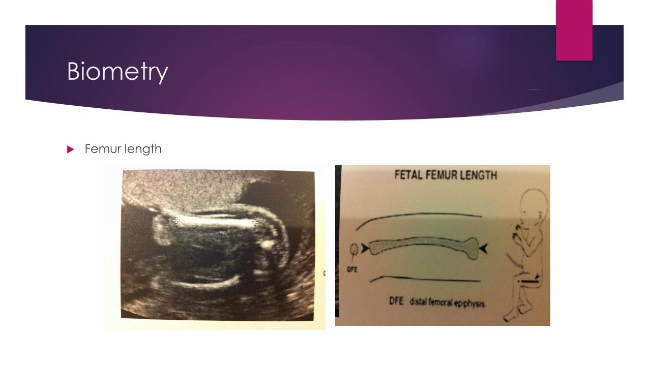
Biometry
Femur length

Dopplers
The Doppler effect is the name given to the perceived change in frequency of a sound wave, detected by an ‘observer’ moving relative to the source of the sound wave.
The frequency is perceived as higher as you approach the source, is identical at the moment of passing the source and it is perceived as lower as you move from the source.
I was first described by Christian Doppler in 1842 and first applied to ultrasound physics in the 1964, when continuous wave Doppler was used to first detect fetal cardiac pulsations. The first fetal pulse detector was marketed in 1965.
Work was begun on pulse wave doppler in the late 60s and the first 2D and M-Mode cardiac echographic machine was developed from 1970-1972.

Umbilical artery dopplers
A measure of down stream placental vascular
resistance
Fetal placenta is usually one of low resistance and
the resistance decreases further approaching term
Increased placental vascular resistance is either the
result of a poorly implanted placenta and/or a loss of
tertiary placental stem villi
There is a subsequent reduction in surface areas for
gas and nutrient exchange in the fetus and therefore
the UmAD can be used to identify the growth
restricted fetus at risk of hypoxia, acidosis and death

Uterine artery dopplers
Can be used as an assessment tool to predict pre-eclampsia
Usually evelated early in pregnancy and when non-pregnant
Uterine arteries become low resistant vessels in normal pregnancy
In normal placental development, there is trophoblast invasion into maternal spiral arteries in two phases
1) decidual and then
2) myometrial, thus establishing placental blood supply.
The uterine arteries concurrently dilate and become low resistant vessels
Uterine arteries become high resistane vessels in Pre-Eclampsia – with notching
In pre-eclampsia, there is impaired placentation, poor invasion of trophoblast into maternal spiral arteries and they remain tight and narrow. Subsequently the uterine arteries remain constricted providing high pressure just to maintain placental perfusion

Uterine Artery Dopplers
Normal
Notch should disappear
Low resistance
Abnormal
Presence of notch after 23 weeks
High resistance waveform as defined by RI or PI (>95%)

KEY TAKE HOME MESSAGES
Be aware of risk factors (RF) for abnormal fetal growth
Both LGA and SGA are RF for fetal loss
Watch for changes in growth velocity
See them regularly (20, 24, 28, 32, 34, 36, 38, 40)
Palpate the belly and do SFH height
it is more relevant when same operator
Refer when concerned
Know your friends – and call for advice!

Resources
SA Perinatal Practice Guidelines
Fetal Growth (restricted)
Fetal Growth (accelerated)
Low PappA
Australian national birthweight percentiles by sex and gestational age. MJA 2012; 197: 291-294
Fetal Growth and Risk of Stillbirth - https://doi.org/10.1371/journal.pmed.1001633
Dating document Dept US WCH
Sonographic Solutions Ultrasound course
Fetal Surveillance – A Practical Guide
Obstetric and Gynaecological Ultrasound Made Easy
Various papers quoted within
Useful tools
SA PPGs
Greentop guidelines
http://perinatology.com/calculators/Estimation%20of%20Fetal%20Weight%20and%20Age.htm#AGECALC