feline lymphomas: immunological and cytochemical ...malignant lymphomas are the most frequent...
TRANSCRIPT
[CANCER RESEARCH 49, 345-351, January 15, 1989)
Feline Lymphomas: Immunological and Cytochemical Characterization1
J. L. Rojko,2G. J. Kociba, J. L. Abkowitz, K. L. Hamilton, W. D. Hardy, Jr., J. N. Ihle, and S. J. O'Brien
Department of Veterinary Pathobiology and Comprehensive Cancer Center, The Ohio State University, Columbus, Ohio 43210 fJ. L. R., G. J. K., K. L. H.J; Division ofHematology, School of Medicine, University of Washington, Seattle, Washington 98195 [J. L. A.]; Laboratory of Veterinary Oncology, Memorial Sloan Kettering, NewYork, New York 10021 [W. D. H.J; Basic Research Program, Litton Bionetics, Frederick Cancer Research Facility, Frederick, Maryland 21701 [J. N. 1.];and Laboratoryof Viral Carcinogenesis, National Cancer Institute, Frederick, Maryland 21701 [S. J. O.J.
ABSTRACT
The ¡mmunologicaland cytochemical phenotypes of five primary felinelymphomas and six feline lymphoma lines are reported. Thymic lympho-mas induced by the Rickard strain of FeLV (FeLV-R) are of prothymo-cyte or (immature) cortical thymocyte origin, as these express terminaldeoxynucleotidyl transferase, the guinea pig erythrocyte rosette receptor,la antigens, partial cortisone sensitivity, and nonspecific esterase. Lymphomas associated with other strains of FeLV form rosettes with guineapig erythrocytes, frequently have la antigens and cytoplasmic nonspecificesterase, and probably originate from helper I -cells, monocyte/macro-phages, or null cells. These data belie previous conclusions that FeLVleukemogenesis is restricted to mature 1-colls; rather, the considerableheterogeneity in the surface and cytochemical phenotype of feline lymphomas probably reflects transformation of multipotent lymphoid ormonocytoid precursors in the bone marrow by FeLV.
INTRODUCTION
Malignant lymphomas are the most frequent malignant neoplasm in the cat in nature (reviewed in Ref. l). Seventy % offeline lymphomas have been causally associated with persistentproductive FeLV3 infection (2). Virus-positive lymphoma cells
have 10 to 20 integrated exogenous FeLV proviruses (3), havecytoplasmic and surface FeLV structural antigens (4), and bearthe FOCMA (3-5). FOCMA, originally described as a tumor-specific antigen, likely is a modified viral envelope M, 70,000glycoprotein (6). FeLV-negative lymphomas have neither exogenous integrated FeLV proviruses nor viral structural antigens (3, 4). Nonetheless, FeLV may be implicated in the causation of some FeLV-negative lymphomas. FeLV-negative lymphomas are more common in cats with a history of natural orexperimental exposure to FeLV, and lymphoma-bearing catshave a higher incidence of latent bone marrow FeLV infectionthan do healthy FeLV-exposed (immune) cats (2, 4, 7). Furthermore, lymphomas from some virus-negative cats containtruncated FeLV-gag sequences or viral structural proteins(Footnote 4; Ref. 8).
Historical characterization of feline lymphomas has beenlimited to histopathology (9, 10) and identification of T-cellsby spontaneous rosette formation with GPE and/or B-cells bydisplay of surface immunoglobulin (4, 11). T-cell identificationby GPE rosette formation is compromised by the recent observation that feline monocytes readily rosette GPE (12). Toaddress the problem of the target cell for transformation byFeLV, we report here extensive characterization of spontaneous
Received 5/12/88; revised 10/7/88; accepted 10/18/88.The costs of publication of this article were defrayed in part by the payment
of page charges. This article must therefore be hereby marked advertisement inaccordance with 18 U.S.C. Section 1734 solely to indicate this fact.
1Supported by ROÕCA-35747, U01 AI-25722, and Program Project GrantCA-16673.
2 Scholar of the Leukemia Society of America, Inc.3The abbreviations used are: FeLV, feline leukemia virus; FOCMA, feline
oncornavirus-associated cell membrane antigen; GPE, guinea pig erythrocytes;PBMC, peripheral blood mononuclear cells; IL-2, interleukin 2; FCS, fetal calfserum; PBS, phosphate-buffered saline; EA, erythrocyte-antibody (designationprefix); PHA, phytohemagglutinin; ED»,50% effective dose; CTLL, cytotoxicT-lymphocyte line mouse cells.
4 L. J. Rezanka, J. W. Casey, and J. L. Rojko, unpublished data.
and experimental feline lymphomas using monoclonal and pol-yclonal antibodies, immunofluorescence, rosette-forming assays, and cytochemistry. Feline PBMC and IL-2-dependent,primary T-cell cultures were used as nonneoplastic control cells.
MATERIALS AND METHODS
Lymphoma Cell Lines. The cat lymphoma cell lines analyzed arelisted in Table 1. FL74 was derived from a renal lymphoma thatdeveloped from in vivopassage of the original Kawakami-Theilen FeLVisolate (13). FL74 cells from three sources (M. Essex, Boston, MA; W.D. Hardy, Jr., New York, NY; R. G. Olsen, Columbus, OH) wereanalyzed and gave identical results in all but one assay (see "Results").
The Boston and New York lines were derived from that propagated byM. Essex since 1970 (5); the Ohio line was obtained from Pfizer underthe Program Resource Program of the National Cancer Institute andwas used at passages 70 to 119. F422 was from a thymic lymphomathat developed from the in vivo passage of the original Rickard FeLVisolate (14); analyzed cells were from passages 170 to 199 and 75 to95. F422 and FL74 were maintained in RPMI 1640:10% FCS:2 HIML-glutamine:0.1 % gentocin. 3272 and 3281 were established from 2FeLV-positive thymic lymphomas in naturally infected pet cats. 3191was established from a FeLV-negative thymic lymphoma from anFeLV-exposed cat; passage of 3191 through a specific-pathogen-freekitten resulted in the development of an intrathoracic lymphoma ofdonor origin. After in vivo passage of 3191, the cell line was reestablished in tissue culture and designated 3201; 3191 and 3201 are indistinguishable by allelic isoenzyme signatures (15). 3191, 3201, 3272,and 3281 were propagated in a 1:1 (v/v) mixture of RPMI 1640 andLeibowitzL-15 medium supplemented with 10% FCS, 2 mMglutamine,and 0.1 % gentocin. All cell lines were from Felis domesticas by isoen-
zyme profiles and karyology (15).Freshly Biopsied Lymphoma Cells. Lymphoma cells were obtained
from biopsies of cats presented to The Ohio State University VeterinaryTeaching Hospital. Both FeLV-positive and FeLV-negative lymphomaswere biopsies. The FeLV-negative lymphomas were from FeLV-exposed cats with latent FeLV infections demonstrable by bone marrowreactivation culture (16). The FeLV-positive lymphoma 1472 was froma specific-pathogen-free cat inoculated with FeLV-R 6 mo previously.
Isolation of Feline PBMC. Blood was collected from 4 specific-pathogen-free cats and 4 conventional cats housed in the Departmentof Veterinary Pathobiology, The Ohio State University, and in theLaboratory of Viral Carcinogenesis, Frederick, MD, respectively. Bloodwas collected by jugular venipuncture into preservative-free heparin(O'Neil, Jones, and Feldman, St. Louis, MO) and centrifuged on one-
step density gradients of Ficoll-metrizoate (specific gravity < 1.077 g/ml; lymphocyte separation medium; Litton Bionetics, Kensington,MD), and the interface cells were harvested and washed as described(17). Typical yield from 10 ml of heparinized blood was 1.5 x IO7cells,
with 80% lymphocytes, 15% monocytes, and 5% granulocytes by morphology.
Establishment of Primary T-Lymphocyte Cultures. PBMC isolated asabove were depleted of adherent monocytes and granulocytes by twocycles of plastic adherence in complete medium (RPMI 1640:15%FCS:1 miviglutamine:0.1% gentocin) at 37°Cin a 5% CO2-humidifiedchamber for 45 min each. Nonadherent cells were adjusted to 2 x IO6cells/ml and panned against rabbit anti-feline IgG 25-cm flasks pre-coated with 0.5 mg of antibody (Cooper Biomedicai, Cochraneville,PA) for 45 min. Nonadherent cells were removed by gentle pipetting,stimulated with soluble Protein A (100 MS/106cells), and incubated in
complete medium supplemented with 15% partly purified, delectinated,
345
on March 5, 2020. © 1989 American Association for Cancer Research. cancerres.aacrjournals.org Downloaded from

PG
CHARACTERIZATIONOF FELINE LYMPHOMAS
Table 1 Origin, morphology, and characteristics of cat lymphomas analyzed
NameFL74F422327232813191
32011472
McRFS
KWFeLVstatusPositivePositivePositivePositiveNegative
NegativePositive
PositiveLatent'
Latent'LocationMulticentric
(kidney,GI)Thymus,
multicentricThymus,
multicentricThymus,
multicentricThymus
ThoraxThymus
MulticentricMulticentric
Thymus, multicentricHistology"L,
NCL,
NCL,
NC,GL,
NC,GM,
NCL,NCL,
NCS,NCL,
NCM, NCCell
line orfreshbiopsyCell
lineCell
lineCell
lineCell
lineCell
lineCelllineBiopsy
BiopsyBiopsy
BiopsySpontaneous
orexperimentalE"ESSS
EE
SSS
Latent' Multicentric L, NC-C, G Biopsy" Classification according to Valli (10) where L is large, M is medium. S is small; C is cleaved, N is noncleaved, G is granulated.* E, experimental; S, spontaneous; GI, gastrointestinal.' Lymphoma negative for FeLV, cultured bone marrow positive for reactivatable FeLV (16).
human 1L-2 (Cellular Products, Buffalo, NY). Cells were maintainedat a density of 0.5 to 2 x IO6 cells/ml and were subcultured once to
twice weekly. Under these conditions, cell doubling time was 3 to 4days. The cells had been in continuous culture 4 to 7 mo before beingused as targets for immunological and cytochemical analysis. The cellswere of feline origin by karotypic analysis.5
Antibodies. The polyclonal and monoclonal antibodies used and theirrecognition specificities are listed in Tables 2 to 4. The reactivity of thepolyclonal rabbit anti-feline thymocyte serum for cat blood, thymus,spleen, bone marrow, and lymph node cells has been described previously ( 17). Rabbit anti-feline thymocyte serum and preexposure (normal) rabbit serum were used at 1:4 dilutions in PBS. The preparationand characterization of the mouse monoclonal anti-feline reagents FT2,FT3, BA4, BA6, ACS, CC4, and F have been described (Footnote 6;Refs. 18 and 19). ISCR3 (20) was the generous gift of N. Shinoharaand D. Sachs, Bethesda, Ml). All primary antibodies were used asundiluted hybridoma supernatant except F which was used as asciticfluid diluted 1:800 in PBS.
Immunofluorescence Assays for Surface Antigen Expression. Analysiswas done by indirect immunofluorescence. Washed cells (IO6) wereincubated with 5 n\ of primary antibody for 30 min at 4°C,washed 3times in PBS:5% FCS:0.01% NaN3, and incubated for 30 min at 4°C
with the secondary antibody (fluorescein isothiocyanate-conjugatedanti-goat IgG, anti-rabbit IgG, or anti-mouse IgG; Cooper Biomedicai,Cochraneville, PA). All secondary reagents were preabsorbed with catPBMC and bone marrow cells and used at 1:40 dilutions in PBS:5%FCS. Following attachment of the secondary reagents, cells werewashed 3 times in PBS:5% FCS:0.05% NaN3 and kept on ice untilexamination by epifluorescence microscopy (Zeiss universal large microscope equipped with a Model 111RS condenser). Assays were donein duplicate with 200 cells counted per replicate.
Rosette-forming Assays. Assays to detect spontaneous rosette formation with GPE pretreated with aminoethylisothiuronium bromideand to detect FcyR, Fc^R, and C3bR using EA-7S, EA-19S, and EA-19S complement were performed according to Rojko et al. (17).
Terminal Deoxynucleotidyl Transferase Assay. Terminal transferaseassays were performed on acetone-fixed cytocentrifuge preparationsusing the indirect immunofluorescence kit available from BethesdaResearch Laboratories, Bethesda, MD. Terminal transferase reactivitycrosses species barriers, and use of the assay to detect terminal trans-ferasc-containing cat lymphocytes has been reported (21).
Assays for IL-2 Production by, and Responsiveness of. CulturedLymphoma Cells. Cloned CTLL and CTB6 mouse cytotoxic T-lympho-cyte lines were the generous gifts of C. Orosz, Columbus, OH, and B.Knowles (22). CTLL and CTB6 were maintained in complete medium
5W. S. Modi, J. L. Rojko, S. J. O'Brien, unpublished data.6J. L. Abkowitz, J. M. Nakamura, and P. J. Martin. Monoclonal antibodies
to feline la-like determinants, manuscript in preparation.
supplemented with 20% conditioned medium prepared from rat spleencells stimulated with concanavalin A. Sequential doubling dilutions oflymphoma supernatants, 48-h feline conditioned medium obtainedfrom PHA (Burroughs-Wellcome; 12 x 10" PBMC) or soluble ProteinA-stimulated lymphocytes (250 n%/2 x IO6 PBMC), and human IL-2were tested for their ability to support the proliferation of 5 x 10<CTB6 or CTLL cells in 24-h, pHlthymidine uptake assays as described(22). Lymphoma cells also were cultured with concanavalin A (5 ¿tg/IO6 PBMC) or phorbol myristic acetate (10 ng/106 PBMC), and thesupernatants assayed for inducible IL-2 production.
To test responsiveness of cat lymphoma cells to IL-2, cells from 5freshly biopsied cat lymphomas also were cultured in the presence ofsequential doubling dilutions of human IL-2 in a modification of theabove 24-h [3H]thymidine uptake assay.
Cortisone Sensitivity Assays. Sensitivity versus resistance to hydro-cortisone hemisuccinate (HydroCort; Abbott Laboratories) was determined via a modification of the assay of Cook (23). Cells were grownfor 7 days in the presence of 10~6,IO"7, 10~8,and 10~9Mhydrocortisone.
Sensitive cells had less than 15% viability at concentrations less than10~8M, and resistant cells had greater than 65% viability when com
pared to untreated parallel cultures. Primary lymphoma cells werecultured in complete medium supplemented with 15% IL-2 to determine relative cortisone sensitivity.
Cytochemistry. Cytochemical characterization of lymphoma andnonneoplastic cultured primary I cells was modeled after Facklam andKociba (24). Cytocentrifuge preparations and positive controls werestained with Wright-Giemsa, Sudan Black B, «-naphthyl butyrate esterase, acid phosphatase, alkaline phosphatase, and chloroacetate esterase. If staining could not be performed within 8 h, the cytocentrifugepreparations were stored unfixed at room temperature in the dark(Wright-Giemsa, «-naphthyl butyrate esterase, Sudan Black B, chloroacetate esterase) or Fixedaccording to the manufacturer's instructions(Sigma Chemical Co., St. Louis, MO) and stored at 4°C(acid phospha
tase, alkaline phosphatase). All slides were stained within 16 h ofcytocentrifuge preparation.
RESULTS
Immunological Phenotype of Nonneoplastic Cat Lymphocytes.The surface marker analysis, terminal transferase activity, andcortisone sensitivity of cat PBMC and cultured T-cells areshown in Table 2. The specificities and percentages of catPBMC recognized by FT2, FT3, BA4, BA6, ACS, CC4, and byrosette assays are in accordance with published reports (17-19). ISCR3 is a murine monoclonal antibody which detects I-Ek epitopes conserved between species as diverse as frogs, dogs,
pigs, mice (20), and cats (Table 1). Monoclonal antibody F,346
on March 5, 2020. © 1989 American Association for Cancer Research. cancerres.aacrjournals.org Downloaded from

CHARACTERIZATIONOF FELINE LYMPHOMAS
Table 2 ¡mmunologicalphenotype of nonneoplastic cat lymphocytes
TypePolyclonal
antibodyMonoclonal
antibodyRosette
assayTerminal
transferaseCortisoneMarkerNo.
(name)Rabbit
antifelinethymocyteFT2FT3BA4BA6ACSCC4ISCR3FEEA
(7S)EA(19S)EA(19S)CRecognizesFeline
T-cellsFeline
TcFelineT-cellsFeline
common leukocyteantigenFelineX(Ig)Feliney 1(Ig)Felinen(Ig)Murine
I-EkFeline
laFeline
T-cells, monocytes,PMNscFc-xRcellsFcjiRcellsC3JRcellsThymocytes,
prothymocytesSensitivity
(S) or resistance (R)%
of cells posit:markerPBMC23
±4"28
±810±25±
110±I15±
110±131±1240
±350
±620±22±234
±80NDive
forPrimary
T-cells17±2*48
±416±29±
I00050
±565±450
±20000S
°Mean ±SE, n = 6, 2 replicates.* Mean ±SE, n = 4, 2 replicates.' PMN, polymorphonuclear leukocyte (neutrophil); ND, not determined.
initially raised against feline lymphoma FL74 cells, detectsfeline la antigens on activated T-cells, B-cells, and erythroidprogenitors. Cultured cat T-cells express epitopes defined by T-cell markers FT2, FT3, and rabbit anti-feline thymocyte serum,and they also express I-Ek and feline la antigens. Half ofprimary cat T-cells form rosettes with guinea pig erythrocytesspontaneously.
Immunological Phenotype of Cat Lymphoma Cell Lines.FeLV-negative lymphoma 3191 and the 3191-derived 3201cells (Table 3) have a paucity of lymphocyte surface markersbut do form spontaneous rosettes with guinea pig erythrocytes.FeLV-positive lymphoma cells express the BA4-defined common leukocyte antigen, I-Ek, feline la antigens, and the guinea
pig erythrocyte receptor. Exceptions are seen in the failure ofF422 and 3281 to express I-Ek and 3281 to express the guinea
pig erythrocyte receptor. Two of the three thymic lymphoma-derived lines (F422, 3272, 3281) are sensitive to cortisone, andthe renal lymphoma-derived FL74 cells are resistant. One ofthree thymic lymphomas (F422) is positive for terminal transferase.
Immunological Phenotype of Freshly Isolated Cat LymphomaCells. Feline lymphoma cells obtained by biopsy were examinedfor expression of surface markers (Table 4). The FeLV-negativelymphomas (PG, FS, KW) were from cats harboring latentFeLV infections in their bone marrows, and all expressed felinela antigens. Two (FS, KW) expressed I-Ek, feline la, and the
guinea pig erythrocyte receptor, and one each had terminaltransferase (FS) and Fc receptors (PG). Both FeLV-positivelymphomas (McR, 1472) had feline la antigens and receptorsfor guinea pig erythrocytes, while only the thymic lymphoma
Table 3 Immunologie phenotype of cat lymphoma cell lines
Marker % of cells positive for marker
TypePolyclonal
antibodyMonoclonal
antibodyRosette
assayTerminal
transferaseCortisoneNo.
(name)Rabbit
antifelinethymocyteFT2FT3BA4BA6ACSCC4ISCR3FEEA
(7S)EA(19S)EA(19S)CRecognizesFeline
T-cellsFeline
TcFelineT-cellsFeline
common leukocyteantigenFeline
X(Ig)Feliney 1(Ig)Feliney(Ig)Murine
I-EkFeline
laFeline
T-cells, monocytes,PMNs*Fc
T RcellsFcMRcellsC3bR
cellsThymocytes,
prothymocytesSensitivity
(S) or resistance (R)FeLV(-)
lymphoma31910"0000000065
±30000R320100000003±15±085±20000RF42200088
±20005±29±
198
±200025
±3S/R'FeLV(-t-)
lymphomaFL7400087
±2000100
±090±435
±1000000R327200060
±500053
±498±2100
±00001
±0S3281000000065
±2013±
10000R
°Mean ±SE, n —¿�6, 2 replicates.* PMN, polymorphonuclear leukocyte (neutrophil).' Susceptible to 10"' and IO"7 M cortisone.
347
on March 5, 2020. © 1989 American Association for Cancer Research. cancerres.aacrjournals.org Downloaded from

CHARACTERIZATIONOF FELINE LYMPHOMAS
Table 4 Immunological phenotype of freshly isolated cat lymphoma cells
TypePolyclonal
antibodyMonoclonal
antibodyRosette
assayTerminal
transferaseCortisoneMarkerNo.
(name)Rabbit
antifelinethymocyteFT2FT3BA4BA6ACSCC41SCR3FEEA
(7S)EA(19S)EA
(19S)C%
of cells positive formarkerRecognizesFeline
T-cellsFeline
TcFelineT-cellsFeline
common leukocyteantigenFeline
X(Ig)Feliney 1(Ig)FelineM(Ig)Murine
1-E*Feline
laFeline
T-cells,mono-cytes,PMNs*Fc-cRcellsFcjiRcellsC3„RcellsThymocytes,
prothy-mocytesSensitivity
(S) or resistance (R)FeLV(-)
lymphomaPC"0e000000050
±370
±522
±311±100RFS"0000000100
±050±493
±200029
±7RKW008±30000100
±0100±0100
+00000RFeLA(-H)
lymphomaMcR"00000000100
±0100
+05±11
±000R1472*0000000100
±0100±093
±000017
±6S/R'
" Spontaneous lymphoma.* Experimental lymphoma, induced by FeLV-R (F422-derived FeLV).' Mean ±SE, 2 replicates.d PMN, polymorphonuclear leukocyte (neutrophil).' Susceptible to 10~' and IO'7 cortisone.
1472 induced by Rickard strain FeLV (FeLV-R) had terminaltransferase and partial sensitivity to cortisone.
Interleukin 2 Responsiveness of Freshly Isolated Cat Lymphoma Cells. Cells from 4 of 5 freshly biopsied cat lymphomacells proliferated in response to 11-2 (Fig. 1). 1472 cells werenonresponsive. Continued 11-2supplementation has allowed theoutgrowth of Il-2-dependent PG and KW lines.7
Absence of Cat Interleukin 2 Production by Cat Lymphomas.All established cat lymphoma lines were tested for the constitutive and inducible production of 11-2 in 24-h proliferationassays using IL-2-dependent CTLL and CTB6 targets. Cat IL-2 obtained from PHA- or soluble Protein A-stimulated PBMCand partly purified, delectinated human IL-2 were the positive
LOOj [iL-z]
Fig. 1. IL-2 dependence of feline lymphoma cells. Freshly biopsied lymphomacells from a latently infected cat (PC) were cultured in the presence of sequentialdoubling dilutions of IL-2 for 24 h with the addition of ('H)thymidine (0.5 Ci/5x IO4cells/well, triplicate assays) for the final 6 h. Points, mean; bars, SE.
7J. L. Rojko and C. M. Cheney, unpublished data.
controls and demonstrated effective doses (ED50s) of 4 Iog2 and6.5 Iog2, respectively, against CTLL cells. ED50s against CTB6were 3.5 Iog2 for cat IL-2 and greater than 9 Iog2 for humanIL-2. IL-2 was not produced by any of the cat lymphoma linestested (F422, FL74, 3191, 3201) in response to concanavalinA (5 Mg/106 PBMC/ml) or phorbol myristic acetate (10 Mg/106
PBMC/ml). To ensure that the FeLV M, 15,000 envelopeprotein was not interfering with CTLL responsiveness to IL-2(22), supernatants from FeLV-positive lymphomas were centri-fuged at 100,000 x g for 2 h at 4°Cprior to use. Removal of
FeLV as shown by loss of infectivity (helper virus activity) forclone 81 target cells did not unmask IL-2-like activity presentin feline lymphoma supernatants (data not shown).
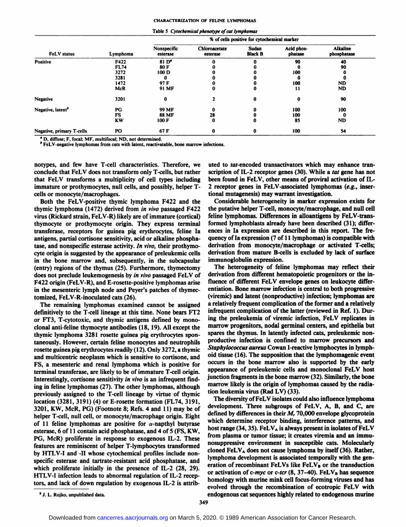
Cytochemical Characterization of Neoplastic and Nonneoplas-tic Feline Lymphocytes. The cytochemical phenotype of felinelymphomas is presented in Table 5. Eight of ten lymphomasand 67% of cultured primary T-cells were positive for nonspecific esterase using a-naphthyl butyrate substrate. Seven of tenlymphomas had demonstrable acid phosphatase, and four ofseven lymphomas had alkaline phosphatase. Cultured primarycat T-cells uniformly were positive for acid phosphatase andpartially positive for alkaline phosphatase.
DISCUSSION
Our workers have used the rosette-forming capability of catlymphomas to infer that FeLV transforms principally T-cells(2, 4, 11). However, we have shown previously that a variety ofcat hematopoietic cells rosette guinea pig erythrocytes (12).Herein, we classify feline T-lymphocytes and lymphomas intosubsets based on their shared surface antigens and enzymecontent. Primary cat T-cells are uniformly positive for acidphosphatase; strongly express feline T-cytotoxic antigen, la,and I-Ek antigens; and may contain nonspecific esterase and
alkaline phosphatase. In contrast, lymphomas from cats withviremic or latent FeLV infections have highly individual phe-
348
on March 5, 2020. © 1989 American Association for Cancer Research. cancerres.aacrjournals.org Downloaded from
CHARACTERIZATION OF FELINE LYMPHOMAS
Table 5 Cytochemical phenotype of cat lymphomas
% of cells positive for cytochemicalmarkerFeLV
statusLymphotnaPositive
F422FL74327232811472McRNonspecific
esterase81D"80F
100 D0
97F91 MFChloroacetate
esterase0
00000Sudan
BlackB0
00000Acid
phos-phatase90
0100
0100
11Alkaline
phosphatase40
9000
NDND
Negative
Negative, latent*
Negative, primary T-cells
3201
PGFSKW
PO
99 MF88 MF
100 F
67F
O28
O
OOo
10010085
100
90
100O
ND
54* D, diffuse; F, focal; MF, multifocal; ND, not determined.* FeLV-negative lymphomas from cats with latent, reactivatable, bone marrow infections.
notypes, and few have T-cell characteristics. Therefore, weconclude that FeLV does not transform only T-cells, but ratherthat FeLV transforms a multiplicity of cell types includingimmature or prothymocytes, null cells, and possibly, helper T-cells or monocyte/macrophages.
Both the FeLV-positive thymic lymphoma F422 and thethymic lymphoma (1472) derived from in vivo passaged F422virus (Rickard strain, FeLV-R) likely are of immature (cortical)thymocyte or prothymocyte origin. They express terminaltransferase, receptors for guinea pig erythrocytes, feline laantigens, partial cortisone sensitivity, acid or alkaline phosphatase, and nonspecific esterase activity. In vivo, their prothymocyte origin is suggested by the appearance of preleukemic cellsin the bone marrow and, subsequently, in the subcapsular(entry) regions of the thymus (25). Furthermore, thymectomydoes not preclude leukemogenesis by in vivo passaged FeLV ofF422 origin (FeLV-R), and E-rosette-positive lymphomas arisein the mesenteric lymph node and Peyer's patches of thymec-
tomized, FeLV-R-inoculated cats (26).The remaining lymphomas examined cannot be assigned
definitively to the T-cell lineage at this time. None bears FT2or FT3, T-cytotoxic, and thymic antigens defined by monoclonal anti-feline thymocyte antibodies (18, 19). All except the
thymic lymphoma 3281 rosette guinea pig erythrocytes spontaneously. However, certain feline monocytes and neutrophilsrosette guinea pig erythrocytes readily (12). Only 3272, a thymicand multicentric neoplasm which is sensitive to cortisone, andFS, a mesenteric and renal lymphoma which is positive forterminal transferase, are likely to be of immature T-cell origin.Interestingly, cortisone sensitivity in vivo is an infrequent finding in feline lymphomas (27). The other lymphomas, althoughpreviously assigned to the T-cell lineage by virtue of thymiclocation (3281, 3191) (4) or E-rosette formation (FL74, 3191,3201, KW, McR, PG) (Footnote 8; Refs. 4 and 11) may be ofhelper T-cell, null cell, or monocyte/macrophage origin. Eightof 11 feline lymphomas are positive for a-napthyl butyraseesterase, 6 of 11 contain acid phosphatase, and 4 of 5 (FS, KW,PG, McR) proliferate in response to exogenous IL-2. Thesefeatures are reminiscent of helper T-lymphocytes transformedby HTLV-I and -II whose cytochemical profiles include nonspecific esterase and tartrate-resistant acid phosphatase, andwhich proliferate initially in the presence of IL-2 (28, 29).HTLV-I infection leads to abnormal regulation of IL-2 receptors, and lack of down regulation by exogenous IL-2 is attrib-
' J. L. Rojko, unpublished data.
uted to tef-encoded transactivators which may enhance transcription of IL-2 receptor genes (30). While a tat gene has notbeen found in FeLV, other means of proviral activation of IL-2 receptor genes in FeLV-associated lymphomas (e.g., inser-tional mutagenesis) may warrant investigation.
Considerable heterogeneity in marker expression exists forthe putative helper T-cell, monocyte/macrophage, and null cellfeline lymphomas. Differences in alloantigens by FeLV-trans-formed lymphoblasts already have been described (31); differences in la expression are described in this report. The frequency of la expression (7 of 11 lymphomas) is compatible withderivation from monocyte/macrophage or activated T-cells;derivation from mature B-cells is excluded by lack of surfaceimmunoglobulin expression.
The heterogeneity of feline lymphomas may reflect theirderivation from different hematopoietic progenitors or the influence of different FeLV envelope genes on leukocyte differentiation. Bone marrow infection is central to both progressive(viremic) and latent (nonproductive) infection; lymphomas area relatively frequent complication of the former and a relativelyinfrequent complication of the latter (reviewed in Ref. l). During the preleukemia of viremic infection, FeLV replicates inmarrow progenitors, nodal germinal centers, and epithelia butspares the thymus. In latently infected cats, preleukemic nonproductive infection is confined to marrow precursors andStaphylococcus aureus Cowan I-reactive lymphocytes in lymph-oid tissue (16). The supposition that the lymphomagenic eventoccurs in the bone marrow also is supported by the earlyappearance of preleukemic cells and monoclonal FeLV hostjunction fragments in the bone marrow (32). Similarly, the bonemarrow likely is the origin of lymphomas caused by the radiation leukemia virus (Rad LV) (33).
The diversity of FeLV isolates could also influence lymphomadevelopment. Three subgroups of FeLV, A, B, and C, aredefined by differences in their M, 70,000 envelope glycoproteinwhich determine receptor binding, interference patterns, andhost range (34,35). FeLVA is always present in isolates of FeLVfrom plasma or tumor tissue; it creates viremia and an immu-nosuppressive environment in susceptible cats. Molecularlycloned FeLVA does not cause lymphoma by itself (36). Rather,lymphoma development is associated temporally with the generation of recombinant FeLVs like FeLVB or the transductionor activation of c-myc or c-tcr (8, 37-40). FeLVB has sequencehomology with murine mink cell focus-forming viruses and hasevolved through the recombination of ecotropic FeLV withendogenous cat sequences highly related to endogenous murine
349
on March 5, 2020. © 1989 American Association for Cancer Research. cancerres.aacrjournals.org Downloaded from

CHARACTERIZATION OF FELINE LYMPHOMAS
retroviral elements (40). Differences in various FeLVB isolatesin the env or gag-poi regions may create viruses with varianttransforming capability as the FeLVB (GM) isolate causes mye-loproliferative disease, whereas the FeLVB (GA) molecularclone causes myelodysplastic anemia (Footnote 9; Refs. 40 and41).
While the role of FeLVB in thymic lymphomagenesis still isspeculative, several observations are cogent. Thymic lymphomaF422 produces replication-competent FeLVA, and, probably,replication-incompetent FeLVB.'°In vivo passage of F422 virus(FeLV-R) produces thymic lymphomas such as 1472 whichproduce FeLVAB. Also, both F422 and its derived thymic lymphomas like 1472 show myc gene alterations. In F422, FeLVhas transduced c-mys to produce an FeL\-myc (37, 39). Intumors induced by FeLV-R like 1472, activation of c-myc hasbeen implicated in lymphoma development (38). While themechanisms of wye-associated lymphomagenesis remain to beelucidated fully, it is interesting that the two FeLV-R-associatedlymphomas (F422 and 1472) examined herein have identicalimmunological and cytochemical phenotypes. The lymphomasinduced by the laboratory strain FeLV-KT (FL74) and bynatural FeLV isolates (3272, 3281, McR) were more heterogeneous.
In the present study, the FeLV-negative lymphomas examined have individual immunological and cytochemical profilesand likely arose by heterogeneous mechanisms. Not all FeLV-negative lymphomas are caused by FeLV. Exogenous FeLVsequences are absent or truncated in the tumor, and sometimesthe lymphoma-bearing cat has never been exposed to FeLV (2-4). However, many FeLV-negative lymphomas are thought toresult from prior or latent FeLV infections (4, 7, 16). Understanding the phenotype of FeLV-positive and FeLV-negativelymphocytes transformed by FeLV and the molecular biologyof the transformation event is critical to both the choice oftherapy and establishment of prognosis. The data presentedhere and studies of T-cell receptor and immunoglobulin generearrangements in progress should allow better understandingof feline lymphomas and their value as models for humanlymphomas.
ACKNOWLEDGMENTS
We thank Dr. M. D. Cooper and Dr. J. Sowder for their generousgift of monoclonal antibodies and for their thoughtful and criticalreview of our data and manuscript.
REFERENCES
1. Rojko, J. I... and Olsen, R. G. The immunobiology of the feline leukemiavirus. Vet. Inumimi!. Immunopathol., 6: 107-165, 1984.
2. Hardy, W. D., Jr., McClelland, A. J., Zuckerman, E. E., Snyder, H. W., Jr.,MacEwen, E. G., Francis, D., and Essex, M. Development of virus non-producer lymphosarcomas in pet cats exposed to FeLV. Nature (lumi.), 288:90-92, 1980.
3. Casey, J. W., Roach. A., Mullins, J. E., Burck, K. B., Nicolson, M. D.,Gardner, M. B.. and Davidson, N. The U3 portion of feline leukemia virusDNA identifies horizontally acquired proviruses in leukemic cats. Proc. Nati.Acad. Sci USA, 78: 7778-7782, 1981.
4. Hardy. W. D., Jr., Zuckerman, E. E., MacEwen, E. G., Hayes, A. A., andEssex, M. A feline leukemia and sarcoma virus-induced tumor-specific antigen. Nature (Lond.), 270: 249-251, 1977.
5. Essex, M., Klein, G., Snyder, S. P., and Harrold, J. B. Feline sarcoma virus-induced tumors: correlation between humoral antibody and tumor regression.Nature (Lond.), 233: 195-197, 1971.
6. Snyder, H. W., Jr., Singhal, M. C, Zuckerman, E. E., Jones, F. R., andHardy, W. D., Jr. The feline oncornavirus-associated cell membrane antigen
" D. E. Onions, personal communication.10L. Rezanka and J. L. Rojko, unpublished data.
7.
8.
9.
10.
11.
12.
13.
14.
15.
16.
17.
18.
19.
20.
21.
22.
23.
24.
25.
26.
27.
28.
29.
30.
31.
32.
33.
34.
35.
36.
(FOCMA) is related to, but distinguishable from, FeLV-C gp70. Virology,131: Ì15-327,1983.Rojko, J. L., Hayes, A. A., and Cheney, C. M. Latent feline leukemia virusinfections: diagnosis and clinical implications. J. Cell. Biochem., 10A: 224,1986.Fulton, R., Forrest, D., McFarlane, R., Onions, D. E., and Neil, J. C.Retroviral transduction of T-cell antigen receptor beta-chain and myc genes.Nature (Lond.), 326:190-193, 1987.Jarrett, W. F. H., and Mackey, L. J.Neoplastic diseases of the haematopoieticand lymphoid tissues. Bull WHO, 50: 21-34, 1974.Valli, V. E., McSherry, B. J., Dunham, B. M., Jacobs, R. M., and Lumsden,J. H. Histocytology of lymphoid tumors in the dog, cat, and cow. Vet.Pathol.,/«: 494-512, 1981.Cockerel!, G. L., Krakowka, S., and Hoover, E. A. Characterization of felineT- and H lymphocytes and identification of an experimentally induced T-cellneoplasm in the cat. J. Nati. Cancer Inst., 57:1095-1099, 1976.Wellman, M. L., Kociba, G. J., and Rojko, J. L. Guinea pig erythrocyterosette formation as a nonspecific cell surface receptor assay in the cat. Am.J. Vet. Res., 47:433-437, 1986.Theilen, G. H., Kawakami, T. G., Rush, J. D., and Munn, R. J. Replicationof the cat leukaemia virus in cell suspension cultures. Nature (Lond.), 222:589-590, 1969.Rickard, C. G., Post, J. E., Noronha, F., and Barr, L. M. A transmissiblevirus-induced lymphocytic leukemia of the cat. J. Nati. Cancer Inst., 42:987-1014, 1969.O'Brien, S. The extent and character of biochemical genetic variation in thedomestic cat. J. Hered., 71: 2-8, 1980.Rojko, J. L., Hoover, E. A., Quackenbush, S. L., and Olsen, R. G. Reactivation of latent feline leukaemia virus infections. Nature (Lond.), 298: 385-388, 1982.Rojko, J. L., Hoover, E. A., Finn, B. L., and Olsen, R. G. Characterizationand mitogenesis of feline lymphocyte populations. Int. Arch. Allergy Appi.Immunol., 68: 226-232, 1982.Klotz, F. J., Gathings, W. E., and Cooper, M. D. Development and distribution of B-lineage cells in the domestic cat: analysis with monoclonalantibodies to cat /<, -,, K and A chains and heterologous anti-monoclonalantibodies. J. Immunol, 134:95-100, 1985.Klotz, F. J., and Cooper, M. D. A feline thymocyte antibody defined by amonoclonal antibody (FT2) identifies a subpopulation of nonhelper cellscapable of specific cytotoxicity. J. Immunol., 136: 2510-2514, 1986.Watanabe, M., Suzuki, T., Taniguchi, M., and Shinohara, N. Monoclonalanti-la murine alloantibodies cross-reactive with the Ia-homologies of othermammalian species including humans. Transplantation, 36: 712-718, 1983.Lafrado, L. J., Mathes, L. E., and Olsen, R. G. Evaluation of terminaldeoxynucleotidyl transferase expression in the bone marrow of clinicallynormal and FeLV-exposed cats. Am. J. Vet. Res., 47: 1-2, 1985.Orosz, C. G., Zinn, N. E., Olsen, R. G., and Mathes, L. E. Retrovirus-mediated immunosuppression. I. FeLV-UV and specific FeLV proteins alterT-lymphocyte behavior by inducing hyporesponsiveness to lymphokines. J.Immunol., 134: 3396-3403, 1985.Cook, W. D. Thymocyte subsets transformed by Abelson murine leukemiavirus. Mol. Cell. Biol., 5: 390-397, 1985.Facklam, N. R., and Kociba, G. J. Cytochemical characterization of felineleukemic cells. Vet. Pathol., 23: 155-161, 1986.Hoover, E. A., Ferryman, L. E., and Kociba, G. J. Early lesions in catsinoculated with feline leukemia virus. Cancer Res., 33: 145-152, 1973.Hoover, E. A., Krakowka, S., Cockerell, G. L., and Olsen, R. G. Influenceof thymectomy on the susceptibility of cats to feline leukemia virus andlymphosarcoma. Am. J. Vet. Res., 39: 993-995, 1978.Bell, R., Cotter, S., Lillquist, S., Sallan, S., and McCaffrey, R. Characterization of glucocorticoid receptors in animal lymphoblastic disease: correlationwith response to single-agent glucocorticoid treatment. Blood, 63: 380-383,1984.Chen, I. S. Y., Quan, S. G., and Golde, D. W. Human T-cell leukemia virustype II transforms normal human lymphocytes. Proc. Nati. Acad. Sci. USA,«0:7006-7010,1980.Markham, P. D., Salahuddin, S. /.., Macchi, B., Robert-Guroff, M., andGallo, R. C. Transformation of different phenotypic types of human bonemarrow T-lymphocytes by human T-cell leukemia virus type I. Int. J. Cancer,33: 13-17, 1984.Krönke,M., Leonard, W. J., Depper, J. M., and Greene, W. C. Deregulationof IL-2 receptor gene expression in HTLV-I-induced adult T-cell leukemia.Science (Wash. DC), 228: 1215-1217, 1985.Grant, C. K., and Michalek, M. T. Feline leukemia: unique and cross-reactingantigens on individual virus-producing tumors identified by complement-dependent antibody. J. Nati. Cancer Inst., 64:1527-1533, 1980.Hoover, E. A., Mullins, J. I., Quackenbush, S. L., and Gasper, P. W.Experimental transmission and pathogenesis of immunodeficiency syndromein cats. Blood, 70: 1880-1892, 1987.Haran-Ghera, N., Rubio, N., Leef, F., and Goldstein, G. Characteristics ofpreleukemic cells in mice. Cell. Immunol., 37: 308-314, 1978.Sarma, P. S., and Log, T. Subgroup classification of feline leukemia andsarcoma viruses by viral interference and neutralization tests. Virology, 54:160-169, 1983.Jarrett, ()., Laird, H., and Hay, D. Determinants of the host range of felineleukaemia viruses. J. Gen. Virol., 20:169-175, 1973.Donahue, P. R., Hoover, E. A., Beltz, G. A., Riedel, N., Hirsch, V. M.,Overbaugh, J., and Mullins, J. I. Strong sequence conservation among
350
on March 5, 2020. © 1989 American Association for Cancer Research. cancerres.aacrjournals.org Downloaded from

CHARACTERIZATION OF FELINE LYMPHOMAS
horizontally transmissible, minimally pathogenic, feline leukemia viruses. J. viruses containing the »i.irgene rapidly produce clonal tumors expressing IVirol., 62: 722-731, 1988. cell antigen receptor transcripts. Int. J. Cancer, 40:40-45, 1987.
37. Neil, J. C, Hughes, D., McFarlane, R., Wilkie, N., Onions, D. E., Lees, G., 40. Stewart, M. A., Warnock, M., Wheeler, A., Wilkie, N., Mullins, J. I., Onions,and Jarrett. O. Transduction and rearrangement of the myc gene by feline D. E., and Neil, J. C. Nucleotide sequences of a feline leukemia virus subgroupleukaemia virus in naturally occurring feline leukaemias. Nature (Lond.), A envelope gene and long terminal repeat and evidence for the recombina-308: 814-820, 1984. tional origin of subgroup B viruses. J. Virol., 58: 825-834, 1986.
38. Forrest, D., Onions, D., Lees. G., and Neil, J. C. Altered structure and 41. Riedel, N., Hoover, E. A., Elder. J. H., and Mullins, J. I. Molecular analysisexpression of c-myc in feline T-cell tumors. Virology, 158: 194-199, 1987. of feline leukemia viruses of diverse pathogenic potential. UCLA Symposium
39. Onions, D. E., Lees, G., Forrest, D., and Neil, J. C. Recombinant feline on Viruses and Human Cancer, Park City, UT, Feb. 3-9, 1986.
351
on March 5, 2020. © 1989 American Association for Cancer Research. cancerres.aacrjournals.org Downloaded from

1989;49:345-351. Cancer Res J. L. Rojko, G. J. Kociba, J. L. Abkowitz, et al. CharacterizationFeline Lymphomas: Immunological and Cytochemical
Updated version
http://cancerres.aacrjournals.org/content/49/2/345
Access the most recent version of this article at:
E-mail alerts related to this article or journal.Sign up to receive free email-alerts
Subscriptions
Reprints and
To order reprints of this article or to subscribe to the journal, contact the AACR Publications
Permissions
Rightslink site. Click on "Request Permissions" which will take you to the Copyright Clearance Center's (CCC)
.http://cancerres.aacrjournals.org/content/49/2/345To request permission to re-use all or part of this article, use this link
on March 5, 2020. © 1989 American Association for Cancer Research. cancerres.aacrjournals.org Downloaded from