fascioliasis: an ongoing zoonotic trematode infection ... · review article fascioliasis: an...
TRANSCRIPT

Review Article
Fascioliasis: an ongoing zoonotic trematode infection
Mramba Nyindo* and Abdul-Hamid Lukambagire
Department of Medical Parasitology and Entomology, Kilimanjaro Christian Medical
University College, P O Box 2240, Moshi, Tanzania
* Email address: [email protected]
Abstract: Zoonotic trematode infections are an area of the neglected tropical diseases that have
become of major interest to global and public health due to their associated morbidity. Human
fascioliasis is a trematode zoonosis of interest in public health. It affects approximately 50 million
people worldwide and over 180 million are at risk of infection both in developed and
underdeveloped countries. The one health paradigm is an area that seeks to address the
problem of zoonotic infections through a comprehensive and sustainable approach. This review
attempts to address the major challenges in managing human and animal fascioliasis with
valuable insights gained from the one health paradigm to global health and multidisciplinary
integration.
1. Introduction
Fascioliasis is a disease of ruminants caused by two major parasitic trematodes, Fasciola
hepatica and F. gigantica. Over the past two decades human fascioliasis has gained notice as a
disease of primary importance. Human fascioliasis is currently classified as a plant/ food-borne
trematode infection, commonly acquired by eating metacercaria encysted on leaves that are
eaten as vegetables [1]. There is a high prevalence of fascioliasis among herding communities in
low income countries because of their constant close association with livestock that they keep.
Fascioliasis was first recorded as early as 2000 BC. Animal fascioliasis causes significant
disease among sheep and cattle, causing severe physical wasting. It contributes to losses of
over $2 billion dollars per annum in the livestock industry in North and South America. Human
fascioliasis also causes significant illness and morbidity, mainly among low income, farming
communities. To date, no human deaths have been directly associated with fascioliasis. This fact
accords the disease a low priority and contributes to its neglect as a significant cause of public
health concern. Human fascioliasis is currently ranked under the food/ plant trematode zoonoses
as a neglected tropical disease (NTD) [2].
2. Materials and methods
We did a literature search on human and animal fascioliasis in the past 15 years We searched
PubMed/MEDLINE resources, HINARI/PubMed, local, regional and institutional e-libraries such
as NIH, CDC, WHO, IHI (Ifakara Health Institute, Tanzania) e-lib, East African Medical Journal,
African Journals of public health as well as Vectors and Zoonoses for the following major
keywords; “fascioliasis”, “human fascioliasis”, “Zoonoses”, “fasciola liver fluke”, “fasciola
hepatica”, “fasciola gigantica”, “fascioliasis, F. hepatica”, “fascioliasis F. gigantica”, “human
fascioliasis epidemiology”, “human fascioliasis distribution”, “trends in fascioliasis research”,
“fascioliasis outbreaks”, We searched published literature from 2000 – 2015. Also we referenced
recognized full textbook articles as well as summaries and abstracts containing discernible
content. We selected references that fitted to be included in the review. We excluded non-
English translated articles and papers/ publications that only focused on animal disease without a
zoonotic aspect. This search allowed us to include 92 most relevant references out of 300.
3. Current Status
Fascioliasis is primarily a disease of ruminants, although over the past two decades human
fascioliasis has gained significance as an important disease in humans [1]. Human fascioliasis is
commonly characterized by a hypo-endemic pattern, with low and stable levels of prevalence
among a defined population and generally shows a focal endemic distribution. However, to-date
there have been reports from every continent except the Antarctica, thereby showing a wide
cosmopolitan distribution [2].
Among the zoonotic parasitic diseases, human fascioliasis is currently classified as a plant/ food-
borne trematode infection, with higher prevalence seen among farming communities in low
income countries [3, 4]. The plant food-borne trematodes comprise Fasciola hepatica, F.
gigantica and Fasciolopsis buski (Family Fasciolidae), Gastrodiscoides hominis (Family
Gastrodiscidae), Watsonius watsoni and Fischoederius elongatus (Family Paramphistomidae).
Fasciola hepatica and F. gigantica infect the liver [5].
The fasciolids and the gastrodiscid cause important zoonoses distributed throughout many
countries, while W. watsoni and F. elongatus have been only accidentally transmitted to humans.
Present climate and global changes appear to increasingly affect the distribution of snail-borne
helminthiases [6]. This provides a good example of emerging/re-emerging parasitic diseases in
many countries as a consequence of various phenomena related to climatic and environmental
changes as well as human socioeconomic activities [7, 5].
Despite the recent developments in diagnostic and surveillance techniques, some countries are
still completely lacking in data on human fascioliasis. This may be because the disease is not
endemic, but is more likely due to under reporting/ diagnosis especially in the resource limited
settings. The underestimated global burden of the disease to date is approximated to be between
35 to 72 million people, with an additional 180 million at risk of infection [8, 9, 10]. Abundant data
supporting animal fasciola infections are available in many tropical developing countries and
regions, with the corresponding presence of snail species responsible for transmission.
Therefore the possibility of transmission of animal fascioliasis to humans is high where close
proximity of humans with domestic animals is common [3, 6].
Although suitable environment and interactions for transmission have been established in many
potentially endemic areas, reliable diagnostic and surveillance methods to establish presence of
human fascioliasis are usually lacking [2, 11, 12]. The apparent rarity of human fascioliasis
infection in such areas underestimates the prevalence of the disease [9]. Because local
physicians may not be fully informed about human fascioliasis, they may mistake fascioliasis for
other diseases with similar clinical picture [13, 3].
Distribution of human fascioliasis
To date, human fascioliasis has been identified in many countries. The highest prevalence has
been reported in Bolivia, Peru, Cuba, China, Spain, Nile delta in Egypt, central areas of Vietnam
and Northern Iran [1, 14]. Bovine fascioliasis accounts for the majority of transmissions and are
evenly spread around the world causing 29% of zoonoses [15]. Hyperendemicity of human
fascioliasis has been noted in the Middle East and North African (MENA) region [16, 17]
particularly in Egypt [10, 18], Ethiopia [19], Iran [20, 14], Iraq [21], Syria [16] and Saudi Arabia [2].
The highest prevalence has been reported in the South American highlands of Bolivia and Peru
[22, 9]. High prevalence of human fascioliasis has been associated with recent climate changes,
human settlement and socioeconomic activities [23, 7, 4]. Also in South America, Argentina [3],
Brazil [24] and Mexico [25] have recently reported epidemics of human fascioliasis. In South East
Asia (SEA) and the Indian subcontinent, Cambodia [7], China [26, 27, 28], Vietnam [5],
Singapore [29], Philippines and India [30, 31, 32] have also recently reported rising case
numbers of the disease that were previously unseen at country or regional health systems.
Human Fascioliasis in Europe and America
Europe has also had a long history of the disease in Italy [33, 34] and Spain [1, 11], Turkey [35,
36, 37], Britain [38, 39], France [6] and Greece [41]. Scattered cases have also been reported
from North America [40] and Cuba [42, 43]. According to recent reviews [44, 46, 1], fascioliasis is
probably the most widespread parasitic infection worldwide. Increased travel, open free-trade,
coupled with economic activities and rural-urban human migration, fascioliasis is set to become a
major disease of interest in public health, travel and trade medicine [45, 46, 47, 48, 15].
Life cycle of fasciola
Knowledge of the life cycle of a parasite may contribute to control strategies focusing either on
the mammalian host or the vector. Infected mammals including cattle, sheep, buffaloes, donkeys
and pigs but also horses, goats, dromedaries, camels, llamas and other herbivores pass
ovulated eggs in stool into fresh-water sources [12, 19]. Since the fasciola worm lives in the bile
ducts of such animals, its unembryonated eggs reach the intestine with bile and are voided with
feces. Fresh water is required for the development of intermediate stages of the fasciola species
in the snail. The ciliated miracidium hatches from the egg. It bores a snail in the genus Lymnae
and develops into a sporocyst. The next developmental stages are redia and cercaria [5, 9]
which later vacate the snail. The cercaria can infect the definitive mammalian host, including
humans passively when the host drinks infected water or it can encyst on leaves and the
mammalian host becomes infected when it eats leaves containing the metacercariae [16].
The ingested metacercariae excyst in the duodenum and migrate into the peritoneal cavity and
finally reach the liver. They bore through the liver capsule and in about 12 weeks enter the bile
ducts where they start to lay eggs. Infected persons develop hyperplasia of the bile ducts.
Clinically patients lose appetite, have nausea and diarrhea. Urticaria, acute epigastric pain,
jaundice, eosinophilia and hepatomegaly are common findings. In the chronic phase of the
disease hyperplasia of the gall bladder and biliary epithelium occurs and this leads to biliary tract
obstruction. When live adult worms in an infected liver lodge in the throat region, they cause
discomfort. After a period of about one to two months, a hypersensitivity reaction in the
pharyngeal area develops. The term Halzoun syndrome describes the resulting suffocative
immunological reaction at the pharyngeal area [6, 11, 16].
Factors noted to contribute to increased human transmission of fascioliasis include; (i) high
density of both human and animal populations living in close proximity, (ii) the presence of
abattoirs and wet markets operating with rudimentary hygiene, limited cold chain for distribution
as well as low levels of meat inspection and bio-safety measures, (iii) widespread consumption of
raw/undercooked blood, meat, organ tissues, offals and consumption of raw leaf vegetables (iv)
the use untreated water sources for household use and/ or use of untreated wastewater and
sewage for agriculture [5, 6].
Snail vectors and distribution of fascioliasis in Africa
The distribution of the F. hepatica and F.gigantica parasites is ubiquitous, mainly attributed to,
and associated with the equally global distribution of the viable, intermediate fresh-water snail
hosts [3, 5]. Species distribution of the Lymnaeid snails may be generalized as mainly temperate,
at a higher altitude over 2500m above sea level for Lymnaea truncatula, L. rubiginosa and their
associated parasite, F. hepatica while L. rupestris and L. natalensis alongside their typical
parasite F. gigantica have a more tropical/ sub-tropical distribution at lower altitudes, below
2000m [17, 49, 50].
In several countries in Africa and Asia it should be considered that F. hepatica and F. gigantica
coexist, notably in areas of the Nile drainage, the great lakes mountain ranges and the rift valley
arms. In such environments, alternating altitudes and climatic conditions favour the respective
snail vectors [11, 4, 50]. The differential specific diagnosis relating to eggs, and specific antigens
is of interest because of their different transmission, epidemiology and control measures [51].
Mixed infections and hybridization have also been cited recently [52, 53, 54].
Surveys done in the hyper endemic Nile delta valley in Egypt [18, 4] and river Tana basin in
Ethiopia [19] found a high association between fascioliasis and schistosomiasis as well as myriad
other intestinal parasites [19]. A gender distribution skewed both in intensity and prevalence
towards girls in the age group of 9 – 11 years among a young key population was also noted.
The co-infection and childhood distribution raises a further differential in the clinical presentation
and etiology of parasitic illnesses on the continent especially in rural, animal rearing areas [4,
19].
Other African countries reporting scattered cases of human fascioliasis include Cameroon [55],
Chad [56], Senegal [57], South Africa [58] and Zimbabwe [59]. Animal fascioliasis on the other
hand has been extensively reported in almost all countries in the African equatorial belt [19],
east, central, west and southern Africa [49, 4, 60, 61, 50]. A favourable climatic and
environmental picture further presents for easy human transmission.
On the other hand, it is strange that no published studies or reports on the disease in humans

have emerged from these potentially endemic regions.
Treatment
Triclabendazole (TCBZ) is the drug of choice in the treatment of fascioliasis. However, in addition
to the changing pattern of disease, reports of resistance to TCBZ have appeared in the literature
[62, 63, 64], although they may not all represent genuine cases of resistance. Nevertheless, any
reports of resistance are a concern, because TCBZ is the only drug that has shown high efficacy
against the migratory and juvenile stages of infection to date [1, 3]. Resistance to the drug could
potentially set back any recent gains made in the efforts to combat and manage human and
animal fascioliasis.
Diagnosis
The diagnosis of fascioliasis has changed considerably in the 3 decades since it became a
disease of primary human importance. Detection of eggs in patients’ stool samples is still
considered the most conclusive diagnosis at the clinical level [2, 11, 9]. New, improved
coprologic antigen tests [65], serological and ELISA tests [52, 66], radiological and imaging
diagnostics [67, 30, 68] as well as highly specific molecular techniques [69, 26] have been
developed and are the current hallmark of sound and scientific studies in the field [1, 70].
Detection of resistant strains, differentiation of the acute from chronic stages of infection, as well
as identifying re-infections after treatment still remain a challenge at the clinical level.
Geospatial distribution of snail vectors and parasite transmission [59, 71, 72] and of zoonotic
epidemiology [3, 27], are recent areas of development in anticipating and planning for epidemics
of human fascioliasis. Model drugs and interventions [62, 73] are also areas of novel research
into the effective management of human and animal fascioliasis.

Issues of immediate concern about fascioliasis in the least developed countries
Although fascioliasis has been established as a disease of human importance, it is still
considered primarily an animal disease particularly of sheep and cattle [74]. Lack of awareness
presents a major obstacle in the effective management of human fascioliasis [1, 2]. Unfortunately
in many of the least developed countries burdened by poverty and infectious disease, human
fascioliasis is not a recognized and reportable disease. Awareness and sensitization are key first
steps to any planned intervention strategies.
Fortunately to date there have been no reported deaths directly associated with human
fascioliasis infection. This inevitably attributes a low emergency health priority to the disease,
making it one of the most neglected tropical diseases [75]. The disease has also been reported
to have a much higher prevalence among the girl child of school going age. The associated
morbidity and Disability Adjusted Life Years (DALYs) impact of the disease is thus of more
significant concern [36, 16]. The consequent morbidities on chronic disease significantly
contribute to poor quality of life, expectancy and productive output since the adult worm can live
for over 10 years in a suitable host [19, 4].
There have been only scattered reports of outbreaks of fascioliasis in irrigated agricultural areas
free of domestic animals [18, 19]. The highest proportions of reported cases are zoonotic in
nature, and so any intervention strategies should ideally address the problem at the veterinary
level as well. Human fascioliasis is hardly reported in those countries where animal fascioliasis is
highly prevalent [76, 77, 4]. Vigilant surveillance and screening programs should be implemented
in such areas, with an emphasis on interdisciplinary involvement across various professions [5,
54].
The global awareness and one health approach to zoonoses is by far the most comprehensive
approach to fascioliasis [46]. Sustained efforts are still required including control measures for
trade and travel to curb the spread of infected animals from one country to another. Improved
water and food hygiene programs are further important components of control programs [74, 44].
Stakeholder involvement and the political will to back such strategies are crucial to the effective
uptake of these interventions.
Definitive reports of drug resistance to Triclabendazole [78] should be rigorously investigated and
alternative treatment options sought. Vaccine development should also be an area of future
research. Mixed infections with other trematode or intestinal parasites [17, 79, 80, 81] confound
early detection of fascioliasis. Delayed and missed diagnoses especially among the young key
population magnify the DALY impact of the disease [82, 83].
Figure 1 shows the global distribution of fascioliasis, 2015 [84].
4. Future Challenges
The 21st
century has seen a dawning in the knowledge on human fascioliasis evidenced by the
number of publications on the subject over the last 10 years. The complete implementation of
this new knowledge and its translation into tangible results remains a challenge in the least
developed countries (LDCs). Populations in LDCs are highest at risk of disease because of the
following reasons; (i) poor access to this new body of knowledge, (ii) limited resources to put it
into practical use, or (iii) have these limited resources dedicated to ‘more threatening’ problems
than fascioliasis [1, 2, 46]. The poverty-disease cycle is indeed a vicious, autocatalytic cascade.
A typical complicating infection control scenario of zoonotic infections including fascioliasis in
sub-Saharan Africa includes; a) global warming and civil unrest, b) close proximity to domestic
animals, c) rural-urban migration with poor personal, water and food hygiene, and d) lax bio-
safety and surveillance systems. Therefore, control programs of human fascioliasis should have
an integrated approach whereby all factors that contribute to the presence of the disease are
considered [44, 75].
Cutting edge advances in diagnostic, surveillance and management techniques of fascioliasis
have been made. Yet the developing countries and particularly the lowest income communities
are not able to access these advances because of poverty. Heavily burdened by disease, civil
unrest and competition for scarce resources, it is not surprising that there are hardly any reports
on human fascioliasis from these regions [4, 2]. Control programs should first consider rigorous
awareness campaigns and sensitization on both the magnitude and impact of human fascioliasis
in humans and animals [44].
The current ‘One Health Integrated Global Approach to Disease’ presents by far the most
comprehensive and participatory solution, not only to human fascioliasis, but the bulk of zoonotic
diseases at large [44, 85]. A classic example of the problems it tackles can best be elaborated in
the recent drive to ‘Go Green,’ as a healthy approach to the modern artificial lifestyle.
Compartmentalized to specific sections like nutrition and preventive medicine, agriculture and
industry, this has seen an unprecedented increase in the consumption of fresh, raw/ green fruit
and vegetables [86, 87]. This is however poorly backed by water safety, fertilizer-pesticide use
control, waste management. The consumption of poorly monitored, produced and stored fresh
green vegetables has contributed to the increased spread of plant food-borne trematodiases
including fascioliasis, among many other health problems [83, 48].
Controlled clinical trials to investigate reported cases of Triclabendazole and Bithionol resistance
of fasciola are areas of immediate research interest. Further development of chemotherapeutic
options like the Myrrh-derived Mirazid® and Nitazoxanide [19, 73, 88], as well as other novel
interventions aimed at the intermediate snail hosts [71, 89] may provide much needed alternative
chemotherapy. Control strategies aimed at the animal reservoirs and active surveillance for
disease hotspots allow early intervention while improved food and water safety combined with
possible vaccine development are vital to prevention strategies of human fascioliasis. In order to
succeed all this needs to be backed by rigorous awareness, sensitization campaigns and political
will to maximize uptake [75, 90].
Human fascioliasis is perceived as a low significance “Neglected Tropical Disease of Poverty”
[42, 46, 16, 87]. As interventions and solutions to the disease are developed in the more
developed countries/ communities, support structures, basic amenities and simple
interdisciplinary collaborations degenerate equally fast in the lowest income communities at
particularly high risk of infection [74, 75]. A case in point is observed in the abundance of
veterinary reports on animal fascioliasis out of sub-Saharan African countries, countered by an
almost total disregard for the human zoonoses among the medical and public health community
[49, 50, 53]. It is surprising that human fascioliasis is still not a reportable disease in many of
these countries.
However, because of easy and fast global travel currently prevailing, open markets and free
trade, cultural tourism and massive cultural and national integration, the problems of the
developing countries may spill to more developed countries [39, 45, 46]. The dawn of
unpredictable climate changes and their effect on eco-biology, civil unrest and the simple natural
laws of evolution are factors that have altered the patterns of spread of zoonoses [47, 48]. For
example the recent global threat from the West African Ebola outbreak is a fresh reminder of the
far reaching ramifications of unexpected disease outbreaks on the continent [75, 91]. Human
fascioliasis is still non-fatal and results from interventions used in most hyperendemic regions
prove that it can be effectively controlled, if not eradicated. As an NTD, this should be a tangible
target [74, 87].
5. Conclusion
The fact that human fascioliasis reporting in the least developed nations is lacking presents a
particularly difficult challenge. These countries are already heavily burdened by different
diseases and lack access to adequate resources. Therefore, despite major progress in the
diagnosis and control of human fascioliasis in the more developed countries, the disease
continues to be a significant public health problem [92, 84].
The breakdown of interdisciplinary collaborations, coupled by political and civil unrest further
perpetuates the prevalence of many diseases including fascioliasis in the developing countries.
We propose that control measures of fascioliasis should include i) general and clinical
awareness, ii) integrated and multidisciplinary collaborations iii) sustained interventions backed
by political will and iv) vaccine and drug development.
Both authors contributed equally to the manuscript formulation.
The authors declare no conflict of interest. This study did not receive any funding from
conceptualization to final submission.
References:
[1] S. Mas-Coma, M. A. Valero, M. D. Bargues, IN: “Digenetic Trematodes, Chapter 4:
Fascioliasis,” Advances in Experimental Medicine and Biology, vol. 766, no. 1, pp 77-114, 2014.
[2] S. Mas-Coma, M. D. Bargues, M. A. Valero, “Diagnosis of human fascioliasis by stool and
blood techniques: update for the present global scenario,” Parasitology, vol. 141, no. 1, pp 1918–
1946, 2014.
[3] R. M. Sierra, V. H. Agramunt, P. Cuervo, S. Mas-Coma, “Human fascioliasis in Argentina:
retrospective overview, critical analysis and baseline for future Research,” Parasites & Vectors,
vol. 4, no. 1, pp 104, 2011.
[4] M. F. M. Soliman, “Epidemiological review of human and animal fascioliasis in Egypt,” J Infect
Developing Countries, vol. 2, no. 3, pp. 182-189, 2008.
[5] J. J. Carrique-Mas, J. E. Bryant, “A Review of Food borne Bacterial and Parasitic Zoonoses in
Vietnam,” EcoHealth, vol. 10, no. 1, pp. 465–489, 2013.
[6] S. Mas-Coma, M. D. Bargues, M. A. Valero, “Fascioliasis and other plant-borne trematode
zoonoses,” International Journal for Parasitology, vol. 35, no.1, pp 1255–1278, 2005.
[7] J. P. Bless, F. Schär, V. Khieu, S. Kramme, S. Muth, H. Marti, P. Odermatt, “High prevalence
of large trematode eggs in school children in Cambodia,” Acta Tropica, vol. 141, no. 1, pp 295–
302, 2015.
[8] C. J. McDaniel, D. M Cardwell, R. B. Moeller Jr, G. C. Gray, “Humans and Cattle: A Review of
Bovine Zoonoses,” Vector-Borne and Zoonotic Diseases, vol 14, no. 1, 2014.
[9] M. A. Valero, I. Perez-Crespo, M. V. Periago et al., “Fluke egg characteristics for the
diagnosis of human and animal fascioliasis by Fasciola hepatica and F. gigantica,” Acta Tropica,
vol. 111, no. 1, pp. 150–159, 2009.
[10] M. V. Periago, M. A. Valero, M. El Sayed et al., “First phenotypic description of Fasciola
hepatica/Fasciola gigantica intermediate forms from the human endemic area of the Nile Delta,
Egypt,” Infection, Genetics and Evolution, vol. 8, no. 1, pp. 51–58, 2008.
[11] S. Mas-Coma, M. A. Valero, M. D. Bargues, “Climate change effects on trematodiases, with
emphasis on zoonotic fascioliasis and schistosomiasis,” Veterinary Parasitology, vol. 163, no. 1,
pp 264–280, 2009.
[12] M. W. Robinson & J. P. Dalton, “Zoonotic helminth infections with particular emphasis on
fasciolosis and other trematodiases,” Philosophical Transactions of the Royal Society B, vol. 364,
no. 1, pp. 2763–2776, 2009.
[13] J. D. La Pook, A. M. Magun, K. G. Nickerson, J. I. Meltzer, “Sheep, watercress, and the
internet,” Lancet, vol. 356, issue 218, 2000.
[14] M. Saberinasab, M. Mohebali, G. Molawi et al., “Seroprevalence of Human Fascioliasis
Using Indirect ELISA in Isfahan District, Central Iran In 2013,” Iranian Journal of Parasitology,
vol. 9, no. 4, pp. 461-465, 2014.
[15] K. E. Jones, N.G. Patel, M. A. Levy et al., “Global trends in emerging infectious diseases,”
Nature, vol. 451, issue 06536, 2008.
[16] P. J. Hotez, L. Savioli & A. Fenwick, “Neglected Tropical Diseases of the Middle East and
North Africa: Review of Their Prevalence, Distribution, and Opportunities for Control,” PLoS
Neglected Tropical Diseases, vol. 6, no. 2, 2012.
[17] A. I. Youssef & S. Uga, “Review of Parasitic Zoonoses in Egypt,” Tropical Medicine and
Health, vol. 42, no. 1, pp. 3-14, 2013.
[18] M. A. Mekky, M. Tolba, M. O. Abdel-Malek, W. A. Abbas, M. Zidan, “Human Fascioliasis: A
Re-emerging Disease in Upper Egypt,” American Journal of Tropical Medicine and Hygeine, vol.
93, no. 1, pp 76-9, 2015.
[19] T. Fentie, S. Erqou, M. Gedefaw, A. Desta, “Epidemiology Of Human Fascioliasis And
Intestinal Parasitosis Among Schoolchildren In Lake Tana Basin, Northwest Ethiopia,”
Transactions of the Royal Society Tropical Medicine and Hygiene, vol. 107, no. 8, pp. 480-6,
2013.
[20] J. Abdi, R. Naserifar, M. N. Rostami, M. Mansouri, “New features of fascioliasis in human
and animal infections in Ilam province, Western Iran,” Gastroenterology & Hepatology from Bed
to Bench, vol. 6, no. 3, pp. 152-155, 2013.
[21] A. H. Hassan, R. A. Majid, N. G. Rashid et al., “Eosinophilic granulomatous gastrointestinal
and hepatic abscesses attributable to basidiobolomycosis and fasciolias: a simultaneous
emergence in Iraqi Kurdistan,” BMC Infectious Diseases,vol. 13, issue 91, 2013.
[22] J. G. Esteban, C. González, M. D. Bargues, “High fascioliasis infection in children linked to a
man-made irrigation zone in Peru,” Tropical Medicine and International Health, vol. 7, no. 4, pp.
339-48, 2002.
[23] M. M. Cabada, M. R. Goodrich, B. Graham et al., “Fascioliasis and Eosinophilia in the
Highlands of Cuzco-Peru, and their Association with Water and Socioeconomic Factors,”
American Journal of Tropical Medicine and Hygiene, vol. 91, no. 5, pp 989–993, 2014.
[24] S. C. Bennema, S. R. G. Carvalho, M. B. Molento et al., “Fasciola hepatica IN bovines in
Brazil: data availability and spatial distribution,” Rev. Institute Medica Tropica, vol. 56, no. 1, pp.
35-41, 2014.
[25] J. L. Zumaquero-Rı´os, J. Sarracent-Pe´rez, R. Rojas-Garcı´a et al., “Fascioliasis and Intestinal
Parasitoses Affecting Schoolchildren in Atlixco, Puebla State, Mexico: Epidemiology and
Treatment with Nitazoxanide,” PLOS Neglected Tropical Diseases; vol. 7, no. 11, 2013.
[26] J. X. Chen, M. X. Chen, L. Ai et al., “An Outbreak of Human Fascioliasis gigantica in
Southwest China,” PLoS One, vol. 8, no. 8, 2013.
[27] W. S. Gu, H. Y. Zou, J. Li et al., “Clinical diagnosis and treatment in an outbreak of Fasciola
gigantica infection in Yunnan Province,” Chinese Journal of Parasitology & Parasitic Diseases,
vol. 30, no. 6, pp. 455-9, 2012.
[28] X. N. Zhou, L. V. Shan, G. J. Yang et al., “Spatial epidemiology in zoonotic parasitic
diseases: insights gained at the 1st International Symposium on Geospatial Health in Lijiang,
China, 2007,” Parasites & Vectors, vol. 2, issue 10, 2007.
[29] S. H. Ahamed, J. Ho, S. K. Venkatesh, “A Fluke Diagnosis,” Annals Academy of Medicine,
vol. 42, no. 7, 2013.
[30] V. Kumari, T. Banerjee, N. Negi et al., “Human fascioliasis with biliary complications,” The
Journal of communicable diseases, vol. 45, issue 1-2, pp. 91-3, 2013.
[31] J. P. Ghildiyal, D. P. Singh, R. K. Goyal, “Cutaneous images of interest of fascioliasis from
India,” Indian Journal of Gastroenterology, vol. 5, no. 3, pp. 499-500, 2014.
[32] J. Ramachandran, S. Ajjampur, A. Chandramohan, G. M. Varghese, “Cases of human
fascioliasis in India: Tip of the iceberg,” Journal of Postgraduate Medicine, vol. 58, no. 1, pp. 150-
2, 2012.
[33] S. Gabrielli, P. Calderini, L. Dall’Oglio et al., “Parasitological and Molecular Observations on
a Little Family Outbreak of Human Fascioliasis Diagnosed in Italy,” The Scientific World Journal,
2014.
[34] D. Otranto & M. L. Eberhard, “Zoonotic helminths affecting the human eye,” Parasites &
Vectors, vol. 4, no. 41, 2011.
[35] M. K. Karahocagil, H. Akdeniz, M. Sunnetcioglu et al., “A familial outbreak of fascioliasis in
Eastern Anatolia: a report with review of literature,” Acta Tropica, vol. 120, no. 1-2, pp. 119 - 29,
2011.
[36] B. Tavil, I. Ok-bozkaya, H. Tezer, B. Tunç, “Severe iron deficiency anemia and marked
eosinophilia in adolescent girls with the diagnosis of human fascioliasis,” The Turkish Journal of
Pediatrics, vol. 56, no. 2, pp. 307-309, 2014.
[37] R. Beştaş, K. Yalçin, M. Çiçek, “Cholestasis Caused by Fasciola gigantica,” Turkish Society
for Parasitology, vol. 38, no. 1, pp. 201-4, 2014.
[38] N. J. Fox, P. C. L. White, C. J. McClean et al., “Predicting Impacts of Climate Change on
Fasciola hepatica Risk,” PLoS ONE, vol. 6, no. 1, 2011.
[39] M. A. Chand, J. S. Herman, P. L. Chiodini et al., “Imported Human Fascioliasis, United
Kingdom,” Emerging Infectious Diseases, vol. 15, No. 11, 2009.
[40] S. A. Weisenberg & D. E. Perlada, “Domestically Acquired Fascioliasis in Northern
California,” American Journal of Tropical Medicine and Hygiene, vol. 89, no.3, pp. 588–591,
2013.
[41] S. F. Assimakopoulos, A. Psilopanagioti, A. Michail, C. Papakonstantinou, C. Gogos, C.
Labropoulou-Karatza, “Severe Eosinophilia and Hepatic Lesion: a Rare Case of Fascioliasis from
Greece,” Journal of Gastrointestinal and Liver Disease Vol.19, No 2, pp 125, 2010.
[42] S. Mas-Coma, “Human fascioliasis. IN: J. A. Cotruvo, A. Dufour, G. Rees, J. Bartram, R.
Carr, D.O. Cliver, G. F. Craun, R. Fayer, V. P. J. Gannon (Eds.), World Health Organization
(WHO), Waterborne Zoonoses: Identification, Causes and Control” International Water
Association Publishing, vol. 9, no. 1, pp. 305–322, 2004.
[43] A. A. Vázquez, J. Sánchez, J. P. Pointier et al., “Fasciola hepatica in Cuba: compatibility of
different isolates with two intermediate snail hosts, Galba cubensis and Pseudosuccinea
columella,” Journal of Helminthology, vol. 88, no. 4, pp. 434- 50, 2014.
[44] S. Bidaisee & C. N. L. Macpherson, “Zoonoses and One Health: A Review of the Literature,”
Journal of Parasitology Research, vol. 10, no. 1155, 2014.
[45] N. C. Banks, D. R. Paini, K. L. Bayliss, M. Hodda, “The role of global trade and transport
network topology in the human-mediated dispersal of alien species,” Ecology Letters, vol. 18, no.
1, pp. 188–199, 2015.
[46] K. Ashrafi, M. D. Bargues, S. O'Neill, S. Mas-Coma, “Fascioliasis: a worldwide parasitic
disease of importance in travel medicine,” Travel Medicine and Infectious Disease, vol. 12, no.
6a, pp. 636-49, 2014.
[47] D. A. Travis, P. Sriramarao, C. Cardona et al., “One Medicine One Science: a framework for
exploring challenges at the intersection of animals, humans, and the environment,” Annals of the
New York Academy Science, vol. 1334, pp. 26–44, 2014.
[48] O. Lev & B. Rager-Zisman, “Protecting Public Health in the Age of Emerging Infections,” The
Israel Medical Association Journal, vol. 16, no. 1, 2014.
[49] J. D. Keyyu, N. C. Kyvsgaard, J. Monrad, A. A. Kassuku, “Effectiveness of strategic
anthelmintic treatments in the control of gastrointestinal nematodes and Fasciola gigantica in
cattle in Iringa region, Tanzania,” Tropical Animal Health Production, vol. 41, no. 1, pp. 25-33,
2009.
[50] A. Howell, L. Mugisha, J. Davies et al., “Bovine fasciolosis at increasing altitudes:
Parasitological and malacological sampling on the slopes of Mount Elgon, Uganda,” Parasites &
Vectors, vol. 5, no.1, pp 196, 2012.
[51] F. Curtale, Y. A. Hassanein, P. Barduagni et al., “Human Fascioliasis Infection: Gender
Differences within School-Age Children from Endemic Areas of the Nile Delta, Egypt,” Parasitic
infections, vol. 101, no. 2, pp. 155-60, 2007.
[52] G. B. Santana, J. P. Dalton, C. V. Camargo et al., “The diagnosis of human fascioliasis by
enzyme-linked immunosorbent assay (ELISA) using recombinant cathepsin L protease,” PLOS
Neglected Tropical Diseases, vol. 7, no. 9, 2013.
[53] H. I. Necati, G. Aktas, H. Savli, et al., “A Fascioliasis Case: a not Rare Cause of
Hypereosinophilia in Developing Countries, Present in Developed too,” Mediterranean Journal of
Hematological Infectious Diseases, vol. 4, no. 1, 2012.
[54] F. Curtale, A. Y. Hassanein, A. E. l. Wakeel et al., “Distribution of Human Fascioliasis by Age
and Gender Among Rural Population in the Nile Delta, Egypt,” Transactions of the Royal Society
of Tropical Medicine and Hygeine, vol. 101, no. 2, pp. 155-60, 2008.
[55] J. V. Mbuh & J. Mbwaye, “Serological changes in goats experimentally infected with Fasciola
gigantica in Buea sub-division of S.W.P. Cameroon,” Veterinary Parasitology, vol. 131, no. 3-5,
pp. 255-9, 2005.
[56] V. Jean-Richard, L. Crump, A. A. Abicho, et al., “Prevalence of Fasciola gigantica infection in
slaughtered animals in south-eastern Lake Chad area in relation to husbandry practices and
seasonal water levels,” BMC Veterinary Research, vol.10, no. 81, pp. 1646-1648, 2014.
[57] M. M. Ka, M Mbengue, B. M Diop et al., “Two unexpected cases of hepatobiliary fascioliasis
in Dakar (Senegal),” Dakar Medical, vol. 47, no. 2, pp 202-5, 2002.
[58] J. Black, N. Ntusi, P. Stead et al., “Human fascioliasis in South Africa,” South African
Medical Journal, vol. 103, no. 9, pp. 658-9, 2013.
[59] U. B. Pedersen, M. Stendel, N. Midzi et al., “Modeling climate change impact on the spatial
distribution of fresh water snails hosting trematodes in Zimbabwe,” Parasites & Vectors, vol. 7,
issue 536, 2014.
[60] N. Maingi, R. O. Otieno, E. H. Weda, V. M. Gichohi, “Effects of three anthelmintic treatment
regimes against Fasciola and nematodes on the performance of ewes and lambs on pasture in
the highlands of Kenya,” Veterinary Research Community, vol. 26, no. 7, pp. 543-52, 2002.

[61] J. M. Kithuka, N. Maingi, F. M. Njeruh, J. N. Ombui, “The prevalence and economic
importance of bovine fasciolosis in Kenya: an analysis of abattoir data,” Onderstepoort Journal of
Veterinary Research, vol. 69, no. 4, pp. 255-62, 2002.
[62] Q. Liu, N. Cheng, Y. Zhou, X.N. Xu, “Research progress on fascioliasis,” Chinese Journal of
Parasitology & Parasitic Diseases, vol. 31, no. 3, pp. 229-34, 2013.
[63] I. Fairweather, “Reducing the future threat from (liver) fluke: realistic prospect or quixotic
fantasy?” Veterinary Parasitology, vol.180, no. 1-2, pp. 133-43, 2011.
[64] C. Boulliat, A. Wolf, K. Gaillard, M. Oliver, “Triclabendazole,” Médecine tropicale: revue du
Corps de santé colonial, vol. 70, no. 4, pp. 341-3, 2010.
[65] M. A. Valero, M. V. Periago, I. Pe´rez-Crespo et al., “Field Evaluation of a Coproantigen
Detection Test for Fascioliasis Diagnosis and Surveillance in Human Hyperendemic Areas of
Andean Countries,” PLoS Neglected Tropical Diseases, vol. 6, no. 9, 2012.
[66] M. B. Rokni, J. Massoud, S. M. O’Neill, M. Parkinson, J. Dalton, “Diagnosis of human
fasciolosis in the Gilan province of Northern Iran: application of cathepsin LELISA,” Diagnostic
Microbiology and Infectious Disease, vol. 44, no. 1, pp. 175–179, 2002.
[67] A. Dusak, M. R. Onur, M. Cicek et al., “Radiological Imaging Features of Fasciola hepatica
Infection – A Pictorial Review,” Journal of Clinical Imaging Science, vol. 2, no. 2, 2012.
[68] C. Behzad, F. Lahmi, M. Iranshahi, and A. H. M. Alizadeh, “Finding of Biliary Fascioliasis by
Endoscopic Ultrasonography in a Patient with Eosinophilic Liver Abscess,” Case Reports in
Gastroenterology, vol. 8, no. 1, pp. 310–18, 2014.
[69] Y. Dar, S. Amer, A. Mercier et al., “Molecular Identification of Fasciola Spcies (Digenea:
Fasciolidae) In Egypt,” Parasites, vol. 19, no. 1, pp.177-182, 2012.
[70] R. K. Prichard, M. G. Basa´n’ez, B. A. Boatin et al., “A Research Agenda for Helminth
Diseases of Humans: Intervention for Control and Elimination,” PLoS Neglected Tropical
Diseases, vol. 6, no. 4, 2012.
[71] U. B. Pedersen, N. Midzi, T. Mduluza, et al., “Modelling spatial distribution of snails
transmitting parasitic worms with importance to human and animal health and analysis of
distributional changes in relation to climate,” Geospatial Health, vol. 8, no. 2, pp. 335-343, 2014.
[72] X. N. Zhou, S. Lv, G. J. Yang et al., “Spatial epidemiology in zoonotic parasitic diseases:
insights gained at the 1st International Symposium on Geospatial Health in Lijiang, China, 2007,”
Parasites & vectors, vol. 2, no. 1, pp. 10-26, 2009.
[73] I. Fairweather & J. C. Boray, "Fasciolicides: Efficacy, Actions, Resistance and Its
Management" Journal of Veterinary Pathology, vol. 158, no. 2, pp. 81–112, 2009.
[74] D. Molyneux, Z. Hallaj, G.T. Keusch, et al., “Zoonoses and marginalised infectious diseases
of poverty: where do we stand,” Parasites & Vectors, vol. 4, no. 1, pp. 106- 12, 2011.
[75] S. Welburn, I. Beange, M. Ducrotoy, A. Okello, “The Neglected Zoonoses – The Case for
Integrated Control and Advocacy”, Clinical Microbiology and Infection, doi:10.1016/j.cmi.04.011,
2015 (in print).
[76] S. M. Walker, A. E Makundi, F. V. Namuba, “The distribution of Fasciola hepatica and
Fasciola gigantica within southern Tanzania: constraints associated with the intermediate host,”
Parasitology, vol. 135, no. 4, pp. 495-503, 2008.
[77] E. S. Swai & E. Ulicky, “An evaluation of the economic losses resulting from condemnation
of cattle livers and loss of carcass weight due to Fasciolosis: a case study from Hai town abattoir,
Kilimanjaro region, Tanzania,” Livestock Research for Rural Development, vol. 21, no. 11, 2009.
[78] J. A. Winkelhagen, T. Mank, J. P. De Vries, R. Soetekouw, “Apparent Triclabendazole-
Resistant Human Fasciola hepatica Infection, the Netherlands,” Emerging Infectious Diseases,
vol. 18, no. 6, pp. 1028-29, 2012.
[79] M. Kaya, R. Beştaş, S. Çetin, “Clinical presentation and management of Fasciola hepatica
infection: Single-center experience,” World Journal of Gastroenterology, vol. 17, no. 44, pp.
4899-4904, 2014.
[80] T. A. Karabuli, M. A. R. Shaikhani, S. H. S. Karadaghi, KH Kasnazan, “Hepatobiliary and
pancreatic Fascioliasis,” Journal of Gastroenterology and Hepatology, vol. 24, no. 1, pp. 1309,
2009.
[81] H. H. Garcia, P. L Morod, P. M Schantzd, “Zoonotic helminth infections of humans:
echinococcosis, cysticercosis and fascioliasis,” Current Opinion in Infectious Diseases, vol. 20,
no.1, pp. 489–494, 2007.
[82] M. Lopez, A. C. White Jr, M. M. Cabada, “Burden of Fasciola hepatica Infection among
Children from Paucartambo in Cusco, Peru,” American Journal of Tropical Medicine and
Hygiene, vol. 86, no. 3, pp. 481–485, 2012.
[83] T. Fürst, J. Keiser, J. Utzinger, “Global burden of human food-borne trematodiasis: a
systematic review and meta-analysis,” Lancet Infectious Diseases, vol. 12, no. 1, pp. 210–21,
2012.
[84] Bio Ventures for Global Health at www.bvgh.org/Current-Programs/Neglected-Disease-Product-
Pipelines/Global-Health-Primer/ Diseases/cid/ViewDetails/ItemID/23.aspx, 2015.
[85] C. J. Standley, A. P. Dobson and J. R. Stothard “Out of Animals and Back Again:
Schistosomiasis as a Zoonosis in Africa,” InTech Shistosomiasis, pp. 209-230, 2012.
[86] A. Broglia and C. Kapel. “Changing dietary habits in a changing world: Emerging drivers for
transmission of food-borne parasitic zoonoses”, Veterinary Parasitology, vol. 182, pp. 2-13, 2011.
[87] P. J. Hotez, M. Alvado, M. G. Basanez, et al., “The Global Burden of Disease Study 2010:
Interpretation and Implications for Neglected Tropical Diseases”, Plos Neglected Tropical
Diseases. vol. 8, Issue 7, 2014.
[88] M. Yakoot, “A Short Review of the Anthelmintic Role of Mirazid,” Arquivos de
Gastroenterologia, vol. 47, no. 4, pp. 393-4, 2010.
[89] R. M. Atlas, “One Health: Its Origins and Future” Current Topics in Microbiology and
Immunology, vol. 223, no. 1, 2012.
[90] N. M. Hung, H. Madsen, and B. Fried, “Global status of food-borne zoonotic trematodiasis in
humans,” Acta Parasitologica, vol. 58, no. 3, pp. 231-258, 2015.
[91] J. Keiser & J. Utzinger, “Food-Borne Trematodiases,” Clinical Microbiology Reviews, vol. 22,
no. 3, pp. 466–483, 2009.
[92] C. M. Adema, C. J. Bayne, J. M. J. Bridger, et al., “Will all scientists working on snails and
the diseases they transmit please stand up?” PLOS Neglected diseases, vol. 6, issue 12, pp. 1-2,
2012.
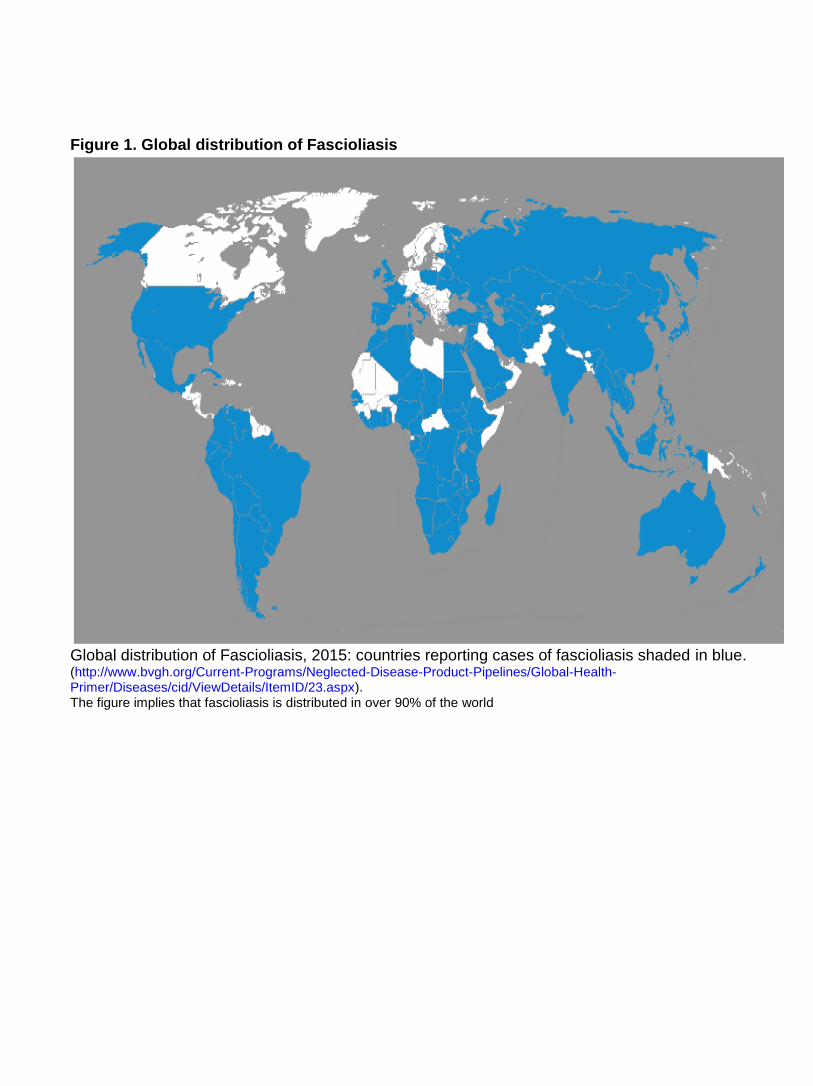
Figure 1. Global distribution of Fascioliasis
Global distribution of Fascioliasis, 2015: countries reporting cases of fascioliasis shaded in blue. (http://www.bvgh.org/Current-Programs/Neglected-Disease-Product-Pipelines/Global-Health-Primer/Diseases/cid/ViewDetails/ItemID/23.aspx). The figure implies that fascioliasis is distributed in over 90% of the world