fascialspaceinfections 140219122023-phpapp01
TRANSCRIPT
Classification of Fascial Spaces
Based on mode of involvement- Primary spaces. Secondary spaces.
Primary maxillary- canine, buccal, infratemporal.
Primary mandibular- submental, sublingual, buccal, submandibular.
Secondary spaces- masseteric, pterygomandibular, superficial & deep temporal, lateral pharyngeal, retropharyngeal, parotid, prevertebral.
INTRODUCTION
Infection of orofacial and neck region, particularly those of odontogenic have been one of most common disease in human beings. Early reconignition of orofacial infection &prompt appropriate therapy is essential .

PATHWAYS OF ODONTOGENIC INFECTION
Serious dental infection spreading beyond tooth socket is more common due to pulpal infection than periodontal infection
Invasion of dental pulp by bacteria after decay of tooth
Inflammation edema &lack of collateral blood supply
Venous congestion or avascular necrosis (pulpal tissue death)
Reservoirs for bacterial growth (anerobic)
Periodic progress of bacteria into surrounding bone (abscess)
May lead to progress according
A ) Host resistance
B) Number and virulence of organism
c) Anatomy of involved area


Classification of Fascial Spaces
Based on mode of involvement- Primary spaces. Secondary spaces.
Primary maxillary- canine, buccal, infratemporal.
Primary mandibular- submental, sublingual, buccal, submandibular.
Secondary spaces- masseteric, pterygomandibular, superficial & deep temporal, lateral pharyngeal, retropharyngeal, parotid, prevertebral.
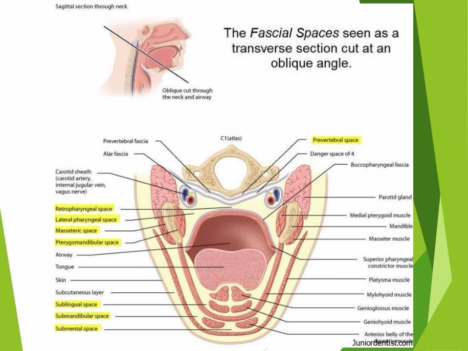
Based on clinical significance-
Face- Buccal, canine, parotid, masticatory.
Suprahyoid- Sublingual, submental, submandibular,
lateral pharyngeal, peritonsillar.
Infrahyoid- Pretracheal.
Spaces of total neck- Retropharyngeal, space of
carotid sheath.
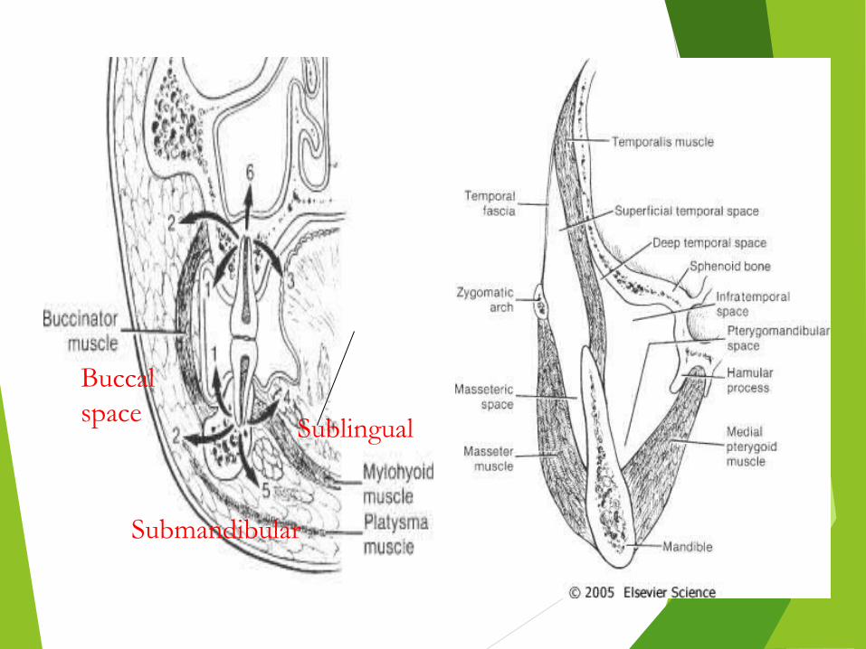
Buccal space Sublingual
Submandibular


Canine Space It is the region between anterior surface of maxilla and overlying
levator muscles of upper lip. Contains angular artery & vein, infraorbital nerve.
Etiology-
Maxillary canine & 1st premolar infection & sometimes mesiobuccal root of first molars.
Boundaries- Superiorly: levator superioris alaque nasi and levator labii superioris Inferiorly: caninus muscle Medially: anterolateral surface of maxilla Posteriorly: buccinator mucsle. Anteriorly: orbicularis oris

10
Clinical Features-
Swelling of cheek, lower eyelid & upper lip.
Drooping of angle of mouth.
Nasolabial fold obliterated.
Odema of lower eyelid

Buccal Space
Boundaries- Superiorly: zygomatic arch. Inferior: inferior border of mandible. Laterally: skin & subcutaneous tissue. Medially: buccinator muscle ,buccopharyngeal fascia. Posteriorly: anterior edge of masseter muscle. Anteriorly: posterior border of zygomaticus major & depressor
anguli oris.
Contents- Buccal fat pad. Stenson’s duct. Facial artery.

Etiology-
Infected mandibular & maxillary premolars & molars.
Clinical Features-
Obliteration of nasolabial fold.
Angle of mouth shifted to opposite side.
Swelling in cheek extending to corner of mouth.
Buccal space associated with temporal space – Dumb bell shaped appearance due to lack of swelling over zygomatic arch.

Buccal Space Infection

Infratemporal Space
Boundaries-
Superiorly: infratemporal surface of greater wing of sphenoid.
Inferiorly: lateral pterygoid muscle.
Laterally: temporalis tendon & coronoid process.
Medially: lateral pterygoid plate & lateral pharyngeal wall.
Posteriorly: condyle & lateral pterygoid muscles.
Anteriorly: infratemporal surface of maxilla & posterior surface of zygomatic bone.
Contents- Pterygoid plexus of veins. Internal maxillary artery. Mandibular nerve & its branches.
Etiology- Infected maxillary 3rd molars. Infected needles or contaminated LA solution.
Clinical Features- Extra-oral swelling over sigmoid notch area. Intra-oral swelling in tuberosity area. Trismus.
Spread of Infection- To temporal space. Cavernous sinus thrombosis- infection spreads via pterygoid plexus
of veins.

Submental Space
Boundaries- Roof: mylohyoid muscle. Inferior: deep cervical fascia, platysma, superficial fascia & skin. Laterally: anterior belly of digastric. Posteriorly: submandibular space.
Contents- Lymph nodes, anterior jugular vein.
Etiology- Infected mandibular incisors. Anterior extension of submandibular space.
Clinical Features-• Chin appears glossy & swollen.• Pain & discomfort on swallowing.

Sublingual Space
Boundaries- Superiorly: mucosa of floor of mouth. Inferior: mylohyoid muscle. Posteriorly: body of hyoid bone. Anteriorly & laterally: inner aspect of mandibular body. Medially: geniohyoid,styloglossus,genioglossus muscle.
Contents- Deep part of Submandibular gland. Wharton’s duct. Sublingual gland. Lingual & hypoglossal nerves. Terminal branches of lingual artery.

Etiology-
Infected mandibular premolar & 1st molar.
Clinical Features-
Swelling of floor of mouth.
Elevated tongue.
Pain & discomfort on swallowing.

Submandibular Space
Boundaries- Superiorly: mylohyoid muscle, inferior border of mandible. Inferior: anterior & posterior belly of digastric. Laterally: deep cervical fascia, platysma, superficial fascia & skin. Medially: hyoglossus,styloglossus,mylohyoid muscle. Posteriorly: to hyoid bone. Anteriorly: submental space.
Contents- Submandibular salivary gland. Proximal portion of Wharton’s duct. Lingual & hypoglossal nerves. Branches of facial artery- palatine,tonsillar,glandular,submental.

Etiology- Infected mandibular 2nd & 3rd molars. From submental,sublingual spaces.
Clinical Features-• Indurated swelling in submandibular region.• Usually bulges over lower border of mandible.
Spread of Infection- Across midline to contralateral space. To contiguous pharyngeal spaces.

Submandibular Space Infection

Pterygomandibular Space
Boundaries- Superiorly: lower head of lateral pterygoid muscle. Laterally: medial surface of ramus. Medially: medial pterygoid muscle. Posteriorly: deep part of parotid. Anteriorly: pterygomandibular raphe.
Contents- Inferior alveolar neurovascular bundle. Lingual & auriculotemporal nerves. Mylohyoid nerve & vessels.
Etiology- Infected mandibular 3rd molars(mesioangular/horizontal) Pericoronitis. Infected needles or contaminated LA solution.
Clinical Features- Absence of extra-oral swelling. Severe trismus. Difficulty in swallowing. Anterior bulging of half of soft palate & tonsillar pillars with
deviation of uvula to unaffected side.
Spread of Infection- Superiorly to infratemporal space. Medially to lateral pharyngeal space. To submandibular space.
Masseteric Space
Boundaries- Superiorly: zygomatic arch. Inferiorly: inferior border of mandible. Laterally: masseter muscle. Medially: ramus of mandible. Posteriorly: parotid gland & its fascia. Anteriorly: buccal space & buccopharyngeal fascia.
Contents- Masseteric artery & vein.
Etiology- Mandibular 3rd molars(pericoronitis).
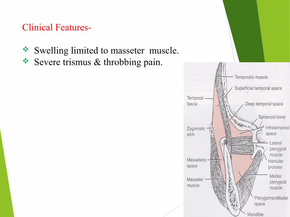
Clinical Features-
Swelling limited to masseter muscle. Severe trismus & throbbing pain.

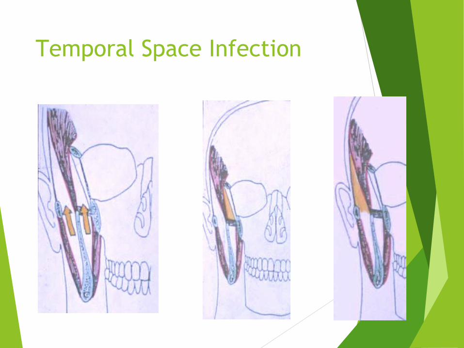
Temporal Spaces
Superficial temporal-
Laterally: temporalis fascia.
Medially: temporalis muscle.
Deep temporal-
Laterally: temporalis muscle.
Medially: temporal bone & greater wing of sphenoid.
Etiology-
From infratemporal or pterygomandibular space.
Clinical Features-
Superficial temporal- swelling limited by outline of temporalis fascia. Trismus. Severe pain.
Deep temporal- less swelling, difficult to diagnose. Trismus.
Temporal Space Infection

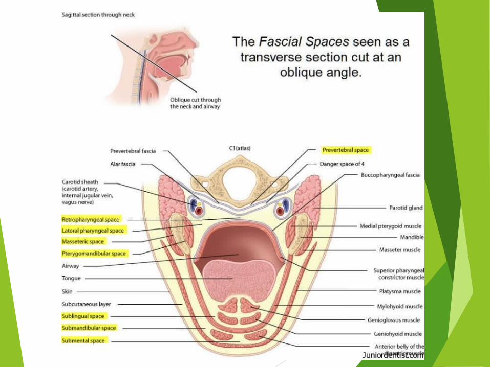
Lateral Pharyngeal Space
Boundaries-
Shape of an inverted cone or pyramid, the base is at sphenoid bone and the apex at hyoid bone.
Anteriorly: pterygomandibular raphe.
Posteriorly: extends to prevertebral fascia.
Laterally: fascia covering medial pterygoid muscle, parotid & mandible.
Medially: buccopharyngeal fascia on lateral surface of superior constrictor muscle.
Styloid process divides the space into anterior muscular and posterior vascular compartment.
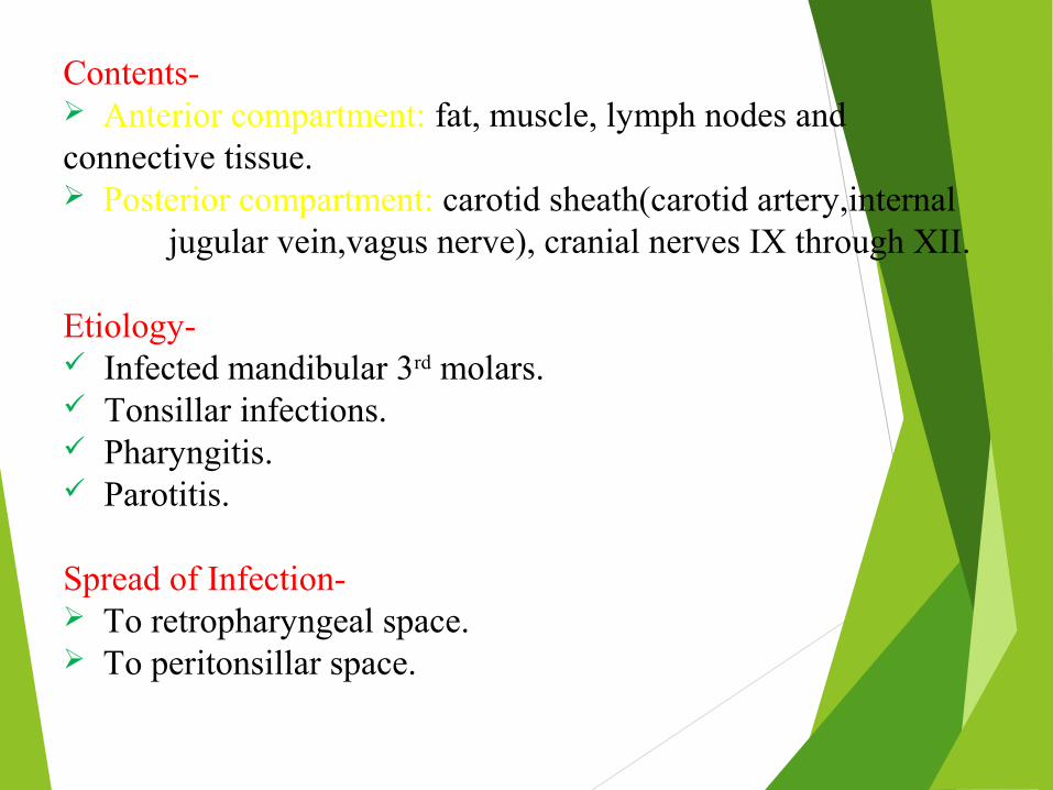
Contents- Anterior compartment: fat, muscle, lymph nodes and connective tissue. Posterior compartment: carotid sheath(carotid artery,internal
jugular vein,vagus nerve), cranial nerves IX through XII.
Etiology- Infected mandibular 3rd molars. Tonsillar infections. Pharyngitis. Parotitis.
Spread of Infection- To retropharyngeal space. To peritonsillar space.

Clinical Features-
Anterior compartment:
Trismus.
Induration & swelling at angle of jaw.
Fever.
Pharyngeal bulging.
Posterior compartment:
Posterior tonsillar pillar deviation.
Neurological involvement.
Thrombosis of internal jugular vein.
Erosion of carotid vessels may occur.
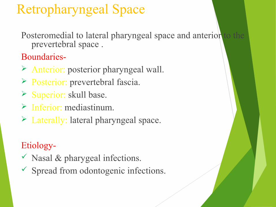
Retropharyngeal Space
Posteromedial to lateral pharyngeal space and anterior to the prevertebral space .
Boundaries- Anterior: posterior pharyngeal wall. Posterior: prevertebral fascia. Superior: skull base. Inferior: mediastinum. Laterally: lateral pharyngeal space.
Etiology- Nasal & pharygeal infections. Spread from odontogenic infections.


Clinical Features-
Stiffness of neck.
Dysponea.
Dysphagia.
Bulging of posterior pharyngeal wall.
Complications-
Airway obstruction.
Aspiration pneumonia.
Acute mediastinitis.
Can spread to Danger space.

Prevertebral Space
Potential space between two layers of prevertebral fascia (alar and prevertebral layers).
Extends from skull base superiorly to the diaphragm inferiorly.
Mediastinitis is concern with prevertebral space infections similarly to retropharyngeal space
infections.

Objectives
Understand the microbiology of odontogenic infections.
Understand the signs symptoms and findings in patients with odontogenic infections.
Review the various pathways of spread with odontogenic infections.
Understand the medical and surgical management of odontogenic infections.

MICROBIOLOGY OF ODONTOGENIC INFECTIONS
Usually caused by endogenous bacteria.
Most odontogenic infections due to mixed flora.
Streptococcus species(alpha hemolytic) are usually the etiologic organisms if aerobic bacteria present.
Anaerobes- prevotella, bacteroids, fusobacterium also involved.

Factors affecting Spread of Infection
General factors-1. Microbial factors-
Level of virulence.
No. of organisms introduced.
2. Host factors-
General state of health.
Integrity of surface defence.
Level of immunity.
Capacity for inflammatory & immune response.
Impact of medical intervention.
3. Combination of both factors.

Routes of Spread
Direct spread-a) Spread into superficial soft tissues as-
Abscess- pathological thick walled cavity filled with pus.
Cellulitis- diffuse subcutaneous/submucous inflammation of soft tissues. Tends to spread along fascial planes.
b) Spread into adjacent fascial spaces.
c) Into deep medullary spaces of bone- osteomyelitis
Indirect spread-a) Lymphatic routes to regional nodes.
b) Hematogenous route to other organs such as brain.

Pathway of Odontogenic Infection
Acute-chronic periapical infection
Intraoral soft tissue abscess
Cellulitis
Deep fascial space infection
Bacteremia- septicemia
Sinus or Fistula
Ascending fascial cerebral infection
Medullary spaces of bone-osteomyelitis

Sites of Localization of Dental Infection
Involved teeth
Usual exit from bone
Relation of muscle to root apices
Site of localization
Upper central incisor
Labial Above Oral vestibule
Upper lateral incisor
Labial Palatal
Above Oral vestibulePalate
Upper canine Labial Above Below
Oral vestibule Canine space
Upper premolars
Buccal Palatal
Above Oral vestibulePalate
Upper molars Buccal
Palatal
AboveBelow
Oral vestibuleBuccal spacePalate

Involved teeth Usual exit from bone
Relation of muscle to root apices
Site of localization
Lower incisors Labial AboveBelow
Submental spaceOral vestibule
Lower canine Labial Below Oral vestibule
Lower premolars
Buccal Below Oral vestibule
Lower 1st molar Buccal
Lingual
Below AboveBelow
Oral vestibuleBuccal spaceSublingual space
Lower 2nd molar Buccal
Lingual
Below AboveBelow Above
Oral vestibuleBuccal spaceSublingual spaceSubmandibular space
Lower 3rd molar Lingual Above Submandibular or pterygomandibular space

Clinical Features
Rubor- (redness) cutaneous surface involved due to vasodilatation
effect of inflammation.
Tumor-(swelling) due to the accumulation of pus or fluid exudate.
Calor-(heat) is the result of increased blood flow to the area due to
the vasodilatation.
Dolor-(or pain) results from pressure on sensory nerve endings from
tisssue distention caused by edema or infection.
Functiolaesa-(loss of function) problems with function.

Lymphadenopathy- nodes enlarged,soft & tender in acute infection.
Firm & enlarged in chronic.
Halitosis.
Fever & headache. Repeated chills.
Presence of draining sinuses/fistulae.
Increased salivation.
Trismus.
Difficulty in swallowing.
Changes in phonation.
Difficulty in breathing.

Investigations
Routine laboratory investigations.
Special laboratory investigations.
Radiological examination- helpful in locating offending teeth or other underlying cause.
IOPA
OPG
Lateral oblique view mandible.
A-P & Lateral view of neck for soft tissues can be useful in detecting retropharyngeal space infection.
Ultrasound of swelling.
CT scan, MRI help in diagnosing extension of infection beyond maxillofacial region.

Management of Odontogenic Infections
Goals of management of odontogenic infection:
1. Airway protection.
2. Surgical drainage.
3. Identification of etiologic bacteria.
4. Selection of appropriate antibiotic therapy.
5. Medical & supportive therapy.

Selection of Antibiotic therapy Parenteral penicillin.
Metronidazole in combination with penicillin can be used in
severe infections.
Clindamycin for penicillin-allergic patients.
Cephalosporins (1st & 2nd generation cephalosporins).
Antibiotics do not substitute for incision and drainage in cases of
significant odontogenic infections.
Causes for clinical failure include inadequate drainage or
antibiotic resistance.

Surgical Management
Surgical treatment may range from simply opening tooth & extirpation of pulp to complex incision & drainage.
Primary goal in surgical management is to remove cause of infection.
Secondary goal is to provide drainage of accumulated pus & necrotic debris.
Extraction provides both removal of cause of infection and drainage of pus & debris.
Incision & Drainage
Incision & drainage helps- To get rid of toxic purulent material. To decompress odematous tissues. To allow better perfusion of blood, containing antibiotics &
defensive elements. To increase oxygenation of infected area.
Removal of the cause; such as infected tooth, a segment of
necrotic bone, a foreign body should be done at the time of
I & D procedure.

Hilton’s method of I & D
1. Topical anesthesia achieved with spray or infiltration.
2. Stab incision given through skin & s/c tissue.
3. If pus is not encountered, further deepening of surgical site done with sinus forceps.
4. Abscess cavity is entered and forceps opened in direction parallel to vital structures.
5. Explore the entire cavity for additional loculi.
6. Cavity irrigated with saline & antiseptic solutions.
7. Placement of drain.
8. Dressing.

Drainage of Fascial Spaces
Canine, Sublingual and Vestibular abscesses are drained
intraorally.
Masseteric, Pterygomandibular, Buccal and Lateral Pharyngeal
space abscesses can be drained with combination of intraoral and
extraoral drainage.
Temporal, Submandibular, Submental, Retropharyngeal and
Parotid space abscesses may mandate extraoral incision and
drainage.

Medical & Supportive Therapy
Administration of antibiotics.
Hydration of patient by I/V route.
Soft or liquid diet rich of high proteins.
Analgesics & NSAIDs.
Antiseptic mouthwashes.
Complete bed rest.

LUDWIG’S ANGINA::
DEFINITION– IT IS A FIRM, ACUTE,TOXIC CELLULITIS OF THE SUBMANDIBULAR,SUBLINGUAL SPACES BILATERLLY & OF THE SUBMENTALIS SPACE.
-- FRIST DISCRIBED BY WILHELM FREDREICH VON LUIDWIG IN 1836 ETIOLOGY: 1. PERIAPICAL,PERICORONAL OR PERIODONTAL INFECTION OF A LOWER THIRD MOLAR 2. TRAUMATIC INJURIES & INFECTED LESIONS 3. INFECTIVE CONDITIONS SUCH AS OSTEOMYELITIS MAY MENIFEST AS LUDWIG’S ANGINA 4. CYSTS OR TUMORS IN THIRD MOLAR REGION PATHOLOGY: 1. INFECTION FROM LOWER THIRD MOLAR REACHES THE SUBMANDIBULAR SPACES 2. FROM HERE INFECTION SPREADS ALONG THE SUMANDIBULAR SALIVARY GLANDS ABOVE THE MYLOHYIOD MUSCLE TO REACH THE SUBLINGUAL SPACE

CLINICAL FEATURES - SYSTEMIC FEATURES- PYREXIA , DEHYDRATION , DYSPHAGIA , DYSPNOEA , HOARSENESS OF VOICE AND STRIDOR
EXTRA ORAL FEATURES – HARD TO FIRM BROWNY INDURATED SWELLING SKIN OVER THE SWELLING APPEARS ERYTHMATOUS AND STRETCHED
SWELLING IS TENDER WITH LOCAL RISE IN TEMPERATURE
Difficulty in closing the mouth and drooling of salivaRespiratory distress
INTRA ORAL FEATURES – Trismus , floor of the mouth is raised , tongue raised upwards , increased salivation

MANAGEMENT - 1.Airway maintainence- Tracheostomy and Cricothyroidectomy
is advisable
2. Parentral antibiotics - Penicillin antibiotic of choice Amoxycillin + Cloxacillin Metronidazole in anaerobic infection 3.Surgical decompression – performed under L.A Decompression improves vascularity and potentiates the action of antibiotics. Bilateral submandibular incision with a midline submental incision pus
should be drained
4.Hydration of the patient – It is necessary to put the pt on i.v. fluids 5. Removal of cause - The offending tooth is removed

COMPLICATIONS – • Death due to airway compromise• septicemia• mediastinit is• carotid blow out

References
Textbook of oral & maxillofacial surgery : Neelima Malik.
Oral & maxillofacial Infections : Topazian
Textbook of oral & maxillofacial surgery : Laskin

THANK YOU!!!!