eye tumour
TRANSCRIPT
Radiosurgery with CyberKnife as an organ preserving
treatment for small localized choroid melanoma
Debnarayan Dutta MD, KR Prasanna Kumar; *, MD; Prativa Mishra^, MS;
Raghunandhan* MD; P Mahadev * MD, DNB; AN Vaidhyswaran* MD; Sanjay
Chandrasekhar* MD;
Janos Stumpf *MD, PhD; Rathnadevi* DMRT; V Murali# PhD; PG Kurup# PhD;
*Department of Radiation Oncology, #Medical Physics,
^Department of Opthalmology,
Apollo Speciality Hospital, Chennai
Aim: To evaluate robotic radiosurgery as an organ preserving treatment for localized choroid
melanoma of the eye.
Case report: Thirty six year old female patient had complaint of progressive dimness of vision of
right eye for six months. Fundoscopy examination showed small lesion (2.5 x 2.5 mm) in macular
region. 320 slices CT scan showed organ confined 2.5 x 2.5 mm nodular lesion in the macular
region (3 mm superior and 2.5 mm temporal to the origin of optic nerve at fovea) of right eye and
was diagnosed with localized choroid melanoma of right eye (visual acuity 6/18 ). Metastatic
workup was normal. She was planned for SRS (CyberKnife) as an organ preserving approach.
Planning CT scan and CyberKnife treatment (dose 18 Gy single fraction, prescription isodose 85%;
treatment time 22 min, GTV 111.6 mm3, 2mm PTV margin, PTV 403 mm3) were done with retro-
bulbar anesthesia. Mean dose to right eye, left eye, right eye lens and pituitary gland was 4.9, 0.4,
0.4 and 1 Gy respectively. Maximum dose to optic chiasm, brainstem, right (2% vol) and left optic
nerve were 1.4, 2.1, 15 and 0.4 Gy respectively. Skull tracking method was used as tumour tracking
method. She completed treatment without any acute complication and visual acuity was preserved.
Conclusion: Robotic radiosurgery is a feasible, acceptable and an appropriate treatment modality
as organ preserving approach in small choroid melanomas.
Keywords: Choroid melanoma, Robotic radiosurgery, Organ preserving approach
Abstract
Case history
36 year old female patient had complaint of progressive dimness of vision of right eye for
six months.
Visual acuity of involved eye was 6/18.
Investigations
Fundoscopy examination showed small lesion (2.5 x 2.5 mm) in macular region (Figure 1).
320 slices CT scan showed organ confined 2.5 x 2.5 mm nodular lesion in the macular
region (3 mm superior and 2.5 mm temporal to the origin of optic nerve at fovea) of right
eye and was diagnosed with localized choroid melanoma of right eye (Figure 2).
Metastatic workup was normal (USG abdomen, Chest X-ray).
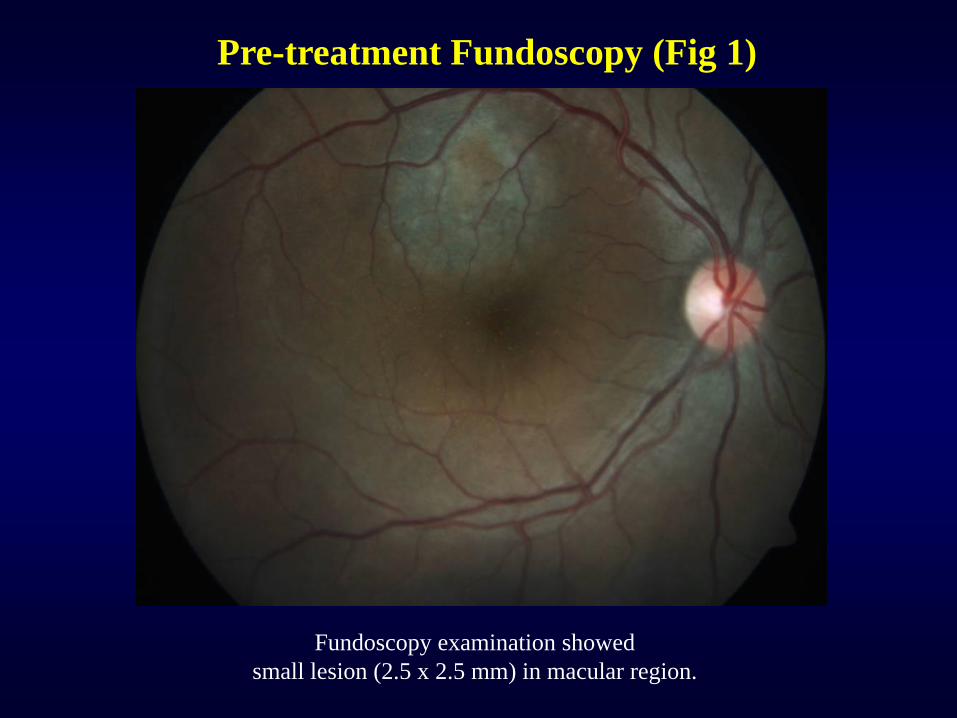
Pre-treatment Fundoscopy (Fig 1)
Fundoscopy examination showed
small lesion (2.5 x 2.5 mm) in macular region.
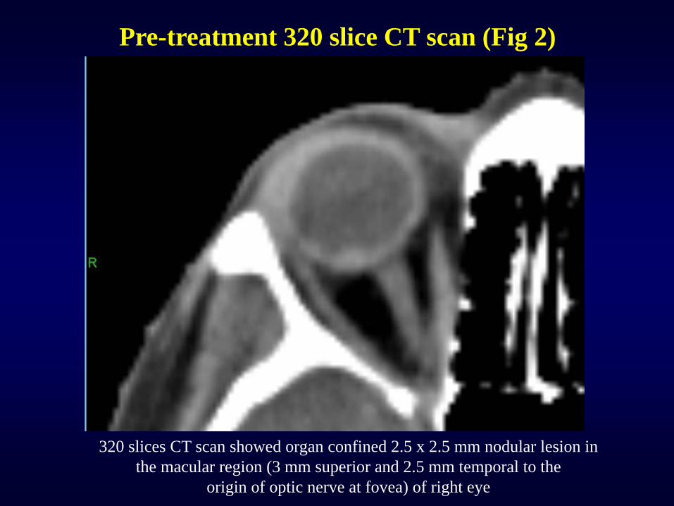
Pre-treatment 320 slice CT scan (Fig 2)
320 slices CT scan showed organ confined 2.5 x 2.5 mm nodular lesion in
the macular region (3 mm superior and 2.5 mm temporal to the
origin of optic nerve at fovea) of right eye
Treatment historyShe was planned for SRS (CyberKnife) as an organ preserving approach .
Planning CT scan done under retro-bulbar anesthesia with thermoplastic mask (Figure 3).
Treatment done under retro-bulbar anesthesia (Figure 6).
Skull tracking method was used as tumour tracking method.
She completed treatment without any acute complication and visual acuity was preserved.
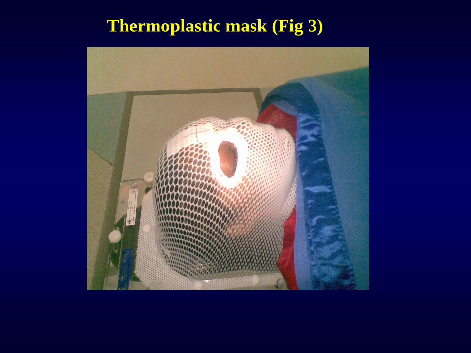
Thermoplastic mask (Fig 3)
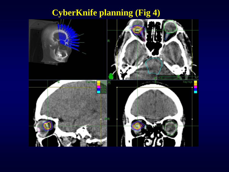
CyberKnife planning (Fig 4)
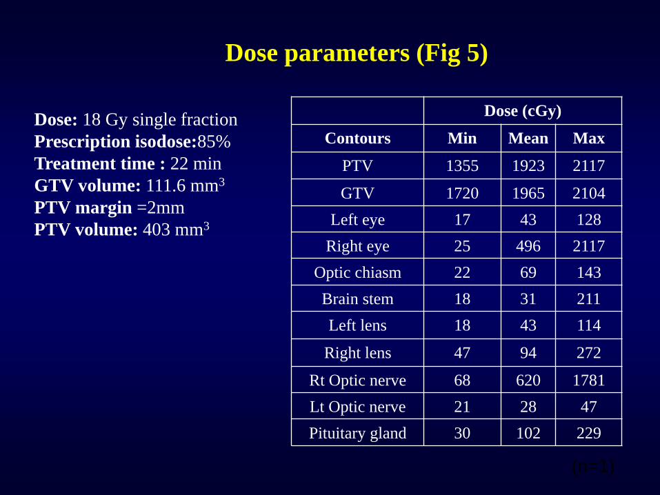
Dose (cGy)
Contours Min Mean Max
PTV 1355 1923 2117
GTV 1720 1965 2104
Left eye 17 43 128
Right eye 25 496 2117
Optic chiasm 22 69 143
Brain stem 18 31 211
Left lens 18 43 114
Right lens 47 94 272
Rt Optic nerve 68 620 1781
Lt Optic nerve 21 28 47
Pituitary gland 30 102 229
Dose parameters (Fig 5)
(n=1)
Dose: 18 Gy single fraction
Prescription isodose:85%
Treatment time : 22 min
GTV volume: 111.6 mm3
PTV margin =2mm
PTV volume: 403 mm3
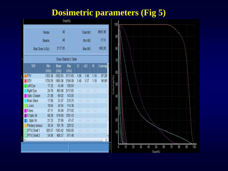
Dosimetric parameters (Fig 5)
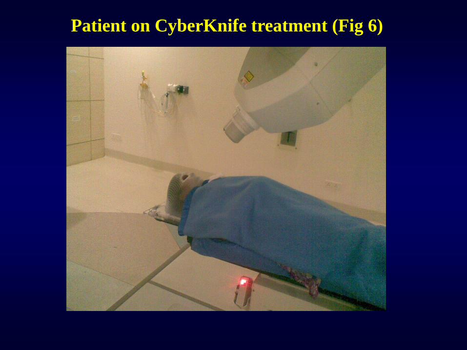
Patient on CyberKnife treatment (Fig 6)
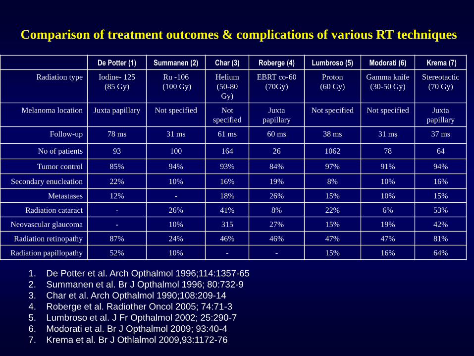
De Potter (1) Summanen (2) Char (3) Roberge (4) Lumbroso (5) Modorati (6) Krema (7)
Radiation type Iodine- 125
(85 Gy)
Ru -106
(100 Gy)
Helium
(50-80
Gy)
EBRT co-60
(70Gy)
Proton
(60 Gy)
Gamma knife
(30-50 Gy)
Stereotactic
(70 Gy)
Melanoma location Juxta papillary Not specified Not
specified
Juxta
papillary
Not specified Not specified Juxta
papillary
Follow-up 78 ms 31 ms 61 ms 60 ms 38 ms 31 ms 37 ms
No of patients 93 100 164 26 1062 78 64
Tumor control 85% 94% 93% 84% 97% 91% 94%
Secondary enucleation 22% 10% 16% 19% 8% 10% 16%
Metastases 12% - 18% 26% 15% 10% 15%
Radiation cataract - 26% 41% 8% 22% 6% 53%
Neovascular glaucoma - 10% 315 27% 15% 19% 42%
Radiation retinopathy 87% 24% 46% 46% 47% 47% 81%
Radiation papillopathy 52% 10% - - 15% 16% 64%
Comparison of treatment outcomes & complications of various RT techniques
1. De Potter et al. Arch Opthalmol 1996;114:1357-65
2. Summanen et al. Br J Opthalmol 1996; 80:732-9
3. Char et al. Arch Opthalmol 1990;108:209-14
4. Roberge et al. Radiother Oncol 2005; 74:71-3
5. Lumbroso et al. J Fr Opthalmol 2002; 25:290-7
6. Modorati et al. Br J Opthalmol 2009; 93:40-4
7. Krema et al. Br J Othlalmol 2009,93:1172-76
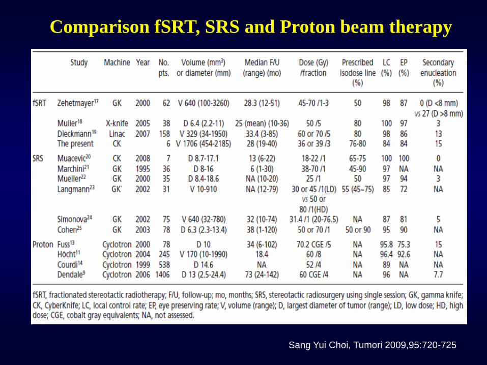
Comparison fSRT, SRS and Proton beam therapy
Sang Yui Choi, Tumori 2009,95:720-725
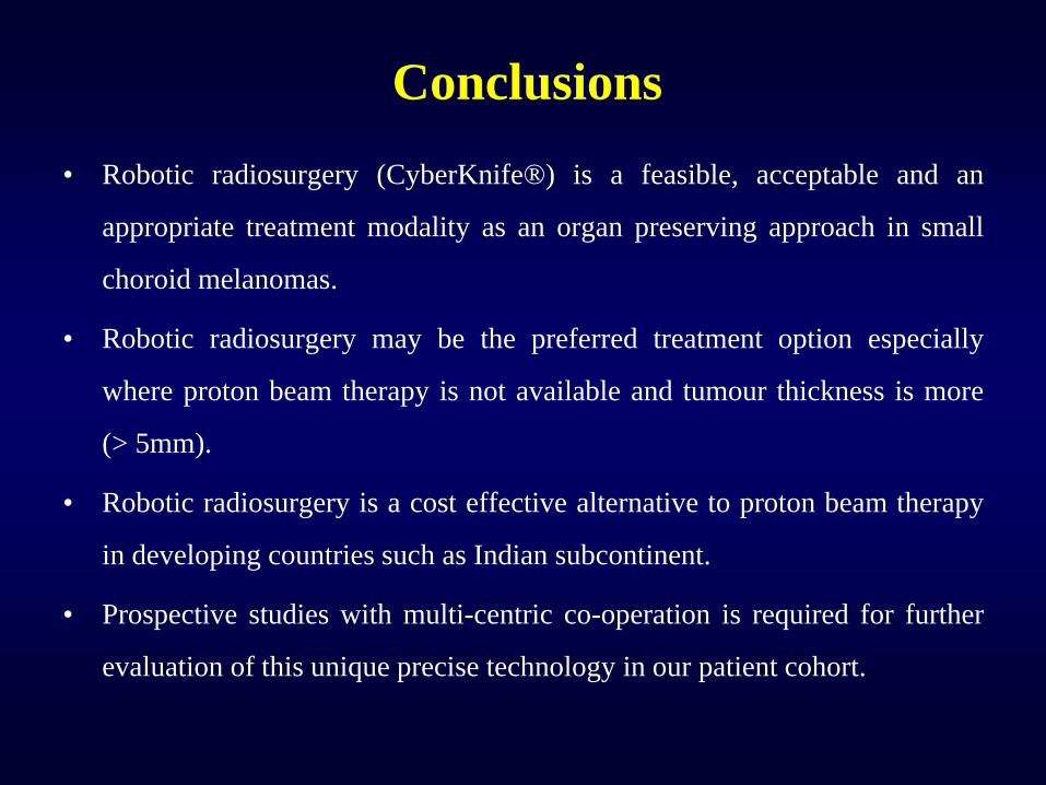
• Robotic radiosurgery (CyberKnife®) is a feasible, acceptable and an
appropriate treatment modality as an organ preserving approach in small
choroid melanomas.
• Robotic radiosurgery may be the preferred treatment option especially
where proton beam therapy is not available and tumour thickness is more
(> 5mm).
• Robotic radiosurgery is a cost effective alternative to proton beam therapy
in developing countries such as Indian subcontinent.
• Prospective studies with multi-centric co-operation is required for further
evaluation of this unique precise technology in our patient cohort.
Conclusions