extended wearing trial of trifield lens device for tunnel...
TRANSCRIPT

Extended wearing trial of Trifield lens devicefor �tunnel vision�Russell L. Woods1, Robert G. Giorgi1, Eliot L. Berson2 and Eli Peli1
1Schepens Eye Research Institute, Harvard Medical School, Boston, MA, 2Berman-Gund Laboratory
for the Study of Retinal Degenerations, Harvard Medical School, Massachusetts Eye and Ear
Infirmary, Boston, MA, USA
Abstract
Severe visual field constriction (tunnel vision) impairs the ability to navigate and walk safely. We
evaluated Trifield glasses as a mobility rehabilitation device for tunnel vision in an extended wearing
trial. Twelve patients with tunnel vision (5–22� wide) due to retinitis pigmentosa or choroideremia
participated in the 5-visit wearing trial. To expand the horizontal visual field, one spectacle lens was
fitted with two apex-to-apex prisms that vertically bisected the pupil on primary gaze. This provides
visual field expansion at the expense of visual confusion (two objects with the same visual direction).
Patients were asked to wear these spectacles as much as possible for the duration of the wearing
trial (median 8, range 6–60 weeks). Clinical success (continued wear, indicating perceived overall
benefit), visual field expansion, perceived direction and perceived visual ability were measured. Of 12
patients, nine chose to continue wearing the Trifield glasses at the end of the wearing trial. Of those
nine patients, at long-term follow-up (35–78 weeks), three reported still wearing the Trifield glasses.
Visual field expansion (median 18, range 9–38�) was demonstrated for all patients. No patient
demonstrated adaptation to the change in visual direction produced by the Trifield glasses (prisms).
For reported difficulty with obstacles, some differences between successful and non-successful
wearers were found. Trifield glasses provided reported benefits in obstacle avoidance to 7 of the 12
patients completing the wearing trial. Crowded environments were particularly difficult for most
wearers. Possible reasons for long-term discontinuation and lack of adaptation to perceived direction
are discussed.
Keywords: choroideremia, low vision rehabilitation, mobility, retinitis pigmentosa, Trifield glasses,
tunnel vision
Introduction
A common definition (e.g. Social Security Administra-tion in the USA) of legal blindness is a constriction ofthe visual field to £20� diameter as measured byGoldmann perimetry using a III4e target size (tunnelvision). Tunnel vision is a debilitating symptom ofretinitis pigmentosa and choroideremia. Retinitis pig-mentosa, the most common cause of inherited blindness,
has a prevalence of about 1 in 4000 people worldwide(Berson, 1993; Hartong et al., 2006).
Patients with tunnel vision have difficulties withnavigating, avoiding obstacles, and performing visualsearch (Marron and Bailey, 1982; Lovie-Kitchin et al.,1990; Haymes et al., 1996; Black et al., 1997; Kuyket al., 1998; Turano et al., 1999a, 2001; Broman et al.,2004; Luo and Peli, 2006; Apfelbaum et al., 2007;Fortenbaugh et al., 2007). The consequent loss ofmobility with increased risk of falls (Felson et al.,1989; Lord et al., 1993; Tinetti and Williams, 1997;Lord and Dayhew, 2001; Biderman et al., 2002;Freeman et al., 2007) is detrimental to patients� inde-pendence and quality of life (Tinetti and Williams, 1997;Turano et al., 1999b; Shinkai et al., 2000; Bidermanet al., 2002; Melzer et al., 2003; Cacciatore et al., 2004).Visual field extent is a significant predictor of mobilityperformance (Marron and Bailey, 1982; Lovie-Kitchin
Received: 30 July 2009
Revised form: 29 October 2009
Accepted: 21 December 2009
Correspondence and reprints requests to: Dr Russell Woods, Schepens
Eye Research Institute, 20 Staniford Street, Boston, MA 02114-2500,
USA.
Tel.: 1 617 912 2589; Fax: 1 617 912 0112.
E-mail address: [email protected]
Ophthal. Physiol. Opt. 2010 30: 240–252
doi: 10.1111/j.1475-1313.2010.00718.x ª 2010 The Authors. Journal compilation ª 2010 The College of Optometrists

et al., 1990; Haymes et al., 1996; Black et al., 1997;Kuyk et al., 1998; Turano et al., 1999a; Broman et al.,2004). Many people with tunnel vision receive orienta-tion and mobility training, though the benefit of thistraining has not been proven (Soong, 2000; Soong et al.,2001; Kuyk et al., 2004; Virgili and Rubin, 2006).Previously, rehabilitation approaches for patients
with tunnel vision included: training to scan (Cohenand Waiss, 1996); minifying devices (reversed telescopes,or hand held negative lenses) (Drasdo, 1976; Hoeftet al., 1985; Weiss, 1992; Szlyk et al., 1998) and variousprism spectacle designs (Cohen, 1993; Onufryk, 1994).Most of these devices have not been commerciallyavailable and others have had limited clinical use due totheir drawbacks. More recently, augmented-vision head-mounted displays (Peli, 2001; Vargas-Martin and Peli,2002) have shown some promise in early testing in visualsearch tasks (Luo and Peli, 2006) and judgements ofpotential collisions (Luo et al., 2009).We are not aware of any publication explaining how
to train patients with tunnel vision to modify theirscanning eye movements or showing that patientsactually increase their scanning following training. Webelieve that in most cases the training simply consistedof instructing patients that they should scan more. Inrecording eye movements of patients with tunnel visionwhile walking, it has been reported that the distributionof eye movements while walking was not larger thanthat of normally-sighted subjects (Vargas-Martin andPeli, 2006) and saccadic amplitudes and directions werevery similar to those of normally-sighted subjects (Luoet al., 2008). Thus, contrary to clinical wisdom and theintent of training, it seems that patients with tunnelvision do not compensate for the restricted visual fieldby making larger scanning eye movements than patientswith a full visual field.Minifying devices shrink the view of the scene so that
a wider section of the world is available within thepatient�s residual visual field but have poor clinicalacceptance, primarily due to the reduction in resolution(visual acuity). The Amorphic lens reversed telescope(Designs for Vision Inc., Ronkonkoma, NY, USA)minified only the horizontal meridian in order to reducethe impact on acuity (Hoeft et al., 1985) at a cost ofimage distortion. Despite some reports of success (Hoeftet al., 1985; Szlyk et al., 1998), they were discontinuedrecently.The use of prisms in treating peripheral visual field
defects has been controversial, as previously proposeddesigns had significant limitations (Cohen, 1993). Thebest known binocular sector prism design (Onufryk,1994), the so-called Channel lens, was based on a field-shifting principle (rather than field expanding). Theseprisms, commercialized briefly in 1998 and 1999 as theInWave� lenses (InWave Inc., Janesville, WI, USA),
had a central prism free channel (corresponding to thewidth of the residual visual field of the patient). As aresult they had no effect in primary position of gaze.When the patient made eye movements towards thesurrounding prismatic areas the effect was to shift(relocate) the image more centrally, rather than expand,the residual visual field. Further, the prism powersavailable laterally (12D � 6� – nasal and temporalbase out) and below the channel (8D � 4� – base down)were probably too small to have an impact on mobility.Also, the prism apex scotomata could interfere withtheir functionality (Giorgi et al., 2009; Ross et al.,2009). Somani et al. (2006) implemented this designusing stick-on Fresnel prisms and claimed smallincreases in visual fields and activities of daily living.
Obstacle detection is likely to be best when the userdoes not need to make a specific action with a visualfield expansion device to search for a potential obstacle.So, to be most effective the device should provide a viewof the potential obstacle with natural use of the device,by providing visual field expansion all of the time. Forpotential obstacle detection, the Channel lens (prisms),the Amorphic lens in a bioptic configuration (Szlyket al., 1998) and hand held negative lenses (Kozlowskiet al., 1984) all require that the user initiate a searchaction by spotting through the device. As such, the useof these devices is unlike the use of bioptics formagnification. With a magnification bioptic the needfor initiating device use is apparent to the user (i.e.cannot resolve object of interest that is detected withoutthe device).
Trifield glasses have been proposed as a mobility aid(Peli, 2001). Trifield glasses expand the visual fieldwithout minification, or prism scotoma (Peli, 2001).Trifield glasses consist of two apex-to-apex prismsmounted in front of one eye, and a conventionalspectacle lens (e.g. single vision with prescription) infront of the other eye (Figure 1). The patient retains theoriginal residual visual field of the non-prism eye, whilethe prism lens provides a laterally shifted visual field.The combination of the original visual field and theshifted visual field provides an expansion. The prismlens is fitted so that the prism apices vertically bisect thepupil when the prism eye is in primary gaze. As the gazeis shifted to the left, the prism eye sees a part of the scenethat is laterally farther to the left compared to that seenby the eye with the conventional lens. Similarly, whenthe gaze is directed to the right, the prism eye seesobjects farther to the right than the non-prism eye. Thecombined percept is double vision (specifically visualconfusion: two objects having the same visual direction).As illustrated in Figure 2, the direction of the lateralshift and expansion is dependent on the direction ofgaze. As the patient looks right (Figure 2b) or left(Figure 2c), the prism eye looks through the right or left
Trifield device for tunnel vision: R. L. Woods et al. 241
ª 2010 The Authors. Journal compilation ª 2010 The College of Optometrists

prism, respectively, and areas the patient would nototherwise see are shifted into their visual field from theright or the left, respectively. If the patient looks directlyforward so that the prism junction is over the pupil,three parts of scene are visible: one through theconventional lens before the non-prism eye and twoparts, laterally shifted from opposite directions, throughthe two prisms of the Trifield lens before the prism eye(Figure 2d). Thus, �tri-field� glasses make three possiblepieces of the visual field available to the wearer, two ofwhich would not be seen without the glasses, andincrease the available visual field extent up to three-fold.While the part (or side) of the field of view that isexpanded varies with gaze direction, the area of thevisual field that is visible is about double at all times.
The lateral shift provided by the prisms is largeenough to shift the extension outside the unchangedvisual field of the non-prism eye, preventing the patientfrom experiencing diplopia (seeing one object twice intwo different apparent directions). Diplopia occurswhen the images of an object fall onto non-correspond-ing points of the two retinas. However, visual confusiondoes occur with the Trifield glasses. Visual confusionoccurs when images of two different objects in the scenefall onto corresponding points of the two retinas and,therefore, have the same apparent direction. To help thepatients differentiate the objects and their directionviewed through the prisms, the right prisms were tintedred and the left prisms were tinted green. While thesecues are helpful, we expected that when wearers werefirst given the glasses, they would have difficulty inter-preting what they saw. One of the two pieces of the fieldof view (the tinted one) brought into view by the prismsdoes not represent the objects in their true direction.However, we also expected that wearers would adapt tothis shift and eventually be able to determine the truedirection of objects viewed through the prisms (Kohler,1964; Pick et al., 1969; Welch et al., 1993).
In this study, patients with severely restricted visualfields were fitted with the Trifield glasses for an extendedperiod (a wearing trial of nominally 6 weeks) duringwhich their benefit as a mobility aid was evaluated.
Methods
Extended wearing trial
The Trifield glasses were evaluated during a prospectivefive-visit extended wearing trial planned for 6-weeksduration (Figure 3). At the initial visit, the patientreceived a full eye examination by one of us (ELB) at theMassachusetts Eye and Ear Infirmary (MEEI) and wasthen further screened for study eligibility at SchepensEye Research Institute (SERI). If eligible (describedbelow), the patient was measured for Trifield glasses.Typically, the Trifield glasses took 6–10 weeks tomanufacture. The Trifield glasses were dispensed at visit2. Follow-up visits were performed nominally 1 (visit 3),3 (visit 4) and 6 (visit 5) weeks after delivery of theTrifield glasses at visit 2. The benefits (or otherwise) ofthe Trifield glasses were evaluated through measure-ments of visual field expansion, change in perceiveddirection through the prisms, perceived visual ability,and through clinical success; that is, the willingness tocontinue wearing the Trifield glasses at the end of thewearing trial. At approximately 1 year following the endof the wearing trial, a long-term follow-up telephoneinterview was conducted with those patients who choseto continue to wear the Trifield glasses at the last visit ofthe wearing trial (Figure 3). There were large individualvariations in the time between visits, and in the totalduration of the wearing trial (Figure 3) that resultedfrom difficulties scheduling the visits, including personalhealth, difficulties with travel (e.g. arranging a compan-ion, short periods of daylight in the winter) and personalschedules.
Eligibility criteria
Eligibility requirements included best-corrected single-letter visual acuity of 6/30 or better in the each eye, astable condition that caused tunnel vision, ability tounderstand verbal instructions in English, and age18 years or older. The primary visual field criterionwas a residual central visual field horizontal diameter of3–20� in each eye as measured by our custom comput-erized perimeter using a white (340 cd m)2), 12 mmtarget on a grey (42 cd m)2) background viewed from1 m. Patients with residual peripheral islands (as deter-mined by Goldmann perimetry, target V4e) were con-sidered for study inclusion if, based on patient report,the islands seemed to be of little functional value inmobility situations. Four patients enrolled in the study
Figure 1. Trifield glasses correction for a patient with �tunnel vision�(severe bilateral visual field constriction). Inset. Top down view of
the right lens showing the two prisms conjoined at each prism�s apex
(34D temporal lens and 22D nasal lens). The temporal prism is tinted
red and the nasal prism is green in the hope that this would assist
with determining the direction of objects. The fellow eye has a
standard ophthalmic lens here shown with a bifocal.
242 Ophthal. Physiol. Opt. 2010 30: No. 3
ª 2010 The Authors. Journal compilation ª 2010 The College of Optometrists

demonstrated such peripheral islands. Lastly, patientswere excluded if they had disruption of binocular visualfunction that could lead to suppression of one eye (e.g.strabismus, amblyopia).
Patients
Patients were recruited from patient databases at theBerman–Gund Laboratory, Massachusetts Eye andEar Infirmary (MEEI), Harvard Medical School andSchepens Eye Research Institute (SERI), and were alsoreferred by the Foundation Fighting Blindness and theChoroideremia Foundation. Twenty-four patients (18male) with tunnel vision, deemed potentially eligiblebased on a telephone interview, were screened for
participation in the study (visit 1). Of those 24 patients,nine were ineligible due to inadequate visual acuity(n = 5), visual fields that were too large (n = 5) andlarge peripheral islands that were reported by the patientto be of use with mobility (n = 1). Of the 15 eligiblepatients, 12 agreed to participate in the study and those12 patients were enrolled in and completed the wearingtrial (Figure 3). As detailed in Table 1, for these 12patients (seven male), the median age was 48 (range 39–59) years; median binocular visual acuity was 6/10(range 6/6–6/30); all 12 had severely restricted visualfields (median width 9.8�, range 5.0–21.5�); due to eitherretinitis pigmentosa (n = 10) or choroideremia (n = 2);eight used a long cane to aid in mobility and seven hadreceived orientation and mobility training.
(a) (b)
(c) (d)
Figure 2. Schematic (not to scale) illustration of the visual field expansion effect of the Trifield glasses. (a) A person with tunnel vision is only
able to see one of the objects at a time when viewing with both eyes simultaneously (in primary gaze, the house). (b) The Trifield lens is placed
over the right eye and the wearer looks to the right, here fixating the coffee pot with the non-prism left eye. The right Trifield prism segment shifts
the view of the prism eye to the right, here to the car. Thus, the car, which would not have been visible without the Trifield glasses, is now visible
to the wearer. However, the binocular percept is that of visual confusion, with the pot and the car both seen in the same apparent visual direction
(see illustration between eyes). (c) Similarly, when the wearer looks to the left, here viewing the fish with the non-prism left eye, the left Trifield
prism segment shifts the view of the prism eye farther to the left so that the prism eye sees the cat. Without the Trifield glasses, the cat would not
have been visible, illustrating the visual field expansion. Again, the binocular percept is that of visual confusion, with the fish and the cat perceived
in the same visual direction. (d) When the wearer looks through the junction between the two Trifield prism segments, the view is more
complicated, with part of the prism-eye view coming from each prism segment (due to the finite size of the pupil). As the gaze shifts across the
junction, the view of the prism eye shifts from one side to the other (note the fading). Learning to interpret the double vision view induced by the
prisms is the most challenging aspect of the Trifield glasses. Note that in this illustration, we assume for simplicity that the person has normal
binocular vision and no phoria.
Trifield device for tunnel vision: R. L. Woods et al. 243
ª 2010 The Authors. Journal compilation ª 2010 The College of Optometrists
The visual status of the 12 patients completing theextended wearing trial remained stable during thewearing trial. Between visit 2 and visit 5, two patientshad reductions in visual acuity of more than one line(0.1 logMAR) in visual acuity ()0.13 and )0.15logMAR) and two patients had improvements of morethan one line in visual acuity (+0.11 and +0.14
logMAR). The measurement error (95% confidencelimits) of visual acuity in healthy eyes is about one line(Bailey et al., 1991; Arditi and Cagenello, 1993), but isgreater when vision is impaired (Reeves et al., 1991;Woods, 1993; Kiser et al., 2005). No patient showed achange in the horizontal diameter of the monocularvisual fields of more than 1� between visits 1 and visit 5.
The study protocol was approved by the InstitutionalReview Boards at MEEI and SERI. All patients read, orhad read to them, the consent to participate forms, hadany questions answered, and then signed the consentform.
Trifield glasses prescription, fitting and training
The Trifield glasses comprised a conventional singlevision or bifocal lens placed over the dominant eye, andthe prism lens over the fellow eye with both prism apicessplitting the pupil (Figure 1). The prism lens includedany required distance optical correction. Ocular domi-nance was determined using polarized goggles and amirror (Peli, 2002). If no dominance was noted with thepolarized goggle dominance, dominance was determinedby comparison of the blurring effect of a + 1.50 dioptrespherical trial lens placed over one or other eye(Benjamin and Borish, 1994; Siejas et al., 2007). If noocular dominance was found with either test, theconventional lens was placed over the eye with thesignificantly better visual acuity or larger visual field. Ifthere was no clinically significant difference in themonocular visual acuities or visual field extents, con-ventional lens placement was based on patient prefer-ence.
As illustrated in Figure 2, the visual field shift of theprism eye provided field expansion at the expense ofvisual confusion (two objects seen in the same direc-tion). The prism powers used were calculated to belarge enough to avoid diplopia (a single object havingtwo apparent visual directions), as would happen with aprism power causing a shift (in degrees) that wassmaller than the residual visual field diameter. Sincesuch separation eliminates all fusional clues, the eyestake up the phoria (resting) position with Trifieldglasses. Phoria measurements taken at two intermediatedistances (3 and 15 feet) were used to adjust the prismpowers to account for the effect of phoria. As anexample, for a patient wearing the prism lens over theright eye, the right (temporal) prism power must besufficient to move the left edge of the right visual fieldto the right of the right edge of the left visual field, thusavoiding overlap of the visual fields (Figure 2b). Thus, ifthose visual field extents were 2 and 4 degrees,respectively, a shift of at least 6 degrees would berequired to avoid diplopia. If that patient wereexophoric, the prism power could be reduced by the
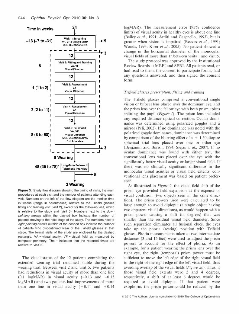
Figure 3. Study flow diagram showing the timing of visits, the main
procedures at each visit and the number of patients attending each
visit. Numbers on the left of the flow diagram are the median time
in weeks (range in parentheses) relative to the Trifield glasses
fitting and training visit (visit 2), except for the follow-up visit, which
is relative to the study end (visit 5). Numbers next to the down
pointing arrows within the dashed box indicate the number of
patients moving to the next stage of the study. The numbers next to
right pointing arrows outside of the dashed box indicate the number
of patients who discontinued wear of the Trifield glasses at that
stage. The formal visits of the study are enclosed by the dashed
rectangle. VA = visual acuity; VF = visual field as measured by
computer perimetry. The * indicates that the reported times are
relative to visit 5.
244 Ophthal. Physiol. Opt. 2010 30: No. 3
ª 2010 The Authors. Journal compilation ª 2010 The College of Optometrists

amount of the phoria. We measured phoria at twodistances that we considered to be about the limits ofthe range of distances of obstacles that we expectedwould be usefully detected with Trifield glasses. In thisexample, if the phorias were 3 and 1D, the prism powercould be reduced by the amount of the smaller phoria(here 1D � 0.6�). Finally, to minimize the risk ofdiplopia, we added 2� of prism power, which wasexpected to ensure that the visual field margins did notoverlap. The final right prism power in this examplewould have been 13D (7.4�). The intent was to have atleast 2� between adjacent visual fields, but measuredvisual fields (e.g. Figure 4b) often had inter-field gapsthat were larger than intended. The difference betweenintended and measured inter-field gap was probablycaused by between-session and between-condition var-iability in phorias and the reduction in the measuredvisual field of the prism eye due to the tint (reduced
transmittance). Prescribed powers for the Trifieldglasses used in this study ranged from 11–44D for thenasal prism and 16–34D for the temporal prism. Theright prism was tinted red with a transmission of about37% and the left was tinted green with a transmissionof about 46%. Each patient was provided with a pair ofclip-on lenses for use in bright conditions, with the non-prism-eye lens having a transmission of 15% and theprism-eye lens having a higher transmission, so thatwhen combined with the tints in the prisms, the totaltransmission was about 15%.
To familiarize a patient in the use of the Trifield glassesthe patient was escorted for a walk through SERIcorridors and into uncluttered rooms. The patient wasinstructed to pay particular attention to doorframes andother potential obstacles. The patient was then escortedboth up and down a flight of stairs, and instructed tomake use of handrails whenever using stairs.
Table 1. Demographic information, clinical vision measurements with and without the Trifield glasses, wearing time and success at the end of
the study (continued to wear after visit 5) for the twelve patients enrolled in the extended wearing trial
Pt. #
Age
(years)/
sex Aetiology
Long
cane
user
O&M
training VA
VF
width
VF width
with Prisms
(degrees)
Prism
Eye
Prisms
Prescribed
(N/T)(D)
Wearing
time per
day (hours)
Clinical
success
1 56/m RP Yes Yes 6/6 7.5 20 Left 15/17 0.61 Yes
2 49/m RP No No 6/9 9.5 28.5 Right 16/27 0.57 Yes
3 39/m CHM Yes Yes 6/9 16 29 Right 27/21 1.77 Yes
4 45/f RP Yes Yes 6/6 21.5 51 Right 44/34 1.18 Yes
5 41/m CHM No No 6/8 15 24 Left 32/19 1.27 Yes
6 45/f RP No No 6/13 8.5 34 Right 19/24 3.32 Yes
7 57/f RP No No 6/15 16.5 41 Right 25/34 0.57 No
8 58/m RP Yes Yes 6/14 7.5 18 Left 11/20 3.81 Yes
9 53/m RP Yes Yes 6/12 10 28 Left 13/28 0.57 Yes
10 43/f RP Yes No 6/30 17 55 Left 42/30 2.61 No
11 46/m RP Yes Yes 6/10 8 26 Left 16/16 1.38 Yes
12 59/f RP Yes Yes 6/11 5 23 Left 17/16 0.84 No
RP, retinitis pigmentosa; CHM, choroideremia; VA, binocular Snellen visual acuity; VF width, horizontal extent of visual field measured in
degrees; N/T, prescription of nasal and temporal prisms.
(a) (b)
Figure 4. Visual field expansion measured using a computerized perimeter for one patient. (a) Left and right eye monocular visual fields. (b) The
visual fields of the same patient when wearing Trifield glasses with the prisms over the right eye. Visual field expansion by the prisms is indicated
by the two hatched areas to the left and right of fixation. Only one of these is available for the patient at any instant depending on the direction of
gaze at that moment. The distance between the leftmost edge of the left expanded area and the rightmost edge of the right expanded area
(shown by the arrow) was defined as the expanded visual field.
Trifield device for tunnel vision: R. L. Woods et al. 245
ª 2010 The Authors. Journal compilation ª 2010 The College of Optometrists

In addition to this general instruction in the use of theTrifield glasses for obstacle detection a visual-directiontraining exercise was demonstrated at visit 2 and wasprescribed for home practice in an effort to improveadaptation to the image shift caused by the prisms. Theexercise was a manual reaching exercise based onKohler�s observation that adaptation to prism inducedfield shifts was hastened by tactile and proprioceptivecues (Kohler, 1964). While the patients looked throughone of the two prisms, an experimenter presented afinger into the prism field, and patients were instructedto quickly reach and touch the experimenter�s fingerwith their own. Initially, reaching was inaccurate due tothe prism-induced change in visual direction, but as theresponse was closed loop (i.e. there was visual feedbackon completion of each ballistic hand movement),reaching became accurate after a few trials, as iscommon in prism adaptation experiments (Welch et al.,1993). The reaching exercise was repeated with the otherprism (monocularly) and then binocularly until thepatient�s hand movements were swift and accurate. Thepatient was asked to repeat the reach and touchexercises at home for objects detected through theprisms as often as possible; initially through the prismeye only (i.e. monocularly) and then with both eyes openwhen the patient felt proficient with the prism-eye onlyexercise. At subsequent visits, the patient was ques-tioned about mobility with the Trifield glasses and thevisual-direction training tasks and further instructionand training was provided if necessary.
Clinical success
Clinical success was measured as the percentage ofpatients who chose to continue to wear the Trifieldglasses on conclusion of the wearing trial (visit 5). Atvisit 5 and long term follow-up (Figure 3), the investi-gators recorded responses to questions covering use,benefits, and difficulties attributed to the glasses. At thevisit 5 interview, a joint (patient and clinician) decisionwas made, based upon the patient�s perceived benefitfrom the Trifield glasses and the clinician�s opinion, as towhether the patient should continue to wear them. If thepatient continued wearing Trifield glasses, there was nocost to the patient, and no further clinical managementwas provided at SERI.
We asked patients to wear the Trifield glasses as muchas possible during the course of the wearing trial. TheTrifield glasses were designed as mobility devices, so theywere only intended for use when walking, not whenperforming other tasks, such as reading, working at adesk or watching TV. Following fitting of the Trifieldglasses (visit 2) patients were given a take-home wearingdiary to record the number of hours per day that theglasses were worn. The form also had an open-ended
comments section in which patients could record anydifficulties or benefits experienced with the Trifieldglasses. Patients were asked to return the diary to us atthe subsequent visit and were given a new diary at visit 3and visit 4 with the same instruction.During the interviewat each visit, the investigator asked the patient for anestimate of the hours worn per day since the last visit.
Visual field expansion
Visual field extent was assessedwith the 12 mm stimuli onthe computerized perimeter at visit 5. Extent of eachmonocular visual field was defined as the distancebetween the farthest lateral edges of the monocular visualfield (Figure 4a). The binocular visual field without theprism was defined as the width of the horizontal extentthat would be covered by either eye (Figure 4a).
The visual field with Trifield glasses (Figure 4b) wasmeasured in two steps. First, the patient fixated with thenon-prism eye (left eye in Figure 4b) and tilted his headslightly to the right. That head shift brought the leftprism in front of his right eye. The visual field of the lefteye was measured under this condition (using standardkinetic perimetry) followed by the measurement of thevisual field of the right eye that covered a field of view tothe left of the fixation target. The patient then turned hishead a little to the left to bring the right prism in front ofhis right eye. The visual field expansion to the right offixation was then measured. While the patient had atany instant only one of the two peripheral extensionareas, we defined the full extent of the visual field as thewidth covered with these three areas (double arrow inFigure 4b) as they represent potential visual field cover-age at different times and available through scanningeye and head movements. As shown in Figure 4b,sometimes this significantly overestimated the amountof visual field afforded by the Trifield glasses. This wasespecially true in the case illustrated where, apparently,the patient was more exophoric at the time of testingthan during the fitting process (visit 1).
The enrolment criterion of no more than 20� diametermonocular central visual field was selected because theprism powers of much above 20� (36D) would impactvisual acuity and are so thick that the prisms couldimpinge on the face and be uncomfortable or cause injury.
Perceived direction
Initially the perceived direction of an object seenthrough a prism is not veridical (i.e. not in its truedirection). People can adapt to the perceived direction ofobjects seen through a prism (Kohler, 1964; Pick et al.,1969; Welch et al., 1993). Kohler (1964) reported a �dual�adaptation after full time wearing a binocular half-prismfor 10 days. Initially, he reported adaptation to the
246 Ophthal. Physiol. Opt. 2010 30: No. 3
ª 2010 The Authors. Journal compilation ª 2010 The College of Optometrists

directional change and visual distortions affected by theprisms. However, when looking through the clearportion of the spectacles, after-effects of adaptation tothe prism were perceived (i.e., he perceived distortionsand reversed directional change). After additionalwearing time, these after-effects dissipated as well, anda �dual� adaptation was noted so that perception wascorrect both through the prism and through the prism-free segment of the lenses. We hypothesized that patientswearing the Trifield glasses may adapt to the directionalchange through the monocular prisms in a similarmanner.To measure adaptation to perceived direction we used
a pointing task at visits 2, 3, 4 and 5. Patients wereseated 1 m from a 64 by 81� rear-projection screen.Large head movements were restricted by a chin rest anda tight fitting headband from a binocular indirectophthalmoscope that was connected to the head rest.This experimental set-up is illustrated in Fig. 5 of Giorgiet al. (2009). Patients indicated the perceived lateraldirection of stimuli on the screen using a large graphicbitpad tablet (90 by 120 cm) and a computer mouse heldin the hand. Patients wore a brace to limit flexion of theelbow and were asked not to flex the elbow or the wrist,so that pointing was from the shoulder. Pointing wasopen-loop with no visual feedback, as a wooden boxover the bitpad prevented a view of the arm, hand ormouse. Using small counter-rotations of the head, asused in visual field testing, the left and right extendedareas were tested separately in consecutive sessions. Ineach session, stimuli were presented one at a time to thevisual field of the non-prism eye or the prism eye. Thefixation cross was always viewed with the non-prism eye.A calibration step conducted without the prism
glasses adjusted for individual differences in pointingresponses. During calibration the patient turned his eyes(but not head) towards each approximately eye-leveltarget. During perceived direction testing the non-prismeye fixated a central target.The difference in the perceived visual direction for
targets presented in the prism field of view, and thosepresented in the visual field of the non-prism eye, wasused as the outcome measure. Adaptation of perceivedvisual direction would appear as no difference in thesetwo directions. Lack of adaptation would appear as apersistent difference in reported direction, with theexpected separation being the prism power. Figures 7and 8 of Giorgi et al. (2009) show, for peripheral prismsfor hemianopia, the effect of adaptation and theoutcomes of similar measurements.
Perceived visual ability
Patients were administered two questionnaires duringthe first and last visits of the wearing trial. Both
questionnaires, an early (1995) version of the NationalEye Institute Visual Functioning Questionnaire-25(NEI-VFQ: Mangione et al., 2001) and the IndependentMobility Questionnaire (IMQ: Turano et al., 1999a),contained items relating to mobility. The patientsreported their perceived levels of difficulty for all itemsin each questionnaire. We performed planned analyseson responses to 27 mobility and obstacle avoidanceitems that we hypothesized would be affected by the useof Trifield glasses. The chosen items from the 1995version of the NEI-VFQ were 19, 23, 31, 33, 34, 41, 43,45 and 47 and from the IMQ were 2, 4, 6, 7, 8, 10, and24–35. During interviews and in diaries, about half thepatients reported increased difficulty with low-lightsituations when using the Trifield glasses. Though all12 patients already had difficulties with low-light situ-ations due to their condition and the prism lens reducedlight transmission due to the tint, as the other eye had anon-tinted conventional spectacle lens, we had notexpected increased problems with low-light situations.Post-hoc analyses were conducted on six lighting-relateditems from the two questionnaires: 22 from the NEI-VFQ and 19–23 from the IMQ (the text of all analyseditems is reported in the Appendix).
Non-parametric tests were used in the data analysis.The Wilcoxon test was used for paired comparisonsand the Mann–Whitney test for unpaired comparisons.We report test results as statistically significant whenp £ 0.05. However, because of our small sample size,we have reported test results as �approaching� signif-icance when 0.05 < p £ 0.10. As there were multiplecomparisons (33 NEI-VFQ and IMQ items), if anexperiment-wise error rate correction was applied the p(a-level) for significance would be p < 0.006, when theBonferroni adjustment is corrected for the correlationbetween items (average r = 0.41) (Sankoh et al.,1997).
Results
Clinical success
As shown in Figure 3, of the fifteen patients who met theeligibility criteria at visit 1, three patients declined toparticipate, and twelve were dispensed a pair of Trifieldglasses at visit 2. Unless otherwise noted, analyses ofdata on outcome measures were restricted to the twelvepatients who completed the wearing trial.
Of the 12 patients who completed the wearing trial,nine were wearing the Trifield glasses when interviewedat visit 5, and all nine (75% of those completing thewearing trial) chose to continue to wear following thewearing trial (visit 5). Male patients were more likely tobe successful (Fisher exact test, p = 0.045). Success wasnot related to the eye fitted with the prisms, long cane
Trifield device for tunnel vision: R. L. Woods et al. 247
ª 2010 The Authors. Journal compilation ª 2010 The College of Optometrists

use or having received orientation and mobility training(Fisher exact test, p > 0.52). There was no significantdifferences between successful and non-successful pa-tients for age, visual acuity, stereoacuity (Titmusstereotest), visual field width or the number of hoursthat the Trifield glasses were worn (Mann–Whitney test,z11 < 1.1, p > 0.37), while non-successful patients hada tendency to have wider visual fields with the Trifieldglasses (Mann–Whitney test, z11 = 1.94, p = 0.064).Patients not wishing to continue to wear the Trifieldglasses returned their glasses at the final visit. At thelong-term telephone follow-up (median 48, range 35–78weeks), three of the nine patients reported still wearingTrifield glasses.
At the visit 5 interview, six of nine (67%) successfulwearers reported benefits of the Trifield glasses asuseful in avoiding collision with objects and passersby,as did one of the three non-successful wearers (7 of all12 patients: 58%). At the same time, five of the nine(56%) successful wearers reported some difficultiesassociated with the Trifield glasses in �street crossing�,six of nine with �crowds�, with one patient describingcrowded environments as �visual noise�. All three non-successful patients cited �crowds� as a difficulty, andthese three patients reported insufficient observedbenefit using the Trifield glasses as the reason todiscontinue use.
During interviews and in diaries, patients madecomments about perceived benefits and difficulties withTrifield glasses. All 12 patients reported improveddetection of objects to the side, including corridors,footpaths (sidewalks) and street crossing. Five patientsreported benefits in supermarkets, with navigation,obstacle avoidance and with locating items on shelves(visual search) and one patient even reported animproved ability to follow games of ice hockey (fastaction). Difficulties that were reported included doublevision (n = 6), steps and stairs (n = 7), vertigo (n = 3)and low-light situations (n = 6).
Visual field expansion
Visual field expansion was recorded for all patientscompleting the wearing trial (Table 1), as illustrated inFig. 4. The median absolute difference between theexpanded visual field and the normal visual field was 18�(range 9–38) for the 12 patients completing the wearingtrial. The median ratio of visual field with-to-withoutthe Trifield glasses was 2.7 (range 1.6–4.6).
Perceived direction
No patient in the study demonstrated adaptation to theprismatic image displacement. Possible reasons for thelack of adaptation are addressed in the discussion.
Perceived visual ability
We hypothesized that Trifield glasses would affect theperceived visual ability of the patients, either benefitingor hindering them in their daily mobility situations.Analyses were performed for all 12 patients. One ofthose patients did not complete the visit five question-naires for health reasons. Planned analyses were per-formed on the 27 items considered pertinent to mobility(Appendix). Comparing wearing trial start (visit 1) andend (visit 5), wearing Trifield glasses may have increasedthe perceived difficulty of visiting with people in theirhomes, at parties, or in restaurants (Wilcoxon signedranks z10 = 2.24, p = 0.025) and the successful patientsmay have been more likely to report that they stay athome most of the time because of my eyesight (Wilcoxonsigned ranks z8 = 1.63, p = 0.10). Comparing success-ful and non-successful patients, for NEI-VFQ items, atthe start of the wearing trial successful patients mayhave been less likely to report that they stay at homemost of the time because of my eyesight (Mann–WhitneyZ11 = 1.90, p = 0.10) than non-successful patients,while there were no significant differences at wearingtrial end. For the IMQ items, at wearing trial start,successful wearers may have reported greater difficultyin moving about at work (Mann–Whitney Z8 = 2.16,p = 0.06), avoiding bumping into walls (Mann–WhitneyZ11 = 1.79, p = 0.10) and avoiding shoulder heightobjects (Mann–Whitney Z11 = 2.06, p = 0.06) thannon-successful patients. At wearing trial end, non-successful patients may have reported more difficultywalking in unfamiliar areas (Mann–Whitney Z10 = 2.08,p = 0.07), being aware of another person�s presence(Mann–Whitney Z10 = 2.13, p = 0.04) and avoidingbumping into knee-high objects (Mann–WhitneyZ10 = 1.97, p = 0.07) than successful patients.
Post-hoc analyses were performed on the six lighting-related items (Appendix). Comparing wearing trial start(visit 1) and end (visit 5), there were no significantchanges in responses for all 12 patients. However, thenine successful patients may have reported less difficultyadjusting to lighting changes during the day outdoor toindoor (Wilcoxon signed ranks z7 = 1.90, p = 0.06)and going down steps, stairs, or curbs in dim light or atnight (Wilcoxon signed ranks z8 = 2.45, p = 0.01) atthe end of the wearing trial, after use of Trifield glasses,compared to visit 1. Thus, despite patients makingcomments about low-light situations with the Trifieldglasses, the perceived level of difficulty in some low-lightsituations may have improved.
Discussion
Initial success with the Trifield glasses appeared to bevery high as 75% (9 of 12 patients who completed the
248 Ophthal. Physiol. Opt. 2010 30: No. 3
ª 2010 The Authors. Journal compilation ª 2010 The College of Optometrists

wearing trial) felt that they benefited from the Trifieldglasses and wanted to continue to wear the Trifieldglasses following about 2 months of experience. How-ever, after about a year, only 25% (3 of the 12 whocompleted the 6-week wearing trial) continued to wearthe Trifield glasses. Even that reduced success rate(25%) may be considered high in view of the lack ofeffective visual aids for patients with tunnel vision. Yet,it is relatively low when compared to about 47%(Bowers et al., 2008) and 42% (Giorgi et al., 2009)found for long term retention of the prismatic correctionfor hemianopia. The decline in use with time suggeststhat the perceived benefits were either eliminated or wereovercome by the perceived limitations and difficulties ofthe Trifield glasses. We believe that it is the latter. Whilepatients with tunnel vision have greater impairment ofmobility than those with hemianopia and thus would beexpected to benefit more from a visual aid, the Trifieldglasses are more difficult to use and may present moredifficulties than the peripheral prism correction forhemianopia. The peripheral prisms do not interfere withcentral vision and thus do not affect visual acuity.Trifield glasses affect central vision and, at least in oneeye, the relatively high power prisms may reduce visualacuity and cause spatial distortions. While the periph-eral prism glasses use higher-power, lower-image-qualityFresnel prisms, their placement out of central visionmakes these limitations less apparent and easier to use.However, it appears that the greatest difficulty inadapting to the Trifield glasses was the central doublevision experienced as visual confusion. Double visionwas reported by six patients in patient diaries or duringinterviews. The visual confusion occurs because objects(potential obstacles) that would not be detected withoutthe prism lens can be detected, but they appear to be in adifferent (prism shifted) direction, superimposed onanother object in the environment seen by the non-prismeye. This double vision is known to be uncomfortable,annoying and disturbing in central vision. Peripheraldouble vision as occurs with physiological diplopia (Peli,2000) is common and thus easier to accept.Based on the findings of Kohler (1964) we hypothe-
sized that at least some patients would obtain veridicalperceived direction of objects seen through the prismsegments. This did not happen. The reason for the lackof adaptation of the type described by Kohler mayinclude the short and intermittent wearing times (0.6–3.8 h each day, even for successful patients) as com-pared to full time wear by Kohler�s subjects, and thatour Trifield prisms were fitted monocularly and notbinocularly as did Kohler, and the higher power of theprisms we fitted, at least for some of the patients.Patients with tunnel vision at the level enrolled in our
study have had many years of slow progression of theirvisual field loss. It appears that many of them have
slowly adapted to the situation by gradually changingtheir life style to reduce the challenges of pedestrianmobility as much as possible. As a result, these patientshave reduced opportunities to walk and to gain expe-rience with the Trifield glasses. Most of these patientsalso suffer from night blindness and that reduces theirmobility further. This is particularly true in a place likeBoston when the daylight hours after work are limitedfor most of the year (Bowers et al., 2003). Thus, ourpatients had fewer opportunities to walk with theTrifield glasses and to learn to benefit from them. Ourpatients wore the glasses for a median of 1.2 h per day(range 0.6–3.8), whereas, patients with hemianopia incomparable studies wore their peripheral prism glassesfor a median of 3.0 h per day (range 1.0–13.4: Giorgiet al., 2009) to 8.0 h per day (range 1.0–16: Bowerset al., 2008).
Using the Trifield glasses when reading may bepossible, but is certainly uncomfortable due to thevisual confusion. Thus, the user has to remove theglasses or shut his prism eye even for spot reading (e.g.reading labels or price tags in stores) or reading distancetext such as street signs. This additional limitation mayhave contributed to the eventual rejection of the Trifieldglasses by some patients. In comparison, the peripheralprisms for hemianopia may be used with small bifocalsegments that permit spot reading with little difficulty.
Long-term discontinuation may have been related toa lack of continued clinical care by the prescriber (us)following the end of the 5-visit wearing trial. At visit 5,patients were advised to continue receiving normalclinical care. However, that clinical care would not haveprovided advice on this experimental device. It ispossible that follow up care from the prescriber, withfurther instruction on the use of Trifield glasses, mighthave resulted in a higher long-term retention rate.
The Trifield lenses expand the visual field onlylaterally, which may be important in avoiding collisionswith other pedestrians and some obstacles. However, thelower visual field is known to be of more importance formobility (Lovie-Kitchin et al., 1990) and was notexpanded at all by the Trifield glasses. An earlier designincluded a lower base-down prism segment for thatpurpose. This was eliminated when it became apparentthat the prism powers practical in our design provided alower visual field expansion that was too small to bemeaningful. This aspect needs to be reconsidered infuture designs of devices for tunnel vision. The use oftinted prism lenses was based on feedback during pilottesting, in which the patients with tunnel vision reporteddifficulty determining the direction of a detected object,particularly which side (i.e. from which prism). As allthe patients in our study had difficulty in low lightconditions (night vision, dark adaptation), there was therisk that the tints would have a negative impact on
Trifield device for tunnel vision: R. L. Woods et al. 249
ª 2010 The Authors. Journal compilation ª 2010 The College of Optometrists

object detection. Two patients reported that the red lenswas better and two patients reported that the green lenswas better. It is not clear whether these differences wererelated to tint or to other issues. Six patients reportedthat the tint seemed to cause a (greater) problem withdark adaptation or they were difficult to use in low lightconditions, however, that is not consistent with theperceived visual ability questionnaire responses, forwhich there was some modest improvement. We didnot assess whether the tints provided a real benefit, andpatients did not report being able to easily make use ofthe different colour appearances from the two prisms,perhaps due to colour adaptation. In the future, the tintsmight be used only during the training period and beremoved once the patient became familiar with themobility device.
We have developed (Vargas-Martin and Peli, 2002)and tested (Vargas-Martin and Peli, 2001; Luo and Peli,2006; Peli et al., 2007; Luo et al., 2009) an augmented-vision system using a head-mounted display which canbe used by patients with one or two eyes. The electronicsystem is much more complicated to maintain andprobably much more expensive and thus, although itmay be an attractive option, we still consider the Trifieldglasses as a potentially useful design perhaps with somemodifications of the design, the fitting procedures andtraining in its use.
Acknowledgements
Supported in part by a grant from the Joint SERI-MEEI Clinical Research Center, by NIH grant#EY12890 (EP), and by the Foundation FightingBlindness (ELB). Dan Stringer and Jenn Segawa helpedwith data collection. Bob Goldstein developed thesoftware for measurement of the perceived direction.Karen Keeney (Chadwick Optical, White River Junc-tion, VT) was instrumental in the development oftechniques to make the Trifield glasses.
References
Apfelbaum, H., Pelah, A. and Peli, E. (2007) Headingassessment by ‘‘tunnel vision’’ patients and control subjects
standing or walking in a virtual reality environment. ACMTrans. Appl. Percept. 4, article 8, pp. 1–16.
Arditi, A. and Cagenello, R. (1993) On the statistical reliability
of letter-chart visual acuity measurements. Invest. Ophthal-mol. Vis. Sci. 34, 120–129.
Bailey, I. L., Bullimore, M. A., Raasch, T. W. and Taylor, H.
R. (1991) Clinical grading and the effects of scaling. Invest.Ophthalmol. Vis. Sci. 32, 422–432.
Benjamin, W. J. and Borish, I. M. (1994) Presbyopia and theinfluence of aging on prescription of contact lenses. In:
Contact Lens Practice (eds M. Rubin and M. Guillon),Chapman & Hall Medical, London, pp. 800–802.
Berson, E. L. (1993) Retinitis pigmentosa: the Friedenwald
lecture. Invest. Ophthalmol. Vis. Sci. 34, 1659–1676.Biderman, A., Cwikel, J., Fried, A. V. and Galinsky, D. (2002)
Depression and falls among community dwelling elderly
people: a search for common risk factors. J. Epidemiol.Community Health 56, 631–636.
Black, A., Lovie-Kitchin, J. E., Woods, R. L., Arnold, N.,
Byrnes, J. and Murrish, J. (1997) Mobility performance inretinitis pigmentosa. Clin. Exp. Optom. 80, 1–12.
Bowers, A. R., Luo,G. and Peli, E. (2003) Functionally relevantillumination levels for evaluation of a new night vision device
(abstract). Invest. Ophthalmol. Vis. Sci. 44, 2772.Bowers, A. R., Keeney, K. and Peli, E. (2008) Community-
based trial of peripheral prism visual expansion device for
hemianopia. Arch. Ophthalmol. 126, 657–664.Broman, A. T., West, S. K., Munoz, B., Bandeen-Roche, K.,
Rubin, G. S. and Turano, K. A. (2004) Divided visual
attention as a predictor of bumping while walking: theSalisbury eye evaluation. Invest. Ophthalmol. Vis. Sci. 45,2955–2960.
Cacciatore, F., Abete, P., Maggi, S., Luchetti, G., Calabrese,
C., Viati, L., Leosco, D., Ferrara, N., Vitale, D. F. andRengo, F. (2004) Disability and 6-year mortality in elderlypopulation. Role of visual impairment. Aging Clin. Exp.
Res. 16, 382–388.Cohen, J. M. (1993) An overview of enhancement techniques
for peripheral field loss. J. Am. Optom. Assoc. 64, 60–70.
Cohen, J. M. and Waiss, B. (1996) Visual field remediation. In:Remediation and Management of Low Vision (ed. B. P.Rosenthal), Mosby, St. Louis, pp. 1–25.
Drasdo, N. (1976) Visual field expanders. Am. J. Optom.Physiol. Opt. 53, 464–467.
Felson, D. T., Anderson, J. J., Hannan, M. T., Milton, R. C.,Wilson, P. W. F. and Kiel, D. P. (1989) Impaired vision and
hip fracture. The Framingham study. J. Am. Geriatr. Soc.37, 495–500.
Fortenbaugh, F. C., Chaudhury, S., Hicks, J. C., Hao, L. and
Turano, K. A. (2007) Gender differences in cue preferenceduring path integration in virtual environments. ACMTrans. Appl. Percept. 4, article 6, pp. 1–18.
Freeman, E. E., Munoz, B., Rubin, G. S. and West, S. K.(2007) Visual field loss increases the risk of falls in olderadults: the Salisbury Eye Evaluation. Invest. Ophthalmol.Vis. Sci. 48, 4445–4450.
Giorgi, R. G., Woods, R. L. and Peli, E. (2009) Clinical andexperimental evaluation of peripheral prism glasses forhemianopia. Optom. Vis. Sci. 86, 492–502.
Hartong, D. T., Berson, E. L. and Dryja, T. P. (2006) Retinitispigmentosa. Lancet 368, 1795–1809.
Haymes, S., Guest, D., Heyes, A. and Johnston, A. (1996)
Mobility of people with retinitis pigmentosa as a function ofvision and psychological variables. Optom. Vis. Sci. 73, 621–637.
Hoeft, W. W., Feinbloom, W., Brilliant, R., Gordon, R.,Hollander, C., Newman, J. and Novak, E. (1985) Amorphiclenses: a mobility aid for patients with retinitis pigmentosa.Am. J. Optom. Physiol. Opt. 62, 142–148.
Kiser, A. K., Mladenovich, D., Eshraghi, F., Bourdeau, D.and Dagnelie, G. (2005) Reliability and consistency of visual
250 Ophthal. Physiol. Opt. 2010 30: No. 3
ª 2010 The Authors. Journal compilation ª 2010 The College of Optometrists

acuity and contrast sensitivity measures in advanced eye
disease. Optom. Vis. Sci. 82, 946–954.Kohler, I. (1964) The formation and transformation of thevisual world. Psychol. Issues 3, 14–173.
Kozlowski, J. M., Mainster, M. A. and Avila, M. P. (1984)Negative-lens field expander for patients with concentricfield constriction. Arch. Ophthalmol. 102, 1182–1184.
Kuyk, T., Elliott, J. and Fuhr, P. S. (1998) Visual correlates ofobstacle avoidance in adults with low vision. Optom. Vis.Sci. 75, 174–182.
Kuyk, T., Elliott, J. L., Wesley, J., Scilley, K., McIntosh, E.,
Mitchell, S. and Owsley, C. (2004) Mobility function inolder veterans improves after blind rehabilitation. J. Reha-bil. Res. Dev. 41, 337–346.
Lord, S. R. and Dayhew, J. (2001) Visual risk factors for fallsin older people. J. Am. Geriatr. Soc. 49, 508–515.
Lord, S. R., Ward, J. A., Williams, P. and Anstey, K. J. (1993)
An epidemiological study of falls in older community-dwelling women: the Randwick falls and fractures study.Aust. J. Public Health 17, 240–245.
Lovie-Kitchin, J., Mainstone, J., Robinson, J. and Brown, B.
(1990) What areas of the visual field are important formobility in low vision patients? Clin. Vis. Sci. 5, 249–263.
Luo, G. and Peli, E. (2006) Use of an augmented-vision device
for visual search by patients with tunnel vision. Invest.Ophthalmol. Vis. Sci. 47, 4152–4159.
Luo, G., Vargas Martin, F. and Peli, E. (2008) The role of
peripheral vision in saccade planning: learning from peoplewith tunnel vision. J. Vis. 8, article 25, 1–8.
Luo, G., Woods, R. L. and Peli, E. (2009) Collision judgement
when using an augmented-vision head-mounted displaydevice. Invest. Ophthalmol. Vis. Sci. 50, 4509–4515.
Mangione, C. M., Lee, P. P., Gutierrez, P. R., Spritzer, K.,Berry, S. and Hays, R. D. (2001) Development of the 25-
Item National Eye Institute Visual Function Questiona-naire. Arch. Ophthalmol. 119, 1050–1058.
Marron, J. A. and Bailey, I. L. (1982) Visual factors and
orientation-mobility performance. Am. J. Optom. Physiol.Opt. 59, 413–426.
Melzer, D., Lan, T. Y. and Guralnik, J. M. (2003) The
predictive validity for mortality of the index of mobility-related limitation - results from the EPESE study. AgeAgeing 32, 619–625.
Onufryk, M.. (1994) Ophthalmic prismatic image relocating
eye glasses for persons having retinitis pigmentosa andhemianopia and method for making same. USA Patent#5,323,190.
Peli, E. (2000) Field expansion for homonymous hemianopiaby optically-induced peripheral exotropia. Optom. Vis. Sci.77, 453–464.
Peli, E. (2001) Vision multiplexing: an engineering approach tovision rehabilitation device development. Optom. Vis. Sci.78, 304–315.
Peli, E. (2002) Treating with spectacle lenses: a novel idea!?Optom. Vis. Sci. 79, 569–580.
Peli, E., Luo, G., Bowers, A. and Rensing, N. (2007)Applications of augmented vision head-mounted systems
in vision rehabilitation. J. Soc. Inf. Disp. 15, 1037–1045.
Pick, H. L., Hay, J. C. and Martin, R. (1969) Adaptation to
split-field wedge prism spectacles. J. Exp. Psychol. 80, 125–132.
Reeves, B. C., Wood, J. M. and Hill, A. R. (1991) Vistech
VCTS 6500 charts - within- and between-session reliability.Optom. Vis. Sci. 68, 728–737.
Ross, N. C., Bowers, A. R. and Peli, E. (2009) Consideration
of optical scotomas in designing visual field expansiondevices (abstract). Invest. Ophthalmol. Vis. Sci. 50, E–4734.
Sankoh, A. J., Huque, M. F. and Dubey, S. D. (1997) Somecomments on frequently used multiple endpoint adjustment
methods in clinical trials. Stat. Med. 16, 2529–2542.Shinkai, S., Watanabe, S., Kumagai, S., Fujiwara, Y., Amano,H., Yoshida, H., Ishizaki, T., Yukawa, H., Suzuki, T. and
Shibata, H. (2000) Walking speed as a good predictor for theonset of functional dependence in a Japanese rural commu-nity population. Age Ageing 29, 441–446.
Siejas, O., Gomez De Liano, P., Gomez De Liano, R.,Roberts, C. J., Piedrahita, E. and Diaz, E. (2007) Oculardominance diagnosis and its influence in monovision. Am. J.Ophthalmol. 144, 209–216.
Somani, S., Brent, M. H. and Markowitz, S. N. (2006) Visualfield expansion in patients with retinitis pigmentosa. Can. J.Ophthalmol. 41, 27–33.
Soong, G. P. (2000) The Effect of Orientation and MobilityTraining on Vision and Mobility Performance in VisuallyImpaired Adults. Unpublished Ph.D. Thesis, Queensland
University of Technology, Brisbane.Soong, G. P., Lovie-Kitchin, J. E. and Brown, B. (2001) Doesmobility performance of visually impaired adults improve
immediately after orientation and mobility training? Optom.Vis. Sci. 78, 657–666.
Szlyk, J. P., Seiple, W., Laderman, D. J., Kelsch, R., Ho, K.and McMahon, T. (1998) Use of bioptic amorphic lenses to
expand the visual field in patients with peripheral loss.Optom. Vis. Sci. 75, 518–524.
Tinetti, M. E. and Williams, C. S. (1997) Falls, injuries due to
falls, and the risk of admission to a nursing home. N. Engl.J. Med. 337, 1279–1284.
Turano, K. A., Geruschat, D. R., Stahl, J. W. and Massof, R.
W. (1999a) Perceived visual ability for independent mobilityin persons with retinitis pigmentosa. Invest. Ophthalmol. Vis.Sci. 40, 865–877.
Turano, K. A., Rubin, G. S. and Quigley, H. A. (1999b)
Mobility performance in glaucoma. Invest. Ophthalmol. Vis.Sci. 40, 2803–2809.
Turano, K. A., Geruschat, D. R., Baker, F. H., Stahl, J. W.
and Shapiro, M. D. (2001) Direction of gaze while walking asimple route: persons with normal vision and persons withretinitis pigmentosa. Optom. Vis. Sci. 78, 667–675.
Vargas-Martin, F. and Peli, E. (2001) Augmented view fortunnel vision: device testing by patients in real environ-ments. In: Digest of Technical Papers SID 01 XXXII (ed
J. Morreale), Society for Information Display, San Jose,CA, pp. 602–605.
Vargas-Martin, F. and Peli, E. (2002) Augmented-view forrestricted visual field: multiple device implementations.
Optom. Vis. Sci. 79, 715–723.
Trifield device for tunnel vision: R. L. Woods et al. 251
ª 2010 The Authors. Journal compilation ª 2010 The College of Optometrists

Vargas-Martin, F. and Peli, E. (2006) Eye movements of
patients with tunnel vision while walking. Invest. Ophthal-mol. Vis. Sci. 47, 5295–5302.
Virgili, G. and Rubin, G. (2006) Orientation and mobility
training for adults with low vision. Cochrane Database Syst.Rev. 3, CD003925.
Weiss, N. J. (1992) Remediation of peripheral visual field
defects in low vision patients. Probl. Optom. 4, 34–54.Welch, R. B., Bridgeman, B., Anand, S. and Browman, K.(1993) Alternating prism exposure causes dual adaptationand generalization to a novel displacement. Percept. Psy-
chophys. 54, 195–204.Woods, R. L. (1993) Reliability of visual performance mea-surement under optical degradation. Ophthalmic Physiol.
Opt. 13, 143–150.
Appendix
The text of items in the two perceived visual abilityquestionnaires that we hypothesised would be affectedby the Trifield glasses. Patients rated all of the followingitems in terms of degree of perceived difficulty on ascale of 1–5. For the 1995 version of the NEI-VFQ(Mangione et al., 2001) the scale for items 19–34 wasdescribed as going from �No difficulty at all� (1) to�Stopped doing this because of your eyesight� (5) and foritems 41–47 was described as going from �Definitelytrue� (1) to �Definitely false� (5). For the IMQ (Turanoet al., 1999a) the scale was described as going from�represents no difficulty� (1) to �represents extremedifficulty� (5).
National Eye Institute Visual Functioning Questionnaire
(NEI-VFQ-25) (1995 version) (Mangione et al., 2001)
19 Because of your eyesight, how much difficulty do youhave finding something on a crowded shelf?
22 Because of your eyesight, how much difficulty do youhave going down steps, stairs, or curbs in dim light orat night?
23 Because of your eyesight, how much difficulty do youhave noticing objects off to the side?
31 Because of your eyesight, how much difficulty do youhave visiting with people in their homes, at parties, orin restaurants?
33 Because of your eyesight, how much difficulty do youhave taking part in active sports or other outdooractivities that you enjoy (like golf, bowling, jogging,or walking)?
34 Because of your eyesight, how much difficulty do youhave going out to see movies, plays or sports events?
41 I stay at home most of the time because of myeyesight.
43 I have much less control over what I do, because ofmy eyesight.
45 I don�t go out of my home alone, because of myeyesight.
47 I need a lot of help from others because of myeyesight.
Independent Mobility Questionnaire (IMQ) (Turanoet al., 1999a).2 Walking in unfamiliar areas?4 Moving about at work?6 Moving about in stores?7 Moving about outdoors?8 Moving about in crowded situations?
10 Moving about using public transportation?19 Adjusting to lighting changes during the day - indoor
to outdoor?20 Adjusting to lighting changes during the day -
outdoor to indoor?21 Adjusting to lighting changes during at night - indoor
to outdoor?22 Adjusting to lighting changes during at night -
outdoor to indoor?23 Walking in dimly lit indoor areas?24 Being aware of another person�s presence?25 Avoiding bumping into people?26 Avoiding bumping into walls?27 Avoiding bumping into head height objects?28 Avoiding bumping into shoulder height objects?29 Avoiding bumping into waist height objects?30 Avoiding bumping into knee height objects31 Avoiding bumping into low lying objects?32 Avoiding tripping over uneven travel surfaces?33 Moving around in social gatherings?34 Finding restrooms in public places?35 Seeing cars at intersections?
252 Ophthal. Physiol. Opt. 2010 30: No. 3
ª 2010 The Authors. Journal compilation ª 2010 The College of Optometrists