expression of noncoding vault rna in human malignant cells and
TRANSCRIPT
1536
Published OnlineFirst September 29, 2010; DOI: 10.1158/1541-7786.MCR-10-0242
DNA Damage and Cellular Stress Responses Molecular
CancerResearch
Expression of Noncoding Vault RNA in Human Malignant Cellsand Its Importance in Mitoxantrone Resistance
Subash C.B. Gopinath, Renu Wadhwa, and Penmetcha K.R. Kumar
Abstract
Authors' AInstitute, NaIbaraki, Jap
CorresponGroup, BioIndustrial SCity 305-861-6095. E
doi: 10.115
©2010 Am
Mol Canc
Dow
Several noncoding RNAs do vital cellular functions, including gene regulation and cell differentiation. Pre-viously, we reported that vault RNA (vRNA) has the ability to recognize chemotherapeutic compounds, suchas mitoxantrone, based on biophysical and biochemical analyses. In the present study, we show that humanglioblastoma-, leukemia-, and osteocarcinoma-derived cell lines overexpress vRNA and exhibit higher resis-tance toward mitoxantrone. Interestingly, when vRNA expression was suppressed by RNA interference inthese cells, the resistance progressively decreased. In agreement with these findings, overexpression ofvRNA-1 caused resistance to mitoxantrone. These results suggest a role of vRNA in mitoxantrone resistancein malignant cells and justify further studies on the importance and application of noncoding RNAs in cancerchemotherapeutics. Mol Cancer Res; 8(11); 1536–46. ©2010 AACR.
Introduction
Vaults are barrel-shaped particles with a mass of ∼13MDaand overall dimensions of 400 × 400 × 700 Å, as determinedby scanning transmission electron microscopy (1). Theseparticles represent the largest ribonucleoprotein complexidentified thus far in eukaryotic cells. They seem to shuttlebetween the cytoplasm and the nucleus (2), and hence havebeen implicated in intracellular (3) and nucleocytoplasmictransport (2, 3). They are widely distributed in eukaryoticspecies, including mammals, avians, and amphibians. Theirconsiderable abundance and striking evolutionary conserva-tion argue for their important role in cell survival (4). Severallines of evidence have suggested that vaults may play animportant role in intracellular detoxification processes,and thus may function in the multidrug resistance (MDR)of cancer cells (5).In mammals, the vault particle is composed of three pro-
teins and RNA. Among these components, the 100-kDamajor vault protein (MVP) accounts for >75% of theparticle mass (1). The other two proteins are the 290-kDatelomerase-associated protein 1 (TEP1) and the 193-kDavault poly(ADP-ribose) polymerase (VPARP). The vault isalso known to contain a noncoding RNA [vault RNA(vRNA); refs. 6-11]. Compared with the other components,
ffiliation: RNA Processing Group, Biomedical Researchtional Institute of Advanced Industrial Science and Technology,an
ding Author: Penmetcha K.R. Kumar, RNA Processingmedical Research Institute, National Institute of Advancedcience and Technology, Central 6, 1-1-1 Higashi, Tsukuba566, Ibaraki, Japan. Phone: 81-298-61-6085; Fax: 81-298--mail: [email protected]
8/1541-7786.MCR-10-0242
erican Association for Cancer Research.
er Res; 8(11) November 2010
on April 1, 2018. ©mcr.aacrjournals.org nloaded from
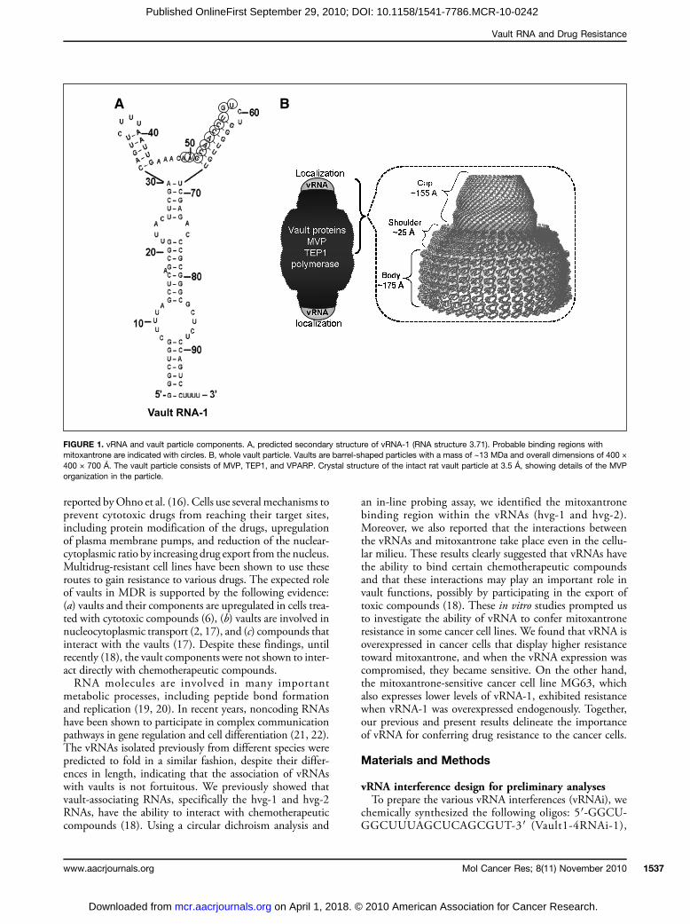
the RNA represents <5% of themass of the vault complex. Inhumans, there are three vRNA genes—the hvg-1, hvg-2, andhvg-3 RNAs—located on chromosome 5, and at least one ofthem is known to associate with the vault complex. Previous-ly, relatively large amounts of hvg-1 in vault particles werereported, whereas small amounts of hvg-2 and hvg-3 werefound (8, 11). Human hvg-1 RNA is 98 bases in length(Fig. 1A), and the other two RNAs are 88 bases long. In ad-dition, hvg-4 is encoded on the X chromosome, but it doesnot seem to be expressed (8). These vRNAs share ∼84%sequence identity and have similar secondary structurearrangements. In the vault particle, the TEP1 protein wasshown to be important for vRNA binding and stabilizationof the vault complex (9, 12). TheTEP1 found to locate at theterminal ends of the vault particle, based on the crystal struc-ture of the whole vault particle, isolated from the rat, at 3.5 Åresolution (13). The vault particle consists of a dimer ofhalf-vaults, comprising 39 identical MVP monomers. EachMVP monomer folds into 12 domains: 9 structural repeatdomains, a shoulder domain, a cap helix domain, and acap-ring domain (Fig. 1B). It was proposed that the inter-actions between the 42-turn-long cap helix domains ofMVP are essential for stabilizing the vault particle (13).MDR, a major cause of failure of cancer treatment, is a
phenomenon whereby cancer cells develop broad resistanceto a wide variety of chemotherapeutic drugs (14). A previousstudy on several non–P-glycoprotein MDR cell lines withincreased MVP levels showed that vRNA and MVP werenot shown to be coordinately regulated, and suggested thatthe entire vault particle is upregulated in MDR and that athreshold level of vaults is required to impart MDR (6). In aclinical scenario, the elevated expression of MVP wasobserved in cell lines resistant to various classes of chemo-therapeutic compounds, including doxorubicin and mitox-antrone (15). Support for the role of vaults in the extrusionof anthracyclines from the nuclei of resistant cells was
2010 American Association for Cancer Research.
Vault RNA and Drug Resistance
Published OnlineFirst September 29, 2010; DOI: 10.1158/1541-7786.MCR-10-0242
reported byOhno et al. (16). Cells use several mechanisms toprevent cytotoxic drugs from reaching their target sites,including protein modification of the drugs, upregulationof plasma membrane pumps, and reduction of the nuclear-cytoplasmic ratio by increasing drug export from the nucleus.Multidrug-resistant cell lines have been shown to use theseroutes to gain resistance to various drugs. The expected roleof vaults in MDR is supported by the following evidence:(a) vaults and their components are upregulated in cells trea-ted with cytotoxic compounds (6), (b) vaults are involved innucleocytoplasmic transport (2, 17), and (c) compounds thatinteract with the vaults (17). Despite these findings, untilrecently (18), the vault components were not shown to inter-act directly with chemotherapeutic compounds.RNA molecules are involved in many important
metabolic processes, including peptide bond formationand replication (19, 20). In recent years, noncoding RNAshave been shown to participate in complex communicationpathways in gene regulation and cell differentiation (21, 22).The vRNAs isolated previously from different species werepredicted to fold in a similar fashion, despite their differ-ences in length, indicating that the association of vRNAswith vaults is not fortuitous. We previously showed thatvault-associating RNAs, specifically the hvg-1 and hvg-2RNAs, have the ability to interact with chemotherapeuticcompounds (18). Using a circular dichroism analysis and
www.aacrjournals.org
on April 1, 2018. ©mcr.aacrjournals.org Downloaded from
an in-line probing assay, we identified the mitoxantronebinding region within the vRNAs (hvg-1 and hvg-2).Moreover, we also reported that the interactions betweenthe vRNAs and mitoxantrone take place even in the cellu-lar milieu. These results clearly suggested that vRNAs havethe ability to bind certain chemotherapeutic compoundsand that these interactions may play an important role invault functions, possibly by participating in the export oftoxic compounds (18). These in vitro studies prompted usto investigate the ability of vRNA to confer mitoxantroneresistance in some cancer cell lines. We found that vRNA isoverexpressed in cancer cells that display higher resistancetoward mitoxantrone, and when the vRNA expression wascompromised, they became sensitive. On the other hand,the mitoxantrone-sensitive cancer cell line MG63, whichalso expresses lower levels of vRNA-1, exhibited resistancewhen vRNA-1 was overexpressed endogenously. Together,our previous and present results delineate the importanceof vRNA for conferring drug resistance to the cancer cells.
Materials and Methods
vRNA interference design for preliminary analysesTo prepare the various vRNA interferences (vRNAi), we
chemically synthesized the following oligos: 5′-GGCU-GGCUUUAGCUCAGCGUT-3′ (Vault1-4RNAi-1),
FIGURE 1. vRNA and vault particle components. A, predicted secondary structure of vRNA-1 (RNA structure 3.71). Probable binding regions withmitoxantrone are indicated with circles. B, whole vault particle. Vaults are barrel-shaped particles with a mass of ∼13 MDa and overall dimensions of 400 ×400 × 700 Å. The vault particle consists of MVP, TEP1, and VPARP. Crystal structure of the intact rat vault particle at 3.5 Å, showing details of the MVPorganization in the particle.
Mol Cancer Res; 8(11) November 2010 1537
2010 American Association for Cancer Research.

Gopinath et al.
1538
Published OnlineFirst September 29, 2010; DOI: 10.1158/1541-7786.MCR-10-0242
5′-CGCUGAGCUAAAGCCAGCCUT-3′ (Vault1-4RNAi-2), 5′-GCGACUCGAUUUCGGUCGGUT-3′ (Vault1-4RNAi-3c), 5′-CCGACCGAAAUCGAGUCGCUT-3′(Vault1-4RNAi-4c), 5′-CAGUUCUUUAAUUGAAA-CAUT-3′ (Vault1RNAi-5), and 5′-UGUUUCAAUUAAA-GAACUGUT-3′ (Vault1RNAi-6). Oligofectamine(Invitrogen) was used to transfect the cancer cell lines. Cellsat ∼90% confluence were collected for the transfection. Fortransient transfection, the cells were treated with 5 μL ofOligofectamine diluted 5-fold in prewarmed, serum-freeoptimum medium. Two complementary vRNAi sequences,each at a 100 nmol/L concentration, were denatured inannealing buffer [100 mmol/L potassium acetate and2 mmol/L magnesium acetate in 30 mmol/L HEPES KOH(pH 7.4)] at 90°C, cooled to 37°C, and kept for 1 hour, andthen the mixture was further diluted to a volume equal tothat of the Oligofectamine mixture. Both the Oligofecta-mine and vRNAi mixtures were mixed together by pipettingand incubated at room temperature for 30 minutes. Thismixture was then used to transfect the cell lines (106 cells)plated in serum-free medium in six-well plates. After a4-hour incubation, serum was added to the cells, and growthwas continued until the optimum confluence was attainedfor analyses.
vRNAi design and vector construction forstable transfectionFor stable transfection, the sequences encoding all four
vRNAs were cloned in-frame into the pSilencer puro(Ambion) vector by the target sequence, according to themanufacturer's instructions. The sequences correspondingto positions 1 to 19 were synthesized and used as thetarget to the vault transcript. Our desired vault sense andantisense strand sequences (Vault1-4RNAi-1: 5′-GGCUGGCUUUAGCUCAGCGUT-3′; Vault1-4RNAi-2:5′-CGCUGAGCUAAAGCCAGCCUT-3′), with BamHIand HindIII restriction digestion sites and sequences forfeatures to create a loop and a human U6 RNA polIII promoter, were inserted into the linearized pSilencer2.1-U6 puro small interfering RNA (siRNA) expressionvector. The clones bearing the hairpin vRNA-coding insertwere screened after ligation. The resulting vector DNAsequences were verified by sequence analysis using anABI Prism 310 Genetic Analyzer. The correct sequencesof the vRNAis were 21 nucleotides long and containedsymmetric 3′ overhangs of deoxyuridine and thymidine.For negative control experiments, the reverse sequencesof the vault transcripts (Vault1-4RNAi-3c: 5′-GCGA-CUCGAUUUCGGUCGGUT-3′; Vault1-4RNAi-4c:5′-CCGACCGAAAUCGAGUCGCUT-3′) were inserted,and in addition, vector sequences alone were also transfected.
Cell culture and stable transfectionHuman cancer cell lines (MG63, U118MG, U937, and
U2OS) were obtained from the American Type CultureCollection and cultured in either RPMI 1640 (U937) orDMEM (forMG63, U118, and U2OS; Invitrogen) supple-mented with 10% fetal bovine serum (Biowest) and penicil-
Mol Cancer Res; 8(11) November 2010
on April 1, 2018. ©mcr.aacrjournals.org Downloaded from
lin, streptomycin, and fungizone. All cells were grown at37°C in a humidified atmosphere with 5% CO2 sup-plementation. The cells were seeded at a density of 5 ×10⁴/cm2 in 10-cm-diameter culture dishes. The U937 cellline was grown as a suspension culture, whereas all of theother cells were adherent. The cells were subcultured atintervals of 1 week after the cell density reached ∼1 × 10⁶/mL,and they grew at the same rate.For the vault knockdown studies, U937, U2OS, and
U2OS/mot-2 cells were used. The vRNAi oligos with ahairpin structure were transfected into these cells. Thescrambled version of the control RNAs that form a hairpinstructure with the reverse sequence, and the vector alone,which does not produce RNA with a hairpin structure,were also transfected. The cells were transfected withFugene 6 (Boehringer Mannheim). Briefly, 4 × 10⁶ cellswere harvested and then resuspended in 200 μL ofserum-free optimum medium. Both the plasmid DNA,constructed as above, and Fugene 6 were then diluted toa 1:3 ratio of mass (3 μg) and volume (9 μL), and addeddirectly to the prepared cell mix. The cells were incubatedwith the DNA encoding the vRNAi sequence and thetransfection reagent at room temperature for 20 minutes.After this incubation, the cells were further diluted withDMEM supplemented with 10% fetal bovine serum andgrown in 1-cm wells. The culture medium was removed byaspiration (or centrifugation for the suspension cultures)after 1 day and replaced with fresh medium containing1 μg/mL of puromycin for selection. After 2 days of drugselection, the dead cells were washed away, and the livecells were collected to monitor the efficiency of transfec-tion. The clones transfected with the silencing vRNAi,the reverse sequence, and the empty vector alone wereselected for further experiments.
Overexpression of vRNA in cancer cells confersresistance to mitoxantroneWe addressed whether the overexpression of vRNA-1 in
cancer cells, which are sensitive to mitoxantrone and alsoexpress lower levels of vRNA-1, confers resistance to mitox-antrone. For this study, we selected the MG63 cancer cellline, as this cell line expresses vRNA-1 at relatively lowerlevels and is also sensitive to mitoxantrone. We preparedan expression vector (pSilencer-vRNA-1) that expressedvRNA-1 on transfection into the MG63 cell line. Thefull-length vault sequence of hvg-1 (5′-GGCUGG-CUUUAGCUCAGCGGUUACUUCGACAGUU-CUUUAAUUGAAACAAGCAACCUGUCUGGGUU-GUUCGAGACCCGCGGGCGCUCUCCAGUC-CUUUU-3′) with BamHI andHindIII restriction digestionsites was inserted into the linearized pSilencer 2.1-U6 puroexpression vector, which features a human U6 RNA pol IIIpromoter. The clones bearing the vRNA-1–coding insertwere screened after ligation, and the resulting vector DNAsequences were verified by sequence analysis using an ABIPrism 310 Genetic Analyzer. The cells were transfected asabove with Fugene 6, and the cells were grown in the pres-ence of puromycin for selection. For control experiments,
Molecular Cancer Research
2010 American Association for Cancer Research.
Vault RNA and Drug Resistance
Published OnlineFirst September 29, 2010; DOI: 10.1158/1541-7786.MCR-10-0242
vector sequences alone and a construct with the comple-mentary sequence of hvg-1 were used.
Detection of vRNA expression by reversetranscription-PCRTotal cellular RNA was isolated using a mirVana miRNA
Isolation kit (Ambion), according to the manufacturer'sinstructions. Using 3 μg of total RNA, reverse transcriptionwas done with the primer 5′-AAAAGGACTGGAGAG-CGCCC-3′ and reverse transcriptase (Wako) at 42°C.PCR analyses were done with the resultant cDNA. Theprimers used for vRNA amplification were 5′-GGCT-GGCTTTAGCTCAGCGG-3′ and 5′-AAAAGGACTG-GAGAGCGCCC-3′, which generated the correct sizes ofvRNAs. The amplification conditions were 20 cycles at95°C for 70 seconds, 50°C for 50 seconds, and 72°C for70 seconds. The double-stranded PCR products thus ob-tained were electrophoresed in a 4% agarose gel. The inten-sities of the PCR products on the gel were quantified usingthe ImageJ software (NIH, Bethesda, MD). Similarly, theU6 promoter regions were amplified with appropriateprimers (forward, 5′-GGCAGCACATATACTAAAATT-GGA-3′; reverse, 5′-CACCGGCGTATAAACGTGGT-CAAA-3′) as the internal control.Similarly, RNA expression of breast cancer resistance pro-
tein (BCRP) andMVP were checked by using the followingsets of primers (forward, 5′-ATGTCTTCCAGTAAT-GTCGAAGTTTTTAT-3′; reverse, 5′-GATATTC-GATAATATTTCTTTCTCAA-3′ and forward, 5′-ATGGCAACTGAAGAGTTCATCATCCG-3′; reverse,5′-GTACCCTCTCATTGTCCTGCCGGATGT-3′).
Western blot analysisThe 10-cm plates with cells at 80% confluence were
washed with PBS, and the cells were collected by tryp-sinization. The total cellular protein was extracted with300 μL of NP40 lysis buffer containing 10 mmol/LTris-HCl (pH 7.4), 150 mmol/L NaCl, 5 mmol/LEDTA, and 1% NP40, supplemented with a proteaseinhibitor cocktail (Complete Mini; Roche DiagnosticsK.K.), at 4°C with mixing. Cell debris was removedby centrifugation at 13,000 × g for 10 minutes at 4°C,and the supernatants were collected. The protein concen-tration was quantified using a Bio-Rad protein assay kit,and bovine plasma γ globulin (Bio-Rad) was used as thestandard. An equal amount of protein (35 μg) from eachsample was fractionated by 5% to 20% gradient gel elec-trophoresis (SuperSep, Wako) and transferred to polyvi-nylidene difluoride membranes (Clear Blot membrane-P,ATTO). After immobilization of the protein on themembrane, it was blocked with 3% nonfat milk inTBS buffer at room temperature for 1 hour and thentreated with a 1:1,000 dilution of the primary antibody(anti-mortalin monoclonal antibody) or anti-actin (Che-micon International) at 4°C overnight. After removingthe unbound primary antibody by washing, the mem-brane was incubated for 1 hour with horseradish perox-idase–conjugated anti-rabbit immunoglobulin G (Santa
www.aacrjournals.org
on April 1, 2018. ©mcr.aacrjournals.org Downloaded from
Cruz Biotechnology). The immunocomplexes thusformed were visualized with an enhanced chemilumines-cence kit (Amersham Pharmacia Biotech).
Cell survival assay with the chemotherapeuticagent mitoxantroneCell viability against the drug was measured using a
MTT assay kit for cell survival and proliferation (Chemi-con International), according to the manufacturer'sinstructions. To determine the cytotoxicity of the mitox-antrone (Sigma) treatment, the cell lines were countedwith a Neubauer hemocytometer and then seeded into96-well plates at a density of 1 × 10⁴ per well. Afterthe cells reached 50% confluence, different concentra-tions of mitoxantrone were added, and no drug wasadded to the control well. The treated cells were incubat-ed at 37°C in a humidified atmosphere containing 5%CO2. Cell viability was analyzed by an MTT assay onthe 3rd day (72 hours) following the mitoxantrone treat-ment. Cell survival with mitoxantrone treatment was cal-culated as the percentage (%) relative to the survival ofcells in the absence of mitoxantrone.
Results
Overexpression of vRNA-1 in cancer cell linesPrevious studies revealed that vRNA-1 is present in cells,
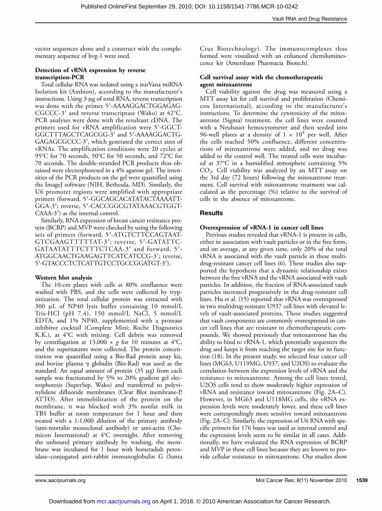
either in association with vault particles or in the free form,and on average, at any given time, only 20% of the totalvRNA is associated with the vault particle in these multi-drug-resistant cancer cell lines (6). These studies also sup-ported the hypothesis that a dynamic relationship existsbetween the free vRNA and the vRNA associated with vaultparticles. In addition, the fraction of RNA-associated vaultparticles increased progressively in the drug-resistant celllines. Hu et al. (15) reported that vRNA was overexpressedin two multidrug-resistant U937 cell lines with elevated le-vels of vault-associated proteins. These studies suggestedthat vault components are commonly overexpressed in can-cer cell lines that are resistant to chemotherapeutic com-pounds. We showed previously that mitoxantrone has theability to bind to vRNA-1, which potentially sequesters thedrug and keeps it from reaching the target site for its func-tion (18). In the present study, we selected four cancer celllines (MG63, U119MG, U937, and U2OS) to evaluate thecorrelation between the expression levels of vRNA and theresistance to mitoxantrone. Among the cell lines tested,U2OS cells tend to show moderately higher expression ofvRNA and resistance toward mitoxantrone (Fig. 2A–C).However, in MG63 and U118MG cells, the vRNA ex-pression levels were moderately lower, and these cell lineswere correspondingly more sensitive toward mitoxantrone(Fig. 2A–C). Similarly, the expression of U6 RNAwith spe-cific primers for 176 bases was used as internal control andthe expression levels seem to be similar in all cases. Addi-tionally, we have evaluated the RNA expression of BCRPand MVP in these cell lines because they are known to pro-vide cellular resistance to mitoxantrone. Our studies show
Mol Cancer Res; 8(11) November 2010 1539
2010 American Association for Cancer Research.
Gopinath et al.
1540
Published OnlineFirst September 29, 2010; DOI: 10.1158/1541-7786.MCR-10-0242
that MVP was expressed at higher levels in MG63,U118MG, and U2OS cells and less in U937 cells. TheBCRP RNA was also expressed abundantly in U937 andU2OS cells but to a lesser extent in other two cell lines(Fig. 2D).
vRNA silencing by a specific siRNATo evaluate the importance of vRNA directly in cells, we
adopted an RNAi approach to knock down the vRNAlevels. In this type of study, it is important to know the
Mol Cancer Res; 8(11) November 2010
on April 1, 2018. ©mcr.aacrjournals.org Downloaded from
sequence of vRNA-1 overexpressed in U937 cells, althoughthe vRNA-1 sequence was previously reported fromSW1573/2R120 and GLC4/ADR cells. Therefore, weisolated total RNA from U937 cells and did a reversetranscription-PCR (RT-PCR) analysis using specificprimers based on the reported vRNA sequences (8). Weobtained PCR products (∼100 bp) for hvg RNA-1 andhvg RNA-3 (Fig. 3A). No PCR product, however, was ob-tained for hvg RNA-2, even with additional PCR cycles(data not shown). To deduce the sequences of the resulting
FIGURE 2. Analyses of vRNA expression and mitoxantrone resistance in the MG63, U118MG, U937, and U2OS cell lines. A, expression of vRNA in differentcell lines. Total RNA from all cell lines was isolated, and the vRNA sequences were amplified by RT, followed by PCR with specific primer sequences.The amplified sequences were fractionated on a 4% gel. U6 RNA expression was used as the internal control. Abundance of total RNA and U6, BCRP,and MVP RNAs was also shown. B, quantitative analysis of vRNA levels. The expressed PCR products were quantitated with the ImageJ program. Columns,mean of three independent experiments; bars, SD. vRNA and U6 RNA expression levels are evaluated statistically (the P values) in MG63 (vault, P < 0.05;U6, P < 0.01), U118MG (vault, P < 0.01; U6, P < 0.01), U937 (vault, P < 0.01; U6, P < 0.03), and U2OS (vault, P < 0.01; U6, P < 0.01) using GraphPadInStat 3 (GraphPad Software, Inc.). C, drug resistance of these cell lines by a cell viability test. An MTT assay was done to monitor the viability in thepresence and absence of mitoxantrone at 50 and 80 nmol/L concentrations. D, quantitative analysis of BCRP and MVP levels. The expressed PCRproducts were quantitated with the ImageJ program.
Molecular Cancer Research
2010 American Association for Cancer Research.
Vault RNA and Drug Resistance
Published OnlineFirst September 29, 2010; DOI: 10.1158/1541-7786.MCR-10-0242
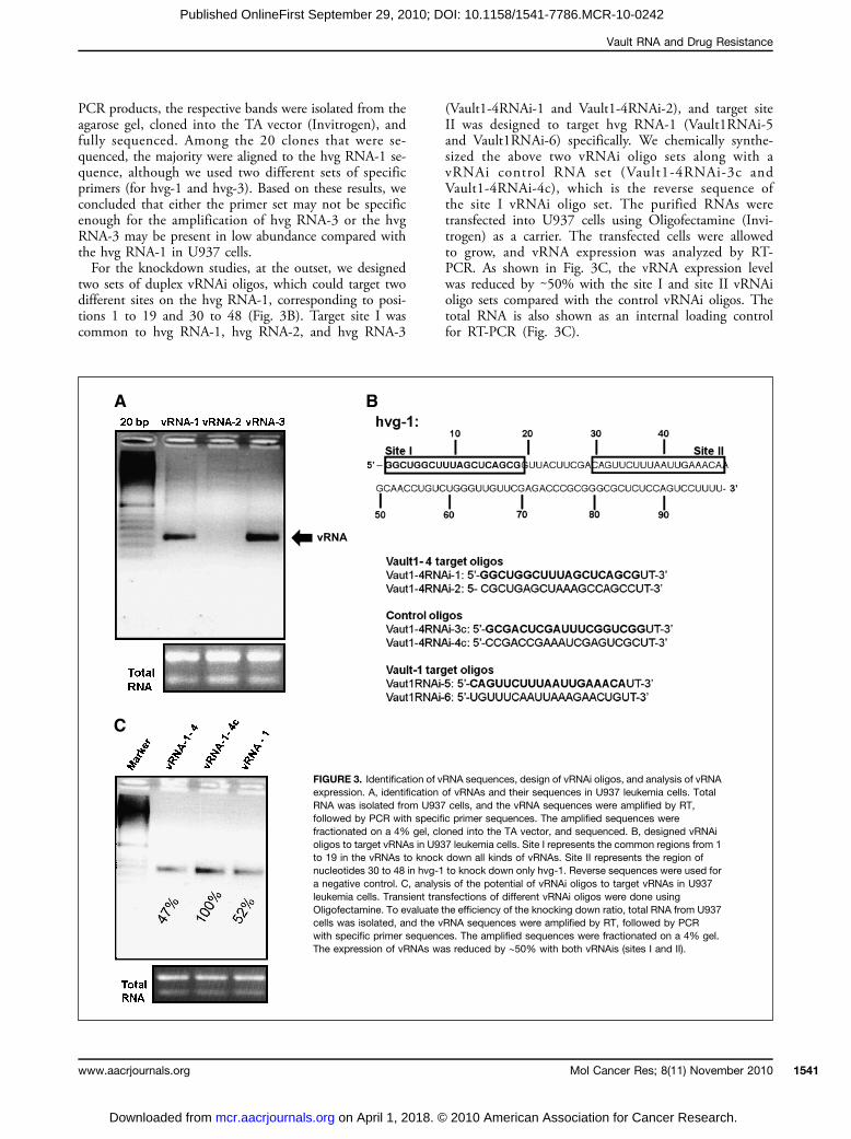
PCR products, the respective bands were isolated from theagarose gel, cloned into the TA vector (Invitrogen), andfully sequenced. Among the 20 clones that were se-quenced, the majority were aligned to the hvg RNA-1 se-quence, although we used two different sets of specificprimers (for hvg-1 and hvg-3). Based on these results, weconcluded that either the primer set may not be specificenough for the amplification of hvg RNA-3 or the hvgRNA-3 may be present in low abundance compared withthe hvg RNA-1 in U937 cells.For the knockdown studies, at the outset, we designed
two sets of duplex vRNAi oligos, which could target twodifferent sites on the hvg RNA-1, corresponding to posi-tions 1 to 19 and 30 to 48 (Fig. 3B). Target site I wascommon to hvg RNA-1, hvg RNA-2, and hvg RNA-3
www.aacrjournals.org
on April 1, 2018. ©mcr.aacrjournals.org Downloaded from
(Vault1-4RNAi-1 and Vault1-4RNAi-2), and target siteII was designed to target hvg RNA-1 (Vault1RNAi-5and Vault1RNAi-6) specifically. We chemically synthe-sized the above two vRNAi oligo sets along with avRNAi control RNA set (Vault1-4RNAi-3c andVault1-4RNAi-4c), which is the reverse sequence ofthe site I vRNAi oligo set. The purified RNAs weretransfected into U937 cells using Oligofectamine (Invi-trogen) as a carrier. The transfected cells were allowedto grow, and vRNA expression was analyzed by RT-PCR. As shown in Fig. 3C, the vRNA expression levelwas reduced by ∼50% with the site I and site II vRNAioligo sets compared with the control vRNAi oligos. Thetotal RNA is also shown as an internal loading controlfor RT-PCR (Fig. 3C).
FIGURE 3. Identification of vRNA sequences, design of vRNAi oligos, and analysis of vRNAexpression. A, identification of vRNAs and their sequences in U937 leukemia cells. TotalRNA was isolated from U937 cells, and the vRNA sequences were amplified by RT,followed by PCR with specific primer sequences. The amplified sequences werefractionated on a 4% gel, cloned into the TA vector, and sequenced. B, designed vRNAioligos to target vRNAs in U937 leukemia cells. Site I represents the common regions from 1to 19 in the vRNAs to knock down all kinds of vRNAs. Site II represents the region ofnucleotides 30 to 48 in hvg-1 to knock down only hvg-1. Reverse sequences were used fora negative control. C, analysis of the potential of vRNAi oligos to target vRNAs in U937leukemia cells. Transient transfections of different vRNAi oligos were done usingOligofectamine. To evaluate the efficiency of the knocking down ratio, total RNA from U937cells was isolated, and the vRNA sequences were amplified by RT, followed by PCRwith specific primer sequences. The amplified sequences were fractionated on a 4% gel.The expression of vRNAs was reduced by ∼50% with both vRNAis (sites I and II).
Mol Cancer Res; 8(11) November 2010 1541
2010 American Association for Cancer Research.
Gopinath et al.
1542
Published OnlineFirst September 29, 2010; DOI: 10.1158/1541-7786.MCR-10-0242
Analysis of knocked down vRNA expression in cancercell linesThe above studies revealed that both sites (sites I and II) of
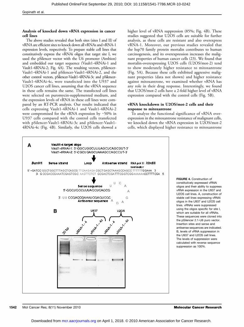
vRNA are efficient sites to knock down all vRNAs and vRNA-1expression levels, respectively. To prepare stable cell lines thatconstitutively express the vRNAi oligos that target site I, weused the pSilencer vector with the U6 promoter (Ambion)and embedded our target sequence (Vault1-4RNAi-1 andVault1-4RNAi-2; Fig. 4A). The resulting vectors, pSilencer-Vault1-4RNAi-1 and pSilencer-Vault1-4RNAi-2, and theother control vectors, pSilencer-Vault1-4RNAi-3c and pSilencer-Vault1-4RNAi-4c, were transfected into the U937 andU2OS cancer cell lines, assuming that the vRNA sequencein these cells remains the same. The transfected cell lineswere selected on puromycin-supplemented medium, andthe expression levels of vRNA in these cell lines were com-pared by an RT-PCR analysis. Our results indicated thatcells expressing Vault1-4RNAi-1 and Vault1-4RNAi-2were compromised for the vRNA expression by ∼50% inU937 cells compared with the control cells transfectedwith pSilencer-Vault1-4RNAi-3c and pSilencer-Vault1-4RNAi-4c (Fig. 4B). Similarly, the U2OS cells showed a
Mol Cancer Res; 8(11) November 2010
on April 1, 2018. ©mcr.aacrjournals.org Downloaded from
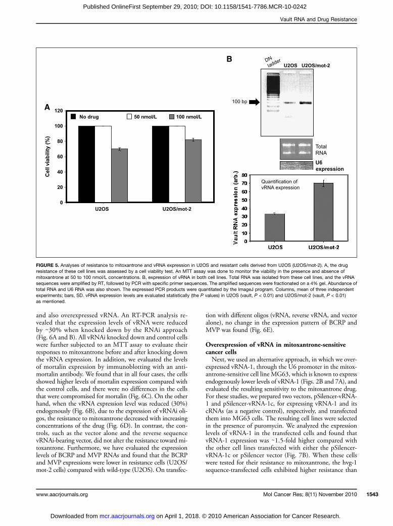
higher level of vRNA suppression (85%; Fig. 4B). Thesestudies suggested that U2OS cells are suitable for furtheranalysis, as these cells are resistant and also overexpressvRNA-1. Moreover, our previous studies revealed thatthe hsp70 family protein mortalin contributes to humancarcinogenesis, and its overexpression increases the malig-nant properties of human cancer cells (23). We found thatmortalin-overexpressing U2OS cells (U2OS/mot-2) tendto show moderately higher resistance to mitoxantrone(Fig. 5A). Because these cells exhibited aggressive malig-nant properties (data not shown) and higher resistanceagainst mitoxantrone, we examined whether vRNA hasany role in their drug response. Interestingly, we foundthat U2OS/mot-2 cells have a 2-fold higher level of vRNAexpression compared with the control cells (Fig. 5B).
vRNA knockdown in U2OS/mot-2 cells and theirresponse to mitoxantroneTo analyze the functional significance of vRNA over-
expression in the mitoxantrone resistance of malignant cells,we knocked down the vRNA expression in U2OS/mot-2cells, which displayed higher resistance to mitoxantrone
M
2010 American Association
FIGURE 4. Construction ofconstitutively expressed vRNAioligos and their ability to suppressvRNA expression in the U937 andU2OS cell lines. A, construction ofstable cell lines expressing vRNAioligos in the U937 and U2OS celllines. vRNAs were suppressedusing the oligos specific for site I,which are suitable for all vRNAs.These sequences were cloned intothe pSilencer 2.1-U6 puro vector.Insertion sites and sense andantisense sequences are indicated.B, levels of vRNA suppression inthe U937 and U2OS cell lines.The levels of suppression werecalculated with reverse sequencesuppression as 100%.
olecular Cancer Research
for Cancer Research.
Vault RNA and Drug Resistance
Published OnlineFirst September 29, 2010; DOI: 10.1158/1541-7786.MCR-10-0242
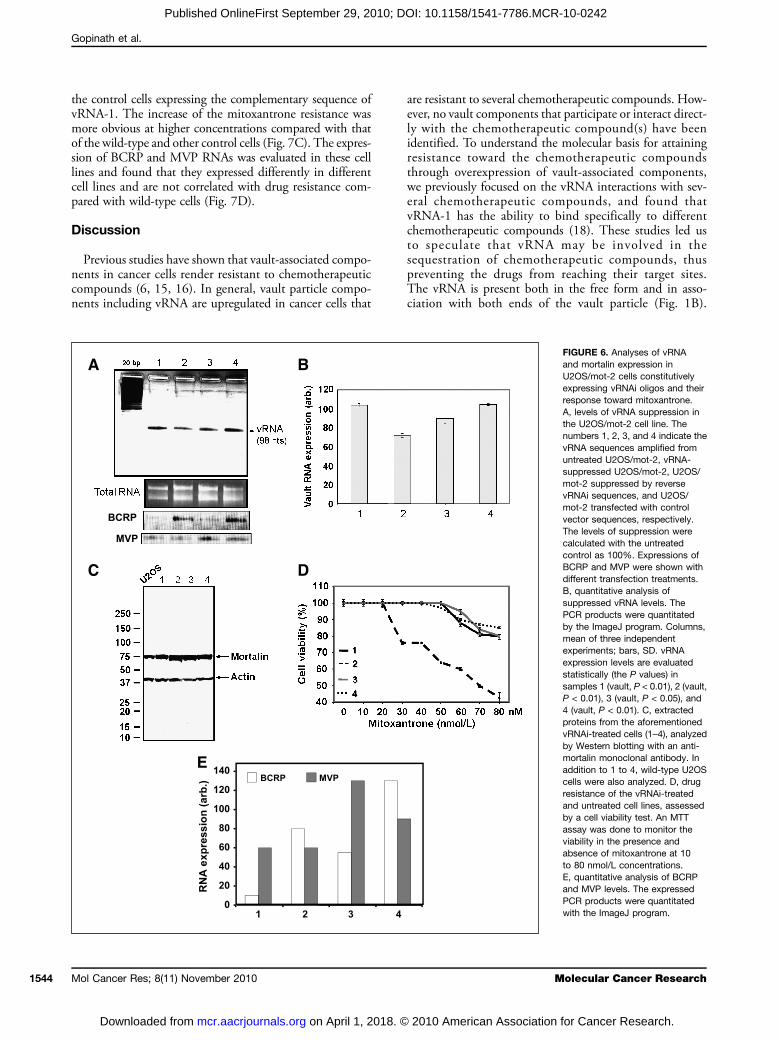
and also overexpressed vRNA. An RT-PCR analysis re-vealed that the expression levels of vRNA were reducedby ∼30% when knocked down by the RNAi approach(Fig. 6A and B). All vRNAi knocked down and control cellswere further subjected to an MTT assay to evaluate theirresponses to mitoxantrone before and after knocking downthe vRNA expression. In addition, we evaluated the levelsof mortalin expression by immunoblotting with an anti-mortalin antibody. We found that in all four cases, the cellsshowed higher levels of mortalin expression compared withthe control cells, and there were no differences in the cellsthat were compromised for mortalin (Fig. 6C). On the otherhand, when the vRNA expression level was reduced (30%)endogenously (Fig. 6B), due to the expression of vRNAi oli-gos, the resistance to mitoxantrone decreased with increasingconcentrations of the drug (Fig. 6D). In contrast, the con-trols, such as the vector alone and the reverse sequencevRNAi-bearing vector, did not alter the resistance toward mi-toxantrone. Furthermore, we have evaluated the expressionlevels of BCRP and MVP RNAs and found that the BCRPand MVP expressions were lower in resistance cells (U2OS/mot-2 cells) compared with wild-type (U2OS). On transfec-
www.aacrjournals.org
on April 1, 2018. ©mcr.aacrjournals.org Downloaded from
tion with different oligos (vRNA, reverse vRNA, and vectoralone), no change in the expression pattern of BCRP andMVP was found (Fig. 6E).
Overexpression of vRNA in mitoxantrone-sensitivecancer cellsNext, we used an alternative approach, in which we over-
expressed vRNA-1, through the U6 promoter in the mitox-antrone-sensitive cell line MG63, which is known to expressendogenously lower levels of vRNA-1 (Figs. 2B and 7A), andevaluated the resulting sensitivity to the mitoxantrone drug.For these studies, we prepared two vectors, pSilencer-vRNA-1 and pSilencer-vRNA-1c, for expressing vRNA-1 and itscRNAs (as a negative control), respectively, and transfectedthem into MG63 cells. The resulting cell lines were selectedin the presence of puromycin. We analyzed the expressionlevels of vRNA-1 in the transfected cells and found thatvRNA-1 expression was ∼1.5-fold higher compared withthe other cell lines transfected with either the pSilencer-vRNA-1c or pSilencer vector (Fig. 7B). When these cellswere tested for their resistance to mitoxantrone, the hvg-1sequence-transfected cells exhibited higher resistance than
nmol/L nmol/L
l vi
FIGURE 5. Analyses of resistance to mitoxantrone and vRNA expression in U2OS and resistant cells derived from U2OS (U2OS/mot-2). A, the drugresistance of these cell lines was assessed by a cell viability test. An MTT assay was done to monitor the viability in the presence and absence ofmitoxantrone at 50 to 100 nmol/L concentrations. B, expression of vRNA in both cell lines. Total RNA was isolated from these cell lines, and the vRNAsequences were amplified by RT, followed by PCR with specific primer sequences. The amplified sequences were fractionated on a 4% gel. Abundance oftotal RNA and U6 RNA was also shown. The expressed PCR products were quantitated by the ImageJ program. Columns, mean of three independentexperiments; bars, SD. vRNA expression levels are evaluated statistically (the P values) in U2OS (vault, P < 0.01) and U2OS/mot-2 (vault, P < 0.01)as mentioned.
Mol Cancer Res; 8(11) November 2010 1543
2010 American Association for Cancer Research.
Gopinath et al.
1544
Published OnlineFirst September 29, 2010; DOI: 10.1158/1541-7786.MCR-10-0242
the control cells expressing the complementary sequence ofvRNA-1. The increase of the mitoxantrone resistance wasmore obvious at higher concentrations compared with thatof the wild-type and other control cells (Fig. 7C). The expres-sion of BCRP and MVP RNAs was evaluated in these celllines and found that they expressed differently in differentcell lines and are not correlated with drug resistance com-pared with wild-type cells (Fig. 7D).
Discussion
Previous studies have shown that vault-associated compo-nents in cancer cells render resistant to chemotherapeuticcompounds (6, 15, 16). In general, vault particle compo-nents including vRNA are upregulated in cancer cells that
Mol Cancer Res; 8(11) November 2010
on April 1, 2018. ©mcr.aacrjournals.org Downloaded from
are resistant to several chemotherapeutic compounds. How-ever, no vault components that participate or interact direct-ly with the chemotherapeutic compound(s) have beenidentified. To understand the molecular basis for attainingresistance toward the chemotherapeutic compoundsthrough overexpression of vault-associated components,we previously focused on the vRNA interactions with sev-eral chemotherapeutic compounds, and found thatvRNA-1 has the ability to bind specifically to differentchemotherapeutic compounds (18). These studies led usto speculate that vRNA may be involved in thesequestration of chemotherapeutic compounds, thuspreventing the drugs from reaching their target sites.The vRNA is present both in the free form and in asso-ciation with both ends of the vault particle (Fig. 1B).
M
2010 American Association
FIGURE 6. Analyses of vRNAand mortalin expression inU2OS/mot-2 cells constitutivelyexpressing vRNAi oligos and theirresponse toward mitoxantrone.A, levels of vRNA suppression inthe U2OS/mot-2 cell line. Thenumbers 1, 2, 3, and 4 indicate thevRNA sequences amplified fromuntreated U2OS/mot-2, vRNA-suppressed U2OS/mot-2, U2OS/mot-2 suppressed by reversevRNAi sequences, and U2OS/mot-2 transfected with controlvector sequences, respectively.The levels of suppression werecalculated with the untreatedcontrol as 100%. Expressions ofBCRP and MVP were shown withdifferent transfection treatments.B, quantitative analysis ofsuppressed vRNA levels. ThePCR products were quantitatedby the ImageJ program. Columns,mean of three independentexperiments; bars, SD. vRNAexpression levels are evaluatedstatistically (the P values) insamples 1 (vault, P < 0.01), 2 (vault,P < 0.01), 3 (vault, P < 0.05), and4 (vault, P < 0.01). C, extractedproteins from the aforementionedvRNAi-treated cells (1–4), analyzedby Western blotting with an anti-mortalin monoclonal antibody. Inaddition to 1 to 4, wild-type U2OScells were also analyzed. D, drugresistance of the vRNAi-treatedand untreated cell lines, assessedby a cell viability test. An MTTassay was done to monitor theviability in the presence andabsence of mitoxantrone at 10to 80 nmol/L concentrations.E, quantitative analysis of BCRPand MVP levels. The expressedPCR products were quantitatedwith the ImageJ program.
olecular Cancer Research
for Cancer Research.
Vault RNA and Drug Resistance
Published OnlineFirst September 29, 2010; DOI: 10.1158/1541-7786.MCR-10-0242
The purpose of this study was to evaluate the physiologicrelevance of the observed vRNA interactions with che-motherapeutic compounds such as mitoxantrone in ma-lignant cells. In this study, we analyzed the expressionlevels of vRNA in five malignant cell lines and foundthat the U2OS and U2OS/mot-2 cell lines expressedmoderately higher levels of vRNA-1 (Figs. 2A and B and5B). These cells tend to show moderate resistance to thechemotherapeutic compound mitoxantrone comparedwith the other cell lines (Figs. 2C and 5A). Although itremains unknown how the malignant U2OS/mot-2 cellsupregulate their vRNA-1, it seems that a higher level ofvRNA-1 expression is involved in their drug-resistant phe-notype, at least to mitoxantrone. The above results sug-gested that there is a correlation between the expression
www.aacrjournals.org
on April 1, 2018. ©mcr.aacrjournals.org Downloaded from
levels of vRNA-1 and the adoption of resistance to che-motherapeutics by cancer cells.Next, to address the functional significance of vRNA-1
overexpression in cells, we used an RNAi approach to knockdown the expression levels of vRNA-1 endogenously andevaluated the resulting phenotypes for their sensitivity tomitoxantrone. This approach was very successful for eluci-dating the functions of many other noncoding RNAs (24).As described above, when the vRNA-1 expression in theU2OS/mot-2 cells was decreased by ∼30%, the cells becameprogressively sensitive to increasing mitoxantrone concen-trations (Fig. 6B and D). However, the vector alone andthe reverse sequence vRNAi-bearing vector did not alterthe resistance toward mitoxantrone (Fig. 6D). These studiessuggested that vRNA-1 expression in cancer cells leads to
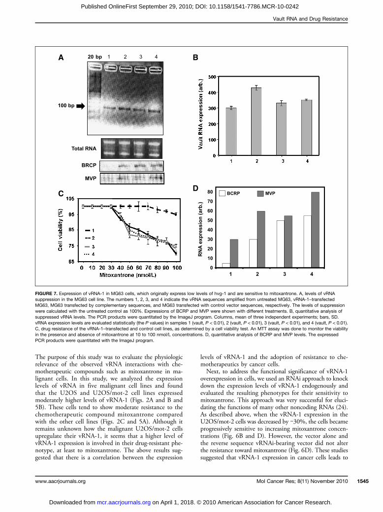
FIGURE 7. Expression of vRNA-1 in MG63 cells, which originally express low levels of hvg-1 and are sensitive to mitoxantrone. A, levels of vRNAsuppression in the MG63 cell line. The numbers 1, 2, 3, and 4 indicate the vRNA sequences amplified from untreated MG63, vRNA-1–transfectedMG63, MG63 transfected by complementary sequences, and MG63 transfected with control vector sequences, respectively. The levels of suppressionwere calculated with the untreated control as 100%. Expressions of BCRP and MVP were shown with different treatments. B, quantitative analysis ofsuppressed vRNA levels. The PCR products were quantitated by the ImageJ program. Columns, mean of three independent experiments; bars, SD.vRNA expression levels are evaluated statistically (the P values) in samples 1 (vault, P < 0.01), 2 (vault, P < 0.01), 3 (vault, P < 0.01), and 4 (vault, P < 0.01).C, drug resistance of the vRNA-1–transfected and control cell lines, as determined by a cell viability test. An MTT assay was done to monitor the viabilityin the presence and absence of mitoxantrone at 10 to 100 nmol/L concentrations. D, quantitative analysis of BCRP and MVP levels. The expressedPCR products were quantitated with the ImageJ program.
Mol Cancer Res; 8(11) November 2010 1545
2010 American Association for Cancer Research.
Gopinath et al.
1546
Published OnlineFirst September 29, 2010; DOI: 10.1158/1541-7786.MCR-10-0242
resistance to mitoxantrone. As a complement to these stud-ies, we did a rescue experiment, in which vRNA-1 was over-expressed in mitoxantrone-sensitive cell lines, such asMG63, and analyzed the resulting phenotypes. In thisstudy, we found that when vRNA-1 was overexpressed to∼1.5 fold higher levels in MG63 cells, the resistance to mi-toxantrone was also increased compared with the controls(Fig. 7B and C). Taken together, both the knockdownand rescue analyses described above suggest that vRNA-1plays an important role in conferring resistance to the cancercells against the chemotherapeutic compounds. We havealso found that in other known proteins such as BCRPand MVP, which are known to render the cellular resistanceto mitoxantrone, expressions could not be correlated fortheir role in mitoxantrone resistance in our studies. Recent-ly, two noncoding RNAs (miR-221/222 and CUDR) havealso been shown to play an important role in mediatingdrug resistance in cancer cells (25, 26). In summary, thisstudy has identified another noncoding RNA (vRNA-1)
Mol Cancer Res; 8(11) November 2010
on April 1, 2018. ©mcr.aacrjournals.org Downloaded from
that plays an important role by mediating the drug-resistantphenotype of malignant cells. Further studies on the impor-tance and applications of noncoding RNAs in cancerchemotherapeutics are warranted.
Disclosure of Potential Conflicts of Interest
No potential conflicts of interest were disclosed.
Grant Support
National Institute of Industrial Science and Technology and Ministry of Educa-tion, Culture, Sports, Science and Technology of Japan (P.K.R. Kumar) and NewEnergy and Industrial Technology Development Organization (R. Wadhwa).
The costs of publication of this article were defrayed in part by the payment ofpage charges. This article must therefore be hereby marked advertisement inaccordance with 18 U.S.C. Section 1734 solely to indicate this fact.
Received 06/03/2010; revised 09/13/2010; accepted 09/21/2010; publishedOnlineFirst 09/29/2010.
References
1. Kedersha NL, Heuser JE, Chugani DC, Rome LH. Vaults. III. Vaultribonucleoprotein particles open into flower-like structures withoctagonal symmetry. J Cell Biol 1991;112:225–35.
2. Abbondanza C, Rossi V, Roscigno A, et al. Interaction of vaultparticles with estrogen receptor in the MCF-7 breast cancer cell.J Cell Biol 1998;141:1301–10.
3. Herrmann C, Golkaramnay E, Inman E, Rome L, Volknandt W.Recombinant major vault protein is targeted to neuritic tips ofPC12 cells. J Cell Biol 1999;144:1163–72.
4. van Zon A, Mossink MH, Scheper RJ, Sonneveld P, Wiemer EAC.The vault complex. Cell Mol Life Sci 2003;60:1828–37.
5. Kitazono M, Sumizawa T, Takebayashi Y, et al. Multidrug resistanceand the lung resistance-related protein in human colon carcinomaSW-620 cells. J Natl Cancer Inst 1999;91:1647–53.
6. Kickhoefer VA, Rajavel KS, Scheffer GL, Dalton WS, Scheper RJ,Rome LH. Vaults are up-regulated in multidrug-resistant cancer celllines. J Biol Chem 1998;273:8971–4.
7. Siva AC, Raval-Fernandes S, Stephen AG, et al. Up-regulation ofvaults may be necessary but not sufficient for multidrug resistance.Int J Cancer 2001;92:195–202.
8. van Zon A, Mossink MH, Schoester M, et al. Multiple human vaultRNAs. Expression and association with the vault complex. J BiolChem 2001;276:23217–20.
9. Poderycki MJ, Rome LH, Harrington L, Kickhoefer VA. The p80homology region of TEP1 is sufficient for its association with thetelomerase and vault RNAs, and the vault particle. Nucleic AcidsRes 2005;33:893–902.
10. Huffman KE, Corey DR. Major vault protein does not play a role inchemoresistance or drug localization in a non-small cell lung cancercell line. Biochemistry 2005;44:2253–61.
11. Steiner E, Holzmann K, Elbling L, Micksche M, Berger W. Cellularfunctions of vaults and their involvement in multidrug resistance.Curr Drug Targets 2006;7:923–34.
12. Kickhoefer VA, Liu Y, Kong LB, et al. The Telomerase/vault-associated protein TEP1 is required for vault RNA stability and itsassociation with the vault particle. J Cell Biol 2001;152:157–64.
13. Tanaka H, Kato K, Yamashita E, et al. The structure of rat liver at 3.5angstrom resolution. Science 2009;322:384–8.
14. Roninson IB. In: Roninson IB, editor. Molecular and cellular biology ofmultidrug resistance in tumour cells. New York: Plenum Press; 1991,p. 1–406.
15. Hu Y, Stephen AG, Cao J, et al. A very early induction of major vaultprotein accompanied by increased drug resistance in U-937 cells. IntJ Cancer 2002;97:149–56.
16. Ohno N, Tani A, Uozumi K, et al. Expression of functional lungresistance-related protein predicts poor outcome in adult T-cellleukemia. Blood 2001;98:1160–5.
17. Mossink MH, van Zon A, Scheper RJ, Sonneveld P, Wiemer EAC.Vaults: a ribonucleoprotein particle involved in drug resistance?Oncogene 2003;22:7458–67.
18. Gopinath SCB, Matsugami A, Katahira M, Kumar PKR. Human vault-associated non-coding RNAs bind to mitoxantrone, a chemothera-peutic compound. Nucleic Acids Res 2005;33:4874–81.
19. Wang HW, Wu HL, Chen DS, Chen PJ. Identification of the functionalregions required for hepatitis virus replication and transcription by linker-scanning mutagenesis of viral genome. Virology 1997;239:119–31.
20. Nissen P, Hansen J, Ban N, Moore PB, Steitz TA. The structural basis ofribosome activity in peptide bond synthesis. Science 2000;289:920–30.
21. Mallory AC, Dugas DV, Bartel DP, Bartel B. MicroRNA regulation ofNAC-domain targets is required for proper formation and separationof adjacent embryonic, vegetative, and floral organs. Curr Biol 2004;14:1035–46.
22. Mandal M, Breaker RR. Adenine riboswitches and gene activation bydisruption of a transcription terminator. Nat Struct Mol Biol 2004;11:29–35.
23. Wadhwa R, Takano S, Kaur K, et al. Upregulation of mortalin/mthsp70/Grp75 contributes to human carcinogenesis. Int J Cancer2006;118:2973–80.
24. Li L, Okino ST, Zhao H, et al. Small dsRNAs induce transcriptional acti-vation in human cells. Proc Natl Acad Sci U S A 2006;103:17337–42.
25. Miller TE, Ghoshal K, Ramaswamy B, et al. MicroRNA-221/222confers tamoxifen resistance in breast cancer by targeting p27kip1.J Biol Chem 2008;283:29897–903.
26. Tsang WP, Wong TWL, Cheung AHH, Co CNN, Kwok TT. Inductionof drug resistance and transformation in human cancer cells by thenon-coding RNA CUDR. RNA 2008;13:890–8.
Molecular Cancer Research
2010 American Association for Cancer Research.

2010;8:1536-1546. Published OnlineFirst September 29, 2010.Mol Cancer Res Subash C.B. Gopinath, Renu Wadhwa and Penmetcha K.R. Kumar Cells and Its Importance in Mitoxantrone ResistanceExpression of Noncoding Vault RNA in Human Malignant
Updated version
10.1158/1541-7786.MCR-10-0242doi:
Access the most recent version of this article at:
Cited articles
http://mcr.aacrjournals.org/content/8/11/1536.full#ref-list-1
This article cites 25 articles, 10 of which you can access for free at:
Citing articles
http://mcr.aacrjournals.org/content/8/11/1536.full#related-urls
This article has been cited by 1 HighWire-hosted articles. Access the articles at:
E-mail alerts related to this article or journal.Sign up to receive free email-alerts
SubscriptionsReprints and
To order reprints of this article or to subscribe to the journal, contact the AACR Publications
Permissions
Rightslink site. (CCC)Click on "Request Permissions" which will take you to the Copyright Clearance Center's
.http://mcr.aacrjournals.org/content/8/11/1536To request permission to re-use all or part of this article, use this link
on April 1, 2018. © 2010 American Association for Cancer Research. mcr.aacrjournals.org Downloaded from
Published OnlineFirst September 29, 2010; DOI: 10.1158/1541-7786.MCR-10-0242