· exploring the glucosylation potential of glucansucrases from enzyme to product phd thesis to...
TRANSCRIPT
University of Groningen
Exploring the glucosylation potential of glucansucrasesDevlamynck, Tim Nick
IMPORTANT NOTE: You are advised to consult the publisher's version (publisher's PDF) if you wish to cite fromit. Please check the document version below.
Document VersionPublisher's PDF, also known as Version of record
Publication date:2017
Link to publication in University of Groningen/UMCG research database
Citation for published version (APA):Devlamynck, T. N. (2017). Exploring the glucosylation potential of glucansucrases: From enzyme toproduct. [Groningen]: University of Groningen.
CopyrightOther than for strictly personal use, it is not permitted to download or to forward/distribute the text or part of it without the consent of theauthor(s) and/or copyright holder(s), unless the work is under an open content license (like Creative Commons).
Take-down policyIf you believe that this document breaches copyright please contact us providing details, and we will remove access to the work immediatelyand investigate your claim.
Downloaded from the University of Groningen/UMCG research database (Pure): http://www.rug.nl/research/portal. For technical reasons thenumber of authors shown on this cover page is limited to 10 maximum.
Download date: 21-02-2020
Exploring the glucosylation potential of
glucansucrases
From enzyme to product
Tim Devlamynck

Cover design: Marta Martínez García
Printed by: University Press
ISBN: 9789463570428
Tim Devlamynck was supported by a fellowship of the Ubbo Emmius Fund
(University of Groningen) and the Special Research Fund (Ghent University).

Exploring the glucosylation potential of
glucansucrases
From enzyme to product
PhD thesis
to obtain the degree of PhD at the
University of Groningen
on the authority of the
Rector Magnificus Prof. E. Sterken
and in accordance with
the decision by the College of Deans
and
to obtain the degree of PhD at
Ghent University
on the authority of the
Rector Prof. R. Van de Walle
and in accordance with
the decision by the Faculty Doctoral Commission
Double PhD degree
This thesis will be defended in public on
Friday 27 October 2017 at 14.30
by
Tim Nick Devlamynck
born on 12 January 1990
in Torhout, Belgium

Supervisors Prof. L. Dijkhuizen Prof. W. Soetaert Copromotor Dr. E. M. te Poele Assessment committee Prof. R. M. Boom Prof. G. J. W. Euverink Prof. D. B. Janssen Prof. J. Van Camp

Table of contents
Chapter 1 General introduction 7
Chapter 2 Glucansucrase Gtf180-ΔN of Lactobacillus reuteri 180:
suppressing α-glucan synthesis results in improved
glycosylation yields
49
Chapter 3 Improving the low operational stability of Gtf180-ΔN from
Lactobacillus reuteri 180 by means of its immobilization
89
Chapter 4 Glucansucrase (mutant) enzymes from Lactobacillus
reuteri 180 efficiently transglucosylate Stevia component
rebaudioside A, resulting in a superior taste
107
Chapter 5 Glucosylation of stevioside by Gtf180-ΔN-Q1140E
improves its taste profile
139
Chapter 6 Biocatalytic production of novel steviol glycosides with
improved taste: scale-up, downstream processing and cost
analysis
169
Chapter 7 Glucosylation of neohesperidin dihydrochalcone from citrus
fruits: glucansucrase Gtf180-ΔN-Q1140E as biocatalyst for
the glycodiversification of sweet glycosides
201
Chapter 8 Summary and future prospects 225
Samenvatting 233
References 243
Dankwoord 261


7
Chapter 1
General introduction
Chapter 1: General introduction
8
Introduction
For many centuries, micro-organisms (and their enzymes) have been employed
for the production of bread, beer, vinegar, etc. without any understanding of the
underlying biochemical principles. At present, we know that enzymes are
nature’s highly efficient and specific catalysts, performing a diverse array of
reactions. Enzymes catalyze all processes essential for life such as DNA
replication and transcription, protein synthesis, metabolism, etc. Conventional
enzyme applications include the addition of proteases and lipases to laundry
detergents, the clarification of fruit juices by pectinases or denim washing by
cellulases (bio-stonewashing).
More recently, enzymes have gained interest as industrial biocatalysts, due to
their ability to perform highly specific chemical reactions in aqueous media with
low energy inputs, which makes biocatalysis more cost effective and eco-friendly
than conventional chemical synthesis1,2. Moreover, the advent of recombinant
DNA technology made it possible to produce enzymes in relatively large
quantities, in order to meet the constantly increasing demand3. Initial drawbacks
of biocatalysts, such as low operational stabilities and limited substrate
specificities, can be overcome by enzyme engineering technologies, such as
directed evolution, high-throughput screening of mutant libraries and in silico
rational design4. In 2014, the global market for industrial enzymes was estimated
to have a value of roughly $4.2 billion. A compound annual growth rate (CAGR)
of approximately 7% was predicted, reaching a market of nearly $6.2 billion by
20205.
A fine example of a biocatalytic process with industrial potential is the enzymatic
glycosylation of small molecules. In vivo, glycosylation is a way to structurally
diversify natural products, such as alkaloids, steroids, flavonoids and antibiotics.
Glycosylated molecules typically display different physicochemical and biological
properties than their non-glycosylated aglycons6. The most obvious effect of
glycosylation is the improved solubility of hydrophobic compounds, which has a
Chapter 1: General introduction
9
direct impact on their bio-availability. Moreover, the stability of labile molecules
against light and oxidation can be enhanced. For example, glycosylated ascorbic
acid is much more stable against oxidative degradation than the aglycon, making
high-value applications of ascorbic acid in cosmetics possible7. Interestingly, the
flavor of many food ingredients is modified by glycosylation. For example, steviol
glycosides, the molecules which give the leaves of Stevia rebaudiana its sweet
taste, display different degrees of sweetness, bitterness and other off-flavors
depending on the number, location and configuration of the attached glycosyl
moieties8. Glycosylation, more specifically galactosylation, offers the possibility to
target compounds towards the liver as a way of site-specific drug delivery9.
Furthermore, it has been demonstrated that glycosylation is an effective tool for
the modulation of the activity spectrum of glycopeptide antibiotics, a process
known as “glycorandomization”10. These examples illustrate the need for cheap
and efficient glycosylation technologies, useful both in the laboratory and in
industry. This PhD study focused on the optimization of glycosylation reactions
catalyzed by glucansucrase Gtf180 from Lactobacillus reuteri 180 and the
characterization of its glycoside products, with an important emphasis on the
glycosylation of steviol glycosides. This introductory chapter explores the state of
the art glycosylation technologies after which glucansucrase-mediated glycoside
synthesis is further elaborated. The scope of the thesis is presented at the end of
this chapter.
Synthesis of glycosides: state of the art technologies
A wide diversity of glycosides occurs in nature and could in theory be extracted
from their production host (mostly plants). However, extraction is a labor-
intensive, low-yielding process, restricting its application to highly priced
compounds such as anthocyanins11 and certain polyphenol glycosides12. The
quest for alternative approaches has led to the development of chemical,
enzymatic –and in vivo (bioconversion and fermentation) synthesis of glycosides,
each briefly discussed below.

Chapter 1: General introduction
10
Chemical glycosylation
Although a large variety of chemical glycosylation protocols has been developed
over the years, chemical glycoside synthesis still largely relies on four reactions,
differing in the glycosyl donors and the activation agents applied13 (Figure 1).
Figure 1. Glycosyl donors and corresponding activation agents applied in chemical
glycoside synthesis. Ac = acetyl.
Chemical glycosylations generally follow a unimolecular SN1 mechanism
(unimolecular nucleophilic substitution). The activation agent assists in the
departure of the leaving group, resulting in the formation of an oxocarbenium ion
which is then attacked by the nucleophilic acceptor substrate14. Chemists face
two big challenges when developing chemical glycosylation reactions: regio –and
linkage (α or β linkage) selectivity. The former is adequately dealt with by the
application of appropriate protective groups. The latter is determined by the
nature of the protecting group on the C-2 of the donor substrate, i.e. by
Chapter 1: General introduction
11
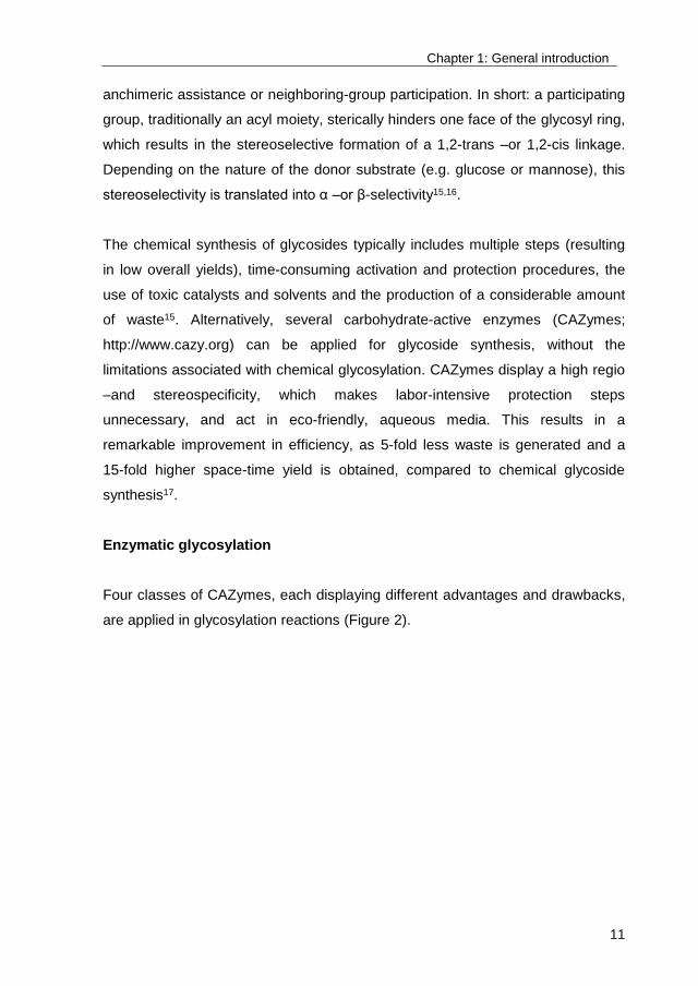
anchimeric assistance or neighboring-group participation. In short: a participating
group, traditionally an acyl moiety, sterically hinders one face of the glycosyl ring,
which results in the stereoselective formation of a 1,2-trans –or 1,2-cis linkage.
Depending on the nature of the donor substrate (e.g. glucose or mannose), this
stereoselectivity is translated into α –or β-selectivity15,16.
The chemical synthesis of glycosides typically includes multiple steps (resulting
in low overall yields), time-consuming activation and protection procedures, the
use of toxic catalysts and solvents and the production of a considerable amount
of waste15. Alternatively, several carbohydrate-active enzymes (CAZymes;
http://www.cazy.org) can be applied for glycoside synthesis, without the
limitations associated with chemical glycosylation. CAZymes display a high regio
–and stereospecificity, which makes labor-intensive protection steps
unnecessary, and act in eco-friendly, aqueous media. This results in a
remarkable improvement in efficiency, as 5-fold less waste is generated and a
15-fold higher space-time yield is obtained, compared to chemical glycoside
synthesis17.
Enzymatic glycosylation
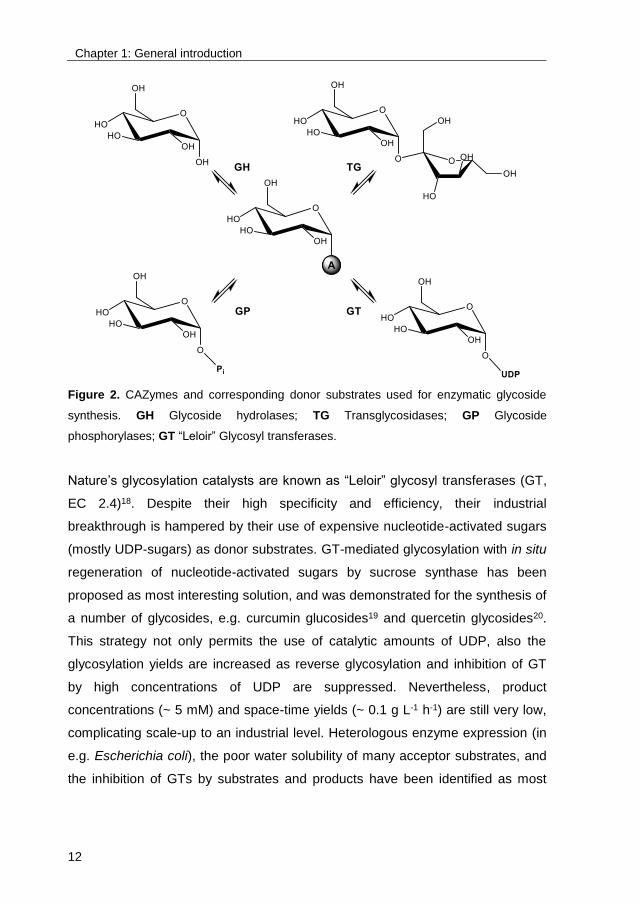
Four classes of CAZymes, each displaying different advantages and drawbacks,
are applied in glycosylation reactions (Figure 2).
Chapter 1: General introduction
12
Figure 2. CAZymes and corresponding donor substrates used for enzymatic glycoside
synthesis. GH Glycoside hydrolases; TG Transglycosidases; GP Glycoside
phosphorylases; GT “Leloir” Glycosyl transferases.
Nature’s glycosylation catalysts are known as “Leloir” glycosyl transferases (GT,
EC 2.4)18. Despite their high specificity and efficiency, their industrial
breakthrough is hampered by their use of expensive nucleotide-activated sugars
(mostly UDP-sugars) as donor substrates. GT-mediated glycosylation with in situ
regeneration of nucleotide-activated sugars by sucrose synthase has been
proposed as most interesting solution, and was demonstrated for the synthesis of
a number of glycosides, e.g. curcumin glucosides19 and quercetin glycosides20.
This strategy not only permits the use of catalytic amounts of UDP, also the
glycosylation yields are increased as reverse glycosylation and inhibition of GT
by high concentrations of UDP are suppressed. Nevertheless, product
concentrations (~ 5 mM) and space-time yields (~ 0.1 g L-1 h-1) are still very low,
complicating scale-up to an industrial level. Heterologous enzyme expression (in
e.g. Escherichia coli), the poor water solubility of many acceptor substrates, and
the inhibition of GTs by substrates and products have been identified as most
Chapter 1: General introduction
13
important bottlenecks, with an urgent need for adequate reaction –and enzyme
engineering21.
Glycoside phosphorylases (GP, EC 2.4), on the other hand, utilize glycosyl
phosphates (e.g. glucose-1-phosphate) for the transfer of a glycosyl moiety,
compounds that are easily synthesized in large quantities. Sucrose
phosphorylase (SP) can even use inexpensive sucrose as glucose donor
substrate for the synthesis of glycosides. Moreover, SP displays activity towards
a wide array of acceptor substrates, which makes it the most interesting GP for
glycoside synthesis22,23. The main disadvantage of GP is that their glycosylation
yields are significantly lower than those of “Leloir” glycosyl transferases, which is
caused by their low affinity for alternative acceptor substrates24. Recently,
improved glycosylation yields were achieved by the construction of two SP
mutants with a better accessibility of the active site, allowing the efficient
synthesis of e.g. resveratrol glycosides25,26.
Glycoside hydrolases (GH, EC 3.2.1), in vivo catalyzing the hydrolysis of
carbohydrates, can be manipulated by dynamic or kinetic control to perform
glycosylation reactions in vitro. The former strategy, also called reverse
hydrolysis, consists of shifting the reaction equilibrium towards glycoside
synthesis by increasing the donor and acceptor substrate concentrations, or by
lowering the water content27. This approach is typically used for the glycosylation
of primary and secondary alcohols by exploiting the high operational stability and
robustness of GH towards acceptor substrates and solvents28. For example, allyl-
β-D-glucoside was synthesized by almond β-glucosidase in a 90% allyl alcohol
solution with high yield29. Kinetic control implies that the donor substrate is
activated by a leaving group, e.g. a para-nitrophenyl moiety. The leaving group is
released, yielding an activated anomeric center which is then attacked by the
acceptor substrate. The resulting yield is consequently higher than the
equilibrium yield. However, the reaction has to be stopped on time, otherwise the
thermodynamically favored hydrolytic reaction takes over28. To cope with the
inherent hydrolytic nature of GH, many successful enzyme engineering strategies
have been developed over the past years, most notably resulting in the
Chapter 1: General introduction
14
development of glycosynthases30. These enzymes constitute a class of GH
mutants that promote glycosidic bond formation, provided a suitable activated
glycosyl donor is supplied, but do not hydrolyze the newly formed glycosidic
linkage. A famous example is the E197S mutant of cellulase Cel7B from
Humicola insolens, capable of efficiently glycosylating several flavonoids with
reaction rates that are comparable with those of “Leloir” glycosyltransferases31.
Last but not least, transglycosidases (TG, EC 2.4) constitute an interesting class
of glycosylation biocatalysts. They only require readily available, non-activated
carbohydrates (e.g. sucrose) as donor substrates for glycoside synthesis. TG are
in fact retaining glycoside hydrolases that are able to avoid water as acceptor
substrate, instead catalyzing the glycosylation of carbohydrates by an intra- or
intermolecular substitution at the anomeric position of a certain glycoside32. In
addition, they can also be applied for glycoside synthesis. Cyclodextrin
glucanotransferases (CGTase, family GH13) have for example been used for the
glycosylation of resveratrol into α-glycosylated products with a conversion degree
of 50%33. Another interesting group of TG are glucansucrase enzymes (family
GH70). The glucansucrase Gtf180-ΔN (an N-terminally truncated version of the
Gtf180 enzyme) from L. reuteri 180 was the glycosylation biocatalyst studied in
this PhD project, therefore glucansucrases are discussed in more detail below.
In vivo glycosylation
The third option to glycosylate small molecules is in vivo synthesis, either by
bioconversion (resting cells) or fermentation (actively growing cells). Most
technologies are based on the overexpression of UDP-glycosyl transferases
(UGTs) in a micro-organism, consequently making use of its intracellular UDP-
sugar pool. As such, this technology intends to exploit the high specificity of
UGTs while circumventing their main constraint for application in vitro (i.e. high
cost of UDP-sugars)34.
Three major types can be distinguished, differing by the number of micro-
organisms used and whether the aglycon is added or not: bacterial coupling,
Chapter 1: General introduction
15
single cell glycosylation and de novo fermentation. As the name suggests, the
bacterial coupling strategy consists of combining different hosts, each fulfilling
one of three steps in the formation of glycosides (i.e. UTP formation, UDP-sugar
formation and UGT-mediated glycosylation).35 Although successfully applied for
the production of oligosaccharides35,36, the inherent complexity of this system
(separate fermentations to obtain high cell densities of each host involved)
makes it an instable and relatively costly process. The development of single cell
glycosylations, merging all steps in one organism, thus is a logical next step.
Depending on the metabolic state of the cell, bioconversion (resting cells) and
fermentation (actively growing cells) can be distinguished. Of the two,
fermentation is preferred as it omits some of the disadvantages associated with
bioconversion. Indeed, the latter often requires permeabilization of the host and
suffers from decreasing productivities over time, caused by cell decay. In
contrast, actively growing cells display enhanced productivities over time34. Later
on, the advances in the field of metabolic engineering even resulted in the
development of de novo fermentation of glycosides, thereby eliminating the need
for the addition of acceptor substrates. A limited number of successful examples,
applying the traditional hosts E. coli and Saccharomyces cerevisiae, are
reported, e.g. vanillin glucoside37, resveratrol glucosides38 and steviol
glycosides8,39.
In general, the currently developed in vivo glycosylation processes all suffer from
very low product concentrations (~ 1 g/L), which can partly be explained by the
low solubility and toxicity of many of the target compounds37-42. On the other
hand, the described processes lack the ability to efficiently (re)generate UDP-
sugars, which results in their rapid depletion and, hence, low product yields.
Much effort is thus still required to turn in vivo glycosylation, in whatever form,
into an economically feasible process.

Chapter 1: General introduction
16
Case study: Glycosylation of steviol glycosides
The various advantages and disadvantages of the previously discussed
glycosylation technologies are nicely illustrated by using the glycosylation of
Stevia glycosides as case study. The steviol glycosides of the plant Stevia
rebaudiana, native in Paraguay and Brazil, were approved for use as high-
intensity sweetener (HIS) in food products by the European Commission in
201143. Although the share of HIS in the global sweetener market, estimated at
US$ 68 billion annually, is currently not significant (Figure 3), the HIS market is
predicted to grow significantly over the next years, due to increased consumption
of low-calorie food products, fueled by increased consumer awareness of diet-
related diseases. Stevia is currently the fastest growing HIS and is expected to
reach a value of US$ 565 million by 2020, reflecting a CAGR of 8.5% during
2014-2020, a significantly faster growth than the total sweetener market,
registering a CAGR of 5.7% during the forecast period. The volume consumption
of stevia is expected to reach 8507 tons on an annual basis by the end of 2020,
the majority of which will be used in beverages and table top sweeteners,
collectively accounting for around 72% of the global stevia market44. Stevia is
projected by the World Health Organization to eventually replace 20% of the
sugar segment, equaling a US$ 10 billion industry45. This is significantly greater
than 2014 sales, estimated at around US$ 347 million44.
Figure 3. Global sweetener market, estimated at $68 billion, and global high-intensity
sweetener (HIS) market in 201444.
Chapter 1: General introduction
17
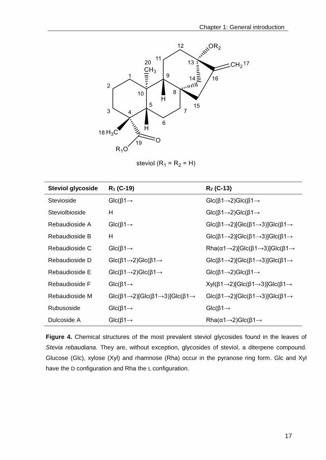
Steviol glycoside R1 (C-19) R2 (C-13)
Stevioside Glc(β1→ Glc(β1→2)Glc(β1→
Steviolbioside H Glc(β1→2)Glc(β1→
Rebaudioside A Glc(β1→ Glc(β1→2)[Glc(β1→3)]Glc(β1→
Rebaudioside B H Glc(β1→2)[Glc(β1→3)]Glc(β1→
Rebaudioside C Glc(β1→ Rha(α1→2)[Glc(β1→3)]Glc(β1→
Rebaudioside D Glc(β1→2)Glc(β1→ Glc(β1→2)[Glc(β1→3)]Glc(β1→
Rebaudioside E Glc(β1→2)Glc(β1→ Glc(β1→2)Glc(β1→
Rebaudioside F Glc(β1→ Xyl(β1→2)[Glc(β1→3)]Glc(β1→
Rebaudioside M Glc(β1→2)[Glc(β1→3)]Glc(β1→ Glc(β1→2)[Glc(β1→3)]Glc(β1→
Rubusoside Glc(β1→ Glc(β1→
Dulcoside A Glc(β1→ Rha(α1→2)Glc(β1→
Figure 4. Chemical structures of the most prevalent steviol glycosides found in the leaves of
Stevia rebaudiana. They are, without exception, glycosides of steviol, a diterpene compound.
Glucose (Glc), xylose (Xyl) and rhamnose (Rha) occur in the pyranose ring form. Glc and Xyl
have the D configuration and Rha the L configuration.
Chapter 1: General introduction
18
Stevia extract mainly consists of the steviol glycosides rebaudioside A (RebA, 2-
4 % of leaf dry weight) and stevioside (5-10% of leaf dry weight) (Figure 4). As
most steviol glycosides, they display a lingering bitterness which has limited their
successful commercialization46. Solving the taste issue of Stevia holds the
potential to greatly expand its use, for example by allowing the creation of zero-
calorie stevia soft drinks. Although the correlation between the structure of steviol
glycosides and their taste quality is still not fully understood, it is clear that the
latter depends on the number, location and configuration of the glycosyl
moieties8. In general, the bitterness is correlated with the total number of
attached glycosyl units: steviol glycosides with fewer glycosyl residues are more
bitter than steviol glycosides with more glycosyl residues46. Glycosylation of
steviol glycosides has consequently been proposed multiple times as bitterness-
eliminating and taste-improving process.
Chemical glycosylation of steviol glycosides
Chemical glycosylation of steviol glycosides has – unsurprisingly – not been
widely reported since this strategy is characterized by a vast complexity of
(de)protection steps and the use of many toxic chemical reagents, which is
undesired for food applications. The importance of these studies is therefore
merely academic8. However, one patent application, reporting the chemical
synthesis of rebaudioside D (RebD) from RebA, has to be noted47,48. RebD is
considered the “holy grail” of Stevia glycosides due to its superior taste profile
compared to RebA and stevioside. Unfortunately, its low presence in the Stevia
plant (around 0.3% of leaf dry weight or 2.5% of total steviol glycosides) makes
RebD extraction impractical and costly, urging the need for its synthesis49. In
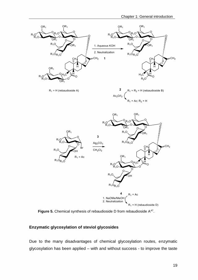
short, the reported chemical synthesis of RebD included 4 main steps (Figure 5):
1 conversion of RebA into rebaudioside B by alkaline treatment, 2 acetylation of
the free hydroxyl groups at the C-13 site, 3 glycosylation of the free C-19
carboxyl group with acetylated α-sophorosyl bromide by activation with silver
carbonate, and 4 deacetylation yielding RebD, resulting in a low overall yield of
8.1%, illustrative for chemical glycosylations in general47,48.
Chapter 1: General introduction
19
Enzymatic glycosylation of steviol glycosides
Due to the many disadvantages of chemical glycosylation routes, enzymatic
glycosylation has been applied – with and without success - to improve the taste
Figure 5. Chemical synthesis of rebaudioside D from rebaudioside A47.

Chapter 1: General introduction
20
profile of steviol glycosides, mainly by using cyclodextrin glucanotransferases
(CGTases)50-56, UDP-glycosyltransferases (UGTs)57-59 and glycoside hydrolases
(GHs)60-65. Although CGTase-catalyzed glycosylations often result in high yields,
a poor C-13/C-19-regiospecificity is obtained51, which has been shown to be of
major influence for the taste quality of the glycosylated products. For example,
(α1→4)-glycosylation of stevioside and rubusoside at the C-13 steviol position
yielded products with improved intensity and quality of sweetness, whereas
(α1→4)-glycosylation at the C-19 position resulted in an increased bitterness52-54.
Moreover, several studies reported that the many multiglycosylated products
synthesized by CGTases were perceived as more bitter than their
monoglycosylated counterparts50,53. This lack of selectivity resulted in a
complicated and costly purification process to obtain the monoglycosylated
product with improved taste, limiting the industrial application of CGTases for the
glycosylation of steviol glycosides. Some progress has been made by using
micro-wave assisted glycosylation with a CGTase from Bacillus firmus (from 46%
to 66% monoglycosylation)55 and, more recently, by application of a CGTase
found in a Paenibacillus sp. isolated from Stevia farmland, yielding a single
monoglycosylated product but, unfortunately, also displaying a low total
conversion56. To this date, the shortcomings of CGTase-catalyzed glycosylation
of steviol glycosides largely remain. Improvement of the product specificity by
mutational engineering is consequently strongly required.
Few studies report the glycosylation of steviol glycosides by UGTs in vitro57-59. Of
industrial interest is the application of UGT76G1 from S. rebaudiana in
combination with sucrose synthase from Arabidopsis thaliana, regenerating the
costly UDP-glucose, for the conversion of stevioside into the better-tasting RebA.
Although a reasonably high conversion degree was obtained (78%), the
productivity was very low: less than 2 mM RebA was synthesized in 30 h59.
Similarly, UGT91D2 from S. rebaudiana has been used for the synthesis of RebD
from RebA, however, only a 4.7% conversion was obtained57. In order to
circumvent the usage of UDP-glucose as donor substrate, UGT76G1 and
UGT91D2 have been applied in vivo for the synthesis of RebD and rebaudioside
M (RebM), as described in detail below.
Chapter 1: General introduction
21
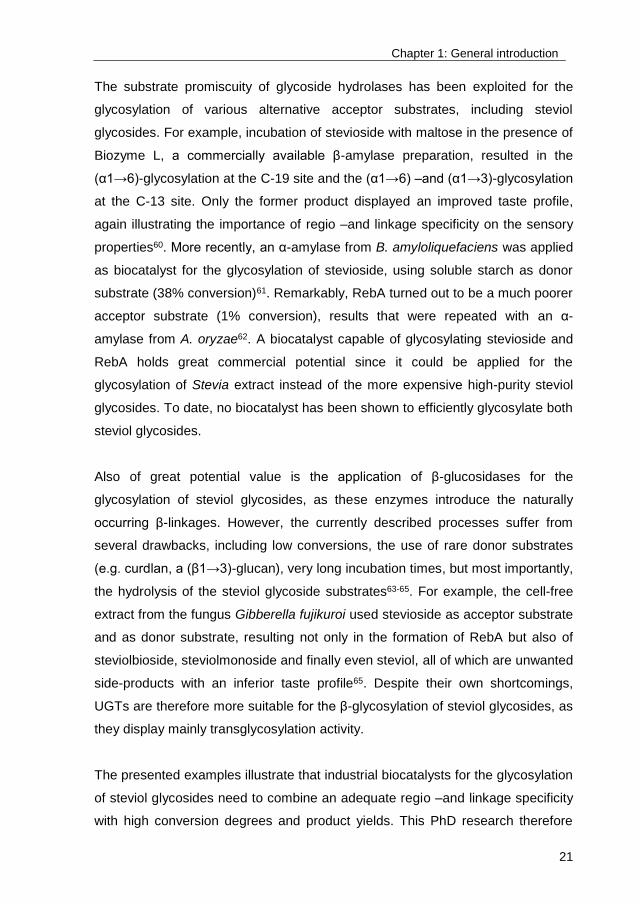
The substrate promiscuity of glycoside hydrolases has been exploited for the
glycosylation of various alternative acceptor substrates, including steviol
glycosides. For example, incubation of stevioside with maltose in the presence of
Biozyme L, a commercially available β-amylase preparation, resulted in the
(α1→6)-glycosylation at the C-19 site and the (α1→6) –and (α1→3)-glycosylation
at the C-13 site. Only the former product displayed an improved taste profile,
again illustrating the importance of regio –and linkage specificity on the sensory
properties60. More recently, an α-amylase from B. amyloliquefaciens was applied
as biocatalyst for the glycosylation of stevioside, using soluble starch as donor
substrate (38% conversion)61. Remarkably, RebA turned out to be a much poorer
acceptor substrate (1% conversion), results that were repeated with an α-
amylase from A. oryzae62. A biocatalyst capable of glycosylating stevioside and
RebA holds great commercial potential since it could be applied for the
glycosylation of Stevia extract instead of the more expensive high-purity steviol
glycosides. To date, no biocatalyst has been shown to efficiently glycosylate both
steviol glycosides.
Also of great potential value is the application of β-glucosidases for the
glycosylation of steviol glycosides, as these enzymes introduce the naturally
occurring β-linkages. However, the currently described processes suffer from
several drawbacks, including low conversions, the use of rare donor substrates
(e.g. curdlan, a (β1→3)-glucan), very long incubation times, but most importantly,
the hydrolysis of the steviol glycoside substrates63-65. For example, the cell-free
extract from the fungus Gibberella fujikuroi used stevioside as acceptor substrate
and as donor substrate, resulting not only in the formation of RebA but also of
steviolbioside, steviolmonoside and finally even steviol, all of which are unwanted
side-products with an inferior taste profile65. Despite their own shortcomings,
UGTs are therefore more suitable for the β-glycosylation of steviol glycosides, as
they display mainly transglycosylation activity.
The presented examples illustrate that industrial biocatalysts for the glycosylation
of steviol glycosides need to combine an adequate regio –and linkage specificity
with high conversion degrees and product yields. This PhD research therefore
Chapter 1: General introduction
22
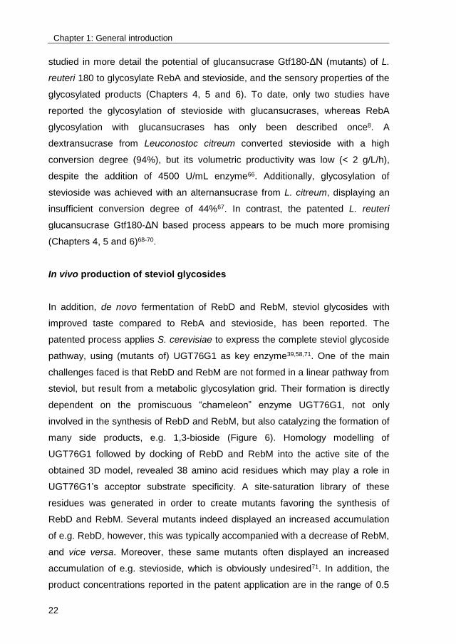
studied in more detail the potential of glucansucrase Gtf180-ΔN (mutants) of L.
reuteri 180 to glycosylate RebA and stevioside, and the sensory properties of the
glycosylated products (Chapters 4, 5 and 6). To date, only two studies have
reported the glycosylation of stevioside with glucansucrases, whereas RebA
glycosylation with glucansucrases has only been described once8. A
dextransucrase from Leuconostoc citreum converted stevioside with a high
conversion degree (94%), but its volumetric productivity was low (< 2 g/L/h),
despite the addition of 4500 U/mL enzyme66. Additionally, glycosylation of
stevioside was achieved with an alternansucrase from L. citreum, displaying an
insufficient conversion degree of 44%67. In contrast, the patented L. reuteri
glucansucrase Gtf180-ΔN based process appears to be much more promising
(Chapters 4, 5 and 6)68-70.
In vivo production of steviol glycosides
In addition, de novo fermentation of RebD and RebM, steviol glycosides with
improved taste compared to RebA and stevioside, has been reported. The
patented process applies S. cerevisiae to express the complete steviol glycoside
pathway, using (mutants of) UGT76G1 as key enzyme39,58,71. One of the main
challenges faced is that RebD and RebM are not formed in a linear pathway from
steviol, but result from a metabolic glycosylation grid. Their formation is directly
dependent on the promiscuous “chameleon” enzyme UGT76G1, not only
involved in the synthesis of RebD and RebM, but also catalyzing the formation of
many side products, e.g. 1,3-bioside (Figure 6). Homology modelling of
UGT76G1 followed by docking of RebD and RebM into the active site of the
obtained 3D model, revealed 38 amino acid residues which may play a role in
UGT76G1’s acceptor substrate specificity. A site-saturation library of these
residues was generated in order to create mutants favoring the synthesis of
RebD and RebM. Several mutants indeed displayed an increased accumulation
of e.g. RebD, however, this was typically accompanied with a decrease of RebM,
and vice versa. Moreover, these same mutants often displayed an increased
accumulation of e.g. stevioside, which is obviously undesired71. In addition, the
product concentrations reported in the patent application are in the range of 0.5

Chapter 1: General introduction
23
to 3 g/L, which should be improved in order for the process to reach a viable
scale58. Nevertheless, a joint-venture of Switzerland-based Evolva, the patent
holder, and Cargill, offering its facilities, has announced to launch fermentation-
based RebD and RebM (EverSweet™) in 2018 (http://www.evolva.com). It
should be noted that the initial launching date was set back several times since
2013. The joint venture has indicated that the production costs are still
problematic, due to inadequate strain characteristics and too high fermentation
and downstream processing costs.
Figure 6. Steviol and the metabolic grid of glycosylation reactions resulting in the
synthesis of rebaudioside D and rebaudioside M71.
Conclusions
From the discussion it is clear that the glycosylation of steviol glycosides holds
great commercial potential, resulting in an ongoing development of novel and
improved glycosylation processes, enzymatic as well as fermentation-based. The
main advantage of de novo fermentation over most enzymatic glycosylation
reactions is that nature-identical steviol glycosides, i.e. RebD and RebM, are
synthesized. On the other hand, these processes may suffer from disputes
concerning their GMO nature, possibly resulting in their (partial) rejection by
consumers72. In contrast, many enzymatic glycosylation reactions yield products
which will be classified as novel food, requiring additional regulatory approval by
Chapter 1: General introduction
24
e.g. the European Food Safety Agency (EFSA). To their advantage, biocatalysts
are generally classified as a processing aid, omitting the obligation for labelling.
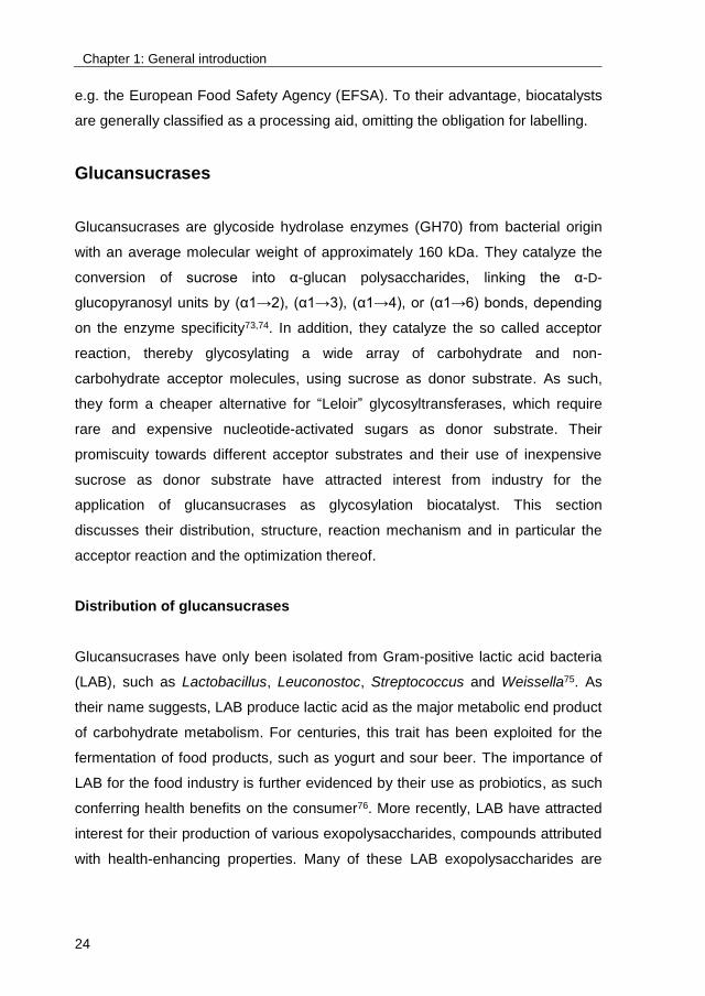
Glucansucrases
Glucansucrases are glycoside hydrolase enzymes (GH70) from bacterial origin
with an average molecular weight of approximately 160 kDa. They catalyze the
conversion of sucrose into α-glucan polysaccharides, linking the α-D-
glucopyranosyl units by (α1→2), (α1→3), (α1→4), or (α1→6) bonds, depending
on the enzyme specificity73,74. In addition, they catalyze the so called acceptor
reaction, thereby glycosylating a wide array of carbohydrate and non-
carbohydrate acceptor molecules, using sucrose as donor substrate. As such,
they form a cheaper alternative for “Leloir” glycosyltransferases, which require
rare and expensive nucleotide-activated sugars as donor substrate. Their
promiscuity towards different acceptor substrates and their use of inexpensive
sucrose as donor substrate have attracted interest from industry for the
application of glucansucrases as glycosylation biocatalyst. This section
discusses their distribution, structure, reaction mechanism and in particular the
acceptor reaction and the optimization thereof.
Distribution of glucansucrases
Glucansucrases have only been isolated from Gram-positive lactic acid bacteria
(LAB), such as Lactobacillus, Leuconostoc, Streptococcus and Weissella75. As
their name suggests, LAB produce lactic acid as the major metabolic end product
of carbohydrate metabolism. For centuries, this trait has been exploited for the
fermentation of food products, such as yogurt and sour beer. The importance of
LAB for the food industry is further evidenced by their use as probiotics, as such
conferring health benefits on the consumer76. More recently, LAB have attracted
interest for their production of various exopolysaccharides, compounds attributed
with health-enhancing properties. Many of these LAB exopolysaccharides are
Chapter 1: General introduction
25
produced by glucansucrases, extracellular enzymes which are, depending on the
bacterial source, either cell wall-attached, free or both77.
Up until the beginning of 2017, 63 GH70 glucansucrases had been
characterized, representing a wide variety of linkage specificities, and are listed
in the CAZy database (http://www.cazy.org). Most of them were obtained from
the genera Leuconostoc (24 of 63) and Streptococcus (21 of 63) and a minority
from Lactobacillus (13 of 63) and Weissella (5 of 63). Some LAB strains express
more than one glucansucrase. For example, L. citreum NRRL B-1299 (originally
L. mesenteroides NRRL B-1299) is known to produce six different
glucansucrases78 whereas Streptococcus mutans produces three79.
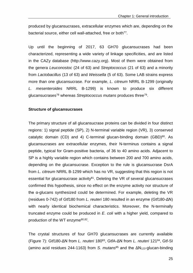
Structure of glucansucrases
The primary structure of all glucansucrase proteins can be divided in four distinct
regions: 1) signal peptide (SP), 2) N-terminal variable region (VR), 3) conserved
catalytic domain (CD) and 4) C-terminal glucan-binding domain (GBD)80. As
glucansucrases are extracellular enzymes, their N-terminus contains a signal
peptide, typical for Gram-positive bacteria, of 36 to 40 amino acids. Adjacent to
SP is a highly variable region which contains between 200 and 700 amino acids,
depending on the glucansucrase. Exception to the rule is glucansucrase DsrA
from L. citreum NRRL B-1299 which has no VR, suggesting that this region is not
essential for glucansucrase activity81. Deleting the VR of several glucansucrases
confirmed this hypothesis, since no effect on the enzyme activity nor structure of
the α-glucans synthesized could be determined. For example, deleting the VR
(residues 0-742) of Gtf180 from L. reuteri 180 resulted in an enzyme (Gtf180-ΔN)
with nearly identical biochemical characteristics. Moreover, the N-terminally
truncated enzyme could be produced in E. coli with a higher yield, compared to
production of the WT enzyme80,82.
The crystal structures of four GH70 glucansucrases are currently available
(Figure 7): Gtf180-ΔN from L. reuteri 18083, GtfA-ΔN from L. reuteri 12184, Gtf-SI
(amino acid residues 244-1163) from S. mutans85 and the ΔN123-glucan-binding
Chapter 1: General introduction
26
domain-catalytic domain 2, a truncated form of DsrE from L. citreum NRRL B-
129986. They all share a common domain organization and a common
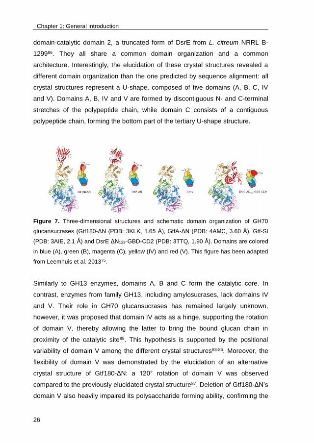
architecture. Interestingly, the elucidation of these crystal structures revealed a
different domain organization than the one predicted by sequence alignment: all
crystal structures represent a U-shape, composed of five domains (A, B, C, IV
and V). Domains A, B, IV and V are formed by discontiguous N- and C-terminal
stretches of the polypeptide chain, while domain C consists of a contiguous
polypeptide chain, forming the bottom part of the tertiary U-shape structure.
Figure 7. Three-dimensional structures and schematic domain organization of GH70
glucansucrases (Gtf180-ΔN (PDB: 3KLK, 1.65 Å), GtfA-ΔN (PDB: 4AMC, 3.60 Å), Gtf-SI
(PDB: 3AIE, 2.1 Å) and DsrE ΔN123-GBD-CD2 (PDB: 3TTQ, 1.90 Å). Domains are colored
in blue (A), green (B), magenta (C), yellow (IV) and red (V). This figure has been adapted
from Leemhuis et al. 201375.
Similarly to GH13 enzymes, domains A, B and C form the catalytic core. In
contrast, enzymes from family GH13, including amylosucrases, lack domains IV
and V. Their role in GH70 glucansucrases has remained largely unknown,
however, it was proposed that domain IV acts as a hinge, supporting the rotation
of domain V, thereby allowing the latter to bring the bound glucan chain in
proximity of the catalytic site85. This hypothesis is supported by the positional
variability of domain V among the different crystal structures83-86. Moreover, the
flexibility of domain V was demonstrated by the elucidation of an alternative
crystal structure of Gtf180-ΔN: a 120° rotation of domain V was observed
compared to the previously elucidated crystal structure87. Deletion of Gtf180-ΔN’s
domain V also heavily impaired its polysaccharide forming ability, confirming the

Chapter 1: General introduction
27
hinge hypothesis yet again88. Domain A adopts a circularly permuted (β/α)8-barrel
fold, as predicted by sequence alignment with GH13 enzymes89, containing the
three catalytic residues (nucleophile, acid/base catalyst and transition state
stabilizer) at loops following β-strands β4, β5 and β7, respectively. The complete
active site is located in a pocket-shaped cavity at the interface of domains A and
B. In fact, several amino acids belonging to domain B assist in shaping the
substrate binding sites, consequently influencing the reaction specificity83.
Additionally, some amino acids between domains A and B form a calcium binding
site near the nucleophilic residue; the Ca2+ ion is absolutely essential for
glucansucrase activity75. The function of domain C is not known yet, although it is
widely distributed within G13 and G70 enzymes. It is composed out of an eight-
stranded β-sheet with a Greek key motif83.
Catalytic mechanism of glucansucrases
According to the CAZy classification system which is based on amino acid
sequence similarity90, glucansucrases are classified as glycoside hydrolase
family GH70. Structurally, mechanistically and evolutionary, they are closely
related to enzymes of the GH13 and GH77 families, together forming the GH-H
clan91. Typical for members of the GH-H clan is their use of the α-retaining
double-displacement reaction mechanism, involving 3 catalytic residues: a
nucleophile, an acid/base catalyst and a transition state stabilizer74,92 (Figure 8).
Firstly, the glycosidic linkage of donor substrate sucrose is cleaved by the
nucleophile. Simultaneously, the acid/base catalyst protonates the fructosyl
moiety, resulting in the release of fructose. An β-glucosyl enzyme intermediate,
stabilized by the transition state stabilizing residue, is consequently formed. In
the next step, this β-glucosyl enzyme intermediate is attacked by the non-
reducing end of the acceptor substrate (i.e. sucrose, or a growing
oligosaccharide or polysaccharide chain), resulting in product formation with
retention of the α-anomeric configuration.
Chapter 1: General introduction
28
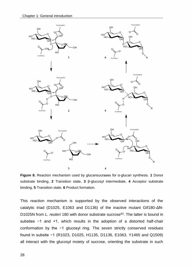
Figure 8. Reaction mechanism used by glucansucrases for α-glucan synthesis. 1 Donor
substrate binding, 2 Transition state, 3 β-glucosyl intermediate, 4 Acceptor substrate
binding, 5 Transition state, 6 Product formation.
This reaction mechanism is supported by the observed interactions of the
catalytic triad (D1025, E1063 and D1136) of the inactive mutant Gtf180-ΔN-
D1025N from L. reuteri 180 with donor substrate sucrose83. The latter is bound in
subsites −1 and +1, which results in the adoption of a distorted half-chair
conformation by the −1 glucosyl ring. The seven strictly conserved residues
found in subsite −1 (R1023, D1025, H1135, D1136, E1063, Y1465 and Q1509)
all interact with the glucosyl moiety of sucrose, orienting the substrate in such
Chapter 1: General introduction
29
manner that the formation of the covalent intermediate is favored. The anomeric
C1 carbon of the glucosyl unit is attacked by the nucleophilic residue (D1025),
resulting in the formation of the covalent β-glucosyl enzyme intermediate via an
oxocarbenium ion-like transition state. Residue E1063 serves as the acid/base
catalyst initially donating a proton to activate fructose as leaving group and
subsequently deprotonating the acceptor substrate to increase its nucleophilicity.
The partly planar transition state is stabilized by interactions with the transition
state stabilizer (D1136), an arginine residue (R1023) and a glutamine residue
(Q1509).
The active site of Gtf180-ΔN stretches no further than subsite −1 due to
adequate “blocking” by residues Q1140, N1411 and D1458. This creates a
pocket-like shape which is also observed in Neisseria polysaccharea
amylosucrase (belonging to GH13) and is the reason why glucansucrases can
only transfer one single glucose moiety per catalytic cycle, while GH13 α-
amylases, which have a longer binding groove, can also transfer
oligosaccharides. In contrast to subsite −1, the residues found in subsite +1 are
much less conserved, which is reflected in the different product specificities that
are displayed by glucansucrases and which can be exploited for the glycosylation
of alternative acceptor substrates.
Reactions catalyzed by glucansucrases
The β-glucosyl enzyme intermediate, formed in the first step of the catalytic
cycle, can not only react with a growing glucan chain to form polysaccharides but
also with the hydroxyl group of several carbohydrate and non-carbohydrate
acceptor substrates, resulting in the synthesis of oligosaccharides and α-D-
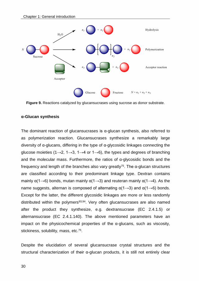
glucosides, respectively. Furthermore, also water can act as acceptor substrate,
with the hydrolysis of sucrose into glucose and fructose as result73,75 (Figure 9).
Chapter 1: General introduction
30
Figure 9. Reactions catalyzed by glucansucrases using sucrose as donor substrate.
α-Glucan synthesis
The dominant reaction of glucansucrases is α-glucan synthesis, also referred to
as polymerization reaction. Glucansucrases synthesize a remarkably large
diversity of α-glucans, differing in the type of α-glycosidic linkages connecting the
glucose moieties (1→2, 1→3, 1→4 or 1→6), the types and degrees of branching
and the molecular mass. Furthermore, the ratios of α-glycosidic bonds and the
frequency and length of the branches also vary greatly75. The α-glucan structures
are classified according to their predominant linkage type. Dextran contains
mainly α(1→6) bonds, mutan mainly α(1→3) and reuteran mainly α(1→4). As the
name suggests, alternan is composed of alternating α(1→3) and α(1→6) bonds.
Except for the latter, the different glycosidic linkages are more or less randomly
distributed within the polymers93,94. Very often glucansucrases are also named
after the product they synthesize, e.g. dextransucrase (EC 2.4.1.5) or
alternansucrase (EC 2.4.1.140). The above mentioned parameters have an
impact on the physicochemical properties of the α-glucans, such as viscosity,
stickiness, solubility, mass, etc.75.
Despite the elucidation of several glucansucrase crystal structures and the
structural characterization of their α-glucan products, it is still not entirely clear

Chapter 1: General introduction
31
how glucansucrases synthesize such a wide array of α-glucans. Essentially, α-
glucan synthesis is the step-wise addition of glucose moieties to a growing α-
glucan chain74. Every catalytic cycle starts with the cleavage of the glycosidic
bond of sucrose which results in the covalent attachment of the glucosyl moiety
at subsite −1, forming the so called β-glucosyl enzyme intermediate. Which type
of glycosidic linkage is subsequently formed depends on the orientation of the
acceptor substrate. Hence, it is the architecture of the active site, and in
particular that of acceptor subsite +1, that determines the glycosidic linkage
specificity95. Indeed, it has repeatedly been shown that mutations in residues of
subsite +1 and +2 lead to the synthesis of α-glucans with altered ratios of
glycosidic linkages96-102. Unfortunately, there is still little understanding about how
the glycosidic linkage specificity is affected by such mutations. Rational design of
glucansucrase mutants for the synthesis of pre-defined α-glucan products is thus
still very complicated.
For many years, it remained unclear how α-glucan synthesis is initiated, or in
other words, which molecule acts as primer. Several studies have since been
performed on the structural characterization of the initially formed products,
revealing that the formation of α-glucans most typically starts with the
glycosylation of sucrose103,104. The latter is thus not only the donor substrate of
glucansucrases but at the same time also the acceptor substrate. The
consequence is that the glycosylation of alternative acceptor substrates will
unavoidably face competition from the synthesis of α-glucans, unless the latter is
adequately suppressed by reaction –or enzyme engineering. It has to be noted
that one study, investigating the mechanism of DsrS from L. mesenteroides, has
proposed both sucrose and glucose as primers for α-glucan synthesis, however,
it is still unknown if this can be extended to other glucansucrases105. During the
initial phase of α-glucan synthesis, hydrolysis of sucrose into fructose and
glucose is the dominant reaction, as the affinity of glucansucrases for sucrose as
acceptor substrate is relatively low. Once the preferred acceptor substrates, i.e.
α-glucan oligosaccharides, have been formed in sufficient quantities, the
hydrolytic activity is suppressed in favor of α-glucan synthesis and α-glucan
polysaccharides are efficiently synthesized103.
Chapter 1: General introduction
32
Whether glucansucrases function as a processive or non-processive enzyme has
been subject to considerable debate. A number of studies revealed that high-
molecular-mass (HMM) glucans reached maximum size after a relatively short
time, suggesting that glucansucrases act processively103,105,106. However, also
oligosaccharides could be detected in the reaction mixture, indicating a non-
processive mode of action105. Interestingly, no intermediate size α-glucan
products were detected. Taking into account all the available information,
Remaud-Siméon et al. suggested that glucansucrases follow a semi-processive
mechanism: in the initial phase of the reaction, oligosaccharides are synthesized
non-processively. When the oligosaccharides reach a certain size,
polysaccharides are formed in a processive mode105. The structural basis of this
mechanism is proposed to lie in both domain V and the acceptor binding sites,
representing remote and close binding sites for glucan chains, respectively. This
was nicely illustrated for Gtf180-ΔN: the truncation of its domain V heavily
impaired polysaccharide synthesis in favor of oligosaccharide formation88.
However, mutations of residues located in the acceptor binding sites (in particular
L940 mutants) partially restored the polysaccharide synthesis of the ΔV-
truncated enzyme98,106. The elucidation of glucansucrase crystal structures with
HMM glucan chains bound to the enzyme is necessary to shed more light on the
mechanism of α-glucan synthesis and will without doubt offer new opportunities
to engineer the reaction specificity of glucansucrases. The following study serves
as good example: the crystal structure of amylosucrase, a special glucansucrase
belonging to family GH13, bound with maltoheptaose revealed the absence of
domains IV and V but the presence of three oligosaccharide binding sites (OB1,
OB2 and OB3)107,108. Molecular modeling and mutational studies confirmed the
importance of OB1 and OB2 for polysaccharide synthesis, suggesting that OB2
provides an anchoring platform for the polysaccharide107,109.
Additionally, the elucidation of several glucansucrase crystal structures has
revealed that their acceptor substrate binding region is reasonably spacious83,84.
Indeed, the synthesis of branched α-glucans demands an acceptor substrate
binding region which is capable of accommodating bulky α-glucan chains. As a
consequence, glucansucrases display a broad acceptor substrate specificity,
Chapter 1: General introduction
33
which is exploited in the glycosylation of alternative acceptor substrates such as
phenolic compounds, sugar alcohols, etc. Here again, it is still not clear how the
formation of branches is triggered in favor of chain elongation.
Hydrolysis
Glucansucrases also are able to catalyze the hydrolysis of sucrose into glucose
an fructose, basically acting as a hydrolase enzyme. Especially at low acceptor
substrate concentrations, hydrolysis is the dominant reaction. When
oligosaccharide products become available, glucansucrases preferentially
transfer the glucosyl moiety to these growing α-glucan chains103. The crystal
structure of the inactive mutant Gtf180-ΔN-D1025N revealed that residue
W1065, located at subsite +2, is an important structural determinant for
hydrolysis, interacting with the carbohydrate acceptor substrate through
hydrophobic stacking. The mutation of W1065 to non-aromatic residues resulted
in a significantly increased hydrolysis102. Also the mutations of residues N1029,
providing a direct hydrogen bond to carbohydrate acceptor substrates at subsite
+1, and L981, strictly conserved in all glucansucrases, substantially enhanced
hydrolysis99. The application of these mutants for the glycosylation of non-
carbohydrates (such as catechol or hexanol) resulted in improved (mono)-
glycosylation yields (Chapter 2)110.
Acceptor reaction
Glucansucrases are not only able to utilize growing α-glucan chains and water as
acceptor substrate. Due to their rather wide acceptor substrate binding region,
they show a relatively high promiscuity towards several other acceptor
substrates111-113. This promiscuity can be exploited for the glycosylation of
carbohydrates and non-carbohydrates, resulting in the synthesis of
oligosaccharides and α-D-glucosides, respectively. Enzymes are particularly
suited for glycosylation reactions as they display a high regio –and
stereospecificity, a feature that is hard to achieve by chemical synthesis13. In
nature, glycosylation is performed by “Leloir” glycosyltransferase enzymes (EC
Chapter 1: General introduction
34
2.4.-.-). Their industrial use is hampered by the high price of their donor
substrates, nucleotide-activated sugars21. Glucansucrases offer a cheaper
alternative, since they only require the energy stored in the glycosidic linkage of
sucrose (~ 27.6 kJ.mol-1) to synthesize their glycosylated products114. In 1953,
this so called acceptor reaction was first reported by Koepsell et al115. Their study
demonstrated the glycosylation of a large number of sugars and sugar
derivatives such as maltose, isomaltose, glucose, and methyl glucoside by a
dextransucrase from L. mesenteroides NRRL B-512F. Since then, many other
carbohydrates were added to the list of acceptor substrates. This makes
glucansucrase-mediated glycosylation an effective tool for the production of a
wide array of interesting oligosaccharides. Isomalto-oligosaccharides (IMO) of
controlled size are produced from sucrose plus maltose or glucose, using a
dextransucrase from L. mesenteroides NRRL B-512F116. They are attributed with
prebiotic properties (i.e. altering the composition and/or activity of the
gastrointestinal microflora, as such conferring health benefits upon the
consumer) and used as a low calorie sweetener in a variety of foods like bakery
and cereal products117. Also lactulosucrose, another prebiotic oligosaccharide, is
effectively synthesized by this dextransucrase enzyme, using lactulose as
acceptor substrate118. Another example is the glycosylation of the bitter prebiotic
gentiobiose with alternansucrase, producing several oligosaccharides with
reduced or even eliminated bitterness119.
Glycosylation of non-carbohydrate compounds
The glycosylation of non-carbohydrates, such as aromatic or aliphatic
compounds, is a valuable tool for the glycodiversification of these molecules (for
examples, see 1. Introduction). A wide range of (poly)phenolic and aliphatic
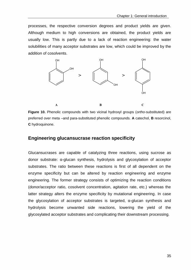
compounds are glycosylated by glucansucrases. The highest conversions are
obtained with phenolic compounds with two vicinal (ortho-substituted) hydroxyl
groups as acceptor substrate (Figure 10). Meta –and para-substituted phenolics
as well as aliphatic compounds are typically not very well glycosylated110,120.
Examples of compounds that are glycosylated by glucansucrases are listed in
Table I. To obtain insight into the economic viability of these glycosylation
Chapter 1: General introduction
35
processes, the respective conversion degrees and product yields are given.
Although medium to high conversions are obtained, the product yields are
usually low. This is partly due to a lack of reaction engineering: the water
solubilities of many acceptor substrates are low, which could be improved by the
addition of cosolvents.
Figure 10. Phenolic compounds with two vicinal hydroxyl groups (ortho-substituted) are
preferred over meta –and para-substituted phenolic compounds. A catechol, B resorcinol,
C hydroquinone.
Engineering glucansucrase reaction specificity
Glucansucrases are capable of catalyzing three reactions, using sucrose as
donor substrate: α-glucan synthesis, hydrolysis and glycosylation of acceptor
substrates. The ratio between these reactions is first of all dependent on the
enzyme specificity but can be altered by reaction engineering and enzyme
engineering. The former strategy consists of optimizing the reaction conditions
(donor/acceptor ratio, cosolvent concentration, agitation rate, etc.) whereas the
latter strategy alters the enzyme specificity by mutational engineering. In case
the glycosylation of acceptor substrates is targeted, α-glucan synthesis and
hydrolysis become unwanted side reactions, lowering the yield of the
glycosylated acceptor substrates and complicating their downstream processing.
Chapter 1: General introduction
36
Reaction engineering
Dealing with the low affinity for alternative acceptor substrates
Although many alternative acceptor substrates are indeed glycosylated by
glucansucrases, very often incomplete conversions, with low to moderate yields,
are obtained. Alternative acceptor substrates are per definition not the natural
acceptor substrates of glucansucrases and, hence, they generally have rather
high Km values (Chapter 2)110. High concentrations of acceptor substrate are
consequently required to outcompete α-glucan synthesis and hydrolysis as
possible glucansucrase reactions. In this way, the relative balance between the 3
reactions may shift towards the acceptor reaction, as was already shown in 1993
by Su and Robyt132. Moreover, high volumetric productivities (space-time yields),
which greatly reduce production costs, can only be achieved if the acceptor
substrate concentrations are sufficiently high. However, glucansucrases may be
inhibited by high concentrations of non-carbohydrate acceptor substrates.
Phenolic compounds, such as catechol, displayed inhibitory effects on
glucansucrase GtfD from S. mutans GS-5 at a concentration of 200 mM but not
at a concentration of 40 mM125. A similar effect was obtained for the glycosylation
of catechol by Gtf180 from L. reuteri 180, which was inhibited at concentrations
of catechol higher than 400 mM (Chapter 2)110. The inhibition of amylosucrase
from N. polysaccharea by several flavonoids was also reported124.
Many (poly)phenolic and aliphatic acceptor substrates have limited water
solubilities, which complicates their glycosylation. A common strategy to increase
the solubility of the acceptor substrate is the addition of organic solvents such as
DMSO, ethanol, acetone, etc. Since the 1980s it has been repeatedly shown that
enzymes can be used in solvent systems with great efficiency133. However,
enzyme activity and stability typically decrease with increasing solvent
concentration. Hence, a compromise between substrate solubility and enzyme
activity needs to be found for each individual case. The determination of the
initial activity of the dextransucrase from L. mesenteroides NRRL B-512F in the
presence of organic solvents, revealed a 50% loss in activity in 20% DMSO, 15%
Chapter 1: General introduction
37
ethanol, 15% acetone, 10% DMF and 7% acetonitrile134. Diglyme or bis(2-
methoxyethyl) ether (MEE) displayed lower inhibitory effects on glucansucrases:
the dextransucrase from L. mesenteroides NRRL B-512F and the
alternansucrase from L. mesenteroides NRRL B-23192 retained more than 50%
of their activity at a MEE concentration of 30%120. It is clear that the combined
use of high concentrations of certain acceptor substrates and high solvent
concentrations will be even more detrimental for glucansucrase activity. Hence,
the optimal balance between acceptor substrate concentration, solvent
concentration and enzyme activity will differ for every individual case.
Dealing with unwanted side reactions
Applying glucansucrases for the glycosylation of alternative acceptor substrates
usually requires high concentrations of donor substrate sucrose to drive the
glycosylation reaction. This is not without negative consequences: the
unavoidable accumulation of relatively high concentrations of fructose results in
competitive inhibition of the desired glycosylation reaction by fructose. In other
words, fructose is under these circumstances used by glucansucrases as
acceptor substrate, resulting in the synthesis of sucrose isomers such as
leucrose and trehalulose135. For example, a Ki value (inhibitor constant) for
fructose as low as 9.3 mM has been observed for GtfD from S. mutans.
Surprisingly, in the same study, glucose did not act as an inhibitor123. It is clear
from the reaction mechanism of glucansucrases that formation of fructose is
inevitable. The solution to this problem is therefore found externally, i.e. by the
addition of a micro-organism that removes fructose from the reaction mixture and
consequently reduces its inhibiting effect. In order for this strategy to work
properly, it is essential that sucrose itself is not metabolized by the micro-
organism. The methylotrophic yeast Pichia pastoris and the mutant S. cerevisiae
T2-3D136 are viable options as both strains are incapable of fermenting sucrose.
In addition, these micro-organisms should not be inhibited themselves by the
presence of acceptor substrates nor metabolize the glycosylated product. The
incubation of P. pastoris and S. cerevisiae T2-3D in a (+)-catechin glycosylation
reaction mixture revealed that their fructose consumption resulted in a

Chapter 1: General introduction
38
prolongation of the transglucosylation activity of GtfD. However, the (+)-catechin
glucoside yield was only slightly improved, indicating that the conversion of
alternative acceptor substrates is dependent on other factors as well123.
The main glucansucrase catalyzed reaction, i.e. the synthesis of α-glucan oligo –
and polysaccharides from sucrose, is the most important side reaction and
strongly impedes the efficient glycosylation of alternative acceptor substrates. As
previously discussed, sucrose acts as primer for α-glucan synthesis, and
increased concentrations of sucrose will result in the formation of more (growing
chains of) α-glucan oligosaccharides, the preferred acceptor substrates of
glucansucrases103,104,135. Their generation initiates a vicious circle of increased α-
glucan synthesis and, hence, needs to be avoided. Suppressing α-glucan
synthesis can be accomplished by performing a “fed-batch” reaction, in which the
donor substrate sucrose is gradually added to the reaction mixture. In this way,
an excess of acceptor substrate relative to sucrose is maintained throughout the
complete reaction, conditions which theoretically favor the glycosylation of the
acceptor substrate by suppressing the synthesis of α-glucans. Successful
attempts include the glycosylation of stevioside with dextransucrase from L.
citreum66 and the glycosylation of rebaudioside A with the Q1140E-mutant of
Gtf180-ΔN from L. reuteri70. However, performing glucansucrase-catalyzed
glycosylation reactions in fed-batch mode is mostly limited to the glycosylation of
highly soluble compounds, which in addition display very little inhibitory effects on
the enzyme. High ratios of non-carbohydrate acceptor substrate over sucrose are
indeed known to strongly inhibit glucansucrases, as described previously110,125.
Furthermore, te Poele et al. have demonstrated that, upon sucrose depletion,
glucansucrases from L. reuteri use several phenolic glucosides as donor
substrate for the synthesis of α-glucans and the further glycosylation of these
phenolic glucosides into multiglycosylated products137. Hence, the incubation
time and enzyme loading (U/mL) of glucansucrase catalyzed glycosylations need
to be carefully selected in order to prevent suboptimal conversion degrees.
Another remarkable characteristic displayed by glucansucrases is their ability to
add multiple α-D-glucosyl moieties to one acceptor substrate, forming α-D-

Chapter 1: General introduction
39
glucosides of different sizes and structures. A prominent example concerns GtfA-
ΔN of L. reuteri 12180: after incubation with catechol and sucrose, several
glycosylated catechol products up to DP5, differing in their combination of
(α1→4) and (α1→6) linkages, were characterized138. From an industrial point of
view, the synthesis of only one glycoside, typically the monoglycosylated product,
is desired in order to facilitate downstream processing. Indeed, the
monoglycosylated product often displays better functional properties than
multiglycosylated products. A comprehensive study on the anti-oxidant activities
of various phenolic glucosides revealed that an increasing level of glycosylation
results in reduced radical-scavenging abilities139. The number of glycosyl
moieties attached to steviol glycosides is also known to have pronounced effects
on their taste8.
Dealing with low operational stability: Enzyme immobilization
Immobilization is an established strategy to increase the operational activity and
stability of enzymes. In this way, immobilization may compensate for the
decrease of enzyme activity and stability provoked by high solvent and acceptor
substrate concentrations140. An additional advantage is the reusability of the
immobilized biocatalyst, which can drastically lower the economic cost of the
enzymatic process141. A number of immobilization methods can be distinguished:
reversible methods (adsorption and affinity binding) and irreversible methods
(entrapment, aggregation and covalent binding). Reversible immobilization
methods typically result in enzyme leaching, preventing biocatalyst reuse and
representing economic loss. On the contrary, irreversible immobilization methods
minimize enzyme leaching due to much stronger interactions between enzyme
and support, which also stabilizes the enzyme more effectively. On the downside,
enzyme activity may decrease due to active site occlusion and inherent diffusion
limitations142.
Glucansucrases have been described as difficult to covalently immobilize, mainly
due to inactivation of the enzyme, e.g. by the participation of a lysine residue in
the active site. Typical immobilization yields (ratio of activity of immobilized
Chapter 1: General introduction
40
enzyme to the activity of enzyme prior immobilization) range from 3% to 22%143-
146. In contrast, encapsulation of glucansucrases in alginate beads has been
more successfully applied as immobilization method. Several studies report
immobilization yields up to 90%147,148. However, this method cannot be used for
the production of α-glucan polysaccharides as their accumulation in alginate
results in rupture of the beads.
Enzyme engineering
Although of value, applying reaction engineering to optimize the acceptor
reaction of glucansucrases is faced with limitations. Enzyme engineering offers
an alternative, more direct optimization method, by altering the reaction
specificity of the enzyme itself. Over the years, many studies have reported
successful attempts to engineer the specificity of enzymes. For example, the
affinity of sucrose phosphorylase (SP) for glucose as acceptor substrate could be
dramatically enhanced by a double mutation, resulting in the efficient synthesis of
the rare disaccharide kojibiose149. Additionally, SP has been engineered twice
towards the more efficient synthesis of polyphenolic glycosides. The first study
attributed the enhanced yields to a better accessibility of the active site, caused
by a single mutation removing a sterically hindering active site loop, more
specifically by the mutation of an arginine residue into an alanine residue (R134A
mutant)25. As such, it forms a good example of loop engineering, i.e. the
alteration of loops, a diverse class of very flexible secondary structures
comprising turns, random coils, and strands connecting the main secondary
protein structures (α-helices and β-strands) and which very often play a vital role
in the catalytic function of the enzyme150. Also the single mutation used in the
second study yielded a loop shift in the active site of SP, as revealed by analysis
of the crystal structure of the mutant. It was argued that the loop shift resulted
from a cascade of structural changes arising from the Q345F exchange,
ultimately causing a widened access channel26. This nicely illustrates how
substantial the effect can be of a – at first sight – simple single mutation,
demonstrating the great power of enzyme engineering, but also revealing one of
the difficulties to rationally design biocatalysts.
Chapter 1: General introduction
41
To date, only a few enzyme engineering studies on glucansucrases have been
dedicated to the glycosylation of non-carbohydrate acceptor substrates. This is
partly due to the lack of available crystal structures (and consequently lack of
docking experiments) with bound acceptor substrates, complicating rational
design of glucansucrase mutants for the synthesis of glycosides. In theory,
glucansucrases can be engineered towards a more efficient acceptor reaction in
three distinguishable ways: lowering their affinity to catalyze side-reactions (in
particular α-glucan synthesis), enhancing their affinity for alternative acceptor
substrates, or both simultaneously. In 2016, Liang et al. expanded the acceptor
substrate promiscuity of GtfD from S. mutans by simultaneous site saturation
mutagenesis of residues Y418 and N469. Significant improvements in
glycosylation yield of several flavonoids were obtained with the best double
mutant (Y418R and N469C), the major products being monoglycosylated.
Docking studies based on the crystal structure of Gtf180-ΔN from L. reuteri 180
suggested that the mutant enzyme formed three additional hydrogen bonds with
the flavonoid acceptor substrate, resulting in an increased catalytic efficiency of
the mutant enzyme compared to the wild type151. Another study reported that the
I228A mutant of the amylosucrase (EC 2.4.1.4) from N. polysaccharea displayed
a significant improvement in luteolin monoglycosylation, compared to the wild-
type enzyme. Docking studies revealed that the introduction of the alanine
residue reduced the steric hindrance, resulting in a better positioning of luteolin in
the catalytic pocket129. Finally, Devlamynck et al. reported that applying Gtf180-
ΔN mutants with an impaired α-glucan synthesis resulted in an improved
glycosylation of several non-carbohydrate acceptor substrates (Chapter 2)110.
Other enzyme engineering studies with glucansucrases have focused on the
formation of oligosaccharides. Their synthesis is of considerable interest for the
food industry, which already produces prebiotic oligosaccharides such as
isomaltooligosaccharides (IMO)152, leucrose153 and isomaltulose (palatinose)154.
Random mutagenesis of the most conserved motif around the transition state
stabilizer in glucansucrases (RAHDSEV motif) yielded variants of GtfR from
Streptococcus oralis with altered reaction specificity. In particular the S628D
mutant displayed a drastic 25-fold increase of conversion degrees for the
Chapter 1: General introduction
42
synthesis of isomaltose and leucrose, using glucose and fructose as acceptor
substrate, respectively. In contrast, this variant lost most of its ability to
synthesize α-glucan polysaccharides. Unfortunately, the absence of the GtfR
crystal structure prevented a rational explanation of the obtained results155. The
mutational improvement of Gtf180-ΔN for the glycosylation of rebaudioside A,
with the steviol glycoside here considered as an oligosaccharide, has also been
reported8,68-70. The increased conversion degree from approximately 50% to 95%
was attributed to the improved deprotonation of the HO-6 of the C-19 glucosyl
moiety of rebaudioside A, caused by the mutation of a glutamine residue into a
glutamate residue (Q1140E mutant)68,70.
Although some progress has been made in the rational design of enzymes,
enzyme engineering still mostly relies on directed evolution, i.e. mimicking
natural selection through random mutagenesis and subsequent screening of the
resulting enzyme libraries156-158. High-throughput screening for the identification
of improved variants is thus a key factor. For glucansucrases, glycosylation
reactions can in theory be screened by measuring the release of fructose from
sucrose. However, no distinction can then be made between the glycosylation of
the desired acceptor substrate and α-glucan synthesis, possibly resulting in an
overestimation of the glycosylation potential. Directly screening the formation of
α-D-glucosides is thus preferred. For example, Seibel et al. identified new
acceptor specificities of GtfR with the aid of substrate microarrays. Firstly, the
acceptor substrates were immobilized on the surface of a microtiter plate. After
incubation of GtfR with sucrose and subsequent removal of the reaction mixture,
fluorescein-labelled (FITC) concanavalin A, a glucose-specific lectin, was added.
Hence, successful glycosylation reactions could be detected by an increase of
fluorescence159.
Conclusions
The broad acceptor substrate specificity of glucansucrases offers a good starting
point to engineer their reaction specificity towards the synthesis of
Chapter 1: General introduction
43
oligosaccharides and α-D-glucosides, using carbohydrates and non-
carbohydrates as acceptor substrate, respectively. The structural diversity of
alternative acceptor substrates requires a similar diversity of optimal reaction
conditions and optimal (mutant) glucansucrases. The combination of reaction –
and enzyme engineering is thus essential to meet the industrial requirements of
high conversion degrees and product yields. The operational stability of enzymes
is an equally crucial factor for industrial applications. Glycosylation biocatalysts in
particular need to be sufficiently robust to withstand the harsh conditions that are
typically applied: high temperatures of 55-60 °C to avoid microbial contamination
and the presence of organic cosolvents, added to the reaction mixture to
solubilize hydrophobic acceptor substrates.
Scope of the thesis
As discussed in this chapter, GH70 glucansucrases have been repeatedly shown
to glycosylate several carbohydrate and non-carbohydrate molecules with low to
moderate yields. From the discussion, it was clear that an important impediment
to the efficient glycosylation of these molecules is the competition from the main
glucansucrase catalyzed reaction, i.e. α-glucan synthesis. Furthermore, it was
illustrated that another drawback in the use of GH70 glucansucrases as
glycosylation biocatalyst is their relatively low operational stability. Chapters 2
and 3 of this PhD thesis address these issues by applying a combination of
reaction –and enzyme engineering. In order to select a GH70 glucansucrase
enzyme from our in-house collection (Gtf180-ΔN from L. reuteri 180, GtfA-ΔN
from L. reuteri 121 and GtfO-ΔN from L. reuteri ATCC 55730), their potential to
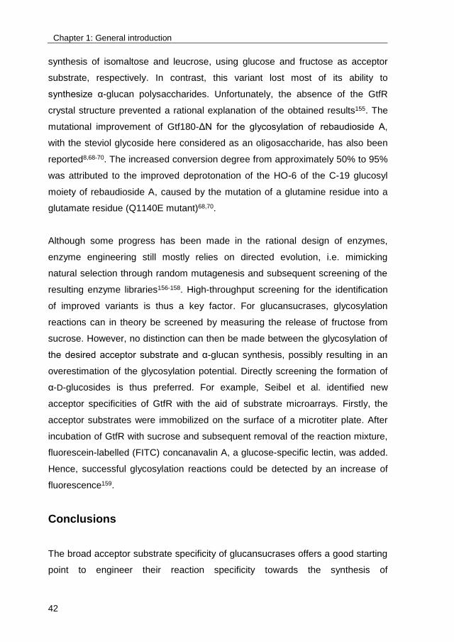
glycosylate catechol was examined (Figure 11). Based on the obtained results,
Gtf180-ΔN was preferred since it demonstrated a higher operational stability
(deactivated by catechol concentrations higher than 400 mM, compared to 300
and 200 mM for GtfA-ΔN and GtfO-ΔN, respectively) and a higher glycosylation
productivity, compared to the other enzymes.
Chapter 1: General introduction
44
Figure 11. Time-course synthesis of monoglycosylated catechol (catechol-G1) by ●
Gtf180-ΔN (400 mM catechol), ○ GtfA-ΔN (300 mM catechol) and ▼ GtfO-ΔN (200 mM
catechol). 1000 mM sucrose, 4 U/mL enzyme (total activity), T = 37 °C, pH = 4.7.
This introductory chapter also illustrated the commercial potential of glycosylating
steviol glycosides to improve their sensory characteristics. Therefore, our in-
house collection of GH70 glucansucrases was screened for their ability to
glycosylate the most prevalent steviol glycosides, rebaudioside A (RebA) and
stevioside. Once again, Gtf180-ΔN demonstrated most potential. In fact, it was
the only GH70 glucansucrase of our collection capable of glycosylating RebA.
The second part of this PhD thesis (Chapters 4, 5 and 6) consequently focusses
on the use of Gtf180-ΔN and derived mutants to glycosylate RebA and
stevioside. Chapter 7 is another illustration of the relatively high acceptor
substrate promiscuity of Gtf180-ΔN; also the high-intensity sweetener
neohesperidin dihydrochalcone (NHDC) was glycosylated in order to improve its
sensory –and physicochemical properties. Conclusively, this PhD thesis
describes the improvement of glucansucrase Gtf180-ΔN from L. reuteri 180 as
glycosylation biocatalyst by applying mutational engineering, optimization of
reaction conditions and development of downstream processing. Furthermore,
the sensory properties of the glycosylated products were determined by a trained
panel.
Chapter 1: General introduction
45
Chapter 1 discusses the state of the art glycosylation technologies, illustrating
their respective advantages and drawbacks by using the glycosylation of steviol
glycosides as case study. Secondly, it reviews current knowledge of GH70
glucansucrases and in particular their use as biocatalyst for the glycosylation of
non-carbohydrate compounds, such as (+)-catechin, and Stevia glycosides, such
as stevioside and rebaudioside A. Conclusively, reaction –and enzyme
engineering are discussed as strategies to improve the glycosylation potential of
GH70 glucansucrases.
Chapter 2 demonstrates that suppressing α-glucan synthesis by mutational
engineering of the Gtf180-ΔN enzyme of Lactobacillus reuteri 180 resulted in the
improved glycosylation of several non-carbohydrate acceptor substrates, such as
catechol and butanol. Three mutants (L938F, L981A and N1029M) were selected
from a previously constructed library of 61 mutants with single amino acid
residue changes, targeting 10 amino acid residues of the Gtf180-ΔN acceptor
binding sites +1 and +293,94. Kinetic analysis revealed that impairing α-glucan
synthesis resulted in a higher affinity of the mutant enzymes for the model
acceptor substrate catechol, explaining the improved monoglycosylation yields.
Finally, the chemical structures of the glycosylated acceptor substrates were
elucidated by NMR spectroscopy.
Chapter 3 discusses solvent engineering strategies (biphasic and cosolvents
systems) for the glycosylation of poorly soluble acceptor substrates by Gtf180-
ΔN of L. reuteri 180. Also the immobilization of this enzyme onto mesoporous
silica particles is described. The covalently immobilized WT enzyme retained
most of its activity at 60 °C, whereas the free WT enzyme lost all activity at
temperatures above 50 °C. Moreover, the covalently immobilized WT enzyme
was much more active at high DMSO concentrations and high acceptor substrate
concentrations than the free WT enzyme, allowing the glycosylation at higher
acceptor substrate concentrations of poorly soluble acceptor substrates, such as
ethyl gallate, gallic acid and (+)-catechin.

Chapter 1: General introduction
46
Chapter 4 reports the glycosylation of rebaudioside A (RebA), a natural high-
intensity sweetener obtained from the leaves of Stevia rebaudiana, by Gtf180-ΔN
and its Q1140E-mutant, which was selected from a mutant library99,100 in view of
its reduced α-glucan synthesis, enhanced RebA glycosylation potential and
introduction of a single extra glucose unit. Structural analysis of the main product
showed that both enzymes exclusively glucosylate RebA at the Glc(β1C-19
position, with the formation of an (α16) linkage. This result was explained by
docking of RebA in the enzyme’s active site, using its high resolution 3D
structure78: only the steviol C-19 β-D-glucosyl moiety is available for
glycosylation. The reaction conditions for the batch process were optimized by
response surface methodology (RSM), revealing the importance of the ratio of
donor substrate sucrose over acceptor substrate RebA to obtain high RebA
conversions. The product yield could be further enhanced by developing a fed-
batch reaction, which further suppressed the remaining and competing α-glucan
synthesis still present in mutant Q1140E. Sensory analysis of the glycosylated
RebA product by a trained taste panel revealed a near complete removal of
bitterness, compared to RebA. In other words, an efficient process for the
synthesis of a novel steviol glycoside (RebA-G1), displaying a superior taste
profile, was developed.
Chapter 5 continues the work on Stevia glycosides by reporting the glycosylation
of stevioside, the most abundant steviol glycoside extracted from the leaves of
Stevia rebaudiana, with the Gtf180-ΔN-Q1140E enzyme. The chemical
structures of the glycosylated stevioside products were elucidated by NMR
spectroscopy, revealing an identical regiospecificity as with RebA glycosylation.
Surprisingly, a minor diglycosylated product with an (α14) linkage was isolated
from the glycosylation reaction mixture, a specificity that is normally not displayed
by the Gtf180-ΔN enzyme. The reaction conditions for the batch process were
again optimized using RSM, indicating that stevioside glycosylation is
significantly less productive than RebA glycosylation. Analysis of the sensory
properties of the glycosylated stevioside product by a trained taste panel
revealed a substantial decrease in bitterness compared to stevioside and RebA.
Chapter 1: General introduction
47
Equally important, its taste profile was nearly identical to that of the glycosylated
RebA product.
Chapter 6 discusses the scale-up of the Gtf180-ΔN-ΔV-Q1140E-catalyzed
glycosylation of RebA and stevioside. Firstly, the enzyme production was
investigated. It was shown that the N- and V-terminally truncated Gtf180-ΔN-ΔV-
Q1140E protein was more efficiently produced than the N-terminally truncated
enzyme, whereas its biocatalytic potential was identical. Secondly, an eco-
friendly and efficient downstream processing of the glycosylated products was
developed and demonstrated at pilot-plant scale, allowing the production of 200 g
glycosylated RebA product. A cost analysis demonstrated the economic
feasibility of the process and pointed out the steviol glycoside substrate (RebA,
stevioside) cost as major factor in the total production cost. The use of low-grade
stevia extract, half the price of high-purity RebA, was therefore proposed as cost
reducing strategy. In this context, the simultaneous glycosylation of stevioside
and RebA was demonstrated, a technology which holds strong commercial
potential.
Chapter 7 illustrates the potential of Gtf180-ΔN-Q1140E to glycosylate
neohesperidin dihydrochalcone (NHDC), a high-intensity sweetener obtained
from citrus fruits. NHDC finds many applications as flavor enhancer (E959) in the
food- and pharmaceutical industry due to its ability to suppress bitterness and
astringency, but also draws interest due to its strong anti-oxidant capacity. The
main drawback of NHDC, its low water solubility, was overcome by its
glycosylation, while the glycosides retained strong anti-oxidant capacities.
Sensory analysis of the flavor enhancing effects of NHDC and the NHDC
glycosides on RebA by a trained panel revealed a slight decrease of bitterness
and an increased sweetness. Although the obtained conversion degree of
roughly 65% was not of the same order as for RebA and stevioside glycosylation
(95%), it forms a good starting point for a profound optimization of the reaction
conditions.
Chapter 8 summarizes and discusses the results presented in this PhD thesis.
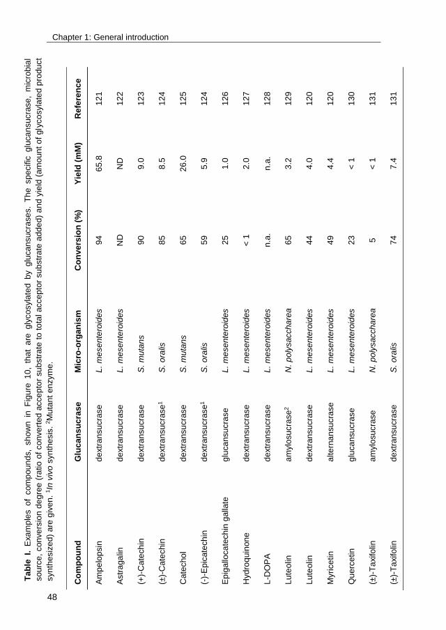
Chapter 1: General introduction
48
Ta
ble
I.
E
xa
mp
les o
f com
pou
nd
s,
sh
ow
n in
F
igu
re 1
0,
tha
t a
re gly
co
syla
ted b
y glu
can
sucra
se
s.
Th
e sp
ecific
g
luca
nsucra
se
, m
icro
bia
l
so
urc
e,
con
ve
rsio
n d
eg
ree
(ra
tio
of
co
nve
rted
acce
pto
r su
bstr
ate
to
to
tal accep
tor
su
bstr
ate
ad
de
d)
an
d y
ield
(am
oun
t o
f gly
co
syla
ted
pro
du
ct
syn
thesiz
ed
) a
re g
iven
. 1In
viv
o s
yn
thesis
. 2M
uta
nt
en
zym
e.
Co
mp
ou
nd
G
luc
an
su
cra
se
Mic
ro-o
rgan
ism
C
on
ve
rsio
n (
%)
Yie
ld (
mM
) R
efe
ren
ce
Am
pe
lop
sin
d
extr
an
su
cra
se
L.
me
se
nte
roid
es
94
65
.8
12
1
Astr
aga
lin
de
xtr
an
su
cra
se
L.
me
se
nte
roid
es
ND
N
D
12
2
(+)-
Cate
ch
in
de
xtr
an
su
cra
se
S.
mu
tan
s
90
9.0
1
23
(±)-
Ca
techin
d
extr
an
su
cra
se
1
S.
ora
lis
85
8.5
1
24
Ca
techo
l d
extr
an
su
cra
se
S.
mu
tan
s
65
26
.0
12
5
(-)-
Ep
icate
ch
in
de
xtr
an
su
cra
se
1
S.
ora
lis
59
5.9
1
24
Ep
iga
lloca
tech
in g
alla
te
glu
ca
nsu
cra
se
L.
me
se
nte
roid
es
25
1.0
1
26
Hyd
roq
uin
on
e
de
xtr
an
su
cra
se
L.
me
se
nte
roid
es
< 1
2
.0
12
7
L-D
OP
A
de
xtr
an
su
cra
se
L.
me
se
nte
roid
es
n.a
. n
.a.
12
8
Lu
teolin
a
mylo
su
cra
se
2
N.
po
lysacch
are
a
65
3.2
1
29
Lu
teolin
d
extr
an
su
cra
se
L.
me
se
nte
roid
es
44
4.0
1
20
Myri
ce
tin
alte
rna
nsu
cra
se
L.
me
se
nte
roid
es
49
4.4
1
20
Qu
erc
etin
glu
ca
nsu
cra
se
L.
me
se
nte
roid
es
23
< 1
1
30
(±)-
Ta
xifo
lin
am
ylo
su
cra
se
N.
po
lysacch
are
a
5
< 1
1
31
(±)-
Ta
xifo
lin
de
xtr
an
su
cra
se
S.
ora
lis
74
7.4
1
31
49
Chapter 2
Glucansucrase Gtf180-ΔN of Lactobacillus reuteri 180:
suppressing α-glucan synthesis results in improved
glycosylation yields
This chapter is published as:
Tim Devlamynck, Evelien M. te Poele, Xiangfeng Meng, Sander S. van Leeuwen,
Lubbert Dijkhuizen (2016) Glucansucrase Gtf180-ΔN of Lactobacillus reuteri 180:
enzyme and reaction engineering for improved glycosylation of non-carbohydrate
molecules. Appl Microbiol Biotechnol 100:7529-7539.

Chapter 2: Suppressing α-glucan synthesis by mutational engineering
50
Abstract
Glucansucrases have a broad acceptor substrate specificity and receive
increased attention as biocatalysts for the glycosylation of small non-
carbohydrate molecules using sucrose as donor substrate. However, the main
glucansucrase catalyzed reaction results in synthesis of α-glucan
polysaccharides from sucrose and this strongly impedes the efficient
glycosylation of non-carbohydrate molecules and complicates downstream
processing of glucosylated products. This chapter reports that suppressing α-
glucan synthesis by mutational engineering of the Gtf180-ΔN enzyme of
Lactobacillus reuteri 180 results in the construction of more efficient glycosylation
biocatalysts. Gtf180-ΔN mutants (L938F, L981A and N1029M) with an impaired
α-glucan synthesis displayed a substantial increase in monoglycosylation yields
for several phenolic and alcoholic compounds. Kinetic analysis revealed that
these mutants possess a higher affinity for the model acceptor substrate catechol
but a lower affinity for its mono-α-D-glucoside product, explaining the improved
monoglycosylation yields. Analysis of the available high resolution 3D crystal
structure of the Gtf180-ΔN protein provided a clear understanding of how
mutagenesis of residues L938, L981 and N1029 impaired α-glucan synthesis,
thus yielding mutants with an improved glycosylation potential.

Chapter 2: Suppressing α-glucan synthesis by mutational engineering
51
1. Introduction
Glycosylation is a versatile tool to enhance the physicochemical and biological
properties of small non-carbohydrate molecules13
. This may result in an
increased solubility of hydrophobic compounds33
and an improved stability of
labile molecules against light and oxidation7. Furthermore, glycosylating medium-
and long-chain alcohols yields alkyl glycosides or alkyl polyglycosides, a class of
eco-friendly and non-ionic surfactants displaying a high surface activity and good
biodegradability160
.
The chemical synthesis of glycosides requires the use of toxic catalysts and
involves many protection and deprotection steps, resulting in low overall yields.
Biocatalysis offers an alternative method circumventing multistep-synthesis and
generating 5-fold less waste17
. In nature, glycosylation is catalyzed by Leloir
glycosyltransferase enzymes (EC 2.4.-.-), using nucleotide-activated sugars as
donor substrates. Despite their high efficiency and specificity, the breakthrough
as glycosylation catalysts is hampered by the high price of their donor
substrates18
. Glycosidases (EC 3.2.-.-) in turn suffer from low yields when applied
in the synthetic direction161
.
Glycoside hydrolase enzymes such as glucansucrases (GS) provide an excellent
alternative for enzymatic glycoside synthesis. These enzymes belong to
glycoside hydrolase family 70 (GH70)90
and catalyze the conversion of the cheap
donor substrate sucrose into α-glucan polysaccharides, thereby linking the α-D-
glucopyranosyl units by (α1→2), (α1→3), (α1→4) or (α1→6) bonds, depending
on the enzyme specificity73,74
. Moreover, GS are promiscuous towards a wide
range of acceptor substrates75,162
. They can use saccharides such as maltose as
acceptor substrate to catalyze the synthesis of various oligosaccharides163
.
Glycosylation of non-carbohydrate acceptor substrates, such as L-ascorbic
acid164
and luteolin120
, also has been reported. The usefulness of GS enzymes as
a glycosylation biocatalyst is further demonstrated by a number of patent
Chapter 2: Suppressing α-glucan synthesis by mutational engineering
52
applications by Auriol et al. (2012), in which the synthesis of a wide array of
phenolic compounds with Leuconostoc glucansucrases is claimed165
.
A remarkable characteristic shared by all GS is their ability to add multiple α-D-
glucopyranosyl moieties to one acceptor substrate, forming α-D-glucosides of
different sizes and structures. A prominent example concerns the glycosylation of
acceptor substrates by the GtfA enzyme of Lactobacillus reuteri 12180
: after
incubation with catechol and sucrose, several glycosylated catechol products up
to DP5, differing in their combination of (α1→4) and (α1→6) linkages, were
characterized138
. From an industrial perspective, the synthesis of only one
glycoside is desired in order to facilitate downstream processing. In addition to
the production of a mixture of α-D-glucosides, glucansucrases also synthesize
rather large amounts of α-glucan polysaccharides from sucrose under these
conditions. This is in fact their main reaction but in this case an unwanted side
reaction lowering the yield of the glycosylated acceptor substrates and
complicating their downstream processing. In this chapter, a combination of
reaction- and enzyme engineering was applied to explore the potential of the N-
terminally truncated glucansucrase Gtf180 from Lactobacillus reuteri 180
(Gtf180-ΔN, retaining wild type activity and specificity)82
as a glycosylation
biocatalyst, aiming to suppress the competing α-glucan synthesis reaction as
much as possible. Screening of a previously constructed mutant library, targeting
10 amino acid residues involved in the acceptor substrate binding subsites +1
and +299,100
, yielded mutants with an impaired α-glucan synthesis. As will be
demonstrated, this substantially enhanced the conversion of a wide range of
phenolic and alcoholic molecules into their α-D-glucosides, and also shifted the
glycoside distribution pattern towards monoglycosylation.
Chapter 2: Suppressing α-glucan synthesis by mutational engineering
53
2. Materials and methods
2.1. Production and purification of recombinant Gtf180-ΔN (mutants)
Recombinant, N-terminally truncated Gtf180-ΔN from Lactobacillus reuteri 180
and derived mutant enzymes (Table S1) were produced and purified as
described previously80,99
.
2.2. Glucansucrase activity assays
Enzyme activity assays were performed at 37°C with 100 mM sucrose in 25 mM
sodium acetate (pH 4.7) and 1 mM CaCl2 unless stated otherwise. Samples of
100 μl were taken every min over a period of 8 min and immediately inactivated
with 20 μl 1 M NaOH for 30 min. The released glucose and fructose were
quantified enzymatically by monitoring the reduction of NADP with the
hexokinase and glucose-6-phosphate dehydrogenase/phosphoglucose
isomerase assay (Roche) as described previously166,167
, allowing the
determination of the total- (fructose release) and hydrolytic (glucose release)
activities, and calculation of the transglycosylation activity. The α-glucan
synthesis potential (α-GSP) is defined as the ratio of transglycosylation activity
over total activity.
One unit (U) of total activity corresponds to the release of 1 μmole fructose from
100 mM sucrose in 25 mM sodium acetate (pH 4.7) and 1 mM CaCl2 at 37 °C.
For the comparison of different reaction conditions and mutants, 4 U/mL enzyme
was added to the incubations, unless stated otherwise.
2.3. Production and purification of glycoside products
The glycosylation of catechol, resorcinol, hydroquinone and butanol was carried
out at 100 mL scale, by incubating 1 U/mL Gtf180-ΔN at 37°C in 25 mM sodium
acetate (pH 4.7) and 1 mM CaCl2 with 400 mM acceptor substrate and 1000 mM

Chapter 2: Suppressing α-glucan synthesis by mutational engineering
54
sucrose for 2 h. Alternatively, hexanol and octanol were glycosylated in a
biphasic system consisting of 20% alcohol, 25 mM sodium acetate (pH 4.7), 1
mM CaCl2 and 1000 mM sucrose, while stirring was achieved in a shaker at 100
rpm. The reactions were terminated by incubating the reaction mixture at 95°C
for 10 min. Most of the fermentable sugars were subsequently removed by
fermentation with the yeast Saccharomyces cerevisiae (Fermentis Ethanol
Red®) at pH 4.0 and 30°C168
. Twenty g/L peptone and 10 g/L yeast extract were
added to support growth. After 24 h incubation the yeast cells were removed by
centrifugation (10000 x g, 4 °C, 10 min) after which the supernatant was
concentrated by evaporating in vacuo. The glycoside products were
subsequently purified from the residue by column chromatography using silica
gel (pore size 60 Å, particle size 230-400 mesh) as the stationary phase. The
eluent consisted of ethyl acetate-methanol-water (30:5:4 by volume) in case
monoglucosides were purified and ethyl acetate-methanol-water (30:6:4 by
volume) for the purification of diglucosides.
2.4. HPLC analysis
HPLC analysis of phenolic acceptor molecules and their α-D-glucosides was
performed on an Adsorbil amine column (250 mm × 4.6 mm, 10 μm) with
acetonitrile (solvent A) and 50 mM ammonium formate (pH 4.4, solvent B) as the
mobile phase. The flow rate and temperature were set at 1.0 mL/min and 35°C,
respectively. The following gradient elution was used: 95% of solvent A (0−5
min), 5-40% solvent B (5−22 min), 80% solvent B (22-25 min) and again 95% of
solvent A (25-29 min). Detection of the phenolic acceptor substrates and their α-
D-glucosides was achieved with an UV detector (276 nm). Before being
subjected to HPLC analysis the samples were diluted 200 times in 80%
methanol. Calibration of the obtained peaks was accomplished using standard
curves of the purified glycosides. All HPLC analyses were performed in duplicate.
Chapter 2: Suppressing α-glucan synthesis by mutational engineering
55
2.5. TLC analysis
TLC analysis was performed on silica gel 60 F254 plates (Merck). The eluent
consisted of ethyl acetate-methanol-water (30:5:4 by volume). Detection was
achieved by UV absorption (254 nm) and/or staining with 10% (v/v) H2SO4
containing 2 g/L orcinol. The concentration of the alkyl α-D-glucosides was
determined by scanning the charred plates with a ChemiDocTM
MP imaging
system and subsequently analyzing the spots with Image Lab 4.0 software.
Calibration of the obtained spots was accomplished using standard curves of the
purified alkyl α-D-glucosides. There was a linear response in the range of 1-10
mM alkyl glucoside (determined experimentally). All TLC analyses were
performed in triplicate.
2.6. Kinetic analysis of Gtf180-ΔN (mutants)
Kinetic analysis of the Gtf180-ΔN (mutants) was based upon the method
described by Dirks-Hofmeister et al. (2015) for the glycosylation of resveratrol
with sucrose phophorylase25
. Kinetic parameters (Km and kcat values) for the
acceptor substrates catechol and the mono-α-D-glucoside of catechol (catechol-
G1), purified as described above, were determined using 10 different catechol(-
G1) concentrations (ranging from 6.25 to 400 mM), while the concentration of the
donor substrate sucrose had a constant value of 1000 mM. One U/mL of Gtf180-
ΔN (mutants) was added. Four samples were taken over a period of 3 min and
immediately inactivated by incubating for 10 min at 95 °C. All samples were
subjected to TLC analysis as described above. The charred plates were scanned
with a ChemiDocTM
MP imaging system allowing analysis of the spots with Image
Lab 4.0 software. Calibration of the obtained spots was accomplished using
standard curves of the purified catechol-G1. Kinetic parameters were calculated
by non-linear regression of the Michaelis-Menten equation with SigmaPlot v12.0.
Chapter 2: Suppressing α-glucan synthesis by mutational engineering
56
2.7. Structural characterization of purified α-D-glucosides
The structures of the purified α-D-glucosides were elucidated by a combination of
1D NMR (1H NMR and
13C NMR) and 2D NMR spectroscopy. Samples were
exchanged twice in 300 μL D2O 99.9 %atom (Cambridge Isotope Laboratories,
Andover, MA) with intermediate lyophilisation. Finally, samples were dissolved in
650 μL D2O, containing acetone as internal standard (δ1H 2.225; δ
13C 31.08).
1H
NMR spectra, including 1H-
1H and
13C-
1H correlation spectra were recorded at a
probe temperature of 298K on a Varian Inova 600 spectrometer (NMR
Department, University of Groningen, The Netherlands). 1D 600-MHz 1H NMR
spectra were recorded with 5000 Hz spectral width at 16k complex data points,
using a WET1D pulse to suppress the HOD signal. 2D 1H-
1H COSY spectra were
recorded in 256 increments in 4000 complex data points with a spectral width of
5000 Hz. 2D 1H-
1H TOCSY spectra were recorded with MLEV17 mixing
sequences with 50, 90, and 150 ms spin-lock times. 2D 13
C-1H HSQC spectra
were recorded with a spectral width of 5000 Hz in t2 and 10,000 Hz in t1 direction.
2D 1H-
1H ROESY spectra with a mixing time of 300 ms were recorded in 128
increments of 4000 complex data points with a spectral width of 5000 Hz. All
spectra were processed using MestReNova 5.3 (Mestrelabs Research SL,
Santiago de Compostela, Spain), using Whittaker Smoother baseline correction.
3. Results
Glucansucrases prefer non-carbohydrate acceptor substrates with two vicinal
hydroxyl groups120
, such as catechol. The latter has a high water solubility at
room temperature, rendering the addition of co-solvents unnecessary.
Glycosylation of catechol with the N-terminally truncated glucansucrase of
Lactobacillus reuteri 180 (Gtf180-ΔN)82
was chosen as the model reaction.
Firstly, the reaction conditions were optimized towards maximal
monoglycosylation and minimal α-glucan synthesis. Subsequently, the mutant
library was screened, applying these optimal reaction conditions. Finally, the

Chapter 2: Suppressing α-glucan synthesis by mutational engineering
57
optimal reaction conditions identified for catechol glycosylation were also tested
for glycosylation of other acceptor substrates.
3.1. Reaction engineering of catechol glycosylation by wild-type Gtf180-ΔN
The catechol acceptor concentration was optimized towards maximal
monoglycosylation and minimal α-glucan synthesis. As shown in Figure 1,
formation of the monoglucoside of catechol (catechol-G1) is kinetically controlled.
Incubation for 20 min was sufficient to reach maximal catechol-G1 production,
coinciding with catechol depletion. Catechol-G1 was subsequently irreversibly
converted into diglucoside (catechol-3`G2 and catechol-6`G2) and further
(catechol-G3+). The donor substrate sucrose was not depleted yet (data not
shown).
Figure 1. Time-course synthesis of α-D-glucosides of catechol by WT Gtf180-ΔN (400 mM
catechol; 1000 mM sucrose; 4 U/mL Gtf180-ΔN). T = 37 °C, pH = 4.7.
Glycosylation reactions catalyzed by glucansucrases suffer from low
thermodynamic favorability as pointed out by Liang et al. (2016)151
. The
production of high catechol-G1 concentrations therefore requires an excess of
donor substrate sucrose to drive the reaction. We observed that the latter also
had a stabilizing effect on the enzyme, allowing addition of relatively high
acceptor substrate concentrations which would otherwise be detrimental for the

Chapter 2: Suppressing α-glucan synthesis by mutational engineering
58
enzyme activity as described previously125
. Therefore, the sucrose concentration
was set at 1000 mM. Kinetic analysis revealed that Gtf180-ΔN follows Michaelis-
Menten kinetics at catechol acceptor substrate concentrations between 6.25 and
400 mM (Figure S1). The Km value of Gtf180-ΔN for catechol was 103.3 mM
which illustrated the need for high acceptor substrate concentrations (Table 1).
Therefore, the catechol concentration was varied from 100 mM to 1000 mM while
the sucrose concentration was kept constant at 1000 mM (Table 2). At catechol
concentrations higher than 600 mM no glycosylated product was formed due to
severe inhibition of enzyme activity by catechol. A catechol concentration of 500
mM and 600 mM only allowed partial conversion of catechol with
monoglycosylation yields of 17% and 7% respectively. Reaction mixtures
containing 400 mM catechol or less, displayed complete conversion of this
acceptor substrate into α-D-glucoside products. Increasing the acceptor
concentration from 100 to 400 mM resulted in an improvement in
monoglycosylation yield from 49% to 60%, whereas the synthesis of
triglucosylated products was reduced (Table 2). At higher catechol
concentrations there indeed is an increased chance that the enzyme glycosylates
a new acceptor substrate rather than glycosylating catechol-G1. Consequently,
400 mM catechol was chosen as the optimal acceptor concentration for the
production of monoglucosides.
The Km value of Gtf180-ΔN for the catechol-G1 acceptor substrate was 88.8 mM,
which is lower than the value for catechol (103.3 mM). The kcat values were 863.3
s-1
and 757.4 s-1
respectively (Table 1). Hence, under these conditions Gtf180-
ΔN glycosylation of catechol-G1 into catechol-G2 and further is inevitable. In the
next step we optimized monoglucoside synthesis by applying Gtf180-ΔN
mutants, aiming to increase the Km value for catechol-G1 and/or decrease the Km
value for catechol.

Chapter 2: Suppressing α-glucan synthesis by mutational engineering
59
Table 1. Kinetic parameters of WT Gtf180-ΔN and mutants derived for catechol (6.25 -
400 mM) and catechol-G1 (6.25 - 400 mM) as acceptor substrates (with sucrose as donor
substrate at 1000 mM), and α-GSP1 of Gtf180-ΔN WT and mutants for sucrose as both
donor and acceptor substrate. T = 37 °C, pH = 4.7.
Enzymes Catechol Catechol-G1
Km
(mM)
kcat
(s-1)
kcat/Km
(s-1.mM
-1)
Km
(mM)
kcat
(s-1)
kcat/Km
(s-1.mM
-1)
α-GSP
WT 103.3 ±
8.5
757.4 ±
12.4 7.4
88.8 ±
17.1
863.3 ±
39.3 10.2 0.556
L938F 85.5 ±
4.0
1872.5 ±
74.7 21.9
91.1 ±
7.3
576.2 ±
44.0 6.3 0.341
N1029M 58.9 ±
6.4
449.4 ±
4.9 7.7
146.9 ±
19.3
126.2 ±
11.0 0.9 0.192
L981A 11.0 ±
1.3
203.2 ±
8.1 18.7
177.4 ±
7.0
69.4 ±
2.4 0.4 0.049
1 α-GSP is defined as the ratio of the transglycosylation activity over the total activity (measured with
1000 mM sucrose only).
Table 2. Effects of acceptor substrate concentration on the glycosylation yields and
glucoside distribution1 of WT Gtf180-ΔN for the model acceptor substrate catechol (1000
mM sucrose; 4 U/mL Gtf180-ΔN). T = 37 °C, pH = 4.7.
Catechol (mM) Catechol glucoside (mM) Catechol glucoside distribution (%)
G1 G2α1→3 G2α1→6 G3+ G1 G2 G3+
600 39.6 - - - 100 - -
500 86.8 < 10.0 < 10.0 - 96 4 4
400 241.1 33.8 68.0 57.3 60 25 14
300 170.1 27.5 56.0 46.3 57 28 15
200 107.2 17.9 37.6 37.2 54 28 19
100 49.4 9.3 18.1 23.1 49 27 23
1 All data given at maximal catechol-G1 yield (20 min incubation).
Chapter 2: Suppressing α-glucan synthesis by mutational engineering
60
3.2. Mutational engineering of the Gtf180-ΔN enzyme
3.2.1. Selection of Gtf180-ΔN mutants
A library of 61 mutants with single amino acid residue changes (Table S1),
targeting 10 amino acid residues of the Gtf180-ΔN acceptor binding sites +1 and
+2, has been constructed previously99,100
. A quick and qualitative screening was
performed to identify mutants displaying a relative increase in monoglycosylation
and a decrease in α-glucan synthesis. For this purpose, 1 U/mL of every mutant
was incubated for 1 h at the optimal reaction conditions (400 mM catechol, 1000
mM sucrose). The resulting reaction mixtures were subsequently spotted on TLC
plates and mutually compared after staining (Figure S2).
Mutants of residues D1085, R1088 and N1089 were not affected in catechol
glycosylation, since their product profiles were nearly identical to those of the WT
Gtf180-ΔN. Mutants of W1065, a residue proven to be essential for both activity
and acceptor binding by interacting with maltose through aromatic stacking83,95
,
displayed a very low total activity. Although the product profiles of these mutants
were improved (more catechol-G1), their low total conversion and low specific
activity rendered them less useful as glycosylation biocatalyst. Mutating D1028
yielded mutants with an enhanced oligosaccharide synthesis, as suggested by
the more intense α-glucan oligosaccharide tail visible on TLC (Figure S2). Since
this was the opposite of what was aimed for, these mutants were not selected for
further analysis. Mutants of L940 all showed a shift in diglucoside linkage type,
forming almost exclusively (α1→6) bonds. Indeed, the crucial role of L940 for
linkage specificity in α-glucan synthesis was demonstrated previously98
.
However, no relative increase in monoglycosylation yield was detected.
Mutants of residues L938, L981 and N1029 provided the most interesting results.
Every L938 mutant tested showed an increased monoglucoside synthesis and a
decreased formation of di- and triglucosides; the strongest effect was observed
for mutant L938F (Figure S2). Similar effects were obtained with L981 mutants,
especially when the leucine residue was replaced by alanine. In case of N1029
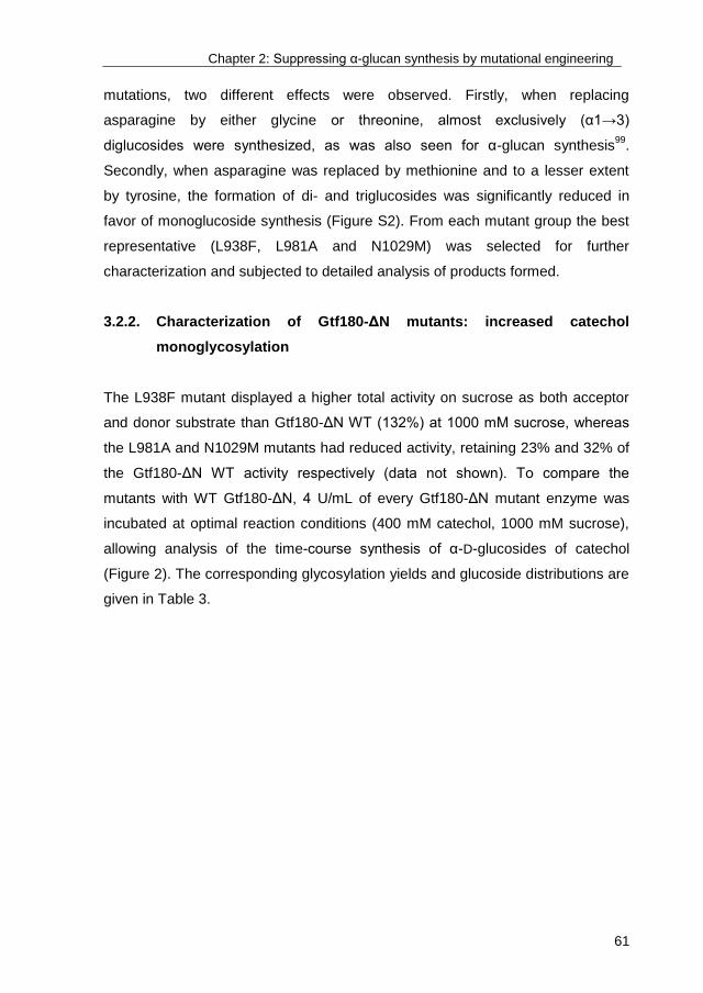
Chapter 2: Suppressing α-glucan synthesis by mutational engineering
61
mutations, two different effects were observed. Firstly, when replacing
asparagine by either glycine or threonine, almost exclusively (α1→3)
diglucosides were synthesized, as was also seen for α-glucan synthesis99
.
Secondly, when asparagine was replaced by methionine and to a lesser extent
by tyrosine, the formation of di- and triglucosides was significantly reduced in
favor of monoglucoside synthesis (Figure S2). From each mutant group the best
representative (L938F, L981A and N1029M) was selected for further
characterization and subjected to detailed analysis of products formed.
3.2.2. Characterization of Gtf180-ΔN mutants: increased catechol
monoglycosylation
The L938F mutant displayed a higher total activity on sucrose as both acceptor
and donor substrate than Gtf180-ΔN WT (132%) at 1000 mM sucrose, whereas
the L981A and N1029M mutants had reduced activity, retaining 23% and 32% of
the Gtf180-ΔN WT activity respectively (data not shown). To compare the
mutants with WT Gtf180-ΔN, 4 U/mL of every Gtf180-ΔN mutant enzyme was
incubated at optimal reaction conditions (400 mM catechol, 1000 mM sucrose),
allowing analysis of the time-course synthesis of α-D-glucosides of catechol
(Figure 2). The corresponding glycosylation yields and glucoside distributions are
given in Table 3.
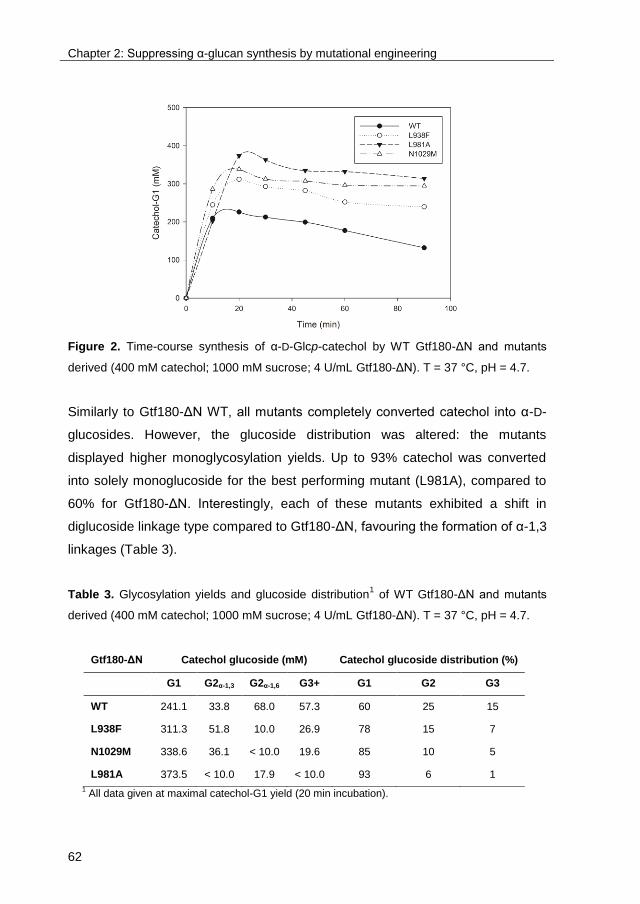
Chapter 2: Suppressing α-glucan synthesis by mutational engineering
62
Figure 2. Time-course synthesis of α-D-Glcp-catechol by WT Gtf180-ΔN and mutants
derived (400 mM catechol; 1000 mM sucrose; 4 U/mL Gtf180-ΔN). T = 37 °C, pH = 4.7.
Similarly to Gtf180-ΔN WT, all mutants completely converted catechol into α-D-
glucosides. However, the glucoside distribution was altered: the mutants
displayed higher monoglycosylation yields. Up to 93% catechol was converted
into solely monoglucoside for the best performing mutant (L981A), compared to
60% for Gtf180-ΔN. Interestingly, each of these mutants exhibited a shift in
diglucoside linkage type compared to Gtf180-ΔN, favouring the formation of α-1,3
linkages (Table 3).
Table 3. Glycosylation yields and glucoside distribution1 of WT Gtf180-ΔN and mutants
derived (400 mM catechol; 1000 mM sucrose; 4 U/mL Gtf180-ΔN). T = 37 °C, pH = 4.7.
1 All data given at maximal catechol-G1 yield (20 min incubation).
Gtf180-ΔN Catechol glucoside (mM) Catechol glucoside distribution (%)
G1 G2α-1,3 G2α-1,6 G3+ G1 G2 G3
WT 241.1 33.8 68.0 57.3 60 25 15
L938F 311.3 51.8 10.0 26.9 78 15 7
N1029M 338.6 36.1 < 10.0 19.6 85 10 5
L981A 373.5 < 10.0 17.9 < 10.0 93 6 1

Chapter 2: Suppressing α-glucan synthesis by mutational engineering
63
Determination of the kinetic parameters (Table 1) revealed that two opposite but
related effects form the basis for the improved monoglycosylation yields. Except
for the L938F mutant, the mutants had lower kcat values for the acceptor reaction
with catechol and sucrose (Table 1), mainly representing a reduction in total
activity with sucrose alone as shown above. However, all mutants displayed
much lower Km values for catechol than the Gtf180-ΔN WT. In particular the
L981A mutant had a low Km value of 11.0 mM for catechol, representing a 9-fold
improvement compared to the Gtf180-ΔN WT. Despite the relatively low total
activity of mutant L981A (23% of Gtf180-ΔN), its catalytic efficiency (kcat/Km) for
the acceptor reaction with catechol (plus sucrose) was 2.5-fold higher than of
Gtf180-ΔN WT. The exact opposite was observed when comparing the kinetic
parameters of the mutants with Gtf180-ΔN WT for catechol-G1 as acceptor
substrate: all mutants displayed higher Km values for catechol-G1 whereas their
catalytic efficiencies were substantially lower.
To elucidate the underlying molecular mechanism, the transglycosylation and
total activities of the Gtf180-ΔN (mutants), incubated with sucrose only, were
determined. Subsequently, the α-glucan synthesis potential (α-GSP) was
calculated, defined as the ratio of the transglycosylation activity over the total
activity, revealing the potential of the enzyme to use the donor substrate sucrose
for α-glucan synthesis (and not for hydrolysis). As shown in Table 1, the mutants
showed a decrease in α-GSP compared to the Gtf180-ΔN WT.
In conclusion, mutant L981A represents a highly efficient biocatalyst for the
glycosylation of catechol, yielding roughly 100 g/L catechol-G1 (373 mM) with a
yield of 93%.
3.2.3. Characterization of Gtf180-ΔN mutants: increased acceptor
substrate conversion
Due to an increased affinity for catechol which resulted from an impaired α-GSP,
the monoglycosylation yield of Gtf180-ΔN mutants for the glycosylation of
catechol was significantly improved. Subsequently, we determined whether the
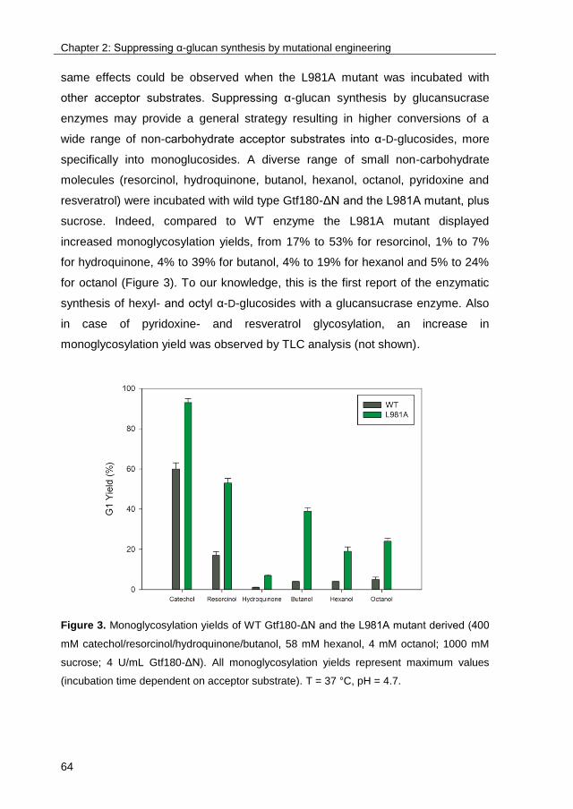
Chapter 2: Suppressing α-glucan synthesis by mutational engineering
64
same effects could be observed when the L981A mutant was incubated with
other acceptor substrates. Suppressing α-glucan synthesis by glucansucrase
enzymes may provide a general strategy resulting in higher conversions of a
wide range of non-carbohydrate acceptor substrates into α-D-glucosides, more
specifically into monoglucosides. A diverse range of small non-carbohydrate
molecules (resorcinol, hydroquinone, butanol, hexanol, octanol, pyridoxine and
resveratrol) were incubated with wild type Gtf180-ΔN and the L981A mutant, plus
sucrose. Indeed, compared to WT enzyme the L981A mutant displayed
increased monoglycosylation yields, from 17% to 53% for resorcinol, 1% to 7%
for hydroquinone, 4% to 39% for butanol, 4% to 19% for hexanol and 5% to 24%
for octanol (Figure 3). To our knowledge, this is the first report of the enzymatic
synthesis of hexyl- and octyl α-D-glucosides with a glucansucrase enzyme. Also
in case of pyridoxine- and resveratrol glycosylation, an increase in
monoglycosylation yield was observed by TLC analysis (not shown).
Figure 3. Monoglycosylation yields of WT Gtf180-ΔN and the L981A mutant derived (400
mM catechol/resorcinol/hydroquinone/butanol, 58 mM hexanol, 4 mM octanol; 1000 mM
sucrose; 4 U/mL Gtf180-ΔN). All monoglycosylation yields represent maximum values
(incubation time dependent on acceptor substrate). T = 37 °C, pH = 4.7.
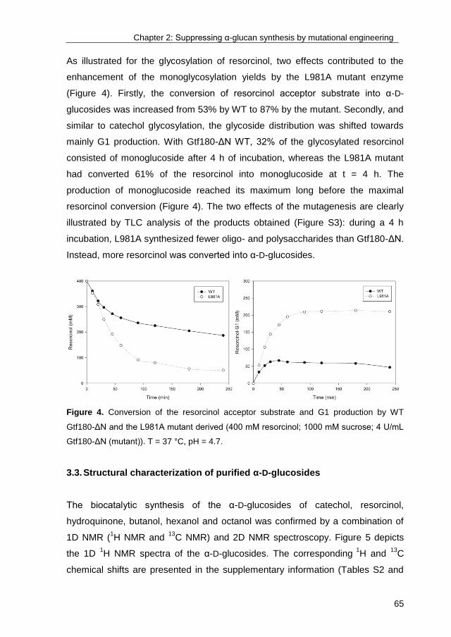
Chapter 2: Suppressing α-glucan synthesis by mutational engineering
65
As illustrated for the glycosylation of resorcinol, two effects contributed to the
enhancement of the monoglycosylation yields by the L981A mutant enzyme
(Figure 4). Firstly, the conversion of resorcinol acceptor substrate into α-D-
glucosides was increased from 53% by WT to 87% by the mutant. Secondly, and
similar to catechol glycosylation, the glycoside distribution was shifted towards
mainly G1 production. With Gtf180-ΔN WT, 32% of the glycosylated resorcinol
consisted of monoglucoside after 4 h of incubation, whereas the L981A mutant
had converted 61% of the resorcinol into monoglucoside at t = 4 h. The
production of monoglucoside reached its maximum long before the maximal
resorcinol conversion (Figure 4). The two effects of the mutagenesis are clearly
illustrated by TLC analysis of the products obtained (Figure S3): during a 4 h
incubation, L981A synthesized fewer oligo- and polysaccharides than Gtf180-ΔN.
Instead, more resorcinol was converted into α-D-glucosides.
Figure 4. Conversion of the resorcinol acceptor substrate and G1 production by WT
Gtf180-ΔN and the L981A mutant derived (400 mM resorcinol; 1000 mM sucrose; 4 U/mL
Gtf180-ΔN (mutant)). T = 37 °C, pH = 4.7.
3.3. Structural characterization of purified α-D-glucosides
The biocatalytic synthesis of the α-D-glucosides of catechol, resorcinol,
hydroquinone, butanol, hexanol and octanol was confirmed by a combination of
1D NMR (1H NMR and
13C NMR) and 2D NMR spectroscopy. Figure 5 depicts
the 1D 1H NMR spectra of the α-D-glucosides. The corresponding
1H and
13C
chemical shifts are presented in the supplementary information (Tables S2 and
Chapter 2: Suppressing α-glucan synthesis by mutational engineering
66
S3). Figures S4-S9 of the supplementary information represent the 1D 1H NMR
spectrum, and 2D 1H-
1H COSY, TOCSY (150 ms mixing time), ROESY (300 ms
mixing time) and 13
C-1H HSQC spectra of butyl glucoside, hexyl glucoside, octyl
glucoside, resorcinol-G1, hydroquinone-G1 and catechol-3`G2, respectively. The
1D 1H NMR spectra of catechol-G1 and catechol-6`G2 matched with those found
previously by te Poele et al. (2016)138
and are presented there. For a detailed
analysis of the NMR spectra, see supplementary information.
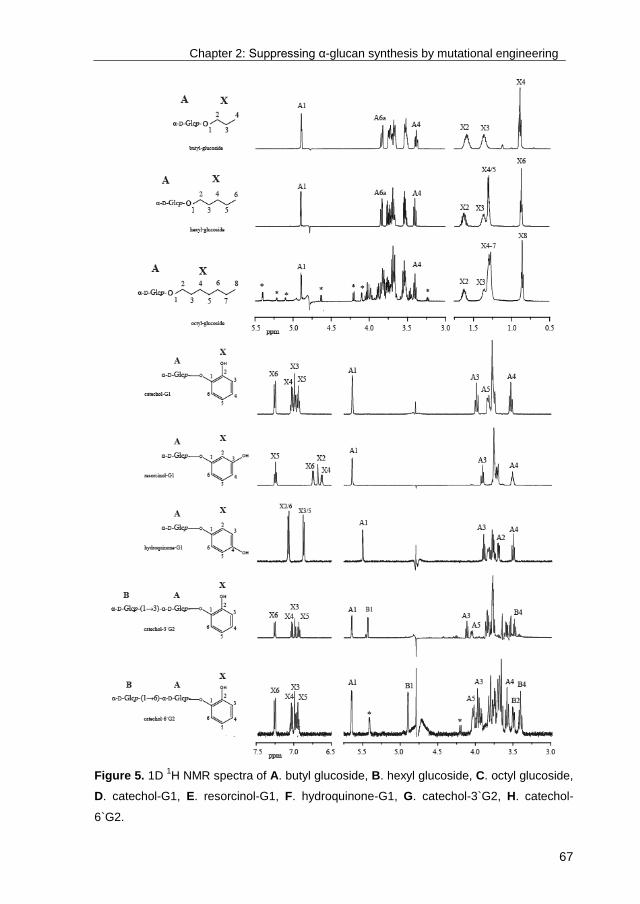
Chapter 2: Suppressing α-glucan synthesis by mutational engineering
67
Figure 5. 1D 1H NMR spectra of A. butyl glucoside, B. hexyl glucoside, C. octyl glucoside,
D. catechol-G1, E. resorcinol-G1, F. hydroquinone-G1, G. catechol-3`G2, H. catechol-
6`G2.

Chapter 2: Suppressing α-glucan synthesis by mutational engineering
68
4. Discussion
In view of their broad acceptor substrate specificity, glucansucrases are
considered promising glycosylation biocatalysts. However, the typical synthesis
of a mixture of α-D-glucosides, oligosaccharides and α-D-glucans remains a
bottleneck in their industrial application. α-Glucan synthesis is the main
glucansucrase reaction but an undesired side reaction when aiming to
glycosylate non-carbohydrate acceptor substrates, lowering glycosylation yields
and complicating downstream processing. For example, when applying salicin
and salicyl alcohol as acceptor substrates, B-1355C2 and B-1299CB-BF563
dextransucrases from Leuconostoc mensenteroides synthesized at least 12 and
9 different kinds of glycosides, respectively169
.
So far, few enzyme engineering studies with glucansucrases have focused on
glycosylation of non-carbohydrate acceptor substrates. In 2014, Malbert et al.
reported a significant improvement of luteolin monoglycosylation by the I228A
NpAS mutant compared to the wild type enzyme. Docking studies attributed this
enhancement to the introduction of a less hindering residue, assisting in a better
positioning of luteolin in the catalytic pocket129
. In 2016, Liang et al. expanded the
acceptor substrate promiscuity of GtfD from S. mutans by simultaneous site-
saturation mutagenesis of residues Y418 and N469. The best mutant (Y418R
and N469C) exhibited a significant improvement in transglycosylation activities
towards several flavonoids, the major products being monoglucosylated. Docking
studies were based on the crystal structure of Gtf180-ΔN and revealed three
additional hydrogen bonds with the flavonoid acceptor substrate compared to the
wild type, resulting in the increased catalytic efficiency of the mutant enzyme151
.
Recently, two substantial improvements were made in sucrose phosphorylase
mediated glycosylation of phenolic compounds. The enhanced performance was
realized by the construction of mutants with a better accessibility of the active
site25,26
.

Chapter 2: Suppressing α-glucan synthesis by mutational engineering
69
In the present study, the aim was to improve glycosylation yields by suppressing
the competing α-glucan synthesis reaction, rather than engineering the active site
to make it more suitable for non-carbohydrate acceptor substrates. As presented
in Results, this resulted in a strong optimization of monoglycosylated product
synthesis by the glucansucrase Gtf180-ΔN. The model acceptor substrate
catechol was almost completely glycosylated into monoglycosylated product by
the L981A mutant (93% compared to 60% for the wild type enzyme),
substantially higher than previously reported for catechol glycosylation by GtfD
from S. mutans (65%)125
. In comparison, the I228A NpAS mutant only displayed
a luteolin monoglycosylation yield of 53%129
, whereas the GtfD mutant showed a
catechin monoglycosylation yield of 90%151
.
Kinetic analysis indicated that the Gtf180-ΔN mutants (partly) lost their ability to
synthesize α-glucan polysaccharides, as was previously shown by Meng et al.
(2015)99
. However, this positively influenced the glycosylation of catechol.
Indeed, a positive correlation could be established between α-GSP and the Km
values for catechol, whereas a negative correlation was found between α-GSP
and the monoglycosylation yields with catechol (Figure 6). This shows that these
mutations (partly) suppressed the competing α-glucan synthesis, yielding
mutants with an improved affinity for catechol as acceptor substrate. Moreover,
the increased Km values of these mutants for catechol-G1 also revealed a
reduced α-GSP. Indeed, in the active site of glucansucrase enzymes, α-D-
glucosides will basically behave like saccharides. Therefore, the affinities of
these mutants for catechol-G1 and for saccharides are positively correlated. The
combination of an improved affinity for catechol with a decreased affinity for
catechol-G1 thus resulted in the higher monoglycosylation yields of these Gtf180-
ΔN mutants.

Chapter 2: Suppressing α-glucan synthesis by mutational engineering
70
Figure 6. Correlation between α-GSP for sucrose as acceptor substrate, Km for catechol
as acceptor substrate and G1 yield of WT Gtf180-ΔN and mutants derived. Data are listed
in Tables 1 and 3. ● G1 yield (%) ○ Km for catechol (mM) □ Km for catechol-G1 (mM)
Moreover, suppressing α-glucan synthesis by mutagenesis of Gtf180-ΔN (L981A)
clearly resulted in improved monoglycosylation yields for all the phenolic and
alcoholic compounds tested here. Mutagenesis of the +1 and +2 acceptor
substrate binding sites thus provides a general strategy to improve the
monoglycosylation yields of non-carbohydrate acceptor substrates of
glucansucrase enzymes.
The architecture of the +1 acceptor substrate binding site is the main determinant
of whether an acceptor substrate will bind to the active site or not, and
consequently react with the covalently attached glucosyl moiety95
. The crystal
structure of Gtf180-ΔN in complex with maltose, representing a typical
saccharide acceptor substrate83
, was studied with the aim to understand how
mutagenesis of the discussed residues impairs α-glucan synthesis (Figure 7).
Firstly, N1029 interacts with the non-reducing end glucosyl moiety of maltose by
means of direct and indirect hydrogen bonds with the C4 and C3 hydroxyl
groups. Mutating the asparagine to a methionine removes this interaction,

Chapter 2: Suppressing α-glucan synthesis by mutational engineering
71
lowering the affinity of the enzyme for maltose. Hence, α-glucan synthesis is
suppressed which improves the glycosylation of non-carbohydrate acceptor
substrates. In contrast, L981 and L938 do not provide maltose with hydrogen
bond interactions. Due to their hydrophobic nature, they contribute by shaping
the active site near subsite +1. Introducing an alanine at position 981 presumably
reduces the hydrophobic interaction with the C6 of the non-reducing end glucosyl
moiety of maltose. Apparently, this severely impairs α-glucan synthesis yielding a
Gtf180-ΔN variant with enhanced glycosylation of non-carbohydrate acceptor
substrates. On the other hand, mutating L938 to a bulky residue like
phenylalanine partially blocks the +1 subsite thereby preventing maltose to
efficiently interact with the other residues. This has a smaller effect on α-glucan
synthesis than L981A and N1029M, resulting in a limited improvement of the
glycosylation of non-carbohydrate acceptor substrates.
Figure 7. Stereo view of Gtf180-ΔN with the acceptor maltose (yellow carbon atoms)
bound in subsites +1 and +2 (PDB: 3KLL). Residue N1029 from domain A (blue) provides
direct and indirect (water-mediated) hydrogen bonds to the non-reducing end glucosyl unit
bound at subsite +1. Residues L938 and L981 from domain B (green) are also near
subsite +1. This figure has been adapted from Meng et al. (2015)99
.
In conclusion, by applying the optimal reaction conditions and using the best
Gtf180-ΔN mutant, a wide range of non-carbohydrate acceptor substrates could
Chapter 2: Suppressing α-glucan synthesis by mutational engineering
72
more efficiently be converted into mainly monoglycosylated products.
Consequently, the glycosylation potential of the Gtf180-ΔN enzyme was strongly
improved. Furthermore, the screening strategy applied in this chapter yielded
mutants that can be used as templates to further engineer the Gtf180-ΔN active
site for improved glycosylation of specific acceptor substrates.
Acknowledgements
The authors wish to thank the Ubbo Emmius Fund of the University of Groningen
and the Special Research Fund (BOF) of Ghent University (PhD-scholarship to
TD), China Scholarship Council (to XM), and EU Project NOVOSIDES FP7-
KBBE-543 2010-4-265854 (to EMtP and LD), for financial support.

Chapter 2: Suppressing α-glucan synthesis by mutational engineering
73
5. Supplementary information
5.1. Detailed analysis of the NMR spectra
5.1.1. Alkyl glucosides
The 1D 1H NMR spectrum of the isolated product from the incubation with
butanol (Figure S4) showed one anomeric signal at δ 4.901 ppm (A H-1; 3J1,2
3.91 Hz), indicative of an α-anomeric residue. The signal at δ 0.908 (X H-4, t)
with an intensity corresponding with 3 protons, fits with the butanol CH3 signal.
Starting from A H-1 for the glucose moiety and X H-4 for the butanol moiety, all
1H chemical shifts with their corresponding
13C chemical shifts could be
determined from 2D 1H-
1H and 2D
13C-
1H NMR spectra (Table S2, Figure S4).
The pattern of 1H and
13C chemical shifts fits with a non-reducing terminal α-D-
Glcp-residue170
. Due to the influence of the Glc-moiety linked to the butanol the
H-1 protons are shifted to X H-1a (δ 3.73) and X H-1b (δ 3.53). The 2D 1H-
1H
ROESY spectrum (Figure S4) showed correlations between A H-1 and X H-1a
and between A H-1 and X H-2. These data confirm the successful coupling of a
Glc-residue to butanol, via an α-linkage.
The 1D 1H NMR spectrum of the isolated product of the reaction with hexanol
(Figure S5) showed a pattern similar to that of α-D-Glcp-butanol, with an
anomeric signal at δ 4.903 (A H-1; 3J1,2 3.87 Hz), fitting with an α-anomeric
residue. The signal at δ 0.868 (X H-6, t), with an intensity corresponding with 3
protons fits with the hexanol CH3 signal. Using 2D 1H-
1H and 2D
13C-
1H NMR
spectroscopy all 1H and
13C chemical shifts were determined (Table S2, Figure
S5). The pattern for residue A fits again with a non-reducing terminal α-D-Glcp-
residue. The 2D 1H-
1H ROESY spectrum revealed correlations between A H-1
and X H-1a and between A H-1 and X H-2, confirming the coupling of an α-D-
Glcp-residue to hexanol.
Chapter 2: Suppressing α-glucan synthesis by mutational engineering
74
The 1D 1H NMR spectrum of the isolated product of the reaction with octanol
(Figure S6) showed a more complex pattern of peaks. Here the signal for A H-1
was observed at δ 4.898 (3J1,2 3.76 Hz), similar to the butanol and hexanol
products. The octanol CH3 signal (X H-8) is found at δ 0.855 (t). Using 2D NMR
spectroscopy all 1H and
13C chemical shifts for A and X were found (Table S2,
Figure S6). The patterns for residue A and X are again comparable to those of
the other alkyl glucosides. The coupling of the α-D-Glcp residue is confirmed by
2D 1H-
1H ROESY cross-peaks between A H-1 and X H-1a and A H-1 and X H-2.
5.1.2. Benzenediol glucosides
The 1D 1H NMR spectra of the reaction products catechol-G1 and catechol-6`G2
from the reaction with catechol match with those found previously for α-D-Glcp-
catechol and α-D-Glcp-(1→6)-α-D-Glcp-catechol, respectively138
. All 1H and
13C
chemical shifts are presented in Table S3.
The 1D 1H NMR spectrum of the structure isolated from the reaction with
resorcinol as acceptor (Figure S7) showed one anomeric signal at δ 5.634 (A1;
3J1,2 3.78 Hz) indicating an α-linked residue. Using 2D
1H-
1H and
13C-
1H NMR
spectroscopy all 1H and
13C chemical shifts were assigned (Table S3, Figure S7).
Compared to free Glc170
the glucose H-2 signal is shifted significantly downfield
(δ 3.722), probably as a result of interactions with the resorcinol aromatic ring, as
observed previously for catechol glucoside138
. The pattern of 1H and
13C chemical
shifts of residue A fit with a non-reducing terminal α-D-Glcp-residue. In the 2D
1H-
1H ROESY spectrum (Figure S7) interactions are observed between A H-1
and X H-2 and between A H-1 and X H-6, confirming the successful coupling of
α-D-Glcp to resorcinol.
The 1D 1H NMR spectrum of the product isolated from the reaction with
hydroquinone (Figure S8) showed one α-anomeric signal at δ 5.490 (A H-1; 3J1,2
3.62 Hz) and hydroquinone signals at δ 7.078 (X H-2 and H-6) and at δ 6.871 (X
H-3 and H-5). Using 2D 1H-
1H and
13C-
1H NMR spectroscopy all
1H and their
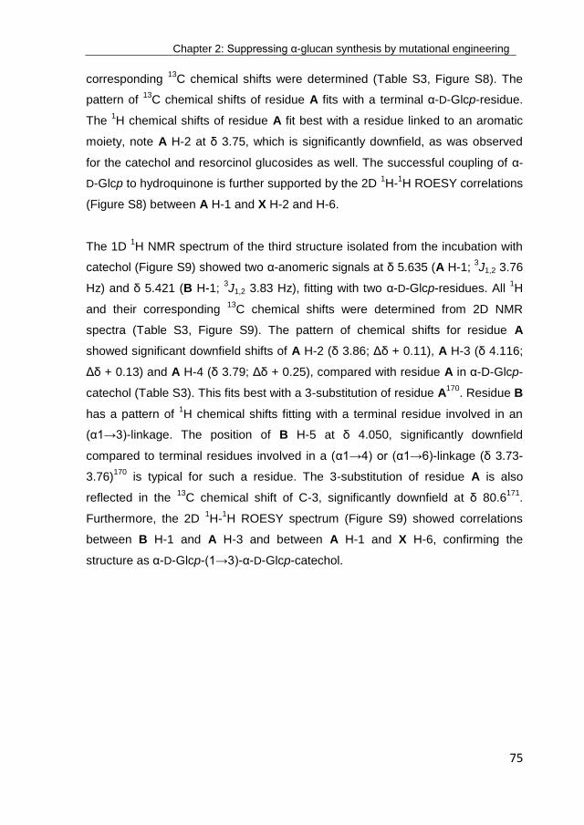
Chapter 2: Suppressing α-glucan synthesis by mutational engineering
75
corresponding 13
C chemical shifts were determined (Table S3, Figure S8). The
pattern of 13
C chemical shifts of residue A fits with a terminal α-D-Glcp-residue.
The 1H chemical shifts of residue A fit best with a residue linked to an aromatic
moiety, note A H-2 at δ 3.75, which is significantly downfield, as was observed
for the catechol and resorcinol glucosides as well. The successful coupling of α-
D-Glcp to hydroquinone is further supported by the 2D 1H-
1H ROESY correlations
(Figure S8) between A H-1 and X H-2 and H-6.
The 1D 1H NMR spectrum of the third structure isolated from the incubation with
catechol (Figure S9) showed two α-anomeric signals at δ 5.635 (A H-1; 3J1,2 3.76
Hz) and δ 5.421 (B H-1; 3J1,2 3.83 Hz), fitting with two α-D-Glcp-residues. All
1H
and their corresponding 13
C chemical shifts were determined from 2D NMR
spectra (Table S3, Figure S9). The pattern of chemical shifts for residue A
showed significant downfield shifts of A H-2 (δ 3.86; Δδ + 0.11), A H-3 (δ 4.116;
Δδ + 0.13) and A H-4 (δ 3.79; Δδ + 0.25), compared with residue A in α-D-Glcp-
catechol (Table S3). This fits best with a 3-substitution of residue A170
. Residue B
has a pattern of 1H chemical shifts fitting with a terminal residue involved in an
(α1→3)-linkage. The position of B H-5 at δ 4.050, significantly downfield
compared to terminal residues involved in a (α1→4) or (α1→6)-linkage (δ 3.73-
3.76)170
is typical for such a residue. The 3-substitution of residue A is also
reflected in the 13
C chemical shift of C-3, significantly downfield at δ 80.6171
.
Furthermore, the 2D 1H-
1H ROESY spectrum (Figure S9) showed correlations
between B H-1 and A H-3 and between A H-1 and X H-6, confirming the
structure as α-D-Glcp-(1→3)-α-D-Glcp-catechol.

Chapter 2: Suppressing α-glucan synthesis by mutational engineering
76
5.2. Tables
Table S1. List of mutants1 of Gtf180-ΔN screened for their glycosylation potential.
1 L938A 14 A978F 27 D1028G 40 D1085Q 53 N1089D
2 L938S 15 A978S 28 D1028N 41 R1088H 54 N1089P
3 L938F 16 A978G 29 N1029Y 42 R1088K 55 W1065F
4 L938K 17 A978L 30 N1029G 43 R1088E 56 W1065K
5 L938M 18 A978P 31 N1029T 44 R1088W 57 W1065L
6 L940A 19 A978Y 32 N1029M 45 R1088T 58 W1065Q
7 L940S 20 L981A 33 N1029R 46 R1088N 59 W1065E
8 L940E 21 L981N 34 D1085Y 47 R1088G 60 W1065M
9 L940F 22 L981E 35 D1085V 48 N1089Y 61 W1065G
10 L940W 23 D1028Y 36 D1085A 49 N1089G
11 L940G 24 D1028W 37 D1085E 50 N1089S
12 L940M 25 D1028L 38 D1085H 51 N1089L
13 L940C 26 D1028K 39 D1085L 52 N1089R
1For construction and partial characterization of these mutants, see Meng et al. (2015
99 and 2016
100).
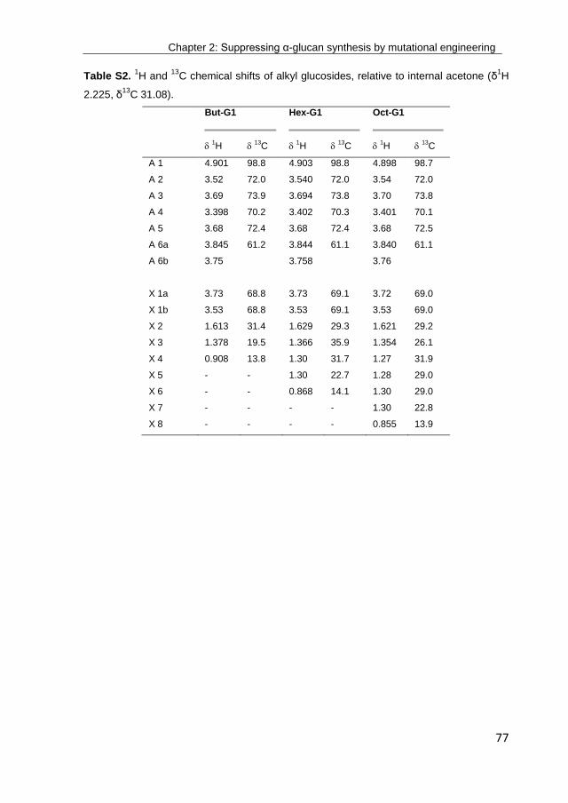
Chapter 2: Suppressing α-glucan synthesis by mutational engineering
77
Table S2. 1H and
13C chemical shifts of alkyl glucosides, relative to internal acetone (δ
1H
2.225, δ13
C 31.08).
But-G1
Hex-G1
Oct-G1
1H
13C
1H
13C
1H
13C
A 1 4.901 98.8 4.903 98.8 4.898 98.7
A 2 3.52 72.0 3.540 72.0 3.54 72.0
A 3 3.69 73.9 3.694 73.8 3.70 73.8
A 4 3.398 70.2 3.402 70.3 3.401 70.1
A 5 3.68 72.4 3.68 72.4 3.68 72.5
A 6a 3.845 61.2 3.844 61.1 3.840 61.1
A 6b 3.75 3.758 3.76
X 1a 3.73 68.8 3.73 69.1 3.72 69.0
X 1b 3.53 68.8 3.53 69.1 3.53 69.0
X 2 1.613 31.4 1.629 29.3 1.621 29.2
X 3 1.378 19.5 1.366 35.9 1.354 26.1
X 4 0.908 13.8 1.30 31.7 1.27 31.9
X 5 - - 1.30 22.7 1.28 29.0
X 6 - - 0.868 14.1 1.30 29.0
X 7 - - - - 1.30 22.8
X 8 - - - - 0.855 13.9
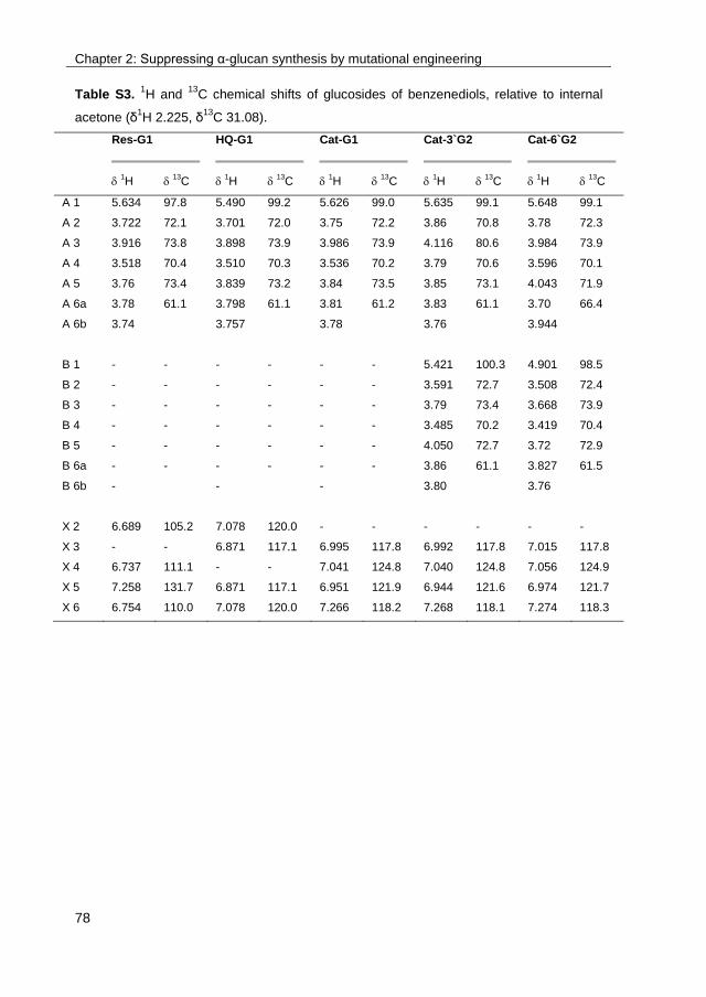
Chapter 2: Suppressing α-glucan synthesis by mutational engineering
78
Table S3. 1H and
13C chemical shifts of glucosides of benzenediols, relative to internal
acetone (δ1H 2.225, δ
13C 31.08).
Res-G1
HQ-G1
Cat-G1
Cat-3`G2
Cat-6`G2
1H
13C
1H
13C
1H
13C
1H
13C
1H
13C
A 1 5.634 97.8 5.490 99.2 5.626 99.0 5.635 99.1 5.648 99.1
A 2 3.722 72.1 3.701 72.0 3.75 72.2 3.86 70.8 3.78 72.3
A 3 3.916 73.8 3.898 73.9 3.986 73.9 4.116 80.6 3.984 73.9
A 4 3.518 70.4 3.510 70.3 3.536 70.2 3.79 70.6 3.596 70.1
A 5 3.76 73.4 3.839 73.2 3.84 73.5 3.85 73.1 4.043 71.9
A 6a 3.78 61.1 3.798 61.1 3.81 61.2 3.83 61.1 3.70 66.4
A 6b 3.74 3.757 3.78 3.76 3.944
B 1 - - - - - - 5.421 100.3 4.901 98.5
B 2 - - - - - - 3.591 72.7 3.508 72.4
B 3 - - - - - - 3.79 73.4 3.668 73.9
B 4 - - - - - - 3.485 70.2 3.419 70.4
B 5 - - - - - - 4.050 72.7 3.72 72.9
B 6a - - - - - - 3.86 61.1 3.827 61.5
B 6b - - - 3.80 3.76
X 2 6.689 105.2 7.078 120.0 - - - - - -
X 3 - - 6.871 117.1 6.995 117.8 6.992 117.8 7.015 117.8
X 4 6.737 111.1 - - 7.041 124.8 7.040 124.8 7.056 124.9
X 5 7.258 131.7 6.871 117.1 6.951 121.9 6.944 121.6 6.974 121.7
X 6 6.754 110.0 7.078 120.0 7.266 118.2 7.268 118.1 7.274 118.3
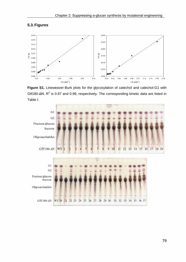
Chapter 2: Suppressing α-glucan synthesis by mutational engineering
79
5.3. Figures
Figure S1. Lineweaver-Burk plots for the glycosylation of catechol and catechol-G1 with
Gtf180-ΔN. R2 is 0.97 and 0.99, respectively. The corresponding kinetic data are listed in
Table I.
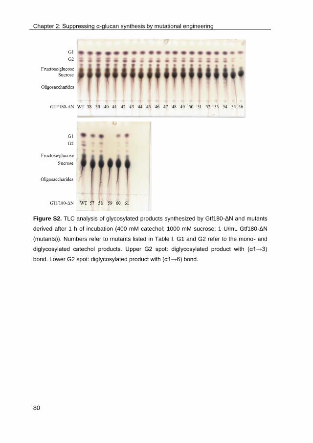
Chapter 2: Suppressing α-glucan synthesis by mutational engineering
80
Figure S2. TLC analysis of glycosylated products synthesized by Gtf180-ΔN and mutants
derived after 1 h of incubation (400 mM catechol; 1000 mM sucrose; 1 U/mL Gtf180-ΔN
(mutants)). Numbers refer to mutants listed in Table I. G1 and G2 refer to the mono- and
diglycosylated catechol products. Upper G2 spot: diglycosylated product with (α1→3)
bond. Lower G2 spot: diglycosylated product with (α1→6) bond.

Chapter 2: Suppressing α-glucan synthesis by mutational engineering
81
Figure S3. TLC analysis of products of resorcinol glycosylation by WT Gtf180-ΔN and the
L981A mutant derived (400 mM resorcinol; 1000 mM sucrose; 4 U/mL Gtf180-ΔN
(mutant)). G1 and G2 refer to the mono- and diglycosylated resorcinol products.
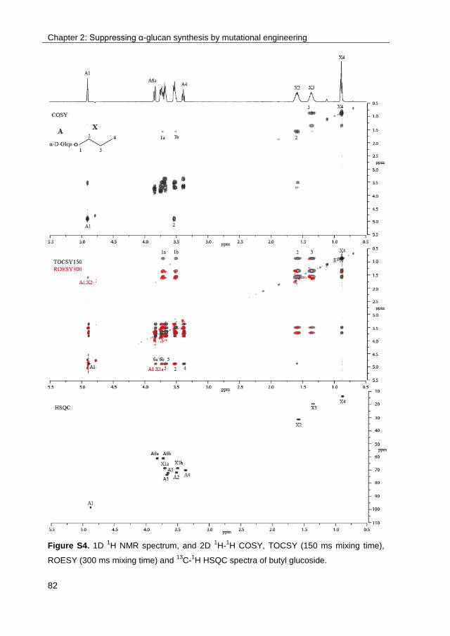
Chapter 2: Suppressing α-glucan synthesis by mutational engineering
82
Figure S4. 1D 1H NMR spectrum, and 2D
1H-
1H COSY, TOCSY (150 ms mixing time),
ROESY (300 ms mixing time) and 13
C-1H HSQC spectra of butyl glucoside.

Chapter 2: Suppressing α-glucan synthesis by mutational engineering
83
Figure S5. 1D 1H NMR spectrum, and 2D
1H-
1H COSY, TOCSY (150 ms mixing time),
ROESY (300 ms mixing time) and 13
C-1H HSQC spectra of hexyl glucoside.
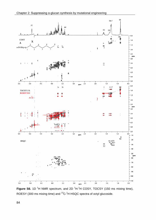
Chapter 2: Suppressing α-glucan synthesis by mutational engineering
84
Figure S6. 1D 1H NMR spectrum, and 2D
1H-
1H COSY, TOCSY (150 ms mixing time),
ROESY (300 ms mixing time) and 13
C-1H HSQC spectra of octyl glucoside.

Chapter 2: Suppressing α-glucan synthesis by mutational engineering
85
Figure S7. 1D 1H NMR spectrum, and 2D
1H-
1H COSY, TOCSY (150 ms mixing time),
ROESY (300 ms mixing time) and 13
C-1H HSQC spectra of resorcinol-G1.
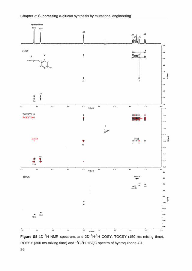
Chapter 2: Suppressing α-glucan synthesis by mutational engineering
86
Figure S8 1D 1H NMR spectrum, and 2D
1H-
1H COSY, TOCSY (150 ms mixing time),
ROESY (300 ms mixing time) and 13
C-1H HSQC spectra of hydroquinone-G1.
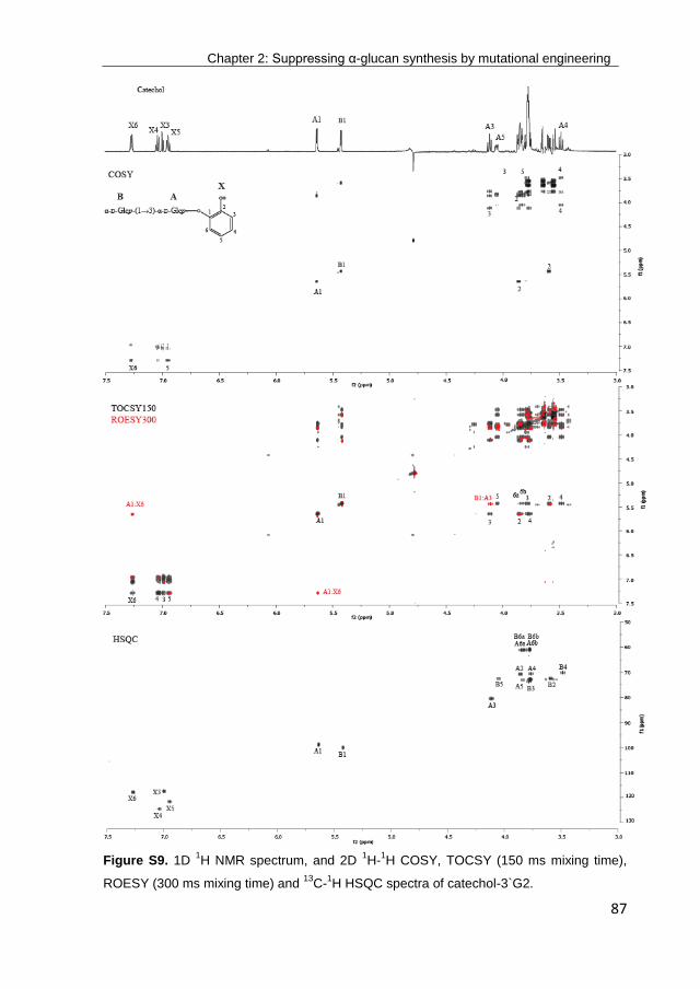
Chapter 2: Suppressing α-glucan synthesis by mutational engineering
87
Figure S9. 1D 1H NMR spectrum, and 2D
1H-
1H COSY, TOCSY (150 ms mixing time),
ROESY (300 ms mixing time) and 13
C-1H HSQC spectra of catechol-3`G2.

89
Chapter 3
Improving the low operational stability of Gtf180-ΔN from
Lactobacillus reuteri 180 by means of its immobilization
Tim Devlamynck1,2
, Wim Soetaert1, Lubbert Dijkhuizen
2
Ghent University1, University of Groningen
2
Chapter 3: Immobilization of Gtf180-ΔN
90
Abstract
Glucansucrases are increasingly targeted as biocatalyst for the glycosylation of
non-carbohydrate acceptor substrates. A major obstacle in their industrial use
remains their relatively low operational stability at high temperatures and in
systems containing cosolvents and high acceptor substrate concentrations. As a
consequence, glycosylation of poorly soluble compounds results in low yields
and productivities as only low concentrations of these acceptor substrates can be
supplied in the reaction mixture. This chapter focused on overcoming the low
operational stability of Gtf180-ΔN from Lactobacillus reuteri 180 by cross-linking
the enzyme to mesoporous silica particles, yielding an immobilized enzyme with
enhanced activity at temperatures above 50 °C and in systems containing 20%
DMSO, reaction conditions detrimental for the free enzyme. As a result, the
glycosylation of gallic acid (GA), caffeic acid (CA) and catechin (CT) could be
performed in reaction mixtures containing much higher concentrations of
acceptor substrate. Their conversion was improved accordingly: from 63% to
82%, from 59% to 80%, and from 4% to 11%, for GA, CA and CT, respectively.
Chapter 3: Immobilization of Gtf180-ΔN
91
1. Introduction
Glucansucrases are glycoside hydrolase enzymes (GH70), catalyzing sucrose
hydrolysis (minor activity) and the conversion of sucrose into α-glucan
polysaccharides (major activity), linking the α-D-glucopyranosyl units by (α1→2),
(α1→3), (α1→4) or (α1→6) bonds, depending on the enzyme specificity73,74
. In
addition, they catalyze so called acceptor reactions, thereby glycosylating various
(poly)phenolic and aliphatic compounds, using sucrose as donor substrate111-114
.
As such, glucansucrases offer a cheap alternative for “Leloir”
glycosyltransferases18
, which require rare and expensive nucleotide-activated
sugars as donor substrate13
. Due to their broad acceptor substrate specificity and
their use of inexpensive sucrose as donor substrate, glucansucrases have
attracted considerable interest from academia and industry for their application
as glycosylation biocatalyst. Glycosylation may result in an increased solubility of
hydrophobic compounds33
, an improved stability of labile molecules against light
and oxidation7, or a modified taste profile
8.
Although several alternative acceptor substrates are indeed glycosylated by
glucansucrases, very often incomplete conversions, with low to moderate yields,
are obtained. Alternative acceptor substrates are per definition not the natural
acceptor substrates of glucansucrases and, hence, they generally have rather
high Km values (Chapter 2)110
. To outcompete α-glucan synthesis and hydrolysis
as possible glucansucrase reactions, high concentrations of acceptor substrate
are consequently required. Shifting the relative balance between the three
glucansucrase reactions towards the acceptor reaction by applying high acceptor
substrate concentrations was demonstrated as early as 1993 by Su and Robyt132
.
Furthermore, high space-time yields, which greatly reduce production costs, can
only be achieved if the acceptor substrate concentrations are correspondingly
high. It was demonstrated in Chapter 2 that suppressing α-glucan synthesis by
mutational engineering enhanced the glycosylation of several phenolic and
aliphatic compounds110
. The biggest improvements were observed for small
molecules such as catechol and butanol. The glycosylation of larger molecules,
Chapter 3: Immobilization of Gtf180-ΔN
92
such as flavonoids, was still problematic, mostly due to the low water solubility of
these compounds, preventing their addition in higher concentrations. Chapter 2
also revealed that glucansucrases were inhibited by high concentrations of non-
carbohydrate acceptor substrates: the glycosylation of catechol by Gtf180-ΔN
from Lactobacillus reuteri 180 was inhibited at concentrations of catechol higher
than 400 mM110
. Similarly, catechol displayed inhibitory effects on glucansucrase
GtfD from Streptococcus mutans GS-5 at a concentration of 200 mM125
. Finally,
also the inhibition of amylosucrase (GH13) from Neisseria polysaccharea by
several flavonoids has been reported124
.
As mentioned earlier, the limited water solubilities of many (poly)phenolic and
aliphatic acceptor substrates complicate their glycosylation by glucansucrases.
Increasing the solubility of acceptor substrates can be achieved by the addition of
organic cosolvents such as DMSO, ethanol, acetone, etc. The main drawback of
this strategy is that enzyme activity –and stability typically decrease with
increasing solvent concentrations172
. Determination of the initial activity of the
dextransucrase from Leuconostoc mesenteroides NRRL B-512F in the presence
of organic solvents revealed a 50% loss in activity in 20% DMSO, 15% ethanol,
15% acetone, 10% DMF and 7% acetonitrile134
. Diglyme or bis(2-methoxyethyl)
ether (MEE) displayed a lower inhibitory effect on glucansucrases: the
dextransucrase from L. mesenteroides NRRL B-512F and the alternansucrase
from L. mesenteroides NRRL B-23192 retained more than 50% activity at an
MEE concentration of 30%120
. The previous examples indicate that the combined
use of high concentrations of certain acceptor substrates and high solvent
concentrations may be even more detrimental for glucansucrase activity. Hence,
a compromise between acceptor substrate concentration, solvent concentration
and enzyme activity needed to be found.
An alternative approach comprises the application of a biphasic glycosylation
system. In this way, the inhibitory effect of cosolvents can be circumvented while
the solubility of the acceptor substrates is still enhanced. In a biphasic
glycosylation system, the aqueous phase contains the biocatalyst and the donor
substrate sucrose, whereas the organic phase contains the hydrophobic acceptor
Chapter 3: Immobilization of Gtf180-ΔN
93
substrates. Upon stirring, the acceptor substrates are transferred to the aqueous
phase, where they are enzymatically glycosylated. Enzyme stability is correlated
with solvent polarity: water miscible cosolvents distort the essential water layer
that stabilizes the enzyme, whereas hydrophobic solvents will leave this layer of
water molecules intact, resulting in a higher enzyme stability. Solvent polarity is
expressed by the logarithm of the octanol/water partition coefficient (log P).
Solvents with a log P < 2 will deactivate enzymes more distinctly than
hydrophobic solvents with a log P > 4173
. Furthermore, the use of a biphasic
glycosylation system minimizes enzyme inhibition by acceptor substrates due to
their low actual concentration in the aqueous phase; hydrophobic compounds are
only slowly released from the organic phase, which acts as a substrate
reservoir174,175
. The use of a biphasic glycosylation system was already
demonstrated for the glycosylation of several phenolic and aliphatic compounds
with sucrose phosphorylase from Bifidobacterium adolescentis, resulting in
enhanced glycosylation yields23
.
Immobilization is a well-known strategy to increase the operational activity and
stability of an enzyme. It may alleviate the decrease of enzyme activity and
stability provoked by high solvent and acceptor substrate concentrations140
. An
additional advantage is the possibility of recycling the immobilized biocatalyst,
which can drastically lower the economic cost of the enzymatic process141
.
Glucansucrases have been described as troublesome to covalently immobilize,
mainly due to inactivation of the enzyme during immobilization, e.g. by the
participation of a lysine residue in the active site. Typical immobilization yields
(ratio of activity of immobilized enzyme to the activity of enzyme prior
immobilization) range from 3% to 22%143-146
. In contrast, encapsulation of
glucansucrases in alginate has been more successfully applied: several studies
report immobilization yields up to 90%147,148
. However, the resulting alginate
beads are not durable, since the accumulation of α-glucan polysaccharides
inside the beads ultimately results in their rupture.
To our knowledge, the inhibitory effects of acceptor substrates and cosolvents on
glucansucrases have never been countered by applying enzyme immobilization
Chapter 3: Immobilization of Gtf180-ΔN
94
nor by using a biphasic system. This chapter investigates both strategies to
overcome the low operational stability of Gtf180-ΔN from L. reuteri 180: the
application of a biphasic system with ethyl acetate as second phase, and the
covalent immobilization of this enzyme on mesoporous silica particles.
2. Materials and methods
2.1. Production and purification of recombinant Gtf180-ΔN
Recombinant, N-terminally truncated Gtf180-ΔN from Lactobacillus reuteri 180
was produced and purified as described previously80
.
2.2. Protein determination
Protein concentration was determined using the BCATM Protein Assay kit from
Pierce. The protocol as described in the kit was used. In short, 200 µL of freshly
prepared assay solution was added to 25 µL of protein sample. After incubation
at 37 °C for 30 min, the absorbance was measured at 562 nm. As standard
series, bovine serum albumin (BSA) was used in a range from 0-2 mg/mL.
2.3. Activity assay for free and immobilized Gtf180-ΔN
Enzyme activity assays were performed at 37°C with 100 mM sucrose in 25 mM
sodium acetate (pH 4.7) and 1 mM CaCl2 unless indicated otherwise. Samples of
100 μL were taken every min over a period of 8 min and immediately inactivated
with 20 μL 1 M NaOH for 30 min. The released glucose and fructose were
quantified enzymatically by monitoring the reduction of NADP with the
hexokinase and glucose-6-phosphate dehydrogenase/phosphoglucose
isomerase assay (Roche) as described previously166,167
, allowing the
determination of the total- (fructose release) and hydrolytic (glucose release)
activities, and calculation of the transglycosylation activity.
Chapter 3: Immobilization of Gtf180-ΔN
95
One unit (U) of total activity corresponds to the release of 1 μmole fructose from
100 mM sucrose in 25 mM sodium acetate (pH 4.7) and 1 mM CaCl2 at 37 °C.
2.4. TLC analysis
TLC analysis of transglycosylation products was performed on silica gel 60 F254
plates (Merck). The eluent consisted of n-butanol-acetic acid-water (2:1:1 by
volume). Detection was achieved by UV absorption (254 nm) and/or staining with
10 % (v/v) H2SO4 containing 2 g/L orcinol.
2.5. HPLC analysis
An Agilent MetaCarb 67H column (300 mm × 6.5 mm) was used under isocratic
conditions with 2.5 mM H2SO4 as the mobile phase. The flow rate and
temperature were set at 0.8 mL/min and 35 °C, respectively. Detection was
achieved with an RID detector. Calibration of the obtained peaks was
accomplished using the corresponding standard curves.
2.6. Solubility measurements
The solubility of gallic acid (GA) was determined in 250 μL of ultrapure water,
incubated in a thermoblock at 20 °C. GA was added until clear precipitation was
noticeable, after which the samples were vortexed multiple times and allowed to
equilibrate for 24 h. The supernatants were diluted in ethanol and subsequently
subjected to HPLC analysis. Calibration was accomplished using the appropriate
standard curve. The analysis was performed in duplicate.
2.7. Immobilization of Gtf180-ΔN on silanized silica particles
Before silanization (the functionalization of the silica surface with alkoxysilane
molecules), the silica particles (10 g) were boiled in distilled water for 30 min. The
wetted particles were then dissolved in 750 mL of a 10% solution of (3-
aminopropyl)triethoxysilane (APTES) at pH 4 and subsequently heated in a water

Chapter 3: Immobilization of Gtf180-ΔN
96
bath at 75 °C for 4 h. The particles were washed three times with water and dried
overnight at 80 °C. The silanized particles were stored at room temperature until
further use.
Immobilization consisted of two steps: (1) adsorption of the enzyme on the
support and (2) cross-linking of the adsorbed enzyme onto the support. In a 1.5
mL-Eppendorf tube, a suspension of 2 mg of silanized particles and 1 mL of
buffered enzyme solution (containing 0.2 mg enzyme/mL) was shaken at room
temperature for 1 h to ensure that adsorption equilibrium was reached.
Subsequently, 10 µL of cross-linker (glutaraldehyde) was added and the
suspension was shaken at room temperature for another hour. Afterwards, the
supernatant was decanted using a benchtop centrifuge (10,000 g, 2 min) and the
particles were washed with distilled water until no enzyme activity was detected
(see 2.3.) in the wash solution.
3. Results and discussion
3.1. Solvent engineering: Applying Gtf180-ΔN in a biphasic –or cosolvent
system
To improve the solubility of acceptor substrates, cosolvents such as DMSO and
acetone are typically added to the glycosylation reaction mixture. The
disadvantage of this strategy is that enzyme activity and stability decrease
substantially. Alternatively, a biphasic glycosylation system can be implemented;
hydrophobic solvents provoke less inhibition than hydrophilic ones. In 2014, De
Winter et al. reported the application of a biphasic glycosylation system with
sucrose phosphorylase, using ethyl acetate as second phase23
. The effect of
using this system on Gtf180-ΔN catalyzed glycosylation reactions was
consequently investigated. The glycosylation of ethyl gallate (EG), added to food
products as antioxidant (E313) and representing a poorly soluble acceptor
substrate with inhibitory effects on Gtf180-ΔN, was demonstrated previously137
and chosen as model reaction. The enzyme was each time incubated with 1000
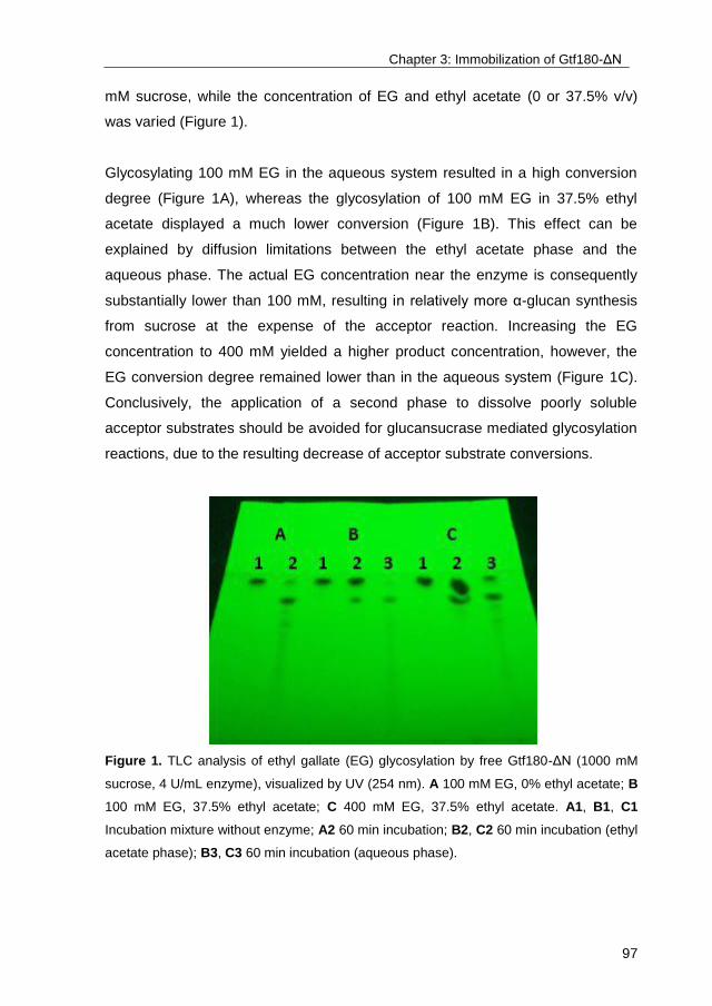
Chapter 3: Immobilization of Gtf180-ΔN
97
mM sucrose, while the concentration of EG and ethyl acetate (0 or 37.5% v/v)
was varied (Figure 1).
Glycosylating 100 mM EG in the aqueous system resulted in a high conversion
degree (Figure 1A), whereas the glycosylation of 100 mM EG in 37.5% ethyl
acetate displayed a much lower conversion (Figure 1B). This effect can be
explained by diffusion limitations between the ethyl acetate phase and the
aqueous phase. The actual EG concentration near the enzyme is consequently
substantially lower than 100 mM, resulting in relatively more α-glucan synthesis
from sucrose at the expense of the acceptor reaction. Increasing the EG
concentration to 400 mM yielded a higher product concentration, however, the
EG conversion degree remained lower than in the aqueous system (Figure 1C).
Conclusively, the application of a second phase to dissolve poorly soluble
acceptor substrates should be avoided for glucansucrase mediated glycosylation
reactions, due to the resulting decrease of acceptor substrate conversions.
Figure 1. TLC analysis of ethyl gallate (EG) glycosylation by free Gtf180-ΔN (1000 mM
sucrose, 4 U/mL enzyme), visualized by UV (254 nm). A 100 mM EG, 0% ethyl acetate; B
100 mM EG, 37.5% ethyl acetate; C 400 mM EG, 37.5% ethyl acetate. A1, B1, C1
Incubation mixture without enzyme; A2 60 min incubation; B2, C2 60 min incubation (ethyl
acetate phase); B3, C3 60 min incubation (aqueous phase).

Chapter 3: Immobilization of Gtf180-ΔN
98
Alternatively, the effect of using DMSO as cosolvent on Gtf180-ΔN catalyzed
glycosylation reactions was investigated. The glycosylation of gallic acid (GA), a
potential medicine for the treatment of Alzheimer’s176
and Parkinson’s disease177
,
served as case study. The solubility of GA in water at 37 °C was determined to
be roughly 80 mM, which could be increased by using DMSO as cosolvent.
Reactions with the free enzyme in 0%, 10% and 20% DMSO however illustrated
the detrimental effect of cosolvent and acceptor substrate on Gtf180-ΔN activity
(Figure 2).
Figure 2. Depletion of gallic acid (GA) due to its glycosylation by free Gtf180-ΔN (1000
mM sucrose, 4 U/mL enzyme): ● 80 mM GA, 0% DMSO; ○ 210 mM GA, 10% DMSO; ▼
325 mM GA, 20% DMSO; and by immobilized Gtf180-ΔN (1000 mM sucrose, 4 U/mL
enzyme): ▽ 325 mM GA, 20% DMSO.
Glycosylation of GA by Gtf180-ΔN without the addition of DMSO (80 mM GA)
resulted in a conversion of 63%. Interestingly, the GA conversion degree in 10%
DMSO (210 mM GA) was significantly higher (80%) than in 0% DMSO (63%),
indicating that the relative balance between α-glucan synthesis, sucrose
hydrolysis and GA glycosylation was shifted towards the latter, due to the
improved availability of the acceptor substrate GA. Glycosylation in 20% DMSO
(325 mM GA) displayed a GA conversion degree of only 14%, which can be
attributed to inactivation of the enzyme. Performing the glycosylation in 20%
Chapter 3: Immobilization of Gtf180-ΔN
99
DMSO might consequently further increase the GA conversion degree, provided
that the enzyme is sufficiently stabilized.
3.2. Immobilization of Gtf180-ΔN on mesoporous silica particles
Previous paragraph was another illustration of the undesired inhibitory effects of
acceptor substrates and cosolvents on Gtf180-ΔN, resulting in suboptimal
glycosylation yields and conversion degrees. Immobilization of Gtf180-ΔN may
alleviate this inhibition, more specifically by improving enzyme activity at high
acceptor substrate –and cosolvent concentrations. Of all immobilization
techniques, covalent immobilization provides the strongest interaction between
enzyme and support, stabilizing the enzyme most thoroughly. Immobilization of
Gtf180-ΔN on mesoporous silica particles was therefore first optimized, after
which the characteristics of the immobilized enzyme were determined.
3.2.1. Optimization of Gtf180-ΔN immobilization
Several factors influence the covalent immobilization of enzymes on mesoporous
silica, the most important being adsorption pH, concentration of cross-linker
(mM), and enzyme loading (mg protein/g support)178
. Immobilizing a single layer
of proteins (2-3 mg protein/m2 support) may result in improved immobilization
yields due to the reduction of substrate diffusion limitation and undesired
conformational changes, both associated with multilayers179
. The specific surface
area of the applied silica particles was roughly 50 m2/g. A single layer of protein
was consequently formed when 100-150 mg Gtf180-ΔN/g or 4000-6000 U
Gtf180-ΔN/g was adsorbed.
The pH was found to be crucial for adsorption –and cross-linking of the
biocatalyst (Figure 3). An enzyme loading of 100 mg/g resulted in a nearly
complete (>95%) adsorption at a pH range of 5.5-6. At a pH of 4.5 not even half
of the enzyme was adsorbed, whereas 75% adsorption was achieved at a pH of
7. These results could only partly explain the sharp immobilization yield optimum:
at a pH lower than 5.5, the enzyme was not efficiently cross-linked (as could be
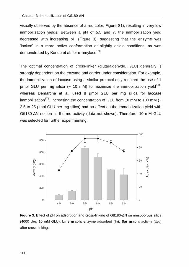
Chapter 3: Immobilization of Gtf180-ΔN
100
visually observed by the absence of a red color, Figure S1), resulting in very low
immobilization yields. Between a pH of 5.5 and 7, the immobilization yield
decreased with increasing pH (Figure 3), suggesting that the enzyme was
‘locked’ in a more active conformation at slightly acidic conditions, as was
demonstrated by Kondo et al. for α-amylase180
.
The optimal concentration of cross-linker (glutaraldehyde, GLU) generally is
strongly dependent on the enzyme and carrier under consideration. For example,
the immobilization of laccase using a similar protocol only required the use of 1
µmol GLU per mg silica (~ 10 mM) to maximize the immobilization yield181
,
whereas Demarche et al. used 8 µmol GLU per mg silica for laccase
immobilization171
. Increasing the concentration of GLU from 10 mM to 100 mM (~
2.5 to 25 µmol GLU per mg silica) had no effect on the immobilization yield with
Gtf180-ΔN nor on its thermo-activity (data not shown). Therefore, 10 mM GLU
was selected for further experimenting.
Figure 3. Effect of pH on adsorption and cross-linking of Gtf180-ΔN on mesoporous silica
(4000 U/g, 10 mM GLU). Line graph: enzyme adsorbed (%). Bar graph: activity (U/g)
after cross-linking.
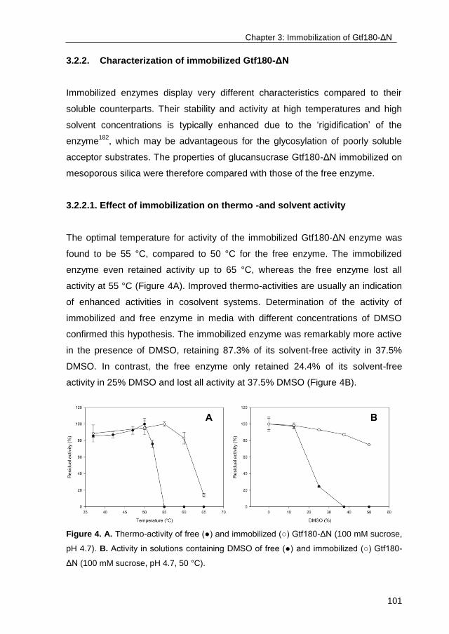
Chapter 3: Immobilization of Gtf180-ΔN
101
3.2.2. Characterization of immobilized Gtf180-ΔN
Immobilized enzymes display very different characteristics compared to their
soluble counterparts. Their stability and activity at high temperatures and high
solvent concentrations is typically enhanced due to the ‘rigidification’ of the
enzyme182
, which may be advantageous for the glycosylation of poorly soluble
acceptor substrates. The properties of glucansucrase Gtf180-ΔN immobilized on
mesoporous silica were therefore compared with those of the free enzyme.
3.2.2.1. Effect of immobilization on thermo -and solvent activity
The optimal temperature for activity of the immobilized Gtf180-ΔN enzyme was
found to be 55 °C, compared to 50 °C for the free enzyme. The immobilized
enzyme even retained activity up to 65 °C, whereas the free enzyme lost all
activity at 55 °C (Figure 4A). Improved thermo-activities are usually an indication
of enhanced activities in cosolvent systems. Determination of the activity of
immobilized and free enzyme in media with different concentrations of DMSO
confirmed this hypothesis. The immobilized enzyme was remarkably more active
in the presence of DMSO, retaining 87.3% of its solvent-free activity in 37.5%
DMSO. In contrast, the free enzyme only retained 24.4% of its solvent-free
activity in 25% DMSO and lost all activity at 37.5% DMSO (Figure 4B).
Figure 4. A. Thermo-activity of free (●) and immobilized (○) Gtf180-ΔN (100 mM sucrose,
pH 4.7). B. Activity in solutions containing DMSO of free (●) and immobilized (○) Gtf180-
ΔN (100 mM sucrose, pH 4.7, 50 °C).

Chapter 3: Immobilization of Gtf180-ΔN
102
3.2.2.2. Effect of immobilization on activity at high acceptor concentrations
Glucansucrases are inhibited by high concentrations of non-carbohydrate
acceptor substrates, as was demonstrated for Gtf180-ΔN: the model substrate
catechol could not be glycosylated at concentrations above 400 mM due to
severe inhibition110
. The immobilized enzyme was incubated with 400, 600 or 800
mM catechol and 1000 mM sucrose to determine the effect of immobilization on
the glycosylation of non-carbohydrate acceptor substrates (Figure 5).
Figure 5. Depletion of catechol (CAT) due to its glycosylation by free and immobilized
Gtf180-ΔN (1000 mM sucrose, 4 U/mL enzyme). ● Immobilized Gtf180-ΔN, 400 mM CAT;
○ Immobilized Gtf180-ΔN, 600 mM CAT; ▼ Free Gtf180-ΔN, 400 mM CAT; ▽ Free
Gtf180-ΔN, 600 mM CAT.
Incubation of the immobilized enzyme with 400 mM catechol resulted in a 78%
conversion of the latter into glycosides compared to 100% for the free enzyme. In
contrast, sucrose was completely converted in both cases, indicating that the
immobilized enzyme underwent undesired changes in the active site, resulting in
a decreased affinity for the acceptor substrate catechol. Furthermore, 600 mM
catechol was converted by the immobilized enzyme into glycosides with a
conversion degree of 81%, slightly better than when incubated with 400 mM and
significantly better than for the free enzyme (7%) (Figure 5). This confirms the
Chapter 3: Immobilization of Gtf180-ΔN
103
results of the thermo –and solvent activity experiments and demonstrates the
improved operational stability of the immobilized enzyme. A concentration of 800
mM catechol resulted in deactivation of not only the free but also the immobilized
enzyme (data not shown). In conclusion, the immobilized enzyme remained
active at higher acceptor substrate concentrations, higher solvent concentrations
and higher temperatures, compared to the free enzyme. However, its acceptor
substrate affinity was altered, possibly due to participation of one or more amino
acid residues in the active site during cross-linking. In fact, this is known to be
one of the main disadvantages related to the use of GLU as cross-linker. Due to
its small size, GLU can penetrate easily into the active site of enzymes and
subsequently react with amino acid residues.
In order to prevent GLU from causing undesired changes in the active site,
protecting agents, typically enzyme substrates, are used183
. Moreover, the
activity of the immobilized enzyme towards the protecting agent/enzyme
substrate can be improved by this ‘molecular imprinting’ of the enzyme’s active
site184
. It is important that the substrate is not converted during immobilization to
obtain the optimal protecting effect; catechol and maltose were consequently
tested in different concentrations (100-400 mM) as protecting agent during
Gtf180-ΔN immobilization. Unfortunately, the addition of catechol prevented
cross-linking of the enzyme, whereas the addition of maltose had no effect on
catechol glycosylation.
3.3. Glycosylation with immobilized Gtf180-ΔN
It was previously shown that GA glycosylation proceeds with improved
conversion degrees in systems containing high GA concentrations, until DMSO
and GA reach a concentration that is inhibitory to Gtf180-ΔN. In order to further
improve GA glycosylation, immobilized Gtf180-ΔN was incubated in 20% DMSO,
containing 325 mM GA (Figure 2).
Immobilization of Gtf180-ΔN alleviated the inhibiting effect of DMSO and GA,
resulting in a GA conversion degree of 82%, which is slightly higher than the
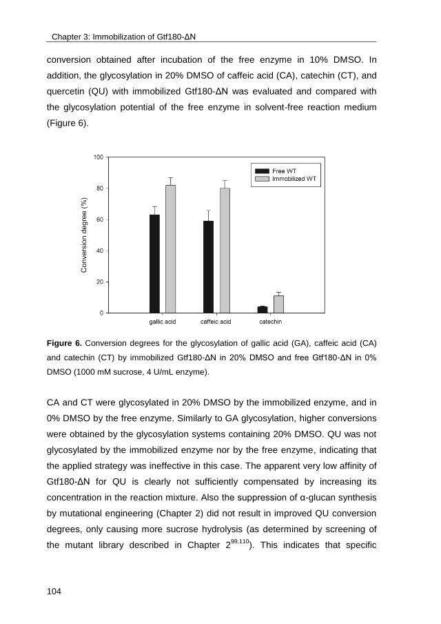
Chapter 3: Immobilization of Gtf180-ΔN
104
conversion obtained after incubation of the free enzyme in 10% DMSO. In
addition, the glycosylation in 20% DMSO of caffeic acid (CA), catechin (CT), and
quercetin (QU) with immobilized Gtf180-ΔN was evaluated and compared with
the glycosylation potential of the free enzyme in solvent-free reaction medium
(Figure 6).
Figure 6. Conversion degrees for the glycosylation of gallic acid (GA), caffeic acid (CA)
and catechin (CT) by immobilized Gtf180-ΔN in 20% DMSO and free Gtf180-ΔN in 0%
DMSO (1000 mM sucrose, 4 U/mL enzyme).
CA and CT were glycosylated in 20% DMSO by the immobilized enzyme, and in
0% DMSO by the free enzyme. Similarly to GA glycosylation, higher conversions
were obtained by the glycosylation systems containing 20% DMSO. QU was not
glycosylated by the immobilized enzyme nor by the free enzyme, indicating that
the applied strategy was ineffective in this case. The apparent very low affinity of
Gtf180-ΔN for QU is clearly not sufficiently compensated by increasing its
concentration in the reaction mixture. Also the suppression of α-glucan synthesis
by mutational engineering (Chapter 2) did not result in improved QU conversion
degrees, only causing more sucrose hydrolysis (as determined by screening of
the mutant library described in Chapter 299,110
). This indicates that specific
Chapter 3: Immobilization of Gtf180-ΔN
105
mutational engineering of Gtf180-ΔN’s active site is required to enhance the
glycosylation of QU and other related flavonoids.
4. Conclusions
The relatively low operational stability of Gtf180-ΔN at high temperatures and in
systems containing cosolvents and high acceptor substrate concentrations
ultimately results in suboptimal acceptor substrate conversion degrees. By
means of its immobilization on mesoporous silica particles, the activity of Gtf180-
ΔN under such conditions was substantially improved. The immobilized enzyme
displayed enhanced activity at temperatures above 50 °C and in systems
containing 20% DMSO, allowing the glycosylation of GA, CA and CT in systems
containing much higher acceptor substrate concentrations. As a result, their
conversion into glycosides was improved substantially: from 63% to 82%, from
59% to 80%, and from 4% to 11%, for GA, CA and CT, respectively. The
glycosylation of QU, a compound which was very poorly glycosylated by the free
enzyme, was also not successful using immobilized Gtf180-ΔN in 20% DMSO.
Suppressing α-glucan synthesis by enzyme engineering (Chapter 2) nor solvent
engineering (Chapter 3) were effective strategies to enhance QU’s glycosylation,
as this most probably requires specific mutational engineering of Gtf180-ΔN in
order to increase its very low affinity for this molecule. Improving the
glycosylation of related flavonoids, such as luteolin, or stilbenoids, such as
resveratrol, will most probably demand the same strategy.

Chapter 3: Immobilization of Gtf180-ΔN
106
5. Supplementary information
Figure S1. Successful cross-linking is indicated by the presence of a red color, caused by
Rayleigh scattering.

107
Chapter 4
Glucansucrase (mutant) enzymes from Lactobacillus reuteri
180 efficiently transglucosylate Stevia component
rebaudioside A, resulting in a superior taste
This chapter is submitted for publication as:
Evelien M. te Poele#, Tim Devlamynck
#, Manuel Jäger, Gerrit J. Gerwig, Davy
Van de Walle, Koen Dewettinck, Anna K.H. Hirsch, Johannis P. Kamerling, Wim
Soetaert, Lubbert Dijkhuizen (2017) Glucansucrase (mutant) enzymes from
Lactobacillus reuteri 180 efficiently transglucosylate Stevia component
rebaudioside A, resulting in a superior taste. Sci Rep.
# these authors contributed equally to this work

Chapter 4: Glucosylation of rebaudioside A
108
Abstract
Steviol glycosides from the leaves of the plant Stevia rebaudiana are high-
potency natural sweeteners but suffer from a lingering bitterness. The
Lactobacillus reuteri 180 wild-type glucansucrase Gtf180-ΔN, and in particular its
Q1140E-mutant, efficiently α-glucosylated rebaudioside A (RebA), using sucrose
as donor substrate. Structural analysis of the products by MALDI-TOF mass
spectrometry, methylation analysis and NMR spectroscopy showed that both
enzymes exclusively glucosylate the Glc(β1→C-19 residue of RebA, with the
initial formation of an (α1→6) linkage. Docking of RebA in the active site of the
enzyme revealed that only the steviol C-19 β-D-glucosyl moiety is available for
glucosylation. Response surface methodology was applied to optimize the
Gtf180-ΔN-Q1140E-catalyzed α-glucosylation of RebA, resulting in a highly
productive process with a RebA conversion of 95% and a production of 115 g/L
α-glucosylated products within 3 h. Development of a fed-batch reaction further
improved the product yield to 270 g/L by suppressing α-glucan synthesis.
Sensory analysis by a trained panel revealed that glucosylated RebA products
have a superior taste profile compared to RebA, showing a significant reduction
in bitterness. The Gtf180-ΔN-Q1140E glucansucrase mutant enzyme thus is an
efficient biocatalyst for generating α-glucosylated RebA variants with improved
edulcorant/organoleptic properties.
Chapter 4: Glucosylation of rebaudioside A
109
1. Introduction
The world-wide increasing incidence of obesity, diabetes type II, cardiovascular
diseases, and dental caries leads to an increased consumer demand for food
products and beverages without high-calorie sugars46
. Steviol glycosides are
excellent natural alternatives for sucrose and synthetic sweeteners185-189
. These
non-calorie compounds are extracted from the leaves of the herb plant Stevia
rebaudiana BERTONI, a rhizomatous perennial shrub belonging to the
Asteraceae [Compositae] family190,191
. Stevioside (~5-20% w/w of dried leaves)
and rebaudioside A (RebA) (~2-5% w/w of dried leaves) are the most abundant
steviol glycosides, followed in lower concentrations by rebaudioside B, C, D, E,
F, M, steviolbioside, rubusoside and dulcoside A (Figure 1). Stevioside and RebA
taste about 300 times sweeter than sucrose (0.4% aqueous solution). Steviol
glycosides are approved as food additives in the USA since 2009 and they are
on the European market (E 960, European Index) since December 201143
. The
main drawback for their more successful commercialization as sweeteners
however, is a lingering bitter aftertaste of several steviol glycosides, experienced
by about half of the human population due to a genetic basis of taste
perception46,192
.
Structurally, steviol glycosides have ent-13-hydroxykaur-16-en-19-oic acid as
aglycon, also called steviol (Figure 1)191,193
. The presence and composition of the
different carbohydrate moieties at the C-19-carboxylic acid group (R1) and at the
C-13-tert-hydroxyl group (R2) of steviol have a relationship with the sweetness as
well as with the quality of taste of the steviol glycosides52
. Sweetness increases
and bitterness perception decreases with the total number of glycosyl residues46
.
It has to be noted however, that the correlations bitterness/structure and
sweetness/structure of steviol glycosides are still not fully understood, particularly
in combination with the interactions with the human taste receptors46,192,194,195
.
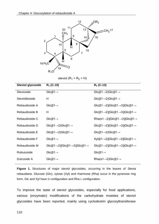
Chapter 4: Glucosylation of rebaudioside A
110
Steviol glycoside R1 (C-19) R2 (C-13)
Stevioside Glc(β1→ Glc(β1→2)Glc(β1→
Steviolbioside H Glc(β1→2)Glc(β1→
Rebaudioside A Glc(β1→ Glc(β1→2)[Glc(β1→3)]Glc(β1→
Rebaudioside B H Glc(β1→2)[Glc(β1→3)]Glc(β1→
Rebaudioside C Glc(β1→ Rha(α1→2)[Glc(β1→3)]Glc(β1→
Rebaudioside D Glc(β1→2)Glc(β1→ Glc(β1→2)[Glc(β1→3)]Glc(β1→
Rebaudioside E Glc(β1→2)Glc(β1→ Glc(β1→2)Glc(β1→
Rebaudioside F Glc(β1→ Xyl(β1→2)[Glc(β1→3)]Glc(β1→
Rebaudioside M Glc(β1→2)[Glc(β1→3)]Glc(β1→ Glc(β1→2)[Glc(β1→3)]Glc(β1→
Rubusoside Glc(β1→ Glc(β1→
Dulcoside A Glc(β1→ Rha(α1→2)Glc(β1→
Figure 1. Structures of major steviol glycosides, occurring in the leaves of Stevia
rebaudiana. Glucose (Glc), xylose (Xyl) and rhamnose (Rha) occur in the pyranose ring
form. Glc and Xyl have D configuration and Rha L configuration.
To improve the taste of steviol glycosides, especially for food applications,
various (enzymatic) modifications of the carbohydrate moieties of steviol
glycosides have been reported, mainly using cyclodextrin glycosyltransferase
Chapter 4: Glucosylation of rebaudioside A
111
(CGTase), α- and β-glucosidase, α- and β-galactosidase and β-fructosidase
transglycosylation and β-glycosyltransferase glycosylation systems as
biocatalysts8. In the context of our study, the reports on trans-α-glucosylation are
of interest. CGTases are attractive enzymes, which catalyze coupling and
disproportionation reactions, transferring glucose residues from starch or
cyclodextrins to acceptor molecules, yielding Glc(α1→4) extensions. Although
often high yields are obtained with steviol glycosides, CGTases have poor steviol
C-13/C-19 site regiospecificity, producing mixtures of steviol glycoside
derivatives with mostly (α1→4)-glucosylation at both carbohydrate moieties51
.
Several early studies have shown that both mono- and di-(α1→4)-glucosylation
of the carbohydrate moiety at the steviol C-13 site of stevioside and rubusoside
gave products with a remarkable improvement in both intensity and quality of
sweetness. However, (α1→4)-glucosylation of the Glc(β1→ residue at the steviol
C-19 site resulted in an increased bitter aftertaste and a lower sweetness
intensity52-54,196
. α-Glucosylation of stevioside using Biozyme L (β-amylase
preparation, probably contaminated with an α-glucosidase) and maltose as donor
substrate resulted in a product with a decreased sweetness, but a remarkable
improvement in the quality of taste [Glc(α1→6) residue attached at the
Glc(β1→C-19 residue], a product with a much lower sweetness [Glc(α1→6)
residue attached to the terminal Glc(β1→2) residue of the β-sophorosyl-C-13
unit] and a product with a bitter taste [Glc(α1→3) residue attached to the terminal
Glc(β1→2) residue of the β-sophorosyl-C-13 unit]60
.
In order to obtain steviol glycoside derivatives with improved organoleptic
properties, we studied the α-glucosylation potential of mutant glucansucrase
enzymes of the generally recognized as safe (GRAS) bacterium Lactobacillus
reuteri 180 on RebA. Glucansucrases (EC 2.1.4.5; glucosyltransferases, Gtfs)
are extracellular enzymes catalyzing the synthesis of α-D-glucan polymers from
the donor substrate sucrose, thereby introducing different ratios of glycosidic
linkages [(α1→2), (α1→3), (α1→4), (α1→6)] in their glucan products, depending
on the enzyme specificities75,162
.

Chapter 4: Glucosylation of rebaudioside A
112
Recently, we have shown that the wild-type Gtf180-ΔN (N-terminally truncated)
glucansucrase enzyme of L. reuteri 180 was able to glucosylate the steviol
glycoside RebA, using sucrose as glucosyl donor substrate69
. About 55% of
RebA was glucosylated with up to eight α-D-glucosyl units attached (RebA-G).
The formed RebA derivatives only had elongations at the steviol C-19 β-D-
glucosyl moiety, mainly with alternating (α1→6)- and (α1→3)-linked
glucopyranose residues, starting with an (α1→6) linkage (RebA-G1). In the
present study, we have screened our in-house collection of mutated Gtf180-ΔN
glucansucrase enzymes for mutants with a better RebA glucosylating activity
than the wild-type Gtf180-ΔN enzyme. One mutant was selected for more
detailed studies and its biochemical characteristics and product profile were
compared to wild-type Gtf180-ΔN. Glucosylated RebA products were isolated by
flash chromatography and their structures were elucidated using MALDI-TOF
mass spectrometry, methylation analysis and 1D/2D 1H/
13C NMR spectroscopy.
Furthermore, docking experiments with RebA and the available high-resolution
3D structure of Gtf18083
were carried out to evaluate the experimental data.
Response surface methodology was applied to optimize the reaction conditions
of RebA glucosylation with the selected mutant. Finally, sensory evaluations
were performed to determine the taste attributes of the novel α-glucosylated
RebA derivatives.
2. Materials and methods
2.1. Glucansucrase enzymes
Gtf180-ΔN is the 117-kDa N-terminally truncated (741 residues) fragment of the
wild-type Gtf180 full-length protein, derived from L. reuteri strain 18080
. In Gtf180-
ΔN-ΔV both the N-terminal variable domain and the N-terminal domain V
fragment (corresponding to the first 793 N-terminal amino acids), and the C-
terminal domain V fragment (corresponding to the last 136 C-terminal amino
acids) have been deleted88
. The glucansucrase Gtf180-ΔN mutant enzymes were

Chapter 4: Glucosylation of rebaudioside A
113
constructed using QuikChange site-directed mutagenesis (Stratagene, La Jolla,
CA)80,88,97-100,102,197
(Table SI).
2.2. Standard reaction buffer
All enzymatic reactions were performed at 37 °C in 25 mM sodium acetate (pH
4.7), containing 1 mM CaCl2.
2.3. Enzyme activity assays
Enzyme activity assays were performed at 37 °C in reaction buffer with 100 mM
sucrose. Samples of 100 μL were taken every min over a period of 8 min and
immediately inactivated with 20 µL 1 M NaOH. The glucose and fructose
concentrations were enzymatically determined by monitoring the reduction of
NADP with the hexokinase and glucose-6-phosphate
dehydrogenase/phosphoglucose isomerase assays (Roche Nederland BV,
Woerden, The Netherlands)167
. Determination of the release of glucose and
fructose from sucrose allowed calculation of the total activity of the
glucansucrase enzymes166
. One unit (U) of enzyme is defined as the amount of
enzyme required for producing 1 μmol fructose per min in reaction buffer,
containing 100 mM sucrose, at 37 °C.
2.4. Screening of (mutant) glucansucrases for α-glucosylation of RebA
In an initial screening, six Gtf180-ΔN-derived mutants were compared to wild-
type Gtf180-ΔN by analyzing reaction products with high-performance liquid
chromatography (HPLC). Then, an additional 76 Gtf180-ΔN mutants from our
collection (Table SII) were screened and analyzed using thin-layer
chromatography (TLC). Incubations of 3 h were performed in reaction buffer,
containing ~1 mg/mL enzyme, 50 mM RebA (Sigma-Aldrich Chemie, Zwijndrecht,
The Netherlands) and 1 M sucrose (for HPLC analysis) or 0.2 M sucrose (for
TLC analysis). For HPLC analysis, 10 µL of the incubation mixture was diluted in
250 µL 80% methanol and centrifuged for 2 min at 15,000 × g. Then, 40 µL of the
Chapter 4: Glucosylation of rebaudioside A
114
upper phase was injected on a Luna 10 μm NH2 column (250 4.6 mm;
Phenomenex, Utrecht, The Netherlands). Separation was obtained at a flow-rate
of 1 mL/min under gradient elution conditions (solvent A = acetonitrile; solvent B
= 0.025% acetic acid in H2O), starting with a 2-min isocratic step of 70% solvent
A followed by a linear gradient from 70 to 55% solvent A over 9 min and a final 3-
min washing step of 20% solvent A. HPLC analyses were performed using an
UltiMate 3000 chromatography system, equipped with a VWD-3000 UV-vis
detector (ThermoFisher Scientific, Amsterdam, The Netherlands; monitoring at
210 nm). For TLC analysis, 1 µL of the enzymatic reaction mixtures was spotted
on TLC sheets (Kieselgel 60 F254, 20 20 cm; Merck, Darmstadt, Germany),
which were developed in n-butanol/acetic acid/water (2:1:1, v/v/v). After drying of
the sheets, the bands were visualized by orcinol/sulfuric acid staining.
2.5. Quantitative preparation of α-glucosylated RebA products
Incubations of 84 mM RebA (Tereos PureCircle Solutions, Lille, France; 97%
purity, HPLC grade) were performed in 50 mL reaction buffer with 282 mM
sucrose donor substrate, using 5 U/mL Gtf180-ΔN-Q1140E enzyme, during 3 h.
Fractionations of RebA-G were carried out by flash chromatography using a
Reveleris X2 flash chromatography system (Büchi Labortechnik AG, Flawil,
Switzerland) with a Reveleris C18 cartridge (12 g, 40 µm) with water (solvent A)
and acetonitrile (solvent B) as the mobile phase (30 mL/min). The following
gradient elution was used: 95% solvent A (0-2 min), 95-50% solvent A (2-20
min), 50-95% solvent B (20-22 min), 95% solvent B (22-25 min). The collected
fractions were evaporated in vacuo and subsequently freeze dried to remove the
residual water.
2.6. Design of response surface methodology experiment
Response surface methodology198
was applied to optimize the glucosylation of
RebA. A Box-Behnken design199
was generated implementing RebA
concentration (mM), sucrose/RebA ratio and agitation rate (rpm) as factors. For
Chapter 4: Glucosylation of rebaudioside A
115
each of them low (-1) and high (+1) level values were assigned as follows: RebA
concentration (50 mM) and (200 mM), sucrose/RebA ratio (1:1) and (4:1),
agitation rate (0 rpm) and (200 rpm). The addition of 5 U/mL Gtf180-ΔN-Q1140E
enzyme ensured a steady-state was reached within 3 h of incubation. The
experimental design was generated and analyzed using JMP software (release
12)200
and consisted of 15 experiments carried out at 50 mL scale in shake
flasks, continuously mixed by shaking (Table SIII). Results were analyzed with
HPLC (see below). The response surface analysis module of JMP software was
applied to fit the following second order polynomial equation:
�̂� = 𝛽0 + ∑ 𝛽𝑖𝑋𝑖
𝐼
𝑖=1
+ ∑ 𝛽𝑖𝑖𝑋𝑖2
𝐼
𝑖=1
+ ∑ ∑ 𝛽𝑖𝑗𝑋𝑖
𝑗𝑖
𝑋𝑗
where �̂� is the predicted response, 𝐼 is the number of factors (3 in this study), 𝛽0
is the model constant, 𝛽𝑖 is the linear coefficient associated to factor 𝑋𝑖, 𝛽𝑖𝑖 is the
quadratic coefficient associated to factor 𝑋𝑖2 and 𝛽𝑖𝑗 is the interaction coefficient
between factors 𝑋𝑖 and 𝑋𝑗. 𝑋𝑖 represents the factor variable in coded form:
𝑋𝑐,𝑖 =[𝑋𝑖 − (𝑙𝑜𝑤 + ℎ𝑖𝑔ℎ)/2]
(ℎ𝑖𝑔ℎ − 𝑙𝑜𝑤)/2
with 1 ≤ 𝑖 ≤ 𝐼, where 𝑋𝑐,𝑖 is the coded variable.
For the HPLC analysis of the RebA and glucosylated RebA products, an Agilent
ZORBAX Eclipse Plus C18 column (100 × 4.6 mm, 3.5 µm) was used with water
(solvent A) and acetonitrile (solvent B) as the mobile phase. The flow rate and
temperature were set at 1.0 mL/min and 40 °C, respectively. The following
gradient elution was used: 5-95% solvent B (0-25 min), 95% solvent B (25-27
min), 95-5% solvent B (27-30 min) and again 95% of solvent A (30-35 min).
Detection was achieved with an ELS detector (evaporation temperature, 90 °C;
nebulization temperature, 70 °C; gas flow rate, 1.6 SLM). Calibration of the
obtained peaks for RebA and mono-α-glucosylated RebA (RebA-G1) was
Chapter 4: Glucosylation of rebaudioside A
116
accomplished using the corresponding standard curves. In this context, the
concentration of multi-α-glucosylated product (RebA-G) at a specific time was
calculated as the initial RebA concentration minus the RebA concentration at that
time. Multi-glycosylated RebA product lacking RebA-G1 (defined as RebA-G2+)
was equal to RebA-G minus RebA-G1.
2.7. Methylation analysis, mass spectrometry and NMR spectroscopy
For details of methylation analysis, gas-liquid chromatography – electron impact
mass spectrometry (GLC-EIMS), matrix-assisted laser-desorption ionization time-
of-flight mass spectrometry (MALDI-TOF-MS), and 1D/2D 1H/
13C (TOCSY,
ROESY, HSQC) NMR spectroscopy, see ref69
.
2.8. Molecular docking of RebA in the active site of Gtf180-ΔN and Gtf180-
ΔN-Q1140E
The X-ray crystal structure of Gtf180-ΔN complexed with maltose (PDB code:
3KLL83
) was used for docking by using LeadIT 2.1.8 from BiosolveIT201
. The
acceptor binding site was defined by manual selection and included the following
amino acid residues: 935–941, 944, 964–968, 975–983, 985, 1023–1032, 1035,
1061–1069, 1082–1093, 1096, 1111, 1129–1142, 1144, 1145, 1155, 1183, 1202,
1204, 1407, 1409, 1411, 1412, 1443, 1446, 1456–1458, 1463–1466, 1504–1509,
1511, 1526, 1527, and the water molecules numbered 7, 15, 45, 83, 106, 144,
145, 172, 401, 432–436, 466, 469, 470, 527, 572, 605, 648, 666, which have at
least two interactions. The crystal structure of rebaudioside A.4H2O.1CH3OH was
taken from the literature (CSD entry: DAWCEL)202
, and the water and methanol
were removed. The default settings were used for docking, except for the
following features: the docking strategy was chosen to be driven by Entropy
(Single Interaction Scan), a hard enzyme was used (maximum allowed overlap
volume: 2 Å) and the maximum number of solutions per fragmentation was
increased to 400. In total, 30 poses were generated with LeadIT, using the
scoring function HYDE in SeeSAR203
, ranked according to their estimated affinity;
finally, poses with torsions flagged in red were removed. Visual inspection of the
Chapter 4: Glucosylation of rebaudioside A
117
residual poses and deletion of unreliable poses resulted in a trustable set of
poses. The model of mutant Q1140E, which was used for the docking
experiment, was built in PyMOL204
and the rotamer showing the smallest number
of steric clashes was chosen.
2.9. Sensory analysis
Panellists were selected on basis of their performance on basic taste
recognitions, ability to ascertain degrees of differences for specific taste stimuli at
different concentrations and repeatability205
, as verified by triangle tests and Wald
sequential analysis206
. Following the selection, the panel was trained over a 6-
month period. In a first session, the panellists had to taste RebA solution at 10%
(w/w) sucrose equivalent level. Their own vocabulary was used to describe taste
and off-tastes as well as after tastes. In the second session, the following
attributes were selected from the first session and from literature207-209
:
sweetness, liquorice, astringency and bitterness. In the following months, training
sessions were alternated with discussion sessions to agree on scoring of
sweetness, off-tastes, aftertastes and lingering on a 15 point hedonic scale, and
evaluation protocol.
The actual Quantitative Descriptive Analysis (QDA) was performed in individual
tasting booths at the UGent Sensolab (Belgium) by the trained panel (9 persons).
The fixed evaluation protocol with standardized vocabulary was applied. Firstly,
taste (sweetness, liquorice, astringency and bitterness) was evaluated by swirling
the sample in the mouth for 5 sec after which the sample was expectorated.
Secondly, aftertaste was evaluated 10 sec after swallowing the solution. Next,
lingering based on the maximum taste intensity was rated 1 min later. Sucrose
reference solutions (5%, 7.5% and 10% sucrose scoring 5, 7.5 and 10,
respectively) were provided. Water (Spa Reine) and plain crackers were used as
palate cleansers between sampling. All samples were evaluated in duplicate.
Statistical analyses were performed with SPSS 23 (SPSS Inc., Chicago, USA).
All tests were done at a significance level of 0.05. One-Way ANOVA was used to

Chapter 4: Glucosylation of rebaudioside A
118
investigate any significant difference between the solutions. Testing for equal
variances was executed with the Modified Levene Test. When conditions for
equal variance were fulfilled, the Tukey test210
was used to determine differences
between samples. In case variances were not equal, the Games-Howell test was
performed211
.
Three different glucosylated products were examined: multi-α-glucosylated
product, containing residual RebA (RebA-G), mono-α-glucosylated product
(RebA-G1) and RebA-G lacking RebA and RebA-G1 (RebA-G2+). Note that
RebA-G, as defined here, contains a very minor amount of residual RebA, in
contrast to the RebA-G as quantified by HPLC analysis.
3. Results
3.1. Screening of wild-type Gtf180-ΔN and mutant glucansucrase enzymes
for α-glucosylation of RebA
The RebA glucosylation activity of 82 mutant enzymes of Gtf180-ΔN (Tables SI
and SII) was compared to that of the Gtf180-ΔN wild-type enzyme. To this end,
50 mM RebA was incubated for 3 h with ~1 mg/mL of each enzyme in reaction
buffer, containing either 0.2 M sucrose (for TLC product analysis) or 1 M sucrose
(for HPLC product analysis). It has to be noted that the in vivo and most
important activity of glucansucrases is the synthesis of α-D-glucan polymers and
oligosaccharides from the donor substrate sucrose75
. During the
transglucosylation reaction with acceptor substrates such as steviol glycosides,
the formation of α-gluco-oligo/polysaccharides is observed as a side-reaction,
occurring to a varying extent depending on the specific (mutant) glucansucrase.
The TLC (Figure S1) and HPLC (Figure S2) profiles obtained after the different
enzyme incubations with RebA and sucrose clearly showed that most of the
Gtf180-ΔN mutants could α-glucosylate RebA in similar amounts (based on spot
intensity) as the wild-type Gtf180-ΔN enzyme (TLC lane 77). Mutant A978P (TLC
lane 64) and mutants Q1140E and S1137Y (Figure S2), however, converted

Chapter 4: Glucosylation of rebaudioside A
119
more RebA than the wild-type enzyme, as can been seen by a smaller amount of
residual RebA after the 3 h incubation. Some mutants, i.e. L981A (TLC lane 31),
W1065L (TLC lane 71) and W1065Q (TLC lane 72), converted comparable
amounts of RebA as the wild-type enzyme, but showed almost no polymerization
activity, as indicated by the absence of the α-gluco-oligo/polysaccharide tails on
TLC. However, previously it has been shown that the mutations L981A, W1065L
and W1065Q had a dramatic effect on the overall enzyme activity, resulting in a
93, 87 and 93% decrease at 100 mM sucrose, respectively99,102
. Of all mutants
tested, Q1140E showed the highest RebA conversion (Figure S2), and was
therefore chosen for further studies, and compared to the wild-type Gtf180-ΔN
enzyme. Mutant Gtf180-ΔN-Q1140E contains a single amino acid substitution
(from a glutamine to a glutamate) close to the transition-state-stabilizing residue
D113697
.
3.2. Analytical details of α-glucosylated RebA products prepared with
Gtf180-ΔN-Q1140E
For structural analysis purposes, a large-scale preparation of α-glucosylated
RebA products was performed using 84 mM RebA, 282 mM sucrose and 5 U/mL
Gtf180-ΔN-Q1140E enzyme (pH 4.7, 3 h, 37 °C). The applied incubation
conditions, shown to be optimal, were taken from the Box-Behnken experimental
design study, as described in the section “Optimization of the synthesis of α-
glucosylated RebA” (see below). The used commercial RebA substrate is of high
purity, as indicated by its 1H NMR spectrum (Figure S3; for the assignment of the
resonances, see ref69
), MALDI-TOF mass spectrum (Figure S4) and methylation
analysis (Table SIV). Contamination with other steviol glycosides was not
detected, which is also of importance for the sensory analysis.
MALDI-TOF-MS analysis of the total RebA-incubation mixture (RebA-G) showed
a series of quasi-molecular ions [M+Na]+, revealing remaining RebA (m/z 989.7)
and extensions of RebA with one (major peak, m/z 1152.9) up to eight glucose
residues (m/z 2287.9) (Figure S4). The 1H NMR spectrum of RebA-G (Figure 2A)
showed the typical steviol core signal pattern as seen for RebA (Figure S3).

Chapter 4: Glucosylation of rebaudioside A
120
Besides the four β-anomeric 1H signals related to RebA (for structure, see Figure
3; Glc1, 5.425; Glc2, 4.700; Glc3, 4.801; Glc4, 4.700), one additional α-
anomeric 1H signal of similar intensity at 4.870 (Glc5) stemming from mono-α-
glucosylated RebA was observed, together with very minor α-anomeric signals
(marked with * in Figure 2A) at 5.42 and 5.27, and a H-5 signal at 4.10,
reflecting the presence of higher α-glucosylated RebA products (<10%).
Flash chromatography fractionations were carried out to separate mono-α-
glucosylated RebA (major fraction RebA-G1; MALDI-TOF-MS analysis: [M+Na]+,
m/z 1152.9; Figure S4) from higher α-glucosylated RebA products (pooled very
minor fractions RebA-G2+) and residual RebA. The 1H NMR spectrum of RebA-
G1 (Figure 2B) is identical to that reported recently for RebA-G1, prepared with
the wild-type Gtf180-N enzyme69
. The very minor pool of higher α-glucosylated
RebA fractions, RebA-G2+, was not used for further structural analysis. In
summary, it can be concluded that the mutant Gtf180-ΔN-Q1140E enzyme
synthesizes as dominant product (77.7% in RebA-G) the same mono-α-
glucosylated RebA derivative RebA-G1 as shown for the wild-type Gtf180-ΔN
enzyme, i.e. a product with a specific elongation of the steviol C-19 β-D-Glcp
moiety of RebA with an α-D-Glcp-(1→6) residue (Figure 3). Taking into account
the structural data of the α-glucosylated RebA products isolated in the wild-type
Gtf180-ΔN/RebA/sucrose incubation study69
, combined with the above-
mentioned extra minor signals in the 1H NMR spectrum of RebA-G (Figure 2A), it
can be concluded that also in the case of the Gtf180-ΔN-Q1140E/RebA/sucrose
incubation, RebA-G1 is further extended at the C-19 site with mainly alternating
(α1→3)- and (α1→6)-linked D-Glcp residues. The methylation analysis data of
RebA-G and RebA-G1, presented in Table SIV, support the NMR data.

Chapter 4: Glucosylation of rebaudioside A
121
Figure 2. 500-MHz 1H NMR spectra of (A) RebA-G and (B) RebA-G1, recorded in D2O at
310 K. The positions of the anomeric protons of the glucose residues (see Figure 3) are
indicated, as well as the steviol C-17 protons in the anomeric region. Products were
synthesized using the mutant Gtf180-ΔN-Q1140E enzyme. Spectrum B is identical to that
of RebA-G1, prepared with the wild-type Gtf180-ΔN enzyme, and recently published with
a complete assignment of resonances69
. * signals stemming from higher α-glucosylated
RebA products. Chemical shifts () are expressed in ppm by reference to internal acetone
( 2.225).

Chapter 4: Glucosylation of rebaudioside A
122
Figure 3. Major reaction product RebA-G1, obtained from the incubation of RebA with
wild-type Gtf180-ΔN and mutant Gtf180-ΔN-Q1140E enzymes, in the presence of
sucrose.
3.3. Optimization of the synthesis of α-glucosylated RebA
Batch reaction. RebA is only sparingly soluble in water (< 10 mM) at room
temperature, however, it readily forms supersaturated solutions in water on
simple stirring212
. It was observed that up to 200 mM RebA could be dissolved at
37 °C before it started precipitating at 90 min. Hence, an efficient conversion of
200 mM RebA into glucosylated products has to be achieved within 90 min in
order to prevent a suboptimal glucosylation yield caused by precipitation of
RebA. Important factors for an optimal conversion of RebA into α-glucosylated
RebA (RebA-G) are the RebA concentration, the ratio of donor substrate sucrose
over acceptor substrate RebA (D/A ratio) and the agitation speed.
A response surface methodology (RSM) using a Box-Behnken experimental
design was performed considering three factors: X1, RebA concentration (mM);
X2, D/A ratio; X3, agitation speed (rpm). The addition of 5 U/mL Gtf180-ΔN-
Q1140E enzyme ensured that a steady state in RebA conversion was obtained
well before precipitation could occur for the highest RebA concentration (90 min).
The results of the Box-Behnken experimental design are summarized in Table
SIII. The analysis of variance (ANOVA) showed R2 values of 98.8%, 78.0% and
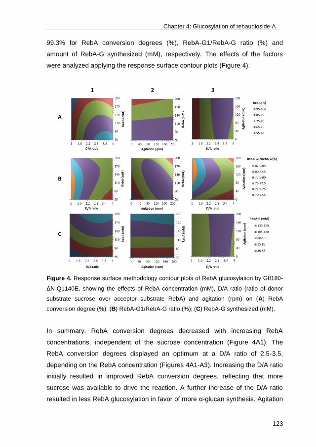
Chapter 4: Glucosylation of rebaudioside A
123
99.3% for RebA conversion degrees (%), RebA-G1/RebA-G ratio (%) and
amount of RebA-G synthesized (mM), respectively. The effects of the factors
were analyzed applying the response surface contour plots (Figure 4).
Figure 4. Response surface methodology contour plots of RebA glucosylation by Gtf180-
ΔN-Q1140E, showing the effects of RebA concentration (mM), D/A ratio (ratio of donor
substrate sucrose over acceptor substrate RebA) and agitation (rpm) on (A) RebA
conversion degree (%); (B) RebA-G1/RebA-G ratio (%); (C) RebA-G synthesized (mM).
In summary, RebA conversion degrees decreased with increasing RebA
concentrations, independent of the sucrose concentration (Figure 4A1). The
RebA conversion degrees displayed an optimum at a D/A ratio of 2.5-3.5,
depending on the RebA concentration (Figures 4A1-A3). Increasing the D/A ratio
initially resulted in improved RebA conversion degrees, reflecting that more
sucrose was available to drive the reaction. A further increase of the D/A ratio
resulted in less RebA glucosylation in favor of more α-glucan synthesis. Agitation
Chapter 4: Glucosylation of rebaudioside A
124
had only a slight effect on RebA conversion degrees and amount of RebA-G
synthesized (Figures 4A2-C2). Agitation influenced the RebA-G1/RebA-G ratio
more strongly: the highest ratios were observed at an agitation speed of
approximately 185 rpm (Figures 4B2-B3). Furthermore, low D/A ratios favored
high RebA-G1/RebA-G ratios, since less donor substrate sucrose was available
to glucosylate RebA-G1 into multi-glucosylated products (Figure 4B1).
The resulting Box-Behnken model (Table SIII) was subsequently used for the
optimization of the reaction conditions. An efficient conversion of RebA into
RebA-G yielding a maximal amount of RebA-G (at least 95%) was aimed for. The
model predicted the synthesis of 80 mM RebA-G in case the following conditions
were applied: 5 U/mL Gtf180-ΔN-Q1140E, 84 mM RebA, 282 mM sucrose and
185 rpm. The validation test resulted in the synthesis of 79 mM (or 115 g/L)
RebA-G with a RebA-G1/RebA-G ratio of 77.7% (Figure 5A), which was in very
good agreement with the prediction. Applying identical conditions for the
glucosylation of RebA with wild-type Gtf180-ΔN resulted only in the conversion of
49.7% RebA, yielding 42 mM RebA-G with a RebA-G1/RebA-G ratio of 54.4%
(Figure 5B). Hence, the Q1140E-mutant not only converted more RebA into
RebA-G than wild-type Gtf180-ΔN (94.5% vs. 49.7%), its RebA-G consisted
mostly of mono-α-glucosylated product RebA-G1 (77.7% vs. 54.4%). Compared
to wild-type (45.6%), Q1140E produced less RebA-G2+ (22.3%), which is the
minor fraction of the RebA glucosides with 2 and more glucose units.
Fed-batch reaction. The main remaining bottleneck for RebA glucosylation with
Gtf180-ΔN-Q1140E is the synthesis of α-glucans at high sucrose concentrations,
preventing complete RebA glucosylation at high RebA concentrations (Figure
4A1). This issue was addressed by performing a fed-batch reaction, in which
sucrose was added to the reaction in fixed intervals of 20 min in order to keep the
sucrose concentration low and hence suppress α-glucan formation as much as
possible. The addition of 50 U/mL enzyme ensured complete usage of sucrose
within 20 min and complete conversion of RebA within 3 h. Figure 5C represents
RebA glucosylation at 200 mM RebA and an average sucrose concentration of
50 mM (fluctuating between 0-100 mM). In comparison to the batch reaction (200
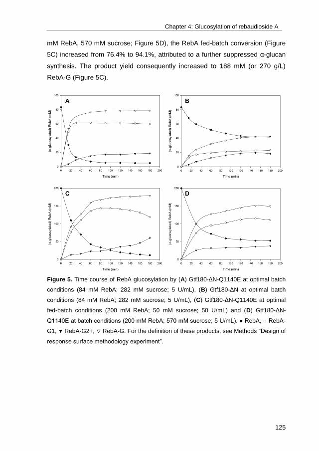
Chapter 4: Glucosylation of rebaudioside A
125
mM RebA, 570 mM sucrose; Figure 5D), the RebA fed-batch conversion (Figure
5C) increased from 76.4% to 94.1%, attributed to a further suppressed α-glucan
synthesis. The product yield consequently increased to 188 mM (or 270 g/L)
RebA-G (Figure 5C).
Figure 5. Time course of RebA glucosylation by (A) Gtf180-ΔN-Q1140E at optimal batch
conditions (84 mM RebA; 282 mM sucrose; 5 U/mL), (B) Gtf180-ΔN at optimal batch
conditions (84 mM RebA; 282 mM sucrose; 5 U/mL), (C) Gtf180-ΔN-Q1140E at optimal
fed-batch conditions (200 mM RebA; 50 mM sucrose; 50 U/mL) and (D) Gtf180-ΔN-
Q1140E at batch conditions (200 mM RebA; 570 mM sucrose; 5 U/mL). ● RebA, ○ RebA-
G1, ▼ RebA-G2+, ▽ RebA-G. For the definition of these products, see Methods “Design of
response surface methodology experiment”.

Chapter 4: Glucosylation of rebaudioside A
126
3.4. Docking of RebA in the active site of wild-type Gtf180-ΔN and mutant
Q1140E
To gain further insight into the α-glucosylation of RebA by the Gtf180-ΔN wild-
type enzyme and its mutant Gtf180-ΔN-Q1140E, in silico docking studies201
were
performed. To this end, the crystal structure of RebA as reported by ref202
was
used. Docking of RebA into the wild-type Gtf180-ΔN active site (X-ray crystal
structure of a complex of Gtf180-ΔN with maltose; PDB code: 3KLL83
) afforded
the pose as depicted in Figure 6A, showing that only the steviol C-19 β-D-
glucosyl moiety (Glc1 in Figure 3) of RebA is available for glucosylation,
especially at the HO-6 group, due to its orientation. Hydrogen bonding of Glc1
HO-6 with the catalytic residue D1136 possibly supports deprotonation in the
glycosylation step (Figure 6C). A π–π-stacking interaction with W1065 as well as
hydrogen bonding of Glc1 HO-4 and HO-3 to the backbone of D1136 was
observed and appears to hold the Glc1 residue in place.
Docking of RebA into the active site of the mutant Gtf180-ΔN-Q1140E afforded
the pose as depicted in Figure 6B. The binding of the steviol C-19 β-D-glucosyl
moiety (Glc1 in Figure 3) is identical to the wild-type enzyme, but fewer hydrogen
bonds of the steviol C-13 trisaccharide moiety (Glc2, Glc3 and Glc4 in Figure 3)
were observed (Figure 6D). The modified binding pocket, resulting from the
mutation, does not influence the binding of the steviol C-19 β-D-glucosyl unit,
given that the mutation is not in direct proximity of the monosaccharide. The
experimental observation that mutant Q1140E leads to more efficient α-
glucosylation could be explained by the possibility that deprotonation of Glc1 HO-
6 is aided by a water-mediated hydrogen bond between Glc1 HO-6 and E1140
as depicted in Figure 6B. The finding that the Q1140E mutant shows mostly
mono-glucosylation instead of oligo-glucosylation is currently difficult to explain
on the basis of these docking results.
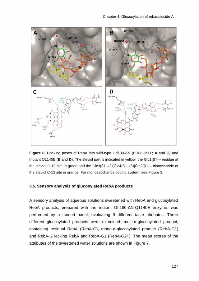
Chapter 4: Glucosylation of rebaudioside A
127
Figure 6. Docking poses of RebA into wild-type Gtf180-ΔN (PDB: 3KLL; A and C) and
mutant Q1140E (B and D). The steviol part is indicated in yellow, the Glc1(β1→ residue at
the steviol C-19 site in green and the Glc3(β1→2)[Glc4(β1→3)]Glc2(β1→ trisaccharide at
the steviol C-13 site in orange. For monosaccharide coding system, see Figure 3.
3.5. Sensory analysis of glucosylated RebA products
A sensory analysis of aqueous solutions sweetened with RebA and glucosylated
RebA products, prepared with the mutant Gtf180-ΔN-Q1140E enzyme, was
performed by a trained panel, evaluating 9 different taste attributes. Three
different glucosylated products were examined: multi-α-glucosylated product,
containing residual RebA (RebA-G), mono-α-glucosylated product (RebA-G1)
and RebA-G lacking RebA and RebA-G1 (RebA-G2+). The mean scores of the
attributes of the sweetened water solutions are shown in Figure 7.
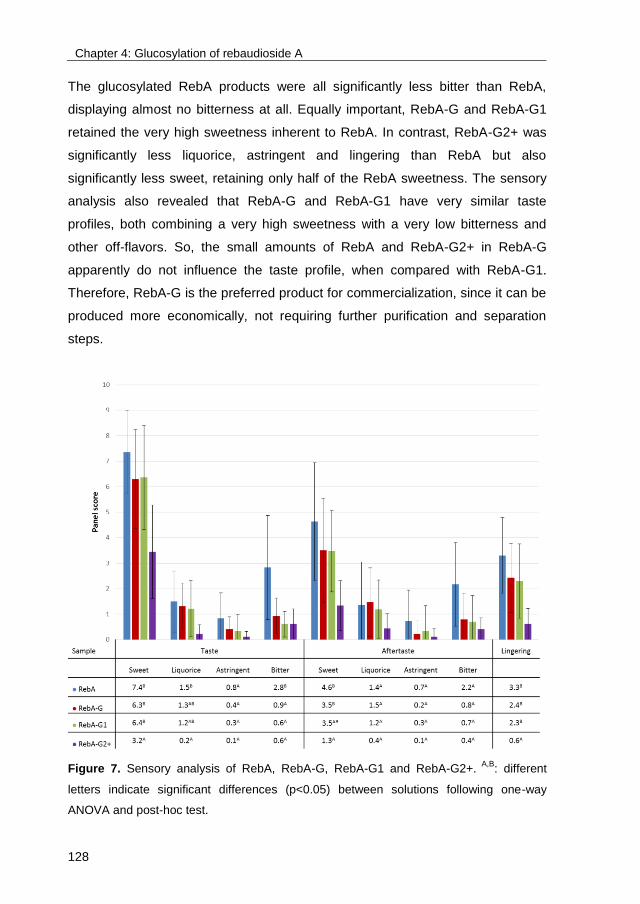
Chapter 4: Glucosylation of rebaudioside A
128
The glucosylated RebA products were all significantly less bitter than RebA,
displaying almost no bitterness at all. Equally important, RebA-G and RebA-G1
retained the very high sweetness inherent to RebA. In contrast, RebA-G2+ was
significantly less liquorice, astringent and lingering than RebA but also
significantly less sweet, retaining only half of the RebA sweetness. The sensory
analysis also revealed that RebA-G and RebA-G1 have very similar taste
profiles, both combining a very high sweetness with a very low bitterness and
other off-flavors. So, the small amounts of RebA and RebA-G2+ in RebA-G
apparently do not influence the taste profile, when compared with RebA-G1.
Therefore, RebA-G is the preferred product for commercialization, since it can be
produced more economically, not requiring further purification and separation
steps.
Figure 7. Sensory analysis of RebA, RebA-G, RebA-G1 and RebA-G2+. A,B
: different
letters indicate significant differences (p<0.05) between solutions following one-way
ANOVA and post-hoc test.
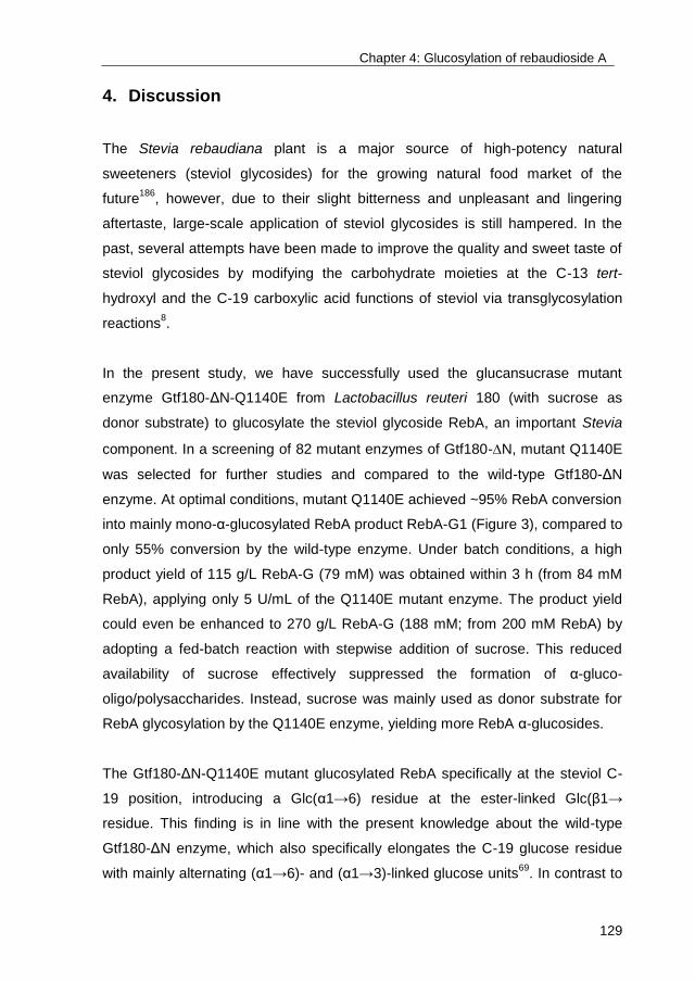
Chapter 4: Glucosylation of rebaudioside A
129
4. Discussion
The Stevia rebaudiana plant is a major source of high-potency natural
sweeteners (steviol glycosides) for the growing natural food market of the
future186
, however, due to their slight bitterness and unpleasant and lingering
aftertaste, large-scale application of steviol glycosides is still hampered. In the
past, several attempts have been made to improve the quality and sweet taste of
steviol glycosides by modifying the carbohydrate moieties at the C-13 tert-
hydroxyl and the C-19 carboxylic acid functions of steviol via transglycosylation
reactions8.
In the present study, we have successfully used the glucansucrase mutant
enzyme Gtf180-ΔN-Q1140E from Lactobacillus reuteri 180 (with sucrose as
donor substrate) to glucosylate the steviol glycoside RebA, an important Stevia
component. In a screening of 82 mutant enzymes of Gtf180-N, mutant Q1140E
was selected for further studies and compared to the wild-type Gtf180-ΔN
enzyme. At optimal conditions, mutant Q1140E achieved ~95% RebA conversion
into mainly mono-α-glucosylated RebA product RebA-G1 (Figure 3), compared to
only 55% conversion by the wild-type enzyme. Under batch conditions, a high
product yield of 115 g/L RebA-G (79 mM) was obtained within 3 h (from 84 mM
RebA), applying only 5 U/mL of the Q1140E mutant enzyme. The product yield
could even be enhanced to 270 g/L RebA-G (188 mM; from 200 mM RebA) by
adopting a fed-batch reaction with stepwise addition of sucrose. This reduced
availability of sucrose effectively suppressed the formation of α-gluco-
oligo/polysaccharides. Instead, sucrose was mainly used as donor substrate for
RebA glycosylation by the Q1140E enzyme, yielding more RebA α-glucosides.
The Gtf180-ΔN-Q1140E mutant glucosylated RebA specifically at the steviol C-
19 position, introducing a Glc(α1→6) residue at the ester-linked Glc(β1→
residue. This finding is in line with the present knowledge about the wild-type
Gtf180-ΔN enzyme, which also specifically elongates the C-19 glucose residue
with mainly alternating (α1→6)- and (α1→3)-linked glucose units69
. In contrast to
Chapter 4: Glucosylation of rebaudioside A
130
the modified steviol glycosides prepared by incubation with CGTases, thereby
introducing only (α1→4)-linked glucose residues, the (α1→3) and (α1→6)
linkages are resistant to hydrolysis by the human amylolytic enzymes in saliva,
which may prolong the sweet taste in the mouth.
Molecular docking studies were performed to gain insight into the glucosylation
mechanism of RebA at the molecular level and to elucidate how a single amino
acid change in Gtf180-ΔN, namely Q1140E, significantly improved RebA
conversion. Docking of RebA into the active site of the Gtf180-ΔN wild-type
enzyme indicated that only its steviol C-19-ester-linked Glc(β1→ residue is
available for glycosylation. The Q1140E mutation is predicted not to affect the
orientation and position of RebA in the active site, supporting the experimental
observation that both enzymes specifically α-glucosylate RebA at the
Glc1(β1→C-19 residue, and not at the Glc3(β1→2)[Glc4(β1→3)]Glc2(β1→C-13
trisaccharide (Figure 3). Furthermore, the docking results showed that Glc1 HO-6
and not Glc1 HO-3 of the Glc1(β1→C-19 residue of RebA is prominently
available for glucosylation. This is in agreement with the experimental results that
both wild-type and mutant Gtf180-ΔN enzymes attach the first Glc residue (Glc5)
exclusively via an (α1→6)-linkage. A faster and more efficient glucosylation of
RebA was obtained by replacement of glutamine with the more negatively
charged glutamate at position 1140 (mutant Q1140E). Conceivably,
deprotonation of Glc1 HO-6 is aided by a water-mediated hydrogen bond
between Glc1 HO-6 and glutamate, which is absent with glutamine at position
1140 (Figure 6). The finding that Q1140E shows mostly mono-glucosylation is
currently difficult to explain on basis of the docking results. Elucidation of the
Q1140E mutant protein 3D structure, followed by a comparison of the Gtf180-ΔN
wild-type and mutant Q1140E structures, ideally in complex with RebA, may
shed more light on the observed differences in RebA glucosylation.
An important finding in our study was that the multi-glucosylated RebA product
(RebA-G) had a significantly reduced bitterness compared to RebA. This
improved steviol glycoside product mixture thus displays appealing sensory
properties and is likely to find application as a functional food ingredient. This
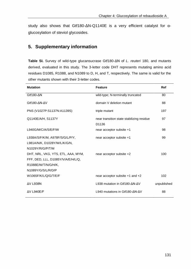
Chapter 4: Glucosylation of rebaudioside A
131
study also shows that Gtf180-ΔN-Q1140E is a very efficient catalyst for α-
glucosylation of steviol glycosides.
5. Supplementary information
Table SI. Survey of wild-type glucansucrase Gtf180-ΔN of L. reuteri 180, and mutants
derived, evaluated in this study. The 3-letter code DHT represents mutating amino acid
residues D1085, R1088, and N1089 to D, H, and T, respectively. The same is valid for the
other mutants shown with their 3-letter codes.
Mutation Feature Ref
Gtf180-ΔN wild-type; N-terminally truncated 80
Gtf180-ΔN-ΔV domain V deletion mutant 88
PNS (V1027P:S1137N:A1139S) triple mutant 197
Q1140E/A/H, S1137Y near transition state stabilizing residue
D1136
97
L940G/M/C/A/S/E/F/W near acceptor subsite +1 98
L938A/S/F/K/M, A978F/S/G/L/P/Y,
L981A/N/K, D1028Y/W/L/K/G/N,
N1029Y/R/G/P/T/M
near acceptor subsite +1 99
DHT, NRL, VKG, YTS, ETL, AAA, MYM,
FFF, DED, LLL, D1085Y/V/A/E/H/L/Q,
R1088E/W/T/N/G/H/K,
N1089Y/G/S/L/R/D/P
near acceptor subsite +2 100
W1065F/K/L/Q/G/T/E/F near acceptor subsite +1 and +2 102
ΔV L938N L938 mutation in Gtf180-ΔN-ΔV unpublished
ΔV L940E/F L940 mutations in Gtf180-ΔN-ΔV 88
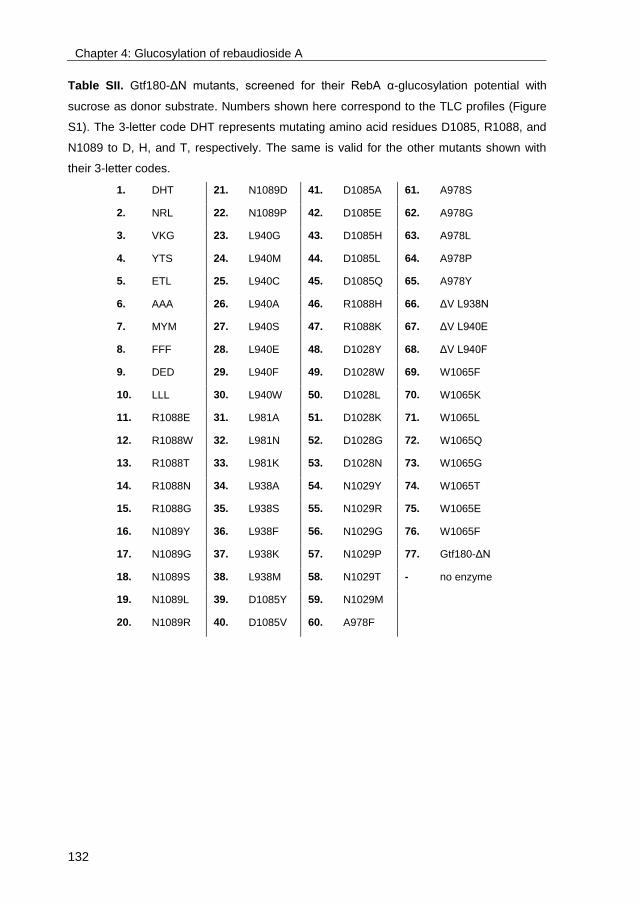
Chapter 4: Glucosylation of rebaudioside A
132
Table SII. Gtf180-ΔN mutants, screened for their RebA α-glucosylation potential with
sucrose as donor substrate. Numbers shown here correspond to the TLC profiles (Figure
S1). The 3-letter code DHT represents mutating amino acid residues D1085, R1088, and
N1089 to D, H, and T, respectively. The same is valid for the other mutants shown with
their 3-letter codes.
1. DHT 21. N1089D 41. D1085A 61. A978S
2. NRL 22. N1089P 42. D1085E 62. A978G
3. VKG 23. L940G 43. D1085H 63. A978L
4. YTS 24. L940M 44. D1085L 64. A978P
5. ETL 25. L940C 45. D1085Q 65. A978Y
6. AAA 26. L940A 46. R1088H 66. ΔV L938N
7. MYM 27. L940S 47. R1088K 67. ΔV L940E
8. FFF 28. L940E 48. D1028Y 68. ΔV L940F
9. DED 29. L940F 49. D1028W 69. W1065F
10. LLL 30. L940W 50. D1028L 70. W1065K
11. R1088E 31. L981A 51. D1028K 71. W1065L
12. R1088W 32. L981N 52. D1028G 72. W1065Q
13. R1088T 33. L981K 53. D1028N 73. W1065G
14. R1088N 34. L938A 54. N1029Y 74. W1065T
15. R1088G 35. L938S 55. N1029R 75. W1065E
16. N1089Y 36. L938F 56. N1029G 76. W1065F
17. N1089G 37. L938K 57. N1029P 77. Gtf180-ΔN
18. N1089S 38. L938M 58. N1029T - no enzyme
19. N1089L 39. D1085Y 59. N1029M
20. N1089R 40. D1085V 60. A978F

Chapter 4: Glucosylation of rebaudioside A
133
Table SIII. Box-Behnken experimental design and results for the variables studied.
Second-degree polynomial equation with coefficients of each factor is given for RebA
conversion (%), RebA-G1/RebA-G (%) and amount of RebA-G synthesized (mM). X1,
RebA concentration (mM); X2, D/A ratio; X3, agitation speed (rpm).
Pattern X1 X2 X3
RebA conversion
(%)
RebA-G1/RebA-G
(%) RebA-G (mM)
1 +0+ 200 2.5 200 73.8 76.3 147.6
2 0−+ 125 1 200 64.3 83.4 80.6
3 −0+ 50 2.5 200 94.1 77.2 47.1
4 0−− 125 1 0 63.4 80 79.3
5 0 200 4 100 66.6 74.8 133.2
6 −0− 50 2.5 0 97 67.7 48.6
7 −+0 50 4 100 96.5 75.8 48.3
8 0+− 125 4 0 82.7 72.6 103.4
9 −−0 50 1 100 67.4 85 33.8
10 0 125 2.5 100 85.5 78.9 106.9
11 +−0 200 1 100 60.7 75.6 121.4
12 0 125 2.5 100 85.4 78 106.8
13 0 125 2.5 100 85.4 78.5 106.8
14 0++ 125 4 200 88 72.2 110
15 +0− 200 2.5 0 69.1 77 138.2
RebA conversion = 85.4333 - 10.6000X1 + 9.7500X2 + 1.000X3 - 5.8000X1X2 +
1.9000X1X3 + 1.1000X2X3 - 1.8667X12 - 10.7667X2
2 - 0.0667X3
2.
RebA-G1/RebA-G = 78.4333 – 0.2375X1 – 3.5375X2 + 1.5250X3 + 2.1250X1X2 –
2.5500X1X3 – 0.8500X2X3 – 1.6292X12 + 0.9708X2
2 - 2.2542X3
2.
RebA-G = 106.8333 + 45.3250X1 + 9.9750X2 + 1.9750X3 - 0.6750X1X2 + 2.7250X1X3 +
1.3250X2X3 – 10.3042X12 – 12.3542X2
2 – 1.1542X3
2.

Chapter 4: Glucosylation of rebaudioside A
134
Table SIV. Methylation analysis of the carbohydrate moieties in RebA and α-glucosylated
RebA products RebA-G and RebA-G1.
Alditol
acetate Rt
a
Structural
feature
Peak area (%)
RebA RebA-G RebA-G1
2,3,4,6-Hexb 1.00 Glcp(1→ 74 58 61
2,4,6-Hex 1.16 →3)Glcp(1→ - 3 -
3,4,6-Hex 1.18 →4)Glcp(1→ - trc tr
c
2,3,4-Hex 1.22 →6)Glcp(1→ - 17 18
4,6-Hex 1.32 →2,3)Glcp(1→ 26 20 21
2,4-Hex 1.39 →3,6)Glcp(1→ - 2 -
a Rt, retention time relative to 1,5-di-O-acetyl-2,3,4,6-tetra-O-methylglucitol (1.00) on GLC.
b 2,3,4,6-Hex = 1,5-di-O-acetyl-2,3,4,6-tetra-O-methylglucitol-1-d. etc.
c tr = trace (<2%).

Chapter 4: Glucosylation of rebaudioside A
135
Figure S1. TLC substrate/product profiles after a 3 h incubation at 37 °C of a buffer
solution (pH 4.7) containing 50 mM RebA, 0.2 M sucrose and ~1 mg/mL wild-type Gtf180-
ΔN (lane 77) or Gtf180-ΔN mutant enzymes.
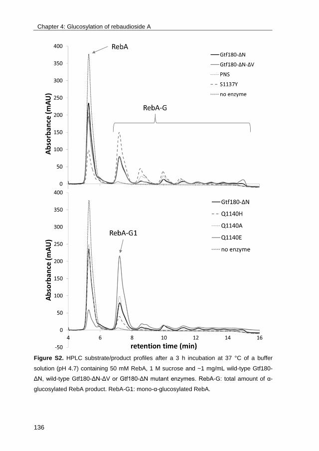
Chapter 4: Glucosylation of rebaudioside A
136
Figure S2. HPLC substrate/product profiles after a 3 h incubation at 37 °C of a buffer
solution (pH 4.7) containing 50 mM RebA, 1 M sucrose and ~1 mg/mL wild-type Gtf180-
ΔN, wild-type Gtf180-ΔN-ΔV or Gtf180-ΔN mutant enzymes. RebA-G: total amount of α-
glucosylated RebA product. RebA-G1: mono-α-glucosylated RebA.
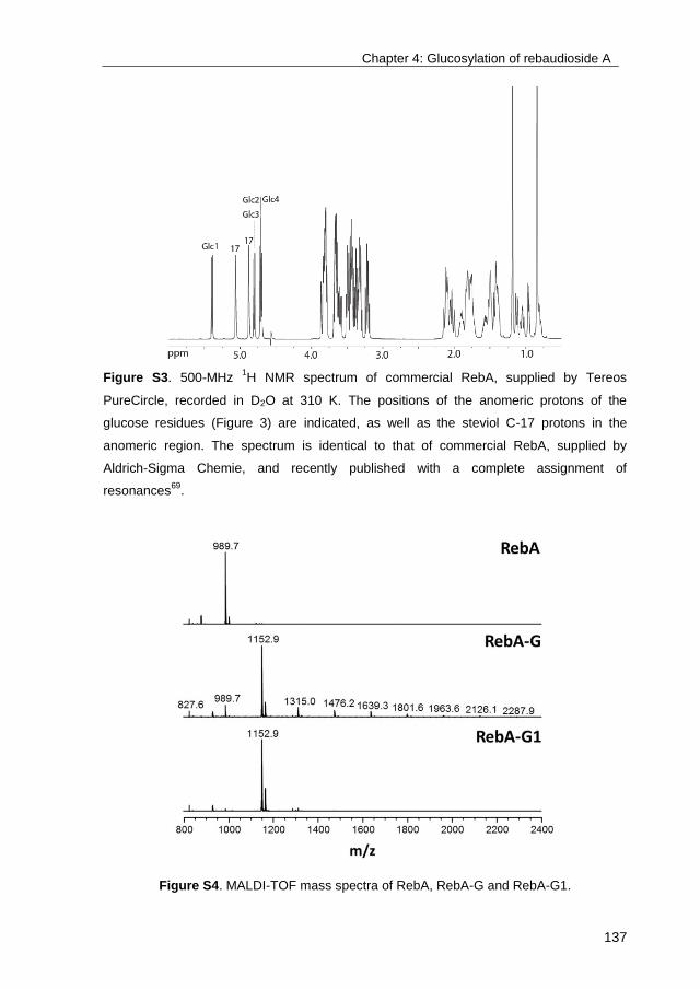
Chapter 4: Glucosylation of rebaudioside A
137
Figure S3. 500-MHz 1H NMR spectrum of commercial RebA, supplied by Tereos
PureCircle, recorded in D2O at 310 K. The positions of the anomeric protons of the
glucose residues (Figure 3) are indicated, as well as the steviol C-17 protons in the
anomeric region. The spectrum is identical to that of commercial RebA, supplied by
Aldrich-Sigma Chemie, and recently published with a complete assignment of
resonances69
.
Figure S4. MALDI-TOF mass spectra of RebA, RebA-G and RebA-G1.

139
Chapter 5
Glucosylation of stevioside by Gtf180-ΔN-Q1140E improves its
taste profile
Tim Devlamynck1, Koen Quataert
1, Evelien M. te Poele
2, Gerrit J. Gerwig
3,
Johannis P. Kamerling3, Wim Soetaert
1, Lubbert Dijkhuizen
2
1Ghent University,
2University of Groningen,
3University of Utrecht
Chapter 5: Glucosylation of stevioside
140
Abstract
The adverse health effects of sucrose overconsumption, typical for diets in
developed countries, necessitate the use of low-calorie sweeteners. Since their
approval by the European Commission in 2011, steviol glycosides have
increasingly been used as high-intensity sweetener in food products. The most
prevalent steviol glycoside in the leaves of Stevia rebaudiana is stevioside. Due
to its lingering bitterness and off-flavors, it has found limited applications in food
products, as the better tasting rebaudioside A (RebA) is preferred. Enzymatic
glucosylation of stevioside is a well-known strategy to reduce this bitterness. Up
to date, many glucosylation reactions of stevioside or RebA suffer from low
productivities. In this chapter, the optimized α-glucosylation of stevioside with
glucansucrase Gtf180-ΔN-Q1140E, previously shown to efficiently glucosylate
RebA as well, is reported. The structures of the novel steviol glycosides were
elucidated by NMR spectroscopy, mass spectrometry, and methylation analysis,
revealing that stevioside was mainly glucosylated at the C-19 glucosyl moiety. In
contrast to RebA glucosylation, minor products were also glucosylated at the C-
13 site. Sensory analysis of the glucosylated stevioside products by a trained
panel revealed significant reductions in bitterness and off-flavors compared to
stevioside, while the typical intensive sweetness of steviol glycosides was
retained.
Chapter 5: Glucosylation of stevioside
141
1. Introduction
Over the past decade, Western society has increasingly been confronted with
lifestyle diseases such as type 2 diabetes, ischaemic heart attacks and various
cardiovascular diseases. The cost for society in Europe is estimated to be 2% to
4% of the total healthcare cost213
. The risk to suffer from lifestyle diseases
increases significantly when the BMI is higher than 25 kg/m2 214,215
. A study from
Calle et al.216
even revealed that 14% to 20% of all cancer deaths may also be
related to overweight or obesity. Important causes of overweight are a decrease
of physical activity and inappropriate dietary patterns. Moreover, an excessive
sugar intake appears to be directly associated with an increase in body weight217
.
A wider array of sweet food products with less or even no sugar content is
consequently a necessity in order to reduce the prevalence of lifestyle diseases.
Consumers are more and more aware of the relationship between diet-related
diseases and healthy foods but are nevertheless not so eager to decrease their
intake of sweet food products218
. In addition, the ‘natural’ character of the applied
sweeteners is increasingly perceived by consumers to be equally important as
their taste219
. The implementation into the market of natural, high-intensity
sweeteners is thus driven by a strong consumer demand. Up to this date, several
candidates have been proposed to take up this role: sweet-tasting proteins such
as monatin and thaumatin220
, and other plant extracts like glycyrrhizin from the
root of Glycyrrhiza glabra221
, mogrosides from monk fruit (Siraitia grosvenorii)222
,
and steviol glycosides (SG) from the leaves of Stevia rebaudiana186
. Since the
European Commission authorized the use of high purity SG (≥95%), such as
rebaudioside A (RebA) and stevioside (Stev), in foods and beverages, stevia-
based products have rapidly expanded across the European market. Recently, it
was shown that SG, by means of their steviol group, potentiate Ca2+
-dependent
activity of TRPM5, a cation channel protein essential for taste transduction of
sweet, bitter, and umami in chemosensory cells223
. As a result, the sweetness of
SG is intensified, whereas their bitterness lingers on. Interestingly, TRPM5 also
facilitates insulin release by the pancreas, preventing high blood glucose
Chapter 5: Glucosylation of stevioside
142
concentrations and consequently the development of type 2 diabetes224
. A study
on mice revealed that TRPM5 potentiation by SG protected them against the
development of high-fat diet-induced hyperglycaemia, prompting the authors to
propose SG as cost-effective antidiabetic drugs225
.
Unfortunately, roughly half of the human population attributes an unpleasant
lingering bitterness to RebA and Stev, as reflected by the considerable sequence
variation in the genes encoding for the bitter receptors hTAS2R4 and
hTAS2R1446
. One strategy to solve this issue consists in the addition of masking
agents such as several sugar alcohols226
. In order to circumvent the use of
masking agents, enzymatic glucosylation of RebA and Stev has been proposed
as a means to (partially) remove their bitterness8. Several enzymes, typically
UDP-glucosyltransferases (UGTases)57-59
and cyclodextrin glucanotransferases
(CGTases)50-56
, have already been applied for this purpose. However, UGTases
require expensive nucleotide-activated sugars as donor substrate13
, whereas
CGTases possess poor C-13/C-19 regiospecificity, producing mixtures of α-
glucosylated SG51
.
Alternatively, glucansucrases (GS) can be applied for the glucosylation of SG.
GS (EC 2.1.4.-) are enzymes found only in lactic acid bacteria, of which most
members, including Lactobacillus reuteri, have the generally-recognized-as-safe
(GRAS) status. They use the donor substrate sucrose to catalyze the synthesis
of α-glucan polysaccharides, thereby introducing different ratios of glycosidic
linkages, depending on the enzyme specificity75
. It was demonstrated that
suppressing this α-glucan synthesis by mutational engineering improved the
glucosylation of non-natural acceptor substrates such as catechol110
. In chapter
4, the glucosylation of RebA with the Q1140E mutant of glucansucrase enzyme
Gtf180-ΔN from Lactobacillus reuteri 180 was reported68-70
. RebA was only
glucosylated at the C-19 glucosyl moiety, producing mainly mono-α-glucosylated
product with an (α1→6)-linkage, but also α-glucosides with two or more glucosyl
units attached. The glucosylation of Stev, the most abundant steviol glycoside,
was not addressed. Here, we report a careful optimization of the enzymatic
glucosylation of Stev by the same enzyme. The structures of the two main α-

Chapter 5: Glucosylation of stevioside
143
glucosylated Stev products were characterized by NMR spectroscopy, mass
spectrometry, and methylation analysis. Sensory analysis by a trained panel
revealed a substantial decrease in bitterness and off-flavors of the glucosylated
products compared to Stev and RebA.
2. Materials and methods
2.1. Stevioside
Stevioside (>85 % purity, HPLC) was obtained from TCI Europe.
2.2. Production and purification of recombinant Gtf180-ΔN-Q1140E
Recombinant, N-terminally truncated Gtf180-ΔN from Lactobacillus reuteri 180
and derived Q1140E mutant were produced and purified as described
previously70,80,99
.
2.3. Gtf180-ΔN-Q1140E activity assays
One unit (U) of enzyme activity corresponds to the conversion of 1 μmole
sucrose (used for hydrolysis and transglycosylation) in a solution containing 100
mM sucrose, 25 mM sodium acetate (pH 4.7) and 1 mM CaCl2, at 37 °C.
Enzyme activity assays were performed at 37 °C with 100 mM sucrose in 25 mM
sodium acetate (pH 4.7) and 1 mM CaCl2. Samples of 150 μL were taken every
min over a period of 8 min and immediately inactivated with 30 µL 1 M NaOH.
The sucrose concentrations of the samples were subsequently quantified by
means of HPLC analysis (see HPLC analysis), allowing the calculation of the
enzyme activity as defined above.

Chapter 5: Glucosylation of stevioside
144
2.4. HPLC analysis
Two HPLC analyses were performed. For the analysis of glucose, fructose and
sucrose, an Agilent MetaCarb 67H column (300 x 6.5 mm) was used under
isocratic conditions with 2.5 mM H2SO4 as the mobile phase. The flow rate and
temperature were set at 0.8 mL/min and 35 °C, respectively. Detection was
achieved with an RI detector. Calibration of the obtained peaks was
accomplished using the corresponding standard curves.
For the analysis of the steviol glycosides, an Agilent ZORBAX Eclipse Plus C18
column (100 x 4.6 mm, 3.5 µm) was used with water (solvent A) and acetonitrile
(solvent B) as the mobile phase. The flow rate and temperature were set at 1.0
mL/min and 40 °C, respectively. Following gradient elution was used: 5-95%
solvent B (0-25 min), 95% solvent B (25-27 min), 95-5% solvent B (27-30 min)
and again 95% of solvent A (30-35 min). Detection was achieved with an ELS
detector (evaporation temperature: 90 °C, nebulization temperature: 70 °C, gas
flow rate: 1.6 SLM). Calibration of the obtained peaks was accomplished using
the corresponding standard curves.
2.5. Design of response surface methodology experiment
Response surface methodology198
was applied to optimize the Gtf180-ΔN-
Q1140E catalyzed glucosylation of stevioside (acceptor substrate) with sucrose
as donor substrate, while minimizing the synthesis of α-glucan oligosaccharides.
All experiments were performed in 25 mM sodium acetate (pH 4.7),
supplemented with 1 mM CaCl2, at 37 °C. The addition of 10 U/mL enzyme
ensured a steady-state was reached within 3 h of incubation. A Box-Behnken
design199
was generated implementing stevioside concentration (mM),
sucrose/stevioside ratio (D/A ratio) and agitation rate (rpm) as factors. For each
of them low (-1) and high (+1) level values were assigned as follows: stevioside
concentration (25 mM) and (100 mM), D/A ratio (1) and (20), agitation rate (0
rpm) and (200 rpm). The experimental design was generated and analyzed using
JMP software (release 12)200
and consisted of 15 experiments carried out at 5
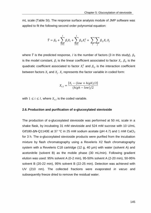
Chapter 5: Glucosylation of stevioside
145
mL scale (Table SI). The response surface analysis module of JMP software was
applied to fit the following second order polynomial equation:
�̂� = 𝛽0 + ∑ 𝛽𝑖𝑋𝑖
𝐼
𝑖=1
+ ∑ 𝛽𝑖𝑖𝑋𝑖2
𝐼
𝑖=1
+ ∑ ∑ 𝛽𝑖𝑗𝑋𝑖
𝑗𝑖
𝑋𝑗
where �̂� is the predicted response, 𝐼 is the number of factors (3 in this study), 𝛽0
is the model constant, 𝛽𝑖 is the linear coefficient associated to factor 𝑋𝑖, 𝛽𝑖𝑖 is the
quadratic coefficient associated to factor 𝑋𝑖2 and 𝛽𝑖𝑗 is the interaction coefficient
between factors 𝑋𝑖 and 𝑋𝑗. 𝑋𝑖 represents the factor variable in coded form:
𝑋𝑐,𝑖 =[𝑋𝑖 − (𝑙𝑜𝑤 + ℎ𝑖𝑔ℎ)/2]
(ℎ𝑖𝑔ℎ − 𝑙𝑜𝑤)/2
with 1 ≤ 𝑖 ≤ 𝐼, where 𝑋𝑐,𝑖 is the coded variable.
2.6. Production and purification of α-glucosylated stevioside
The production of α-glucosylated stevioside was performed at 50 mL scale in a
shake flask, by incubating 31 mM stevioside and 524 mM sucrose with 10 U/mL
Gtf180-ΔN-Q1140E at 37 °C in 25 mM sodium acetate (pH 4.7) and 1 mM CaCl2
for 3 h. The α-glucosylated stevioside products were purified from the incubation
mixture by flash chromatography using a Reveleris X2 flash chromatography
system with a Reveleris C18 cartridge (12 g, 40 µm) with water (solvent A) and
acetonitrile (solvent B) as the mobile phase (30 mL/min). Following gradient
elution was used: 95% solvent A (0-2 min), 95-50% solvent A (2-20 min), 50-95%
solvent B (20-22 min), 95% solvent B (22-25 min). Detection was achieved with
UV (210 nm). The collected fractions were evaporated in vacuo and
subsequently freeze dried to remove the residual water.
Chapter 5: Glucosylation of stevioside
146
2.7. Alkaline hydrolysis of α-glucosylated stevioside and TLC analysis
To release the carbohydrate moiety linked to the C-19 carboxyl group, a 1 h
stevioside incubation with Gtf180-ΔN-Q1140E (50 mM stevioside, 100 mM
sucrose in 25 mM sodium acetate (pH 4.7) and 1 mM CaCl2, 37 °C) was
subjected to alkaline hydrolysis. Briefly, the stevioside incubation mixture was
transferred into 1.0 M NaOH and heated at 80 °C for 2.5 h, then cooled down,
and neutralized with 6 M HCl. Samples were spotted in lines of 1 cm on a TLC
sheet (Merck Kieselgel 60 F254, 20x20 cm), which was developed in n-
butanol:acetic acid:water = 2:1:1. Bands were visualized by orcinol/sulfuric acid
staining and compared with a simultaneous run of standard compounds.
2.8. Methylation analysis
Steviol glycoside samples were permethylated using CH3I and solid NaOH in
(CH3)2SO, as described previously227
, then hydrolyzed with 2 M trifluoroacetic
acid (2 h, 120 °C) to give a mixture of partially methylated monosaccharides.
After evaporation to dryness, the mixture was dissolved in H2O and reduced with
NaBD4 (2 h, room temperature). Subsequently, the solution was neutralized with
4 M acetic acid and boric acid was removed by repeated co-evaporation with
methanol. The obtained partially methylated alditol samples were acetylated with
1:1 acetic anhydride-pyridine (30 min, 120 °C). After evaporation to dryness, the
mixtures of partially methylated alditol acetates (PMAAs) were dissolved in
dichloromethane and analyzed by GLC-EI-MS on an EC-1 column (30 m x 0.25
mm; Alltech), using a GCMS-QP2010 Plus instrument (Shimadzu Kratos Inc.,
Manchester, UK) and a temperature gradient (140-250 °C at 8 °C/min)228
.
2.9. Mass spectrometry
Matrix-assisted laser desorption ionization time-of-flight mass spectrometry
(MALDI-TOF-MS) was performed on an AximaTM mass spectrometer (Shimadzu
Kratos Inc.), equipped with a nitrogen laser (337 nm, 3 ns pulse width). Positive-
ion mode spectra were recorded using the reflector mode at a resolution of 5000
Chapter 5: Glucosylation of stevioside
147
FWHM and delayed extraction (450 ns). Accelerating voltage was 19 kV with a
grid voltage of 75.2%. The mirror voltage ratio was 1.12 and the acquisition mass
range was 200-6000 Da. Samples were prepared by mixing on the target 1 μL
sample solutions with 1 μL aqueous 10% 2,5-dihydroxybenzoic acid in 70%
acetonitrile as matrix solution.
2.10. NMR spectroscopic analysis
Resolution-enhanced 1D/2D 500-MHz 1H/
13C NMR spectra were recorded in D2O
on a Bruker DRX-500 spectrometer (Bijvoet Center, Department of NMR
Spectroscopy, Utrecht University). To avoid overlap of anomeric signals with the
HOD signal, the 1D and 2D spectra were run at 310 K. Data acquisition was
done with Bruker Topspin 2.1. Before analysis, samples were exchanged twice in
D2O (99.9 atom% D, Cambridge Isotope Laboratories, Inc., Andover, MA) with
intermediate lyophilisation, and then dissolved in 0.6 mL D2O. Fresh solutions of
~4 mg/mL were used for all NMR measurements. Suppression of the HOD signal
was achieved by applying a WEFT (water eliminated Fourier transform) pulse
sequence for 1D NMR experiments and by a pre-saturation of 1 s during the
relaxation delay in 2D experiments. The 2D TOCSY spectra were recorded using
an MLEV-17 (composite pulse devised by Levitt et al.229
) mixing sequence with
spin-lock times of 20, 50, 100 and 200 ms. The 2D 1H-
1H ROESY spectra were
recorded using standard Bruker XWINNMR software with a mixing time of 200
ms. The carrier frequency was set at the downfield edge of the spectrum in order
to minimize TOCSY transfer during spin-locking. Natural abundance 2D 13
C-1H
HSQC experiments (1H frequency 500.0821 MHz,
13C frequency 125.7552 MHz)
were recorded without decoupling during acquisition of the 1H FID. The NMR
data were processed using the MestReNova 9 program (Mestrelab Research SL,
Santiago de Compostella, Spain). Chemical shifts (δ) are expressed in ppm by
reference to internal acetone (δH 2.225 for 1H and δC 31.07 for
13C).
Chapter 5: Glucosylation of stevioside
148
2.10 Sensory analysis
Sensory analysis was performed in individual tasting booths at the UGent
Sensolab (Belgium) by a trained panel (7 persons), as described previously in
chapter 470
. In short, taste (sweetness, liquorice, astringency and bitterness) was
evaluated by swirling the sample in the mouth for 5 sec after which the sample
was expectorated. Aftertaste was evaluated 10 sec after swallowing the solution.
Lingering based on the maximum taste intensity was rated 1 min later. Sucrose
reference solutions (5%, 7.5% and 10% sucrose, scoring 5, 7.5 and 10,
respectively) were provided. Water (Spa Reine) and plain crackers were used as
palate cleansers between sampling. All samples were evaluated in duplicate.
Statistical analyses were performed with SPSS 23 (SPSS Inc., Chicago, USA).
All tests were done at a significance level of 0.05. One-Way ANOVA was used to
investigate any significant difference between the solutions. Testing for equal
variances was executed with the Modified Levene Test. When conditions for
equal variance were fulfilled, the Tukey test210
was used to determine differences
between samples. In case variances were not equal, Games-Howell was
performed211
.
Three different solutions sweetened with Stev products were examined: 588
mg/L mono-α-glucosylated product (Stev-G1), 588 mg/L multi-α-glucosylated
product, containing residual Stev, Stev-G1 and higher α-glucosides (Stev-G) and
1176 mg/L multi-α-glucosylated product (Stev-G’).
3. Results
3.1. Synthesis of α-glucosylated stevioside with Gtf180-ΔN-Q1140E
Gtf180-ΔN and the derived Gtf180-ΔN-Q1140E mutant also readily glucosylated
stevioside. Optimization of the reaction conditions for rebaudioside A (RebA)
glucosylation with Gtf180-ΔN-Q1140E revealed the importance of selecting

Chapter 5: Glucosylation of stevioside
149
adequate concentrations of donor substrate sucrose and acceptor substrate
RebA. The addition of too much sucrose resulted in suboptimal yields due to
increased α-glucan synthesis70
. The glucosylation of stevioside (Stev) was
therefore also optimized by response surface methodology (RSM), using a Box-
Behnken experimental design. Following factors were considered: X1 Stev
concentration (mM); X2 the ratio of donor substrate sucrose over acceptor
substrate Stev (D/A ratio); X3 agitation speed (rpm). The addition of 10 U/mL
enzyme ensured that a steady state in Stev conversion was obtained within 3 h.
The results of the Box-Behnken experimental design are summarized in Table
SI. The analysis of variance (ANOVA) showed R2 values of 98.75% and 98.72%
for Stev conversion degree (%) and amount of α-glucosylated Stev (Stev-G)
synthesized (mM), respectively. The effects of the factors were analyzed
applying the response surface contour plots (Figure 1).
Higher Stev conversion degrees were obtained at decreasing Stev
concentrations, independent on the concentration of donor substrate sucrose.
The effect of the D/A ratio on Stev conversion degrees displayed a distinct
optimum, similarly to RebA glucosylation with Gtf180-ΔN-Q1140E70
. An increase
of D/A ratio initially resulted in improved Stev conversion degrees, indicating that
sucrose drives the reaction. However, as sucrose also acts as primer for α-
glucan synthesis, a further increase of D/A ratio resulted in less Stev
glucosylation in favor of more α-glucan synthesis. This confirmed that the
concentrations of sucrose and Stev need to be carefully optimized. In contrast,
the effect of agitation on Stev conversion degrees and amount of Stev-G
synthesized was negligible.
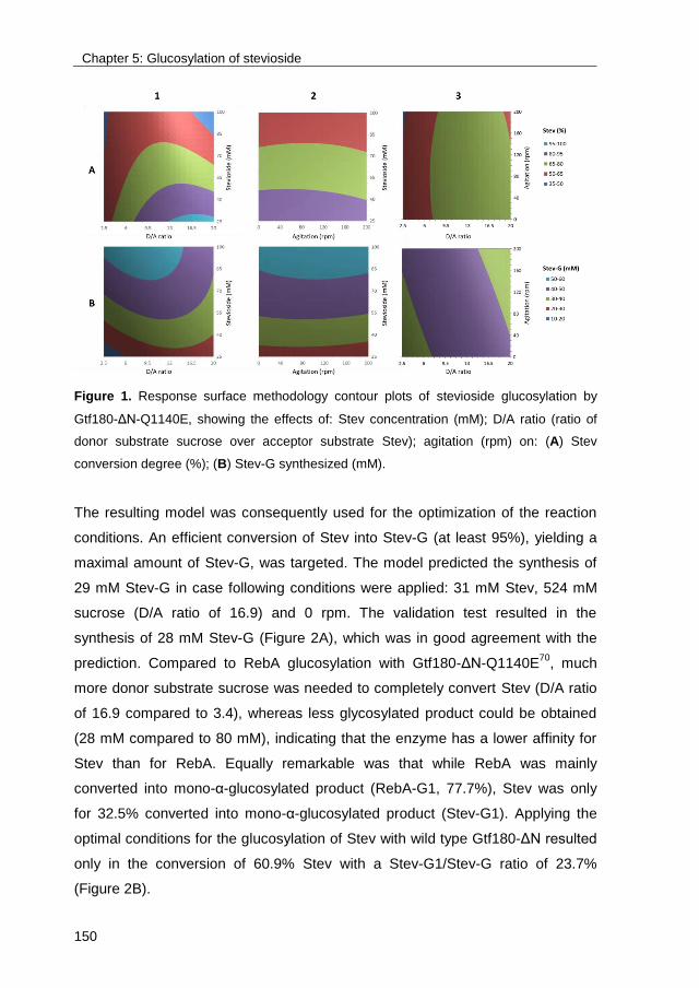
Chapter 5: Glucosylation of stevioside
150
Figure 1. Response surface methodology contour plots of stevioside glucosylation by
Gtf180-ΔN-Q1140E, showing the effects of: Stev concentration (mM); D/A ratio (ratio of
donor substrate sucrose over acceptor substrate Stev); agitation (rpm) on: (A) Stev
conversion degree (%); (B) Stev-G synthesized (mM).
The resulting model was consequently used for the optimization of the reaction
conditions. An efficient conversion of Stev into Stev-G (at least 95%), yielding a
maximal amount of Stev-G, was targeted. The model predicted the synthesis of
29 mM Stev-G in case following conditions were applied: 31 mM Stev, 524 mM
sucrose (D/A ratio of 16.9) and 0 rpm. The validation test resulted in the
synthesis of 28 mM Stev-G (Figure 2A), which was in good agreement with the
prediction. Compared to RebA glucosylation with Gtf180-ΔN-Q1140E70
, much
more donor substrate sucrose was needed to completely convert Stev (D/A ratio
of 16.9 compared to 3.4), whereas less glycosylated product could be obtained
(28 mM compared to 80 mM), indicating that the enzyme has a lower affinity for
Stev than for RebA. Equally remarkable was that while RebA was mainly
converted into mono-α-glucosylated product (RebA-G1, 77.7%), Stev was only
for 32.5% converted into mono-α-glucosylated product (Stev-G1). Applying the
optimal conditions for the glucosylation of Stev with wild type Gtf180-ΔN resulted
only in the conversion of 60.9% Stev with a Stev-G1/Stev-G ratio of 23.7%
(Figure 2B).
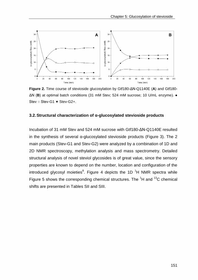
Chapter 5: Glucosylation of stevioside
151
Figure 2. Time course of stevioside glucosylation by Gtf180-ΔN-Q1140E (A) and Gtf180-
ΔN (B) at optimal batch conditions (31 mM Stev; 524 mM sucrose; 10 U/mL enzyme). ●
Stev ○ Stev-G1 ▼ Stev-G2+.
3.2. Structural characterization of α-glucosylated stevioside products
Incubation of 31 mM Stev and 524 mM sucrose with Gtf180-ΔN-Q1140E resulted
in the synthesis of several α-glucosylated stevioside products (Figure 3). The 2
main products (Stev-G1 and Stev-G2) were analyzed by a combination of 1D and
2D NMR spectroscopy, methylation analysis and mass spectrometry. Detailed
structural analysis of novel steviol glycosides is of great value, since the sensory
properties are known to depend on the number, location and configuration of the
introduced glycosyl moieties8. Figure 4 depicts the 1D
1H NMR spectra while
Figure 5 shows the corresponding chemical structures. The 1H and
13C chemical
shifts are presented in Tables SII and SIII.

Chapter 5: Glucosylation of stevioside
152
Figure 3. HPLC separation profiles of a 3 h incubation of 31 mM Stev and 524 mM
sucrose with 10 U/mL Gtf180-ΔN-Q1140E. A 0 min, B 30 min, C 180 min of incubation at
37 °C. Suc sucrose, fru fructose, glu glucose.
3.2.1. Stev-G1
Methylation analysis (Table SIV) of Stev-G1 (Stev+1Glc, according to MALDI-
TOF-MS: m/z 989.7 [M+Na]+ (Figure 6B)) showed terminal Glcp, 2-substituted
Glcp and 6-substituted Glcp (molar ratio 2:1:1), indicating that transglucosylation
had resulted in elongation but not in branching. The 1H NMR spectrum of Stev-
G1 (Figure 4B) showed resonances of one main α-glucosylated Stev product,
however, with five small (anomeric) signals (indicated with *: δH 5.35, 4.45, 4.14,
3.98, 3.16 in Figure 4B) of extra compounds (<10%). The spectrum between 0.8
and 2.2 ppm represented the typical steviol core signal pattern as seen for Stev
(Figure 4A). Besides the three β-anomeric 1H signals related to Stev (Glc1, δH
5.415; Glc2, δH 4.725; Glc3, δH 4.675), one extra anomeric 1H resonance (δH
4.862; J1,2 3.7 Hz), partially overlapping with one steviol C-17 proton, was

Chapter 5: Glucosylation of stevioside
153
observed, stemming from a new α-linked Glc residue (Glc4). The latter 1H signal
correlated with a 13
C resonance at δC 99.3 in the HSQC spectrum (Figure S1).
Figure 4. 500-MHz 1H NMR spectrum of Stev (A), Stev-G1 (B), and Stev-G2 (C),
recorded in D2O at 310 K. * Resonances stemming from additional product(s).
Chapter 5: Glucosylation of stevioside
154
Applying 2D NMR spectroscopy (TOCSY with different mixing times, ROESY and
HSQC), the 1H/
13C chemical shifts of the steviol core (Table SII) and the four Glc
residues (Table SIII) of Stev-G1 were assigned (Figure S1). The 1H and
13C
chemical shift sets of Glc2 and Glc3 correspond to those of Stev, suggesting that
no modifications had occurred in the carbohydrate moiety at the steviol C-13 site.
The TOCSY Glc4 H-1 track (δH 4.862) showed the complete scalar coupling
network H-1,2,3,4,5,6a,6b, and combined with the Glc4 C-1–C-6 set of chemical
shifts (HSQC), a terminal Glc(α1→6) unit is indicated. Based on the inter-residual
ROESY cross-peaks between Glc4 H-1 (δH 4.862) and Glc1 H-6a/b (δH
3.89/3.70), combined with the 13
C downfield shift of 4.5 ppm for Glc1 C-6 (δC
66.7; Stev Glc1 C-6: δC 62.2) (HSQC spectrum, Figure S1), a Glc4(α1→6)Glc1
disaccharide element could be established69
.
The conclusive structure of the main compound in Stev-G1 was consequently
determined as Stev elongated with a Glcp(α1→6) residue at the Glc1(β1→ on
the C-19 site of the steviol core (Figure 5B).
3.2.2. Stev-G2
Methylation analysis (Table SIV) of Stev-G2 (Stev+2Glc, according to MALDI-
TOF-MS: m/z 1152.0 [M+Na]+ (Figure 6C)) showed terminal Glcp and 2-
substituted Glcp, 4-substituted Glcp, and 6-substituted Glcp (molar ratio
2:1:1:1), together with a trace amount (<2%) of 2,6-substituted Glcp. The 1H
NMR spectrum of Stev-G2 (Figure 4C) exhibited the typical steviol core signal
pattern as seen for Stev (Figure 4A). Besides the three β-anomeric 1H
carbohydrate signals related to Stev (Glc1, δH 5.415; Glc2, δH 4.727; Glc3, δH
4.674), two α-anomeric 1H resonances of equal intensity (δH 4.863; J1,2 3.9 Hz
and δH 5.292; J1,2 3.7 Hz) were observed, stemming from two new α-linked Glc
residues (Glc4 and Glc5). The 1H NMR spectrum indicated the presence of one
main di-α-glucosylated Stev product, together with very minor products (<10%)
represented by four small anomeric signals (indicated with *: δH 5.43, 4.91, 4.75,
4.71 in Figure 4C).
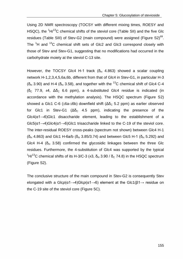
Chapter 5: Glucosylation of stevioside
155
Using 2D NMR spectroscopy (TOCSY with different mixing times, ROESY and
HSQC), the 1H/
13C chemical shifts of the steviol core (Table SII) and the five Glc
residues (Table SIII) of Stev-G2 (main compound) were assigned (Figure S2)69
.
The 1H and
13C chemical shift sets of Glc2 and Glc3 correspond closely with
those of Stev and Stev-G1, suggesting that no modifications had occurred in the
carbohydrate moiety at the steviol C-13 site.
However, the TOCSY Glc4 H-1 track (δH 4.863) showed a scalar coupling
network H-1,2,3,4,5,6a,6b, different from that of Glc4 in Stev-G1, in particular H-3
(δH 3.90) and H-4 (δH 3.58), and together with the 13
C chemical shift of Glc4 C-4
(δC 77.9, 44, ΔδC 6.6 ppm), a 4-substituted Glc4 residue is indicated (in
accordance with the methylation analysis). The HSQC spectrum (Figure S2)
showed a Glc1 C-6 (16a-16b) downfield shift (ΔδC 5.2 ppm) as earlier observed
for Glc1 in Stev-G1 (ΔδC 4.5 ppm), indicating the presence of the
Glc4(α1→6)Glc1 disaccharide element, leading to the establishment of a
Glc5(α1→4)Glc4(α1→6)Glc1 trisaccharide linked to the C-19 of the steviol core.
The inter-residual ROESY cross-peaks (spectrum not shown) between Glc4 H-1
(δH 4.863) and Glc1 H-6a/b (δH 3.85/3.74) and between Glc5 H-1 (δH 5.292) and
Glc4 H-4 (δH 3.58) confirmed the glycosidic linkages between the three Glc
residues. Furthermore, the 4-substitution of Glc4 was supported by the typical
1H/
13C chemical shifts of its H-3/C-3 (43, δH 3.90 / δC 74.8) in the HSQC spectrum
(Figure S2).
The conclusive structure of the main compound in Stev-G2 is consequently Stev
elongated with a Glcp(α1→4)Glcp(α1→6) element at the Glc1(β1→ residue on
the C-19 site of the steviol core (Figure 5C).

Chapter 5: Glucosylation of stevioside
156
Figure 5. Chemical structures of Stev (A), Stev-G1 (B), and Stev-G2 (C).
3.2.3 Stev-G
Besides Stev-G1 and Stev-G2, Stev-G was composed of a wide array of α-
glucosylated Stev products (Stev-G2+), as demonstrated by MALDI-TOF mass
spectrometry, showing the m/z [M+Na]+ peaks of Stev+1Glc up to Stev+9Glc
(Figure 6D). It has to be noted that each peak may contain more than one
compound, as different glycosidic linkage types may be present. Indeed,
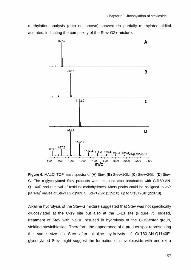
Chapter 5: Glucosylation of stevioside
157
methylation analysis (data not shown) showed six partially methylated alditol
acetates, indicating the complexity of the Stev-G2+ mixture.
Figure 6. MALDI-TOF mass spectra of (A) Stev, (B) Stev+1Glc, (C) Stev+2Glc, (D) Stev-
G. The α-glycosylated Stev products were obtained after incubation with Gtf180-ΔN-
Q1140E and removal of residual carbohydrates. Mass peaks could be assigned to m/z
[M+Na]+ values of Stev+1Glc (989.7), Stev+2Glc (1152.0), up to Stev+9Glc (2287.8).
Alkaline hydrolysis of the Stev-G mixture suggested that Stev was not specifically
glucosylated at the C-19 site but also at the C-13 site (Figure 7). Indeed,
treatment of Stev with NaOH resulted in hydrolysis of the C-19-ester group,
yielding steviolbioside. Therefore, the appearance of a product spot representing
the same size as Stev after alkaline hydrolysis of Gtf180-ΔN-Q1140E-
glycosylated Stev might suggest the formation of steviolbioside with one extra
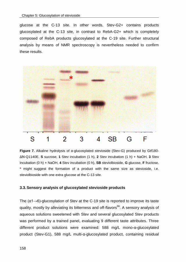
Chapter 5: Glucosylation of stevioside
158
glucose at the C-13 site. In other words, Stev-G2+ contains products
glucosylated at the C-13 site, in contrast to RebA-G2+ which is completely
composed of RebA products glucosylated at the C-19 site. Further structural
analysis by means of NMR spectroscopy is nevertheless needed to confirm
these results.
Figure 7. Alkaline hydrolysis of α-glucosylated stevioside (Stev-G) produced by Gtf180-
ΔN-Q1140E. S sucrose, 1 Stev incubation (1 h), 2 Stev incubation (1 h) + NaOH, 3 Stev
incubation (0 h) + NaOH, 4 Stev incubation (0 h), SB steviolbioside, G glucose, F fructose.
* might suggest the formation of a product with the same size as stevioside, i.e.
steviolbioside with one extra glucose at the C-13 site.
3.3. Sensory analysis of glucosylated stevioside products
The (α1→6)-glucosylation of Stev at the C-19 site is reported to improve its taste
quality, mostly by alleviating its bitterness and off-flavors66
. A sensory analysis of
aqueous solutions sweetened with Stev and several glucosylated Stev products
was performed by a trained panel, evaluating 9 different taste attributes. Three
different product solutions were examined: 588 mg/L mono-α-glucosylated
product (Stev-G1), 588 mg/L multi-α-glucosylated product, containing residual

Chapter 5: Glucosylation of stevioside
159
Stev, Stev-G1 and higher α-glucosides (Stev-G) and 1176 mg/L multi-α-
glucosylated product (Stev-G’). The mean scores of the taste attributes of the
sweetened solutions are shown in Figure 8.
Figure 8. Sensory analysis of stevioside (Stev), Stev-G, Stev-G1 and Stev-G’. A,B
:
different letters indicate significant differences (p<0.05) between solutions following one-
way ANOVA and post-hoc test.
All α-glucosylated stevioside products were significantly less bitter than
stevioside. Stev-G1 retained the very high sweetness typical for steviol
glycosides such as stevioside. In contrast, Stev-G was significantly less sweet
than stevioside, which can be explained by the relatively large proportion of multi-
α-glucosylated products. Multi-α-glucosylation resulted not only in a further
decrease of bitterness, but simultaneously decreased sweetness as well. In
addition, Stev-G was also significantly less liquorice and lingering than stevioside
(Figure 8). Doubling the concentration of Stev-G from 588 mg/L to 1176 mg/L

Chapter 5: Glucosylation of stevioside
160
roughly resulted in a duplication of the sweetness, equaling the sweetness level
of stevioside, whereas bitterness and off-flavors were still equally suppressed.
Glucosylation of stevioside with Gtf180-ΔN-Q1140E is thus a very adequate
method to improve its sensory properties, i.e. by reducing the typical bitterness to
a very low level.
4. Discussion
Even though Stev is the most abundant of all steviol glycosides extracted from
the leaves of the Stevia plant, its lingering bitterness prevents applications in low-
calorie foods and beverages. All current Stevia food products are based on
RebA, perceived as less bitter than Stev, implying that the latter is discarded as a
“side product”. This chapter demonstrated that the α-glucosylation of Stev with
Gtf180-ΔN-Q1140E offers a viable method to significantly reduce its bitterness. A
very high Stev conversion of 95%, yielding 50 g/L Stev-G within 3 h, while using
only 10 U/mL enzyme, was obtained after optimization of the reaction conditions
by RSM. Structural analysis revealed that Stev was mostly glucosylated at the C-
19 site, initially through an (α1→6)-linkage, after which the synthesized Stev-G1
was glucosylated through an (α1→4)-linkage, yielding Stev-G2 (Figure 5).
MALDI-TOF and MS analysis indicated that a complex mixture of many other
multi-glucosylated products (> 50% of Stev-G) was formed by the enzyme, as
opposed to RebA α-glucosylation by Gtf180-ΔN-Q1140E, resulting in the
synthesis of mostly mono-α-glycosylated product (77%)70
. As a consequence,
Stev-G was perceived half as sweet as Stev, an undesired effect which could be
compensated by doubling the dose of Stev-G. Remarkably, this did not affect the
significantly reduced perception of bitterness nor of other off-flavors.
A previous study already reported the application of a dextransucrase from
Leuconostoc citreum KM2066
for the glucosylation of Stev: a high conversion
degree (94%) was obtained, however, much more enzyme (4500 U/mL vs. 10
U/mL) and a longer incubation time (5 days vs. 3 h) was needed. The volumetric
productivity per U enzyme of the mutant Gtf180-ΔN-Q1140E enzyme reaction is
Chapter 5: Glucosylation of stevioside
161
consequently more than 2,000 times higher. Transglucosylation of stevioside,
using sucrose as donor substrate, was also achieved with an alternansucrase
(EC 2.4.1.140) from L. citreum SK24.002, an enzyme that also makes (α1→6)
and (α1→3) linkages67,230,231
. Under optimized reaction conditions, a maximum
conversion degree of only 44% was achieved. Stevioside was elongated at the
terminal Glc(β1→2) residue of the β-sophorosyl unit at the steviol C-13 site with
an (α1→6) linkage. Also a tri-glucosyl-stevioside was structurally characterized
and was shown to be a (α1→3)-(α1→6)-(α1→3) extension at the terminal
Glc(β1→2) residue at C-13. A taste comparison of the products was not reported.
In addition, a mono-glucosylated stevioside product, containing a Glc(α1→6)
residue at the steviol C-19-ester-linked Glc(β1→ residue (comparable with Stev-
G1), has been synthesized with β-amylase Biozyme L and maltose as glucose
donor. This also led to an improvement in quality of taste60
, just like shown here
for the Gtf180-ΔN-Q1140E products. However, also products elongated at the C-
13 site were synthesized, resulting in a decreased quality of taste.
These examples illustrate the three main requirements for any enzymatic Stev
glucosylation process: an adequate product specificity, a complete Stev
conversion and a high space-time yield. The here described process is clearly
superior to the other glucansucrase-catalyzed Stev glucosylation reactions,
adequately meeting all three requirements.

Chapter 5: Glucosylation of stevioside
162
5. Supplementary information
Table SI. Box-Behnken experimental design and results for the variables studied. Second-
degree polynomial equation with coefficients of each factor is given for amount of Stev-G
synthesized (mM) and stevioside conversion degree (%). X1 stevioside (mM), X2 D/A ratio,
X3 agitation (rpm).
Pattern X1 X2 X3 Stev-G (mM) 1 Stev conversion (%)
2
1 +0+ 100 11.25 200 55.5 55.5
2 0−+ 62.5 2.5 200 28.3 45.3
3 −0+ 25 11.25 200 22.5 90.0
4 0−− 62.5 2.5 0 30.3 48.5
5 ++0 100 20 100 38.1 38.1
6 −0− 25 11.25 0 23.2 92.9
7 −+0 50 20 20 23.7 94.8
8 0+− 62.5 20 0 42.9 68.6
9 −−0 25 2.5 100 14.8 59.3
10 000 62.5 11.25 100 44.9 71.8
11 +−0 100 2.5 100 51.6 51.6
12 000 62.5 11.25 100 45.2 72.4
13 000 62.5 11.25 100 45.6 72.9
14 0++ 62.5 20 200 39.8 63.6
15 +0− 100 11.25 0 53.2 53.2
1 Stev-G = 45.0500 + 14.2725X1 – 0.1875X2 – 0.1900X3 – 5.6000X1X2 + 0.7550X1X3 – 5.5250X2X3 –
5.1100X12 – 7.8900X2
2 – 1.3350X3
2
2 Stev conversion = 72.1000 - 17.3225X1 + 7.5500X2 - 1.0975X3 – 12.2500X1X2 + 1.2950X1X3 –
0.4500X2X3 – 2.6225X12 - 13.7725X2
2 – 1.8275X3
2
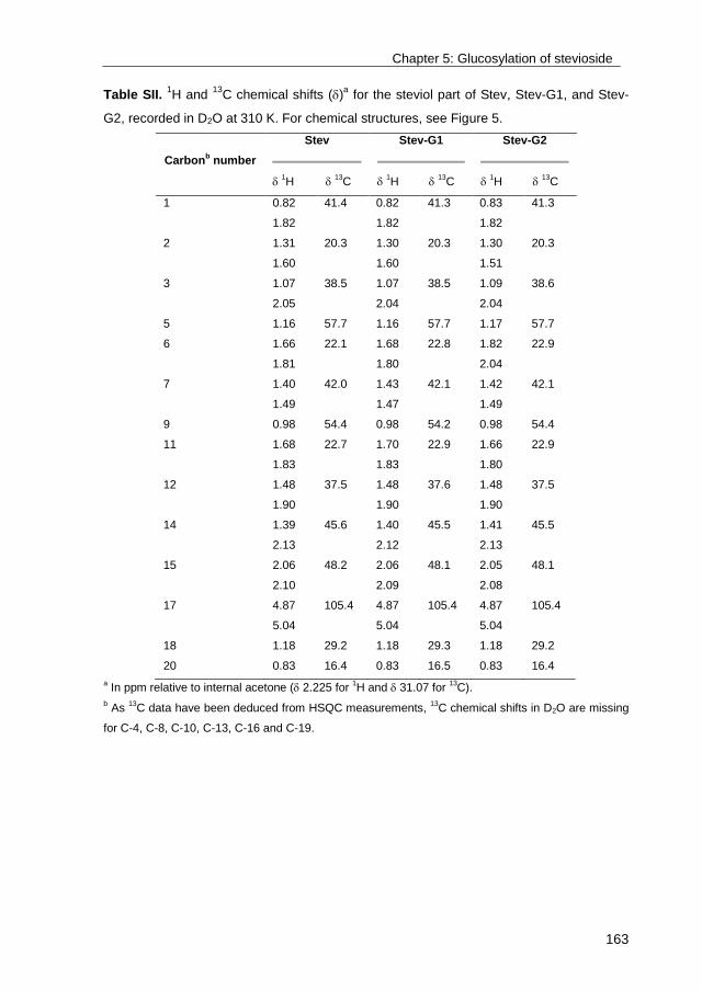
Chapter 5: Glucosylation of stevioside
163
Table SII. 1H and
13C chemical shifts ()
a for the steviol part of Stev, Stev-G1, and Stev-
G2, recorded in D2O at 310 K. For chemical structures, see Figure 5.
Carbonb number
Stev
Stev-G1
Stev-G2
1H
13C
1H
13C
1H
13C
1 0.82 41.4 0.82 41.3 0.83 41.3
1.82 1.82 1.82
2 1.31 20.3 1.30 20.3 1.30 20.3
1.60 1.60 1.51
3 1.07 38.5 1.07 38.5 1.09 38.6
2.05 2.04 2.04
5 1.16 57.7 1.16 57.7 1.17 57.7
6 1.66 22.1 1.68 22.8 1.82 22.9
1.81 1.80 2.04
7 1.40 42.0 1.43 42.1 1.42 42.1
1.49 1.47 1.49
9 0.98 54.4 0.98 54.2 0.98 54.4
11 1.68 22.7 1.70 22.9 1.66 22.9
1.83 1.83 1.80
12 1.48 37.5 1.48 37.6 1.48 37.5
1.90 1.90 1.90
14 1.39 45.6 1.40 45.5 1.41 45.5
2.13 2.12 2.13
15 2.06 48.2 2.06 48.1 2.05 48.1
2.10 2.09 2.08
17 4.87 105.4 4.87 105.4 4.87 105.4
5.04 5.04 5.04
18 1.18 29.2 1.18 29.3 1.18 29.2
20 0.83 16.4 0.83 16.5 0.83 16.4
a In ppm relative to internal acetone ( 2.225 for
1H and 31.07 for
13C).
b As
13C data have been deduced from HSQC measurements,
13C chemical shifts in D2O are missing
for C-4, C-8, C-10, C-13, C-16 and C-19.
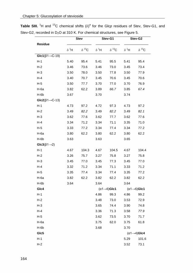
Chapter 5: Glucosylation of stevioside
164
Table SIII. 1H and
13C chemical shifts ()
a for the Glcp residues of Stev, Stev-G1, and
Stev-G2, recorded in D2O at 310 K. For chemical structures, see Figure 5.
Residue
Stev
Stev-G1
Stev-G2
1H
13C
1H
13C
1H
13C
Glc1(β1→C-19)
H-1 5.40 95.4 5.41 95.5 5.41 95.4
H-2 3.46 73.6 3.46 73.0 3.45 73.4
H-3 3.50 78.0 3.50 77.8 3.50 77.9
H-4 3.40 70.7 3.45 70.6 3.45 70.6
H-5 3.50 77.7 3.70 77.0 3.70 76.9
H-6a 3.82 62.2 3.89 66.7 3.85 67.4
H-6b 3.67 3.70 3.74
Glc2(β1→C-13)
H-1 4.73 97.2 4.72 97.3 4.73 97.2
H-2 3.49 82.2 3.49 82.2 3.49 82.1
H-3 3.62 77.6 3.62 77.7 3.62 77.6
H-4 3.34 71.2 3.34 71.1 3.35 71.0
H-5 3.33 77.2 3.34 77.4 3.34 77.2
H-6a 3.80 62.2 3.80 62.2 3.80 62.2
H-6b 3.63 3.63 3.65
Glc3(β1→2)
H-1 4.67 104.3 4.67 104.5 4.67 104.4
H-2 3.26 75.7 3.27 75.8 3.27 75.8
H-3 3.45 77.0 3.45 77.3 3.45 77.0
H-4 3.32 71.2 3.34 71.1 3.33 71.2
H-5 3.35 77.4 3.34 77.4 3.35 77.2
H-6a 3.82 62.2 3.82 62.2 3.82 62.2
H-6b 3.64 3.64 3.64
Glc4 (α1→6)Glc1 (α1→6)Glc1
H-1 4.86 99.3 4.86 99.2
H-2 3.48 73.0 3.53 72.9
H-3 3.65 74.4 3.90 74.8
H-4 3.36 71.3 3.58 77.9
H-5 3.62 73.5 3.70 71.7
H-6a 3.75 62.0 3.75 61.8
H-6b 3.68 3.70
Glc5 (α1→4)Glc4
H-1 5.29 101.6
H-2 3.52 73.1
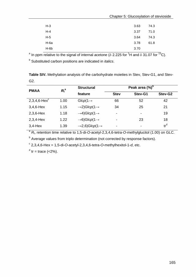
Chapter 5: Glucosylation of stevioside
165
H-3 3.63 74.3
H-4 3.37 71.0
H-5 3.64 74.3
H-6a 3.78 61.8
H-6b 3.70
a In ppm relative to the signal of internal acetone ( 2.225 for
1H and 31.07 for
13C).
b Substituted carbon positions are indicated in italics.
Table SIV. Methylation analysis of the carbohydrate moieties in Stev, Stev-G1, and Stev-
G2.
PMAA Rta
Structural
feature
Peak area (%)b
Stev Stev-G1 Stev-G2
2,3,4,6-Hexc 1.00 Glcp(1→ 66 52 42
3,4,6-Hex 1.15 →2)Glcp(1→ 34 25 21
2,3,6-Hex 1.18 →4)Glcp(1→ - - 19
2,3,4-Hex 1.22 →6)Glcp(1→ - 23 18
3,4-Hex 1.39 →2,6)Glcp(1→ - - trd
a Rt, retention time relative to 1,5-di-O-acetyl-2,3,4,6-tetra-O-methylglucitol (1.00) on GLC.
b Average values from triplo determination (not corrected by response factors).
c 2,3,4,6-Hex = 1,5-di-O-acetyl-2,3,4,6-tetra-O-methylhexitol-1-d, etc.
d tr = trace (<2%).
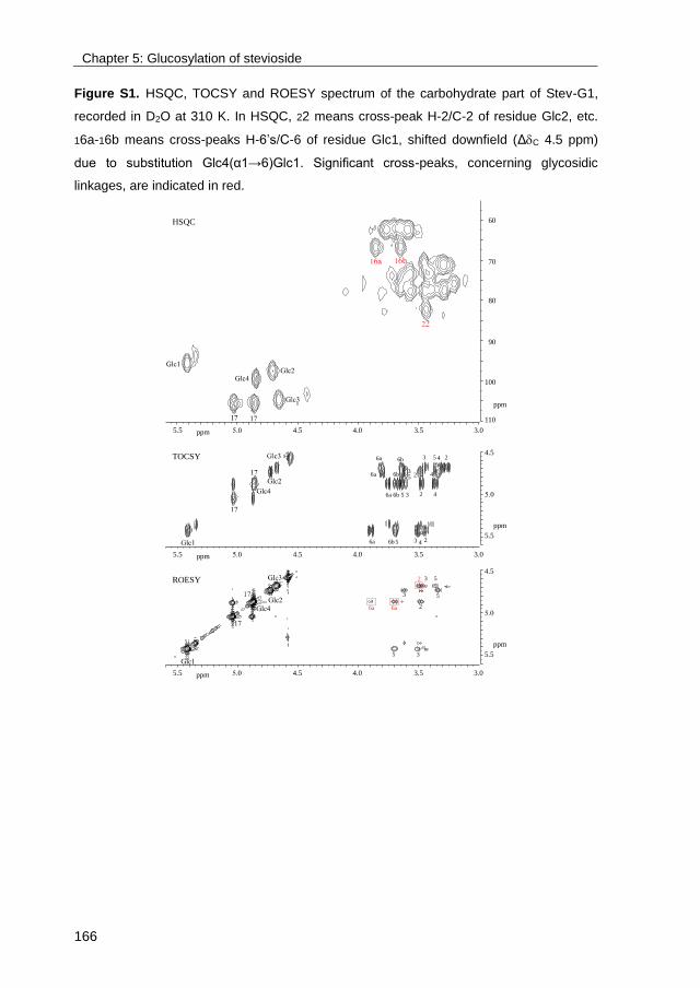
Chapter 5: Glucosylation of stevioside
166
Figure S1. HSQC, TOCSY and ROESY spectrum of the carbohydrate part of Stev-G1,
recorded in D2O at 310 K. In HSQC, 22 means cross-peak H-2/C-2 of residue Glc2, etc.
16a-16b means cross-peaks H-6’s/C-6 of residue Glc1, shifted downfield (ΔC 4.5 ppm)
due to substitution Glc4(α1→6)Glc1. Significant cross-peaks, concerning glycosidic
linkages, are indicated in red.
5.5 4.5 4.0 3.5 3.0ppm
100
90
110
80
70
60
ppm
5.5 4.5 4.0 3.5 3.0ppm
5.5
4.5
ppm
23 456a 6b
23 456a 6b
23 456a 6b
23
456a 6b
5.5 4.5 4.0 3.5 3.0ppm
5.5
4.5
ppm
5 3
2
3 5
3 5
6a 6a
2
HSQC
TOCSY
ROESY

Chapter 5: Glucosylation of stevioside
167
Figure S2. HSQC, TOCSY and ROESY spectra of the carbohydrate part of Stev-G2,
recorded in D2O at 310 K. In the HSQC spectrum, 22 means cross-peak H-2/C-2 of
residue Glc2, etc. Assignments in red reflect the substituted positions of the residues. In
the ROESY spectrum, the inter-residual cross-peaks confirming the Glc3(β12)Glc2 and
Glc5(α14)Glc4(α16)Glc1 linkages are indicated with red boxes.

169
Chapter 6
Biocatalytic production of novel steviol glycosides with
improved taste: scale-up, downstream processing and cost
analysis
Tim Devlamynck1,2
, Evelien M. te Poele2, Wim Soetaert
1, Lubbert Dijkhuizen
2
Ghent University1, University of Groningen
2
Chapter 6: Biocatalytic production of novel steviol glycosides
170
Abstract
Previously we reported the efficient laboratory scale glucosylation of the Stevia
components rebaudioside A (RebA) and stevioside with the Q1140E mutant of
glucansucrase Gtf180-ΔN from Lactobacillus reuteri 180. Sensory analysis by a
trained panel revealed that the glucosylated products possess a superior taste
profile, displaying a significant reduction in bitterness compared to RebA and
stevioside. As the developed technology holds excellent commercial potential,
this chapter focused on the scale-up of the Gtf180-ΔN-ΔV-Q1140E catalyzed
glucosylation of RebA and stevioside. An eco-friendly and efficient downstream
processing of the glucosylated products was developed and demonstrated at 7.5
L scale, allowing the production of 250 g glucosylated RebA product. Estimates
of the production costs indicated the economic feasibility of the overall process.
The major factor in the total production cost was the acceptor substrate, i.e.
RebA or stevioside; in contrast, the biocatalyst cost represented only a minor
part. In an attempt to lower the overall production cost even further, the
glucosylation of low-grade stevia extract (roughly a 50:50 mixture of stevioside
and RebA) was demonstrated. The sensory properties of the resulting product
mixture were perceived as excellent by a trained panel, mainly due to a
significant reduction in bitterness.
Chapter 6: Biocatalytic production of novel steviol glycosides
171
1. Introduction
The steviol glycosides extracted from the plant Stevia rebaudiana (Figure 1),
native in Paraguay and Brazil, were approved for use as high-intensity sweetener
(HIS) in food products by the USA in 2009 and by the European Commission in
201143
. The share of HIS in the global sweetener market, estimated at $68 billion
in 2014, is currently not significant44
, however, the world-wide increasing
incidence of obesity and other diet-related diseases is expected to increase the
consumer demand for low-calorie food products and beverages46
. The HIS
market is consequently predicted to grow at a fast compound annual growth rate
(CAGR) of more than 5%. The natural character of stevia HIS gives it a
competitive edge over synthetic HIS such as aspartame and saccharin, suffering
from a bad reputation among consumers. As a result, stevia is currently the
fastest growing HIS on the market, displaying a CAGR of roughly 8.5%44
. The
World Health Organization even projects that stevia will eventually replace 20%
of the sugar segment, equaling a $10 billion industry45
, which is significantly
greater than current stevia sales, estimated at $347.0 million in 201444
.
The main impediment to accomplish these projections is the lingering bitterness
displayed by most steviol glycosides, including stevioside (5-10% of leaf dry
weight) and rebaudioside A (2-4% of leaf dry weight, RebA), the only steviol
glycosides which can be economically extracted from the Stevia plant190-192
. Not
surprisingly, several companies have launched next-generation stevia products
with improved taste. For example, USA-based MycoTechnology produces
mushroom-derived enzymes (MycoZyme) which can be added to stevia products
to remove their bitterness (http://www.mycotechcorp.com). In addition, a joint
venture of Cargill and Evolva aims to develop a process for the fermentative
production of rebaudioside D and M, two steviol glycosides perceived as less
bitter than RebA, but difficult to extract from the Stevia leaves, in 2018
(http://www.evolva.com)39,58,71
. However, the relatively high production costs,
caused by inadequate strain characteristics and associated fermentation and
downstream processing costs, have forced this joint venture to set back the

Chapter 6: Biocatalytic production of novel steviol glycosides
172
launching date several times since 2013. Dutch multinational DSM is developing
a similar technology, applying a recombinant yeast (Yarrowia lipolytica) to
produce steviol glycosides (http://www.dsm.com)232
. However, no new
announcements have followed the initial 2014 press release.
Steviol glycoside R1 (C-19) R2 (C-13)
Stevioside Glc(β1→ Glc(β1→2)Glc(β1→
Steviolbioside H Glc(β1→2)Glc(β1→
Rebaudioside A Glc(β1→ Glc(β1→2)[Glc(β1→3)]Glc(β1→
Rebaudioside B H Glc(β1→2)[Glc(β1→3)]Glc(β1→
Rebaudioside C Glc(β1→ Rha(α1→2)[Glc(β1→3)]Glc(β1→
Rebaudioside D Glc(β1→2)Glc(β1→ Glc(β1→2)[Glc(β1→3)]Glc(β1→
Rebaudioside E Glc(β1→2)Glc(β1→ Glc(β1→2)Glc(β1→
Rebaudioside F Glc(β1→ Xyl(β1→2)[Glc(β1→3)]Glc(β1→
Rebaudioside M Glc(β1→2)[Glc(β1→3)]Glc(β1→ Glc(β1→2)[Glc(β1→3)]Glc(β1→
Rubusoside Glc(β1→ Glc(β1→
Dulcoside A Glc(β1→ Rha(α1→2)Glc(β1→
Figure 1. Chemical structures of the most prevalent steviol glycosides found in the leaves
of Stevia rebaudiana. Glucose (Glc), xylose (Xyl) and rhamnose (Rha) occur in the
pyranose ring form. Glc and Xyl have the D configuration and Rha the L configuration.
Chapter 6: Biocatalytic production of novel steviol glycosides
173
Alternatively, enzymatic glucosylation has been proposed as a bitterness-
eliminating process8. The main challenges faced here are to obtain an adequate
product specificity, a complete RebA and stevioside conversion and a high
space-time yield. The majority of described processes fail in fulfilling at least one
of these three requirements. Glucosylations catalyzed by cyclodextrin
glucanotransferases, α-amylases and β-amylases, are characterized by relatively
high product yields, however, their lack of C-13/C-19-regiospecificity (Figure 1)
renders them less useful as industrial biocatalyst: products with enhanced and
reduced bitterness are obtained50-56,60-62
. For example, (α1→4)-glucosylation of
stevioside at the C-13 steviol position yielded products with improved intensity
and quality of sweetness, whereas (α1→4)-glucosylation at the C-19 position
resulted in an increased bitterness52-54,196
. In contrast, the in vitro use of UDP-
glycosyltransferases, catalyzing the conversion of RebA into rebaudioside D and
M, yielded glycosides with improved sensory properties but suffered from very
low productivities57-59
. The application of β-glucosidases, introducing the naturally
occurring β-linkages, resulted not only in the glucosylation of the steviol
glycosides substrates but also in their hydrolysis, yielding products with an
inferior taste profile63-65
.
As illustrated in chapters 4 and 5, the α-glucosylation of RebA and stevioside
with the glucansucrase Gtf180-ΔN-Q1140E from Lactobacillus reuteri 180, using
sucrose as donor substrate, offers a viable method for the production of next-
generation stevia products68-70
. The Q1140E mutation improved the conversion
from roughly 50% to 95%, yielding 115 g/L and 50 g/L α-glucosylated product, for
RebA and stevioside glucosylation, respectively. Structural analysis and sensory
analysis by a trained panel revealed that introducing a single (α1→6) linked
glucosyl moiety at their Glc(β1→C-19 residues yielded monoglucosylated
products with a superior taste profile compared to their respective substrates.
The multiglucosylated products displayed even better taste profiles (reduced
bitterness and off-flavors), however, this was accompanied with an undesirable
decrease in sweetness. Altogether, the mixtures of mono –and multiglucosylated
RebA and Stev products showed most potential: an intensive sweetness
combined with a very limited bitterness (see 3.4. Sensory analysis).

Chapter 6: Biocatalytic production of novel steviol glycosides
174
The RebA and stevioside glucosylation reactions were demonstrated only at
laboratory scale, whereas chromatography was applied as purification method, a
technology which is better avoided as it is too costly for industrial use. This
chapter therefore aimed to develop this laboratory scale process with commercial
potential into a cheap and straightforward pilot-plant process, allowing the
synthesis of product samples for food safety analysis and for testing and tasting
in several food products, such as chocolate, candy, etc. Furthermore, an
accelerated shelf life study was performed to determine the stability of the
introduced (α1→6) linkage in buffer solutions mimicking the acidic conditions in
soft drinks. Finally, cost analysis of the glucosylation processes provided
guidelines for future research and development activities in order to decrease the
production costs.
2. Materials and methods
2.1. Stevioside, rebaudioside A and low-grade stevia extract
Stevioside was obtained from TCI Europe (> 85% pure, HPLC), rebaudioside A
(RebA) from Tereos PureCircle Solutions (97% pure, HPLC). Low-grade stevia
extract (Steviasol, 95% steviol glycosides) was mainly composed of RebA and
stevioside (~ 50:50 mixture, as determined with HPLC).
2.2. Production and purification of recombinant Gtf180-ΔN-ΔV-Q1140E
Initial experiments with Escherichia coli BL21 (DE3) strains (Invitrogen, Carlsbad,
USA), expressing either the N-terminally truncated Gtf180-ΔN-Q1140E70,80,97
or
the N- and V-terminally truncated88
Gtf180-ΔN-ΔV-Q1140E70,97
from
Lactobacillus reuteri 180, were performed in shake flasks at 1 L scale, after
which the biocatalyst production was performed at 7.5 L scale in a Labfors 5
bioreactor. The inoculum was routinely grown at 37 °C in shake flasks (200 rpm)
containing LB medium (10 g/L tryptone, 5 g/L yeast extract, 5 g/L NaCl, pH 7.0)
supplemented with 100 mg/L ampicillin. After overnight growth, 200 mL inoculum
Chapter 6: Biocatalytic production of novel steviol glycosides
175
was added to 5 L of LB medium supplemented with 30 g/L glucose and 100 mg/
L ampicillin. The temperature, agitation rate and aeration rate were set at 37 °C,
350 rpm and 1 vvm, respectively. The dissolved oxygen concentration was
maintained above 30% by gradually and automatically increasing the agitation
rate to 1200 rpm. The pH was maintained at 7.0 by automatic addition of 25 %
NH4OH and 1 M H2SO4. Anti-foam was added occasionally to prevent foaming.
Optical density measurements were performed in a spectrophotometer at 600 nm
(OD600). At an OD600 of 5 the culture was induced with IPTG (final concentration
of 0.1 mM). After 9 h of fermentation the glucose was depleted and the cells
harvested by centrifugation (3000 g, 10 min). The cells were resuspended in lysis
buffer and subsequently homogenized (800 bar, 3 cycles). Cell debris was
removed by centrifugation (3000 g, 10 min) followed by microfiltration (0.5 µm).
The crude enzyme was stable at 4 °C for several months and used accordingly
for the production of glucosylated RebA (RebA-G).
2.3. Glucansucrase activity assay
One unit (U) of enzyme activity corresponds to the conversion of 1 μmole
sucrose (used for hydrolysis and transglycosylation) in a solution of 100 mM
sucrose, 25 mM sodium acetate (pH 4.7) and 1 mM CaCl2 at 37 °C.
Enzyme activity assays, using approximately 50 mg/mL enzyme, were performed
at 37°C with 100 mM sucrose in 25 mM sodium acetate (pH 4.7) and 1 mM
CaCl2. Samples of 150 μl were taken every min over a period of 8 min and
immediately inactivated with 30 µl 1 M NaOH. The sucrose concentrations of the
samples were subsequently quantified by means of HPLC analysis (see 2.4.
HPLC analysis), allowing the calculation of the enzyme activity as defined above.
2.4. HPLC analysis
Two different types of HPLC analyses were performed. For the analysis of
glucose, fructose and sucrose, an Agilent MetaCarb 67H column (300 mm × 6.5
mm) was used under isocratic conditions with 2.5 mM H2SO4 as the mobile

Chapter 6: Biocatalytic production of novel steviol glycosides
176
phase. The flow rate and temperature were set at 0.8 mL/min and 35°C,
respectively. Detection was achieved with an RID detector. Calibration of the
obtained peaks was accomplished using the corresponding standard curves.
For the analysis of the steviol glycosides an Agilent ZORBAX Eclipse Plus C18
column (100 mm × 4.6 mm, 3.5 µm) was used with water (solvent A) and
acetonitrile (solvent B) as the mobile phase. The flow rate and temperature were
set at 1.0 mL/min and 40 °C, respectively. The following gradient elution was
used: 5-95% solvent B (0-25 min), 95% solvent B (25-27 min), 95-5% solvent B
(27-30 min) and again 95% of solvent A (30-35 min). Detection was achieved
with an evaporative light scattering detector (ELSD) (evaporation temperature: 90
°C, nebulization temperature: 70 °C, gas flow rate: 1.6 standard liter per min,
SLM). Calibration of the obtained peaks was accomplished using the
corresponding standard curves, obtained from previously purified glucosylated
RebA (RebA-G) and stevioside (Chapters 4 and 5).
2.5. Yeast fermentation
RebA glucosylation reaction mixture (50 mM RebA, 125 mM sucrose) was
incubated with 5, 10, 20 and 30 g/L (wet cell weight) fresh baker’s yeast
(Saccharomyces cerevisiae, AB Mauri) to remove sucrose, glucose, and
fructose. Samples were taken at 0, 2, 4, 6 and 8 h, and subjected to HPLC
analysis to determine the removal rate of both saccharides and RebA glucosides.
The experiments were performed in duplicate.
2.6. Determination of adsorption and desorption characteristics
Adsorption experiments were carried out by adding a fixed amount of adsorbent
(Lewatit® VP OC 1064 MD PH from LANXESS, 1 g) to six 50 ml Falcon tubes
containing 10 mL dilutions of the RebA glucosylation mixture (with RebA-G
concentrations ranging from 10 g/L to 60 g/L). The Falcon tubes were
subsequently placed in a shaker at a temperature of 30 °C, 35 °C and 40 °C and

Chapter 6: Biocatalytic production of novel steviol glycosides
177
an agitation rate of 200 rpm for 60 min. The adsorption capacity qe (g RebA-G/kg
resin) and adsorption efficiency A (%) were calculated as follows:
qe = (C0 − Ce)V
W
A =C0 − Ce
C0
where C0 is the initial RebA-G concentration (g/L), Ce is the RebA-G
concentration at equilibrium (g/L), V is the volume of the solution (10 mL) and W
is the mass of the adsorbent (1 g).
The obtained adsorption isotherms were analyzed using the Langmuir adsorption
isotherm, which is represented by the following equation:
qe = qmaxKCe
1 + KCe
where qmax is the maximum adsorption capacity (g RebA-G/kg resin) and K is the
Langmuir adsorption constant (L/g).
For the desorption experiments 10 mL of solvent was added to 4 g of adsorbent
(equivalent to 1.5 bed volumes) loaded with a known amount of RebA-G. The 50
mL Falcon tubes were subsequently placed in a shaker at a room temperature
and an agitation rate of 200 rpm for 60 min. The desorption capacity qd (g RebA-
G/kg resin) was calculated as follows:
qd = CdV
W
where Cd is the RebA-G concentration at equilibrium (g/L), V is the volume of the
solution (10 mL) and W is the mass of the adsorbent (4 g). The experiments were
performed in duplicate.
C0, Ce and Cd were determined applying HPLC analysis as described before.
Chapter 6: Biocatalytic production of novel steviol glycosides
178
2.7. Continuous adsorption of glucosylated rebaudioside A
After optimization of the static adsorption of RebA-G onto Lewatit® VP OC 1064
MD PH resin, its continuous adsorption was evaluated at laboratory scale (120 g
of resin, bed volume (BV) of 200 mL), and finally at pilot-plant scale (2 kg of
resin, BV of 3.34 L). After supplying the RebA glucosylation reaction mixture, 3
BV of water were immediately supplied to remove any residual sugars and α-
glucans. Subsequently, six BV of 70% isopropanol were added to elute the
adsorbed RebA-G. Several flow rates (3, 4.5 and 6 BV/h) and adsorbate loadings
(50-100 g RebA-G/kg resin) were tested at laboratory scale, after which the
optimal continuous adsorption was performed at pilot-plant scale (see also 2.8.).
2.8. Production and purification of glucosylated rebaudioside A
The production of glucosylated RebA (RebA-G) was performed at 7.5 L scale in a
Labfors 5 bioreactor. The temperature and agitation rate were set at 37 °C and
185 rpm, respectively. The pH was maintained at 4.7 by automatic addition of 1
M NH4OH and 1 M H2SO4. No buffer agent was added. The medium contained
the optimal substrate- and enzyme concentrations as determined in chapter 4 (84
mM RebA, 282 mM sucrose, 5 U/mL Gtf180-ΔN-ΔV-Q1140E) supplemented with
1 mM CaCl2. RebA (97% purity, HPLC grade) was obtained from Tereos
PureCircle Solutions.
After completion of the reaction, the protein was removed by briefly (10 min)
incubating the reaction mixture at 95 °C, after which the precipitated protein was
removed by filtration. RebA-G was isolated from the reaction mixture by
adsorption onto Lewatit® VP OC 1064 MD PH macroporous resin. Washing
thoroughly with water removed any residual sugars and α-glucans. RebA-G was
desorbed with 70% isopropanol. The resulting mixture was concentrated by
evaporation in vacuo and ultimately freeze-dried, yielding pure RebA-G.
Chapter 6: Biocatalytic production of novel steviol glycosides
179
2.9. Cost analysis
The base case used for the cost analysis was RebA-G production in batch mode.
The production costs were estimated by using the process mass balances as
obtained in chapter 4 (RebA glucosylation)70
and the current chapter (enzyme
production (see also 2.1.) and downstream processing (DSP) of RebA-G (see
also 2.6. and 2.7.). Guidelines as presented by Tufvesson et al.233
were followed
for the cost estimation. Several assumptions were made in order to simplify the
economic model (see supplementary information for detailed information). In
short: The production scale of the enzyme fermentation and the enzymatic
glucosylation were both fixed at 10 m3 (75% working volume). To run the
fermentation, including set-up, harvesting and cleaning, 96 man-hours were
allocated. The cost to obtain crude enzyme (homogenization, centrifugation to
remove cell debris, and finally microfiltration) was assigned a value of €200/kg
enzyme233
. For the execution of the enzymatic glucosylation, including product
recovery, 120 man-hours was considered. The cost of direct labor was assigned
a value of €30/h, based on data from Eurostat (http://ec.europa.eu/eurostat).
Supervision costs and indirect operating costs such as quality control
corresponded to 100% of the direct labor costs.
Tables SI and SII summarize the various raw materials needed to run one
production cycle (for definition, see supplementary information). The prices of the
raw materials were obtained from the respective suppliers. Tables SIII-VII
summarize the equipment related costs, utilities costs, and finally labor costs.
2.10. Sensory analysis
The sensory analysis was performed in individual tasting booths at the UGent
Sensolab (Belgium) by a trained panel (9 persons), as described in chapter 470
.
In short, all solutions contained 588 mg/L of sweetener. Their taste (sweetness,
liquorice, astringency and bitterness) was evaluated by swirling the sample in the
mouth for 5 sec after which the sample was expectorated. Aftertaste was
evaluated 10 sec after swallowing the solution. Lingering based on the maximum
Chapter 6: Biocatalytic production of novel steviol glycosides
180
taste intensity was rated 1 min later. Sucrose reference solutions (5%, 7.5% and
10% sucrose scoring 5, 7.5 and 10, respectively) were provided. Water (Spa
Reine) and plain crackers were used as palate cleansers between sampling. All
samples were evaluated in duplicate.
Statistical analyses were performed with SPSS 23 (SPSS Inc., Chicago, USA).
All tests were done at a significance level of 0.05. One-Way ANOVA was used to
investigate any significant difference between the solutions. Testing for equal
variances was executed with the Modified Levene Test. When conditions for
equal variance were fulfilled, the Tukey test was used to determine differences
between samples210
. In case variances were not equal, a Games-Howell post-
hoc test was performed211
.
2.11. Stability analysis
The stability of RebA and RebA-G was determined by conducting an accelerated
shelf life study at 80 °C in acidic buffer solutions (50 mM citric acid (pH 2.8 and
3.8) and 50 mM phosphate (pH 2.8 and 3.8)). Five mg of product was added to
10 ml buffer. All sample solutions were transferred to 1.5 mL Eppendorf tubes
and incubated for 72 h in a thermoblock at 80 °C. Samples were taken at 0, 24,
48 and 72 h, and subjected to HPLC analysis.
3. Results and discussion
3.1. Biocatalyst production
The biocatalyst production generally forms an important factor of the total cost of
a biocatalytic process at industrial scale233
. A productive enzyme fermentation,
yielding a highly active biocatalyst, are thus two essential features of any cost-
effective enzymatic process.
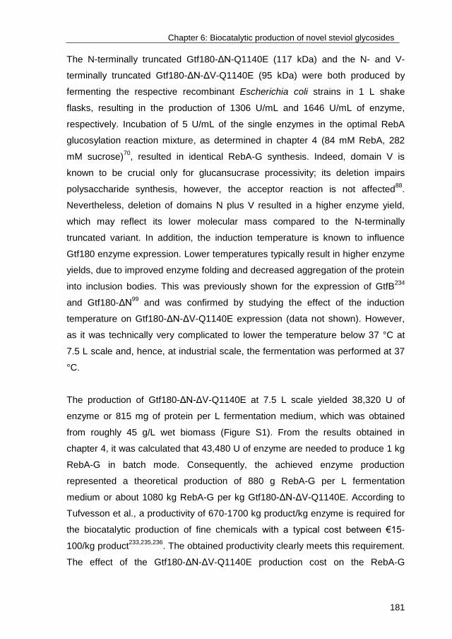
Chapter 6: Biocatalytic production of novel steviol glycosides
181
The N-terminally truncated Gtf180-ΔN-Q1140E (117 kDa) and the N- and V-
terminally truncated Gtf180-ΔN-ΔV-Q1140E (95 kDa) were both produced by
fermenting the respective recombinant Escherichia coli strains in 1 L shake
flasks, resulting in the production of 1306 U/mL and 1646 U/mL of enzyme,
respectively. Incubation of 5 U/mL of the single enzymes in the optimal RebA
glucosylation reaction mixture, as determined in chapter 4 (84 mM RebA, 282
mM sucrose)70
, resulted in identical RebA-G synthesis. Indeed, domain V is
known to be crucial only for glucansucrase processivity; its deletion impairs
polysaccharide synthesis, however, the acceptor reaction is not affected88
.
Nevertheless, deletion of domains N plus V resulted in a higher enzyme yield,
which may reflect its lower molecular mass compared to the N-terminally
truncated variant. In addition, the induction temperature is known to influence
Gtf180 enzyme expression. Lower temperatures typically result in higher enzyme
yields, due to improved enzyme folding and decreased aggregation of the protein
into inclusion bodies. This was previously shown for the expression of GtfB234
and Gtf180-ΔN99
and was confirmed by studying the effect of the induction
temperature on Gtf180-ΔN-ΔV-Q1140E expression (data not shown). However,
as it was technically very complicated to lower the temperature below 37 °C at
7.5 L scale and, hence, at industrial scale, the fermentation was performed at 37
°C.
The production of Gtf180-ΔN-ΔV-Q1140E at 7.5 L scale yielded 38,320 U of
enzyme or 815 mg of protein per L fermentation medium, which was obtained
from roughly 45 g/L wet biomass (Figure S1). From the results obtained in
chapter 4, it was calculated that 43,480 U of enzyme are needed to produce 1 kg
RebA-G in batch mode. Consequently, the achieved enzyme production
represented a theoretical production of 880 g RebA-G per L fermentation
medium or about 1080 kg RebA-G per kg Gtf180-ΔN-ΔV-Q1140E. According to
Tufvesson et al., a productivity of 670-1700 kg product/kg enzyme is required for
the biocatalytic production of fine chemicals with a typical cost between €15-
100/kg product233,235,236
. The obtained productivity clearly meets this requirement.
The effect of the Gtf180-ΔN-ΔV-Q1140E production cost on the RebA-G

Chapter 6: Biocatalytic production of novel steviol glycosides
182
production cost is discussed in more detail later in this chapter (3.3. Cost
analysis).
3.2. Downstream processing of glucosylated RebA
Glucosylated RebA (RebA-G) was selected as model product for the
development of the downstream processing (DSP). Glucosylated stevioside
(Stev-G) was purified following the same principle as applied for RebA-G
purification. The two products are consequently interchangeable in the context of
DSP.
3.2.1. Yeast fermentation
In addition to the synthesis of RebA-G, two types of by-products are produced by
the enzyme: fermentable sugars such as fructose and glucose (and some
remaining sucrose substrate), and α-glucan oligo- and polysaccharides. A
common strategy to remove fermentable sugars is Saccharomyces cerevisiae
fermentation237
, a very cheap and sustainable DSP option, provided that the
desired product (RebA-G) is not metabolized. As illustrated in Figure 2, S.
cerevisiae was indeed able to ferment sucrose, fructose and glucose, while no
loss of RebA-G was observed. Besides CO2, ethanol and glycerol were produced
by the yeast as side-products. A yellow color was observed after fermentation,
suggesting the formation of other side-products.
3.2.2. Selective precipitation
As the α-glucans were not metabolized by the yeast, their removal required an
additional step. In theory, the differences in solubility between α-glucans and
RebA-G in organic solvents may result in their selective precipitation. The
precipitate can then be easily separated from the supernatant. A patent of Cargill,
describing the use of antisolvent crystallization (precipitation) to separate steviol
glycosides, offered a useful protocol238
. In short, the yeast-treated reaction
mixture was evaporated until a sugar content of 30 °Bx (degrees Brix, the sugar
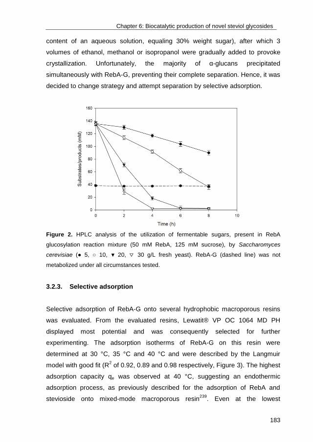
Chapter 6: Biocatalytic production of novel steviol glycosides
183
content of an aqueous solution, equaling 30% weight sugar), after which 3
volumes of ethanol, methanol or isopropanol were gradually added to provoke
crystallization. Unfortunately, the majority of α-glucans precipitated
simultaneously with RebA-G, preventing their complete separation. Hence, it was
decided to change strategy and attempt separation by selective adsorption.
Figure 2. HPLC analysis of the utilization of fermentable sugars, present in RebA
glucosylation reaction mixture (50 mM RebA, 125 mM sucrose), by Saccharomyces
cerevisiae (● 5, ○ 10, ▼ 20, ▽ 30 g/L fresh yeast). RebA-G (dashed line) was not
metabolized under all circumstances tested.
3.2.3. Selective adsorption
Selective adsorption of RebA-G onto several hydrophobic macroporous resins
was evaluated. From the evaluated resins, Lewatit® VP OC 1064 MD PH
displayed most potential and was consequently selected for further
experimenting. The adsorption isotherms of RebA-G on this resin were
determined at 30 °C, 35 °C and 40 °C and were described by the Langmuir
model with good fit (R2 of 0.92, 0.89 and 0.98 respectively, Figure 3). The highest
adsorption capacity qe was observed at 40 °C, suggesting an endothermic
adsorption process, as previously described for the adsorption of RebA and
stevioside onto mixed-mode macroporous resin239
. Even at the lowest
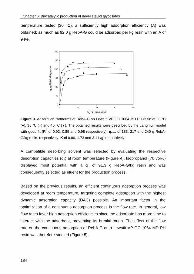
Chapter 6: Biocatalytic production of novel steviol glycosides
184
temperature tested (30 °C), a sufficiently high adsorption efficiency (A) was
obtained: as much as 92.0 g RebA-G could be adsorbed per kg resin with an A of
94%.
Figure 3. Adsorption isotherms of RebA-G on Lewatit VP OC 1064 MD PH resin at 30 °C
(●), 35 °C (○) and 40 °C (▼). The obtained results were described by the Langmuir model
with good fit (R2 of 0.92, 0.89 and 0.98 respectively). qmax of 183, 217 and 240 g RebA-
G/kg resin, respectively. K of 0.80, 1.73 and 3.1 L/g, respectively.
A compatible desorbing solvent was selected by evaluating the respective
desorption capacities (qd) at room temperature (Figure 4). Isopropanol (70 vol%)
displayed most potential with a qd of 91.3 g RebA-G/kg resin and was
consequently selected as eluent for the production process.
Based on the previous results, an efficient continuous adsorption process was
developed at room temperature, targeting complete adsorption with the highest
dynamic adsorption capacity (DAC) possible. An important factor in the
optimization of a continuous adsorption process is the flow rate. In general, low
flow rates favor high adsorption efficiencies since the adsorbate has more time to
interact with the adsorbent, preventing its breakthrough. The effect of the flow
rate on the continuous adsorption of RebA-G onto Lewatit VP OC 1064 MD PH
resin was therefore studied (Figure 5).
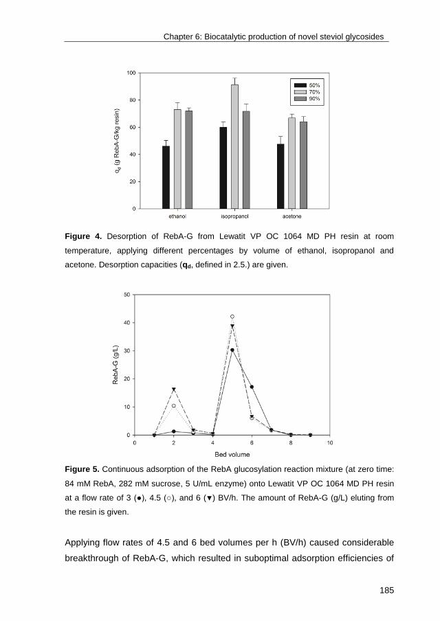
Chapter 6: Biocatalytic production of novel steviol glycosides
185
Figure 4. Desorption of RebA-G from Lewatit VP OC 1064 MD PH resin at room
temperature, applying different percentages by volume of ethanol, isopropanol and
acetone. Desorption capacities (qd, defined in 2.5.) are given.
Figure 5. Continuous adsorption of the RebA glucosylation reaction mixture (at zero time:
84 mM RebA, 282 mM sucrose, 5 U/mL enzyme) onto Lewatit VP OC 1064 MD PH resin
at a flow rate of 3 (●), 4.5 (○), and 6 (▼) BV/h. The amount of RebA-G (g/L) eluting from
the resin is given.
Applying flow rates of 4.5 and 6 bed volumes per h (BV/h) caused considerable
breakthrough of RebA-G, which resulted in suboptimal adsorption efficiencies of
Chapter 6: Biocatalytic production of novel steviol glycosides
186
79% and 68%, respectively. The application of a flow rate of 3 BV/h resulted in a
near complete adsorption (96%) and a DAC of 95.7 g RebA-G/kg resin, similar to
the optimal static adsorption capacity (92.0 g RebA-G/kg resin). Only 3 bed
volumes of 70% isopropanol were subsequently needed for a near complete
recovery of the product. The successful separation of RebA-G from the
fermentable sugars and α-glucans is clearly shown by TLC analysis (Figure S2).
The developed continuous adsorption was consequently used to treat 4 L RebA
glucosylation reaction mixture (containing 60 g/L RebA-G), yielding 250 g of
amorphous RebA-G, composed of (by mass) 4% RebA, 60% monoglucosylated
RebA (RebA-G1) and 36% multiglucosylated RebA (RebA-G2+) (Figure S3). As
the applied desorbing solvent (70% isopropanol) can be reused after
evaporation, the developed DSP is not only efficient (almost no loss of RebA-G)
but also eco-friendly.
3.3. Cost analysis of stevia glucosylation
3.3.1. Base case: RebA glucosylation
Any new production process must meet a number of criteria to be successfully
implemented at industrial scale. Besides analyzing safety matters and
environmental and legal issues, the process economics need to be evaluated240
.
Assessing the economic feasibility of a process is achieved by performing a
production cost estimation, a powerful tool to guide research and development
activities in order to turn lab-scale processes into commercially viable ones.
Production costs can be divided into two categories: capital investment (CapEx)
and operation costs (OpEx), both considered for the cost estimation of the base
case (for detailed information, see supplementary information). A production
scale of 10 m3 for both the enzyme fermentation and the enzymatic glucosylation
was assumed. It should be noted that production volume has a major impact on
the production costs: increasing the volume from 1 m3 to multiple cubic meters is
accompanied by a cost reduction of several orders of magnitude233
. The
assignment of an adequate production scale is typically based on the expected
sales, however, that was outside the scope of this chapter.

Chapter 6: Biocatalytic production of novel steviol glycosides
187
Tables I and II summarize the results obtained for the base case analysis. For
the detailed results, see supplementary information (Tables SI-VII). Figure 6
illustrates the cost contribution of the different resources on the enzyme
production (A) and RebA-G production, including DSP (B).
Table I. Cost analysis of Gtf180-ΔN-ΔV-Q1140E production in batch mode.
Resources Cost
(€/kg enzyme)
Relative cost
(%)
Fermentation 2106 91
Raw materials 114 5
Utilities 34 1
Labor 950 41
Equipment 1008 44
Enzyme recovery 200 9
Total 2306 100
Table II. Cost analysis of RebA-G production and glucosylated steviol glycosides (GSG)
production in batch mode, using RebA and low-grade stevia extract (steviol glycosides,
SG) as acceptor substrate, respectively.
Resources Cost
(€/kg RebA-G)
Relative cost
(%)
Cost
(€/kg GSG)
Relative cost
(%)
Biocatalysis 70.7 79 41.3 66
Enzyme 2.1 2 3.5 6
Sucrose 0.6 1 1.8 3
RebA/SG 56.5 63 17.1 27
Utilities < 1 < 1 < 1 < 1
Labor 4.2 5 6.9 11
Equipment 7.1 8 11.6 19
DSP 18.5 21 21.3 34
Resin 12.7 14 12.7 20
IPA 1.1 1 1.1 2
Utilities < 1 1 < 1 1
Labor 4.2 5 6.9 11
Total 89.2 100 62.6 100
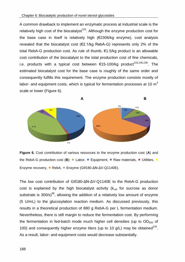
Chapter 6: Biocatalytic production of novel steviol glycosides
188
A common drawback to implement an enzymatic process at industrial scale is the
relatively high cost of the biocatalyst233
. Although the enzyme production cost for
the base case in itself is relatively high (€2306/kg enzyme), cost analysis
revealed that the biocatalyst cost (€2.1/kg RebA-G) represents only 2% of the
total RebA-G production cost. As rule of thumb, €1.5/kg product is an allowable
cost contribution of the biocatalyst to the total production cost of fine chemicals,
i.e. products with a typical cost between €15-100/kg product233,235,236
. The
estimated biocatalyst cost for the base case is roughly of the same order and
consequently fulfills this requirement. The enzyme production consists mostly of
labor- and equipment costs, which is typical for fermentation processes at 10 m3
scale or lower (Figure 6).
Figure 6. Cost contribution of various resources to the enzyme production cost (A) and
the RebA-G production cost (B): ● Labor, ● Equipment, ● Raw materials, ● Utilities, ●
Enzyme recovery, ● RebA, ● Enzyme (Gtf180-ΔN-ΔV-Q1140E).
The low cost contribution of Gtf180-ΔN-ΔV-Q1140E to the RebA-G production
cost is explained by the high biocatalyst activity (kcat for sucrose as donor
substrate is 300/s)98
, allowing the addition of a relatively low amount of enzyme
(5 U/mL) to the glucosylation reaction medium. As discussed previously, this
results in a theoretical production of 880 g RebA-G per L fermentation medium.
Nevertheless, there is still margin to reduce the fermentation cost. By performing
the fermentation in fed-batch mode much higher cell densities (up to OD600 of
100) and consequently higher enzyme titers (up to 10 g/L) may be obtained233
.
As a result, labor- and equipment costs would decrease substantially.
Chapter 6: Biocatalytic production of novel steviol glycosides
189
The total RebA-G production cost equals €89.2/kg RebA-G, representing an
extra cost of 12% compared to high-purity RebA, sold at €80/kg (Tereos
PureCircle Solutions). Since the price of sucrose is roughly €0.70/kg and RebA-G
is around 150 times sweeter than sucrose, a selling price of €105/kg RebA-G can
still be considered as competitive. If RebA-G is sold at this price, a profit of
€15/kg RebA-G is obtained.
As demonstrated in chapter 4, glucosylating RebA in fed-batch mode resulted in
higher production yield compared to the batch production (270 vs 115 g/L RebA-
G). However, the much lower productivity per kg enzyme (1080 vs 253 kg RebA-
G/kg enzyme), due to the addition of 10 times more enzyme, was translated into
an undesirable production cost increase of €11.1/kg RebA-G. From an economic
perspective, the batch reaction is thus preferred. It has to be noted that further
optimization of the fed-batch reaction, thereby decreasing the applied enzyme
activity (U/mL), may allow further reduction of the production cost.
3.3.2. Alternative case: glucosylation of low-grade stevia extract
Remarkably, the base case analysis shows that RebA-G production cost is
dominated by the cost of the acceptor substrate RebA (63%). A major cost
reduction consequently may be achieved by changing the acceptor substrate:
instead of high-purity RebA, low-grade stevia extract could be used as substrate,
which is commonly marketed for roughly €40/kg (Tereos PureCircle Solutions, or
half of the price for high-purity RebA (€80/kg, Tereos PureCircle Solutions).
Moreover, glucosylating mixtures of steviol glycosides also offers the possibility
to valorize stevioside, whose glucosylation by Gtf180-ΔN-Q1140E was already
demonstrated in chapter 5. The glucosylation of different self-made
stevioside/RebA mixtures and of low-grade stevia extract (Steviasol), basing the
experimental design on the optimal reaction conditions for stevioside
glucosylation, as determined in chapter 5 (31 mM steviol glycosides, 524 mM
sucrose), was also successful (data not shown), yielding roughly 50 g/L
glucosylated steviol glycosides (GSG). The economic model of the base case
was changed accordingly, resulting in a production cost of €62.7/kg GSG, a
Chapter 6: Biocatalytic production of novel steviol glycosides
190
substantial reduction of almost 30% compared to RebA-G production (Table II).
The glucosylation of a mixture of steviol glycosides consequently provides a
substantial economic advantage over the glucosylation of high-purity RebA.
3.4. Sensory analysis of glucosylated stevioside/RebA mixture
The previous paragraph demonstrated the economic advantage of glucosylating
a mixture of steviol glycosides. However, the sensory properties of the obtained
product must at least be similar to those of RebA-G to justify this change of
acceptor substrate. As proof of concept, a sensory analysis of a glucosylated
mixture (obtained from glucosylating low-grade stevia extract), containing 50%
Stev-G and 50% RebA-G (RebA-G/Stev-G), was performed by a trained panel,
evaluating 9 different taste attributes. The mean scores of the attributes of the
sweetened water solutions are shown in Figure 7 and were compared with the
sensory properties of RebA-G and Stev-G.
The sensory properties of the RebA-G/Stev-G mixture, compared to RebA and
stevioside, were clearly improved, mainly due to a significant reduction in
bitterness. Interestingly, the relatively low sweetness of Stev-G was
compensated by the high sweetness of RebA-G, yielding a product with excellent
edulcorant properties. RebA-G was identified as the most promising high-
intensity sweetener, combining a high sweetness with a very low off-flavor
intensity. If taste has the highest priority, the glucosylation of high-purity RebA is
thus preferred, as the presence of stevioside deteriorated the sensory properties
of the RebA-G/Stev-G.
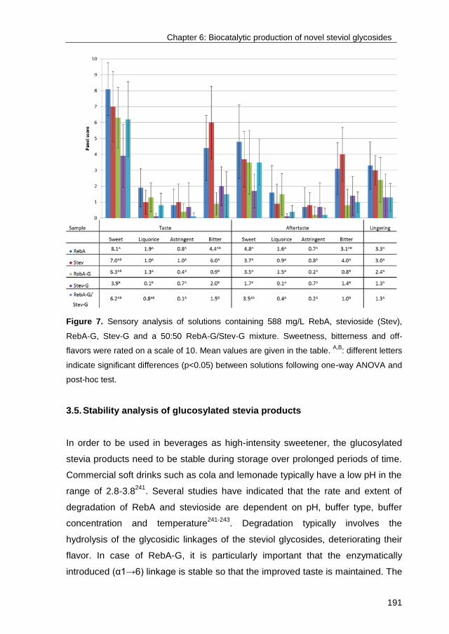
Chapter 6: Biocatalytic production of novel steviol glycosides
191
Figure 7. Sensory analysis of solutions containing 588 mg/L RebA, stevioside (Stev),
RebA-G, Stev-G and a 50:50 RebA-G/Stev-G mixture. Sweetness, bitterness and off-
flavors were rated on a scale of 10. Mean values are given in the table. A,B
: different letters
indicate significant differences (p<0.05) between solutions following one-way ANOVA and
post-hoc test.
3.5. Stability analysis of glucosylated stevia products
In order to be used in beverages as high-intensity sweetener, the glucosylated
stevia products need to be stable during storage over prolonged periods of time.
Commercial soft drinks such as cola and lemonade typically have a low pH in the
range of 2.8-3.8241
. Several studies have indicated that the rate and extent of
degradation of RebA and stevioside are dependent on pH, buffer type, buffer
concentration and temperature241-243
. Degradation typically involves the
hydrolysis of the glycosidic linkages of the steviol glycosides, deteriorating their
flavor. In case of RebA-G, it is particularly important that the enzymatically
introduced (α1→6) linkage is stable so that the improved taste is maintained. The
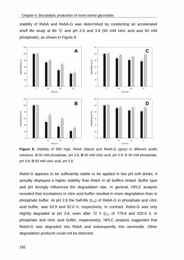
Chapter 6: Biocatalytic production of novel steviol glycosides
192
stability of RebA and RebA-G was determined by conducting an accelerated
shelf life study at 80 °C and pH 2.8 and 3.8 (50 mM citric acid and 50 mM
phosphate), as shown in Figure 8.
Figure 8. Stability of 500 mg/L RebA (black) and RebA-G (grey) in different acidic
solutions. A 50 mM phosphate, pH 2.8; B 50 mM citric acid, pH 2.8; C 50 mM phosphate,
pH 3.8; D 50 mM citric acid, pH 3.8.
RebA-G appears to be sufficiently stable to be applied in low pH soft drinks, it
actually displayed a higher stability than RebA in all buffers tested. Buffer type
and pH strongly influenced the degradation rate. In general, HPLC analysis
revealed that incubations in citric acid buffer resulted in more degradation than in
phosphate buffer. At pH 2.8 the half-life (t1/2) of RebA-G in phosphate and citric
acid buffer, was 63.9 and 52.0 h, respectively. In contrast, RebA-G was only
slightly degraded at pH 3.8, even after 72 h (t1/2 of 779.8 and 520.6 h, in
phosphate and citric acid buffer, respectively). HPLC analysis suggested that
RebA-G was degraded into RebA and subsequently into stevioside. Other
degradation products could not be detected.
Chapter 6: Biocatalytic production of novel steviol glycosides
193
4. Conclusions
This chapter demonstrates the technical and economic feasibility of glucosylating
steviol glycosides with the Gtf180-ΔN-ΔV-Q1140E enzyme. The strength of the
technology lies in the high activity of this biocatalyst, suppressing the associated
costs, as revealed by the cost analysis. Furthermore, selective adsorption of the
glucosylated products onto macroporous resin offers a straightforward and
efficient method for their purification. Based on the cost analysis, it was
suggested to glucosylate mixtures of steviol glycosides rather than high-purity
RebA or stevioside, as this results in a 30% cost reduction. From a taste quality
perspective, the glucosylation of high-purity RebA is nevertheless preferred.
Finally, the glucosylation reaction was successfully scaled up to 7.5 L scale,
allowing the production of samples for food safety analysis and testing in several
food products, such as chocolate, candy, etc. In future work demonstration of the
complete process at pilot-plant scale will be an essential requirement to survive
the so-called valley of death, which is faced by many novel technologies.
Chapter 6: Biocatalytic production of novel steviol glycosides
194
5. Supplementary information
5.1. Cost analysis: Assumptions and economic model
The base case used for the cost analysis is the glucosylation of high-purity RebA
in batch mode at optimal reaction conditions (84 mM RebA, 282 mM sucrose, 5
U/mL Gtf180-ΔN-ΔV-Q1140E)70
.
General assumptions
Plant is located in Western Europe.
The facilities are part of a multi-purpose plant with shared utilities, services,
offices, quality control, etc.
Capital investment included equipment, instrumentation and piping needed
for RebA-G production, excluding DSP.
Scale: One bioreactor and one blending tank of 10 m3 with 75% working
volume, shared for fermentation and biocatalysis.
Costs are given in €.
Production is as follows:
One optimal fermentation at 10 m3 yields enzyme to perform 8 enzymatic
glucosylations at 10 m3. As a result, 1 week of fermentation is followed by 8
weeks of biocatalysis, defined as 1 production cycle.
Equipment is used to perform 5 production cycles per year, yielding 34.5
metric tons of RebA-G.
The volume consumption of stevia is expected to reach 8,507 metric tons per
year by the end of 202044
. The assumed production would consequently
represent 0.4% of the total stevia market.
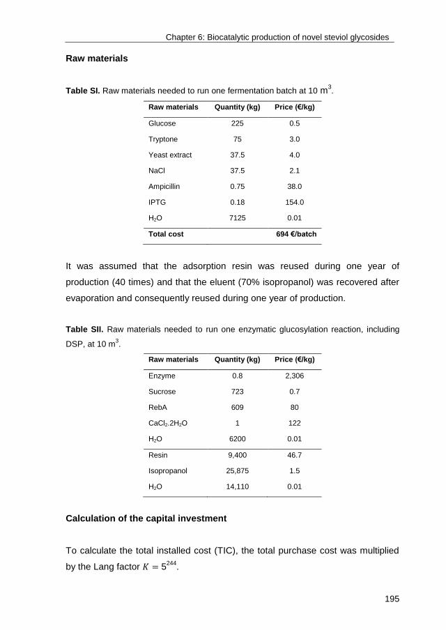
Chapter 6: Biocatalytic production of novel steviol glycosides
195
Raw materials
Table SI. Raw materials needed to run one fermentation batch at 10 m3.
Raw materials Quantity (kg) Price (€/kg)
Glucose 225 0.5
Tryptone 75 3.0
Yeast extract 37.5 4.0
NaCl 37.5 2.1
Ampicillin 0.75 38.0
IPTG 0.18 154.0
H2O 7125 0.01
Total cost 694 €/batch
It was assumed that the adsorption resin was reused during one year of
production (40 times) and that the eluent (70% isopropanol) was recovered after
evaporation and consequently reused during one year of production.
Table SII. Raw materials needed to run one enzymatic glucosylation reaction, including
DSP, at 10 m3.
Raw materials Quantity (kg) Price (€/kg)
Enzyme 0.8 2,306
Sucrose 723 0.7
RebA 609 80
CaCl2.2H2O 1 122
H2O 6200 0.01
Resin 9,400 46.7
Isopropanol 25,875 1.5
H2O 14,110 0.01
Calculation of the capital investment
To calculate the total installed cost (TIC), the total purchase cost was multiplied
by the Lang factor 𝐾 = 5244
.
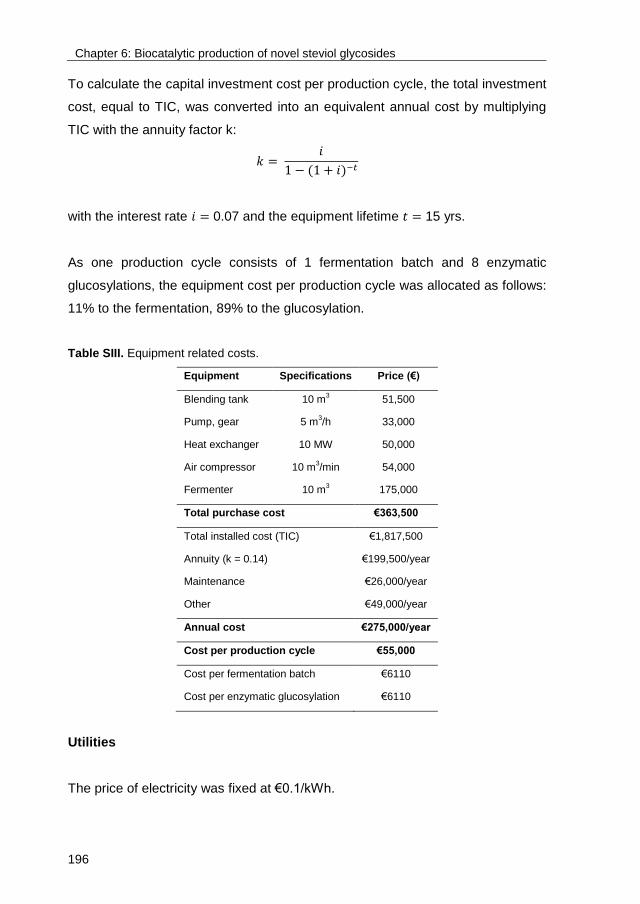
Chapter 6: Biocatalytic production of novel steviol glycosides
196
To calculate the capital investment cost per production cycle, the total investment
cost, equal to TIC, was converted into an equivalent annual cost by multiplying
TIC with the annuity factor k:
𝑘 = 𝑖
1 − (1 + 𝑖)−𝑡
with the interest rate 𝑖 = 0.07 and the equipment lifetime 𝑡 = 15 yrs.
As one production cycle consists of 1 fermentation batch and 8 enzymatic
glucosylations, the equipment cost per production cycle was allocated as follows:
11% to the fermentation, 89% to the glucosylation.
Table SIII. Equipment related costs.
Equipment Specifications Price (€)
Blending tank 10 m3 51,500
Pump, gear 5 m3/h 33,000
Heat exchanger 10 MW 50,000
Air compressor 10 m3/min 54,000
Fermenter 10 m3 175,000
Total purchase cost €363,500
Total installed cost (TIC) €1,817,500
Annuity (k = 0.14) €199,500/year
Maintenance €26,000/year
Other €49,000/year
Annual cost €275,000/year
Cost per production cycle €55,000
Cost per fermentation batch €6110
Cost per enzymatic glucosylation €6110
Utilities
The price of electricity was fixed at €0.1/kWh.

Chapter 6: Biocatalytic production of novel steviol glycosides
197
Table SIV. Utilities needed to run one fermentation batch at 10 m3.
Utilities Specifications kWh
Sterilization (20-140 °C) 4,187 J/kg.K 1047
Aeration (1 vvm) 5 kW/m3 400
Agitation (500 rpm) 5 kW/m3 400
Waste treatment €2/m3
Total utilities cost €207/batch
Table SV. Utilities needed to run one enzymatic glucosylation at 10 m3.
Utilities Specifications kWh
Agitation (185 rpm) 1.9 kW/m3 57
Heating (37 °C) 4,187 J/kg.K 148
Waste treatment €2/m3
Total utilities cost €26/batch
At kg scale, the last step of RebA-G production consisted of freeze-drying to
remove the residual water. As freeze-drying is technically and economically not
feasible at ton scale, spray drying was considered as last purification step for the
cost analysis. Firstly, the mixture obtained after elution (2% solids) was
evaporated until a solid content of 50% by means of a mechanical vapor
recompression (MVR). Energy consumption of MVR and spray drying were
based on Fox et al.245
.
Table SVI. Utilities needed to perform DSP of RebA-G.
Utilities kWh
Evaporation 476
Spray drying 1917
Total utilities cost €249/batch

Chapter 6: Biocatalytic production of novel steviol glycosides
198
Labor
Table SVII. Labor cost.
Utilities Man-hours
Fermentation 96
Biocatalysis + DSP 120
5.2. Figures
Figure S1. Biocatalyst production: growth of the recombinant Escherichia coli BL21 (DE3)
strain, expressing Gtf180-ΔN-ΔV-Q1140E, in LB medium supplemented with 30 g/L
glucose. Induction with IPTG after 4 h of incubation. ● OD600, ○ glucose.
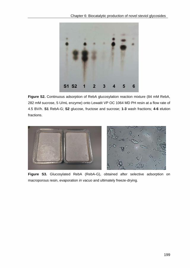
Chapter 6: Biocatalytic production of novel steviol glycosides
199
Figure S2. Continuous adsorption of RebA glucosylation reaction mixture (84 mM RebA,
282 mM sucrose, 5 U/mL enzyme) onto Lewatit VP OC 1064 MD PH resin at a flow rate of
4.5 BV/h. S1 RebA-G; S2 glucose, fructose and sucrose; 1-3 wash fractions; 4-6 elution
fractions.
Figure S3. Glucosylated RebA (RebA-G), obtained after selective adsorption on
macroporous resin, evaporation in vacuo and ultimately freeze-drying.

201
Chapter 7
Enzymatic transglucosylation of neohesperidin
dihydrochalcone: glucansucrase Gtf180-ΔN-Q1140E as
biocatalyst for the glycodiversification of sweet glycosides
Tim Devlamynck1,2
, Gerrit Gerwig3, Wim Soetaert
1, Lubbert Dijkhuizen
2
University of Ghent1, University of Groningen
2, University of Utrecht
3
Chapter 7: Glycosylation of NHDC
202
Abstract
Increasing rates of obesity and diabetes type 2 lead to a rapidly growing number
of health-conscious consumers, fueling the demand for low-calorie food products.
Natural high-intensity sweeteners (HIS), of which steviol glycosides [e.g.
rebaudioside A (RebA)] extracted from the leaves of Stevia rebaudiana are best
known, have a competitive edge over synthetic HIS such as aspartame and
acesulfame K, suffering from a bad reputation among consumers. Over the
years, several other natural compounds have been proposed as possible
products for the HIS market of the future. An interesting target is the flavor
enhancer neohesperidin dihydrochalcone (NHDC). The main disadvantages of
NHDC are its low water solubility at room temperature, resulting in a relatively
low bioavailability, and an unpleasant lingering aftertaste. This chapter reports
the α-glucosylation of NHDC with glucansucrase Gtf180-ΔN-Q1140E from
Lactobacillus reuteri 180, using sucrose as donor substrate, in an attempt to
improve its sensory and physicochemical properties. Structure elucidation of the
novel glucosylated products revealed that NHDC was glucosylated at the β-D-
glucosyl unit and the α-L-rhamnosyl unit, through an (α1→6) and (α1→4) linkage,
respectively. The obtained conversion degree of 65% offers a good starting point
for future improvements by reaction engineering. The α-glucosylated NHDC
products displayed much higher water solubilities than NHDC and retained
strong anti-oxidant capacities, enabling their use as nutraceuticals. Sensory
analysis by a trained panel revealed limited bitterness suppressing effects of
NHDC and its α-glucosylated products on RebA, in contradiction to previous
reports.
Chapter 7: Glycosylation of NHDC
203
1. Introduction
Previously we demonstrated the potential of the glucansucrase enzyme Gtf180-
ΔN and in particular its Q1140E mutant97
to trans-α-glucosylate rebaudioside A
(RebA) and stevioside as a means to improve their sensory properties68-70
. In
addition to these steviol glycosides from Stevia rebaudiana, also neohesperidin
dihydrochalcone (NHDC, Figure 1) has attracted considerable attention from the
food industry, due to its intensive sweetness. Later on, NHDC’s remarkable
properties as flavor enhancer, as such contributing to the mouth feel of several
food products and/or suppressing the bitterness displayed by other compounds,
expanded the interest in this versatile molecule246-248
.
First discovered in 1963 by Horowitz and Gentili246
, NHDC is industrially
produced by hydrogenation of neohesperidin (hesperetin 7-O-neohesperidoside),
a flavanone glycoside isolated from citrus fruits such as mandarin, orange and
grapefruit249
. Recently, NHDC was found to naturally occur as a minor constituent
in the plant Oxytropis myriophylla250
and in the bark of the tree Eysenhardtia
polystachya251
. Classified as a semisynthetic sweetener, NHDC is roughly 340
times sweeter than sucrose and was approved as sweetener by the European
Union in 1994. Similarly to other highly sweet glycosides such as glycyrrhizin
(Glycyrrhiza glabra, liquorice) and most steviol glycosides, NHDC displays a
lingering liquorice aftertaste limiting its application as the sole sweetener in foods
and beverages252
. NHDC is best applied in sweetener blends: it displays strong
synergistic effects when combined with other sweeteners such as saccharin,
aspartame, and RebA253
. The total sweetness intensity of such mixtures is
greater than the theoretical sum of the intensities of the individual components254
.
The use of NHDC thus provides a substantial economic benefit, as much less of
the other sweetener needs to be supplied.
In addition, NHDC finds many applications as a flavor enhancer in a wide range
of food –and pharmaceutical products, as such identified by the E number
E959255-257
. A typical example concerns the enhancement of the creaminess of

Chapter 7: Glycosylation of NHDC
204
dairy products such as yogurt and ice cream. Moreover, NHDC is particularly
effective in masking the bitterness displayed by several other compounds, such
as limonin and naringin from citrus fruits. A patent application from 2012 even
claims that the bitterness and astringency exhibited by RebA and monk fruit
extract are very effectively suppressed by the addition of 10 ppm NHDC258
. Also
the pharmaceutical industry makes good use of this remarkable NHDC
characteristic to reduce the bitterness of several drugs in tablet form, such as the
antipyretic paracetamol259
. More recently, several studies have attributed strong
antioxidant260-262
, hepatoprotective (preventing damage to the liver)263
and
prebiotic264
properties to NHDC. In the context of all these possible applications,
the main disadvantage of NHDC is its low water solubility at room temperature
(0.4 g/L), resulting in a relatively low bioavailability and limiting its application as
sweetener in water based dispersions, such as syrups and jams265
.
Figure 1. Chemical structure of neohesperidin dihydrochalcone (NHDC) with carbon atom
numbering as applied in the structure elucidation. The numbering used is arbitrary (similar
to Caccia et al. 1998266
) as there is no consensus in literature.
NHDC contains 9 hydroxy groups (Figure 1): 3 of rhamnose, 3 of glucose and 3
of the aglycon (2 on the A-ring and 1 on the B-ring). In theory, all these hydroxy
groups can be enzymatically glycosylated, depending on the biocatalyst and its
substrate specificity and regioselectivity. To our knowledge, NHDC glycosylation
has been reported only once: by applying maltogenic amylase from Bacillus
stearothermophilus, NHDC was maltosylated at the β-D-glucosyl unit through an
Chapter 7: Glycosylation of NHDC
205
(α1→6) linkage, resulting in a 700-fold increase of solubility and a 7-fold
decrease of sweetness267
. Previously, we reported the trans-α-glucosylation of a
wide variety of compounds, ranging from catechol to more complex molecules
such as steviol glycosides, with Gtf180-ΔN from Lactobacillus reuteri 180 and
derived mutants, highlighting the broad acceptor substrate specificity of this
enzyme68-70,110
. This chapter reports the transglucosylation of NHDC with Gtf180-
ΔN-Q1140E, yielding three new-to-nature α-glucosylated NHDC derivatives. A
detailed structural analysis is provided, a necessity for the evaluation of the
quality and safety of novel food products. In addition, the sensory properties,
anti-oxidant capacities and solubilities of the α-glucosylated NHDC products were
compared to NHDC.
2. Materials and methods
2.1. Production and purification of glucosylated NHDC
The production of glucosylated NHDC (Ingrizo) was performed at 50 mL scale in
a shake flask, in duplicate. Samples were analyzed by HPLC. The temperature
and agitation rate were set at 37 °C and 150 rpm, respectively. Gtf180-ΔN-
Q1140E97
(5 U/mL), produced as described previously70
, was incubated in a 20%
ethanol solution containing 20 g/L NHDC and 125 mM sucrose supplemented
with 25 mM sodium acetate (pH 4.7) and 1 mM CaCl2. One unit (U) of enzyme
activity corresponds to the conversion of 1 μmole sucrose (used for hydrolysis
and transglucosylation) in a solution of 100 mM sucrose, 25 mM sodium acetate
(pH 4.7) and 1 mM CaCl2 at 37 °C.
After completion of the reaction (i.e. when a steady state was reached),
glucosylated NHDC was isolated from the reaction mixture by adsorption onto
Lewatit VP OC 1064 MD PH macroporous resin. A washing step with water
removed the residual sugars and α-glucan oligosaccharides. Desorption of
glucosylated NHDC was achieved with 70% isopropanol. The resulting mixture
was evaporated in vacuo and subsequently applied onto a Reveleris X2 flash
Chapter 7: Glycosylation of NHDC
206
chromatography system, applying a Reveleris amino cartridge (40 µm) with water
(solvent A) and acetonitrile (solvent B) as the mobile phase. Following gradient
elution was used: 99% solvent B (0-2 min), 99-35% solvent B (2-20 min), 35%
solvent B (20-25 min). The collected fractions were evaporated in vacuo and
subsequently freeze dried to remove the residual water.
2.2. MALDI-TOF-mass spectrometry
Matrix-assisted laser desorption ionization time-of-flight mass spectrometry
(MALDI-TOF-MS) was performed on an AximaTM mass spectrometer (Shimadzu
Kratos Inc., Manchester, UK), equipped with a Nitrogen laser (337 nm, 3 ns pulse
width). Positive-ion mode spectra were recorded using the reflector mode at a
resolution of 5000 FWHM and delayed extraction (450 ns). Accelerating voltage
was 19 kV with a grid voltage of 75.2%. The mirror voltage ratio was 1.12 and the
acquisition mass range was 200-2000 Da. Samples were prepared by mixing on
the target 1 μL sample solutions with 1 μL aqueous 10% 2,5-dihydroxybenzoic
acid in 40% acetonitrile as matrix solution, containing 0.1% TFA.
2.3. Methylation analysis
Samples were permethylated using CH3I and solid NaOH in DMSO, as described
by Ciucanu and Kerek (1984)227
, then hydrolyzed with 2 M TFA (2 h, 120 °C) to
give the mixture of partially methylated monosaccharides. After evaporation to
dryness and redissolving in H2O, reduction was performed with NaBD4 (2 h, room
temperature). After neutralization with 4 M acetic acid and removal of boric acid
by co-evaporation with methanol, the samples were acetylated with acetic
anhydride/pyridine (1:1, 30 min, 120 °C). After evaporation to dryness and re-
dissolving in dichloromethane, the mixtures of partially methylated alditol
acetates (PMAAs) were analyzed by gas-liquid chromatography – electron
ionization – mass spectrometry (GLC-EI-MS) on an EC-1 column (30 m x 0.25
mm, Alltech), using a gas chromatograph mass spectrometer (GCMS-QP2010
Plus from Shimadzu Kratos Inc.) and a temperature gradient (140-250 °C at 8
°C/min)228
.
Chapter 7: Glycosylation of NHDC
207
2.4. NMR spectroscopy
Resolution-enhanced 1D/2D 500-MHz 1H/
13C NMR spectra were recorded in D2O
on a Bruker DRX-500 spectrometer (Bijvoet Center, Department of NMR
Spectroscopy, Utrecht University) at a probe temperature of 310 K. Data
acquisition was done with Bruker Topspin 2.1. Before analysis, samples were
exchanged twice in D2O (99.9 atom% D, Cambridge Isotope Laboratories, Inc.,
Andover, MA) with intermediate lyophilization, and then dissolved in 0.6 mL D2O.
Suppression of the deuterated water signal (HOD) was achieved by applying a
WEFT (water eliminated Fourier transform) pulse sequence for 1D NMR
experiments and by a selective pre-saturation pulse of 1 s during the relaxation
delay in 2D experiments. The 2D TOCSY spectra were recorded using an MLEV-
17 (composite pulse devised by Levitt et al., 1982) mixing sequence with spin-
lock times of 20, 50, 100 and 200 ms. The 2D 1H-
1H ROESY spectra were
recorded using standard Bruker XWINNMR software with a mixing time of 200
ms. The carrier frequency was set at the downfield edge of the spectrum in order
to minimize TOCSY transfer during spin-locking. Natural abundance 2D 13
C-1H
HSQC experiments (1H frequency 500.0821 MHz,
13C frequency 125.7552 MHz)
were recorded without decoupling during acquisition of the 1H FID. The NMR
data were processed using the MestReNova 9 program (Mestrelab Research SL,
Santiago de Compostella, Spain). Chemical shifts (δ) are expressed in ppm by
reference to internal acetone (δH 2.225 for 1H and δC 31.07 for
13C).
2.5. Solubility measurements
The solubilities of NHDC and α-glucosylated NHDC products were determined in
250 μL of ultrapure water, incubated in a thermoblock at 25 °C. Products were
added until clear precipitation was noticeable, after which the samples were
vortexed multiple times and allowed to equilibrate for 24 h. The supernatants
were diluted in ethanol and subsequently subjected to HPLC analysis. Calibration
was accomplished using the appropriate standard curves, obtained after
purification of the glycosylated products as described in 2.1. All analyses were
performed in duplicate.
Chapter 7: Glycosylation of NHDC
208
2.6. Determination of 2,2-diphenyl-1-picrylhydrazyl (DPPH) radical-
scavenging activity
The 2,2-diphenyl-1-picrylhydrazyl (DPPH) assay is widely used to determine the
anti-oxidant potential of various compounds. DPPH possesses an unpaired
valence electron susceptible to scavenging by molecules displaying anti-oxidant
activity, a chemical reaction which can be followed spectrophotometrically268
.
The DPPH radical-scavenging activity was evaluated by adding 100 μL of
methanol, containing various concentrations of NHDC and α-glucosylated NHDC
products (10-1000 μM), to 200 μL of methanol supplemented with 200 μM DPPH.
The samples were incubated at room temperature during 15 min in complete
darkness, after which their absorbance was measured at 517 nm. The
(glucosylated) NHDC concentration required to reduce the absorbance by 50%
(EC50) was calculated by linear regression (of the linear part) of the absorption
curves. Analyses were performed in duplicate. Butylated hydroxytoluene (BHT)
was included in the experiments to serve as comparison.
2.7. Sensory analysis
The sensory analysis was performed in individual tasting booths at the UGent
Sensolab (Belgium) by a trained panel (9 persons). In short, taste (sweetness,
liquorice, astringency and bitterness) was evaluated by swirling the sample in the
mouth for 5 sec after which the sample was expectorated. Aftertaste was
evaluated 10 sec after swallowing the solution. Lingering based on the maximum
taste intensity was rated 1 min later. Sucrose reference solutions (5%, 7.5% and
10% sucrose, scoring 5, 7.5 and 10, respectively) were provided. Water (Spa
Reine) and plain crackers were used as palate cleansers between sampling.
Following sweetened solutions were tasted: 588 mg/L rebaudioside A (RebA,
Tereos PureCircle Solutions), 588 mg/L RebA + 10 ppm NHDC, 588 mg/L RebA
+ 10 ppm monoglucosylated NHDC (NHDC-G1), and 588 mg/L RebA + 10 ppm
diglucosylated NHDC (NHDC-G2). All samples were evaluated in duplicate.

Chapter 7: Glycosylation of NHDC
209
Statistical analyses were performed with SPSS 23 (SPSS Inc., Chicago, USA).
All tests were done at a significance level of 0.05. One-Way ANOVA was used to
investigate any significant difference between the solutions. Testing for equal
variances was executed with the Modified Levene Test. When conditions for
equal variance were fulfilled, the Tukey test was used to determine differences
between samples210
. In case variances were not equal, a Games-Howell post-
hoc test was performed211
.
3. Results and discussion
3.1. Production of glucosylated NHDC
Figure 2. HPLC analysis of the synthesis of α-D-glucosides of NHDC by Gtf180-ΔN-
Q1140E in time (33 mM NHDC; 125 mM sucrose; 20% v/v EtOH; 5 U/mL enzyme). ●
NHDC, ○ NHDC-G1, ▼ NHDC-G2. Sucrose was depleted after 180 min.
As NHDC is only sparingly soluble in water (0.4 g/L), a cosolvent had to be
supplied to the glycosylation reaction mixture in order to avoid suboptimal yields
and conversion degrees. Indeed, as demonstrated in chapter 3, higher
concentrations of acceptor substrate generally result in higher conversion
degrees. Gtf180-ΔN-Q1140E retained its activity in solutions with up to 20% v/v

Chapter 7: Glycosylation of NHDC
210
EtOH (Figure S1). The reaction mixture was consequently composed of 20% v/v
EtOH, containing 33 mM NHDC (maximal amount that could be dissolved), and
125 mM sucrose, enough donor substrate to drive the reaction to completion.
The incubation of 5 U/mL Gtf180-ΔN-Q1140E in this reaction mixture resulted in
a NHDC conversion degree of 64.4% (Figure 2). HPLC analysis also revealed
that at least two major products were synthesized, 62% NHDC-G1 and 38%
NHDC-G2.
3.2. Structural characterization of glucosylated NHDC products
NHDC was enzymatically transglycosylated by Gtf180-ΔN-Q1140E, at first sight
resulting in the formation of 2 major products (Figure 2). However, purifying the
reaction mixture with flash chromatography resulted in the isolation of three
major glucosylated products (P1, P2, P3), of which P1 and P2 were formed in
equal amounts. MALDI-TOF mass spectrometric analysis revealed the same
quasi-molecular mass peaks m/z 796.5 [M+Na]+ and m/z 812.5 [M+K]
+ for P1
and P2, indicating NHDC+1Glc (MW = 774 Da), as compared to NHDC (MW =
612 Da) showing m/z 634.5 [M+Na]+ and m/z 650.6 [M+K]
+. P3 showed m/z
958.6 [M+Na]+ and m/z 974.6 [M+K]
+, indicating NHDC+2Glc (MW = 936 Da). In
other words, NHDC-G1 consists of products P1 and P2, whereas NHDC-G2 was
equal to P3.
In order to obtain information about the linkage pattern of the carbohydrate
moieties of the products, methylation analysis was performed (Table SI). For
NHDC, the expected terminal Rhamnose and 2-substituted Glucose in molar
ratio 1:1 was found. Additionally, in P1 and P2, the presence of 4-substituted
Rhamnose and 2,6-disubstituted Glucose was observed, respectively. P3
showed both of the latter residues. The minor amounts of →6)Glcp(1→ and
→2)Glcp(1→ found by methylation analysis indicate that the products were not
100% pure.
Different NMR spectroscopic techniques (1H- and
13C-NMR, TOCSY, ROESY
and HSQC) were used to investigate the structures of the transglucosylated
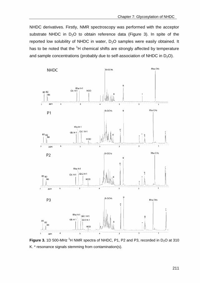
Chapter 7: Glycosylation of NHDC
211
NHDC derivatives. Firstly, NMR spectroscopy was performed with the acceptor
substrate NHDC in D2O to obtain reference data (Figure 3). In spite of the
reported low solubility of NHDC in water, D2O samples were easily obtained. It
has to be noted that the 1H chemical shifts are strongly affected by temperature
and sample concentrations (probably due to self-association of NHDC in D2O).
Figure 3. 1D 500-MHz 1H NMR spectra of NHDC, P1, P2 and P3, recorded in D2O at 310
K. * resonance signals stemming from contamination(s).

Chapter 7: Glycosylation of NHDC
212
The 500-MHz 1H NMR spectrum (Figure 3) of NHDC showed the signals of the
aromatic protons at δ 6.87 [d, J=8.2 Hz, B5], at δ 6.72 [s, J=2.0 Hz, B2], and at δ
6.70 [d, J=8.2 Hz, B6], together with two methylene groups [δ 3.29 (C8) and δ
2.81 (C9)] and a methoxy group [δ 3.75 (B-OCH3)], typical for the
dihydrochalcone skeleton266
. The aromatic proton signals of A2 and A6 (see
NHDC structure, Figure 1), initially found at δ 5.99, had disappeared due to
exchange with deuterium from the solvent (D2O) after prolonged time at 310 K269
.
Also, the C8 proton, in α position to the carbonyl group (C7), is susceptible to
deuterium exchange via the keto-enol route, consequently, 13
C correlations in
HSQC experiments will not be observed. Concerning the carbohydrate moiety,
the 1H NMR spectrum exhibited signals due to one β-D-glucopyranosyl unit [δ
5.185 (d, J=7.6 Hz, Glc H-1)] and one α-L-rhamnopyranosyl unit [δ 5.059 (br s, J
<2 Hz, Rha H-1) and δ 1.168 (d, J=6.1 Hz, Rha CH3)]. The assignment of the
signals (Table SII) was made through 2D NMR TOCSY experiments, using
different mixing times, and in combination with ROESY and HSQC (Figures S2
and S3). Indeed, the obtained NMR data of NHDC (Table SII) were in agreement
with She et al. (2011)250
and Caccia et al. (1998)266
.
The 500-MHz 1H NMR spectrum (Figure 3) of fractions P1 and P2 showed an
extra α-anomeric proton at δ 4.936 (J=3.7 Hz) and at δ 4.845 (J=3.7 Hz),
respectively, stemming from two differently attached glucose units, denoted as
Glc1 and Glc2. Compared to NHDC, the NMR spectrum of fraction P3 showed
signals stemming from both Glc1 and Glc2 (δ 4.955: J=3.7 Hz and δ 4.855: J=3.7
Hz). The resonance signals indicated with * in Figure 3 (e.g. p, q and r, having
mutual TOCSY connections) are stemming from contamination(s). The total
assignment of NMR signals (Table SII) was made through 2D NMR TOCSY,
HSQC and ROESY experiments in a similar way as for NHDC (Figures S4-9),
revealing that Glc1 and Glc2 were attached to NHDC at the α-L-rhamnopyranosyl
unit through an (α1→4) linkage and at the β-D-glucopyranosyl unit through an
(α1→6) linkage, respectively (Figure 4). In other words, P3 contained both the
newly formed (α1→4) linkage present in P1 and the (α1→6) linkage introduced in
P2. The results are in agreement with the methylation analyses (Table …). To

Chapter 7: Glycosylation of NHDC
213
our knowledge, these α-glucosylated NHDC derivatives have not been described
before and can thus be considered new-to-nature.
Figure 4. Chemical structures of α-glucosylated NHDC products: A NHDC-P1, B NHDC-
P2 and C NHDC-P3.
3.3. Sensory analysis of glucosylated NHDC products
NHDC is not only a high-intensity sweetener (340 times sweeter than sucrose), it
is reported to suppress the bitterness displayed by other sweeteners such as
RebA. A sensory analysis of aqueous solutions sweetened with RebA and 10
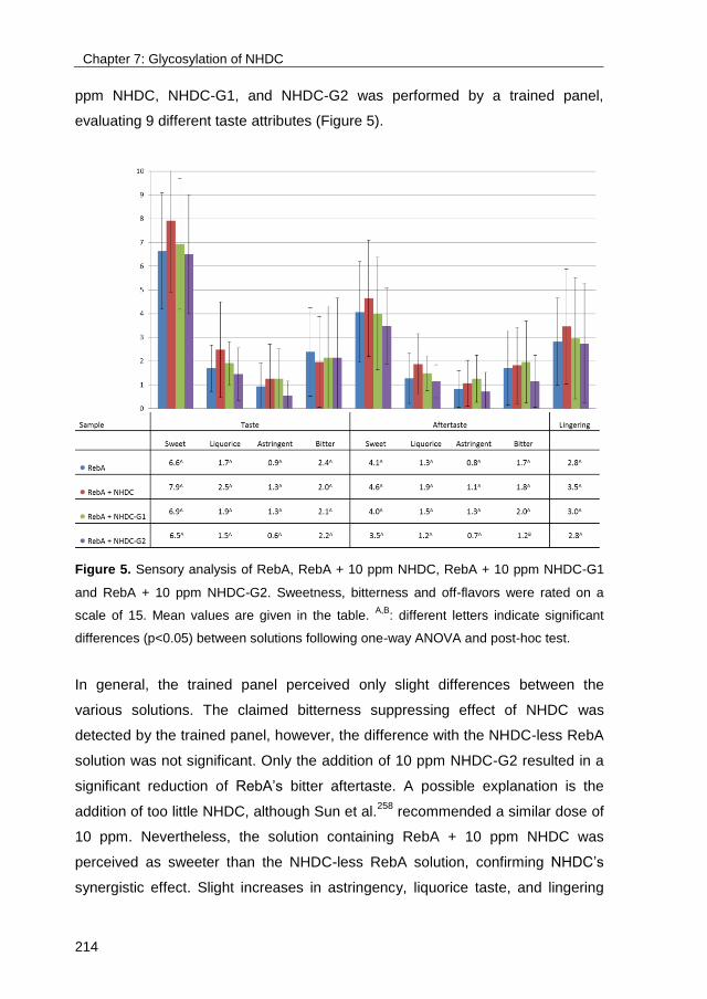
Chapter 7: Glycosylation of NHDC
214
ppm NHDC, NHDC-G1, and NHDC-G2 was performed by a trained panel,
evaluating 9 different taste attributes (Figure 5).
Figure 5. Sensory analysis of RebA, RebA + 10 ppm NHDC, RebA + 10 ppm NHDC-G1
and RebA + 10 ppm NHDC-G2. Sweetness, bitterness and off-flavors were rated on a
scale of 15. Mean values are given in the table. A,B
: different letters indicate significant
differences (p<0.05) between solutions following one-way ANOVA and post-hoc test.
In general, the trained panel perceived only slight differences between the
various solutions. The claimed bitterness suppressing effect of NHDC was
detected by the trained panel, however, the difference with the NHDC-less RebA
solution was not significant. Only the addition of 10 ppm NHDC-G2 resulted in a
significant reduction of RebA’s bitter aftertaste. A possible explanation is the
addition of too little NHDC, although Sun et al.258
recommended a similar dose of
10 ppm. Nevertheless, the solution containing RebA + 10 ppm NHDC was
perceived as sweeter than the NHDC-less RebA solution, confirming NHDC’s
synergistic effect. Slight increases in astringency, liquorice taste, and lingering
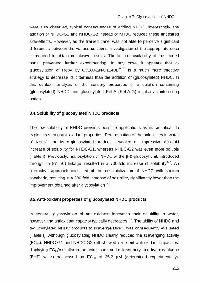
Chapter 7: Glycosylation of NHDC
215
were also observed, typical consequences of adding NHDC. Interestingly, the
addition of NHDC-G1 and NHDC-G2 instead of NHDC reduced these undesired
side-effects. However, as the trained panel was not able to perceive significant
differences between the various solutions, investigation of the appropriate dose
is required to obtain conclusive results. The limited availability of the trained
panel prevented further experimenting. In any case, it appears that α-
glucosylation of RebA by Gtf180-ΔN-Q1140E68-70
is a much more effective
strategy to decrease its bitterness than the addition of (glucosylated) NHDC. In
this context, analysis of the sensory properties of a solution containing
(glucosylated) NHDC and glucosylated RebA (RebA-G) is also an interesting
option.
3.4. Solubility of glucosylated NHDC products
The low solubility of NHDC prevents possible applications as nutraceutical, to
exploit its strong anti-oxidant properties. Determination of the solubilities in water
of NHDC and its α-glucosylated products revealed an impressive 800-fold
increase of solubility for NHDC-G1, whereas NHDC-G2 was even more soluble
(Table I). Previously, maltosylation of NHDC at the β-D-glucosyl unit, introduced
through an (α1→6) linkage, resulted in a 700-fold increase of solubility267
. An
alternative approach consisted of the cosolubilization of NHDC with sodium
saccharin, resulting in a 200-fold increase of solubility, significantly lower than the
improvement obtained after glycosylation265
.
3.5. Anti-oxidant properties of glucosylated NHDC products
In general, glycosylation of anti-oxidants increases their solubility in water,
however, the antioxidant capacity typically decreases139
. The ability of NHDC and
α-glucosylated NHDC products to scavenge DPPH was consequently evaluated
(Table I). Although glucosylating NHDC clearly reduced the scavenging activity
(EC50), NHDC-G1 and NHDC-G2 still showed excellent anti-oxidant capacities,
displaying EC50’s similar to the established anti-oxidant butylated hydroxytoluene
(BHT) which possessed an EC50 of 35.2 µM (determined experimentally).
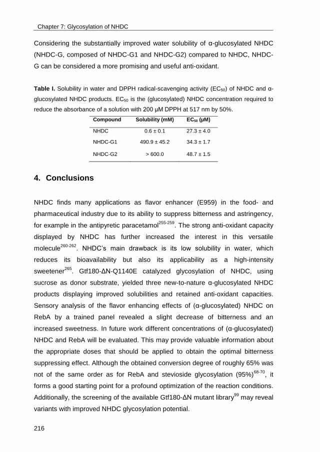
Chapter 7: Glycosylation of NHDC
216
Considering the substantially improved water solubility of α-glucosylated NHDC
(NHDC-G, composed of NHDC-G1 and NHDC-G2) compared to NHDC, NHDC-
G can be considered a more promising and useful anti-oxidant.
Table I. Solubility in water and DPPH radical-scavenging activity (EC50) of NHDC and α-
glucosylated NHDC products. EC50 is the (glucosylated) NHDC concentration required to
reduce the absorbance of a solution with 200 μM DPPH at 517 nm by 50%.
Compound Solubility (mM) EC50 (µM)
NHDC 0.6 ± 0.1 27.3 ± 4.0
..
NHDC-G1 490.9 ± 45.2 34.3 ± 1.7
NHDC-G2 > 600.0 48.7 ± 1.5
4. Conclusions
NHDC finds many applications as flavor enhancer (E959) in the food- and
pharmaceutical industry due to its ability to suppress bitterness and astringency,
for example in the antipyretic paracetamol255-259
. The strong anti-oxidant capacity
displayed by NHDC has further increased the interest in this versatile
molecule260-262
. NHDC’s main drawback is its low solubility in water, which
reduces its bioavailability but also its applicability as a high-intensity
sweetener265
. Gtf180-ΔN-Q1140E catalyzed glycosylation of NHDC, using
sucrose as donor substrate, yielded three new-to-nature α-glucosylated NHDC
products displaying improved solubilities and retained anti-oxidant capacities.
Sensory analysis of the flavor enhancing effects of (α-glucosylated) NHDC on
RebA by a trained panel revealed a slight decrease of bitterness and an
increased sweetness. In future work different concentrations of (α-glucosylated)
NHDC and RebA will be evaluated. This may provide valuable information about
the appropriate doses that should be applied to obtain the optimal bitterness
suppressing effect. Although the obtained conversion degree of roughly 65% was
not of the same order as for RebA and stevioside glycosylation (95%)68-70
, it
forms a good starting point for a profound optimization of the reaction conditions.
Additionally, the screening of the available Gtf180-ΔN mutant library99
may reveal
variants with improved NHDC glycosylation potential.
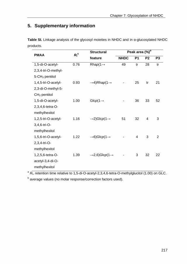
Chapter 7: Glycosylation of NHDC
217
5. Supplementary information
Table SI. Linkage analysis of the glycosyl moieties in NHDC and in α-glucosylated NHDC
products.
PMAA Rta
Structural
feature
Peak area (%)b
NHDC P1 P2 P3
1,5-di-O-acetyl-
2,3,4-tri-O-methyl-
5-CH3 pentitol
0.76 Rhap(1→ 49 tr 28 tr
1,4,5-tri-O-acetyl-
2,3-di-O-methyl-5-
CH3 pentitol
0.93 →4)Rhap(1→ - 25 tr 21
1,5-di-O-acetyl-
2,3,4,6-tetra-O-
methylhexitol
1.00 Glcp(1→ - 36 33 52
1,2,5-tri-O-acetyl-
3,4,6-tri-O-
methylhexitol
1.16 →2)Glcp(1→ 51 32 4 3
1,5,6-tri-O-acetyl-
2,3,4-tri-O-
methylhexitol
1.22 →6)Glcp(1→ - 4 3 2
1,2,5,6-tetra-O-
acetyl-3,4-di-O-
methylhexitol
1.39 →2,6)Glcp(1→ - 3 32 22
a Rt, retention time relative to 1,5-di-O-acetyl-2,3,4,6-tetra-O-methylglucitol (1.00) on GLC.
b average values (no molar response/correction factors used).
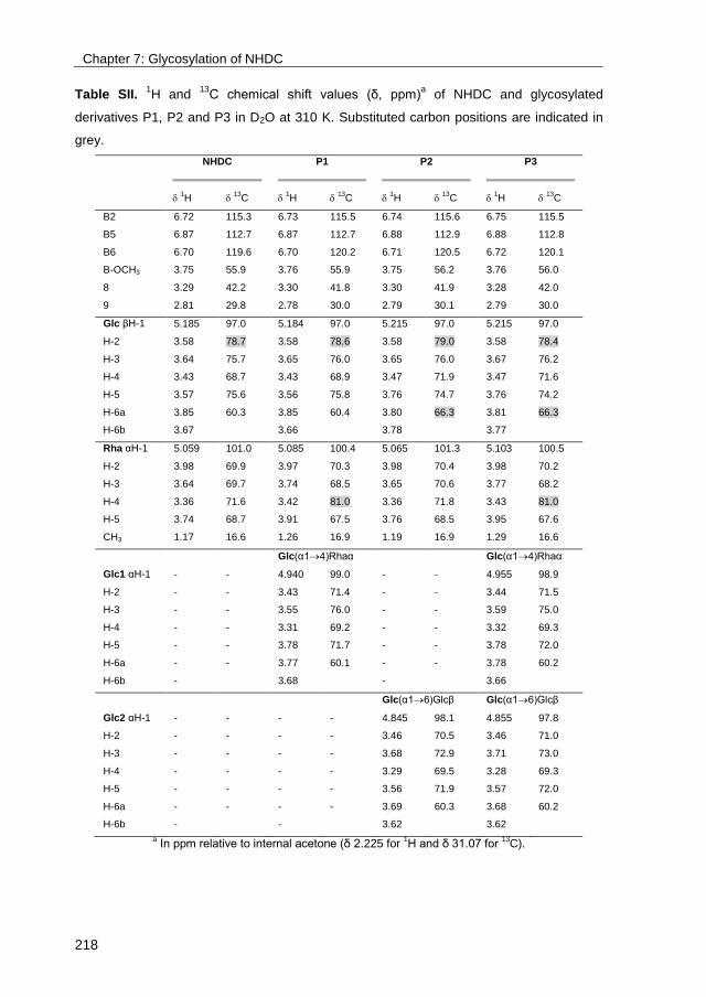
Chapter 7: Glycosylation of NHDC
218
Table SII. 1H and
13C chemical shift values (δ, ppm)
a of NHDC and glycosylated
derivatives P1, P2 and P3 in D2O at 310 K. Substituted carbon positions are indicated in
grey.
NHDC
P1
P2
P3
1H
13C
1H
13C
1H
13C
1H
13C
B2 6.72 115.3 6.73 115.5 6.74 115.6 6.75 115.5
B5 6.87 112.7 6.87 112.7 6.88 112.9 6.88 112.8
B6 6.70 119.6 6.70 120.2 6.71 120.5 6.72 120.1
B-OCH3 3.75 55.9 3.76 55.9 3.75 56.2 3.76 56.0
8 3.29 42.2 3.30 41.8 3.30 41.9 3.28 42.0
9 2.81 29.8 2.78 30.0 2.79 30.1 2.79 30.0
Glc βH-1 5.185 97.0 5.184 97.0 5.215 97.0 5.215 97.0
H-2 3.58 78.7 3.58 78.6 3.58 79.0 3.58 78.4
H-3 3.64 75.7 3.65 76.0 3.65 76.0 3.67 76.2
H-4 3.43 68.7 3.43 68.9 3.47 71.9 3.47 71.6
H-5 3.57 75.6 3.56 75.8 3.76 74.7 3.76 74.2
H-6a 3.85 60.3 3.85 60.4 3.80 66.3 3.81 66.3
H-6b 3.67 3.66 3.78 3.77
Rha αH-1 5.059 101.0 5.085 100.4 5.065 101.3 5.103 100.5
H-2 3.98 69.9 3.97 70.3 3.98 70.4 3.98 70.2
H-3 3.64 69.7 3.74 68.5 3.65 70.6 3.77 68.2
H-4 3.36 71.6 3.42 81.0 3.36 71.8 3.43 81.0
H-5 3.74 68.7 3.91 67.5 3.76 68.5 3.95 67.6
CH3 1.17 16.6 1.26 16.9 1.19 16.9 1.29 16.6
Glc(α14)Rhaα Glc(α14)Rhaα
Glc1 αH-1 - - 4.940 99.0 - - 4.955 98.9
H-2 - - 3.43 71.4 - - 3.44 71.5
H-3 - - 3.55 76.0 - - 3.59 75.0
H-4 - - 3.31 69.2 - - 3.32 69.3
H-5 - - 3.78 71.7 - - 3.78 72.0
H-6a - - 3.77 60.1 - - 3.78 60.2
H-6b - 3.68 - 3.66
Glc(α16)Glcβ Glc(α16)Glcβ
Glc2 αH-1 - - - - 4.845 98.1 4.855 97.8
H-2 - - - - 3.46 70.5 3.46 71.0
H-3 - - - - 3.68 72.9 3.71 73.0
H-4 - - - - 3.29 69.5 3.28 69.3
H-5 - - - - 3.56 71.9 3.57 72.0
H-6a - - - - 3.69 60.3 3.68 60.2
H-6b - - 3.62 3.62
a In ppm relative to internal acetone (δ 2.225 for
1H and δ 31.07 for
13C).
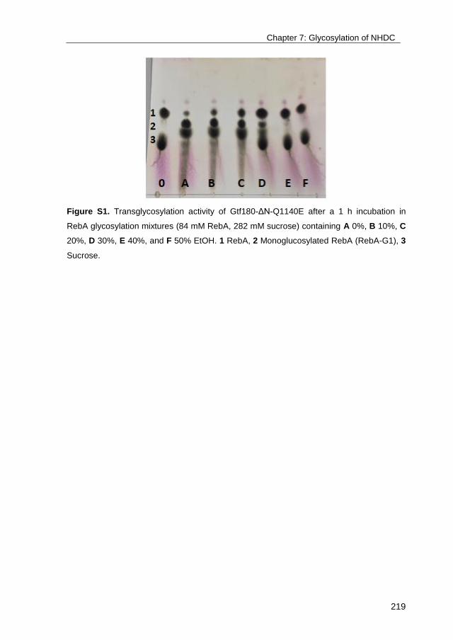
Chapter 7: Glycosylation of NHDC
219
Figure S1. Transglycosylation activity of Gtf180-ΔN-Q1140E after a 1 h incubation in
RebA glycosylation mixtures (84 mM RebA, 282 mM sucrose) containing A 0%, B 10%, C
20%, D 30%, E 40%, and F 50% EtOH. 1 RebA, 2 Monoglucosylated RebA (RebA-G1), 3
Sucrose.
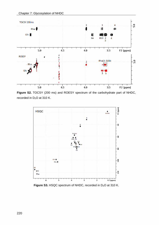
Chapter 7: Glycosylation of NHDC
220
Figure S2. TOCSY (200 ms) and ROESY spectrum of the carbohydrate part of NHDC,
recorded in D2O at 310 K.
Figure S3. HSQC spectrum of NHDC, recorded in D2O at 310 K.
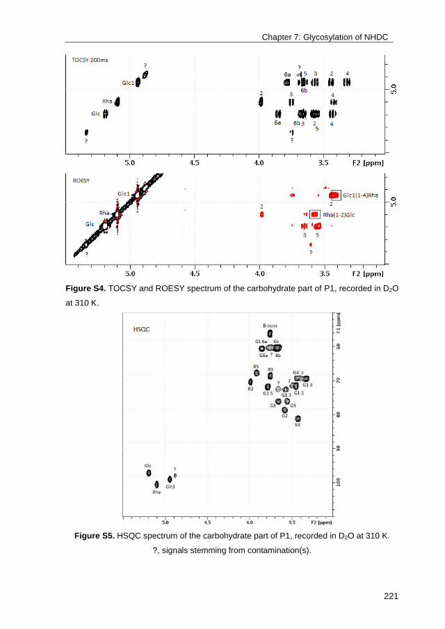
Chapter 7: Glycosylation of NHDC
221
Figure S4. TOCSY and ROESY spectrum of the carbohydrate part of P1, recorded in D2O
at 310 K.
Figure S5. HSQC spectrum of the carbohydrate part of P1, recorded in D2O at 310 K.
?, signals stemming from contamination(s).
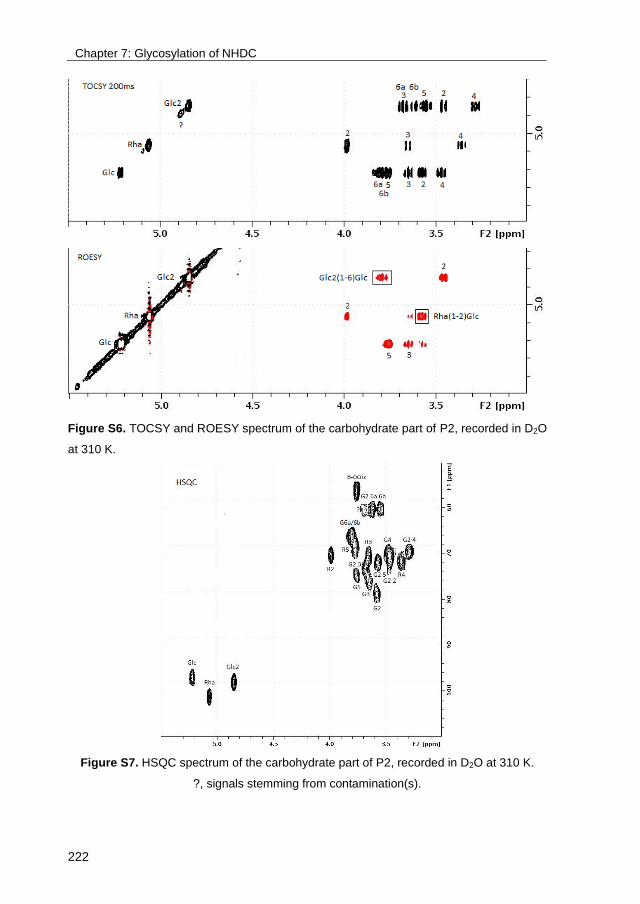
Chapter 7: Glycosylation of NHDC
222
Figure S6. TOCSY and ROESY spectrum of the carbohydrate part of P2, recorded in D2O
at 310 K.
Figure S7. HSQC spectrum of the carbohydrate part of P2, recorded in D2O at 310 K.
?, signals stemming from contamination(s).
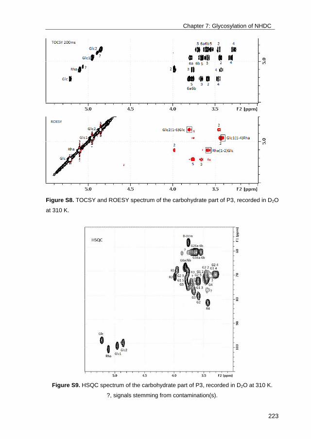
Chapter 7: Glycosylation of NHDC
223
Figure S8. TOCSY and ROESY spectrum of the carbohydrate part of P3, recorded in D2O
at 310 K.
Figure S9. HSQC spectrum of the carbohydrate part of P3, recorded in D2O at 310 K.
?, signals stemming from contamination(s).


225
Chapter 8
Summary and future prospects

Chapter 8: Summary and prospects
226
Part 1: Dealing with the limitations of glucansucrases
The introduction of a glycosyl moiety can influence the physicochemical and
biological properties of organic molecules such as anti-oxidants, antibiotics, and
flavors6. The chemical synthesis of glycosides is characterized by multistep
routes generating lots of waste. One-step enzymatic glycosylation, thereby taking
advantage of the high specificity of enzymes, is preferred as 5-fold less waste is
produced compared to chemical glycosylation17. Among all carbohydrate-active
enzymes that can be applied as glycosylation biocatalyst, glucansucrases have
received a considerable part of the attention due to their broad acceptor
substrate specificity and use of inexpensive sucrose as donor substrate.
Glucansucrases are glycoside hydrolase enzymes (GH70) originating from
Gram-positive lactic acid bacteria. They catalyze the conversion of sucrose into
α-glucan polysaccharides, linking the α-D-glucopyranosyl units by (α1→2),
(α1→3), (α1→4), or (α1→6) bonds, depending on the enzyme specificity73,74. In
addition, they are able to transfer glucosyl groups to a wide array of carbohydrate
and non-carbohydrate acceptor molecules, catalyzing the so-called acceptor
reaction75. To this date, most research on the acceptor reaction of
glucansucrases has focused on the discovery of new acceptor substrate
specificities111-113,120-131. Less progress has been made on the detailed structural
characterization of products obtained, or the improvement of glucansucrases as
industrial glycosylation biocatalyst, neither by reaction –nor enzyme engineering
strategies.
Glucansucrases display a number of drawbacks, in particular if the acceptor
reaction is targeted. First of all, the main glucansucrase catalyzed reaction is the
synthesis of α-glucan polysaccharides from sucrose. It strongly impedes the
efficient glycosylation of alternative acceptor substrates and complicates
downstream processing of the glycosylated products. Chapter 2 explored the
potential of the N-terminally truncated glucansucrase Gtf180 from Lactobacillus
reuteri 180 (Gtf180-ΔN)80 and derived mutants99,100 as glycosylation
Chapter 8: Summary and prospects
227
biocatalysts110. Three Gtf180-ΔN mutants (L938F, L981A and N1029M) were
selected from a mutant library on the basis of their impaired α-glucan synthesis.
Analysis of the glycosylation of the model acceptor substrate catechol by these
mutants revealed that this apparent imperfection resulted in a substantial
increase in monoglycosylation yield. Also several other phenolic and alcoholic
compounds were more efficiently converted into glycosylated products by the
selected mutants. For example, the resorcinol conversion degree of the L981A
variant tripled compared to the WT enzyme, reaching a substantial 53%. Further
analysis showed that these mutants possess a higher affinity for the model
acceptor substrate catechol but a lower affinity for its mono-α-D-glucoside
product, explaining the improved monoglycosylation yields. An explanation of
how mutagenesis of residues L938, L981 and N1029 impaired α-glucan
synthesis was provided by analyzing the available high resolution 3D crystal
structure of the Gtf180-ΔN protein. On the downside, the kcat of the best
performing mutant (L981A) displayed a 4-fold decrease compared to the WT
enzyme. In addition, the glycosylation of larger non-carbohydrate acceptor
substrates such as resveratrol or quercetin remained problematic, partly due to
the low water solubility of these compounds. The L981A mutant could
nevertheless serve as template for further mutational engineering of the Gtf180-
ΔN enzyme, targeting the improved glycosylation of specific acceptor substrates.
A second drawback related to the use of glucansucrases is their relatively low
operational stability at high temperatures and in systems containing high
acceptor substrate concentrations and cosolvents110,120,125. Immobilization of
Gtf180-ΔN on mesoporous silica particles enhanced its activity at temperatures
above 50 °C and high concentrations of DMSO, conditions detrimental for the
free enzyme (Chapter 3). Covalent cross-linking of Gtf180-ΔN with
glutaraldehyde resulted in an undesired side-effect: less catechol was converted
by the immobilized enzyme compared to the free enzyme, in favor of sucrose
hydrolysis. Stabilizing Gtf180-ΔN by the developed immobilization protocol
nevertheless allowed its incubation in 20% DMSO systems containing several
poorly soluble acceptor substrates such as gallic acid and (+)-catechin, resulting
in their improved glycosylation.

Chapter 8: Summary and prospects
228
Part 2: Glycosylation of steviol glycosides
The steviol glycosides extracted from the leaves of the plant Stevia rebaudiana
were approved for use as high-intensity sweetener (HIS) in food products by the
European Commission in 201143. Stevia extract consists mainly of the steviol
glycosides stevioside and rebaudioside A (RebA), however, they display a bitter
(after)taste which prevents the creation of zero-calorie stevia soft drinks and a
complete sugar substitution in other food products46. Solving this taste issue
could therefore greatly expand stevia sales and enforce its position on the HIS
market. The correlation between the structure of steviol glycosides and their taste
quality is still not fully understood, however, it is clear that the latter depends on
the number, location and configuration of the glycosyl moieties8. Hence, the
enzymatic glycosylation of steviol glycosides has been proposed as effective tool
to improve their sensory properties50-70.
Screening of our in-house collection of glucansucrases revealed that only
Gtf180-ΔN from L. reuteri 180 is able to glycosylate RebA, displaying a RebA
conversion of roughly 50% (Chapter 4). As this is insufficient for industrial
applications, the ability of several Gtf180-ΔN mutants to glycosylate RebA was
explored, revealing that the Q1140E mutant is the most promising variant.
Structural analysis of the products showed that both enzymes exclusively
glycosylate RebA at the Glc(β1→C-19 residue, with the formation of an (α1→6)
linkage. Docking of RebA in the enzyme’s active site provided an explanation for
these results: only the steviol C-19 β-D-glucosyl moiety is available for
glycosylation. Several previous studies already demonstrated the importance of
an adequate C-13/C-19-regiospecificity for the taste quality of the glycosylated
products. For example, (α1→4)-glycosylation of stevioside and rubusoside at the
C-13 steviol position yielded products with improved intensity and quality of
sweetness, whereas (α1→4)-glycosylation at the C-19 position resulted in an
increased bitterness52-54. Alternatively, (α1→6)-glycosylation at the C-19 site of
stevioside improved its taste profile60.
Chapter 8: Summary and prospects
229
Optimizing the reaction conditions of the Gtf180-ΔN-Q1140E-catalyzed
glycosylation of RebA by response surface methodology (RSM) yet again
identified the (low) remaining α-glucan synthesis as an important impediment to
obtain high conversion degrees and high product concentrations. Nevertheless, a
highly productive process with a RebA conversion of 95% and a production of
115 g/L glycosylated product (RebA-G) within 3 h was achieved at optimal
reaction conditions. Development of a fed-batch reaction with continuous addition
of relatively low sucrose levels further improved the product yield to 270 g/L by
adequately further suppressing α-glucan synthesis. Sucrose acts as primer for α-
glucan synthesis103,104,135; a constant excess of RebA relative to sucrose
therefore favored the glycosylation of RebA. The continuous addition of sucrose
ensured that enough donor substrate was available to drive the reaction. On the
downside, 10 times more enzyme had to be supplied to the reaction mixture in
order to obtain a 95% conversion within 3 h, which substantially adds to the
production costs. Additionally, performing the fed-batch reaction required the
addition of 34% more sucrose compared to performing the glycosylation in batch
mode. Altogether, the batch reaction is thus preferred over the fed-batch reaction
as performed in this work.
Sensory analysis of RebA and the glycosylated RebA products by a trained panel
showed that RebA-G has a superior taste profile, displaying a significant
reduction in bitterness compared to RebA. Glycosylation of RebA at the
Glc(β1→C-19 residue with the introduction of an (α1→6) linkage is thus a very
appropriate method to improve its sensory properties. Whether the off-flavors are
sufficiently reduced to allow the production of zero-calorie soft drinks, jams,
yoghurts, etc. is currently unknown. Answering this question requires the analysis
of the sensory properties of the end product, including for example the bulking
agent. In addition, the physicochemical properties of the ingredients, such as
viscosity, density, etc. play an important role for the taste quality of the end
product.
Stevioside is the most prevalent steviol glycoside (5-10% of leaf dry weight) and
is perceived by consumers as more bitter than RebA (2-4% of leaf dry weight).

Chapter 8: Summary and prospects
230
This explains why all commercial stevia products are high-purity variants of
RebA, while stevioside is considered as an undesired side-product. Improvement
of the sensory properties of stevioside by means of its glycosylation
consequently offers an opportunity to valorize this “waste” stream. Incubation of
stevioside with Gtf180-ΔN-Q1140E indeed resulted in its glycosylation, although
at a lower efficiency than RebA glycosylation (as determined by RSM): 50 g/L
glycosylated stevioside product (Stev-G) was produced with a 95% conversion,
using 5 times more sucrose than needed for RebA glycosylation (Chapter 5).
Elucidation of the glycosylated stevioside product structures revealed that, in
contrast to RebA glycosylation, stevioside was not exclusively glycosylated at the
C-19 site; minor products were also glycosylated at the C-13 site. Similarly to
RebA glycosylation, the main product was glycosylated at the C-19 site, with the
introduction of an (α1→6) linkage (Stev-G1). Remarkably, the most prevalent
diglycosylated product contained an (α1→4) linkage and not the expected
(α1→3) –or (α1→6) linkages, reflecting the regular linkage specificity of Gtf18082.
Sensory analysis of the glycosylated stevioside products showed a significant
reduction in bitterness compared to stevioside. The trained panel perceived Stev-
G1 as sweet as stevioside but also as significantly less bitter. The large
proportion of multiglycosylated products in Stev-G (67.5%) was translated into a
significantly reduced sweetness, in contrast to RebA-G (22.3% multiglycosylated
products) which maintained the sweetness of RebA. Interestingly, a double dose
of Stev-G displayed a sweetness level similar to that of stevioside whereas
bitterness remained equally suppressed. Comparison of the sensory properties
of RebA-G and Stev-G identified the former as the superior product.
As the glycosylation of RebA and stevioside with Gtf180-ΔN-Q1140E showed
great potential at laboratory scale, an efficient overall process at kg scale,
including downstream processing, was developed (Chapter 6). Ultimately,
adsorption of the glycosylated product with removal of sucrose, fructose, glucose
and α-glucan oligo –and polysaccharides was applied as main purification step.
An estimation of the production costs demonstrated the main strength of the
process: the biocatalyst represents a minor part of the total cost, which is mostly

Chapter 8: Summary and prospects
231
determined by the cost of the acceptor substrate, i.e. RebA or stevioside. A major
cost reduction of 30% can consequently be achieved by using low-grade stevia
extract as acceptor substrate instead of the more costly high-purity steviol
glycosides. An additional advantage is that the side-product stevioside is
valorized, representing an extra economic profit. If taste quality is priority, RebA
is preferred as acceptor substrate as it displays a superior taste profile.
Gtf180-ΔN-Q1140E also displayed activity towards neohesperidin
dihydrochalcone (NHDC), even though a lower conversion degree (64%) was
obtained (Chapter 7). Use of 5-10 ppm NHDC with RebA is known to result in a
decreased bitterness perception. Unfortunately, this is accompanied by an
unpleasant lingering sensation, caused by NHDC247. Its glycosylation by Gtf180-
ΔN-Q1140E resulted in a decreased perception of the off-flavors by the trained
panel. In general, the bitterness suppressing effect of (glycosylated) NHDC on
RebA was limited; RebA glycosylation reduced its bitterness much more
effectively. Very recently, NHDC was attributed with potent anti-oxidant,
hepatoprotective and prebiotic properties260-264. NHDC’s low water solubility265,
restricting its application as nutraceutical, was overcome by its glycosylation,
while its anti-oxidant potential remained very high.
Conclusions
This thesis explored the potential of Gtf180-ΔN from L. reuteri 180 and derived
mutants to glycosylate a wide range of alternative acceptor substrates. It was
shown that the Q1140E mutant is particularly suited to glycosylate steviol
glycosides. High conversion degrees and product yields were obtained, and the
glycosylated products displayed a superior taste profile compared to RebA and
stevioside. The developed processes consequently hold excellent potential to be
implemented at industrial scale.
The Q1140E mutant was also able to glycosylate other glycosides, such as
NHDC, although at a lower efficiency than the glycosylation of steviol glycosides.
Chapter 8: Summary and prospects
232
This is another illustration of the broad carbohydrate acceptor substrate
specificity of Gtf180-ΔN and derived mutants. The high water solubility of
carbohydrates forms an additional advantage for their Gtf180-ΔN catalyzed
glycosylation, thus avoiding the use of inhibitory cosolvents in the reaction
mixture.
The glycosylation of non-carbohydrate acceptor substrates was achieved with
more difficulty, partly due to their low water solubility and their inhibition of
Gtf180-ΔN. α-Glucan synthesis was identified as important impediment for the
glycosylation of these compounds, however, its suppression by mutational
engineering only partially solved the problem. In other words, specific mutants
will need to be constructed to further improve the Gtf180-ΔN glycosylation
potential, similarly to the approach followed for the glycosylation of steviol
glycosides by the Q1140E mutant of Gtf180-ΔN.
In conclusion, the glucansucrase enzyme Gtf180-ΔN holds considerable potential
as glycosylation biocatalyst, in particular for the glycosylation of carbohydrate
acceptor substrates. The development of industrial processes will first and
foremost depend on the construction of adequate mutants displaying high
conversion degrees, and secondly, on the optimization of the reaction conditions,
thereby suppressing α-glucan synthesis from sucrose as much as possible.

233
Samenvatting en conclusies
Samenvatting en conclusies
234
Deel 1: Omgaan met de limitaties van glucansucrases
De introductie van een glycosylgroep kan zowel de fysicochemische als
biologische eigenschappen van organische moleculen, zoals antioxidanten,
antibiotica en smaakstoffen, beïnvloeden6. De chemische synthese van
glycosiden is een meerstapsproces dat gekenmerkt wordt door de productie van
een grote hoeveelheid afvalproducten. Enzymatische glycosylatie geniet de
voorkeur; de hoge specificiteit van enzymen leidt immers tot de productie van 5
keer minder afval17. Van alle koolhydraat-actieve enzymen die gebruikt kunnen
worden als biokatalysator voor de glycosylatie van moleculen zijn
glucansucrases meerdere keren naar voren geschoven als uitstekend alternatief.
Deze enzymen bezitten een brede acceptorsubstraatspecificiteit en gebruiken
sucrose als donorsubstraat, een goedkope en gemakkelijk te verkrijgen
grondstof.
Glucansucrases behoren tot de glycoside hydrolase GH70-familie. Ze
katalyseren de conversie van sucrose in α-glucan polysachariden, en daarbij
verbinden ze de α-D-glucopyranosyl eenheden met (α1→2), (α1→3), (α1→4), of
(α1→6) bindingen, afhankelijk van de enzymspecificiteit73,74. Daarnaast zijn
glucansucrases ook in staat om glucosylgroepen te transfereren van sucrose
naar een brede waaier van koolhydraat en niet-koolhydraat acceptormoleculen,
de zogenaamde acceptorreactie75. Tot op vandaag heeft het meeste onderzoek
naar de acceptorreactie van glucansucrases zich toegespitst op de ontdekking
van nieuwe acceptorsubstraatspecificiteiten111-113,120-131. Er is echter minder
vooruitgang geboekt op het vlak van de gedetailleerde structuurbepaling van de
geglycosyleerde producten, of wat betreft het verbeteren van deze enzymen als
industriële biokatalysator, noch door reactie noch door enzym-engineering
strategieën.
Het toepassen van de acceptorreactie van glucansucrases voor de glycosylatie
van organische moleculen wordt getypeerd door een aantal nadelen.
Glucansucrases katalyseren in principe de synthese van α-glucan
Samenvatting en conclusies
235
polysachariden vanuit sucrose, in dit geval zowel het donor als
acceptorsubstraat. Dit verhindert uiteraard de efficiënte glycosylatie van
alternatieve acceptorsubstraten en compliceert de opzuivering van de
geglycosyleerde producten. Hoofdstuk 2 verkende het potentieel van het N-
terminaal getrunceerde glucansucrase Gtf180 van Lactobacillus reuteri 180
(Gtf180-ΔN)80 en afgeleide mutanten99,100 als glycosylatiebiokatalysator110. Drie
Gtf180-ΔN mutanten (L938F, L981A and N1029M) werden geselecteerd uit een
mutantenbibliotheek op basis van hun verzwakte α-glucansynthese. Analyse van
de glycosylatie van het acceptorsubstraat catechol door deze mutanten onthulde
dat deze ogenschijnlijke imperfectie resulteerde in een substantiële verbetering
van de monoglycosylatie-opbrengst. Ook verscheidene andere fenolische en
alcoholische verbindingen werden efficiënter geglycosyleerd door de
geselecteerde mutanten. In vergelijking met het WT-enzym was de omzetting
van bijvoorbeeld resorcinol in geglycosyleerde producten door de L981A-variant
verdrievoudigd van 17% tot 53%. Deze resultaten werden andermaal bevestigd
door de kinetische analyse van de mutanten; ze hadden een hogere affiniteit
voor het acceptorsubstraat catechol maar een lagere affiniteit voor het
monogeglycosyleerde catecholproduct. Analyse van de beschikbare 3D-structuur
van het Gtf180-ΔN-proteïne verklaarde hoe mutagenese van residuen L938,
L981 en N1029 de α-glucansynthese verzwakte in het voordeel van de
acceptorreactie. Toch bleef de glycosylatie van grotere (niet-koolhydraat)
acceptorsubstraten, zoals resveratrol en quercetine, problematisch, deels door
de lage oplosbaarheid in water van deze moleculen. De L981A-mutant vormt
desalniettemin een goed vertrekpunt voor verdere mutationele engineering om
de glycosylatie van vooraf gedefinieerde acceptorsubstraten te verbeteren.
Een tweede nadeel inherent aan het gebruik van glucansucrases is hun relatief
lage operationele stabiliteit bij hoge temperaturen en in systemen met hoge
acceptorsubstraatconcentraties en cosolventen110,120,125. Immobilisatie van
Gtf180-ΔN op mesoporeuze silicapartikels verbeterde de enzymactiviteit bij
temperaturen hoger dan 50 °C en in systemen met hoge DMSO-concentraties,
condities die normalerwijs zeer schadelijk zijn voor het enzym (Hoofdstuk 3).
Het cross-linken van Gtf180-ΔN met glutaaraldehyde resulteerde echter in een
Samenvatting en conclusies
236
ongewenst neveneffect: minder catechol werd geconverteerd door het
geïmmobiliseerde enzym in vergelijking met het vrije enzym, ten gunste van
meer sucrosehydrolyse. Incubatie van geïmmobiliseerd Gtf180-ΔN in systemen
met 20% DMSO leidde desalniettemin tot de verbeterde glycosylatie van een
aantal slecht oplosbare verbindingen, zoals catechine en galluszuur.
Deel 2: Glycosylatie van steviolglycosiden
Het gebruik van steviolglycosiden, verbindingen die geëxtraheerd worden uit de
bladeren van de plant Stevia rebaudiana, als intensieve zoetstof is in 2011
goedgekeurd door de Europese Commissie43. Steviaextract bestaat voornamelijk
uit de steviolglycosiden stevioside en rebaudioside A (RebA), wiens bittere
(na)smaak de creatie van calorieloze frisdrank en een volledige suikervervanging
in andere voedingsproducten in de weg staat46. Het oplossen van deze
smaakkwestie zou de populariteit en bijgevolg het verkoopsucces van stevia
substantieel kunnen verhogen en daarmee een sterke positie voor stevia op de
zoetstofmarkt vrijwaren. Hoe de chemische structuur van steviolglycosiden hun
smaakkwaliteit beïnvloedt is nog steeds niet helemaal duidelijk. Het staat echter
vast dat deze laatste afhangt van het aantal, de locatie en configuratie van de
glycosylgroepen8. Het spreekt dus voor zich dat de enzymatische glycosylatie
van steviolglycosiden een veelbelovend middel is om diens
smaakeigenschappen te verbeteren50-70.
Screening van onze in-house glucansucrasecollectie onthulde dat alleen Gtf180-
ΔN van L. reuteri 180 in staat is om RebA te glycosyleren en daarbij een 50%
conversie bereikt (Hoofdstuk 4). Omdat deze omzetting vanuit industrieel
perspectief te laag is, werd een mutantenbibliotheek gescreend op basis van een
verbeterde RebA-conversie, wat leidde tot de ontdekking van de Q1140E-
mutant. Structurele analyse van de geglycosyleerde producten toonde aan dat
beide enzymen RebA exclusief glycosyleren op het Glc(β1→C-19 residu, met de
introductie van een (α1→6)-binding. Docking van RebA in het actieve centrum
van het enzym gaf een visuele interpretatie voor deze resultaten: enkel de steviol
Samenvatting en conclusies
237
C-19 β-D-glucosylgroep is beschikbaar voor glycosylatie. Voorgaande studies
hadden het belang van de C-13/C-19-regiospecificiteit op de smaakkwaliteit van
de geglycosyleerde producten al aangetoond. Bijvoorbeeld, (α1→4)-glycosylatie
van stevioside en rubusoside op de C-13-steviolpositie resulteerde in producten
met een betere en intensievere zoete smaak. De introductie van dezelfde
(α1→4)-binding op de C-19-positie leidde dan weer tot meer bitterheid52-54.
Hiermee in tegenstelling: (α1→6)-glycosylatie op de C-19-positie van stevioside
verbeterde diens smaakprofiel60.
Optimalisatie van de reactiecondities d.m.v. response surface methodology
(RSM) duidde eens te meer de lage maar nog steeds aanwezige α-
glucansynthese aan als belangrijkste belemmering in het behalen van hoge
omzettingen en productconcentraties. Toch kon er een zeer productief proces
ontwikkeld worden: het toepassen van de optimale condities stond de productie
toe van 115 g/L geglycosyleerd product (RebA-G) binnen 3 uur, met een hoge
RebA-conversie van 95%. Het aannemen van een fed-batchsysteem, waarbij de
concentratie van donorsubstraat sucrose constant op een laag niveau wordt
gehouden door middel van diens continue toevoeging, leidde tot een verdere
verbetering van de productconcentratie tot 270 g/L. Omdat sucrose de primer is
voor α-glucansynthese103,104,135, creëert de constante overmaat van RebA t.o.v.
sucrose immers condities ten gunste van RebA-glycosylatie. Tegelijkertijd zorgt
de continue toevoeging van sucrose ervoor dat er voldoende donorsubstraat
aanwezig is om de reactie voort te drijven.
Smaakanalyse van RebA en de geglycosyleerde RebA-producten door een
getraind panel liet zien dat RebA-G beschikt over een superieur smaakprofiel,
met een significant gereduceerde bitterheid, in vergelijking tot RebA. Glycosylatie
van RebA op het Glc(β1→C-19-residu, met de introductie van een (α1→6)-
binding, is dus een zeer geschikte methode om diens smaakeigenschappen te
verbeteren. Of het smaakprofiel al dan niet voldoende verbeterd is voor de
productie van calorieloze frisdrank, confituur, yoghurt, e.d. vereist uiteraard de
smaakanalyse van het respectievelijke eindproduct.

Samenvatting en conclusies
238
Stevioside is het meest voorkomende steviolglycoside (5-10% van het droge
bladgewicht) maar wordt door consumenten als bitterder bevonden dan RebA (2-
4% van het droge bladgewicht). Dit verklaart waarom alle commerciële
steviaproducten zeer zuivere varianten zijn van RebA, terwijl stevioside wordt
beschouwd als een ongewenst nevenproduct. Het verbeteren van stevioside’s
smaakeigenschappen d.m.v. glycosylatie biedt dus een opportuniteit om ook dit
nevenproduct te valoriseren. Hoewel incubatie van stevioside met Gtf180-ΔN-
Q1140E inderdaad resulteerde in diens glycosylatie, verliep de reactie minder
efficiënt dan RebA-glycosylatie (bepaald door RSM): om 50 g/L geglycosyleerd
product (Stev-G) te produceren binnen 3 uur, met een conversie van 95%, was
er 5 keer meer sucrose en 2 keer meer enzym nodig (Hoofdstuk 5). Structurele
analyse van de geglycosyleerde producten onthulde dat, in tegenstelling tot
RebA-glycosylatie, stevioside niet exclusief geglycosyleerd werd op de C-19-
positie; een kleine hoeveelheid producten bleek geglycosyleerd op de C-13-
positie. Net als bij RebA-glycosylatie was het belangrijkste product
geglycosyleerd op de C-19-positie, met de introductie van een (α1→6)-binding
(Stev-G1). Het meest voorkomende digeglycosyleerde product bevatte een
(α1→4)-binding en niet een verwachte (α1→3) –of (α1→6)-binding (specifiek
voor de Gtf180-ΔN-producten met sucrose alleen82).
Smaakanalyse van de geglycosyleerde producten toonde een significante
bitterheidsreductie aan ten opzichte van stevioside. Het getrainde panel
oordeelde dat Stev-G1 net zo zoet was als stevioside maar ook significant
minder bitter. Het grote aandeel aan multigeglycosyleerde producten in Stev-G
(67.5%) vertaalde zich in een significant gereduceerde zoetheid, in tegenstelling
tot RebA-G (22.3% multigeglycosyleerde producten) dat RebA’s zoetheid
behield. Dit probleem kon echter opgelost worden door het verdubbelen van de
Stev-G dosis want daardoor werd een gelijkwaardige zoetheid als bij stevioside
bereikt maar was er nog steeds sprake van een significante bitterheidsreductie.
In vergelijking met Stev-G was RebA-G duidelijk het superieure product.
Omdat de glycosylatie van RebA en stevioside met Gtf180-ΔN-Q1140E veel
potentieel toonde op laboratoriumschaal, werd een efficiënt proces, inclusief

Samenvatting en conclusies
239
productopzuivering, ontwikkeld op kg-schaal (Hoofdstuk 6). Uiteindelijk werd
gekozen voor specifieke adsorptie van het geglycosyleerde product om sucrose,
fructose en de α-glucanoligo –en polysachariden te verwijderen. Analyse van de
productiekosten onthulde de grote kracht van het proces: het enzym
vertegenwoordigt slechts een klein deel van de totale kosten, die vooral worden
bepaald door de kosten van het acceptorsubstraat, RebA of stevioside. Een
substantiële kostenreductie van 30% kan dus bewerkstelligd worden door stevia-
extract van lagere kwaliteit te gebruiken als acceptorsubstraat i.p.v. de duurdere
steviolglycosiden van hoge zuiverheid. Een bijkomend voordeel is dat het
nevenproduct stevioside gevaloriseerd wordt, een extra economische winst. Als
smaakkwaliteit de prioriteit is, dan vormt RebA-glycosylatie de beste keuze
aangezien RebA-G een superieure smaak heeft.
Daarnaast kon Gtf180-ΔN-Q1140E ook neohesperidine dihydrochalcon (NHDC)
glycosyleren, ook al werd een relatief lage omzetting (64%) bereikt (Hoofdstuk
7). Het is bekend dat het gebruik van 5-10 ppm NHDC in een RebA-oplossing
een vermindering van de bitterheid tot gevolg heeft. Jammer genoeg gaat dit
gepaard met een onprettige en slepende nasmaak, veroorzaakt door NHDC247.
Diens glycosylatie door Gtf180-ΔN-Q1140E resulteerde in een geringe
verbetering van de ongewenste nasmaak. In het algemeen was het
bitterheidsonderdrukkende effect van (geglycosyleerd) NHDC op RebA vrij
miniem; RebA-glycosylatie bleef overeind als meest effectieve manier om diens
smaak te verbeteren. Recent onderzoek kende ook krachtige antioxidant –en
prebiotische eigenschappen toe aan NHDC260-264. De lage wateroplosbaarheid
van NHDC265, wat diens toepassing als nutraceutical in de weg staat, werd
verbeterd d.m.v. glycosylatie met Gtf180-ΔN-Q1140E, terwijl de sterke
antioxidanteigenschappen door glycosylatie behouden bleven.

Samenvatting en conclusies
240
Conclusies
Deze thesis verkende het potentieel van L. reuteri 180 Gtf180-ΔN en afgeleide
mutanten om een brede waaier aan alternatieve acceptorsubstraten te
glycosyleren. Hierbij werd aangetoond dat de Q1140E-mutant bijzonder geschikt
is om steviolglycosiden te glycosyleren. Niet alleen werden hoge conversies en
productconcentraties bekomen, de geglycosyleerde producten beschikten over
een superieur smaakprofiel in vergelijking met RebA en stevioside. De
ontwikkelde processen hebben dus een uitstekend potentieel om
geïmplementeerd te worden op een industriële schaal.
De Q1140E-mutant was daarnaast ook in staat om andere glycosiden, in het
bijzonder NHDC, te glycosyleren, hoewel deze reactie minder efficiënt was dan
de glycosylatie van steviolglycosiden. Toch is dit nogmaals een illustratie van de
brede acceptorsubstraatspecificiteit van Gtf180-ΔN en afgeleide mutanten wat
betreft de glycosylatie van koolhydraten. De uitstekende wateroplosbaarheid van
koolhydraten is hiervoor uiteraard een extra voordeel aangezien de additie van
inhiberende cosolventen niet nodig is.
De glycosylatie van niet-koolhydraten verliep met meer moeite, mede door hun
lage wateroplosbaarheid en hun inhibitie van Gtf180-ΔN. α-Glucansynthese werd
aangeduid als belangrijkste struikelblok voor de efficiënte glycosylatie van deze
verbindingen. Onderdrukking van de α-glucansynthese door mutationele
engineering zorgde voor een beperkt verbeterde omzetting. Er zullen dus
specifieke mutanten geconstrueerd moeten worden om Gtf180-ΔN’s
glycosylatiepotentieel verder uit te bouwen en te verbeteren. Een gelijkwaardige
benadering zoals gevolgd voor de glycosylatie van steviolglycosiden zou het
meest succesvol moeten zijn.
Als finale conclusie kan dus gesteld worden dat glucansucrase Gtf180-ΔN zeker
een toekomst heeft als glycosylatiebiokatalysator, in het bijzonder voor de
glycosylatie van koolhydraten. Het succes van industriële processen zal eerst en

Samenvatting en conclusies
241
vooral afhangen van de constructie van gepaste mutanten, leidend tot hoge
omzettingen, en ten tweede, de nauwkeurige optimalisatie van de
reactiecondities, daarbij α-glucansynthese zoveel mogelijk onderdrukkend.

243
References

References
244
1. Li S, Yang X, Yang S, Zhu M, Wang X (2012) Technology prospecting on enzymes:
application, marketing and engineering. Comput Struct Biotechnol J 2:1-11.
2. Choi JM, Han SS, Kim HS (2015) Industrial applications of enzyme biocatalysis: current
status and future aspects. Biotechnol Adv 33:1443-1454.
3. Liu L, Yang H, Shin HD, Chen RR, Li J, Du G, Chen J (2013) How to achieve high-level
expression of microbial enzymes: strategies and perspectives. Bioengineered 4:212-223.
4. Damborsky J, Brezovsky J (2014) Computational tools for designing and engineering
enzymes. Curr Opin Chem Biol 19:8-16.
5. Singh R, Kumar M, Mittal A, Mehta PK (2016) Microbial enzymes: industrial progress in 21st
century. 3 Biotech 6:174.
6. Křen V, Martínková L (2001) Glycosides in medicine: The role of glycosidic residue in
biological activity. Curr Med Chem 8:1303-1328.
7. Yamamoto I, Muto N, Nagata E, Nakamura T, Suzuki Y (1990) Formation of a stable L-
ascorbic acid α-glucoside by mammalian α-glucosidase-catalyzed transglucosylation. Biochim
Biophys Acta 1035:44-50.
8. Gerwig GJ, te Poele EM, Dijkhuizen L, Kamerling JP (2016) Stevia glycosides: Chemical and
enzymatic modifications of their carbohydrate moieties to improve the sweet-tasting quality.
Adv Carbohydr Chem Biochem 73:1-72.
9. Wong A, Toth I (2001) Lipid, sugar and liposaccharide based delivery systems. Curr Med
Chem 8:1123-1136.
10. Křen V, Řezanka T (2008) Sweet antibiotics – the role of glycosidic residues in antibiotic and
antitumor activity and their randomization. FEMS Microbiol Rev 32:858-889.
11. Schwarz M, Hillebrand S, Habben S, Degenhardt A, Winterhalter P (2003) Application of
high-speed countercurrent chromatography to the large-scale isolation of anthocyanins.
Biochem Eng J 14:179-189.
12. Bai XL, Yue TL, Yuan YH, Zhang HW (2010) Optimization of microwave-assisted extraction
of polyphenols from apple pomace using response surface methodology and HPLC analysis.
J Sep Sci 33:3751-3758.
13. Desmet T, Soetaert W, Bojarova P, Kren V, Dijkhuizen L, Eastwick-Field V, Schiller A (2012)
Enzymatic glycosylation of small molecules: Challenging substrates require tailored catalysts.
Chem Eur J 18:10786-10801.
14. Demchenko AV (2008) Handbook of chemical glycosylation: Advances in stereoselectivity
and therapeutic relevance. Wiley-VCH.
15. Zhu X, Schmidt RR (2009) New principles for glycoside-bond formation. Angew Chem Int Ed
48:1900-1934.
16. Guo J, Ye X (2010) Protecting groups in carbohydrate chemistry: influence on
stereoselectivity of glycosylations. Molecules 15:7235-7265.
17. de Roode BM, Franssen MCR, van der Padt A, Boom RM (2003) Perspectives for the
industrial enzymatic production of glycosides. Biotechnol Prog 19:1391-1402.
18. Lairson LL, Henrissat B, Davies GJ, Withers SG (2008) Glycosyltransferases: structures,
functions, and mechanisms. Annu Rev Biochem 77:521-555.

References
245
19. Masada S, Kawase Y, Nagatoshi M, Oguchi Y, Terasaka K, Mizukami H (2007) An efficient
chemoenzymatic production of small molecule glucosides with in situ UDP-glucose recycling.
FEBS Lett 581:2562-2566.
20. Terasaka K, Mizutani Y, Nagatsu A, Mizukami H (2012) In situ UDP-glucose regeneration
unravels diverse functions of plant secondary product glycosyltransferases. FEBS Lett
586:4344-4350.
21. Schmölzer K, Gutmann A, Diricks M, Desmet T, Nidetzky B (2016) Sucrose synthase: A
unique glycosyltransferase for biocatalytic glycosylation process development. Biotechnol
Adv 34:88-111.
22. Aerts D, Verhaeghe TF, Roman BI, Stevens CV, Desmet T, Soetaert W (2011)
Transglucosylation potential of six sucrose phosphorylases toward different classes of
acceptors. Carbohydr Res 346:1860-1867.
23. De Winter K, Desmet T, Devlamynck T, Van Renterghem L, Verhaeghe T, Pelantová H, Křen
V, Soetaert W (2014) Biphasic catalysis with disaccharide phosphorylases: chemoenzymatic
synthesis of α-D-glucosides using sucrose phosphorylase. Org Process Res Dev 18:781-787.
24. Luley-Goedl C, Nidetzky B (2010) Carbohydrate synthesis by disaccharide phosphorylases:
Reactions, catalytic mechanisms and application in the glycosciences. Biotechnol J 5:1324-
1338.
25. Dirks-Hofmeister ME, Verhaeghe T, De Winter K, Desmet T (2015) Creating space for large
acceptors: Rational biocatalyst design for resveratrol glycosylation in an aqueous system.
Angew Chem Int Ed Engl 54:9289-9292.
26. Kraus M, Grimm C, Seibel J (2016) Redesign of the active site of sucrose phosphorylase
through a clash-induced cascade of loop shifts. Chembiochem 17:33-36.
27. Balogh T, Baross L, Kosáry J (2004) Novel reaction systems for the synthesis of O-
glucosides by enzymatic reverse hydrolysis. Tetrahedron 60:679-682.
28. van Rantwijk F, Woudenberg-van Oosterom M, Sheldon RA (1999) Glycosidase-catalysed
synthesis of alkyl glycosides. J Mol Catal 6:511-532.
29. Vic G, Crout DHG (1995) Synthesis of allyl and benzyl β-D-glucopyranosides, and allyl β-D-
galactopyranoside from D-glucose or D-galactose and the corresponding alcohol using
almond β-D-glucosidase. Carbohydr Res 279:315-319.
30. Wang LX, Huang W (2009) Enzymatic transglycosylation for glycoconjugate synthesis. Curr
Opin Chem Biol 13:592-600.
31. Yang M, Davies GJ, Davis BG (2007) A glycosynthase catalyst for the synthesis of flavonoid
glycosides. Angew Chem Int Ed 46:3885-3888.
32. Desmet T, Soetaert W (2011) Enzymatic glycosyl transfer: mechanisms and applications.
Biocat Biotransform 29:1-18.
33. Torres P, Poveda A, Jimenez-Barbero J, Parra JL, Comelles F, Ballesteros A, Plou F (2011)
Enzymatic synthesis of α-glucosides of resveratrol with surfactant activity. Adv Synth Catal
353:1077-1086.
34. De Bruyn F, Maertens J, Beauprez J, Soetaert W, De Mey M (2015) Biotechnological
advances in UDP-sugar based glycosylation of small molecules. Biotechnol Adv 33:288-302.

References
246
35. Endo T, Koizumi S, Tabata K, Kakita S, Ozaki A (1999) Large-scale production of N-
acetyllactosamine through bacterial coupling. Carbohydr Res 316:179-183.
36. Koizumi S, Endo T, Tabata K, Ozaki A (1998) Large-scale production of UDP-galactose and
globotriose by coupling metabolically engineered bacteria. Nat Biotechnol 16:847-850.
37. Brochado AR, Matos C, Moller BL, Hansen J, Mortensen UH, Patil KR (2010) Improved
vanillin production in baker’s yeast through in silico design. Microb Cell Fact 9:84.
38. Choi O, Lee JK, Kang SY, Pandey RP, Sohng JK, Ahn JS, Hong YS (2014) Construction of
artificial biosynthetic pathways for resveratrol glucoside derivatives. J Microbiol Biotechnol
24:614-618.
39. Kishore GM, Motion M, Hicks PM, Hansen J, Houghton-Larsen J, Hansen EH, Mikkelsen MD,
Tavares S, Blom C (2011) Production of steviol glycosides in microorganisms.
WO2011153378.
40. Kim BG, Kim HJ, Ahn JH (2012) Production of bioactive flavonol rhamnosides by expression
of plant genes in Escherichia coli. J Agric Food Chem 60:11143-11148.
41. Pandey RP, Parajuli P, Koirala N, Lee JH, Park YI, Sohng JK (2014) Glucosylation of
isoflavonoids in engineered Escherichia coli. Mol Cells 37:172-177.
42. De Bruyn F, De Paepe B, Maertens J, Beauprez J, De Cocker P, Mincke S, Stevens C, De
Mey M (2015) Development of an in vivo glucosylation platform by coupling production to
growth: Production of phenolic glucosides by a glycosyltransferase of Vitis vinifera. Biotech
Bioeng 112:1594-1603.
43. Commission Regulation (EU) No 1131/2011. Official Journal of the European Union (2011).
44. Future Market Insights (2014) Stevia market: Global industry analysis and opportunity
assessment 2014 – 2020. http://www.futuremarketinsights.com
45. World Health Organization (2016) Evaluation of certain food additives. http://www.who.int
46. Hellfritsch C, Brockhoff A, Stähler F, Meyerhof W, Hofmann T (2012) Human psychometric
and taste receptor responses to steviol glycosides. J Agric Food Chem 60:6782-6793.
47. Shi J, Feng Y, Zhao C, Wang H (2010) Process for rebaudioside D. WO 2010146463.
48. Chen P, Li Y, Peng S (2014) Novel process for the preparation of rebaudioside D and other
related naturally occurring sweeteners. US20140296499.
49. Prakash I, Markosyan A, Bunders C (2014) Development of next generation stevia
sweetener: Rebaudioside M. Foods 3:162-175.
50. Li S, Li W, Xiao Q, Xia Y (2013) Transglycosylation of stevioside to improve the edulcorant
quality by lower substitution using cornstarch hydrolyzate and CGTase. Food Chem
138:2064-2069.
51. Abelyan V, Balayan A, Ghochikyan V, Markosyan A (2004) Transglycosylation of stevioside
by cyclodextrin glucanotransferases of various groups of microorganisms. Appl Biochem
Microbiol 40:129-134.
52. Darise M, Mizutani K, Kasai R, Tanaka O (1984) Enzymic transglucosylation of rubusoside
and the structure- sweetness relationship of steviol- bisglycosides. Agric Biol Chem 102483-
2488.
References
247
53. Fukunaga Y, Miyata T, Nakayasu N, Mizutani K, Kasai R, Tanaka O (1989) Enzymatic
transglucosylation products of stevioside – separation and sweetness-evaluation. Agric Biol
Chem 53:1603-1607.
54. Ohtani K, Aikawa Y, Ishikawa H, Kasai R, Kitahata S, Mizutani K, Doi S, Nakaura M, Tanaka
O (1991) Further study on the 1,4-alpha-transglucosylation of rubusoside, a sweet steviol-
bisglucoside from Rubus suavissimus. Agric Biol Chem 55:449-453.
55. Jaitak V, Kaul VK, Bandna, Kumar N, Singh B, Savergave LS, Jogdand VV, Nene S (2009)
Simple and efficient enzymatic transglycosylation of stevioside by β-cyclodextrin
glucanotransferase from Bacillus firmus. Biotechnol Lett 31:1415-1420.
56. Yu X, Yang J, Li B, Yuan H (2015) High efficiency transformation of stevioside into a single
mono-glycosylated product using a cyclodextrin glucanotransferase from Paenibacillus sp.
CGMCC 5316. World J Microbiol Biotechnol 31:1983-1991.
57. Markosyan A, Prakash I, Chaturvedula VSP (2013) High purity steviol glycosides.
WO2013176738.
58. Mikkelsen MD, Hansen J, Simon E, Brianza F, Semmler A, Olsson K, Carlsen S, Düring L,
Ouspenski A, Hicks P (2014) Methods for improved production of rebaudioside D and
rebaudioside M. WO2014122227.
59. Wang Y, Chen L, Li Y, Li Y, Yan M, Chen K, Hao N, Xu L (2015) Efficient enzymatic
production of rebaudioside A from stevioside. Biosci Biotechnol Biochem 80:67-73.
60. Lobov SV, Kasai R, Ohtani K, Yamasaki K (1991) Enzymic production of sweet stevioside
derivatives: Transglucosylation by glucosidases. Agric Biol Chem 55:2959-2965.
61. Ye F, Yang R, Hua X, Shen Q, Zhao W, Zhang W (2013) Modification of stevioside using
transglucosylation activity of Bacillus amyloliquefaciens α-amylase to reduce its bitter
aftertaste. LWT – Food Sci Technol 51:524-530.
62. Ye F, Yang R, Hua X, Shen Q, Zhao W, Zhang W (2014) Modification of steviol glycosides
using α-amylase. LWT – Food Sci Technol 57:400-405.
63. Kusama S, Kusakabe I, Nakamura Y, Eda S, Murakami K (1986) Transglucosylation into
stevioside by the enzyme system from Streptomyces sp. Agric Biol Chem 50:2445-2451.
64. Kusakabe I, Watanabe S, Morita R, Terahara M, Murakami K (1992) Formation of a transfer
product from stevioside by the cultures of Actinomycete. Biosci Biotechnol Biochem 56:233-
237.
65. De Oliveira BH, Packer JF, Chimelli M, de Jesus DA (2007) Enzymatic modification of
stevioside by cell-free extract of Gibberella fujikuroi. J Biotechnol 131:92-96.
66. Ko JA, Nam SH, Park JY, Wee Y, Kim D, Lee WS, Ryu YB, Kim YM (2016) Synthesis and
characterization of glucosyl stevioside using Leuconostoc dextransucrase. Food Chem
211:577-582.
67. Musa A, Miao M, Zhang T, Jiang B (2014) Biotransformation of stevioside by Leuconostoc
citreum SK24.002 alternansucrase acceptor reaction. Food Chem 146:23-29.
68. te Poele EM, Dijkhuizen L, Gerwig GJ, Kamerling JP (2016) Methods for the enzymatic
modification of steviol glycosides, modified steviol glycosides obtainable thereby, and the use
thereof as sweeteners. WO 2016144175.
References
248
69. Gerwig GJ, te Poele EM, Dijkhuizen L, Kamerling JP (2017) Structural analysis of
rebaudioside A derivatives obtained by Lactobacillus reuteri 180 glucansucrase-catalyzed
trans-α-glucosylation. Carbohydr Res 440-441:51-62.
70. te Poele EM, Devlamynck T, Jäger M, Gerwig GJ, Van de Walle D, Dewettinck K, Hirsch
AKH, Kamerling JP, Soetaert W, Dijkhuizen L (2017) Glucansucrase (mutant) enzymes from
Lactobacillus reuteri 180 efficiently transglucosylate Stevia component rebaudioside A,
resulting in a superior taste. Sci Reports. Submitted for publication.
71. Olsson K, Carlsen S, Semmler A, Simon E, Mikkelsen MD, Møller BL (2016) Microbial
production of next-generation stevia sweeteners. Microb Cell Fact 15, DOI: 10.1186/s12934-
016-0609-1.
72. Costa-Font M, Gil JM, Traill WB (2008) Consumer acceptance, valuation of and attitudes
towards genetically modified food: Review and implications for food policy. Food Policy
33:99-111.
73. Monchois V, Willemot RM, Monsan P (1999) Glucansucrases: mechanism of action and
structure-function relationships. FEMS Microbiol Rev 23: 131-151.
74. van Hijum SAFT, Kralj S, Ozimek LK, Dijkhuizen L, van Geel-Schutten IGH (2006) Structure-
function relationships of glucansucrase and fructansucrase enzymes from lactic acid bacteria.
Microbiol Mol Biol Rev 70:157-176.
75. Leemhuis H, Pijning T, Dobruchowska JM, van Leeuwen SS, Kralj S, Dijkstra BW, Dijkhuizen
L (2013) Glucansucrases: three-dimensional structures, reactions, mechanism, α-glucan
analysis and their implications in biotechnology and food applications. J Biotechnol 163:250-
272.
76. Kumari A, Catanzaro R, Marotta F (2011) Clinical importance of lactic acid bacteria: a short
review. Acta Biomed 82:177-180.
77. Bounaix MS, Gabriel V, Morel S, Robert H, Rabier P, Remaud-Siméon M, Gabriel B,
Fontagné-Faucher C (2009) Biodiversity of exopolysaccharides produced from sucrose by
sourdough lactic acid bacteria. J Agric Food Chem 57:10889-10897.
78. Passerini D, Vuillemin M, Ufarte L, Morel S, Loux V, Fontagné-Faucher C, Remaud-Siméon
M, Moulis C (2015) Inventory of the GH70 enzymes encoded by Leuconostoc citreum NRRL
B-1299 – identification of three novel α-tranglucosylases. FEBS J 282:2115-2130.
79. Ooshima T, Matsumura M, Hoshino T, Kawabata S, Sobue S, Fujiwara T (2001)
Contributions of three glucosyltransferases to sucrose-dependent adherence of
Streptococcus mutans. J Dent Res 80:1672-1677.
80. Kralj S, van Geel-Schutten IGH, Dondorff MMG, Kirsanovs S, van der Maarel MJEC,
Dijkhuizen L (2004) Glucan synthesis in the genus Lactobacillus: isolation and
characterization of glucansucrase genes, enzymes and glucan products from six different
strains. Microbiology 150:3681-3690.
81. Monchois V, Willemot RM, Remaud-Simeon M, Croux C, Monsan P (1996) Cloning and
sequencing of a gene coding for a novel dextransucrase from Leuconostoc mesenteroides
NRRL B-1299 synthesizing only alpha (1-6) and alpha (1-3) linkages. Gene 182:23-32.
References
249
82. Pijning T, Vujicic-Zagar A, Kralj S, Eeuwema W, Dijkhuizen L, Dijkstra BW (2008)
Biochemical and crystallographic characterization of a glucansucrase from Lactobacillus
reuteri 180. Biocat Biotrans 26:12-17.
83. Vujicic-Zagar A, Pijning T, Kralj S, Lopez CA, Eeuwema W, Dijkhuizen L, Dijkstra BW (2010)
Crystal structure of a 117 kDa glucansucrase fragment provides insight into evolution and
product specificity of GH70 enzymes. Proc Natl Acad Sci USA 107:21406-21411.
84. Pijning T, Vujičić-Žagar A, Kralj S, Dijkhuizen L, Dijkstra BW (2012) Structure of the alpha-
1,6/alpha-1,4-specific glucansucrase GtfA from Lactobacillus reuteri 121. Acta Crystallogr
Sect F Struct Biol Cryst Commun 68:1448-1454.
85. Ito K, Ito S, Shimamura T, Weyand S, Kawarasaki Y, Misaka T, Abe K, Kobayashi T,
Cameron AD, Iwata S (2011) Crystal structure of glucansucrase from the dental caries
pathogen Streptococcus mutans. J Mol Biol 408:177-186.
86. Brison Y, Pijning T, Malbert Y, Fabre É, Mourey L, Morel S, Potocki-Véronèse G, Monsan P,
Tranier S, Remaud-Siméon M, Dijkstra BW (2012) Functional and structural characterization
of α-(1→2) branching sucrase derived from DSR-E glucansucrase. J Biol Chem 287:7915-
7924.
87. Pijning T, Vujicic-Zagar A, Kralj S, Dijkhuizen L, Dijkstra BW (2014) Flexibility of truncated
and full-length glucansucrase Gtf180 enzymes from Lactobacillus reuteri 180. FEBS J
281:2159-2171.
88. Meng X, Dobruchowska JM, Pijning T, Gerwig GJ, Kamerling JP, Dijkhuizen L (2015)
Truncation of domain V of the multidomain glucansucrase Gtf180 of Lactobacillus reuteri 180
heavily impairs its polysaccharide-synthesizing ability. Appl Microbiol Biotechnol 99:5885-
5894.
89. MacGregor EA, Jespersen HM, Svensson B (1996) A circularly permuted alpha-amylase-type
alpha/beta-barrel structure in glucan-synthesizing glucosyltransferases. FEBS Lett 378:263-
266.
90. Lombard V, Golaconda Ramulu H, Drula E, Coutinho PM, Henrissat B (2014) The
Carbohydrate-active enzymes database (CAZy) in 2013. Nucleic Acids Res 42:490-495.
91. Henrissat B, Davies G (1997) Structural and sequence-based classification of glycoside
hydrolases. Curr Opin Struct Biol 7:637-644.
92. Uitdehaag JC, Mosi R, Kalk KH, van der Veen BA, Dijkhuizen L, Withers SG, Dijkstra BW
(1999) X-ray structures along the reaction pathway of cyclodextrin glycosyltransferase
elucidate catalysis in the alpha-amylase family. Nat Struct Biol 6:432-436.
93. van Leeuwen SS, Kralj S, van Geel-Schutten IH, Gerwig GJ, Dijkhuizen L, Kamerling JP
(2008) Structural analysis of the alpha-D-glucan (EPS180) produced by the Lactobacillus
reuteri strain 180 glucansucrase GTF180 enzyme. Carbohydr Res 343:1237-1250.
94. van Leeuwen SS, Kralj S, van Geel-Schutten IH, Gerwig GJ, Dijkhuizen L, Kamerling JP
(2008) Structural analysis of the alpha-D-glucan (EPS35-5) produced by the Lactobacillus
reuteri strain 35-5 glucansucrase GTFA enzyme. Carbohydr Res 343:1251-1265.

References
250
95. Leemhuis H, Pijning T, Dobruchowska JM, Dijkstra BW, Dijkhuizen L (2012) Glycosidic bond
specificity of glucansucrases: on the role of acceptor substrate binding residues. Biocat
Biotrans 30:366-376.
96. Kralj S, van Geel-Schutten IGH, Faber EJ, van der Maarel MJEC, Dijkhuizen L (2005)
Rational transformation of a Lactobacillus reuteri 121 reuteransucrase into a dextransucrase.
Biochemistry 44:9206-9216.
97. van Leeuwen SS, Kralj S, Eeuwema W, Gerwig GJ, Dijkhuizen L, Kamerling JP (2009)
Structural characterization of bioengineered alpha-D-glucans produced by mutant
glucansucrase GTF180 enzymes of Lactobacillus reuteri strain 180. Biomacromolecules
10:580-588.
98. Meng X, Dobruchowska JM, Pijning T, Lopez CA, Kamerling JP, Dijkhuizen L (2014) Residue
Leu940 has a crucial role in the linkage and reaction specificity of the glucansucrase GTF180
of the probiotic bacterium Lactobacillus reuteri 180. J Biol Chem 289:32773-32782.
99. Meng X, Pijning T, Dobruchowska JM, Gerwig GJ, Dijkhuizen L (2015) Characterization of
the functional roles of amino acid residues in acceptor binding subsite +1 in the active site of
the glucansucrase GTF180 enzyme of Lactobacillus reuteri 180. J Biol Chem 290:30131-
30141.
100. Meng X, Dobruchowska JM, Pijning T, Gerwig GJ, Dijkhuizen L (2016) Synthesis of new
hyper-branched α-glucans from sucrose by Lactobacillus reuteri 180 glucansucrase mutants.
J Agr Food Chem 64:433-442.
101. Meng X, Pijning T, Dobruchowska JM, Yin H, Gerwig GJ Dijkhuizen L (2016) Structural
determinants of alternating (α1→4) and (α1→6) linkage specificity in reuteransucrase of
Lactobacillus reuteri. Sci Rep 6:35261.
102. Meng X, Pijning T, Tietema M, Dobruchowska JM, Yin H, Gerwig GJ, Kralj S, Dijkhuizen L
(2017) The essential role of W1065 in the glucansucrase GTF180 of Lactobacillus reuteri 180
for activity, polysaccharide synthesis and linkage specificity. Food Chem 217:81-90.
103. Dobruchowska JM, Meng X, Leemhuis H, Gerwig GJ, Dijkhuizen L, Kamerling JP (2013)
Gluco-oligomers initially formed by the reuteransucrase enzyme of Lactobacillus reuteri 121
incubated with sucrose and malto-oligosaccharides. Glycobiology 23:1084-1096.
104. Cheetham NWH, Slodki ME, Walker GJ (1991) Structure of the linear, low-molecular-weight
dextran synthesized by a D-glucosyltransferase (Gtf-S3) of Streptococcus sobrinus.
Carbohydr Polym 16:342-353.
105. Moulis C, Joucla G, Harrison D, Fabre E, Potocki-Véronèse G, Monsan P, Remaud-Siméon
M (2006) Understanding the polymerization mechanism of glycoside-hydrolase family 70
glucansucrases. J Biol Chem 281:31254-31267.
106. Ebert KH, Schenk G (1968) Mechanisms of biopolymer growth: the formation of dextran and
levan. Adv Enzymol Relat Areas Mol Biol 30:179-221.
107. Albenne C, Skov LK, Mirza O, Gajhede M, Feller G, D’ Amico S, André G, Potocki-Véronèse
G, van der Veen BA, Monsan P, Remaud-Siméon M (2004) Molecular basis of the amylose-
like polymer formation catalyzed by Neisseria polysaccharea amylosucrase. J Biol Chem
279:726-734.
References
251
108. Skov LK, Mirza O, Sprogoe D, Dar I, Remaud-Siméon M, Albenne C, Monsan P, Gajhede M
(2002) Oligosaccharide and sucrose complexes of amylosucrase. Structural implications for
the polymerase activity. J Biol Chem 277:47741-47747.
109. Albenne C, Skov LK, Tran V, Gajhede M, Monsan P, Remaud-Siméon M, André-Leroux G
(2007) Towards the molecular understanding of glycogen elongation by amylosucrase.
Proteins 66:118-126.
110. Devlamynck T, te Poele EM, Meng X, van Leeuwen S, Dijkhuizen L (2016) Glucansucrase
Gtf180-ΔN of Lactobacillus reuteri 180: Enzyme and reaction engineering for improved
glycosylation of non-carbohydrate molecules. Appl Microbiol Biotechnol 100:7529-7539.
111. Robyt JF, Walseth TF (1978) The mechanism of acceptor reactions of Leuconostoc
mesenteroides B-512F dextransucrase. Carbohydr Res 61:433-445.
112. Robyt JF, Eklund SH (1983) Relative, quantitative effects of acceptors in the reaction of
Leuconostoc mesenteroides B-512F dextransucrase. Carbohydr Res 121:279-286.
113. Fu DT, Robyt JF (1990) Acceptor reactions of maltodextrins with Leuconostoc mesenteroides
B-512FM dextransucrase. Arch Biochem Biophys 283:379-387.
114. Monsan PF, Ouarné F (2009) Oligosaccharides derived from sucrose. Prebiotics and
probiotics science and technology 293-336 (Charalampopoulos D, Rastall RA, Eds.) Springer
New York.
115. Koepsell HJ, Tsuchiya HM, Hellman NN, Kazenko A, Hoffman CA, Sharpe ES, Jackson RW
(1953) Enzymatic synthesis of dextran: acceptor specificity and chain initiation. J Biol Chem
200:793-801.
116. Moulis C, Medina GV, Suwannarangsee S, Monsan P, Remaud-Siméon M, Potocki-Véronèse
G (2008) One-step synthesis of isomalto-oligosaccharide syrups and dextrans of controlled
size using engineered dextransucrase. Biocat Biotrans 26:141-151.
117. Rycroft CE, Jones MR, Gibson GR, Rastall RA (2001) A comparative in vitro evaluation of the
fermentation properties of prebiotic oligosaccharides. J Appl Microbiol 91:878-887.
118. Díez-Municio M, Herrero M, Jimeno ML, Olano A, Moreno FJ (2012) Efficient synthesis and
characterization of lactulosucrose by Leuconostoc mesenteroides B-512F dextransucrase. J
Agric Food Chem 60:10564-10571.
119. Côté GL (2009) Acceptor products of alternansucrase with gentiobiose. Production of novel
oligosaccharides for food and feed and elimination of bitterness. Carbohydr Res 344:187-
190.
120. Bertrand A, Morel S, Lefoulon F, Rolland Y, Monsan P, Remaud-Simeon M (2006)
Leuconostoc mesenteroides glucansucrase synthesis of flavonoid glucosides by acceptor
reactions in aqueous-organic solvents. Carbohydr Res 341:855-863.
121. Woo HJ, Kang HK, Nguyen TTH, Kim GE, Kim YM, Park JS, Kim D, Cha J, Moon YH, Nam
SH, Xia YM, Kimura A, Kim D (2012) Synthesis and characterization of ampelopsin
glucosides using dextransucrase from Leuconostoc mesenteroides B-1299CB4:
Glucosylation enhancing physicochemical properties. Enzyme Microb Technol 51:311-318.
122. Kim GE, Kang HK, Seo ES, Jung SH, Park JS, Kim DH, Kim DW, Ahn SA, Sunwoo C, Kim D
(2012) Glucosylation of the flavonoid, astragalin by Leuconostoc mesenteroides B-512FMCM
References
252
dextransucrase acceptor reactions and characterization of the products. Enzyme Microb
Technol 50:50-56.
123. Meulenbeld GH, Zuilhof H, van Veldhuizen A, van den Heuvel RH, Hartmans S (1999)
Enhanced (+)-catechin transglucosylating activity of Streptococcus mutans GS-5
glucosyltransferase-D due to fructose removal. Appl Environ Microbiol 65:4141-4147.
124. Overwin H, Wray V, Hofer B (2015) Flavonoid glucosylation by non-Leloir
glycosyltransferases: formation of multiple derivatives of 3,5,7,3′,4′-pentahydroxyflavane
stereoisomers. Appl Microbiol Biotechnol 99:9565-9576.
125. Meulenbeld GH, Hartmans S (2000) Transglycosylation by Streptococcus mutans GS-5
glucosyltransferase-D: Acceptor specificity and engineering of reaction conditions. Biotechnol
Bioeng 70:363-369.
126. Moon YH, Lee JH, Ahn JS, Nam SH, Oh DK, Park DH, Chung HJ, Kang S, Day DF, Kim D
(2006) Synthesis, structure analyses, and characterization of novel epigallocatechin gallate
(EGCG) glycosides using the glucansucrase from Leuconostoc mesenteroides B-1299CB. J
Agric Food Chem 54:1230-1237.
127. Seo ES, Kang J, Lee JH, Kim GE, Kim GJ, Kim D (2009) Synthesis and characterization of
hydroquinone glucoside using Leuconostoc mesenteroides dextransucrase. Enzyme Microb
Technol 45:355-360.
128. Yoon SH, Fulton DB, Robyt JF (2010) Enzymatic synthesis of L-DOPA α-glycosides by
reaction with sucrose catalyzed by four different glucansucrases from four strains of
Leuconostoc mesenteroides. Carbohydr Res 345:1730-1735.
129. Malbert Y, Pizzut-Serin S, Massou S, Cambon E, Laguerre S, Monsan P, Lefoulon F, Morel
S, André I, Remaud-Simeon M (2014) Extending the structural diversity of α-flavonoid
glycosides with engineered glucansucrases. ChemCatChem 6:2282-2291.
130. Moon YH, Lee JH, Jhon DY, Jun WJ, Kang SS, Sim J, Choi H, Moon JH, Kim D (2007)
Synthesis and characterization of novel quercetin-α-glucopyranosides using glucansucrase
from Leuconostoc mesenteroides. Enzyme Microb Technol 40:1124–1129.
131. Overwin H, Wray V, Seeger M, Sepulveda-Boza S, Hofer B (2016) Flavanone and isoflavone
glucosylation by non-Leloir glycosyltransferases. J Biotech 233:121-128.
132. Su D, Robyt JF (1993) Control of the synthesis of dextran and acceptor-products by
Leuconostoc mesenteroides B-512FM dextransucrase. Carbohydr Res 248:339-348.
133. Brink LES, Tramper J, Luyben KCAM, Van ’t Riet K (1988) Biocatalysis in organic media.
Enzyme Microb Technol 10:736-743.
134. Girard E, Legoy MD (1999) Activity and stability of dextransucrase from Leuconostoc
mesenteroides NRRL B-512F in the presence of organic solvents. Enzyme Microb Technol
24:425-432.
135. Meng X, Dobruchowska JM, Gerwig GJ, Kamerling JP, Dijkhuizen L (2015) Synthesis of
oligo- and polysaccharides by Lactobacillus reuteri 121 reuteransucrase at high
concentrations of sucrose. Carbohydr Res 414:85-92.

References
253
136. Wenzel TJ, van den Berg MA, Visser W, van den Berg JS, Steensma HY (1992)
Characterization of Saccharomyces cerevisiae mutant lacking the E1α subunit of the pyruvate
dehydrogenase complex. Eur J Biochem. 209:697-705.
137. te Poele EM, Valk V, Devlamynck T, van Leeuwen S, Dijkhuizen L (2017) Catechol
glucosides act as donor/acceptor substrates of glucansucrase enzymes of Lactobacillus
reuteri. Appl Microbiol Biotechnol 101:4495-4505.
138. te Poele EM, Grijpstra P, van Leeuwen SS, Dijkhuizen L (2016) Glucosylation of catechol
with the GtfA glucansucrase enzyme from Lactobacillus reuteri and sucrose as donor
substrate. Bioconjug Chem 27:937-946.
139. De Winter K, Dewitte G, Dirks-Hofmeister ME, De Laet S, Pelantova H, Kren V, Desmet T
(2015) Enzymatic glycosylation of phenolic antioxidants: Phosphorylase-mediated synthesis
and characterization. J Agric Food Chem 63:10131-10139.
140. Prodanovic R, Milosavic N, Jovanovic S, Cirkovic T, Vujcic Z, Jankov R (2006) Stabilization of
α-glucosidase in organic solvents by immobilization on macroporous poly(GMA-co-EGDMA)
with different surface characteristics. J Serb Chem Soc 71:339-347.
141. Graebin NG, Schöffer JDN, Andrades DD, Hertz PF, Ayub MA, Rodriques RC (2016)
Immobilization of glycoside hydrolase families GH1, GH13, and GH70: State of the art and
perspectives. Molecules 21:1074.
142. Karav S, Cohen JL, Barile D, de Moura Bell JM (2017) Recent advances in immobilization
strategies for glycosidases. Biotechnol Prog 33:104-112.
143. Alcalde M, Plou FJ, Gómez de Segura A, Remaud-Simeon M, Willemot RM, Monsan P,
Ballesteros A (1999) Immobilization of native and dextran-free dextransucrases from L.
mesenteroides NRRL B-512F for the synthesis of glucooligosaccharides. Biotechnol Tech 13
749:755.
144. Kaboli H, Reilly PJ (1980) Immobilization and properties of Leuconostoc mesenteroides
dextransucrase. Biotechnol Bioeng 22:1055-1069.
145. Monsan P, Lopez A (1981) On the production of dextran by free and immobilized
dextransucrase. Biotechnol Bioeng 23:2027-2037.
146. Gomez de Segura A, Alcalde M, Yates M, Rojas-Cervantes ML, Lopez-Cortes N, Ballesteros
A, Plou FJ (2004) Immobilization of dextransucrase from Leuconostoc mesenteroides NRRL
B-512F on Eupergit C Supports. Biotechnol Prog 20:1414-1420.
147. Kothari D, Baruah R, Goyal A (2012) Immobilization of glucansucrase for the production of
gluco-oligosaccharides from Leuconostoc mesenteroides. Biotechnol Lett 34:2101-2106.
148. Tanriseven A, Dogan S (2002) Production of isomalto-oligosaccharides using dextransucrase
immobilized in alginate fibres. Proc Biochem 37:1111-1115.
149. Verhaeghe T, De Winter K, Berland M, De Vreese R, D’hooghe M, Offmann B, Desmet T
(2016) Converting bulk sugars into prebiotics: semi-rational design of a transglucosylase with
controlled selectivity. Chem Commun 52:3687-3689.
150. Nestl BM, Hauer B (2014) Engineering of flexible loops in enzymes. ACS Catal 4:3201-3211.
References
254
151. Liang C, Zhang Y, Jia Y, Wang W, Li Y, Lu S, Jin JM, Tang SY (2016) Engineering a
carbohydrate-processing transglycosidase into glycosyltransferase for natural product
glycodiversification. Sci Reports 6:21051.
152. Buchholz K, Seibel J (2003) Isomaltooligosaccharides. Oligosaccharides in Food and
Agriculture 63-75 (Eggleston G, Cote GL, Eds.), Oxford University Press, Washington DC.
153. Buchholz K, Noll-Borchers M, Schwengers D (1998) Production of leucrose by
dextransucrase. Starch 50:162-164.
154. Lina BA, Jonker D, Kozianowski G (2002) Isomaltulose (palatinose): A review of biological
and toxicological studies. Food Chem Toxicol 40:1375-1381.
155. Hellmuth H, Wittrock S, Kralj S, Dijkhuizen L, Hofer B, Seibel J (2008) Engineering the
glucansucrase GtfR enzyme reaction and glycosidic bond specificity: Toward tailor-made
polymer and oligosaccharide products. Biochemistry 47:6678-6684.
156. Kelly RM, Dijkhuizen L, Leemhuis H (2009) Starch and α-glucan acting enzymes, modulating
their properties by directed evolution. J Biotechnol 140:184-193.
157. Leemhuis H, Kelly RM, Dijkhuizen L (2009) Directed evolution of enzymes: Library screening
strategies. IUBMB Life 61:222-228.
158. Turner NJ (2009) Directed evolution drives the next generation of biocatalysts. Nat Chem Biol
5:567-573.
159. Seibel J, Hellmuth H, Hofer B, Kicinska AM, Schmalbruch B (2006) Identification of new
acceptor specificities of glycosyltransferase R with the aid of substrate microarrays.
ChemBioChem 7:310-320.
160. Ojha S, Mishra S, Kapoor S, Chand S (2013) Synthesis of hexyl α-glucoside and α-
polyglucosides by a novel Microbacterium isolate. Appl Microbiol Biotechnol 97:5293–5301.
161. Desmet T, Soetaert W (2012) Broadening the synthetic potential of disaccharide
phosphorylases through enzyme engineering. Proc Biochem 47:11-17.
162. Monsan P, Remaud-Siméon M, André I (2010) Transglucosidases as efficient tools for
oligosaccharide and glucoconjugate synthesis. Curr Opin Microbiol 13:293-300.
163. Yoon SH, Robyt JF (2002) Synthesis of acarbose analogues by transglycosylation reactions
of Leuconostoc mesenteroides B-512FMC and B-742CB dextransucrases. Carbohydr Res
337:2427-2435.
164. Kim YMM, Yeon MJ, Choi NSS, Chang YHH, Jung MY, Song JJ, Kim JS (2010) Purification
and characterization of a novel glucansucrase from Leuconostoc lactis EG001. Microbiol Res
165:384-391.
165. Auriol D, Nalin R, Robe P, Lefevre F (2012) Phenolic compounds with cosmetic and
therapeutic applications. EP2027279.
166. Van Geel-Schutten GH, Faber E, Smit E, Bonting K, Smith M, Ten Brink B, Kamerling J,
Vliegenthart J, Dijkhuizen L (1999) Biochemical and structural characterization of the glucan
and fructan exopolysaccharides synthesized by the Lactobacillus reuteri wildtype strain and
by mutant strains. Appl Environ Microbiol 65:3008-3014.
167. Mayer RM (1987) Dextransucrase: a glucosyltransferase from Streptococcus sanguis.
Methods Enzymol 138:649-661.
References
255
168. De Winter K, Cerdobbel A, Soetaert W, Desmet T (2011) Operational stability of immobilized
sucrose phosphorylase: continuous production of α-glucose-1-phosphate at elevated
temperatures. Proc Biochem 46:2074-2078.
169. Seo ES, Lee JH, Park JY, Kim D, Han HJ, Robyt JF (2005) Enzymatic synthesis and anti-
coagulant effect of salicin analogs by using the Leuconostoc mesenteroides glucansucrase
acceptor reaction. J Biotechnol 117:31-38.
170. Van Leeuwen SS, Leeflang BR, Gerwig GJ, Kamerling JP (2008) Development of a 1H NMR
structural-reporter-group concept for the primary structural characterisation of α-D-glucans.
Carbohydr Res 343:1114-1119.
171. Bock K, Thøgersen H (1982) Nuclear magnetic resonance spectroscopy of mono- and
oligosaccharides. Annu Rep NMR Spectrosc 13:2-57.
172. De Winter K, Verlinden K, Kren V, Weignerova L, Soetaert W, Desmet T (2013) Ionic liquids
as cosolvents for glycosylation by sucrose phosphorylase: balancing acceptor solubility and
enzyme stability. Green Chem 15:1949-1955.
173. Laane C, Boeren S, Vos K, Veeger C (1987) Rules for optimization of biocatalysis in organic
solvents. Biotech Bioeng 30:81-87.
174. Carrea G (1984) Biocatalysis in water-organic solvent two-phase systems. Trends Biotechnol
2:102-106.
175. Eggers DK, Blanch HW, Prausnitz JM (1989) Extractive catalysis: Solvent effects on
equilibria of enzymatic reactions in two-phase systems. Enzyme Microb Technol 11:84-89.
176. Hajipour S, Sarkaki A, Farbood Y, Eidi A, Mortazavi P, Valizadeh Z (2016) Effect of gallic acid
on dementia type of Alzheimer disease in rats: Electrophysiological and histological studies.
Basic Clin Neurosci 7:97-106.
177. Ortega-Arellano HF, Jimenez-Del-Rio M, Velez-Pardo C (2013) Dmp53, basket and drICE
gene knockdown and polyphenol gallic acid increase life span and locomotor activity in a
Drosophila Parkinson’s disease model. Genet Mol Biol 36:608-615.
178. Demarche P, Junghanns C, Mazy N, Agathos SN (2012) Design-of-experiment strategy for
the formulation of laccase biocatalysts and their application to degrade bisphenol A. N
Biotechnol 30:96-103.
179. Brash JL, Horbett TA (1995) Proteins at interfaces: an overview. American Chemical Society
pp. 1-23.
180. Kondo A, Urabe T (1995) Relationship between molecular states (conformation and
orientation) and activities of α-amylase adsorbed on ultrafine silica particles. Appl Microbiol
Biotechnol 43:801-807.
181. Hommes G, Gasser CA, Howald CB, Goers R, Schlosser D, Shahgaldian P, Corvini PF
(2012) Production of a robust nanobiocatalyst for municipal wastewater treatment. Bioresour
Technol 115:8-15.
182. Mateo C, Grazú V, Pessela BC, Montes T, Palomo JM, Torres R, López-Gallego F,
Fernández-Lafuente R, Guisán JM (2007) Advances in the design of new epoxy supports for
enzyme immobilization-stabilization. Biochem Soc Trans 35:1593-1601.
References
256
183. Li XD, Wu J, Jia DC, Wan YH, Yang N, Qiao M (2016) Preparation of cross-linked
glucoamylase aggregates immobilization by using dextrin and xanthan gum as protecting
agents. Catalysts 6, DOI:10.3390/catal6060077.
184. De Winter K, Soetaert W, Desmet T (2012) An imprinted cross-linked enzyme aggregate
(iCLEA) of sucrose phosphorylase: Combining improved stability with altered specificity. Int J
Mol Sci 13:11333-11342.
185. Geuns J (2003) Stevioside. Phytochemistry 64:913-921.
186. Goyal SK, Samsher, Goyal RK (2010) Stevia (Stevia rebaudiana) a bio-sweetener: a review.
Int J Food Sci Nutr 61:1-10.
187. Puri M, Sharma D, Tiwari AK (2011) Downstream processing of stevioside and its potential
applications. Biotechnol Adv 29:781-791.
188. DuBois GE, Prakash I (2012) Non-caloric sweeteners, sweetness modulators, and sweetener
enhancers. Annu Rev Food Sci Technol 3:353-380.
189. Yadav SK, Guleria P (2012) Steviol glycosides from Stevia: biosynthesis pathway review and
their application in foods and medicine. Crit Rev Food Sci Nutr 52:988-998.
190. Brandle JE, Starratt AN, Gijzen M (1998) Stevia rebaudiana: Its agricultural, biological, and
chemical properties. Can J Plant Sci 78:527-536.
191. Ceunen S, Geuns JMC (2013) Steviol glycosides: chemical diversity, metabolism, and
function. J Nat Prod 76:1201-1228.
192. Risso D, Morini G, Pagani L, Quagliariello A, Giuliani C, De Fanti S, Sazzini M, Luiselli D,
Tofanelli S (2014) Genetic signature of differential sensitivity to stevioside in the Italian
population. Genes Nutr 9:401.
193. Wölwer-Rieck U (2012) The leaves of Stevia rebaudiana (Bertoni), their constituents and the
analyses thereof: a review. J Agric Food Chem 60:886-895.
194. Li X, Staszewski L, Xu H, Durick K, Zoller M, Adler E (2002) Human receptors for sweet and
umami taste. Proc Natl Acad Sci USA 99:4692-4696.
195. Nelson G, Hoon MA, Chandrashekar J, Zhang Y, Ryba NJ, Zuker CS (2001) Mammalian
sweet taste receptors. Cell 106:381-390.
196. Ohtani K, Aikawa Y, Fujisawa Y, Kasai R, Tanaka O, Yamasaki K (1991) Solubilization of
steviolbioside and steviolmonoside with gamma-cyclodextrin and its application to selective
syntheses of better sweet glycosides from stevioside and rubusoside. Chem Pharm Bull
(Tokyo) 39:3172-3174.
197. van Leeuwen SS, Kralj S, Gerwig GJ, Dijkhuizen L, Kamerling JP (2008) Structural analysis
of bioengineered α-D-glucan produced by a triple mutant of the glucansucrase GTF180
enzyme from Lactobacillus reuteri strain 180: generation of (α1→4) linkages in a native
(1→3)(1→6)-α-D-glucan. Biomacromolecules 9:2251-2258.
198. Box G, Wilson K (1951) On the experimental attainment of optimum conditions (with
discussion). J R Stat Soc Series B13:1-45.
199. Box G, Behnken D (1960) Some new three level designs for the study of quantitative
variables. Technometrics 2:455-475.
200. JMP®, Version 12. SAS Institute Inc., Cary, NC, 1989-2007.

References
257
201. LeadIT 2.1.8. (2014) BioSolveIT GmbH.
202. Upreti M, Smit JP, Hagen EJ, Smolenskaya VN, Prakash I (2012) Single crystal growth and
structure determination of the natural ‘high potency’ sweetener rebaudioside A. Cryst Growth
Des 12:990-993.
203. SeeSAR 3.3. (2015) BioSolveIT GmbH.
204. Schrödinger L (2013) The PyMOL Molecular Graphics System, Version 1.6.x.
205. Damásio MH, Costell E (1991) Analisis sensorial descriptivo: Generacion de descriptores y
seleccion de catadores. Rev Agroquim Y Tecnol Aliment 31:165-178.
206. Amerine MA, Pangborn RM, Roessler EB (1965) Principles of sensory evaluation of food.
Academic Press.
207. Schiffman SS, Sattely-Miller EA, Bishay IE (2007) Time to maximum sweetness intensity of
binary and ternary blends of sweeteners. Food Qual Prefer 18:405-415.
208. Jamieson P (2008) The sugarfree toolbox – Bulk ingredients and intense sweeteners. Manuf
Confect 88:33-46.
209. Fujimaru T, Park JH, Lim J (2012) Sensory characteristics and relative sweetness of tagatose
and other sweeteners. J Food Sci 77:323-328.
210. Tukey JW (1953) The problem of multiple comparisons. Princeton University.
211. Games P, Howell J (1976) Pairwise multiple comparison procedures with unequal N’s and/or
variances: a Monte Carlo study. J Educ Stat 1:113-125.
212. Prakash I, DuBois GE, Clos JF, Wilkens KL, Fosdick LE (2008) Development of Rebiana, a
natural, non-caloric sweetener. Food Chem Toxicol 46:75-82.
213. World Health Organization (2007) The challenge of obesity in the WHO European Region
and the strategies for response. http://www.who.int
214. Field AE, Coakley EH, Must A, Spadano JL, Laird N, Dietz WH, Rimm E, Colditz GA (2001)
Impact of overweight on the risk of developing common chronic diseases during a 10-year
period. Arch Intern Med 161:1581-1586.
215. Gregg EW, Cheng YJ, Cadwell BL, Imperatore G, Williams DE, Flegal KM, Narayan KM,
Williamson DF (2005) Secular trends in cardiovascular disease risk factors according to body
mass index in US adults. JAMA 293:1868-1874.
216. Calle EE, Rodriguez C, Walker-Thurmond K, Thun MJ (2003) Overweight, obesity, and
mortality from cancer in a prospectively studied cohort of US adults. N Engl J Med 348:1625-
1638.
217. Te Morenga LA, Mann J, Mallard S (2013) Dietary sugars and body weight: systematic review
and meta-analyses of randomised controlled trials and cohort studies. BMJ 346:e7492.
218. Sun YH (2008) Health concern, food choice motives, and attitudes toward healthy eating: The
mediating role of food choice motives. Appetite 51:42-49.
219. Bearth A, Cousin ME, Siegrist M (2014) The consumer's perception of artificial food additives:
Influences on acceptance, risk and benefit perceptions. Food Qual Prefer 38:14-23.
220. Faus I (2000) Recent developments in the characterization and biotechnological production of
sweet-tasting proteins. Appl Microbiol Biotechnol 53:145-151.

References
258
221. Liu HM, Sugimoto N, Akiyama T, Maitani T (2000) Constituents and their sweetness of food
additive enzymatically modified licorice extract. J Agr Food Chem 48: 6044-6047.
222. Murata Y, Yoshikawa S, Suzuki YA, Sugiura M, Inui H, Nakano Y (2006) Sweetness
characteristics of the triterpene glycosides in Siraitia grosvenori. J Jpn Soc Food Sci 53:527-
533.
223. Prawitt D, Monteilh-Zoller MK, Brixel L, Spangenberg C, Zabel B, Fleig A, Penner R (2003)
TRPM5 is a transient Ca2+
-activated cation channel responding to rapid changes in [Ca2+
]i.
Proc Natl Acad Sci USA 100:15166-15171.
224. Colsoul B, Schraenen A, Lemaire K, Quintens R, Van Lommel L, Segal A, Owsianik G,
Talavera K, Voets T, Margolskee RF, Kokrashvili Z, Gilon P, Nilius B, Schuit FC, Vennekens
R (2010) Loss of high-frequency glucose-induced Ca2+
oscillations in pancreatic islets
correlates with impaired glucose tolerance in Trpm5−/−
mice. Proc Natl Acad Sci USA
107:5208-5213.
225. Philippaert K, Pironet A, Mesuere M, Sones W, Vermeiren L, Kerselaers S, Pinto S, Segal A,
Antoine A, Gysemans C, Laureys J, Lemaire K, Gilon P, Cuypers E, Tytgat J, Mathieu C,
Schuit F, Rorsman P, Talavera K, Voets T, Vennekens R (2017) Steviol glycosides enhance
pancreatic beta-cell function and taste sensation by potentiation of TRPM5 channel activity.
Nat Commun 8:14733.
226. Sips NCAP, Vercauteren RLM (2011). Sweetener compositions with reduced bitter off taste
and methods of preparing. WO 2011143465.
227. Ciucanu I, Kerek F (1984) A simple and rapid method for the permethylation of
carbohydrates. Carbohydr Res 131:209-217.
228. Kamerling JP, Gerwig GJ (2007) Strategies for the structural analysis of carbohydrates. In
Kamerling JP, Boons GJ, Lee YC, Suzuki A, Taniguchi N, Voragen AGJ (Eds),
Comprehensive Glycoscience - From Chemistry to Systems Biology, Elsevier Ltd, Oxford,
Vol. 2, pp1-68.
229. Levitt MH, Freeman R, Frenkiel T (1982) Broad-band heteronuclear decoupling. J Magn
Reson 47:328-330.
230. Musa A, Gasmalla MAA, Miao M, Zhang T, Aboshora W, Eibaid A, Jiang B (2014) Separation
and structural characterization of tri-glucosyl-stevioside. J Acad Ind Res 2:593-598.
231. Musa A, Miao M, Gasmalla MAA, Zhang T, Eibaid A, Aboshora W, Jiang B (2015) Effect of
shaking velocity on mono-glycosyl-stevioside productivity via alternansucrase acceptor
reaction. J Mol Catal B: Enzym 116:106-112.
232. Broers NJ, Boer VM, Lawrence AG (2015) Diterpene production in Yarrowia.
WO2015011209.
233. Tufvesson P, Lima-Ramos J, Nordblad M, Woodley JM (2011) Guidelines and cost analysis
for catalyst production in biocatalytic processes. Org Process Res Dev 15:266-274.
234. Bai Y, van der Kaaij RM, Woortman AJJ, Jin Z, Dijkhuizen L (2015) Characterization of the
4,6-α-glucanotransferase GTFB enzyme of Lactobacillus reuteri 121 isolated from inclusion
bodies. BMC Biotechnol 15:49.
References
259
235. Jørgensen OB, Karlsen LG, Nielsen NB, Pedersen S, Rugh S (1988) A new immobilized
glucose isomerase with high productivity produced by a strain of Streptomyces murinus.
Starch Stärke 40:307-313.
236. Kobayashi M, Nagasawa T, Yamada H (1992) Enzymatic synthesis of acrylamide: a success
story not yet over. Trends Biotechnol 10:402-408.
237. Yoon S, Mukerjea R, Robyt JF (2003) Specificity of yeast (Saccharomyces cerevisiae) in
removing carbohydrates by fermentation. Carbohydr Res 338:1127-1132.
238. Hahn JJ, Evans JC, Myerson AS, Oolman T, Rhonemus TA, Tyler CA, Storo KM (2008)
Method of producing purified rebaudioside A compositions using solvent/antisolvent
crystallization. WO2008091547.
239. Liu Y, Di D, Bai Q, Li J, Chen Z, Lou S, Ye H (2011) Preparative separation and purification of
rebaudioside A from steviol glycosides using mixed-mode macroporous adsorption resins. J
Agr Food Chem 59:9629-9636.
240. Rozzell JD (1999) Commercial scale biocatalysis: myths and realities. Bioorg Med Chem
7:2253-2261.
241. Prakash I, Clos JF, Chaturvedula VSP (2012) Stability of rebaudioside A under acidic
conditions and its degradation products. Food Res Int 48:65-75.
242. Musa A, Miao M, Gasmalla MAA, Zhang T, Eibaid A, Aboshora W, Jiang B (2014) Stability of
stevioside and glucosyl-stevioside under acidic conditions and its degradation products. J
Food Nutr Res 2:198-203.
243. Gong Q, Bell LN (2013) Degradation kinetics of rebaudioside A in various buffer solutions. Int
J Food Sci Tech 48:2500-2505.
244. Lang H (1948) Simplified approach to preliminary cost estimates. Chem Eng 55:112.
245. Fox M, Akkerman C, Straatsma H, de Jong P (2010) Energy reduction by high dry matter
concentration and drying. Spray drying, NIZO Food Research, pages 6-8.
246. Horowitz RM, Gentili B (1963) Dihydrochalcone derivatives and their use as sweetening
agents. US3087821.
247. Borrego F, Castillo J, Benavente-García O, Del Rio JA (1991) Application potential of the
citrus origin sweetener neohesperidin dihydrochalcone. Intl Food Ingredients 2:23-26.
248. Frydman A, Weisshaus O, Huhman DV, Sumner LW, Bar-Peled M, Lewinsohn E, Fluhr R,
Gressel J, Eyal Y (2005) Metabolic engineering of plant cells for biotransformation of
hesperidin into neohesperidin, a substrate for production of the low-calorie sweetener and
flavor enhancer NHDC. J Agric Food Chem 53:9708-9712.
249. Roowi S, Crozier A (2012) Flavonoids in tropical Citrus species. J Agric Food Chem
60:12217-12225.
250. She G, Wang S, Liu B (2011) Dihydrochalcone glycosides from Oxytropis myriophylla. Chem
Central J 5:71-73.
251. Perez Gutierrez RM, Garcia Campoy AH, Muñiz Ramirez A (2016) Properties of flavonoids
isolated from the bark of Eysenhardtia polystachya, and their effect on oxidative stress in
streptozotocin-induced diabetes mellitus in mice. Oxid Med Cell Longev,
DOI:10.1155/2016/9156510.
References
260
252. DuBois GE, Crosby GA, Stephenson RA (1981) Dihydrochalcone sweeteners. A study of the
atypical temporal phenomena. J Med Chem 24:408-428.
253. Schiffman SS, Sattely-Miller EA, Graham BG, Booth BJ, Gibes KM (2000) Synergism among
ternary mixtures of fourteen sweeteners. Chem Senses 25:131-140.
254. Schiffman SS, Booth BJ, Carr BT, Losee ML, Sattely-Miller EA, Graham BG (1995)
Investigation of synergism in binary mixtures of sweeteners. Brain Res Bull 38:105-120.
255. Engel LD, Stagnitti G (1997) Flavor modifying composition. WO1996017527.
256. Felisaz D, Jacquier Y (1999) Taste masking powders for pharmaceuticals. WO1999027915.
257. Felisaz D, Sinet N, Thieblemont C (2007) Powdered sweetener for human nutrition.
WO2006012763.
258. Sun B, Tang L, Lu W, Chen Z, Yi Y, Wu B (2012) Sweetener composition. WO2011066754.
259. Abraham J, Mathew F (2014) Taste masking of peadiatric formulation: A review on
technologies, recent trends and regulatory aspects. Int J Pharm Pharm Sci 6:12-19.
260. Shi Q, Song XF, Fu JL, Su CY, Xia XM, Song EQ, Song Y (2015) Artificial sweetener
neohesperidin dihydrochalcone showed antioxidative, anti-inflammatory and anti-apoptosis
effects against paraquat-induced liver injury in mice. Int Immunopharmacol 29:722-729.
261. Su CY, Xia XM, Shi Q, Song XF, Fu JL, Xiao CX, Chen HJ, Lu B, Sun ZY, Wu SM, Yang SY,
Li XG, Ye XL, Song EQ, Song Y (2015) Neohesperidin dihydrochalcone versus CCl4-induced
hepatic injury through different mechanisms: the implication of free radical scavenging and
Nrf2 activation. J Agric Food Chem 63:5468-5475.
262. Choi JM, Yoon BS, Lee SK, Hwang JK, Ryang R (2007) Antioxidant properties of
neohesperidin dihydrochalcone: inhibition of hypochlorous acid-induced DNA strand
breakage, protein degradation, and cell death. Biol Pharm Bull 30:324-330.
263. Xia XM, Fu JL, Song XF, Shi Q, Su CY, Song EQ, Song Y (2015) Neohesperidin
dihydrochalcone down-regulates MyD88-dependent and -independent signaling by inhibiting
endotoxin-induced trafficking of TLR4 to lipid rafts. Free Radic Biol Med 89:522-532.
264. Daly K, Darby AC, Hall N, Nau A, Bravo D, Shirazi-Beechey SP (2014) Dietary
supplementation with lactose or artificial sweetener enhances swine gut Lactobacillus
population abundance. Br J Nutr 111:S30-S35.
265. Benavente-García O, Castillo J, Del Baño MJ, Lorente J (2001) Improved water solubility of
neohesperidin dihydrochalcone in sweetener blends. J Agric Food Chem 49:189-191.
266. Caccia F, Dispenza R, Fronza G, Fuganti C, Malpezzi L, Mele A (1998) Structure of
neohesperidin dihydrochalcone/β-cyclodextrin inclusion complex: NMR, MS, and X-ray
spectroscopic investigation. J Agric Food Chem 46:1500-1505.
267. Cho JS, Yoo SS, Cheong TK, Kim MJ, Kim Y, Park KH (2000) Transglycosylation of
neohesperidin dihydrochalcone by Bacillus stearothermophilus maltogenic amylase. J Agric
Food Chem 48:152-154.
268. Sharma OP, Bhat TK (2009) DPPH antioxidant assay revisited. Food Chem 113:1202-1205.
269. Maltese F, Erkelens C, Van der Kooy F, Choi YH, Verpoorte R (2009) Identification of natural
epimeric flavone glycosides by NMR spectroscopy. Food Chem 116:575-579.

261
Dankwoord
Dankwoord
262
Een doctoraat is nooit het werk van de promovendus alleen, al zeker niet wanneer
het gaat over een gezamenlijk doctoraat. Ik heb het geluk gehad om onderzoek te
mogen doen in twee landen, twee universiteiten én een pilot plant. Er is
waarschijnlijk niemand die de afgelopen 4 jaar meer gereisd heeft van Gent naar
het Verre Noorden en omgekeerd. Én er is waarschijnlijk niemand die beter weet
waar de beste koffie en lekkerste hamburgers te vinden zijn op de schier eindeloze
A6 doorheen Flevoland. Gelukkig heb ik me altijd thuis gevoeld in zowel Gent als
Groningen, de gezelligste steden van respectievelijk België en Nederland. Daarom
is het heel passend om een aantal mensen te bedanken die meegeholpen hebben
aan mijn doctoraat maar ook aan het gevoel van thuiskomen dat ik altijd ervaar
nadat ik weer eens ettelijke uren in de auto heb versleten.
Eerst en vooral zou ik graag mijn promoteren, prof. Lubbert Dijkhuizen en prof.
Wim Soetaert, bedanken. Enerzijds om mij voldoende vertrouwen te schenken om
te doen wat ik dacht dat het beste was, met als gevolg enkele successen maar
ook veel mislukkingen en, daaraan gekoppeld, veel leermomenten. Daarnaast ook
om twee uitstekende doch verschillende voorbeelden te zijn van de ondernemende
wetenschapper, iets waar ik veel van opgestoken heb. Mijn dank gaat ook uit naar
de leden van de leescommissie, prof. Remko Boom, prof. Gert-Jan Euverink, prof.
Dick Janssen, en prof. John Van Camp, voor het kritisch nalezen van mijn thesis.
Aangezien ik de eerste twee jaren van mijn doctoraat doorgebracht heb in
Groningen en er bovendien niets boven Groningen gaat, moet ik dit dankwoord
wel verderzetten met de mensen die ik in de Parel van het Noorden heb ontmoet.
Eerst en vooral moet ik Evelien te Poele bedanken voor de dagelijkse begeleiding
gedurende deze eerste twee jaren, inclusief gezamenlijk HPLC-gevloek, en de
vruchtbare samenwerking nadien. Wat mij altijd het meest gecharmeerd heeft aan
de Microbiële Fysiologie-groep is de ongedwongen en multiculturele sfeer die er
heerste, en natuurlijk de vrijdagse borrels. Het was een plezier om een
kantoorruimte te delen met Yuxiang, Ana, en Markus. Het ventileren van onze
gelijkaardige frustraties liet me toe om alles te relativeren en niet op te geven. Een
speciaal woord van dank gaat uit naar Xiangfeng, wiens onderzoek het dichtst
aansloot bij het mijne. Jouw kennis over glucansucrases was onovertroffen, net
Dankwoord
263
als je vermogen om onvermoeibaar nieuwe enzymmutanten te produceren.
Structuurbepaling vormde een essentieel deel van mijn doctoraat en was niet
mogelijk geweest zonder de hulp van experten prof. Hans Kamerling, dr. Gerrit
Gerwig en dr. Sander van Leeuwen. Ook bedankt voor het kritisch nalezen van
mijn manuscripten. Het lab werd al die tijd draaiende gehouden door Pieter die
nooit te verlegen was om iedereen, mezelf inclusief, te wijzen op hun
tekortkomingen, waarvoor mijn excuses en dank. Laura, bedankt voor jouw
luisterend oor, zowel binnen als buiten het lab. Jouw vriendschap en opgewektheid
vormden belangrijke steunpilaren gedurende al deze jaren. Joana, als baken van
rust en kalmte vormde je tevens een vat vol levenswijsheden en enzymknowhow.
Bedankt ook voor het herhaaldelijk uitlenen van buffers wanneer ik vanuit Gent
naar Groningen kwam om vlug een aantal experimenten te doen. Lara en Ana,
twee handen op één buik waarmee ik veel plezier heb beleefd maar ook
moeilijkere momenten heb gedeeld, bedankt hiervoor. En dan zou ik bijna HaJö
vergeten: ik spreek voor velen als ik zeg dat jouw humor een typisch grijze
Groningse dag in één klap veel zonniger kon maken. Ik mag zeker ook Bea, Manon
en later Anmara niet vergeten te bedanken voor de altijd vriendelijke
behulpzaamheid bij allerhande administratieve rompslomp. Ook Hien, Huifang,
Justyna, Elena, Mirjan, Alicia, Maarten, Jelle, Cecile, Geralt en Vincent: bedankt
voor de goeie sfeer op het lab! Daarnaast wil ik ook enkele mensen van buiten het
lab bedanken: Guti, wij vormden een onnavolgbaar duo op de flanken van VVK
Groningen Zaterdag 2 en hebben samen menig verdediging geterroriseerd,
tenminste als we niet geblesseerd waren. Natuurlijk wil ik ook de andere
medespelers bedanken voor de altijd welgekomen ontspanning op donderdag en
zaterdag. Vele vrijdagavonden, soms ook zaterdagavonden, heb ik doorgebracht
in etablissementen zoals Papa Joe, Ribhouse Bronco, Chupitos, Het Feest en mijn
favoriete Grieks eetcafé Fortuna. Bedankt aan iedereen die hier bij was: Javi, Joost
en alle anderen die al vermeld zijn.
Dit brengt me bij mijn periode in Gent die ik deels gespendeerd heb in het lab van
Bio Base Europe Pilot Plant (BBEPP), deels in het lab van InBio. Het was niet
evident om na twee jaar in Groningen het onderzoek eigenlijk opnieuw op te
starten. De mensen van BBEPP die het nauwst betrokken waren bij mijn

Dankwoord
264
onderzoek waren Rakesh, bedankt om mij te helpen met de fermentaties, Emile,
bedankt voor de discussies met betrekking tot DSP, en An, bedankt om mij bij te
staan bij de HPLC-analyses. Ook een dankjewel aan alle anderen die altijd klaar
stonden om mijn vragen te beantwoorden op de meest ongeschikte momenten.
Natuurlijk moet ik vooral Koen bedanken: dankzij jouw inzet en werklust is mijn
thesis sterker en completer geworden! Het doet me plezier dat we nu opnieuw
collega’s zijn. Daarnaast ben ik ook dankbaar voor de productieve samenwerking
met Davy, Allison en Griet van SensoLab in het kader van het Finesweet-project.
De resulterende data maken van mijn thesis een veel mooier geheel. Naar het
einde van mijn doctoraat toe begon ik ook meer tijd door te brengen in het lab van
InBio. Het was vreemd om na vier jaar terug te zijn op de plek waar het voor mij
allemaal ooit begon en (opnieuw) vele nieuwe gezichten te zien. Maar ook hier
was de sfeer in het lab en vooral in de Happy Side fantastisch! Robin, Veerle, Mol,
Yatti, Nico, Marilyn, Lisa, Sofie, Sofie, Sophie, Anke, Gilles, Isabelle, Anneleen en
Barbara: jullie waren allen heel behulpzaam wanneer ik weer eens in de war was
over de lokale labgewoontes en hebben ervoor gezorgd dat ik me ook bij InBio
onmiddellijk thuis voelde. Bedankt hiervoor en veel succes met jullie doctoraten!
In het bijzonder wil ik Dominique bedanken voor het verlichten van mijn kopzorgen
bij al het gedoe rond het administratieve aspect van het gezamenlijk doctoraat.
Het wordt veel gezegd maar is daarom niet minder waar: Mijn ouders zijn altijd
heel ondersteunend geweest in al de beslissingen die ik de laatste jaren genomen
heb, niet alleen financieel maar zeker ook door middel van veel goede raad. Ook
de grote volumes meeneemstoofvlees werden erg geapprecieerd! In die zin
hebben ze misschien wel het meest bijgedragen aan het afwerken van dit
doctoraat. Bedankt hiervoor!
Tenslotte wil ik ook Marta bedanken voor alle steun en liefde de afgelopen drie
jaar. We hebben ons doctoraat ongeveer in dezelfde periode afgewerkt en de
resulterende wetenschappelijke discussies hebben veel bijgedragen aan dit werk.
Maar vooral bedankt om er te zijn tijdens de moeilijke momenten, mijn culturele
horizon te verbreden en me een beter mens te maken. Merci!
Dankwoord
265
Over de jaren heen heb ik veel gemijmerd over hoe een mogelijk dankwoord er
zou kunnen uitzien. Nu het eindelijk is neergeschreven betekent dit dat het einde
van een mooie periode nabij is. Nogmaals merci aan iedereen voor al de mooie
herinneringen!
Tim
26 september 2017
Curriculum vitae
Tim Devlamynck (° 12 January 1990)
Education
2015 - 2017 PhD student, Faculty of Bioscience Engineering, Ghent University,
Ghent, Belgium
2013 - 2015 PhD student, Groningen Biomolecular Sciences and Biotechnology
Institute, University of Groningen, Groningen, Netherlands
2008 - 2013 Master in Bioscience Engineering, option Chemistry and
Bioprocess Technology, Faculty of Bioscience Engineering,
Ghent University, Ghent, Belgium
2006 - 2008 Latin-Sciences, Sint-Jozefsinstituut-College, Torhout, Belgium
2002 - 2006 Latin, Margareta-Maria-Instituut, Kortemark, Belgium
List of publications
te Poele EM, Devlamynck T, Jäger M, Gerwig GJ, Van de Walle D,
Dewettinck K, Hirsch AKH, Kamerling JP, Soetaert W, Dijkhuizen L (2017)
Glucansucrase (mutant) enzymes from Lactobacillus reuteri 180 efficiently
transglucosylate Stevia component rebaudioside A, resulting in a superior
taste. Sci Reports. Submitted for publication.
te Poele EM, Valk V, Devlamynck T, van Leeuwen S, Dijkhuizen L (2017)
Catechol glucosides act as donor/acceptor substrates of glucansucrase
enzymes of Lactobacillus reuteri. Appl Microbiol Biotechnol 101:4495-4505.
Devlamynck T, te Poele EM, Meng X, van Leeuwen S, Dijkhuizen L (2016)
Glucansucrase Gtf180-ΔN of Lactobacillus reuteri 180: Enzyme and reaction

engineering for improved glycosylation of non-carbohydrate molecules. Appl
Microbiol Biotechnol 100:7529-7539.
De Winter K, Desmet T, Devlamynck T, Van Renterghem L, Verhaeghe T,
Pelantová H, Křen V, Soetaert W (2014) Biphasic catalysis with disaccharide
phosphorylases: chemoenzymatic synthesis of α-D-glucosides using sucrose
phosphorylase. Org Process Res Dev 18:781-787.