examining porous bio-active glass as a potential osteo-odonto-keratoprosthetic skirt material
TRANSCRIPT

Examining porous bio-active glass as a potentialosteo-odonto-keratoprosthetic skirt material
Reeta Huhtinen • Susan Sandeman • Susanna Rose • Elsie Fok • Carol Howell •
Linda Froberg • Niko Moritz • Leena Hupa • Andrew Lloyd
Received: 8 October 2012 / Accepted: 28 January 2013 / Published online: 6 February 2013
� Springer Science+Business Media New York 2013
Abstract Bio-active glass has been developed for use as
a bone substitute with strong osteo-inductive capacity and
the ability to form strong bonds with soft and hard tissue.
The ability of this material to enhance tissue in-growth
suggests its potential use as a substitute for the dental
laminate of an osteo-odonto-keratoprosthesis. A pre-
liminary in vitro investigation of porous bio-active glass as
an OOKP skirt material was carried out. Porous glass
structures were manufactured from bio-active glasses 1-98
and 28-04 containing varying oxide formulation (1-98,
28-04) and particle size range (250–315 lm for 1-98 and
28-04a, 315–500 lm for 28-04b). Dissolution of the porous
glass structure and its effect on pH was measured. Struc-
tural 2D and 3D analysis of porous structures were per-
formed. Cell culture experiments were carried out to
study keratocyte adhesion and the inflammatory response
induced by the porous glass materials. The dissolution
results suggested that the porous structure made out of 1-98
dissolves faster than the structures made from glass 28-04.
pH experiments showed that the dissolution of the porous
glass increased the pH of the surrounding solution. The cell
culture results showed that keratocytes adhered onto the
surface of each of the porous glass structures, but cell
adhesion and spreading was greatest for the 98a bio-glass.
Cytokine production by all porous glass samples was
similar to that of the negative control indicating that the
glasses do not induce a cytokine driven inflammatory
response. Cell culture results support the potential use of
synthetic porous bio-glass as an OOKP skirt material in
terms of limited inflammatory potential and capacity to
induce and support tissue ingrowth.
1 Introduction
A keratoprosthesis (KPro) is a corneal implant which is
used in the eye to replace the central area of an opacified
cornea in patients who are unresponsive to donor corneal
transplant. Typically, the keratoprosthesis comprises a
transparent part which is capable of transmitting light from
the exterior of the eye to the retina. The optical central part
is surrounded by a supporting skirt which keeps the kera-
toprosthesis anchored within the cornea. The early kera-
toprostheses were fabricated using glass or quartz glass to
provide a convex disc in the central part of the kerato-
prosthesis, allowing light to travel to the back of the eye. In
the early designs keratoprostheses were made totally out of
glass or alternatively the central part was made from glass
and metal rings or flanges were used to anchor the kera-
toprothesis within the eye. Supporting structures for glass
were made out of metals like gold, platinum, tantalum and
stainless steel. One problem with the early KPro designs
was the loss of tissue around the prosthetic rim. In 1862,
Abbate used a keratoprosthesis consisting of a glass disk
surrounded by a skirt made of two successive rings: gutta-
percha and casein, both natural polymers [1]. Although the
R. Huhtinen � N. Moritz
BioCity Turku Biomaterials Research Program, Institute
of Dentistry, Turku Clinical Biomaterials Centre (TCBC),
University of Turku, Turku, Finland
S. Sandeman (&) � S. Rose � E. Fok � C. Howell � A. Lloyd
Biomaterials and Medical Devices Research Group,
School of Pharmacy and Biomolecular Sciences,
University of Brighton, Huxley Building, Lewes Road,
Brighton, East Sussex BN2 4GJ, UK
e-mail: [email protected]
L. Froberg � L. Hupa
Process Chemistry Centre, Abo Akademi University,
Turku, Finland
123
J Mater Sci: Mater Med (2013) 24:1217–1227
DOI 10.1007/s10856-013-4881-x
choice of peripheral prosthetic materials was not very
successful, the approach indicated the need for a skirt
material to promote better incorporation of the prosthesis
into the host cornea. In 1937 Salzer made two revolu-
tionary suggestions. Firstly, keratoprostheses should be
made of materials lighter than glass and, secondly, the
prosthetic skirt should be made of materials which are able
to bond tightly with the host tissue [1].
The modern period in the development of keratopros-
thesis began with the use of poly(methyl methacrylate)
(PMMA). PMMA is transparent and lighter than glass.
Typical density values are 1.1 for PMMA and 2.5 g/cm3
for glass. At present, PMMA is one of the most commonly
used core materials in so called ‘‘core and skirt’’ KPro
design where the core consists of an optically transparent
material while the skirt forms the supporting structure for
the optical part. Several other polymers have been tested in
KPro designs with varying success. These include silicone
(Aachen-KPro) [2], poly(2-hydroxyethyl methacrylate)
PHEMA (AlphaCor� previously known as Chirila KPro)
[3], polyurethanes (Seoul KPro) [4, 5], polytetrafluoroeth-
ylene (PTFE) Teflon� [6, 7], polyethylene terephthalate
Dacron� [8], a fibrous melt-blown web of polybutylene and
polypropylene [9, 10] and carbon fibres [11]. Teflon is no
longer considered a suitable material in corneal surgery due
to several shortcomings; it has poor adhesion to the sur-
rounding tissue and, especially if used in porous form, it
provides a potential route for infection.
Theoretically, the KPro should integrate with the cor-
neal epithelium, stroma, endothelium, or a combination of
these corneal layers. Integration of the KPro with the
corneal epithelium, which is the outmost layer of cornea,
may act as a barrier to infections, but offers little structural
support. Stromal adhesion has been reported to increase the
structural integrity of a KPro [12]. The Stroma comprises
about 90 % of the cornea thickness and it contains mainly
collagen fibres, glycosaminoglycans and keratocytes. The
advantage of a porous skirt material is that it could allow
stromal keratocytes to penetrate, proliferate, and synthesize
connective tissue proteins inside the skirt structure. When
this happens, ‘‘healing’’ will occur, creating a natural
anchor between the synthetic material and host tissue.
Additionally, there are no blood vessels in the cornea and
the porous structure would also allow the transport of
nutrients into the eye even if the material itself is non-
permeable.
There have been various attempts to enhance the bond
between the KPro and surrounding tissues through the
promotion of better cell adhesion. In order to promote
epithelial cell adhesion the surface has been modified with
naturally occurring extracellular matrix proteins like col-
lagen [13, 14], fibronectin [15, 16] and laminin [17].
Another way to increase the biocompatibility of the
prosthesis is to use autologous tissue, like tooth and bone,
as a skirt material around a PMMA core. In Strampelli’s
osteo-odontokeratoprosthesis (OOKP) [18] the patient’s
own tooth and part of the jaw bone are used to form a
biocompatible skirt around the PMMA core. A relatively
good, long term clinical outcome can be achieved with the
OOKP, but the disadvantages are that the OOKP surgery
has to be done in several steps, it is time consuming and the
patient’s own tissues have to be sacrificed. It has been
asserted that as long as the grafted tissue stays viable in the
eye the prosthesis will stay in the right position. As both
tooth and bone are porous they enhance tissue ingrowth
into the skirt matrix and thus increase the integration of the
OOKP in the eye. However, a long lasting inflammation
can locally decrease the pH of the tissue and cause the
degradation of tooth and bone leading to loosening of the
prosthesis and finally the loss of the OOKP [19].
After the early development of KPro materials, so-called
bio-active glasses have been developed and tested in vitro and
in vivo. They are synthetic, silica derived materials, which are
able to bond with living tissues. Bio-active glasses also have
osteoinductive capacity. Currently bio-active glasses are used
clinically as fillers for bone cement and in dental restorative
composites mainly in the form of granulates or plates. Certain
glass compositions can also be manufactured into structures
with interconnected porosity [20]. In bone replacement appli-
cations the bio-active glass slowly resorbs and is finally
replaced by newly formed tissue. The influence of glass com-
position on in vitro reactions was recently reported [21]. Bio-
active glasses are interesting material options for OOKPs, as
they bond with living tissue. However, one problem with typ-
ical bio-active glasses is that they resorb with time, thus lim-
iting their use in OOKP design. It is known that the dissolution
of glasses in neutral and acidic solutions commences by an ion
exchange of alkalis and alkaline earths ions from the glass to
hydrogen in the solution. This reaction increases the pH of the
immersion solution and also gives a silica rich layer on the glass
surface. However, the in vivo dissolution of glass showing
bioactivity is not properly established. Soft tissue bonding has
been reported only for glasses which show a high degree of
bioactivity, and thus a relatively rapid resorption [22]. Bio-
active glass–ceramic coating on titanium in an ocular envi-
ronment in rabbits was found to cause problems due to
degradation and detachment of the coating. The detachment
was assumed to be partially due to coating thickness [23]. Some
bio-active glass compositions have been reported that react
slowly in vitro [21]. Among these, several compositions allow
the manufacture of porous implants without interfering with
simultaneous crystallization of the glass [24]. These composi-
tions could be of interest in OOKP skirt design.
This study investigated the suitability of porous bio-
active glasses as replacement OOKP skirt materials. The
most suitable glass would be a relatively stable base
1218 J Mater Sci: Mater Med (2013) 24:1217–1227
123

material which promotes peripheral apatite deposition to
allow bonding to surrounding tissue without the danger of
widespread implant erosion and extrusion of the implant by
newly forming tissue. An interwoven backbone material
may be necessary to hold the OOKP optic in place if a
slowly resorbing bio-glass is found to be the most suitable
option. Skirt structures with interconnected porosity were
manufactured of two glass compositions and tested in an
in vitro environment. Degradation of the porous structures
was studied in simulated aqueous humour and also changes
in pH were measured. 2D porosity measurements are based
on SEM-image analysis and 3D structural analysis on
micro-computed tomography. The interaction of kerato-
cytes with the porous glass was investigated in order to
assess the possibility that bio-active glass may induce
corneal in-growth and integration without exacerbating
corneal inflammation and, in this respect, be suitable as an
OOKP skirt material. Inflammatory response to the glass
samples was measured by cell expression of the cytokines
IL-6 and IL-8 which were found to be expressed by cul-
tured human keratocytes and are known to modulate the
corneal response to inflammation.
2 Materials and methods
2.1 Preparation of porous skirts
Two glass types 1-98 and 28-04, which according to in vitro
experiments using simulated body fluid (SBF), show medium
and low level bioactivity respectively, were chosen [21]. Both
glass compositions allow a viscous sintering of particles into
porous bodies without crystallization [25]. The oxide compo-
sition of the glasses is presented in Table 1. The glasses were
produced by melting of commercial grade Belgian sand and
analytical grade Na2CO3, K2CO3, MgO, CaCO3, H3BO3 and
CaHPO4�2H2O in a Pt crucible at 1360 �C for 3 h. After casting
and annealing, the glasses were remelted to improve homo-
geneity. The glasses were crushed and sieved into particles
within the size ranges 250–315 and 315–500 lm. The particles
were sintered in a graphite mould into ring shaped structures
with interconnected porosity in an electrical furnace in a
nitrogen atmosphere at 710 �C. The structures were given the
codes 98a, 04a and 04b according to Table 2. The porosity of
the skirt structures was adjusted by using two different sintering
times 60 and 90 min. The sintering time was calculated from
the moment the sample reached the preset temperature, i.e.
roughly 5 min after inserting the sample into the furnace. After
sintering, the porous structures were cooled in flowing nitrogen.
2.2 In vitro dissolution studies
Dissolution of ions from the porous glass structures was
measured in simulated aqueous humour. Simulated aqueous
humour mimics the inorganic ion composition of human
aqueous humour [26]. The ion concentration of the human
and simulated aqueous humour is presented in Table 3 [27].
Zinc is present in the human aqueous humour, but in sim-
ulated aqueous humour zinc precipitates and is thus exclu-
ded. The following chemicals were used to prepare
the simulated aqueous humour: sodium chloride 99.5 %
(Sigma), potassium chloride ACS reagent (Sigma), sodium
bicarbonate 99.5 % (Sigma), potassium phosphate dibasic
trihydrate 99 % (Sigma) and tris(hydroxymethyl)amino-
methane 99.8 % (Aldrich). Tris buffer was used to adjust
the pH to 7.4. Dissolution experiments were done at 37 �C
in a shaking water bath at 150 rpm (Heto, Heto Lab
Equipments, Denmark). Silica and calcium ion concentra-
tions in the simulated aqueous humour were measured
colorimetrically. Calcium ion concentration measurement is
based on an ortho-cresolphthalein complex method and
silica measurement on the molybdenum blue complex
method. Absorbances were measured using a UV-1601
spectrophotometer (Shimadzu, Australia) and Multiskan
MS ELISA plate reader (Labsystems, Finland) respectively.
The measured silica and calcium ion values were used to
calculate the dissolution of silica and CaO from the glass.
The weight of each porous glass sample varied between
0.99–1.20 g and the volume of simulated aqueous humour
was 100 ml per sample in the beginning of the in vitro
dissolution experiment. Part of the solution was replaced
during the experiment to avoid saturation and absolute Ca2?
and silica concentrations at each time points were mea-
sured. The cumulative release of silica and calcium oxide is
calculated and results are presented as percent of loss of the
nominal glass composition. The measured values are given
as average values of three samples.
2.3 Change in the pH of simulated aqueous humour
Although the aqueous humor is a buffered solution, dis-
solution of bio-active glass is likely to increase its pH.
Table 1 Nominal oxide composition in wt% (mol%)
Glass Na2O K2O MgO CaO B2O3 P2O5 SiO2
1-98 6 (5.9) 11 (7.1) 5 (7.6) 22 (23.9) 1 (0.9) 2 (0.9) 53 (53.8)
28-04 5 (4.9) 11.25 (7.2) 6 (9.0) 15 (16.2) 3 (2.6) 0 (0) 59.75 (60.1)
J Mater Sci: Mater Med (2013) 24:1217–1227 1219
123

The pH of the solution during immersion of the porous
glass structures was measured at different time points. The
glass to aqueous humour ratio was 20 mg/ml. The
immersion solution was not changed during the experi-
ment. The experiment was carried out at 37 �C in a shaking
water bath at 150 rpm (Heto, Heto Lab Equipments,
Denmark). Simulated aqueous humour without porous
glass was used as a control solution and the pH of the
control solution was measured during the experiment. The
pH was measured with a PHM220 Lab pH Meter (Radi-
ometer) using a pHC2401 combination pH electrode. The
solution temperature was measured with a T201 ther-
mometer (Radiometer). Prior to experiments the pH elec-
trode was calibrated with Radiometer analytical buffer
solutions at pH 7.00 and pH 10.00.
2.4 Structural analysis of porous samples
2D porosity of the glass structures 98a, 04a and 04b was
measured by image analysis of SEM micrographs of the
sample cross-section. Three to six samples of each glass
structure were cast in EpoFix Kit (Struers) and cut in order
to reveal the cross-sections. The cross-sectional surfaces
were polished. Scanning electron microscope (SEM)
measurements were obtained with a Leo Gemini 1530
SEM using a Thermo NORAN Vantage X-ray detector.
Adobe Photoshop Elements 6.0- and ImageJ- programs
were used to edit the SEM-images into black and white
images, which were used to analyse the relative 2D pore/
glass surface area ratio.
3D structural analysis was done using micro-computed
tomography (micro-CT, Skyscan 1072, Skyscan n.v.
Kontich, Belgium) for porous structures 98a, 04a and 04b.
To obtain reasonable resolution with micro-CT imaging, a
sector of around 90� was cut away from each of the original
ring-shaped samples. The samples were cut using a high-
speed diamond disc attached to a dental drill. Thereafter,
each sample was mounted on the standard sample holder
and imaged separately. The specimens were imaged with
the step angle of 0.675� within the full angle of 180�.
Source voltage was 53 kV, source current was 189 lA, and
no filters were used. In image acquisition, a single 16 bit
grayscale shadow projection image was obtained for
each step angle, whilst no frame averaging was used. All
imaging and reconstruction parameters were kept identical
for each of the specimens imaged. For each specimen, the
acquired shadow projection images were reconstructed into
an array of cross-sectional 8 bit grayscale images using
NRecon software (Skyscan n.v. Kontich, Belgium). Auto-
matic post-alignment and beam hardening correction were
used in the reconstruction. The resulting spatial resolution
of the cross-sectional images was 5.86 lm per voxel.
Arrays of cross-sectional images were further used to study
structural features of the specimens.
3D structural analysis of the three-dimensional data
arrays was performed using CTAn software (Skyscan n.v.
Kontich, Belgium). As the first step of the analysis, the
contour of the specimen was manually traced on the top
and bottom cross-sectional images in the image arrays. The
three-dimensional volumes of interest (VOI) obtained
consequently served as bounding VOIs in the analysis. The
volume of these bounding VOIs served as a measure of the
total volume (TV) of the specimen. Material volume (BV)
was established as a result of global thresholding. There-
after, the software provided direct calculations of mor-
phometric parameters including material volume fraction
(BV/TV), mean trabecular thickness (Tb�Th), trabecular
separation (Tb.Sp), and trabecular number (Tb�N). Non-
metric parameters, such as trabecular bone pattern factor
(Tb.Pf), structure model index (SMI) were also calculated.
3D porosity was calculated as (TV-BV)/TV expressed as
percentage and presented in the results section together
with mean pore sizes. It is also possible to calculate the
material surface area (BS) per material volume as BS/BV.
The analysis software also gave the distribution of tra-
becular thicknesses (distribution of distances between the
pores) and the distribution of trabecular separations (dis-
tribution of pore sizes), both are presented in the results
section.
Table 2 Particle size fractions and sintering parameters of porous
glass skirts
Material
code
Glass Particle
size (lm)
Sintering
temperature (�C)
Sintering
time (min)
98a 1-98 250–315 710 60
04a 28-04 250–315 710 60
04b 28-04 315–500 710 90
Table 3 Ion concentration of human aqueous humour and simulated
aqueous humour
Element Human aqueous
humor (mmol/l)
Simulated aqueous
humor (mmol/l)
Sodium (Na?) 111 125
Potassium (K?) 3.2 3.2
Chloride (Cl-) 107 107
Bicarbonate (HCO3-) 20.6 20
Phosphorus (P) 0.62 0.62
Zinc (Zn) 1.59 –
Tris – 50
1220 J Mater Sci: Mater Med (2013) 24:1217–1227
123

2.5 In vitro assessment of keratocyte adhesion
and cytokine production
2.5.1 Cell culture and characterisation of cytokine
expression profile
A human keratocyte cell strain established at the University
of Brighton from donated corneal tissue to the East
Grinstead Eye Bank was used for the in vitro cell studies.
Cells were grown in DMEM supplemented with 10 % foetal
calf serum and 1 % penicillin/streptomycin and were pas-
saged by standard trypsinisation using 19 trypsin/EDTA.
Cells at both early and mid-phase cumulative population
doublings were assessed for the expression of vimentin and
cytokeratin by immunocytochemistry to determine main-
tenance of fibroblast phenotype in culture. The secretion of
cytokines IL-1b, TNF, IL-8 and IL-6 were measured by
enzyme linked immunosorbent assay (ELISA). Keratocytes
were seeded into the wells of a 24 well plate at a concen-
tration of 1 9 105 cells in 1 ml of media per well. Wells
were set up for each time point with either no LPS, 10 ng,
100 ng or 1,000 ng/ml LPS added. Samples of cell condi-
tioned media were collected after 1, 2, 6, 24, 30 and 48 h.
After centrifugation at 3009g for 5 min to remove debris,
samples were frozen at -80 �C 96 well ELISA plates were
coated in capture antibody diluted 1:250 in carbonate buffer
and incubated at 4 Æ C overnight. Samples were diluted in
assay diluent (10 % FCS in PBS) at a range of different
concentrations from 1:2 to 1:200 and assayed for cytokine
content according to manufacturer’s instructions (BD Bio-
sciences). Media control (without cells) was also assessed to
determine any background levels of cytokine.
2.5.2 Cell adhesion and cytokine production in response
to bio-glass exposure
Three of each porous glass structures 98a, 04a and 04b were
placed in a 24 well plate and incubated in 1 ml of supple-
mented DMEM (10 % FCS, 1 % penicillin/streptomycin)
for 24 h at 37 �C. The media was removed and the samples
were washed in PBS three times. A human keratocyte cell
strain was passaged and 1 9 105 cells suspended in 100 ll
supplemented DMEM was added to each sample. Cells
were added in clockwise fashion directly onto the surface of
the samples in 10 amounts of 10 ll. Bacterial lipopoly-
saccharide (LPS) positive and LPS negative controls were
set up in triplicate. For the controls 100 ll cell suspension
was added directly to the tissue culture well and 0.9 ml of
media was added immediately to prevent cell drying. The
glass samples were incubated for 1 h to allow cell adhesion.
0.9 ml of media was added to each of the sample wells.
10 ll of a 1 lg/ml LPS solution was added to 1 ml of media
in the LPS positive control wells to give 10 ng/ml LPS final
concentration. The plates were incubated for a further 5 h.
Media was removed and stored at -20 �C for measurement
of cytokine production by ELISA. A further 1 ml of media
was added to the wells and plates were incubated overnight.
Media was removed and wells were rinsed with PBS. 1 ml
0
20
40
60
80
100
120
0 100 200 300 400 500
Time [h]
98a
04a
04b
0
10
20
30
40
50
60
0 100 200 300 400 500
Time [h]
98a04a04b
0
2
4
6
8
10
0 100 200 300 400 500
Time [h]
98a
04a
04b
0
2
4
6
8
10
0 100 200 300 400 500
Time [h]
98a
04a
04b
a
b
c
d
Fig. 1 a Silica concentration in simulated aqueous humour as a
function of immersion time b Ca2? concentration in simulated
aqueous humour as a function of immersion time c Cumulative
dissolution of SiO2 from porous glass structures d Cumulative
dissolution of CaO from porous glass structures
J Mater Sci: Mater Med (2013) 24:1217–1227 1221
123

of a 1 lg/ml calcein-am solution in media was added to
each well and plates were incubated at 37 �C for 30 min.
Cell adhesion was observed using fluorescence microscopy,
counting cell adhesion in a total of 15 fields for each sample
in clockwise fashion. Cell penetration into the materials was
observed using confocal microscopy following DAPI
staining of the cell nuclei. Cell conditioned media was
assayed for the presence of cytokines IL-8 and IL-6
according to manufacturer’s instructions (BD Biosciences).
Samples underwent a 1:100 dilution in assay diluent.
3 Results
3.1 In vitro dissolution
Concentrations of silica and calcium ions in the simulated
aqueous humour increased as a function of immersion time
(Fig. 1a, b). Amorphous silica has a solubility of 100–
140 ppm in water. Thus Fig. 1a suggests that for all samples
the dissolution of the glasses took place under saturated
conditions, but not in sink conditions. Structure 98a showed
the highest SiO2 and Ca2? concentrations at all time points.
The calculated cumulative dissolution of CaO and SiO2 from
the porous glass structures is shown in Fig. 1c, d. The relative
dissolution of CaO from all structures was higher than the
relative dissolution of SiO2, especially in the case of the
structure 98a. The dissolution of porous structures decreased
in the following order: 98a [ 04a [ 04b.
3.2 Change in the pH of simulated aqueous humour
The pH change caused by immersion of the glass structures
in simulated aqueous humour is presented in Fig. 2. During
the first 100 h 98a gave the highest increase in pH (0.27
pH-units) followed by 04a (0,25) and 04b (0.20). After
100 h the highest increase in pH was caused by 04a. Also
the pH of the pure simulated aqueous humour increased
with immersion time. This has been taken into account in
the results presented in Fig. 2 as a background subtraction.
The increase of the pH in the blank solution was likely due
to carbon dioxide evaporation and thus shifts in the car-
bonate equilibrium value during the pH experiment.
3.3 Structural analysis
Fig. 3 shows edited black and white SEM images of the
polished cross-sections of 98a, 04a and 04b. The glassy
parts can be seen as a white structure interspersed with a
black matrix indicating the pores. Average 2D porosity was
calculated from edited SEM-images as the relative surface
area of pores versus the total area of pores and glass
(Table 4). In the Table 4 is also given the average 3D
porosity and the average pore sizes as well as the surface
area to material volume ratios calculated from the values
obtained with micro-computed tomography analysis.
Although the absolute values are smaller for the 2D
porosity than the 3D porosity for each sample the porosity
increased in the same order: 98a \ 04b \ 04a. Where as
the 3D average pore size increased in the following order:
98a \ 04a \ 04b. The average distance between pores is
longer in 04b then in 98a and 04a as clearly seen also in the
SEM-images. Distribution of pore sizes and distances
between pores are presented in Figs. 4 and 5 respectively.
3.4 Keratocyte adhesion and inflammatory response
The keratocyte cell strain reached 35 cumulative popula-
tion doublings (cpd) prior to senescence of the cultures.
Cells at early and mid-phase cpd showed positive expres-
sion of vimentin and negative expression of cytokeratin.
Since cytokeratin is found in corneal epithelial cells but not
in stromal keratocytes and vimentin is present in cells of
mesenchymal origin the results confirmed that the cultures
were uncontaminated keratocytes. No IL-1b or TNF cyto-
kine secretion by the keratocytes was measured over a
period of 48 h in the absence and presence of LPS. Sig-
nificant IL-6 and IL-8 production occurred at the 6 h time
point and reached a plateau at 24 h. IL-6 and IL-8 secretion
was much greater in the presence of LPS (Fig. 6). How-
ever, increasing the LPS concentration from 10 to
10,000 ng/ml did not increase the concentration of IL-6 or
IL-8 secreted any further. Therefore an LPS concentration
of 10 ng/ml was used for further studies. Keratocyte
adhesion was seen on the surface of each of the bio-active
glass samples. Cells were generally spread out in typical
fibroblast type morphology. Surface cell adhesion was
greatest for porous glass structure 98a (Fig. 7). A typical
field of view is shown for each of the porous structures in
Fig. 8. Confocal scans carried out on calcein-am and DAPI
7,40
7,50
7,60
7,70
7,80
0 100 200 300 400 500
Time [h]
pH
98a
04a
04b
Fig. 2 The pH change caused by the porous glass structures in
simulated aqueous humour as a function of immersion time
1222 J Mater Sci: Mater Med (2013) 24:1217–1227
123

stained cells showed the presence of cells within the porous
glass matrix as well as at the surface (Fig. 8).
Surface cell adhesion counts may be influenced by
sample pore size differences and interparticulate spacing as
well as differences in the compatibility of the materials
themselves to keratocyte adhesion. While cell counts were
taken of cells adherent to the surface it was evident that, for
04a and even more so for 04b, there were many cells
present deeper into the matrix of the material. The surface
adherent cells appeared to be sitting on islands surrounded
by the open structure of the porous glass. In 04b where pore
diameter appears greatest and cells cannot span across the
surface, these cells may adhere further within the matrix
influencing the reduced surface cell counts observed.
Cytokine production by each of the bio-active glass
structures was similar to that of the negative control indi-
cating that the glasses do not induce a cytokine driven
inflammatory response (Fig. 9a, b). Cytokine production on
all of the materials matched that of the LPS negative
control of cells on TC plastic. The levels of IL-8 and IL-6
produced in response to LPS, used to indicate a cellular
inflammatory response, were significantly higher than
those of the keratocytes on the materials or on TC plastic.
4 Discussion
Finding a synthetic replacement for the dental laminate of
the OOKP remains an unsolved problem since it is diffi-
cult to mimic the properties of bone in synthetic form.
Fig. 3 Edited SEM images of a 98a, b 04a and c 04b. In the images
white represents glass, black is the pore area and grey is background
Table 4 Comparison between 2D and 3D porosity values. 2D values
are based on SEM-analysis and 3D on mirco-CT analysis
Material
code
Glass Particle size
of used glass
granulas (lm)
2D pore
area (%)
Mean 3D pore
volume (%)
Mean pore
size (lm)
98a 1-98 250–315 6,8 12,0 92,5
04a 28-04 250–315 23,5 32,9 98,9
04b 28-04 315–500 11,7 21,6 135,4
Also the mean pore size based on Mirco CT-measurement is presented in table
0
2
4
6
8
10
12
14
0,00 50 100 150 200 250 300 350 400 450
Pore size (µm)
98a
04a
04b
Fig. 4 Distribution of pore sizes measured with micro-computed
tomography
0
1
2
3
4
5
6
7
8
9
0,00 100 200 300 400 500 600
Trabecular thickness (µm)
98a
04a
04b
Fig. 5 Distribution of distances between pores measured with micro-
computed tomography
J Mater Sci: Mater Med (2013) 24:1217–1227 1223
123
Bio-glass can be tuned to match the inorganic/mineral
phase of bone, forming a hydroxyapatite like surface layer
through ion exchange processes which allow strong
localised tissue bonding following implantation. In this
respect it may be possible to use bio-glass to replace the
OOKP skirt. A bio-glass or composite skirt which quickly
stimulates cellular ingrowth would limit complications
such as epithelial downgrowth which lead to implant
extrusion. However, the biological toxicity effects of sol-
uble ionic species release from the chemically degrading
bio-glass are unknown. The study investigated the impact
of three bio-glass formulations on ionic species release,
pH change, rate of dissolution, cell adhesion and inflam-
matory cytokine release.
In vitro dissolution results show that the porous struc-
ture 98a sintered from glass 1-98 dissolved faster than the
porous structures 04a and 04b from glass 28-04. Glass 1-98
contains a higher concentration of network modifiers Na2O
and CaO and less silica than 28-04. This leads to a more
open silicate structure of glass 1-98 than 28-04. Thus the
dissolution of the non-bridging ions is more likely from
glass 1-98 than from 28-04 as is also suggested by the
higher calcium ion concentration measured for the struc-
ture 98a than for the structures 04a and 04b in Fig. 1d.
Structures 04a and 04b have the same chemical composi-
tion, but different porosity. The difference in the dissolu-
tion behaviour of these structures depends mainly on the
difference in their surface area. The concentration of
the surrounding solution also affects the dissolution rate of
the glasses. After the first 100 h of immersion the SiO2
concentration is close to saturation in the case of 98a and
this may slow down the dissolution of the glass. On the
other hand, the ion concentration changes caused by the
dissolving implant in vivo in the vicinity of the implant are
rarely known and it depends on, for example, the size of
implant, implantation site, implant dissolution rate and
liquid flow in the tissue next to implant. It should be
pointed out that the in vitro dissolution results give only a
trend between the porous glasses. Their in vivo dissolution
rates may vary compared to these in vitro results.
pH measurements show that the dissolution of the porous
structures 98a, 04a and 04b increased the pH of the sur-
rounding solution. Dissolution of alkali and alkaline earth
ions from the glass is compensated by hydrogen bonding
which causes the increase of the pH. During the first 100 h
the biggest increase in pH was caused by 98a, which showed
also the highest relative dissolution of Ca2?. After 100 h the
highest pH values were measured for 04a. Porous struc-
tures 04a and 04b are made from the same bio-active glass
28-04, but their different effect on pH could be explained by
the higher material surface per volume ratio in 04a. Glass
1-98 has higher reactivity in vitro than 28-04 [21], which
indicates that calcium phosphate precipitates form faster on
98a than 04a and 04b. pH was measured in a system where
immersion solution was not changed during the experiment.
This could give a saturated solution with increasing
immersion time, and thus prevent a further dissolution of the
porous glass structures. The stable pH values after 300 h
of immersion can be explained partly by saturation of
the solution. In the cell culture experiments pH increase
caused by the bio-active glass was taken into account by
pre-incubating the glass samples for 24 h. After that porous
glasses were washed and used in contact with cells. It is
0
5
10
15
20
25
30
35
1 2 6 24 30 48
Time in hours
0 ng/mL LPS10 ng/mL LPS
100 ng/mL LPS
1000 ng/mL LPS
Fig. 6 Secretion of IL-6 over time obtained from cells adherent to
tissue culture plastic in the presence of varying concentrations of LPS.
Mean of n = 3 ± SD. Insert fluorescent micrograph shows kerato-
cytes stained positive for vimentin confirming a pure fibroblast
culture
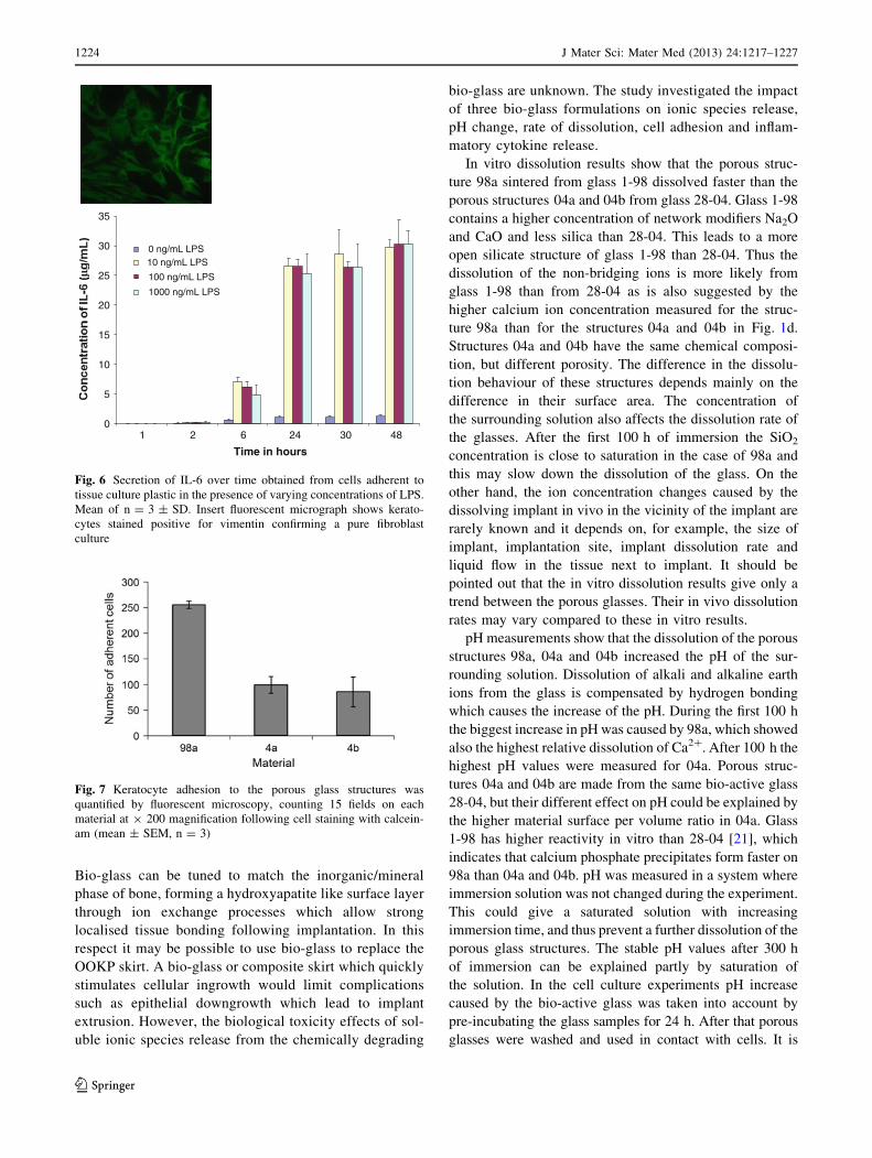
Fig. 7 Keratocyte adhesion to the porous glass structures was
quantified by fluorescent microscopy, counting 15 fields on each
material at 9 200 magnification following cell staining with calcein-
am (mean ± SEM, n = 3)
1224 J Mater Sci: Mater Med (2013) 24:1217–1227
123

known that cells are sensitive to drastic pH changes and in this
way the initial pH increase during cell contact is avoided.
It is known that in vivo chronic inflammation plays an
important role in OOKP skirt degradation. The acutely
inflamed tissue can create an acidic environment. Using bio-
active glass structures in such an environment can partly
counterbalance the pH changes, and thus have an antibac-
terial effect. Powdered bio-active glasses have been reported
to show antibacterial effect against several bacterium types,
mainly because the dissolving glasses effectively increase
the pH of the surrounding solution [28]. Figure 5 indicates
that the rise in pH produced by bio-glass dissolution levels
off below a pH of 7.8 indicating that no extreme rise in pH
will occur to the detriment of surrounding tissue.
3D porosity values were higher than corresponding 2D
values. The pores are not equally distributed in the struc-
tures and 3D analysis is likely to give a more accurate
estimation of the porosity. The porous structure 04a
showed the highest porosity followed by 04b and 98a,
where as 04b had the biggest average pore size. The
chemical composition of glass, particle size fraction and
sintering time and temperature control the final porosity of
the sintered glass structures. In the manufacturing of the
porous structures 04a and 04b there were differences in the
particle size fraction and the sintering time. Structure 04b
was sintered from bigger particles than 04a, thus explaining
the bigger average pore size in 04b. When sintering time is
increased the particles will grow closer together leading to
Surface layer
Internal
a b
c
d
Fig. 8 Calcein-am fluorescent images of live keratocyte adhesion to a 04a b 04b c 98a porous glass structures. d Confocal microscope z stack
image of DAPI stained keratocyte nuclei showing cells on the porous glass surface of glass 04b and cells within the internal pores
J Mater Sci: Mater Med (2013) 24:1217–1227 1225
123

a more compact structure, thus explaining the lower total
porosity of 04b compared to 04a. The sintering parameters
were the same for 98a and 04a, but the glass 1-98 used for
98a structure had a lower silica content and thus a lower
viscosity during sintering than glass 28-04 in 04a structure
leading to lower porosity of 98a than 04a when using the
same sintering parameters for both compositions.
Cell invasion deeper into the matrix depends on the pore
size of the glass structure. The initial cell adhesion results
indicate that cells moved deeper into the structures with
large pores. The biggest average pore size (135 lm) and
trabecular distance (around 300–450 lm) was measured in
04b. In the two other structures with smaller average pore
size, the cells were observed closer to the outer surfaces
during the initial cell adhesion period. Both the average pore
size and chemical composition of the glasses affected cell
adhesion. The cell culture time was relatively short because
in this paper only acute cell behaviour was investigated. The
effect of long term ion release caused by dissolution of the
glasses was not studied. It is known that Ca2?,, Na? and K?
are important cell signalling ions and concentration changes
of these ions in the tissue might change the cell behaviour
[29]. Cell adhesion was highest to the 98a bio-glass which
also showed greatest bioactivity and levels of Ca2? release.
These ionic species are thought to quicken formation of HA
surface coating followed by enhanced cell adhesion and
deposition of extracellular matrix. This may provide a partial
explanation for the greater numbers of keratocytes adhered
to the surface of the 98a bio-glass.
5 Conclusion
Three bio-glasses of varying bioactivity, 98a, 4a and 4b,
were investigated for use as an OOKP skirt substitute. 98a
showed the highest rate of calcium ion release, CaO and
SiO2 dissolution over 500 h. The pH level rose for up to
100 h and then stabilised. 98a also showed the highest
human keratocyte cell adhesion to the surface indicating
that a rise in local ionic species may influence rapid cell
adhesion. None of the porous bio-active glass structures
induced a cytokine driven inflammatory response and the
adherent keratocytes showed a typical elongated, spindle
shaped morphology suggestive of good adhesive potential.
This supports their use as synthetic OOKP skirt in this
respect. However, dissolution of the bio-glass over time
may destabilise the OOKP indicating that a composite
system using a stable backbone structure may be necessary
to maintain the PMMA optic following bio-glass chemical
degradation. Future in vivo studies will explore the sys-
temic effects of the bio-glasses and the impact of ion dis-
solution and pH change in the eye.
Acknowledgments Academy of Finland (Project no: 114117) is
acknowledged for financial support. Jessica Alm is thanked for advice
on cell culture on bio-active glasses.
References
1. Chirila TV, et al. Artificial cornea. Prog Polym Sci. 1998;23:
447–73.
2. Langefeld S, Kompa S, Redbrake C, Brenman K, Kirchhof B,
Schrage NF. Aachen keratoprosthesis as temporary implant for
combined vitreoretinal surgery and keratoplasty: report on 10
clinical applications. Graefe’s Arch Clin Exp Ophthalmol. 2000;
238:722–6.
3. Chirila TV. An overview of the development of artificial corneas
with porous skirts and the use of PHEMA for such an application.
Biomaterials. 2001;22:3311–7.
4. Kim MK, Lee JL, Wee WR, Lee JH. Seoul-type keratoprosthesis.
Arch Ophthalmol. 2002;120:761–6.
5. Bruin P, Meeuwsen EAJ, van Andel MV, Worst JGF, Pennings
AJ. Autoclavable highly cross-linked polyurethane networks in
ophthalmology. Biomaterials. 1993;14:1089–97.
6. Pruitt LA. Fluorocarbon polymers in biomedical engineering.
Encycl Mat Sci Tech. 2008;3216-21.
7. Renard G, Cetinel B, Legeais JM, Savoldelli M, Durand J,
Pouliquen Y. Incorporation of a fluorocarbon polymer implanted
at the posterior surface of the rabbit cornea. J Biomed Mat Res.
1996;31:193–9.
8. Caiazza S, Fanizza C, Mazziotti I, Pintucci S, Tomaino MA.
Light and scanning electron microscopy evaluation of the Dacron
felt as the haptic part of an improved keratoprosthesis. An in vitro
and in vivo study. Clin Mat. 1988;3:33–40.
9. Trinkaus-Randall V, Capecchi J, Sammon L, Gibbons D,
Leibowitz HM, Franzblau C. In vitro evaluation of fibroplasia in
a porous polymer. Invest Ophthalmol Vis Sci. 1990;31:1321–6.
10. Trinkaus-Randall V, Banwatt R, Capecchi J, Leibowitz HM,
Franzblau C. In vivo fibroplasia of a porous polymer in the
cornea. Invest Ophthalmol Vis Sci. 1991;32:3245–51.
Fig. 9 Production of cytokines a IL-6 and b IL-8 by keratocytes on
different porous glass structures and by keratocytes on control TC
plastic in response to LPS or no LPS
1226 J Mater Sci: Mater Med (2013) 24:1217–1227
123

11. Kain HL. The development of the silicone-carbon keratoprothe-
sis. Refract Corneal Surg. 1993;9:209–10.
12. Ciolino JB, Dohlman CH. Biologic keratoprosthesis materials. Int
Ophthalmol Clin. 2009;49(1):1–9.
13. Miyashita H, et al. Collagen-immobilized poly(vinyl alcohol) as
an artificial cornea scaffold that supports a stratified corneal
epithelium. J Biomed Mat Res. 2006;76B:56–63.
14. Xie RZ, Sweeney DF, Beumer GJ, Johnson G, Griesser HJ, Steele
JG. Effects of biologically modified surfaces of synthetic lenti-
cules on corneal epithelialization in vivo. Aust N Z J Ophthalmol.
1997;25(Suppl):46–9.
15. Kobayashi H, Ikada Y. Corneal cell adhesion and proliferation on
hydrogel sheets bound with cell-adhesive proteins. Curr Eye Res.
1991;10:899–908.
16. Pettit DK, Horbett TA, Hoffman AS, Chan KY. Quantitation of
rabbit corneal epithelial cell outgrowth on polymeric substrates
in vitro. Invest Ophthalmol Vis Sci. 1990;31:2269–77.
17. Ohji M, Mandarino L, SundarRaj N, Thoft RA. Corneal epithelial
cell attachment with endogenous laminin and fibronectin. Invest
Ophthalmol Vis Sci. 1993;34:2487–92.
18. Strampelli B. Osteo-odonto-keratoprosthesis. Ann Ottalmol Clin
Ocul. 1963;89:1039.
19. Ricci R, Pecorella I, Ciardi A, Della RC, Di Tondo U, Marchi V.
Strampelli’s osteo-odonto-keratoprosthesis. Clinical and histo-
logical long-term features of three prostheses. British J Oph-
thalmol. 1992;76:232–4.
20. Karlsson KH, Ylanen H, Aro H. Porous bone implants. Ceram
Int. 2000;26:897–900.
21. Zhang D, Vedel E, Hupa L, Aro HT. Predicting physical and
chemical properties of bio-active glasses from chemical
composition. Part III. In vitro reactivity of glasses. Glass Tech
Eur J Glass Sci Tech. 2009;50(1):1–8.
22. Hench LL, Andersson O. ‘‘Bio-active glasses’’ an introduction to
bioceramics. In: Hench LL, Wilson J, editors. Advanced series in
ceramics – vol. 1. Singapore: World Scientific; 1993.
23. Linnola RJ, Happonen RP, Andersson OH, Vedel E, Yli-Urpo
AU, Krause U, Laatikainen L. Titanium and bio-active glass–
ceramic coated titanium as materials for keratoprosthesis. Exp
Eye Res. 1996;63:471–8.
24. Arstila H, Vedel E, Hupa L, Hupa M. Predicting physical and
chemical properties of bio-active glasses from chemical compo-
sition. Part II. Devitrification characteristics. Glass Tech Eur J
Glass Sci Tech. 2008;49(6):251–9.
25. Arstila H, Vedel E, Hupa L, Hupa M. Predicting physical and
chemical properties of bio-active glasses from chemical compo-
sition. Part 2: Devitrification characteristics. Glass Tech Eur J
Glass Sci Tech. 2009;49(6):260–5.
26. Lentner C. Geigy scientific tables, vol. 1. Units of measurements,
Body fluid, Composition of body, and Nutrition. West Galdwell:
Giba-Geigy; 1981.
27. Viitala R, Franklin V, Green D, Liu C, Lloyd A, Tighe B.
Towards a synthetic osteo-odonto-keratoprosthesis. Acta Bio-
mater. 2009;5:438–52.
28. Zhang D, Lepparanta O, Munukka E, Ylanen H, Viljanen MK,
Eerola E, Hupa M, Hupa L. Antibacterial effects and dissolution
behavior of six bio-active glasses. J Biomed Mat Res. doi:
10.1002/jbm.a.32564.
29. Hoppe A, Guldal NS, Boccaccini AR. A review of the biological
response to ionic dissolution products from bio-active glasses and
glass–ceramics. Biomaterials. 2011;32:2757–74.
J Mater Sci: Mater Med (2013) 24:1217–1227 1227
123