evolution of root canal sealers: an insight story
TRANSCRIPT

|| 199 || | European Journal of General Dentistry | Vol 2 | Issue 3 | September-December 2013 |
ABSTRACTAttainment of ideal root canal treatment comprises various essential factors such as proper instrumentation, biomechanical preparation, obturation, and ultimately depending upon the case, post‑endodontic restoration. Main objective of the treatment is to get absolute rid of microbial entity and prevent any future predilection of re‑infection. In order to achieve that, proper seal is required to cut down any chance of proliferation of bacteria and future occurrence of any pathology. Although gutta‑percha has been the standard obturating material used in root canal treatment, it does not reinforce endodontically treated roots owing to its inability to achieve an impervious seal along the dentinal walls of the root canal. Gutta‑percha does not from a monoblock even with the use of a resin‑based sealer such as AH Plus because the sealer does not bind to gutta‑percha. As a result, a monoblock is formed (consisting of Resilon core material, Resin sealer, bonding agent/primer, and dentin). Another reason of Resilon being a better obturating material could be that the removal of smear layer by ethylenediaminetetraacetic acid (EDTA) after biomechanical preparation may have allowed the root canal filling material and root canal sealers to contact the canal wall and penetrate in the dentinal tubules, which may increase the strength of roots. New silicone‑based sealers like Roekoseal automix and the most recent GuttaFlow have some affirmative results regarding solubility and biocompatibility, as compared to other sealers. Methacrylate resin–based sealers and mineral trioxide aggregate (MTA)‑based sealers have opened a new horizon for sealers.
Key wordsBiocompatibility, contemporary sealers, cytotoxicity, leakage, monoblock
Sanjeev Tyagi, Priyesh Mishra, Parimala Tyagi1
Departments of Conservative Dentistry and Endodontics, 1Pedodontics and Preventive Dentistry, Peoples Dental Academy, Bhopal, Madhya Pradesh, India
Address for correspondence: Dr. Sanjeev Tyagi,
Department of Conservative Dentistry and Endodontics,
Peoples Dental Academy, Bhopal, Madhya Pradesh, India.
E-mail: [email protected]
INRODUCTION
Accomplishment of ideal root canal treatment is attributed to various essential factors such as proper instrumentation, biomechanical preparation, obturation, and ultimately depending upon the case post‑endodontic restoration. The pertinent aim of this treatment is to do away with the microbial entity and any future predilection of re‑infection. In order to achieve this, proper seal is required to denigrate any chance of proliferation of bacteria and future occurrence of any pathology. Sealer along with solid obturating material acts synergistically to create hermetic seal.[1,2]
The quality of the seal obtained with gutta‑percha (GP) and conventional zinc oxide eugenol (ZOE) sealers is
quite far from being perfect.[3,4] Also, despite its multiple strong points, GP and conventional sealer combination still has its own shortcomings, like its inability to strengthen root, as it does not adhere to dentin, inability to control microleakage, and the solubility of sealer makes prognosis dilemmatic and un‑assuring. Although few materials are capable enough to swap GP on multiple parameters, research continues to find alternatives that may seal better and mechanically reinforce compromised roots by forming monoblock, which has been suggested to reduce bacterial ingress pathways and strengthen the root to some extent.[5,6,10] Hence, several new resin cement sealants have been developed to be used instead of ZOE, thereby improving the root canal seal and imparting it more strength as compared to the conventional materials.[3,4] These include silicon‑based sealers which are well tolerated by tissues, have low water sorption, and have a potential of forming monoblock, thus reinforcing root canal,[7] epoxy resin–based sealers with the possibility of adhesion to dentin and with lower rates of water solubility,[7,8] and mineral trioxide aggregate (MTA)‑based sealers which have the predilection toward mineralization along with all the viable properties of orthodox sealers. Nevertheless, resin‑based and silicon‑based materials are also soluble, which may endanger a proper seal,
Evolution of root canal sealers: An insight story
rEvIEw ArTIClE
Access this article onlineQuick Response Code:
Website:www.ejgd.org
DOI:10.4103/2278-9626.115976
Published online: 2021-11-01

Tyagi, et al.: Evolution of root canal sealers
| European Journal of General Dentistry | Vol 2 | Issue 3 | September-December 2013 | || 200 ||
although the solubility of resin‑based materials is usually lesser than that of ZOE (which is reported as between 1% and 7%)[9] and does not exceed a maximum weight loss of 3% within 24 h of distilled water storage (in accordance with the standards for Root Canal treatment sealer (R Cl T).[7,8] Accordingly, availability of so many sealers makes it impossible for the clinician to decide what to avail and when. So, the purpose of the article is to create awareness about the different types of sealers and their pros and cons. Every manufacturer claims its product to be the ideal one, but only the clinical results can give the affirmation or negation of that particular sealer. Till date, none of the sealers has proved to be the ideal except a few which can come closer to being one. The objectives of this review are to delineate the behavior of contemporary sealers and juxtapose it with that of conventional sealers and their future clinical use based on all the parameters required for ascendancy.
ConvEnTIonAl rooT CAnAl SEAlEr
Early sealers were modified zinc oxide–eugenol (ZOE) cements based on Grossman or Rickerts’s formula that were widely used throughout the world. Unlike the resin‑based sealers, setting reaction of ZOE‑based sealers is a chelation reaction occurring between eugenol and the zinc ion of the zinc oxide. This reaction might also occur with the zinc oxide phase of GP along with the calcium ions of dentin. This might explain the decreased setting shrinkage associated with the ZOE‑based sealers.[11] Components are given in Table 1.1.
Michaud et al.[12] evaluated the three‑dimensional expansion of GP at various powder/liquid ratios of Pulp Canal Sealer extended working time (EWT) (ZOE‑based sealer) by using spiral (helical) computed tomography (SCT). They concluded that increasing the ratio of eugenol in sealer resulted in volumetric increase of GP [Figure 1].[13] It is cerebrated that the free eugenol component of freshly mixed ZOE sealer can seep out and cause various cytotoxic effects on human gingival fibroblasts, periodontal ligament (PDL) cells, and osteoblast‑like cells.[13,14] However, Haseih et al.[15] reported that leakage of eugenol into periapical tissues is very low, and it dramatically decreases over time.
Sealing properties of ZOE ZnOE sealers were inferior in comparison to other sealers due to the relatively high solubility of the ZOE sealer; so, adhesion between GP and ZOE is weak [Figure 2].[16] Eugenol is cytotoxic and the same has been shown frequently for ZOE with different cell culture systems, especially after mixing, but also in a set state. Even higher cytotoxicity was observed with formaldehyde‑containing ZOE sealers, which were classified as highly/extremely cytotoxic.[17] An ZOE sealer in the pulp chamber disinfected the dental tubules to a depth of 250 μm[18] and had a good antimicrobial property compared to other sealers.[19,20]
CONTEMPORARY SEALERS
• AH Plus• GuttaFlow• MTA‑based sealers• EndoSequence bioceramic sealer• Methacrylate‑based resin sealer
figure 1: Effects of altered powder/liquid ratios on volumetric change of gutta-percha at the end of 1-month interval. Control group (no sealer group) exhibited no visible expansion. Significant difference (P<0.05) between ZE 1:2 and ZE 1:3 groups when compared with ZE 1:1 and ZE 1:4 groups. SD, standard deviation (courtesy: Chandrasekhar et al. 2011)
figure 2: The adhesion between gutta-percha and zinc oxide eugenol is weak, and hence a gap remains (courtesy: Upadhyay et al. 2011)

Tyagi, et al.: Evolution of root canal sealers
|| 201 || | European Journal of General Dentistry | Vol 2 | Issue 3 | September-December 2013 |
Tabl
e 1:
Lis
t of t
he ro
ot c
anal
sea
lers
, the
ir c
ompo
siti
on, m
anuf
actu
rer,
adv
anta
ges
and
disa
dvan
tage
sr
oot c
anal
se
aler
sB
rand
Com
posi
tion
of
seal
ers
man
ufac
ture
rA
dvan
tage
sD
isad
vant
ages
1.1
Zinc
oxi
de
euge
nol
Roth
sea
ler
Kerr
PCS
Proc
osea
lEn
dom
etha
sone
Pow
der:
Zinc
oxi
de (
42%
)St
aybe
lite
resi
n (2
7%)
Bism
uth
subc
arbo
nate
(15
%)
Bariu
m s
ulfa
te (
15%
)So
dium
bor
ate,
an
hydr
ous
(1%
)Li
quid
:Eu
geno
l (4-
ally
l-2-
met
hoxy
phen
ol)
Roth
Inc.
, Chi
cago
, IL,
USA
Kerr
PCS
, silv
er K
err,
Rom
ulus
, M
I, U
SAPr
ocoS
ol, D
en-t
al-e
z, L
anca
ster
, PA
, USA
Sept
odon
t, S
aint
-Mau
r de
s Fo
sse'
s, F
ranc
e
1. L
owes
t sh
rinka
ge (
0.14
%)
com
pare
d to
res
in b
ased
sea
lers
.[16]
2. L
ong
last
ing
antim
icro
bial
pro
pert
y.
ZOE
seal
ers
have
dem
onst
rate
d an
timic
robi
al p
rope
rtie
s on
a
varie
ty o
f m
icro
orga
nism
s, in
clud
ing
Ente
roco
ccus
fae
calis
sus
pens
ions
and
an
aero
bic
bact
eria
eve
n 7
days
aft
er
mix
ing.
[20]
3. Z
OE-
base
d se
aler
s ar
e ea
sy t
o ha
ndle
.4.
The
rad
iopa
city
of
diff
eren
t ZO
E se
aler
s w
as 5
-7.9
7 m
m A
l and
thu
s ca
n be
reg
arde
d as
suffi
cien
t.[2
1]
5. P
owde
r/Liq
uid
ratio
of
1:3
caus
es
volu
met
ric e
xpan
sion
of
gutt
a pe
rcha
w
hich
fur
ther
sea
ls t
he c
anal
.[12]
6. D
imen
sion
al c
hang
es is
ver
y le
ss
0.41
9±0.
298
as c
ompa
red
to o
ther
se
aler
s.[1
6]
1. S
ever
al s
tudi
es s
how
ed a
pica
l lea
kage
ar
ound
ZO
E se
aler
s th
at in
crea
sed
with
st
orag
e tim
e (m
easu
red
up t
o 2
year
s) in
th
ick
laye
rs m
ore
than
in t
hin
laye
rs. S
ealin
g pr
oper
ties
of Z
OE
seal
ers
wer
e in
ferio
r in
co
mpa
rison
to
othe
r se
aler
s (r
esin
or
calc
ium
hy
drox
ide
seal
ers)
.[22]
2. F
orm
alde
hyde
, whi
ch is
rel
ease
d fr
om
cert
ain
ZOE
seal
ers,
is a
lso
a kn
own
alle
rgen
w
hich
wer
e cl
assi
fied
as h
ighl
y/ex
trem
ely
cyto
toxi
c. F
orm
alde
hyde
con
tain
ing
seal
ers
sugg
est
perm
anen
t da
mag
e of
the
ner
ve
in v
ivo.
[23]
3. E
ugen
ol in
hibi
ted
nerv
e co
nduc
tanc
e in
vitr
o in
exp
erim
ents
with
diff
eren
t ne
rve
tissu
es.[2
4]
4. H
ighe
st s
olub
ility
as
com
pare
d to
oth
er
cont
empo
rary
sea
ler
thus
mak
ing
mor
e pr
one
to c
ause
mic
role
akag
e 2,
426±
0,73
3 th
ough
w
ithin
the
with
in t
he li
mits
of
ISO
sta
ndar
ds
(wei
ght
loss
-3%
of
mas
s).[1
7]
1.2
Epox
y re
sin
base
d se
aler
AH
Plu
sA
H26
TopS
eal
2-Se
al
For A
HPl
us E
poxi
de
past
eD
iepo
xide
Calc
ium
tun
gsta
teZi
rcon
ium
oxi
deA
eros
ilPi
gmen
tA
min
e pa
ste
1-ad
aman
tane
am
ine
N, N
’-dib
enzy
l-5-
oxa-
nona
ndia
min
e-1,
9TC
D-D
iam
ine
Calc
ium
tun
gsta
teZi
rcon
ium
oxi
deA
eros
ilSi
licon
e oi
lFo
r AH
26A
H 2
6, p
owde
r:
Bism
uth
oxid
e,
Met
hena
min
e, S
ilver
, Ti
tani
um d
ioxi
de
Den
tspl
y M
aille
fer,
Balla
igue
s,
Switz
erla
ndD
ents
ply
Mai
llefe
r, Ba
llaig
ues,
Sw
itzer
land
]V
DW
, End
odon
tic S
yner
gy,
Mun
chen
, Ger
man
y
1. R
adio
paci
ty-1
3. 6
mm
of A
l of A
H
Plus
, and
AH
-26
has
9.3
mm
of A
l.[25]
2. D
imen
sion
al s
tabi
lity-
poly
mer
isat
ion
shrin
kage
of A
H P
lus
is 1
.76
V%
and
A
H-2
6 is
1.4
6 V
%.[2
5]
3. S
olub
ility
is v
ery
less
for
AH
Plu
s bu
t fo
r AH
-26
it is
mor
e th
an R
oeko
seal
an
d A
H P
lus.
[25]
4. T
he li
near
exp
ansi
on o
f AH
Plu
s is
ve
ry lo
w (
0.12
9±0.
08)
very
less
tha
n ot
her
seal
er.[2
5]
5. A
H-2
6 an
d A
H P
lus
is a
ble
to
flow
into
the
orifi
ces
of t
he d
entin
al
tubu
les,
whi
ch is
the
rea
son
for
the
com
para
tivel
y go
od a
dhes
ion
of A
H-2
6 to
den
tin.[2
5]
6. H
andl
ing
prop
ertie
s ar
e us
ually
co
nsid
ered
to
be g
ood.
7. R
elea
se o
f fo
rmal
dehy
de-O
nly
a m
inim
um r
elea
se w
as o
bser
ved
for A
H
Plus
(3.
9 pp
m).[2
9]
8. A
H P
lus
prod
uced
slig
ht in
hibi
tion
on S
trep
toco
ccus
mut
ants
at
20 d
ays
and
on A
ctin
omyc
es is
rael
ii at
eve
ry
time
inte
rval
.[28]
1. A
H-2
6 ha
s ha
rmfu
l am
ount
of
form
alde
hyde
re
leas
e is
134
7 pp
m.[2
9]
2. R
ever
sibl
e ac
ute
infla
mm
atio
n of
the
ora
l m
ucos
a af
ter
cont
act
with
the
uns
et p
aste
. In
indi
vidu
al c
ases
, loc
al a
nd s
yste
mic
alle
rgic
re
actio
ns h
ave
been
rep
orte
d.[2
5]
3. B
isph
enol
A d
igly
cidy
l eth
er w
as id
entifi
ed
as a
mut
agen
ic c
ompo
nent
of
resi
n-ba
sed
mat
eria
ls, w
hich
may
als
o be
cyt
otox
ic.[2
5]
4. E
poxy
res
in-b
ased
sea
lers
adh
ere
bett
er t
o th
e de
ntin
wal
ls, m
akin
g th
eir
rem
oval
with
ro
tary
inst
rum
ents
diffi
cult.
[36]
5. L
ess
frac
ture
res
ista
nce
whe
n us
ed
with
gut
ta p
erch
a as
com
pare
d to
Re
silo
n/Re
alse
al.[3
7]
6. W
ith t
he e
poxy
-bas
ed s
eale
r ei
ther
no
diff
eren
ce (
shea
r) o
r lo
wer
bon
d st
reng
th in
th
in fi
lms
was
fou
nd, a
nd a
ppea
red
to r
esul
t fr
om n
umer
ous
void
s cr
eate
d du
ring
mix
ing.
[38]
Cont
d...
Tyagi, et al.: Evolution of root canal sealers
| European Journal of General Dentistry | Vol 2 | Issue 3 | September-December 2013 | || 202 ||
Tabl
e 1:
Con
tinu
edr
oot c
anal
se
aler
sB
rand
Com
posi
tion
of
seal
ers
man
ufac
ture
rA
dvan
tage
sD
isad
vant
ages
AH
26
silv
erfr
ee,
pow
der:
Bis
mut
h ox
ide,
Met
hena
min
eA
H 2
6 re
sin:
Epo
xy
resi
n
9. T
issu
e co
mpa
tibili
ty-n
o ge
noto
xici
ty
and
mut
agen
icity
wer
e re
veal
ed b
y A
H
Plus
.[25]
10.
Rem
ovab
ility
-If A
H P
lus
is u
sed
in
com
bina
tion
with
gut
ta-p
erch
a po
ints
, th
e ro
ot c
anal
filli
ngs
can
be r
emov
ed
usin
g co
nven
tiona
l tec
hniq
ues
for
the
rem
oval
of
gutt
a-pe
rcha
.[36]
11.
2-se
al h
as lo
wes
t so
lubi
lity
follo
wed
by
Tops
eal a
nd A
H26
has
m
axim
um s
olub
ility
.[33]
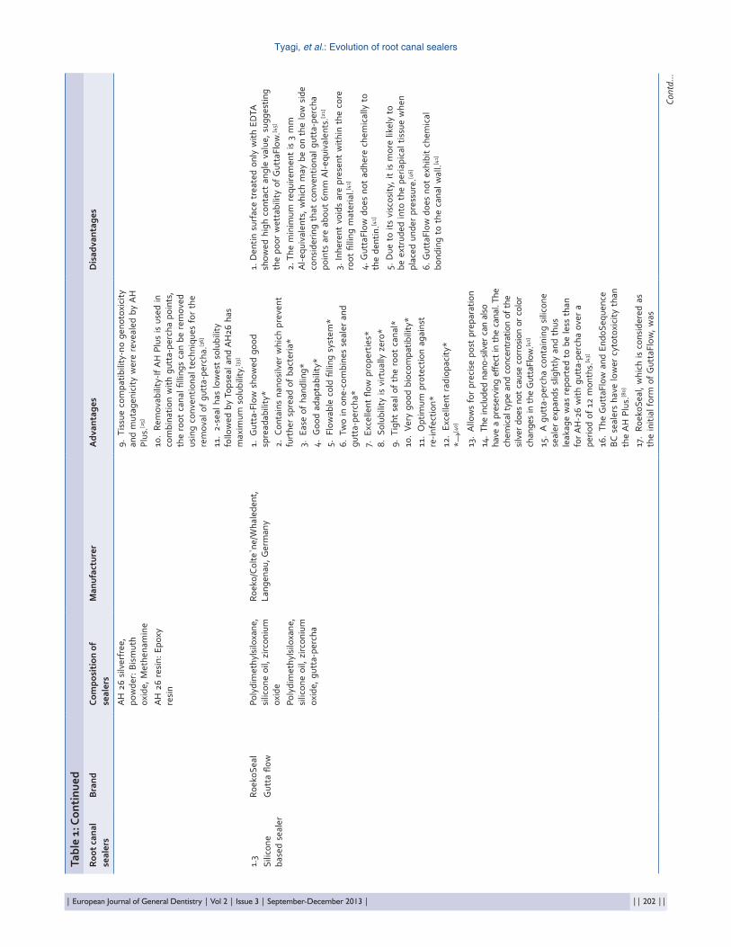
1.3
Silic
one
base
d se
aler
Roek
oSea
lG
utta
flow
Poly
dim
ethy
lsilo
xane
, si
licon
e oi
l, zi
rcon
ium
ox
ide
Poly
dim
ethy
lsilo
xane
, si
licon
e oi
l, zi
rcon
ium
ox
ide,
gut
ta-p
erch
a
Roek
o/Co
lte`n
e/W
hale
dent
, La
ngen
au, G
erm
any
1. G
utta
-Flo
w s
how
ed g
ood
spre
adab
ility
*2.
Con
tain
s na
nosi
lver
whi
ch p
reve
nt
furt
her
spre
ad o
f ba
cter
ia*
3. E
ase
of h
andl
ing*
4. G
ood
adap
tabi
lity*
5. F
low
able
col
d fil
ling
syst
em*
6. T
wo
in o
ne-c
ombi
nes
seal
er a
nd
gutt
a-pe
rcha
*7.
Exc
elle
nt fl
ow p
rope
rtie
s*8.
Sol
ubili
ty is
virt
ually
zer
o*9.
Tig
ht s
eal o
f th
e ro
ot c
anal
*10
. Ve
ry g
ood
bioc
ompa
tibili
ty*
11.
Opt
imum
pro
tect
ion
agai
nst
re-in
fect
ion*
12.
Exce
llent
rad
iopa
city
* *→
[40]
13.
Allo
ws
for
prec
ise
post
pre
para
tion
14.
The
incl
uded
nan
o-si
lver
can
als
o ha
ve a
pre
serv
ing
effec
t in
the
can
al. T
he
chem
ical
typ
e an
d co
ncen
trat
ion
of t
he
silv
er d
oes
not
caus
e co
rros
ion
or c
olor
ch
ange
s in
the
Gut
taFl
ow.[4
1]
15.
A g
utta
-per
cha
cont
aini
ng s
ilico
ne
seal
er e
xpan
ds s
light
ly a
nd t
hus
leak
age
was
rep
orte
d to
be
less
tha
n fo
r AH
-26
with
gut
ta-p
erch
a ov
er a
pe
riod
of 1
2 m
onth
s.[4
3]
16.
The
Gut
taFl
ow a
nd E
ndoS
eque
nce
BC s
eale
rs h
ave
low
er c
ytot
oxic
ity t
han
the
AH
Plu
s.[8
0]
17.
Roek
oSea
l, w
hich
is c
onsi
dere
d as
th
e in
itial
for
m o
f Gut
taFl
ow, w
as
1. D
entin
sur
face
tre
ated
onl
y w
ith E
DTA
sh
owed
hig
h co
ntac
t an
gle
valu
e, s
ugge
stin
g th
e po
or w
etta
bilit
y of
Gut
taFl
ow.[4
5]
2. T
he m
inim
um r
equi
rem
ent
is 3
mm
A
l-equ
ival
ents
, whi
ch m
ay b
e on
the
low
sid
e co
nsid
erin
g th
at c
onve
ntio
nal g
utta
-per
cha
poin
ts a
re a
bout
6m
m A
l-equ
ival
ents
.[21]
3. In
here
nt v
oids
are
pre
sent
with
in t
he c
ore
root
filli
ng m
ater
ial.[4
1]
4. G
utta
Flow
doe
s no
t ad
here
che
mic
ally
to
the
dent
in.[4
1]
5. D
ue t
o its
vis
cosi
ty, i
t is
mor
e lik
ely
to
be e
xtru
ded
into
the
per
iapi
cal t
issu
e w
hen
plac
ed u
nder
pre
ssur
e.[4
6]
6. G
utta
Flow
doe
s no
t ex
hibi
t ch
emic
al
bond
ing
to t
he c
anal
wal
l.[41]
Cont
d...

Tyagi, et al.: Evolution of root canal sealers
|| 203 || | European Journal of General Dentistry | Vol 2 | Issue 3 | September-December 2013 |
Tabl
e 1:
Con
tinu
edr
oot c
anal
se
aler
sB
rand
Com
posi
tion
of
seal
ers
man
ufac
ture
rA
dvan
tage
sD
isad
vant
ages
rem
oved
mor
e ea
sily
fro
m t
he c
anal
s th
an a
res
in-b
ased
sea
ler.[4
5]
1.4
MTA
bas
ed
seal
er
Endo
-CPM
-Sea
ler,
MTA
Obt
ura
ProR
oot
Endo
Se
aler
MTA
filla
pex
EGEO
srl,
Bue
nos
Aire
s, A
rgen
tina
Ang
elus
, Lon
drin
a PR
, Bra
zil
Den
tspl
y M
aille
fer,
Balla
igue
s,
Switz
erla
ndM
TA-F
; Ang
elus
, Lo
ndrin
a, B
razi
l
50%
MTA
(SiO
2, K
2O, A
l2O
3,
SO3,
CaO
and
Bi2
O3-
50%
SiO
2-7%
,Ca
CO3-
10%
,Bi
2O3-
10%
,Ba
SO4-
10%
,pr
opyl
ene
glyc
ol a
lgin
ate-
1%,
prop
ylen
e gl
ycol
-1%
, sod
ium
ci
trat
e-1%
cal
cium
chl
orid
e-10
%
1. H
ighl
y bi
ocom
patib
le.*
2. S
timul
ate
min
eral
izat
ion.
*3.
Enc
oura
ge a
patit
e-lik
e cr
ysta
lline
de
posi
ts a
long
the
api
cal a
nd m
iddl
e th
irds
of c
anal
wal
ls.*
*→
[50]
4. T
hese
mat
eria
ls e
xhib
ited
high
er
push
-out
str
engt
hs a
fter
sto
rage
.[57]
5. F
luor
ide-
dope
d M
TA d
emon
stra
ted
stab
le s
ealin
g up
to
6 m
onth
s an
d si
gnifi
cant
ly b
ette
r th
an c
onve
ntio
nal
MTA
sea
lers
.[58]
6. I
t ha
s an
ade
quat
e ca
lciu
m r
elea
sing
pr
oper
ty7.
End
o- C
PM w
as a
lso
repo
rted
to
have
a s
imila
r or
bet
ter
seal
ing
abili
ty
to r
esin
-bas
ed s
eale
rs.[5
9]
8. P
roRo
ot E
ndo
Seal
er d
emon
stra
ted
the
supe
rior
seal
ing
abili
ty o
f th
is
mat
eria
l com
para
ble
to r
esin
-bas
ed
seal
ers.
[59]
9. A
fter
set
ting,
the
cyt
otox
icity
of
MTA
-F d
ecre
ases
and
the
sea
ler
pres
ents
sui
tabl
e bi
oact
ivity
to
stim
ulat
e H
ydro
xyap
atite
cry
stal
nu
clea
tion.
[60]
10.
MTA
Fill
apex
yie
lds
an
impr
essi
ve, h
erm
etic
sea
l in
whi
ch t
he M
TA p
artic
les
expa
nd,
prev
entin
g m
icro
infil
trat
ion.
And
, M
TA s
imul
tane
ousl
y re
leas
es f
ree
calc
ium
ions
[Ca2+
] to
acc
eler
ate
the
heal
ing
proc
ess
by s
timul
atin
g th
e re
gene
ratio
n of
the
adj
acen
t tis
sues
.[64]
11.
Endo
-CPM
sea
ler
show
ed t
he
high
est
valu
es o
f bo
nd s
tren
gth
to r
oot
dent
in (
8.26
5 M
Pa)
(P<.
05).
The
valu
es
of p
ush-
out
test
wer
e si
mila
r fo
r M
TA
Filla
pex
(2.0
41 M
Pa)
and
AH
Plu
s (3
.034
M
Pa.[6
1]
1. D
o no
t bi
nd t
o de
ntin
and
cor
e m
ater
ial
2. M
TA F
illap
ex h
ad t
he lo
wes
t pu
sh-o
ut b
ond
valu
es t
o ro
ot d
entin
e co
mpa
red
with
oth
er
seal
ers.
[61]
3. M
TA F
illap
ex®
set
ting
time,
whi
ch h
as
resi
n in
its
com
posi
tion
cons
eque
ntly
re
duci
ng t
he m
ediu
m a
lkal
inis
atio
n he
nce
less
m
iner
alis
atio
n th
en o
ther
MTA
sea
lers
.[71]
4. T
he a
lkal
inity
of
MTA
can
the
oret
ical
ly
wea
ken
root
den
tin s
imila
r to
the
find
ings
on
calc
ium
hyd
roxi
de.[6
9]
5. In
cas
es o
f M
TA-b
ased
mat
eria
ls e
xtru
sion
ou
tsid
e th
e ro
ot c
anal
is a
ssoc
iate
d w
ith
seve
re p
ain
felt
by t
he p
atie
nt.[8
2]
Cont
d...

Tyagi, et al.: Evolution of root canal sealers
| European Journal of General Dentistry | Vol 2 | Issue 3 | September-December 2013 | || 204 ||
Tabl
e 1:
Con
tinu
edr
oot c
anal
se
aler
sB
rand
Com
posi
tion
of
seal
ers
man
ufac
ture
rA
dvan
tage
sD
isad
vant
ages
1.5
Calc
ium
- Si
licat
e-Ph
osph
ate-
base
d bi
ocer
amic
Seal
er
Endo
sequ
ence
/iro
ot S
PIro
ot B
PBi
o ag
greg
ate
Bras
sele
r U
SA,
Sava
nnah
, GA
;In
nova
tive
BioC
eram
ix In
c.,
Van
couv
er, B
C,
Cana
da)
Inno
vativ
e Bi
oCer
amix
Inc.
Tric
alci
um s
ilica
te, d
ical
cium
si
licat
e, c
alci
um p
hosp
hate
s,
collo
idal
sili
ca, a
nd c
alci
um
hydr
oxid
e zi
rcon
ium
oxi
de a
s th
e ra
diop
acifi
er
1. B
ioco
mpa
tible
and
do
not
indu
ce
criti
cal c
ytot
oxic
eff
ects
.[81]
2. F
orm
atio
n of
a n
ano-
com
posi
te
netw
ork
of g
el-li
ke c
alci
um
silic
ate
hydr
ate
intim
atel
y m
ixed
with
hy
drox
yapa
tite,
bio
cera
mic
, and
for
ms
a he
rmet
ic s
eal w
hen
appl
ied
insi
de
the
root
can
al.*
3. P
reci
pita
tes
calc
ium
pho
spha
te
on h
ydra
tion
with
sam
e st
reng
th a
s hu
man
bon
e
1. C
hang
es in
env
ironm
enta
l wat
er c
onte
nt
adve
rsel
y aff
ect
the
sett
ing
time
and
mic
roha
rdne
ss o
f En
doSe
quen
ceBC
Sea
ler.[8
1]
2. C
onve
ntio
nal r
etre
atm
ent
tech
niqu
es a
re
not
able
to
fully
rem
ove
Bioc
eram
ic s
eale
r.[83]
4. iR
oot
BP is
non
-mut
agen
ic, d
oes
not
caus
e an
alle
rgen
ic p
oten
tial a
fter
m
ultip
le u
ses
and
has
a go
od t
oler
ance
by
sub
cuta
neou
s tis
sue*
5. H
igh
alka
linity
incr
ease
s its
m
iner
alis
atio
n pr
oces
s al
so it
s ba
cter
icid
al p
rope
rtie
s (p
H 1
2.8)
*6.
Hyd
roph
ilic,
roo
t ca
nal h
ydra
tion
aids
in t
he f
orm
atio
n of
cal
cium
ph
osph
ate
henc
e gi
ves
stre
ngth
*7.
Low
con
tact
ang
le h
ence
the
se
feat
ures
allo
w t
hem
to
spre
ad e
asily
ov
er t
he d
entin
wal
ls o
f th
e ro
ot c
anal
an
d to
get
insi
de a
nd fi
ll th
e la
tera
l m
icro
can
als*
8. T
hese
new
bio
cera
mic
sea
lers
als
o fo
rm c
hem
ical
bon
d w
ith t
he c
anal
’s de
ntin
wal
ls. T
hat
is w
hy n
o sp
ace
is le
ft
betw
een
the
seal
er a
nd d
entin
wal
ls.*
9. T
hey
are
also
oss
eo-c
ondu
ctiv
e10
. Ver
y go
od r
adio
paci
ty (
3.8
mm
of
Al).
[78]
11. S
ettin
g tim
e is
3-4
hrs
hen
ce
it gi
ves
ampl
e am
ount
of
time
for
plac
emen
t of
roo
t ca
nal.*
12. B
ioce
ram
ics
do n
ot s
hrin
k up
on
sett
ing.
In f
act,
the
y ac
tual
ly e
xpan
d sl
ight
ly u
pon
com
plet
ion
of t
he s
ettin
g pr
oces
s.*
13. F
urth
erm
ore
(and
thi
s is
ver
y im
port
ant
in e
ndod
ontic
s), b
ioce
ram
ics
will
not
res
ult
in a
sig
nific
ant
infla
mm
ator
y re
spon
se if
an
over
fill
occu
rs d
urin
g th
e ob
tura
tion
proc
ess.
[82]
Cont
d...

Tyagi, et al.: Evolution of root canal sealers
|| 205 || | European Journal of General Dentistry | Vol 2 | Issue 3 | September-December 2013 |
Tabl
e 1:
Con
tinu
edr
oot c
anal
se
aler
sB
rand
Com
posi
tion
of
seal
ers
man
ufac
ture
rA
dvan
tage
sD
isad
vant
ages
14. R
emar
kabl
e flo
wab
ility
of
the
BC
Seal
er. T
his
is a
res
ult
of it
s pa
rtic
le
size
and
hyd
roph
ilici
ty. (
27 m
m).[8
2]
15. B
ioce
ram
ic s
eale
r ha
s m
ore
frac
ture
re
sist
ance
the
n co
nven
tiona
l sea
ler.[8
2]
16. W
hen
bioc
eram
ic-b
ased
sea
lers
Bi
oAgg
rega
te o
r iR
oot
SP a
re
extr
uded
, the
pai
n is
rel
ativ
ely
smal
l or
tota
lly a
bsen
t.*
*-[8
5]
1.6
Met
hacr
ylat
e re
sin
base
d se
aler
Hyd
ron-
Firs
t ge
nera
tion
Endo
REZ-
Seco
nd
gene
ratio
nRe
alse
alEp
ipha
ny-T
hird
ge
nera
tion
Fibr
efill‑
Third
ge
nera
tion
Real
seal
SE
Met
asea
l SE
- Fo
urth
ge
nera
tion
Smar
tsea
l
Hyd
ron
tech
nolo
gies
, In
c, P
ompa
no B
each
, FL U
ltrad
ent
prod
uct
inc,
Sou
th J
orda
n U
TSy
bron
Endo
, Ora
nge,
CA
/Pen
tron
Clin
ical
tec
hnol
ogie
sPe
ntro
n cl
inic
al
tech
nolo
gies
,W
allin
gfor
d, C
TSy
bron
Endo
, Ora
nge,
CA Pa
rkel
l inc
Smar
t se
al (
DRF
P Lt
d, S
tam
ford
, U
nite
d K
ingd
om),
bisp
heno
l-A-g
lyci
dyld
imet
hacr
ylat
e (B
isG
MA
),Et
hoxy
late
d Bi
sGM
A,
uret
hane
dim
etha
cryl
ate
(UD
MA
) an
d H
ydro
phili
c di
func
tiona
l m
etha
cryl
ates
.Ca
lciu
m h
ydro
xide
, bar
ium
su
lfate
, bar
ium
gla
ss, a
nd s
ilica
.Th
e pr
imer
-a s
elf-
etch
prim
er t
hat
cont
ains
sul
foni
c ac
id-t
erm
inat
ed
func
tiona
l mon
omer
,H
ydro
xyet
hylm
etha
cryl
ate
(HEM
A),
wat
er a
nd a
po
lym
eriz
atio
n in
itiat
or.
Smar
tpoi
nt, a
rad
iopa
que
non-
gu
tta-
perc
ha c
ore
with
a
radi
oluc
ent
hydr
ophi
lic
poly
mer
coa
ting
(cop
olym
er o
f vi
nylp
yrro
lidon
e an
d ac
rylo
nitr
ile,
met
hyl m
etha
cryl
ate,
or
HEM
A)
and
Smar
t pa
ste,
a r
adio
luce
nt
seal
er t
hat
cont
ains
an
activ
e po
lym
er
1. W
hen
used
with
res
ilon
form
s ‘M
onob
lock
’ whi
ch f
urth
er im
prov
es
the
seal
.[68]
2. R
eals
eal h
as g
reat
er r
oot
frac
ture
re
sist
ance
com
pare
d to
AH
Plu
s.[3
7]
3. G
ood
radi
opac
ity b
ut le
ss t
hen
AH
Pl
us.[2
5]
4. S
low
pol
ymer
izat
ion
of t
he
dual
-cur
able
sea
lers
wou
ld im
prov
e th
e ch
ance
for
the
rel
ief
of s
hrin
kage
str
ess
via
resi
n flo
w.[1
04]
5. T
hey
show
ed t
hat
root
s fil
led
with
Re
silo
n/Ep
ipha
ny e
xhib
ited
sign
ifica
ntly
hi
gher
fra
ctur
e lo
ad v
alue
s th
an t
hose
fil
led
with
gut
ta‑p
erch
a/A
H‑2
6 w
hen
the
spec
imen
s w
ere
subj
ecte
d to
ve
rtic
al lo
adin
g fo
rces
.[100
]
6. E
ndoR
EZ w
as f
ound
to
be
wel
l-tol
erat
ed b
y co
nnec
tive
tissu
es
and
bone
tis
sue.
[103
,109
]
7. M
etha
cryl
ate
resi
n–ba
sed
seal
ers
used
with
Res
ilon
or g
utta
-per
cha
wer
e m
ore
effec
tivel
y re
mov
ed, w
ith
less
rem
nant
filli
ng m
ater
ial t
han
conv
entio
nal s
eale
r/gu
tta-
perc
ha
com
bina
tions
.[105
]
8. S
mar
tpoi
nt e
xpan
ds o
nly
late
rally
on
abs
orbi
ng w
ater
fro
m t
he t
ooth
, ad
optin
g th
e ca
nal s
hape
. Sm
art
past
e al
so e
xpan
ds o
n hy
drat
ion
to f
orm
a
perf
ect
seal
[106
]
9. F
ibre
Fill
R.C.
S. is
rep
orte
d to
hav
e go
od s
ealin
g an
d ad
hesi
ve p
rope
rtie
s to
rad
icul
ar d
entin
.[107
]
1. E
piph
any
and
met
asea
l is
cyto
toxi
c ev
en
afte
r di
lutio
ns.[1
01]
2. R
esilo
n/Ep
ipha
ny (
Real
Seal
)‑fil
led
cana
ls
also
con
tain
ed s
igni
fican
tly m
ore
void
s an
d ga
ps t
han
thos
e fil
led
with
gut
ta‑p
erch
a an
d co
nven
tiona
l sea
lers
.[97]
3. L
ower
pus
h-ou
t st
reng
ths
than
gut
ta
perc
ha/c
onve
ntio
nal n
onbo
ndin
g se
aler
co
mbi
natio
ns.[1
30]
4. G
reat
er C
- fa
ctor
cau
sing
mor
e po
lym
eris
atio
n sh
rinka
ge h
ence
mor
e ga
p fo
rmat
ion
and
mic
role
akag
e.[9
8]
5. T
he c
hem
ical
cou
plin
g be
twee
n co
ntem
pora
ry m
etha
cryl
ate
resi
n–ba
sed
seal
ers
to r
oot
fillin
g m
ater
ials
is g
ener
ally
w
eak
or in
suffi
cien
tly o
ptim
ized
.[98]
6. C
reep
ing
of in
com
plet
ely
poly
mer
ized
resi
nous
sea
lers
, whi
ch r
esul
ts in
fai
lure
alo
ng
the
seal
er-d
entin
inte
rfac
e.[1
26]
7. P
rese
nce
of r
esid
ual m
onom
ers
in t
he r
oot
cana
ls.[1
27]
8. E
piph
any
in b
oth
fres
hly
mix
ed a
nd s
et
cond
ition
s sh
owed
a s
ever
e to
mod
erat
e cy
toto
xic
effec
t, a
nd it
s cy
toto
xici
ty a
ctua
lly
incr
ease
d w
ith t
ime,
pos
ing
sign
ifica
nt
cyto
toxi
c ris
ks.[3
9,13
1]
9. E
piph
any
is in
solu
ble
in t
he s
olve
nts
com
mon
ly u
sed
in d
entis
try.
Thu
s, r
emov
al o
f re
sin
seal
ers
from
fins
, acc
esso
ry c
anal
s, o
r ca
nal i
sthm
i rem
ains
a c
halle
nge.
[91]
10. S
olub
ility
val
ues
for
Epip
hany
and
AH
Plu
s w
ere
3.41
% b
ut a
ccor
ding
to
AD
A it
sho
uld
be
less
tha
n 3%
.[25]
Cont
d...

Tyagi, et al.: Evolution of root canal sealers
| European Journal of General Dentistry | Vol 2 | Issue 3 | September-December 2013 | || 206 ||
Tabl
e 1:
Con
tinu
edr
oot c
anal
se
aler
sB
rand
Com
posi
tion
of
seal
ers
man
ufac
ture
rA
dvan
tage
sD
isad
vant
ages
10. F
or m
etha
cryl
ate
resi
n-ba
sed
seal
ers,
thi
n fil
ms
had
high
er b
ond
stre
ngth
tha
n th
ick.
[38]
11. T
he c
hem
ical
uni
on b
etw
een
the
poly
isop
rene
com
pone
nt o
f th
e gu
tta-
perc
ha a
nd t
he p
olyb
utad
iene
en
d of
the
End
oRez
res
in c
oatin
g
11. U
nrea
cted
mon
omer
s, le
acha
ble
mon
omer
s fr
om t
he in
com
plet
ely
poly
mer
ized
Sm
art
past
e se
aler
can
leak
thr
ough
the
api
cal
fora
men
aft
er w
ater
sor
ptio
n an
d sw
ellin
g an
d ca
use
inad
vert
ent
harm
ful d
etrim
enta
l eff
ects
on
the
per
iodo
ntal
tis
sues
.[132
]
12. D
iffus
ion
of w
ater
into
res
in m
atric
es
mig
ht r
esul
t in
the
rap
id d
eter
iora
tion
of t
he
phys
ical
/mec
hani
cal p
rope
rtie
s of
a r
esin
, co
mpr
omis
ing
the
dura
bilit
y of
res
in-d
entin
bo
nds
by h
ydro
lysi
s an
d m
icro
crac
k fo
rmat
ion.
[133
]
mol
ecul
e ap
pear
s to
be
stro
nger
tha
n th
e co
uplin
g be
twee
n th
e m
etha
cryl
ate
end
of t
he m
olec
ule
to t
he r
esin
se
aler
.[93]
12. E
ndoR
ez s
how
ed in
crea
sed
intr
atub
ular
pen
etra
tion
com
pare
d to
A
HPl
us a
nd E
ndo
CPM
- se
aler
.[63]
13. D
ecre
ased
den
tin t
hick
ness
, lac
k of
po
lym
eriz
atio
n, o
r ex
tend
ed e
xpos
ure
times
m
ight
incr
ease
the
ris
k of
cyt
otox
ixity
of
HEM
A s
igni
fican
tly[1
34]
14. E
ndoR
EZ w
ith a
gut
ta-p
erch
a po
int
into
a
drie
d ro
ot c
anal
pro
duce
s po
or a
dapt
atio
n of
th
e se
aler
to
dent
in w
ith a
lack
of
resi
n ta
g fo
rmat
ion.
[92]
15. R
eals
eal h
as t
he p
oten
tial t
o ca
use
toot
h st
aini
ng a
s it
is s
usce
ptib
le t
o en
zym
atic
and
al
kalin
e hy
drol
ysis
.[99]
16. M
ETA
seal
is f
ound
to
be m
ost
cyto
toxi
c
whe
n co
mpa
red
with
AH
Plu
s, E
piph
any
and
Endo
REZ.
[108
]
1.7
Calc
ium
ph
osph
ate
seal
er
Caps
eal I
Caps
eal I
ISa
nkin
Ape
tite
Seal
er; S
anki
n Ko
gyo,
Tok
yo, J
apan
Pow
der:
Tric
alci
um p
hosp
hate
Dic
alci
um d
ihyd
rate
Port
land
cem
ent
Zirc
oniu
m o
xide
Liqu
id:
Sodi
um p
hosp
hate
sol
utio
n
1. C
APS
EAL
I and
II s
how
less
cy
toto
xici
ty a
nd in
flam
mat
ory
med
iato
rs c
ompa
red
with
oth
er s
eale
rs
and
have
the
pot
entia
l to
prom
ote
bone
reg
ener
atio
n as
roo
t ca
nal
seal
ers.
[135
]
2. C
APS
EAL
I and
II f
acili
tate
the
pe
riapi
cal d
ento
alve
olar
and
alv
eola
r he
alin
g by
con
trol
ling
cellu
lar
med
iato
rs f
rom
PD
L ce
lls a
nd
oste
obla
st d
iffer
entia
tion
of p
recu
rsor
ce
lls.[1
36]
3. C
APS
EAL
I and
II s
eale
rs w
ere
wel
l-ada
pted
to
the
cana
l wal
l and
in
filtr
ated
into
the
den
tinal
tub
ules
.[139
]
1. F
ract
ure
resi
stan
ce is
yet
to
eval
uate
.[137
]
2. C
PS-1
sea
ler
is n
ot b
ioco
mpa
tible
.[138
]

Tyagi, et al.: Evolution of root canal sealers
|| 207 || | European Journal of General Dentistry | Vol 2 | Issue 3 | September-December 2013 |
• Calcium phosphate–based sealer• Calcium‑enriched mixture (CEM).
AH PLUS
AH Plus consists of a paste–paste system, delivered in two tubes in a new double barrel syringe. The components of AH Plus are given in Table 1.2. The epoxide paste contains radiopaque fillers and aerosil. The amine paste consists of three different types of amines, radiopaque fillers, and aerosil.[25]
AH Plus has shown positive results when compared to other sealers [Figures 3‑6].[25] It showed significantly lowest weight loss among the different root canal sealers in water and in artificial saliva with different pH values, independent of the solubility medium used. Furthermore, AH Plus showed the greatest stability in solution, as compared to the conventional sealers.[26]
AH Plus has a film thickness of 26 mm, which is clearly below the value of less than 50 mm required by the ISO standard for root canal sealing materials.[25] AH Plus has been designed to be slightly thixotropic. A flow of 36 mm also perfectly meets the requirements of the ISO standard (>25 mm).
It is known from the literature that pure epoxy resins develop mutagenic activities under the conditions of the Ames test. Therefore, the epoxide paste (paste A) and amine paste (paste B) were studied in the Ames test, in which the aqueous extracts did not induce any mutagenic effects. In numerous in vivo studies, the pure epoxy resins never showed any genotoxic effects.[27]
Recently, the antimicrobial effects of endodontic sealers (Endion, AH‑26, AH‑Plus, Procosol, and Ketac
Endo) were investigated after 2, 20, and 40 days. AH Plus produced slight inhibition on Streptococcus mutants at 20 days and on Actinomyces israelii at every time interval. No effect was found on Candida albicans and Staphylococcus aureus.[28]
The studies showed that AH26 and Endomethasone sealers released formaldehyde after setting. Only a minimum release was observed for AH Plus (3.9 ppm), followed by EZ‑Fill (540 ppm) endodontic cement and AH26 (1347 ppm) endodontic cement which yielded the greatest formaldehyde release.[29]
AH Plus has greater adhesion to root dentin than Epiphany as it is an epoxy resin–based sealer. AH Plus has better penetration into the micro‑irregularities because of its creep capacity and long setting time, which increases the mechanical interlocking between sealer and root dentin and the cohesion of sealer causes Resilon to be more resistant to fracture.[30]
Kirsten et al.[31] investigated the mutagenicity of resin‑based endodontic sealers by evaluating their potential to induce DNA double‑strand breaks (DSBs) on extrusion into the periapical tissue and found that there were no indications for increased risk of genotoxicity of resin‑based root canal sealers caused by the induction of DNA DSBs.
The strong link between sealer solubility and periapical re‑infection indicates that water solubility of new sealers should be studied. So, Azadi et al.[32] studied the water solubility of five root canal sealers [AH26, Topseal, 2‑Seal, Acroseal, and Roeko Seal Automix (RSA)] and found that the solubilitiesof the sealers AH26, Acroseal, Topseal, 2‑Seal, and RSA were 0.28%, 0.36%, 0.07%, 0.037%, and 0.141%, respectively, after 24 h. After
figure 4: Polymerization shrinkage of root canal sealers
figure 5: Solubility in different storage media over 28 days (Schafer 2003)
figure 3: Radiopacity of root canal sealers
figure 6: Adhesion to root canal dentine after various pre-treatment

Tyagi, et al.: Evolution of root canal sealers
| European Journal of General Dentistry | Vol 2 | Issue 3 | September-December 2013 | || 208 ||
28 days, their solubilities were 1.75%, 0.746%, 0.082%, 0.04%, and 0.517%, respectively, and the authors came to the conclusion that all the tested materials met the standards (maximum weight loss of 3% within 24 h). However, the results of 2‑Seal followed by Topseal were the most favorable ones.
According to Franco et al.,[33] the oxygen inhibits vinyl polymerization in composite resins. Pecora et al.[34] found an adhesion of 4 MPa for AH Plus to dentin. After Er: YAG laser treatment of the root canal, the adhesion increased to about 7 MPa. Recently, Gogos demonstrated that a product identical to AH Plus exhibits a significant self‑adhesion to dentin of 6.24 ± 1.43 MPa [Figure 7].[35]
Due to its excellent properties, such as low solubility, small expansion, adhesion to dentin, and very good sealing ability, AH Plus is considered as a benchmark “Gold Standard.”[25]
GUTTAFLOW
In 1984, silicone was first introduced as a root canal sealer. A‑silicones show comparatively little leakage, are virtually non‑toxic, but display no antibacterial activity.
GP powder with a particle size of less than 30 nm has been introduced into a silicone matrix (polydimethylsiloxane (PDMS)). Silver particles have been added as preservative.[33,39] Working time is 15 min and setting time is 25‑30 min. Components are given in Table 1.3 GuttaFlow is a cold, fluid obturation system that combines sealer and GP in a single material. It consists of a PDMS matrix which is highly filled with very finely ground GP. PDMS has only limited dimensional change in setting (about 0.6%‑0.15%) and low water sorption. The finely ground GP powder and the silicone‑based matrix are distributed homogeneously after mixing. GuttaFlow has very promising properties because of its insolubility, biocompatibility, post‑setting expansion, great fluidity, and ability for providing a thin film of sealer,[40] and hence greater adhesion with the dentinal wall [Figure 7].[16]
GuttaFlow has nanosilver in its composition. Nanosilver is metallic silver which is distributed uniformly on the surface of the filling. It do not cause corrosion or color changes in the GuttaFlow. There is sufficient nanosilver in the material to prevent further spread of bacteria and is highly biocompatible.[41] GuttaFlow also showed poor wetting on the root dentin surface because of the presence of silicone, which possibly produces high surface tension forces, making the spreading of these materials more difficult.[42]
GuttaFlow showed good spreadability in the group where root dentin surface was treated with both ethylenediaminetetraacetic acid (EDTA) and sodium hypochlorite (NaOCl). The reason for this could be the increase in the surface energy of the root dentinal wall which was free of the smear layer.[42] A GP containing silicone sealer expands slightly, and thus leakage was reported to be less than for AH26 with GP over a period of 12 months.[43]
Dentin surface treated only with EDTA showed high contact angle value, suggesting the poor wettability of GuttaFlow. The high concentration of EDTA could have caused mild etching of the dentin surface leading to the exposure of collagen fibers, and the exposure of this hydrophobic moiety could have resulted in the increased contact angle.[44]
No data for systemic toxicity and allergy are available. However, based on the composition of the material, no adverse type reaction is to be expected.[39]
MTA‑BASED SEALERS
This sealer produces calcium hydroxide,[47] which is released in solution[48] and induces formation of hydroxyapatite structures in simulated body fluid.[49] Newer developments of MTA include its use as a root canal sealer. Currently, three MTA sealer formulations are available: Endo CPM Sealer (EGEO SRL, Buenos Aires,
figure 7: The homogeneity and adaption of a GuttaFlow to root canal walls and it was found that GuttaFlow completely filled the prepared root canal but small voids were frequently present within the core of the filling material (Upadhyay et al. 2011)

Tyagi, et al.: Evolution of root canal sealers
|| 209 || | European Journal of General Dentistry | Vol 2 | Issue 3 | September-December 2013 |
Argentina), MTA Obtura (Angelus, Londrina PR, Brazil), and ProRoot Endo Sealer (Dentsply Maillefer, Ballaigues, Switzerland). Components are given in Table 1.4.
The composition of CPM sealer after mixing is reported to be 50% MTA (SiO2, K2O, Al2O3, SO3, CaO, and Bi2O3), 7% SiO2, 10% CaCO3, 10% Bi2O3, 10% BaSO4, 1% propylene glycol alginate, 1% propylene glycol, 1% sodium citrate, and 10% calcium chloride.[50]
MTA Obtura is a mixture of white MTA with a proprietary viscous liquid.[51] ProRoot Endo Sealer is calcium silicate–based endodontic sealer. The major components of the powder of ProRoot Endo Sealer are tricalcium silicate and dicalcium silicate, with inclusion of calcium sulfate as setting retardant, bismuth oxide as radiopacifier, and a small amount of tricalcium aluminate. Tricalcium aluminate is necessary for the initial hydration reaction of the cement. The liquid component consists of viscous aqueous solution of a water‑soluble polymer and to improve The liquid component consists of viscous aqueous solution of a water soluble polymer to improve the workability.[52‑55]
When placed in the canal, it releases calcium activity and causes cell attachment and proliferation, increases the pH, modulates cytokines like interleukin (IL) 4, IL6, IL8, IL10, and hence causes proliferation, migration, and differentiation of hard tissue producing hydroxyapatite which aids in the formation of physical bond between sealer and MTA.
The polymer did not seem to affect the biocompatibility of the materials and the hydration characteristics were similar to those reported for MTA.[56] Sealers based on MTA have been reported to be biocompatible, stimulate mineralization,[50] and encourage apatite‑like crystalline deposits along the apical‑ and middle‑thirds of canal walls.[52] These materials exhibited higher push‑out strengths after storage in simulated body fluid[57] and had similar sealing properties to epoxy resin–based sealer when evaluated using the fluid filtration system.[50]
Fluoride‑doped MTA demonstrated stable sealing up to 6 months, and was significantly better than conventional MTA sealers and comparable to AH Plus. The study supports the suitability of MTA sealers in association with warm GP for root filling.[58] Loise et al. evaluated the biocompatibility and bioactivity of a new MTA‑based endodontic sealer, MTA Fillapex (MTA‑F; Angelus, Londrina, Brazil), in human cell culture and came to the conclusion that after setting, the cytotoxicity of MTA‑F decreases and the sealer presents suitable bioactivity to stimulate hydroxyapatite crystal nucleation.[60]
Sagsen et al. assessed the push‑out bond strengths of two new calcium silicate–based endodontic sealers MTA Fillapex and iRoot SP and compared them with AH Plus
in the root canals of extracted teeth and found that in the coronal specimens, there was no significant difference between the sealers. In the middle and apical segments, there was no significant difference between IRoot SP and AH Plus groups. However, the IRoot SP and AH Plus had significantly higher bond strength values than the MTA Fillapex. So, they concluded that MTA Fillapex had the lowest push‑out bond values to root dentine compared with other sealers.[61]
Gomes‑Filho et al. evaluated the rat subcutaneous tissue reaction to implanted polyethylene tubes filled with MTA Fillapex and compared it with MTA‑Angelus, and concluded that MTA Fillapex was biocompatible and stimulated mineralization.[62]
Bortolini et al.[63] evaluated in vitro the intratubular penetration and permeability of Endo CPM Sealer in teeth contaminated with Enterococcus faecalis and concluded that Endo CPM sealer showed greater permeability to E. faecalis [Figure 8].
Morgental et al.[64] found that MTA Fillapex and Endo CPM Sealer has a good antibacterial effect on E. feacalis before setting, but not after setting despite having high pH.
Bin et al.[65] studied the cytotoxicity and genotoxicity of MTA canal sealer (Fillapex) compared with white MTA cement and AH Plus, and found that white MTA group was the less cytotoxic material in this study. Both AH Plus and Fillapex MTA sealer showed the lowest cell viability rates and caused an increased micronucleus formation.
Vidotto et al.[66] did the comparison of MTA Fillapex radiopacity with five root canal sealers (Endomethasone‑N, AH Plus, Acroseal, Epiphany SE, and RoekoSeal) and concluded that in a decreasing order of radiopacity, AH Plus® (9.4 mm Al) was the most radiopaque sealer, followed by Epiphany SE (7.8 mm Al), MTA Fillapex (6.5 mm Al), RoekoSeal (5.8 mm Al), Endomethasone‑N (4.5 mm Al), and Acroseal (3.5 mm Al). MTA Fillapex™ was the third most radiopaque sealer among all the tested sealers. Also, MTA Fillapex has the radiopacity degree in agreement with ADA specification No. 57.
figure 8: (a) Middle third with Endo CPM sealer: low intratubular penetration; (b) cervical third with EndoREZ: good intratubular penetration; and (c) apical third with AH Plus: regular intratubular penetration (1000 magnification) (courtesy: Bertolini et al. 2010)
cba

Tyagi, et al.: Evolution of root canal sealers
| European Journal of General Dentistry | Vol 2 | Issue 3 | September-December 2013 | || 210 ||
Considering the elastic modulus of dentin which is about 14‑18.6 GPa,[67] the reinforcing effect of MTA may be explained by its similar elastic modulus to dentin. This hypothesis also explains the gradual increase in the fracture resistance of MTA‑filled teeth found by Hatibovic‑Kofman et al.[68] Aalso, fracture resistance of MTA‑filled teeth is time dependant.
The alkalinity of MTA can theoretically weaken root dentin similar to the findings on calcium hydroxide.[69‑71] Another hypothesis is that a combination of little tensile strength of MTA and lack of bonding to dentin can weaken the dentin.[68] Regardless of the excellent biologic properties of MTA, the thin dentinal walls still make these teeth more prone to fracture and a reinforcing technique in these weak roots is necessary.
The novel sealer based on MTA has efficacious sealing ability. In contact with a simulated body fluid, the MTAs release calcium ions in solution and encourage the deposition of calcium phosphate crystals.
ENDOSEQUENCE BIOCERAMIC SEALER
EndoSequence BC Sealer (Brasseler, Savannah, GA, USA), also known as iRoot SP Injectable Root Canal Sealer (Innovative BioCeramix Inc., Vancouver, BC, Canada), is an example of a calcium phosphate silicate–based cement.[72] Its major inorganic components include tricalcium silicate, dicalcium silicate, calcium phosphates, colloidal silica, and calcium hydroxide. It uses zirconium oxide as the radiopacifier and contains water‑free thickening vehicles to enable the sealer to be delivered in the form of a premixed paste.[73] Components are given in Table 1.5.
Hydroxyapatite is co‑precipitated within the calcium silicate hydrate phase to produce a composite‑like structure, reinforcing the set cement.[74] The introduction of a premixed calcium phosphate silicate–based sealer eliminates the potential of heterogeneous consistency during on‑site mixing. Because the sealer is premixed with non‑aqueous but water‑miscible carriers, the water‑free paste will not set during storage in the syringe and only hardens on exposure to an aqueous environment.[75]
EndoSequence BC Sealer uses the moisture within the dentinal tubules after canal irrigation to initiate and complete the setting reaction. Moreover, the presence of smear plugs and/or tubular sclerosis can affect the amount of moisture present.[76] The setting time of EndoSequence BC Sealer is 4 h and it may be extended in overly dry canals.[73] The pH of EndoSequence BC Sealer during the setting process is higher than 12 (Material Safety Data Sheet information), which increases its bactericidal properties.[77] The amount of Ca2+ released from EndoSequence BC Sealer was far higher (2.585 mg/l)
than that released from AH Plus (0.797 mg/l), mainly after 7 days.[78]
Loushine et al.[79] investigated the setting time and micohardness of a premixed calcium phosphate silicate–based sealer in the presence of different moisture contents (0%‑9 wt%). The moisture content that produced the most optimal setting properties was used to prepare set EndoSequence BC Sealer for cytotoxicity in comparison with AH Plus, and they concluded that cytotoxicity of AH Plus gradually decreased and became noncytotoxic, whereas BC Sealer remained moderately cytotoxic over the 6‑week period. Hence, it shows bioceramic sealer is non‑toxic and biocompatible.
Zoufan et al.[80] conducted a study which evaluated the cytotoxicity of GuttaFlow and EndoSequence BC sealers and compared them with AH Plus and Tubli‑Seal sealers. The GuttaFlow and EndoSequence BC sealers had lower cytotoxicity than the AH Plus and Tubli‑Seal sealers.
Hess et al.[83] evaluated the efficacy of solvent and rotary instrumentation in the removal of bioceramic sealer (BCS) when used in combination with GP as compared with AH Plus sealer and found that the working length ( WL) was not regained in 70% of samples with BCS/master cone short of the WL. Patency was not re‑established in 20% of samples with BCS/master cone to the WL or in 70% of samples with BCS/master cone short of the WL. Hence, it was concluded that conventional retreatment techniques are not able to fully remove BCS.
According to Ghoneim et al.,[84] bioceramic‑based sealer (i.e., iRoot SP) is a promising sealer in terms of increasing in vitro resistance to the fracture of endodontically treated roots, particularly when accompanied with ActiV GP cones.
Deyan Kossev and Valeri Stefanov[85] found that when bioceramic‑based sealers BioAggregate or iRoot SP are extruded, the pain is relatively small or totally absent. Such lack of pain may be explained based on the characteristics of these new materials. During hardening, they “produce” hydroxylapatite and after the end of hardening process they exhibit the same features as non‑resorbable hydroxylapatite‑based bioceramics used for bone replacement in oral surgery. Due to the hydroxylapatite formed, they are also osseo‑conductive. During setting, hard ceramic‑based sealers expand. Expansion of BioAggregate and iRoot SP and iRoot BP is significant (0.20%). These new bioceramic sealers also form chemical bond with the canal’s dentin walls. That is why no space is left between the sealer and dentin walls [Figure 9].[85]
Borges et al.[86] compared the changes in the surface structure and elemental distribution, as well as the percentage of ion release, of four calcium silicate–

Tyagi, et al.: Evolution of root canal sealers
|| 211 || | European Journal of General Dentistry | Vol 2 | Issue 3 | September-December 2013 |
containing endodontic materials with a well‑established epoxy resin–based sealer, submitted to a solubility test, and found that AH Plus and MTA‑A were in accordance with ANSI/ADA’s requirements regarding solubility, while iRoot SP, MTA Fillapex, and Sealapex did not fulfil ANSI/ADA’s protocols. High levels of Ca2+ ion release were observed in all materials except AH Plus. Scanning electron microscopy (SEM)/Energy‑dispersive X‑ray spectroscopy (EDX) analysis revealed that all samples had morphological changes in both outer and inner surfaces after the solubility test. High levels of calcium and carbon were also observed at the surface of all materials except AH Plus and MTA‑A.
Further studies should be conducted to evaluate the by‑product components produced during setting to accurately assess the cytotoxicity of EndoSequence BC Sealer.
METHACRYLATE RESIN–BASED SEALER
Classification:1. Hydron: First generation2. EndoREZ: Second generation3. RealSeal/Epiphany, Fibrefill: Third generation4. RealSeal SE/MetaSEAL SE: Fourth generation
These are the bondable sealers, and therefore bond the core material along with the root canal wall, thus forming monoblock. Here we will be discussing about the formation of monoblock and where it pretermits along with other physical and compatibility properties. Components are given in Table 1.6.
Monoblock conceptResilon is a synthetic polymer. The resin sealer attaches to it, as well as to the bonding agent/primer used to penetrate into the dentin tubules. As a result, a “monoblock” is formed, consisting of filling material resins sealer‑bonding agent/primer‑dentin. GP does not form a monoblock, even with the use of a resin‑based sealer, because the sealer does not bind to GP. Moreover, the sealer tends to pull away from the GP on setting [Figures 10 and 11].[87]
The intent of a root canal monoblock is to achieve a total bond, and hence a total seal of the canal space has been hampered by the lack of chemical union between the polyisoprene component of GP and methacrylate‑based resins. To evade this problem, coating GP cones with a polybutadiene di‑isocyanate‑methacrylate adhesive is done.[88] This is the first strategy. This adhesive resin includes a hydrophobic portion that chemically binds with hydrophobic polyisoprene substrate and a hydrophilic portion that is chemically compatible with a hydrophilic dentinal wall. With the use of this adhesive resin coating, a strong chemical union is achieved
between the GP and the MRBS. This thermoplastic resin‑coated GP cone is recommended for use with
figure 9: Bioceramic sealer iRoot SP. D-dentinal tubules of root canal wall. White arrow shows the interface between sealer and dentin without the presence of any voids because of chemical bond between dentin and sealer (courtesy: Deyan Kossev and Valeri Stefanov 2009)
figure 10: RealSeal/Resilon and gutta-percha/AH26 (courtesy Rosenberg et al. 2007)
figure 11: (a) Graphic illustration of dentinal tubules after smear layer removal. (b) Graphic illustration of Resilon primer penetration. (c) Graphic illustration of Resilon sealer penetration and Resilon point creating a monoblock of resin. (d) Resilon “monoblock” (×40). (e) Resilon “monoblock” (×650). (f) Sealer tags and Resilon (×1000) (Takagi S, Chow LC, Hirayama S, et al. 2003)
dc
b
f
a
e

Tyagi, et al.: Evolution of root canal sealers
| European Journal of General Dentistry | Vol 2 | Issue 3 | September-December 2013 | || 212 ||
the EndoREZ system.[89] The second scheme uses a polycaprolactone and dimethacrylate‑containing resin blend to form a filled thermoplastic composite (Resilon) that replaces GP as an alternative root filling material.[90]
The introduction of adhesive endodontics flings assurance, but also has some minuses.[91] For the second‑generation EndoREZ system, gaps and silver leakage were identified between the GP resin coating and the EndoREZ sealer, even though a thin layer of hybridized dentin created by EDTA demineralization could be identified together with long resin tags.[92] When considering that the interface between the GP resin coating and the resin sealer is the only truly bondable interface in this system, this interface is a weak link that failed during polymerization shrinkage of the sealer. The chemical union between the polyisoprene component of the GP and the polybutadiene end of the resin coating molecule appears to be stronger than the coupling between the methacrylate end of the molecule and the resin sealer. Removal of the oxygen inhibition layer[93] from the surface of resin‑coated GP cones during packaging has been hypothesized for their weak adhesion to the methacrylate resin–based root canal sealer, resulting in their frequent delamination from the sealer after root canal obturation. Hiraishi et al.[94] attempted to improve the shear strength of the resin‑coated GP to the EndoREZ sealer by generating active free radicals for chemical coupling via in situ application of a dual‑cured dentin adhesive to the resin‑coated GP. They observed a fivefold increase in shear strength after adhesive application, with complex interfacial failures instead of complete sealer delamination from the resin coating.
The adhesive strength of Resilon to a third‑generation MRBS was 4‑5 times lower than the bond strength of a composite resin to the same sealer,[95] suggesting that the coupling of MRBSs to Resilon is very weak. This occurrence might be attributed to the phase separation of the emulsified dimethacrylate phase within a continuous polycaprolactone phase.[96] The bond strength of Epiphany to Resilon was reported to be lower than the bond strength of AH26, an epoxy resin–based sealer to Resilon.[100]
The fourth‑generation self‑adhesive type root canal sealers are still relatively new, and detailed information on their adhesive properties to root filling materials is limited or lacking. For the 4‑META containing sealer MetaSEAL, a recent report identified a hybrid layer‑like structure along the GP–sealer interface.[97] However, no data are currently available on the adhesive strength of MetaSEAL to GP via this hybrid layer‑like interface. Taken together, these data suggest that the chemical coupling between contemporary MRBSs to root filling materials is generally weak or insufficiently optimized. In view of the extremely high C‑factor encountered in long, narrow root canals,[98] it is doubtful whether
the core material–sealer bond is capable of resisting polymerization shrinkage stresses that develop during the setting of the resin sealer to permit the realization of the goal of creating a monoblock in the root canal system.
The existence of monoblock throughout the entire root canal system was not seen in a study by Tay et al.[99] SEM evaluated the ultrastructural quality of the apical seal of canals obturated using the Resilon system compared to canals obturated with GP and a resin sealer. Excellent coupling was found between the Resilon and sealer; both gap‑free and gap‑containing segments were viewed along the dentin–sealer interface. Similar gap‑free and gap‑containing segments were observed in the GP group. Gap formation was likely created due to the polymerization contraction of the methacrylate‑based resin sealer.[99,100]
Studies on different physical properties of methacrylate-based sealersWhile the low interfacial bond strengths found in the research of Tay et al. and Gesi et al. cast doubts on the ability of Resilon to strength roots, the initial study by Teixeira et al. found that Resilon obturated teeth were more than 20% stronger than the teeth obturated with GP and resin sealer.[100]
The retention mechanisms suggested by the manufacturers of methacrylate resin–based root canal sealers (i.e., dentin hybridization and profuse resin tag formation) are likely to be contributed by the combined dentin demineralization effects of EDTA[101] and the sealer system.
When EDTA was used as the final rinse, the smear layer was completely dissolved and a thin layer of partially demineralized dentin could be identified on the intact dentin surface, irrespective of whether the sealer was non‑etching (EndoREZ) or self‑etching (RealSeal, Meta‑SEAL, and RealSeal SE).[102]
For methacrylate resin‑based sealers, thin films had higher bond strength than thick films (P<0.001 for both tensile and shear bond strength). With the epoxy‑based sealer, either no difference (shear) or lower bond strength in thin films (tensile; P<0.05) was found, and appeared to result from numerous voids created during mixing.[103]
It is normally seen that polymerization shrinkage occurs more when resin sealer is sparsely filled, and used in low viscosity which creates the gap in sealer–dentin interface and can allow the microorganism to penetrate and multiply. So, slow polymerization of the dual‑curable sealers would improve the chance for the relief of shrinkage stress via resin flow. The slow self‑curing mechanism of some of these sealers is supposed to promote stress relief via prolonged gelation time during the initial setting stage.[104]

Tyagi, et al.: Evolution of root canal sealers
|| 213 || | European Journal of General Dentistry | Vol 2 | Issue 3 | September-December 2013 |
Epiphany (RealSeal)‑filled canals also contained significantly more voids and gaps than those filled with GP and conventional sealers.[105]
Pulling of resin sealer tags out of the tubules during polymerization shrinkage of the sealer might create gaps along the sealer–dentin interface.[110] Heat generation during warm vertical compaction and searing of the sealer from the canal orifices with a heat source could have expedited the setting of the sealers, defeating the purpose of incorporating delayed polymerization mechanisms and preventing relief of polymerization stresses by slow flow.[111,112]
While excellent coupling was found between the Resilon and sealer, both gap‑free and gap‑containing segments were viewed along the dentin‑sealer interface. Similar gap‑free and gap‑containing segments were observed in the gutta‑percha group. Further apical leakage was observed in all gutta‑percha obturated canals and 9 of 10 Resilon obturated canals. The authors speculated that gap formation was likely created due to the polymerization contraction of the methacrylate‑based resin sealer.[113]
It is known that polymers degrade over time through physical and chemical processes.[114] As the bond degrades, interfacial leakage increases, which resembles in vivo aging. In addition, Resilon is susceptible to alkaline[115] and enzymatic[116] hydrolysis. Therefore, biodegradation of Resilon by bacterial/salivary enzymes and endodontically relevant bacteria might occur in the event of apical or coronal leakage. Many studies have been performed and it is seen that leakage results vary too much [Figure 12].
The chemical coupling between contemporary MRBSs and root filling materials is generally weak or insufficiently optimized. In view of the extremely high C‑factor encountered in long, narrow root canals,[117] it is doubtful whether the core material–sealer bond is capable of resisting polymerization shrinkage stresses that develop during the setting of the resin sealer to permit the
realization of the goal of creating a monoblock in the root canal system.
Teixeira et al.[118] showed that roots filled with Resilon/Epiphany exhibited significantly higher fracture load values than those filled with GP/AH26 when the specimens were subjected to vertical loading forces. This finding was supported by other studies demonstrating that roots filled with MRBSs exhibited higher resistance to fracture than those filled with GP and sealers[119] [Figure 13].
Hammad et al.[120] reported that Epiphany and EndoREZ groups showed significantly higher fracture loads than GP and GuttaFlow (Coltene/Whaledent Inc., Cuyahoga Falls, OH, USA) groups. However, opposing results were reported by other studies showing that bondable root filling materials did not improve the overall mechanical properties of the root dentin. In those studies, the combined use of Epiphany (RealSeal)/Resilon was unable to reinforce endodontically treated teeth against horizontal fracture forces[121‑123] as well as vertical loading forces.[122‑124]
It is perceived that MRBSs are not able to influence the mechanical properties of root canal dentin might be due to the following factors: (1) polymerization along the sealer–dentin interface in the coronal part of the root is possibly affected by oxygen inhibition;[125] (2) creeping of incompletely polymerized resinous sealers, which results in failure along the sealer–dentin interface;[126] (3) presence of residual monomers in the root canals;[127] and most importantly, (4) the low cohesive, tensile, compressive strengths and modulus of elasticity of the currently available root filling materials when compared with dentin, with the former behaving as elastomers that dissipate instead of transmitting stresses.[124]
Toxicity of Epiphany might be explained by the presence of unpolymerized hydrophilic monomers (such as 2‑hydroxyethyl methacrylate (HEMA)) that can easily diffuse into the cell and elicit significant toxicity. Epiphany
020
4060
80
No differnce
Weaken root
Strengthen root
No differnce, 61.5
Weaken root, 15.4Strengthen root,
23.1
figure 13: Results of in vitro studies to examine whether the use of methacrylate resin–based sealers and bondable root filling materials is able to improve the fracture resistance of root‑filled teeth (courtesy: Kim et al. 2009)
0
10
20
30
40
Good seal
No difference
Worse seal
36.8
28.934.2
Bondable v/s non bondable sealer
figure 12: The extent of leakage between teeth that were filled with methacrylate resin–based sealers versus conventional nonbonding sealers (courtesy: Kim et al. 2009)

Tyagi, et al.: Evolution of root canal sealers
| European Journal of General Dentistry | Vol 2 | Issue 3 | September-December 2013 | || 214 ||
requires body temperature and total elimination of air contact to polymerize. It polymerized within 30 min in an anaerobic environment, but in the presence of air, material setting took up to 7 days.[128]
There is a general consensus that MRBSs used with Resilon or GP were more effectively removed, with fewer remnant filling materials than conventional sealer/FP combinations. Easier removal and less remnant materials would imply that MRBSs do not bond well to sclerotic dentin that is present in the apical part of the canal walls. Epiphany is insoluble in the solvents commonly used in dentistry. Thus, removal of resin sealers from fins, accessory canals, or canal isthmi remains a challenge. Ezzie et al.[129] found that Resilon left less residual debris in the apical third of the root canal; this may be due to the fact that effective removal of the smear layer and subsequent bonding is difficult to achieve in this area.
Al‑hiyasat (2010) investigated the cytotoxic effects of four resin‑based root canal sealers, namely, AH Plus, an epoxy resin; EndoREZ, a single‑methacrylate–based sealer; Epiphany, a multi‑methacrylate resin–based sealer; and MetaSEAL, one of the latest generation methacrylate 4‑META–containing resin‑based sealers, and found that MetaSEAL was most cytotoxic and AH Plus was least cytotoxic.[108]
Javaheri et al.[149] conducted a study to evaluate the fracture resistance of teeth restored with two obturation and two filling systems. They found that composite resin restorations may recover significantly more fracture resistance than those bonded with amalgam. Resilon–Epiphany may have slightly, but not significantly superior results in terms of fracture resistance, as shown in Figure 14.
CALCIUM PHOSPHATE SEALER
Bae et al.[135] investigated the cellular effects of newly developed calcium phosphate–based sealers (CAPSEAL I and II) using cultured human periodontal ligament
cells (HPDLCs), in comparison with epoxy resin sealer (AH26; Dentsply, DeTrey, Konstanz, Germany), ZOE sealer (EWT; Kerr Corporation, Orange, CA, USA), and CPC sealer (Sankin apatite sealer; Sankin‑kogyo, Tokyo, Japan), and found that both CAPSEAL I and II show less cytotoxicity and inflammatory mediators compared with the other sealers and have the potential to promote bone regeneration as root canal sealers. Components are given in Table 1.7.
Shon et al.[136] examined the biological effects of new calcium phosphate–based root canal sealers, CAPSEAL I and CAPSEAL II (CPS), on human periodontal fibroblast cells by examining the expression levels of inflammatory mediators and compared the effects of CPS on the viability and osteogenic potential of human osteoblast MG63 cells, with those of other commercially available calcium phosphate sealers [Apatite Root Sealer type I (ARS I)] and [Apatite Root Sealer III (ARS III); Sankin Kogyo, Tokyo, Japan] and an ZOE‑based sealer [Pulp Canal Sealer EWT (PCS EWT); Kerr, Detroit, MI, USA) and came to the conclusion that CAPSEAL I and II facilitate the periapical dentoalveolar and alveolar healing by controlling cellular mediators from PDL cells and osteoblast differentiation of precursor cells.
Khashaba et al.[138] evaluated the histopathologic biocompatibility of two new calcium phosphate–based sealers (CPS‑1 and CPS‑2) with a commercially available calcium hydroxide–based sealer (Acroseal) and found that CPS‑1 sealer was not biocompatible. CPS‑2 sealer and Acroseal had a favorable biocompatibility level based on the histological findings.
Accordingly, Yang et al.[139] did field emission‑scanning electron microscopy and found that both CAPSEAL I and II sealers were well adapted to the canal wall and infiltrated into the dentinal tubules.
CALCIUM‑ENRICHED MIXTURE
White et al. showed weakening of dentinal structure in short term and attributed this effect to the structural alteration of proteins caused by the alkalinity of MTA.[140] Recently, a new biomaterial, CEM cement has been introduced.[141] This cement consists mainly of CaO, SO3, P2O5, and SiO2. CEM cement releases calcium hydroxide during and after setting.[141,142] This cement has antibacterial features similar to calcium hydroxide and better than MTA.[142,143] On comparison with MTA, this novel cement was found to have similar sealing ability and pH and increased flow, but decreased working time and film thickness.[144] It has shown its capacity in regenerating PDL and induction of cementogenesis.[145]
Milani et al.[146] evaluated the strengthening effect of MTA and CEM and found it to be the same for MTA and CEM. Andreasen et al.[147] have advocated placing calcium
figure 14: Mean fracture loads and standard deviations (N) of the studied groups. Control = no obturation; 1A = AH26–gutta-percha + bonded amalgam; 1B = AH26–gutta percha + composite restoration; 2A = epiphany–resilon + amalgam bond; 2B = epiphany–resilon + composite restoration. Control = intact tooth (courtesy: Javaheri et al. 2012) 148

Tyagi, et al.: Evolution of root canal sealers
|| 215 || | European Journal of General Dentistry | Vol 2 | Issue 3 | September-December 2013 |
hydroxide for a maximum of 4 weeks followed by filling the canal with MTA. This abbreviates the duration of the high fracture risk phase of calcium hydroxide dressing and allows much earlier placement of strength enhancing restorative materials. In contrast to the aforementioned studies, other investigators believe that the alkalinity of MTA can theoretically weaken root dentin, similar to the findings on calcium hydroxide.[148] Lack of data on modulus elasticity of CEM, the mechanism of reinforcing effect of CEM remains to be elucidated. Lack of data on modulus elasticity of CEM, the mechanism of reinforcing effect of CEM when used as a sealer remains to be elucidated.
An important issue neglected in the studies on fracture strength of MTA‑filled teeth is the role of fatigue. None of these studies applied cyclic loads prior to fracture testing. However, it is recommended to consider this issue in future studies on fracture strength of immature teeth.
CONCLUSION
The evolution of sealers is from the conventional ZOE to the contemporary ones like epoxy‑based resin and MRBS, and to the most recent MTA sealer and bioceramic sealer, which have the predilection to change the perception the way sealers have been used in the near future. MTA and bioceramic sealer have opened a new dimension on how apart from creating hermetic seal, a sealer can also have the propensity toward mineralization through the formation of hydroxyapatite crystals.
It is seen that in contact with a simulated body fluid, the MTA sealer and bioceramic sealer released calcium in solution and encouraged the deposition of calcium phosphate crystals, and have superior sealing ability as compared to resin‑based sealer though more study needs to be done as far as retreatment and fracture resistance is concerned.
rEfErEnCES
1. Ingle JI. Ingle’s endodontics. 6th ed.. Connecticut: Pmph USA Ltd; 2008.
2. Torabinejad M, Walton RE. Endodontics: Principles and practice. Philadelphia: WB Saunders Co; 2009.
3. Schäfer E, Zandbiglari T. Solubility of root‑canal sealers in water and artificial saliva. Int Endod J 2003;36:660‑9.
4. Bouillaguet S, Shaw L, Barthelemy J, Krejci I, Wataha JC. Long‑term sealing ability of pulp canal sealer, AH‑Plus, GuttaFlow and epiphany. Int Endod J 2008;41:219‑26.
5. Hammad M, Qualtrough A, Silikas N. Effect of new obturating material on vertical root fracture resistance of endodontically treated teeth. J Endod 2007;33:7332‑6.
6. Teixeira FB, Teixeira EC, Thompson JY, Trope M. Fracture resistance of roots endodontically treated with a new resin filling material. J Am Dent Assoc 2004;135:646‑52.
7. Hargreaves KM, Cohen S, Berman LH, Service S. Cohen's pathways of the pulp. Mosby Elsevier; 2011.
8. Poggio C, Arciola CR, Dagna A, Colombo M, Bianchi S, Visai L. Solubility of root canal sealers: A comparative study. Int J Artif Organs 2011;33:676‑81.
9. Camps J, Pommel L, Bukiet F, About I. Influence of the powder/liquid ratio on the properties of zinc oxide‑eugenol‑based root canal sealers. Dent Mater 2004;20:915‑23.
10. Lerticheirakaran V, Timayam A, Messer HH. Effects of root canal sealers on vertical root fracture resistance of endodontically treated teeth. J Endod 2002;28:217‑9.
11. Kwang‑Won Lee MC, Williams BS, Camps JC, Pashley DH. Adhesion of endodontic sealers to dentin and gutta‑percha. J Endod 2002;10:684‑8.
12. Michaud RA, Burgess J, Barfield RD, Cakir D, McNeal SF, Eleazer PD. Volumetric expansion of gutta‑percha in contact with eugenol. J Endod 2008;12:1528‑32.
13. Chandrasekhar V, Morishetty PK, Metla SL, Raju RV. Expansion of Gutta‑percha in Contact with Various Concentrations of Zinc Oxide–Eugenol Sealer: A Three‑dimensional Volumetric Study. J Endod 2011;37:697‑700.
14. Hug Fan M, Tai KW, Chou MY, Chang YC. Cytotoxicity of resin, zinc oxide eugenol and calcium hydroxide‑based root canal sealers on human periodontal ligament cells and permanent V79 cells. Int Endod J 2002;35:153–8.
15. Hashieh IA, Pommel L, Camps J. Concentration of eugenol apically released from zinc‑oxide eugenol based sealers. J Endod 1999;24:713–5.
16. Upadhyay V, Upadhyay M, Panday RK, Chturvedi TP, Bajpai U. A SEM evaluation of dentinal adaptation of root canal obturation with GuttaFlow and conventional obturating material. Indian J Dent Res 2011;22:1‑6.
17. Barnett F, Trope M, Rooney J, Tronstad L. In vivo sealing ability of calcium hydroxide‑containing root canal sealers. Endod Dent Traumatol 1989;5:23‑6.
18. Arenholt‑Bindslev D, Hørsted‑Bindslev P. A simple model for evaluating relative toxicity of root filling materials in cultures of human oral fibroblasts. Endod Dent Traumatol 1989;5:219‑26.
19. Ørstavik D. Antibacterial properties of endodontic materials. Int Endod J 1988;21:161‑9.
20. Ørstavik D. Antibacterial properties of root canal sealers, cements and pastes. Int. Endod. J. 1981;14:125–33.
21. Shah PM, Chong BS, Sidhu SK, Ford TR. Radiopacity of potential root‑end filling materials. Oral Surg. Oral Med. Oral Pathol. Oral Radiol. Endod. 1996;81:476–9.
22. Kontakiotis EG, Wu MK, Wesselink PR. Effect of sealer thickness on long‑term sealing ability: A 2‑year follow‑up study. Int. Endod. J. 1997;30:307–12.
23. El‑Sayed F, Seite‑Bellezza D, Sans B, Bayle‑Lebey P, Marguery MC, Bazex J. Contact urticaria from formaldehyde in a root canal dental paste. Contact Dermatitis 1995;33:353.
24. Brodin P. Neurotoxic and analgesic effects of root canal cements and pulp‑protecting dental materials. Endod. Dent. Traumatol. 1988;4:1–11.
25. Roggendorf M. Bayerisches Zahnärzteblatt. Sept. München Germany, Bavarian Dental Journal 2004. p. 32‑4.
26. McMichen FR, Pearson G, Rahbaran S, Gulabivala K. A comparative study of selected physical properties of five root‑canal sealers. Int Endod J 2003;36:629‑35.
27. Saleh IM, Ruyter IE, Haapasalo M, Ørstavik D. Survival of Enterococcus faecalis in infected dentinal tubules after root canal filling with different root canal sealers in vitro. Int Endod J 2004;37:193‑8.
28. Kaplan AE, Picca M, Gonzalez MI, Macchi RL, Molgatini SL. Antimicrobial effect of six endodontic sealers: an in vitro evaluation. Endod Dent Traumatol 1999;15:42‑5.

Tyagi, et al.: Evolution of root canal sealers
| European Journal of General Dentistry | Vol 2 | Issue 3 | September-December 2013 | || 216 ||
29. Cohen BI, Pagnillo MK, Musikant BL, Deutsch AS. Formaldehyde evaluation from endodontic materials. Oral Health 1998;88:37‑9.
30. Azar NG, Heidari M, Bahrami ZS, Shokri F. In vitro cytotoxicity of a new epoxy resin root canal sealer. J Endod 2000;26:462‑5.
31. Van Landuyt KL, Geebelen B, Shehata M, Furche SL, Durner J, Van Meerbeek B, et al. No Evidence for DNA Double‑strand Breaks Caused by Endodontic Sealers. J Endod 2012;38:636‑41.
32. Niloofar Azadi, Arzhang Fallahdoost, Payman Mehrvarzfar, Vahid Rakhshan. A four‑week solubility assessment of AH‑26 and four new root canal sealers. Dent Res J. 2012;9:31‑5.
33. Franco EB, Lopes LG, D'Alpino PH, Pereira JC, Mondelli RF, Navarro MF. Evaluation of compatibility between different types of adhesives and dual‑cured resin cement. J Adhes Dent 2002;4:271‑5.
34. Pécora JD, Cussioli AL, Guerişoli DM, Marchesan MA, Sousa‑Neto MD, Brugnera Júnior A. Evaluation of Er:YAG Laser and EDTAC on dentin adhesion of six endodontic sealers. Braz Dent J 2001; 12:27‑30.
35. Gogos C, Economides N, Stavrianos C, Kolokouris I, Kokorikos I. Adhesion of a new methacrylate resin‑based sealer to human dentin. J Endod 2004;30:238‑40.
36. Xu LL, Zhang L, Zhou XD, Wang R, Deng YH, Huang DM. Residual Filling Material in Dentinal Tubules after Gutta‑percha Removal Observed with Scanning Electron Microscopy. J Endod 2012;38:293‑6.
37. Jeanne Monteiro, Ida de Noronha de Ataide, Paul Chalakkal, and Pavan Kumar Chandra. In Vitro Resistance to Fracture of Roots Obturated with Resilon or Gutta‑percha. J Endod 2011;37:828–31.
38. Pane ES, Palamara JE, Messer HH. Behaviour of resin‑based endodontic sealer cements in thin and thick films. Dent Mater. 2012;28:150‑9.
39. Bouillaguet S, Wataha JC, Tay FR, Brackett MG, Lockwood PE. Initial in vitro biological response to contemporary endodontic sealers. J. Endod. 2006;32:989–92.
40. Available from: http://www.guttaflow.com 2007. [Last accessed on 2012 Nov 22].
41. De‑Deus G, Brandão MC, Fidel RA, Fidel SR. The sealing ability of GuttaFlow in oval‑shaped canals: an ex vivo study using a polymicrobial leakage model. Int Endod J. 2007;40:794‑9.
42. Nakashima K, Terata R. Effect of pH modified EDTA solution to the properties of dentin. J Endod 2005;31:47‑9.
43. Kontakiotis EG, Tzanetakis GN, Loizides AL. A 12‑month longitudinal in vitro leakage study on a new silicon‑based root canal filling material (Gutta‑Flow). Oral Surg. Oral Med. Oral Pathol. Oral Radiol. Endod. 2007;103:854‑9.
44. Attal JP, Asmussen E, Degrange M. Effects of surface treatment on the free surface energy of dentin. Dent Mater 1994;10:259‑64.
45. E. Kosti, T. Lambrianidis, N. Economides, and C. Neofitou, “Ex vivo study of the efficacy of H‑files and rotary Ni‑Ti instruments to remove gutta‑percha and four types of sealer,” International Endodontic Journal 2006;39:48‑54.
46. Willershausen I, Callaway A, Briseño B, Willershausen B. In vitro analysis of the cytotoxicity and the antimicrobial effect of four endodontic sealers. Head Face Med 2011;7:15.
47. Camilleri J. Characterization of hydration products of mineral trioxide aggregate. International Endodontic Journal. 2008;41: 408–17.
48. Fridland M, Rosado R. Mineral trioxide aggregate (MTA) solubility and porosity with different water‑powder ratios. Journal of Endodontics. 2003;29:814‑7.
49. Sarkar NK, Caicedo R, Ritwik P, Moiseyeva R, Kawashima I. Physicochemical basis of the biologic properties of mineral trioxide aggregate. J Endod 2005;31:97‑100.
50. Gomes‑Filho JE, Watanabe S, Bernabe´ PF, de Moraes Costa MTA. mineral trioxide aggregate sealer stimulated mineralization. Journal
of Endodontics. 2009;35:256–60.51. Monteiro Bramante C, Demarchi AC, de Moraes IG, Bernadineli
N, Garcia RB, Spångberg LS, et al. Presence of arsenic in different types of MTA and white and gray Portland cement. Oral Surgery Oral Medicine Oral Pathology Oral Radiology and Endodontics 2008;106; 909‑13.
52. Camilleri J, Montesin FE, Brady K, Sweeney R, Curtis RV, Ford TR. The constitution of mineral trioxide aggregate. Dent Mater. 2005;21:297‑3.
53. Camilleri J. The physical properties of accelerated Portland cement for endodontic use. Int Endod J. 2008;41:151‑7.
54. Camilleri J. Characterization and chemical activity of the Portland cement and two experimental cements with potential for use in dentistry. Int Endod J. 2008;41:791‑9.
55. Camilleri J. Modification of MTA. Physical and mechanical properties. Int Endod J. 2008;41:843–9.
56. Camilleri J. Evaluation of selected properties of MTA sealer cement. J Endod. 2009;35;1412–7.
57. Huffman BP, Mai S, Pinna L, Weller RN, Primus CM, Gutmann JL, et al. Dislocation resistance of ProRoot Endo Sealer, a calcium silicate‑based root canal sealer, from radicular dentine. Int Endod J 2009;41:34‑46.
58. Gandolfi MG, Prati C. MTA and F‑doped MTA cements used as sealers with warm gutta‑percha. Long‑term study of sealing ability. Int Endod J. 2010;43:889‑901.
59. Weller RN, Tay KC, Garrett LV, Mai S, Primus CM, Gutmann JL, et al. Microscopic appearance and apical seal of root canals filled with gutta‑percha and ProRoot Endo Sealer after immersion in a phosphate‑containing fluid. International Endodontic Journal. 2008;41:977–86.
60. Salles LP, Gomes‑Cornélio AL, Guimarães FC, Herrera BS, Bao SN, Rossa‑Junior C, et al. Mineral trioxide aggregate‑based endodontic sealer stimulates hydroxyapatite nucleation in human osteoblast‑like cell culture. J Endod 2012;38:971‑6.
61. Sagsen B, Ustün Y, Demirbuga S, Pala K. Push‑out bond strength of two new calcium silicate‑based endodontic sealers to root canal dentine. Int Endod J. 2011:44;1088‑91.
62. Gomes‑Filho JE, Watanabe S, Lodi CS, Cintra LT, Nery MJ, Filho JA, et al. Rat tissue reaction to MTA FILLAPEX. Dent Traumatol 2011;28:452‑6.
63. Bortolini MC, Ferreira dos Santos SS, Habitante SM, Rodrigues JR, Vance R, Jorge AO. Endodontic sealers: Intratubular penetration and permeability to Enterococcus faecalis. 2010;21:40‑43.
64. Morgental RD, Vier‑Pelisser FV, Oliveira SD, Antunes FC, Cogo DM, Kopper PM. Antibacterial activity of two MTA‑based root canal sealers. Int Endod J 2011;44:1128‑33.
65. Bin CV, Valera MC, Camargo SE, Rabelo SB, Silva GO, Balducci I, et al. Cytotoxicity and Genotoxicity of Root Canal Sealers Based on Mineral Trioxide Aggregate. J Endod 2012;38:495‑500.
66. Vidotto AP, Cunha RS, Zeferino EG, Rocha DG, Martin AS, Bueno CE. Comparison of MTA Fillapex radiopacity with five root canal sealers. RSBO. 2011;8:404‑9.
67. Tay FR, Pashley DH. Monoblocks in root canals: A hypothetical or a tangible goal. J Endod 2007;33:391‑8.
68. Hatibovic‑Kofman S, Raimundo L, Zheng L, Chong L, Friedman M, Andreasen JO. Fracture resistance and histological findings of immature teeth treated with mineral trioxide aggregate. Dent Traumatol 2008;24:272‑6.
69. Andreasen JO, Farik B, Munksgaard EC. Long‑term calcium hydroxide as a root canal dressing may increase risk of root fracture. Dent Traumatol 2002;18:134‑7.
70. Grigoratos D, Knowles J, Ng YL, Gulabivala K. Effect of exposing dentine to sodium hypochlorite and calcium hydroxide on its flexural strength and elastic modulus. Int Endod J 2001;34:113‑9.

Tyagi, et al.: Evolution of root canal sealers
|| 217 || | European Journal of General Dentistry | Vol 2 | Issue 3 | September-December 2013 |
71. Rosenberg B, Murray PE, Namerow K. The effect of calcium hydroxide root filling on dentin fracture strength. Dent Traumatol 2007; 23:26‑9.
72. Takagi S, Chow LC, Hirayama S, Eichmiller FC. Properties of novel resorbable chitosan calcium phosphate composites. Dent Mater 2003;19:797‑804.73.
73. Yang Q, Lu D. Premix biological hydraulic cement paste composition and using the same. United States Patent Application 2008029909, 2008.
74. Yang Q, Troczynski T, Liu DM. Influence of apatite seeds on the synthesis of calcium phosphate cement. Biomaterials 2002; 23:2751–60.
75. Xu HH, Carey LE, Simon CG Jr, Takagi S, Chow LC. Premixed calcium phosphate cements: Synthesis, physical properties, and cell cytotoxicity. Dent Mater 2007;23:433‑41.
76. Paqué F, Luder HU, Sener B, Zehnder M. Tubular sclerosis rather than the smear layer impedes dye penetration into the dentine of endodontically instrumented root canals. Int Endod J 2006;39:18‑25.
77. Zhang H, Shen Y, Ruse ND, Haapasalo M. Antibacterial activity of endodontic sealers by modified direct contact test against Enterococcus faecalis. J Endod 2009;35:1051‑5.
78. Candeiro GT, Correia FC, Duarte MA, Ribeiro‑Siqueira DC, Gavini G. Evaluation of Radiopacity, pH, Release of Calcium Ions, and Flow of a Bioceramic Root Canal Sealer. J Endod 2012;38:842‑5.
79. Loushine BA, Bryan TE, Looney SW, Gillen BM, Loushine RJ, Weller RN, et al. Setting Properties and Cytotoxicity Evaluation of a Premixed Bioceramic Root Canal Sealer. J Endod 2011;37:673‑7.
80. Zoufan K, Komabayashi T, Safavi KE, Zhu Q. Cytotoxicity evaluation of Gutta Flow and Endo Sequence BC sealers. Oral Surg Oral Med Oral Pathol Oral Radiol Endod. 2011;112:657‑61.
81. De‑Deus G, Canabarro A, Alves GG, Marins JR, Linhares AB, Granjeiro JM. Cytocompatibility of the ready‑to‑use bioceramic putty repair cement iRoot BP Plus with primary human osteoblasts. Int Endod J. 2012;45:508‑13.
82. Kenneth A. Koch, DMD, and Dennis G. Brave, Bioceramics, Part 1: The Clinician's Viewpoint; 13 January 2012.
83. Hess D, Solomon E, Spears R, He J. Retreatability of a Bioceramic Root Canal Sealing Material. J Endod 2011;37:1‑3.
84. Ghoneim AG, Lutfy RA, Sabet NE, Fayyad DM. Resistance to Fracture of Roots Obturated with Novel Canal‑filling Systems. J Endod 2011;37:1590‑2.
85. Kossev D, Stefanov V. Ceramics‑based sealers as new alternative to currently used endodontic sealers. research ceramics‑based sealers 2009;1:42‑48.
86. Borges RP, Sousa‑Neto MD, Versiani MA, Rached‑Júnior FA, De‑Deus G, Miranda CE, et al. Changes in the surface of four calcium silicate‑containing endodontic materials and an epoxy resin‑based sealer after a solubility test. Int Endod J 2012;45:419‑28.
87. Mounce R, Glassman G. Bonded Endodontic Obturation: Another Quantum Leap Forward for Endodontics.Oralhealthgroup.com. Aavailable from: http://www.oralhealthgroup.com/news/bonded‑endodontic‑obturation‑another‑quantum‑leap‑forward‑for‑ endodontics/1000148863 [Last accessed on 2004 Jul].
88. Haschke E. Methods of filling a root canal with adhesive endodontic cones and polymerizable filling and sealing materials. United States Patent and Trademark Office. Patent number 7,261,563, 2007.
89. Bergmans L, Moisiadis P, De Munck J, Van Meerbeek B, Lambrechts P. Effect of polymerization shrinkage on the sealing capacity of resin fillers for endodontic use. J Adhes Dent 2005;7:321‑9.
90. Jia WT, Trope M, Alpert B. Dental filling material. United States Patent and Trademark Office. Patent number 7,211,136, May 1, 2007.
91. Schwartz RS. Adhesive dentistry and endodontics: Part 2‑bonding
in the root canal system: The promise and the problems‑a review. J Endod 2006;32:1125‑34.
92. Tay FR, Loushine RJ, Monticelli F, Weller RN, Breschi L, Ferrari M, et al. Effectiveness of resin‑coated gutta‑percha cones and a dual‑cured, hydrophilic methacrylate resin‑based sealer in obturating root canals. J Endod 2005;31:659‑64.
93. Ruyter IE. Unpolymerized surface layers on sealants. Acta Odontol Scand 1981;39:27‑32.
94. Hiraishi N, Loushine RJ, Vano M, Chieffi N, Weller RN, Ferrari M, et al. Is an oxygen inhibited layer required for bonding of resin‑coated gutta‑percha to a methacrylate‑based root canal sealer? J Endod 2006;32:429‑33.
95. Hiraishi N, Papacchini F, Loushine RJ, Weller RN, Ferrari M, Pashley DH, et al. Shear bond strength of Resilon to a methacrylate‑based root canal sealer. Int Endod J 2005;38:753‑63.
96. Tay FR, Hiraishi N, Pashley DH, Loushine RJ, Weller RN, Gillespie WT, et al. Bondability of Resilon to a methacrylate‑based root canal sealer. J Endod 2006;32:133‑7.
97. Ori T, Otsuki H, Wakamatsu S, Yamamoto N, Kawashima T, Matsushima K, and Ikemi T et al. Chemical surface analyses of a 4‑meta‑containg methacrylate resin‑based sealer. J Dent Res 2009;88A[Spec Iss]: Abstr 2335.
98. Tay FR, Loushine RJ, Lambrechts P, Weller RN, Pashley DH. Geometric factors affecting dentin bonding in root canals: A theoretical modeling approach. J Endod 2005;31:584‑9.
99. Tay FR, Loushine RJ, Weller RN, Kimbrough WF, Pashley DH, Mak YF, et al. Ultrastructural evaluation of the apical seal in roots filled with a polycaprolactone‑based root canal filling material. J Endod 2005;31:514‑9.
100. Teixeira FB, Teixeira EC, Thompson JY, Trope M. Fracture resistance of roots endodontically treated with a new resin filling material. J Am Dent Assoc 2004;135:646‑52.
101. Tay FR, Gutmann JL, Pashley DH. Microporous, demineralized collagen matrices in intact radicular dentin created by commonly used calcium‑depleting endodontic irrigants. J Endod 2007;33:1086‑90.
102. Mai S, Kim YK, Hiraishi N, Ling J, Pashley DH, Tay FR. Evaluation of the true self etching potential of a fourth generation self‑adhesive methacrylate resin‑based sealer. J Endod 2009;35:870‑4.
103. Zmener O. Tissue response to a new methacrylate‑based root canal sealer: Preliminary observations in the subcutaneous connective tissue of rats. J Endod 2004;30:348‑51.
104. Davidson CL, Van Zeghbroeck L, Feilzer AJ. Destructive stresses in adhesive luting cements. J Dent Res 1991;70:880‑2.
105. Hammad M, Qualtrough A, Silikas N. Evaluation of root canal obturation: A three dimensional in vitro study. J Endod 2009;35:541‑4.
106. Smartseal home page. Available at: http://www.smart‑seal.co.uk. [Last accessed on 2009 Nov 5].
107. Gogos C, Economides N, Stavrianos C, Kolokouris I, Kokorikos I. Adhesion of a new methacrylate resin‑based sealer to human dentin. J Endod 2004;30:238‑40.
108. Al‑Hiyasat AS, Tayyar M, Darmani H. Cytotoxicity evaluation of various resin based root canal sealers. Int Endod J. 2010;43:148‑53.
109. Zmener O, Banegas G, Pameijer CH. Bone tissue response to a methacrylate‑based endodontic sealer: A histological and histometric study. J Endod 2005;31:457–9.
110. Pashley DH, Ciucchi B, Sano H, Carvalho RM, Russell CM. Bond strength versus dentine structure: A modelling approach. Arch Oral Biol 1995;40:1109‑18.
111. Fisher MA, Berzins DW, Bahcall JK. An in vitro comparison of bond strength of various obturation materials to root canal dentin using a push‑out test design. J Endod 2007;33:856‑8.
112. Lawson MS, Loushine B, Mai S, Weller RN, Pashley DH, Tay FR, et al.

Tyagi, et al.: Evolution of root canal sealers
| European Journal of General Dentistry | Vol 2 | Issue 3 | September-December 2013 | || 218 ||
Resistance of a 4‑META‑containing, methacrylate‑ based sealer to dislocation in root canals. J Endod 2008;34:833‑7.
113. Resilon LK. What Is the Latest Research? northwest dentistry. Journal of Minnesota association. 2006;85:3.
114. Santerre JP, Shajii L, Leung BW. Relation of dental composite formulations to their degradation and the release of hydrolyzed polymeric‑resin‑derived products. Crit Rev Oral Biol Med 2001;12:136‑51.
115. Tay FR, Pashley DH, Williams MC, Raina R, Loushine RJ, Weller RN, et al. Susceptibility of a polycaprolactone‑base root canal filling material to degradation: I‑alkaline hydrolysis. J Endod 2005;31:593‑8.
116. Tay FR, Pashley DH, Yiu CK, Yau JY, Yiu‑fai M, Loushine RJ, et al. Susceptibility of a polycaprolactone‑based root canal filling material to degradation: II‑gravimetric evaluation of enzymatic hydrolysis. J Endod 2005;31:737‑41.
117. Tay FR, Loushine RJ, Lambrechts P, Weller RN, Pashley DH. Geometric factors affecting dentin bonding in root canals: A theoretical modeling approach. J Endod 2005;31:584‑9.
118. Teixeira FB, Teixeira EC, Thompson JY, Trope M. Fracture resistance of roots endodontically treated with a new resin fi lling material. J Am Dent Assoc 2004;135:646‑52.
119. Schafer E, Zandbiglari T, Schafer J. Influence of resin‑based adhesive root canal fillings on the resistance to fracture of endodontically treated roots: An in vitro preliminary study. Oral Surg Oral Med Oral Pathol Oral Radiol Endod 2007;103:274‑9.
120. Hammad M, Qualtrough A, Silikas N. Effect of new obturating materials on vertical root fracture resistance of endodontically treated teeth. J Endod 2007;33:732‑6.
121. Stuart CH, Schwartz SA, Beeson TJ. Reinforcement of immature roots with a new resin filling material. J Endod 2006;32:350‑3.
122. Hemalatha H, Sandeep M, Kulkarni S, Yakub SS. Evaluation of fracture resistance in simulated immature teeth using Resilon and Ribbond as root reinforcements: An in vitro study. Dent Traumatol 2009;25:433‑8.
123. Karapinar Kazandag M, Sunay H, Tanalp J, Bayirli G. Fracture resistance of roots using different canal filling systems. Int Endod J 2009;42:705‑10.
124. Jainaen A, Palamara JE, Messer HH. The effect of resin‑based sealers on fracture properties of dentine. Int Endod J 2009;42:136‑43.
125. Rueggeberg FA, Margeson DH. The effect of oxygen inhibition on an unfilled/filled composite system. J Dent Res 1990;69:1652‑8.
126. Nunes VH, Silva RG, Alfredo E, Sousa‑Neto MD. Silva‑Sousa. Adhesion of Epiphany and AH Plus sealers to human root dentin treated with different solutions. Braz Dent J 2008;19:46‑59.
127. Filipov IA, Vladimirov SB. Residual monomer in a composite resin after lightcuring with different sources, light intensities and spectra of radiation. Braz Dent J 2006;17:34‑8.
128. Nielsen BA, Beeler WJ, Vy C, Baumgartner JC. Setting times of Resilon and other sealers in aerobic and anaerobic environments. J Endod 2006;32:130‑2.
129. Ezzie E, Fleury A, Solomon E, Spears R, He J. Efficacy of retreatment techniques for a resin based root canal obturation material. J Endod 2006;32:341‑4.
130. Jainaen A, Palamara JE, Messer HH. Push‑out bond strengths of the dentine‑sealer interface with and without a main cone. Int Endod J 2007;40:882–90.
131. Heitman EP, Joyce AP, McPherson JC 3rd, Roberts S, Chuang A. An in vitro evaluation of the growth of human periodontal ligament fibroblasts after exposure to a methacrylate‑based endodontic sealer.
J Endod 2008;34:186–9.132. Janke V, von Neuhoff N, Schlegelberger B, Leyhausen G, Geurtsen
W. TEGDMA causes apoptosis in primary human gingival fibroblasts. J Dent Res 2003;82:814‑8.
133. Ito S, Hashimoto M, Wadgaonkar B, Svizero N, Carvalho RM, Yiu C, et al. Effects of resin hydrophilicity on water sorption and changes in modulus of elasticity. Biomaterials 2005;26:6449‑59.
134. Bouillaguet S, Wataha JC, Hanks CT, Ciucchi B, Holz J. In vitro cytotoxicity and dentin permeability of HEMA. J Endod 1996; 22:244‑8.
135. Bae WJ, Chang SW, Lee SI, Kum KY, Bae KS, Kim EC. Human periodontal ligament cell response to a newly developed calcium phosphate‑based root canal sealer. J Endod 2010;36:1658‑63.
136. Shon WJ, Bae KS, Baek SH, Kum KY, Han AR, Lee W. Effects of calcium phosphate endodontic sealers on the behavior of human periodontal ligament fibroblasts and MG63 osteoblast‑like cells. J Biomed Mater Res B Appl Biomater 2012;100:2141‑7.
137. Ambard AJ, Mueninghoff L. Calcium phosphate cement: review of mechanical and biological properties. J Prosthodont 2006;15:321‑28.
138. Khashaba RM, Moussa MM, Chutkan NB, Borke JL. The response of subcutaneous connective tissue to newly developed calcium phosphate‑based root canal sealers. Int Endod J 2011;44:342‑52.
139. Yang SE, Baek SH, Lee W, Kum KY, Bae KS. In vitro evaluation of the sealing ability of newly developed calcium phosphate‑based root canal sealer. J Endod 2007;33:978‑81.
140. White JD, Lacefield WR, Chavers LS, Eleazer PD. The effect of three commonly used endodontic materials on the strength and hardness of root dentin. J Endod 2002;28:828‑30.
141. Asgary S, Shahabi S, JafarzadehT, Amini S, Kheirieh S. The properties of a new endodontic material. J Endod 2008;34:990‑3.
142. Asgary S, Eghbal MJ, Parirokh M, Ghoddusi J, Kheirieh S, Brink F. Comparison of mineral trioxide aggregate’s composition with Portland cements and a new endodontic cement. J Endod 2009;35:243‑50.
143. Asgary S, Kamrani FA. Antibacterial effects of five different root canal sealing materials. J Oral Sci 2008;50:469‑74.
144. Asgary S, Eghbal MJ, Parirokh M. Sealing ability of a novel endodontic cement as a root‑end filling material. J Biomed Mater Res A 2008;87:706‑9.
145. Asgary S, Eghbal MJ, Ehsani S. Periradicular regeneration after endodontic surgery with calcium‑enriched mixture cement in dogs. J Endod 2010;36:837‑41.
146. Milani AS, Rahimi S, Borna Z, Jafarabadi MA, Bahari M, Deljavan AS. Fracture resistance of immature teeth filled with mineral trioxide aggregate or calcium‑enriched mixture cement: An ex vivo study. Dent Res J 2012;9:299‑304.
147. Andreasen JO, Munksgaard EC, Bakland LK. Comparison of fracture resistance in root canals of immature sheep teeth after filling with calcium hydroxide or MTA. Dent Traumatol 2006;22:154‑6.
148. Bortolini MCT, Santos S, Habitante S, Dias JR, Rodrigues D, Vance R, et al. Endodontic sealers: Intratubular penetration and permeability to Enterococcus faecalis. International journal of clinical research. 2010;21:40‑43.
149. Javaheri M, Bahmani‑Zanjani L, Rakhshan V, Foroozia M. Vertical fracture resistance of endodontically treated teeth restored with four sets of obturation and filling materials. J Dent Sci 2012;7:130‑6.
How to cite this article: Tyagi S, Mishra P, Tyagi P. Evolution of root canal sealers: An insight story. Eur J Gen Dent 2013;2:199-218.
Source of Support: Nil, Conflict of Interest: None declared.