antibacterial activity of two mta-based sealers root canal...
TRANSCRIPT

www.pucrs.br/repositorio
Antibacterial activity of two MTA-based sealers root canal sealers
Morgental RD, Vier-Pelisser FV, Oliveira SD, Antunes FC, Cogo DM, Kopper PMP.
Antibacterial activity of two MTA-based sealers root canal sealers. Int Endod J
2011; 44:1128-33.
PMID: 21895702 [PubMed - indexed for MEDLINE]
http://dx.doi.org/10.1111/j.1365-2591.2011.01931.x.
International Endodontic Journal (ISSN: 0143-2885)
The PDF publisher version of this paper is not available in the PUCRS Institutional
Repository because this is a RoMEO yellow journal (Archiving Policy: Can archive
pre-print) but author cannot archive publisher's version/PDF.
A versão PDF da editora não está disponível no Repositório Institucional da PUCRS
porque este é um periódico classificado como RoMEO amarelo (Política de
Arquivamento: Pode arquivar a versão preprint ), mas o autor não pode arquivar a
versão/PDF do editor.
Antibacterial activity of two MTA-based sealers root canal sealers
R. D. Morgental1
F. V. Vier-Pelisser1
S. D. Oliveira2
F. C. Antunes2
D. M. Cogo1
P. M. P. Kopper3
1Department of Endodontics, School of Dentistry, Pontifical Catholic University of
Rio Grande do Sul (PUCRS), Porto Alegre, RS, Brazil;
2Laboratory of Immunology and Microbiology, Faculty of Biosciences, Pontifical
Catholic University of Rio Grande do Sul (PUCRS), Porto Alegre, RS, Brazil;
3Department of Endodontics, School of Dentistry, Federal University of Rio Grande
do Sul (UFRGS), Porto Alegre, RS, Brazil
Address Correspondence to:
Profa. Dra. Fabiana Vieira Vier-Pelisser
E-mail: [email protected]
Abstract
Aim This laboratory study evaluated the pH and antibacterial activity of Endo CPM
Sealer and MTA Fillapex by two different methods, using White MTA and Endofill
as references for comparison.
Methodology Antibacterial activity was evaluated against Enterococcus faecalis
(ATCC 29212). The agar diffusion test (ADT) was performed to evaluate the effect
before setting. The materials were placed in four equidistant wells made in ten
agar plates. After incubation at 37ºC for 48 h, the inhibition zones were measured
using a digital paquimeter. The direct contact test (DCT) was performed to assess
the antibacterial effect after setting. Suspensions of crushed materials were
prepared and mixed with E. faecalis. After different periods of time (1, 6, 15 and
60 min), the survival of bacteria was assessed by using 10-fold serial dilution and
cultivated on agar plates in triplicate. Colony-forming units (CFU)/mL were
calculated after incubation. pH values were also measured in triplicate. Comparison
between sealers in the ADT and DCT were performed by the Kruskal-Wallis test.
Results In the ADT, inhibition zones were found with MTA Fillapex and Endofill.
They were similar to each other and greater than the other sealers (P<0.05). None
of the tested sealers demonstrated antibacterial activity in the DCT, thus all sealers
had similar bacterial counts compared to the negative control group (P>0.05).
White MTA and Endo CPM Sealer suspensions had pH values greater than 11,
while MTA Fillapex and Endofill had lower values.
Conclusions MTA Fillapex and Endofill had an antibacterial effect against E.
faecalis before setting, but none of the sealers maintained antibacterial activity
after setting, despite the high pH of the MTA-based materials.
Key Words: antibacterial activity; Endo CPM Sealer; Endofill; MTA Fillapex; White
MTA.
Introduction
Mineral trioxide aggregate (MTA) is a biomaterial that has been investigated
for endodontic applications since the early 1990s (Roberts et al. 2008). First, it was
suggested to treat root perforations and in root-end fillings (Lee et al. 1993,
Torabinejad et al. 1993). Currently, it is being used also in conservative pulpal
treatments, repair of root resorption and apexification procedures (Menezes et al.
2004b, Jacobovitz & Lima 2008, 2009). MTA is widely accepted for its
biocompatibility and excellent sealing capacity (Torabinejad & Chivian 1999,
Scarparo et al. 2010).
However, despite favorable characteristics, MTA has physical properties that
hinder its use for root canal filling (Roberts et al. 2008). The need for a
biocompatible material that induces the formation of mineralized tissue, and also
has suitable flow rate and manipulation, led to the development of MTA-based root
canal sealers. Thus, a new formulation was created: Endo CPM Sealer (EGEO,
Buenos Aires, Argentina). The powder consists of fine hydrophilic particles that
form a colloidal gel in the presence of moisture, similar to the original MTA (Orosco
et al. 2008, Gomes-Filho et al. 2009).
Another MTA-based root canal sealer with enhanced consistency, MTA
Fillapex (Angelus, Londrina, PR, Brazil) is now available. It has resinous
components and its manufacturer claims that it has excellent radiopacity, easy
handling and great working time. However, there is a lack of scientific information
about this new sealer.
Numerous studies have evaluated the effect of MTA on microorganisms
associated with endodontic disease with divergent methodologies and results
(Torabinejad et al. 1995, Estrela et al. 2000, Al-Hezaimi et al. 2009, Ribeiro et al.
2010). There are few investigations about the antimicrobial activity of Endo CPM
Sealer (Tanomaru et al. 2008) and none about MTA Fillapex.
The persistence of microorganisms in dentinal tubules, lateral canals and
apical ramifications after root canal treatment has been reported (Sjögren et al.
1997, Peters et al. 2001, Nair et al. 2005). If the filling provides a good seal, it will
only impair the exit of bacteria entrapped in the root canal system. However, to
eradicate the remaining microorganisms, the antimicrobial activity of the sealer
could play an important role (Spangberg et al. 1973, Nawal et al. 2011).
The agar diffusion test (ADT) is used extensively to assess the
antimicrobial effect of endodontic sealers, despite its well-known limitations
(Cobankara et al. 2004). Its results are influenced by the solubility and diffusibility
of the material in the culture medium. Also, this test cannot distinguish the
microbiostatic and microbicidal properties of the material (Tobias 1988). On the
other hand, the direct contact test (DCT) does not have these disadvantages and it
can be used to assess the antimicrobial effect of water-insoluble materials,
providing quantitative and reproducible results (Weiss et al. 1996, Zhang et al.
2009).
Enterococcus faecalis is often used in research which aims to evaluate the
antimicrobial properties of endodontic materials. It seems to play a significant role
in the aetiology of persistent periradicular lesions (Gomes et al. 2006). E. faecalis
possesses several virulence factors that contribute to its ability to survive the
effects of conventional root canal therapy (Kayaoglu & Ørstavik 2004). Besides,
this Gram-positive facultative anaerobe is able to invade dentine tubules and bind
to collagen (Love 2001).
The aim of this study was to evaluate the effect of two MTA-based root
canal sealers (Endo CPM Sealer and MTA Fillapex) against E. faecalis by two
different methods: the agar diffusion test (ADT) and the direct contact test (DCT)
to assess the antibacterial activity before and after setting, respectively. White
MTA and Endofill were used as references for comparison. The pH values were
also recorded and correlated to the antibacterial activity results.
Materials and methods
Materials
White MTA, Endo CPM Sealer, MTA Fillapex and Endofill were tested and
compared (Table 1). The materials were prepared in accordance to the
manufacturer’s recommendations.
Agar diffusion test (ADT)
The microbiological assays were carried out under aseptic conditions in a
laminar flow chamber (Quimis, Diadema, SP, Brazil). The antibacterial activity was
evaluated using a standard strain of E. faecalis (ATCC 29212). The microorganisms
were cultivated in Brain Heart Infusion – BHI broth (Merck, Darmstadt, Germany)
at 37ºC for 18 h. Then, a bacterial suspension was prepared with 0.85% saline
solution to match the turbidity equivalent to 1.0 McFarland standard tube,
corresponding to 3 X 10⁸ CFU/mL.
Ten replica plates containing BHI agar were spread with 0.1 mL of the
bacterial suspension, using a Drigalsky’s loop. Thereafter, four wells of 6 mm in
diameter and 4 mm in depth (one for each material) were made with a punch by
removing the agar at equidistant points and then filled immediately with the
materials to be evaluated. Two plates did not receive the bacterial suspension; one
did not receive the sealers and aimed to control the sterilization of the culture
medium, while the other received the sealers and aimed to control their
contamination.
All plates were maintained at room temperature for 2 h for prediffusion of
the materials, and then incubated at 37ºC for 48 h under aerobic conditions. The
inhibition zones around each one of the wells were then measured in millimetres
using a digital paquimeter (Digimess, São Paulo, SP, Brazil).
Direct contact test (DCT)
The methodology used was adapted from Zhang et al. (2009). The
endodontic sealers were manipulated according to manufacturers’ instructions and
inserted in a glass device with four orifices of 5 mm in diameter and 5 mm in
depth (one for each material). They were allowed to set at 37ºC in 100% humidity
for 7 days. Next, the blocks of set sealers were crushed to powder with ceramic
mortar and pestle (CoorsTek, Golden, CO, USA). The powder was sterilized by
ethylene oxide gas (Esteriliplus LTDA, Porto Alegre, RS, Brazil). Suspensions of
each crushed material were prepared with saline solution at concentrations of 50
mg/mL.
Bacterial suspensions were prepared as described for the ADT and mixed
with sealers suspensions in equal volumes (500 µL) inside polypropylene
microtubes. Saline solution without sealers served as a negative control. After
incubation at room temperature for 1, 6, 15 and 60 min, the survival of the
bacteria in the solutions was assessed by 10-fold serial dilutions to 10-9 and culture
on BHI agar plates. After incubation at 37°C for 48 h, colonies on the plates were
counted and CFU/mL was determined. All experiments were made in triplicate.
Measurements of pH
Suspensions of each crushed material were prepared with deionized water
at concentrations of 50 mg/mL. The pH of the supernatant of each sealer
suspension was measured with a pH meter (Digimed, São Paulo, SP, Brazil)
previously calibrated at room temperature (25ºC). Before the measurements, the
suspensions were mixed by vortexing for 30 s and centrifuged for 30 s to allow
measurement of the clear supernatant. The pH values were evaluated 1, 6, 15 and
60 min after preparing the suspensions. Deionized water was used as a control. All
experiments were performed in triplicate. The mean values of pH with the
standard deviation were calculated.
Statistical analysis
To analyze data obtained in the ADT and for comparisons among sealers at
each experimental period in the DCT, Kruskal-Wallis and Dunn tests were applied.
For comparisons between experimental periods at each sealer in the DCT,
Friedman test was applied. The level of significance was established at 5%. Data
from pH analysis were submitted to descriptive statistics. Statistical analysis was
performed with the software BioEstat 5.0 (CNPq, Brasília, DF, Brazil).
Results
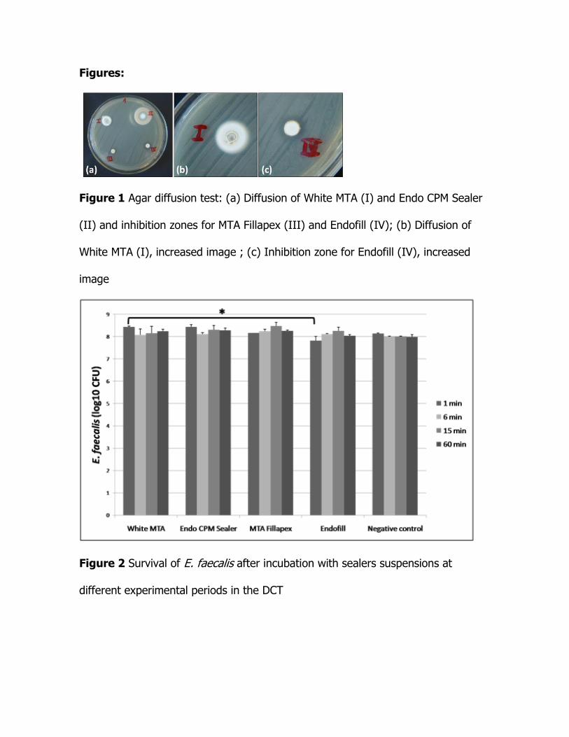
Table 2 shows data obtained in the ADT for each sealer. Endofill (positive
control) had the largest inhibition zone (Figure 1a,c), similar to MTA Fillapex
(Figure 1a) and greater than the other sealers (P<0.05). White MTA (Figure 1a,b)
and Endo CPM Sealer (Figure 1a) themselves resulted in diffusion in the agar, but
they were not able to inhibit E. faecalis. There was no bacterial growth on the two
control plates.
The results of the DCT are showed in Figure 2. None of the set materials
had antimicrobial activity against E. faecalis. There was a significant difference
between the bacterial counts of White MTA and Endofill in the first minute period
(P<0.05). However, the bacterial counts of all sealers were similar to the negative
control group in all experimental periods (P>0.05). Moreover, there were no
significant differences throughout the experimental periods for any sealers
(P>0.05).
The mean pH values for the sealers suspensions are described in Table 3.
White MTA and Endo CPM Sealer suspensions presented pH values greater than 11
at all experimental periods. MTA Fillapex and Endofill had lower but also alkaline
values. Deionized water had a neutral pH.
Discussion
This study investigated the pH and antibacterial activity of different
endodontic sealers against E. faecalis. In the ADT, it was possible to observe
inhibition zones with MTA Fillapex and Endofill. This fact could be explained by the
presence of resin and eugenol, respectively. In regard to MTA Fillapex, there is no
other data available about its antimicrobial effect. On the contrary, zinc oxide and
eugenol-based sealers, such as Endofill, have been investigated extensively and
have been used as positive controls in antimicrobial activity assays (Gomes et al.
2004, Tanomaru et al. 2008, Pinheiro et al. 2009).
White MTA and Endo CPM Sealer did not have inhibition zones against E.
faecalis. Similar results were reported previously by Estrela et al. (2000), who used
gray MTA. On the other hand, Tanomaru et al. (2008) verified that white MTA and
Endo CPM Sealer had inhibition zones of 15 and 12 mm against E. faecalis,
respectively. These two sealers showed visible diffusion in the agar medium, which
could lead to misinterpretation of their antibacterial activity.
None of the set sealers had antibacterial activity in the DCT. White MTA and
Endo CPM Sealer allowed the survival of E. faecalis, despite their high pH.
According to McHugh et al. (2004), E. faecalis is unable to live at the pH of 11.5 or
greater. As the pH shown by the above mentioned sealer was between 11 and 12,
it can be assumed that its alkalinity was not enough to make the environment
improper to the survival of that microorganism. Its proton pump is probably the
key factor in its resistance to alkaline agents (Stuart et al. 2006).
These findings contrasts with those by Zhang et al. (2009), who reported a
significant decreased in bacterial viability within 6 minutes of contact with grey
MTA powder. White MTA powder was employed in the present study, thus the
results could not be directly compared. Holt et al. (2007) reported that grey MTA
showed greater E. faecalis growth inhibition than white MTA and this could explain
the divergence.
An important goal of root canal treatment is to eliminate or prevent the
introduction of microorganisms into the root canal system (Siqueira & Roças 2008).
It is well known, however, that chemomechanical preparation is not able to

completely eradicate the endodontic infection (Nair et al. 2005). Residual bacteria
may remain untouched by instruments, irrigants and medicaments (Sjögren et al.
1997). To prevent new bacteria growth, filling materials and sealers should have
antimicrobial properties upon contact with microorganisms and biofilms especially
before setting. After this period, the most important property of the endodontic
sealer should be its sealing ability.
E. faecalis was chosen as the target microorganism due to its high
prevalence in persistent endodontic infections, ranging from 24 to 77% (Stuart et
al. 2006). E. faecalis can compete with other microorganisms and adapt to adverse
conditions, such as nutritional deprivation (Kayaoglu & Ørstavik 2004). This
microorganism is resistant to several irrigants and intracanal medicaments used in
endodontics (Menezes et al. 2004a, Zehnder & Guggenheim 2009). Therefore, the
antibacterial activity of endodontic sealers against E. faecalis is important in clinical
practice.
Historically, two different assays have been used to test the antimicrobial
characteristics of endodontic sealers: the ADT and the DCT. In this study, the first
test was used, despite its limitations, to evaluate fresh sealers immediately after
their manipulation. The second test was performed to analyze set sealers, seven
days after their mixture. In the ADT, the size of the inhibition zones from a certain
substance depends on its diffusibility in the culture medium used. This fact is the
main disadvantage of this semi quantitative method (Nawal et al. 2011). However,
the ADT is suitable to indicate the activity of freshly mixed materials and its
inclusion is interesting for comparative reasons with previous studies.
In turn, the DCT relies on direct contact between the microorganism and
the tested material. This method is virtually independent of the diffusion and
solubility properties of both the material and the media (Weiss et al. 1996). In
contrast with the ADT, the DCT is capable of showing the antibacterial activity of
insoluble components. When new materials are in test, more than one method
should be employed (Nawal et al. 2011).
To improve the assessment of the antibacterial activity of root canal sealers,
new methods should be developed where there is no interference from the
diffusivity and solubility of the material in the culture medium.
Conclusion
MTA Fillapex and Endofill had an effect against E. faecalis before setting,
but they did not maintain the antibacterial activity seven days after mixture.
Despite their alkaline pH, White MTA and Endo CPM Sealer did not have
antibacterial activity either before or after setting.
References
Al-Hezaimi K, Al-Shalan TA, Naghshbandi J, Simon JHS, Rotstein I (2009) MTA
preparations from different origins may vary in their antimicrobial activity. Oral
Surgery, Oral Medicine, Oral Pathology, Oral Radiology, and Endodontology
107, 85-8.
Cobankara FK, Altinöz HC, Ergani O, Kav K, Belli S (2004) In vitro antibacterial
activities of root-canal sealers by using two different methods. Journal of
Endodontics 30, 57-60.
Estrela C, Baummann LL, Estrela CRA, Silva RS, Pécora JD (2000) Antimicrobial
and chemical study of MTA, Portland cement, calcium hydroxide paste,
Sealapex and Dycal. Brazilian Dental Journal 11, 3-9.
Gomes BPFA, Pedroso JA, Jacinto RC, et al. (2004) In vitro evaluation of the
antimicrobial activity of five root canal sealers. Brazilian Dental Journal 15, 30-
5.
Gomes BPFA, Pinheiro ET, Sousa ELR, et al. (2006) Enterococcus faecalis in dental
root canals detected by culture and by polymerase chain reaction analysis. Oral
Surgery, Oral Medicine, Oral Pathology, Oral Radiology, and Endodontology
102, 247-53.
Gomes-Filho JE, Watanabe S, Bernabe PF, Moraes Costa MT (2009) A mineral
trioxide aggregate sealer stimulated mineralization. Journal of Endodontics 35,
256-60.
Holt DM, Watts JD, Beeson TJ, Kirkpatrick TC, Rutledge RE (2007) The anti-
microbial effect against Enterococcus faecalis and the compressive strength of
two types of mineral trioxide aggregate mixed with sterile water or 2%
chlorhexidine liquid. Journal of Endodontics 33, 844-7.
Jacobovitz M, Lima RKP (2008) Treatment of inflammatory internal root resorption
with mineral trioxide aggregate: a case report. International Endodontic Journal
41, 905-12.
Jacobovitz M, Lima RKP (2009) The use of calcium hydroxide and mineral trioxide
aggregate on apexification of a replanted tooth: a case report. Dental
Traumatology 25, e32-6.
Kayaoglu G, Ørstavik D (2004) Virulence factors of Enterococcus faecalis:
relationship to endodontic disease. Critical Reviews in Oral Biology & Medicine
15, 308-20.
Lee ES, Monsef M, Torabinejad M (1993) Sealing ability of a mineral trioxide
aggregate for repair of lateral root perforations. Journal of Endodontics 19,
541-4.
Love RM (2001) Enterococcus faecalis: a mechanism for its role in endodontic
failure. International Endodontic Journal 34, 399-405.
McHugh CP, Zhang P, Michalek S, Eleazer PD (2004) pH required to kill
Enterococcus faecalis in vitro. Journal of Endodontics 30, 218-9.
Menezes MM, Valera MC, Jorge AOC, Koga-Ito CY, Camargo CHR, Mancini MNG
(2004a) In vitro evaluation of the effectiveness of irrigants and intracanal
medicaments on microorganisms within root canals. International Endodontic
Journal 37, 311-9.
Menezes R, Bramante CM, Letra A, Carvalho VG, Garcia RB (2004b) Histologic
evaluation of pulpotomies in dog using two types of mineral trioxide aggregate
and regular and white Portland cements as wound dressings. Oral Surgery, Oral
Medicine, Oral Pathology, Oral Radiology, and Endodontology 98, 376-9.
Nair PN, Henry S, Cano V, Vera J (2005) Microbial status of apical root canal
system of human mandibular first molars with primary apical periodontitis after
"one-visit" endodontic treatment. Oral Surgery, Oral Medicine, Oral Pathology,
Oral Radiology, and Endodontology 99, 231-52.
Nawal RR, Parande M, Sehgal R, Naik A, Rao NR (2011) A comparative evaluation
of antimicrobial efficacy and flow properties for Epiphany, Guttaflow and AH-
Plus sealer. International Endodontic Journal 44, 307-13.
Orosco FA, Bramante CM, Garcia RB, Bernadineli N, Moraes IG (2008) Sealing
ability of gray MTA AngelusTM, CPMTM and MBPc used as apical plugs. Journal of
Applied Oral Science 16, 50-4.
Peters LB, Wesselink PR, Bujis JF, Van Winkelhoff AJ (2001) Viable bacteria in root
dentinal tubules of teeth with apical periodontitis. Journal of Endodontics 27,
76-81.
Pinheiro CR, Guinesi AS, Pizzolitto AC, Bonetti-Filho I (2009) In vitro antimicrobial
activity of Acroseal, Polifil and Epiphany against Enterococcus faecalis. Brazilian
Dental Journal 20, 107-11.
Ribeiro CS, Scelza MF, Hirata Júnior H, Oliveira LMB (2010) The antimicrobial
activity of gray-colored mineral trioxide aggregate (GMTA) and white-colored
MTA (WMTA) under aerobic and anaerobic conditions. Oral Surgery, Oral
Medicine, Oral Pathology, Oral Radiology, and Endodontology 109, e109-12.
Roberts HW, Toth JM, Berzins DW, Charton DG (2008) Mineral trioxide aggregate
material use in endodontic treatment: a review of the literature. Dental
Materials 24, 149-64.
Scarparo RK, Haddad H, Acasigua GA, Fossati ACM, Fachin EVF, Grecca FS (2010)
Mineral trioxide aggregate-based sealer: analysis of tissue reactions to a new
endodontic material. Journal of Endodontics 36, 1174-8.
Siqueira JF Jr, Roças IN (2008) Clinical implications and microbiology of bacterial
persistence after treatment procedures. Journal of Endodontics 34, 1291-301.
Sjögren U, Figdor D, Persson S, Sundqvist G (1997) Influence of infection at the
time of root filling on the outcome of endodontic treatment of teeth with apical
periodontitis. International Endodontic Journal 30, 297-306.
Spangberg L, Engstrom B, Langeland K (1973) Biologic effects of dental materials.
Toxicity and antimicrobial effect of endodontic antiseptics in vitro. Oral Surgery,
Oral Medicine, and Oral Pathology 36, 856-71.
Stuart CH, Schwartz AS, Beeson TJ, Owatz CB (2006) Enterococcus faecalis: its
role in root canal treatment failure and current concepts in retreatment. Journal
of Endodontics 32, 93-8.
Tanomaru JMG, Tanomaru-Filho M, Hotta J, Watanabe E, Ito IY (2008)
Antimicrobial activity of endodontic sealers based on calcium hydroxide and
MTA. Acta Odontologica Latinoamericana 21, 147-51.
Tobias RS (1988) Antibacterial properties of dental restorative materials: a review.
International Endodontic Journal 21, 155-60.
Torabinejad M, Chivian N (1999) Clinical applications of mineral trioxide aggregate.
Journal of Endodontics 25, 197-205.
Torabinejad M, Hong CU, Pitt Ford TR, Kettering JD (1995) Antibacterial effects of
some root end fillings materials. Journal of Endodontics 21, 403-6.
Torabinejad M, Watson TF, Pitt Ford TR (1993) Sealing ability of a mineral trioxide
aggregate when used as a root end filling material. Journal of Endodontics 19,
591-5.
Weiss E, Shahlav M, Fuss Z (1996) Assessment of antimicrobial activity of
endodontic sealers by a direct contact test. Endodontics & Dental Traumatology
12, 179-84.
Zehnder M, Guggenheim B (2009) The mysterious appearance of enterococci in
filled root canals. International Endodontic Journal 42, 277-87.
Zhang H, Pappen FG, Haapasalo M (2009) Dentin enhances the antibacterial effect
of Mineral Trioxide Aggregate and BioAggregate. Journal of Endodontics 35,
221-4.
Figures:
Figure 1 Agar diffusion test: (a) Diffusion of White MTA (I) and Endo CPM Sealer
(II) and inhibition zones for MTA Fillapex (III) and Endofill (IV); (b) Diffusion of
White MTA (I), increased image ; (c) Inhibition zone for Endofill (IV), increased
image
Figure 2 Survival of E. faecalis after incubation with sealers suspensions at
different experimental periods in the DCT
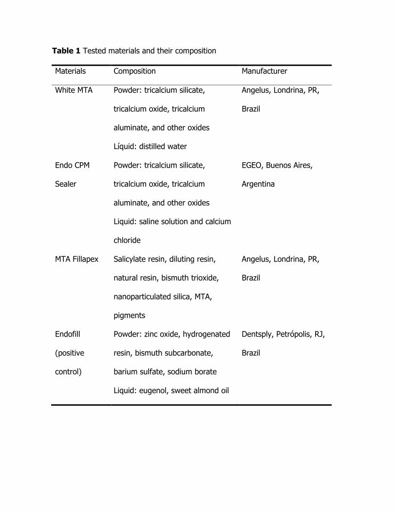
Table 1 Tested materials and their composition
Materials Composition Manufacturer
White MTA Powder: tricalcium silicate,
tricalcium oxide, tricalcium
aluminate, and other oxides
Líquid: distilled water
Angelus, Londrina, PR,
Brazil
Endo CPM
Sealer
Powder: tricalcium silicate,
tricalcium oxide, tricalcium
aluminate, and other oxides
Liquid: saline solution and calcium
chloride
EGEO, Buenos Aires,
Argentina
MTA Fillapex Salicylate resin, diluting resin,
natural resin, bismuth trioxide,
nanoparticulated silica, MTA,
pigments
Angelus, Londrina, PR,
Brazil
Endofill
(positive
control)
Powder: zinc oxide, hydrogenated
resin, bismuth subcarbonate,
barium sulfate, sodium borate
Liquid: eugenol, sweet almond oil
Dentsply, Petrópolis, RJ,
Brazil
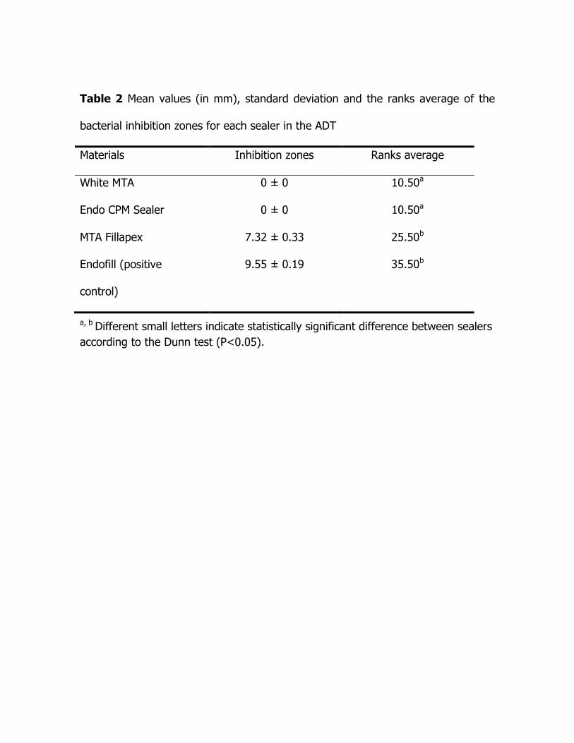
Table 2 Mean values (in mm), standard deviation and the ranks average of the
bacterial inhibition zones for each sealer in the ADT
Materials Inhibition zones Ranks average
White MTA 0 ± 0 10.50a
Endo CPM Sealer 0 ± 0 10.50a
MTA Fillapex 7.32 ± 0.33 25.50b
Endofill (positive
control)
9.55 ± 0.19 35.50b
a, b Different small letters indicate statistically significant difference between sealers
according to the Dunn test (P<0.05).

Table 3 Mean values of pH for each sealer at different experimental periods
Time White MTA Endo CPM Sealer
MTA Fillapex
Endofill Control
1 min 11.64 ±
0.17
11.39 ± 0.31 10.49 ±
0.07
8.22 ±
0.29
6.33 ±
0.04
6 min 11.83 ±
0.18
11.23 ± 0.12 10.5 ±
0.02
8.56 ±
0.26
7.82 ±
0.42
15 min 11.84 ±
0.2
11.36 ± 0.24 10.46 ±
0.02
8.63 ±
0.35
7.79 ±
0.54
60 min 11.84 ±
0.2
11.19 ± 0.09 10.14 ±
0.21
8.27 ±
0.54
7.07 ±
0.37