evidence on the male reproductive toxicity of
TRANSCRIPT

OFFICE OF ENVIRONMENTAL HEALTH HAZARD ASSESSMENT
Proposition 65
Evidence on the Male Reproductive Toxicity of Perfluorononanoic Acid (PFNA) and Its Salts and Perfluorodecanoic Acid (PFDA) and Its Salts
October 2021
Reproductive and Cancer Hazard Assessment Branch Office of Environmental Health Hazard Assessment California Environmental Protection Agency

PFNA and its salts and i OEHHA PFDA and its salts October 2021
CONTRIBUTORS
The Office of Environmental Health Hazard Assessment’s (OEHHA) Reproductive and Cancer Hazard Assessment Branch was responsible for the preparation of this document.
Authors (listed alphabetically by last name)
Marlissa Campbell, Ph.D. Staff Toxicologist
Poorni Iyer, DVM, Ph.D., DABT Staff Toxicologist
Farla Kaufman, Ph.D. Staff Toxicologist
Allegra Kim, Ph.D. Research Scientist III
Ling-Hong Li, Ph.D. Staff Toxicologist
Francisco Moran, Ph.D. Staff Toxicologist
Yassaman Niknam, Ph.D. Staff Toxicologist
Acknowledgments The valuable support of the following OEHHA staff is also acknowledged: Nancy Firchow, MLS for conducting the literature search and Elizabeth Boxer for assistance with the epidemiologic data.
Internal OEHHA Reviewers
Martha S. Sandy, Ph.D., M.P.H. Chief, Reproductive and Cancer Hazard Assessment Branch
Vince Cogliano, Ph.D. Deputy Director, Division of Scientific Programs
David Edwards, Ph.D. Chief Deputy Director
Director
Lauren Zeise, Ph.D.

PFNA and its salts and ii OEHHA PFDA and its salts October 2021
PREFACE
Proposition 651 requires the publication of a list of chemicals “known to the state” to cause cancer or reproductive toxicity. The Office of Environmental Health Hazard Assessment (OEHHA) of the California Environmental Protection Agency maintains this list in its role as lead agency for implementing Proposition 65. The Developmental and Reproductive Toxicant Identification Committee (DARTIC) advises and assists OEHHA, and adds chemicals to the Proposition 65 list of chemicals that cause reproductive toxicity, as required by Health and Safety Code section 25249.8. The DARTIC serves as the state’s qualified experts for determining whether a chemical has been clearly shown to cause reproductive toxicity.
The Committee also provides advice and consultation regarding which chemicals should receive their review. At their meeting in December 2020, the DARTIC recommended that perfluorononanoic acid (PFNA) and its salts and perfluorodecanoic acid (PFDA) and its salts be placed in a ‘high’ priority group for future listing consideration. OEHHA selected PFNA and its salts and PFDA and its salts for consideration for listing by the DARTIC, and in March 2021 OEHHA solicited from the public information relevant to the assessment of the evidence on the reproductive toxicity of these chemicals. No information was received on either PFNA and its salts or PFDA and its salts in response to this request. This document presents evidence relevant to the evaluation of the male reproductive toxicity of these chemicals. Documents presenting evidence relevant to the evaluation of these chemicals for other reproductive toxicity endpoints (e.g., female reproductive, developmental) may be developed in the future.
.
1 The Safe Drinking Water and Toxic Enforcement Act of 1986 (California Health and Safety Code section 5249.5 et seq.)
PFNA and its salts and iii OEHHA PFDA and its salts October 2021
TABLE OF CONTENTS PREFACE ........................................................................................................................ii
list of abbreviations..........................................................................................................ix
EXECUTIVE SUMMARY ............................................................................................... xiii
Systematic Literature Review Approach .................................................................... xiii
Pharmacokinetics of PFNA and PFDA ...................................................................... xiii
Endocrine System Involvement in Development and Function of the Male Reproductive System ................................................................................................. xiv
PFNA and its salts: Male reproductive toxicity .......................................................... xiv
Studies in Humans ............................................................................................. xiv
Studies in Animals ...............................................................................................xv
Mechanistic Considerations ................................................................................ xvi
Key characteristics of male reproductive toxicants and endocrine disrupting chemicals .......................................................................................................... xvii
PFDA and its salts: Male reproductive toxicity ........................................................ xviii
Studies in Humans ........................................................................................... xviii
Studies in Animals ............................................................................................ xviii
Mechanistic Considerations ................................................................................ xix
Key characteristics of male reproductive toxicants and endocrine disrupting chemicals ............................................................................................................xx
1. Introduction ........................................................................................................... 1
1.1 Identity of perfluorononanoic acid (PFNA) and its salts and perfluorodecanoic acid (PFDA) and its salts ..................................................................................................... 1
1.2 Uses, occurrence, and exposure ........................................................................... 2
1.3 Reviews by other health agencies ......................................................................... 7
1.4 Overview of systematic literature review approach ................................................ 8
Search process ..................................................................................................... 8
Data sources: ....................................................................................................... 8
Literature screening process ................................................................................ 9
2. Pharmacokinetics of PFNA and PFDA ............................................................... 10
2.1 Absorption ........................................................................................................... 10

PFNA and its salts and iv OEHHA PFDA and its salts October 2021
2.2 Distribution ........................................................................................................... 10
Transport ............................................................................................................ 11
Maternal-fetal transfer ........................................................................................ 11
Breast milk transfer ............................................................................................. 12
Sex differences in distribution ............................................................................. 12
2.3 Metabolism .......................................................................................................... 13
2.4 Excretion .............................................................................................................. 13
2.5 Species differences in PFNA and PFDA serum half-lives .................................... 13
PFNA .................................................................................................................. 13
PFDA .................................................................................................................. 14
3. Endocrine System Involvement in Development and Function of the MaleReproductive System .................................................................................... 14
3.1 Hypothalamus-pituitary-gonad axis ...................................................................... 14
3.2 Thyroid hormones ................................................................................................ 15
4. PFNA and its salts: Male reproductive toxicity .................................................... 16
4.1 PFNA: Human studies of male reproductive effects ............................................ 16
Anogenital distance ............................................................................................ 16
Male reproductive function ................................................................................. 17
Cancer ................................................................................................................ 18
4.2 PFNA: Animal studies of male reproductive toxicity ............................................. 29
Organ Weight and Histopathology ...................................................................... 30
Sperm Parameters ............................................................................................. 34
Hormonal Effects ................................................................................................ 34
Fertility or Reproductive Performance ................................................................ 36
Development of the Male Reproductive System ................................................. 37
4.3 PFNA: Mechanistic considerations and other relevant data ................................ 46
General toxicity ................................................................................................... 46
In vitro study in testicular cells ............................................................................ 46
Effects on the hypothalamic-pituitary-gonadal-(liver) axis .................................. 46
Effects on the thyroid .......................................................................................... 56
Possible involvement of peroxisome proliferator-activated receptors ................. 63

PFNA and its salts and v OEHHA PFDA and its salts October 2021
4.4 PFNA: Summary of evidence on male reproductive toxicity ................................ 63
Human studies .................................................................................................... 63
Animal studies .................................................................................................... 64
Coherence of results in human and animal studies ............................................ 65
Mechanistic considerations................................................................................. 66
Key characteristics of male reproductive toxicants and endocrine-disrupting chemicals ........................................................................................................... 68
5. PFDA and its salts: Male Reproductive Toxicity ................................................. 74
5.1 PFDA: Human studies of male reproductive effects ............................................ 74
Anogenital distance ............................................................................................ 74
Male reproductive function ................................................................................. 75
Cancer ................................................................................................................ 75
5.2 PFDA: Animal studies of male reproductive toxicity ............................................. 82
Organ weights & histopathology ......................................................................... 82
Sperm parameters .............................................................................................. 86
Hormonal effects ................................................................................................ 86
Effects on fertility ................................................................................................ 87
5.3 PFDA: Mechanistic considerations and other relevant data ................................ 93
General toxicity ................................................................................................... 93
Cytotoxicity ......................................................................................................... 93
Effects on the hypothalamic-pituitary-gonadal-(liver) axis .................................. 94
Effects on the thyroid ........................................................................................ 102
Possible involvement of peroxisome proliferator-activated receptors ............... 110
5.4 PFDA: Summary of evidence on male reproductive toxicity ............................. 110
Human studies .................................................................................................. 110
Animal studies .................................................................................................. 111
Coherence of results in human and animal studies .......................................... 111
Mechanistic considerations............................................................................... 112
Key characteristics of male reproductive toxicants and endocrine-disrupting chemicals ......................................................................................................... 113
6. REFERENCES ................................................................................................. 117

PFNA and its salts and vi OEHHA PFDA and its salts October 2021
Appendix A. Literature Search Approach on the Male Reproductive Toxicity of PFNA and Its Salts and PFDA and Its Salts .......................................................... 128
Search process ........................................................................................................ 128
Data sources: .......................................................................................................... 128
Literature screening process ................................................................................... 129
Use of Health Assessment Workspace Collaborative .............................................. 129

PFNA and its salts and vii OEHHA PFDA and its salts October 2021
LIST OF TABLES Table 1.1 Identifiers for PFNA and some salts. ............................................................... 1
Table 1.2 Identifiers for PFDA and some salts. ............................................................... 2
Table 1.3 PFNA serum concentrations (ng/ml) in studies of California residents. ........... 4
Table 1.4 PFDA serum concentrations (ng/ml) in studies of California residents. ........... 6
Table 4.1 PFNA: Epidemiologic studies of male reproductive toxicity. .......................... 19
Table 4.2.1 PFNA or Wyeth-14,643: Relevant key findings in male rats treated by gavage for 28 days (NTP 2019) ............................................................................. 31
Table 4.2.2 PFNA: Evidence on the male reproductive toxicity in animal studies. ........ 38
Table 4.3.1 PFNA:......................................................................................................... 50
Table 4.3.2 PFNA Effects on thyroid hormones. ........................................................... 59
Table 4.4.2 Key characteristics of endocrine-disrupting chemicals ............................... 70
Table 5.1 PFDA: Epidemiologic studies of male reproductive toxicity. .......................... 76
Table 5.2.2 PFDA: Evidence on the male reproductive toxicity in animal studies. ........ 88
Table 5.3.1 PFDA: Effects on the hypothalamic-pituitary-gonadal-(liver) axis in animals and in vitro studies. ................................................................................................ 97
Table 5.3.2 .PFDA: Effects on thyroid hormones. ...................................................... 105
Table A. 1 Human DART Study Searches ................................................................. 131
Table A. 2 Animal DART Study Searches ................................................................... 137

PFNA and its salts and viii OEHHA PFDA and its salts October 2021
LIST OF FIGURES
Figure 1.1 Structure of PFNA ......................................................................................... 1
Figure 1.2 Structure of PFDA ......................................................................................... 1

PFNA and its salts and ix OEHHA PFDA and its salts October 2021
LIST OF ABBREVIATIONS Abbreviation Full name
β regression coefficient µM micromolar 11-KT 11-ketotestosterone17β-HSD 17β-Hydroxysteroid dehydrogenase3β-HSD 3β-Hydroxysteroid dehydrogenase6:2 Cl-PFESA 6:2 chlorinated polyfluorinated ether sulfonate8:2 Cl-PFESA 8:2 chlorinated polyfluorinated ether sulfonateACTH adrenocorticotropic hormoneAGD anogenital distanceAGDAP anopenile distanceAGDAS anoscrotal distanceAhR arylhydrocarbon receptorAMH antimüllerian hormone or Müllerian inhibiting substanceAR androgen receptorATSDR Agency for Toxic Substances and Disease RegistryBax Bcl2 associated XBcl-2 B-cell lymphoma 2Bcrp1 breast cancer resistance proteinBMI body mass indexBZRP benzodiazepine receptorCASA computer-aided sperm analysisCI confidence intervalCYP11a cytochrome P450 family 11 subfamily ACYP17 cytochrome P450 family 17CYP19a cytochrome P450 family 19 subfamily ADART developmental and reproductive toxicity/toxicantDARTIC Developmental and Reproductive Toxicant Identification CommitteeDFI DNA fragmentation indexDFI DNA fragmentation indexDGML DNA global methylationDHT dihydrotestosteroneDpf days post-fertilizationE2 estradiolECHA European Chemicals AgencyEDCs endocrine-disrupting chemicalsEGR 3 early growth receptor 3ER estrogen receptorERα estrogen receptor alphaERβ estrogen receptor betaEt-PFOSA-AcOH 2-(N-Ethyl-perfluorooctane sulfonamido) acetic acid

PFNA and its salts and x OEHHA PFDA and its salts October 2021
Abbreviation
FAI FasL FCM FDA FDR FSH FSHR GD GHR GLUT-3 GnRH GST GWG HAWC hCG HDS hERα
Full name
free androgen index Fas ligand flow cytometric Food and Drug Administration false discovery rate p-value follicle-stimulating hormone follicle-stimulating hormone receptor gestation day growth hormone receptor glycose transporter 3 gonadotrophin-releasing hormone glutathione-s-transferase gestational weight gain Health Assessment Workspace Collaborative human chorionic gonadotropin high DNA stainability human estrogen receptor α
HMG CoA synthase 1 3-hydroxy-3-methylglutaryl coenzyme A synthase 1HO-1 heme oxygenase-1 HPG hypothalamus-pituitary-gonad HPGL hypothalamic-pituitary-gonadal-liver HPT hypothalamic-pituitary-thyroid HSA human serum albumin i.p. intraperitoneal IC50 half maximal inhibitory concentration IGF-1 insulin like growth factor 1 IGF-1R insulin like growth factor 1 receptor IKKβ inhibitor of NF-κB kinase IQR interquartile range IVF in vitro fertilization KCs key characteristics LH luteinizing hormone LHDC lactate dehydrogenase type c LHR luteinizing hormone receptor Ln natural log LOD limit of detection LOQ limit of quantification Me-PFOSA-AcOH 2-(N-Methyl-perfluorooctane sulfonamido) acetic acid MIS Müllerian inhibiting substance mRNA messenger ribonucleic acid

PFNA and its salts and xi OEHHA PFDA and its salts October 2021
Abbreviation Full name
MTT 3‐(4, 5‐dimethylthiazol‐2‐yl)‐2, 5 diphenyltetrazolium bromide N number of participants NF-κB nuclear factor kappa B NHANES National Health and Nutrition Examination Survey NIOSH National Institute for Occupational Safety and Health Nrf2 nuclear factor-erythroid-2-related factor-2 NTP National Toxicology Program OD optical density OEHHA Office of Environmental Health Hazard Assessment OR odds ratio PCNA proliferating cell nuclear antigen PFASs per- and polyfluoroalkyl substances PFBS perfluorobutane sulfonic acid PFDA perfluorodecanoic acid PFDoA perfluorododecanoic acid PFHpA perfluoroheptanoic acid PFHpS perfluoroheptane sulfonic acid PFHxA perfluorohexanoic acid PFHxS perfluorohexane sulfonic acid PFNA perfluorononanoic acid PFOA perfluorooctanoic acid PFOS perfluorooctane sulfonic acid PFPA perfluorophosphonic acid PFTrA perfluorotridecanoic acid PFUnDA perfluoroundecanoic acid p-mTOR phosphorylated mammalian target of rapamycin PND postnatal day PPAR peroxisome proliferator-activated receptor PPARα peroxisome proliferator-activated receptor alpha ppm parts per million PSA prostate-specific antigen PVDF polyvinylidene fluoride qRT-PCR quantitative real-time PCR r correlation coefficient RAC Committee for Risk Assessment [ECHA] ROS reactive oxygen species RP relative potency RR relative risk rT3 reverse triiodothyronine SCSA sperm chromatin structure assay SD Sprague Dawley or standard deviation

PFNA and its salts and xii OEHHA PFDA and its salts October 2021
Abbreviation
SF1 SHBG SOD SR-B1 StAR T T3 T4 TBG TCDD TRs TRH TSH TSPO TTR TUNEL US EPA VCL VSL VtG WT1
Full name
steroidogenic factor 1 sex hormone-binding globulin superoxide dismutase scavenger receptor class B type 1 steroidogenic acute regulatory protein testosterone triiodothyronine or tri-iodo-L-thyronine thyroxine thyroid binding globulin 2,3,7,8-tetrachlorodibenzo-p-dioxin thyroid hormone receptors thyrotropin-releasing hormone thyroid stimulating hormone mitochondrial translocator protein, also peripheral-type benzodiazepine receptor transthyretin terminal deoxynucleotidyl transferase dUTP nick end labeling US Environmental Protection Agency curvilinear velocity straight-line velocity vitellogenin Wilms’ tumor gene

PFNA and its salts and xiii OEHHA PFDA and its salts October 2021
EXECUTIVE SUMMARY This document presents evidence relevant to the evaluation of the male reproductive toxicity of perfluorononanoic acid (PFNA) and its salts and perfluorodecanoic acid (PFDA) and its salts. PFNA and PFDA are perfluoroalkyl carboxylic acids containing nine and ten carbons, respectively, and are members of a large group of substances with surfactant properties collectively called per- and polyfluoroalkyl substances (PFASs). PFNA and its salts and PFDA and its salts have been used in various industries, including as processing aids in fluoropolymer manufacture. PFNA and PFDA are also used in some cosmetic products. PFASs, including PFNA and PFDA, are global environmental pollutants of air, water, soil and wildlife, and are very persistent in the environment. Recent data from Biomonitoring California (https://biomonitoring.ca.gov) have shown that PFNA and PFDA are readily detected in virtually all Californians. See Section 1.2 for additional information on uses, occurrence and exposure to PFNA and its salts and PFDA and its salts.
The evidence summarized in this document includes studies of PFNA and PFDA, as well as studies of their salts, which dissociate to form the corresponding organic acids.
Systematic Literature Review Approach
Using a systematic approach, the Office of Environmental Health Hazard Assessment (OEHHA) conducted literature searches on the developmental and reproductive toxicity of PFNA and its salts, and PFDA and its salts, and then focused on literature relevant to male reproductive toxicity (last comprehensive search, February 2021). An overview of the systematic literature review approach is presented in Section 1.4 of this document, and more detailed information can be found in Appendix A.
Pharmacokinetics of PFNA and PFDA
PFNA and PFDA are not known to be metabolized in animals or humans. PFNA and PFDA are well absorbed with oral administration in animals, bind to serum proteins, and are widely distributed throughout the body in both humans and animals. The estimated half-lives of PFNA and PFDA are significantly longer in humans (3.1 years and 7.1 years, respectively) than in rodents (30-55 days and 36-109 days, respectively). In a study of PFASs in human tissues, the highest levels of PFNA were observed in brain and kidney, with lower levels in lung and liver, and for PFDA the highest levels were observed in the brain, with lower levels in lung and kidney. PFNA and PFDA can cross the blood brain barrier and the placenta, and both have been detected in fetal tissues, cord serum, and breast milk. PFNA and PFDA excretion pathways in humans include urinary and fecal excretion and incorporation into nails and hair. See Section 2 for more detailed information on pharmacokinetics.

PFNA and its salts and xiv OEHHA PFDA and its salts October 2021
Endocrine System Involvement in Development and Function of the Male Reproductive System
Reproductive biology is under close control from the endocrine system. Environmental factors, including exposures to certain chemicals, may influence the endocrine system and the reproductive processes controlled by it. An overview of the role of the hypothalamus-pituitary-gonad axis and the thyroid hormone system in male reproductive system development and function is presented in Section 3 of this document.
PFNA and its salts: Male reproductive toxicity
Studies in Humans
OEHHA identified 17 epidemiologic studies of effects of PFNA on the male reproductive system. The levels of exposure to PFNA in the available epidemiologic studies were generally low, with little variability in concentrations.
The epidemiologic evidence for an effect of PFNA on anogenital distance (AGD) was mixed, with one study reporting an association with longer anoscrotal distance (AGDAS), and another reporting non-significant associations with shorter AGDAS and anopenile distance (AGDAP) that did not remain at 12 months of age.
Three of seven studies of effects of PFNA levels on serum testosterone (T) levels reported decreases (two of which were statistically significant), while the other four studies reported inconsistent results across locations or no associations with T.
A small study with relatively high plasma PFNA concentrations and variability reported a substantial reduction in sperm concentration, while other studies reported no associations with sperm concentration or count. Sperm quality studies reported decreases, increases, and no change in the proportion of sperm with normal morphology. A study that examined both semen and serum PFNA concentrations reported that semen PFNA was associated with decreased motility, curvilinear velocity (VCL), and straight line velocity (VSL), while other studies that used serum PFNA measurements reported no associations with these parameters. Compromised sperm DNA integrity was associated with semen PFNA in a study where infertile men were overrepresented, but not in a study using serum PFNA and in which infertile men were underrepresented. PFNA exposure was not associated with prostate cancer or prostate-specific antigen (PSA) concentration.
See Section 4.1 for more detailed information on male reproductive toxicity studies in humans.

PFNA and its salts and xv OEHHA PFDA and its salts October 2021
Studies in Animals
PFNA has been evaluated for its male reproductive toxicity in a number of animal studies in vivo, including four studies in rats, five studies in mice and one study in zebrafish. All the studies in rats or mice treated the animals by oral gavage, and the study in zebrafish exposed the animals to PFNA for 180 days. Two of the studies in mice evaluated the effects of PFNA on development of the male reproductive system after gestational treatment.
PFNA induced dose-dependent reductions in epididymal weight at ≥0.615 mg/kg-day and testis weight at ≥1.25 mg/kg-day in adult Sprague-Dawley (SD) rats exposed for 28 days. Treatment with PFNA at ≥2 mg/kg-day for 14 days resulted in an apparent decrease in testis weight in prepubertal mice, but the reduction did not reach statistical significance.
Histopathological changes in the testis (e.g., germ cell degeneration) were observed in three rat studies and three mouse studies. Testicular lesions observed in rats treated with PFNA at ≥2 mg/kg-day for 14 days included cell degeneration in spermatocytes and spermatogonia, and cytoplasmic vacuolization in Sertoli cells. In the NTP study in rats, exposure to PFNA for 28 days caused dose-related increases in germ cell degeneration (≥2.5 mg/kg-day), interstitial cell atrophy (≥2.5 mg/kg-day), and spermatid retention (≥2.5 mg/kg-day). In prepubertal mice, treatment with PFNA at ≥2 mg/kg-day for 14 days or 0.5 mg/kg-day for 90 days resulted in increased germ cell degeneration as well as other changes consistent with these effects, e.g., decreased relative population size of 4C germ cells and decreased spermatogonial cells in G2 phase.
In studies that measured sperm parameters, a dose-dependent reduction in epididymal sperm counts was observed in rats treated with PFNA for 28 days (≥ 1.25 mg/kg-day), and in mice dose-related reductions in sperm counts, motility and viability were observed following exposure to PFNA at 0.2 and 0.5 mg/kg-day for 90 days.
Reduced serum levels of T were consistently observed in rats and mice at dosing levels that caused histopathological lesions or changes in sperm parameters. On the other hand, increased serum T levels were observed at 1 mg/kg-day dosing level (but decreased T at 5 mg/kg-day) in pubertal rats treated for 14 days and in zebrafish treated for 180 days at 0.01 mg/L in water (but not 0.1 and 1.0 mg/L exposure levels).
Two studies provided limited data on the male fertility effects of PFNA, one study in mice and another in zebrafish. In mice, reductions in fertility index and litter size were observed when PFNA-exposed males were mated with unexposed females, but detailed data and information on the design of the fertility assessment studies were not reported. In the zebrafish study both males and females were exposed to PFNA prior to mating, and reductions in egg production and hatching rate were observed with PFNA treatment.

PFNA and its salts and xvi OEHHA PFDA and its salts October 2021
Gestational exposure to PFNA at 5 mg/kg-day reduced intratesticular levels of T in mice on postnatal day (PND) 3. In this same study gestational exposure to PFNA at 2 and 5 mg/kg-day reduced testicular protein levels of steroidogenic acute regulatory protein (StAR) and WT1, and at 5 mg/kg-day reduced testicular protein levels of SF1, CYP11A1, 3β-HSD, 17β-HSD, and PCNA (a marker of cell proliferation). These proteins are critical for either steroidogenesis in Leydig cells or for Sertoli cell function and proliferation during the perinatal period. In another study of male mice, a dose-related delay in preputial separation was observed following gestational exposures to PFNA at 3.0 and 5.0 mg/kg-day.
See Section 4.2 for more detailed information on male reproductive toxicity studies in animals.
Mechanistic Considerations
Potential mechanistic pathways involved in PFNA mediated male reproductive toxicity include effects on the HPG axis, and effects on thyroid homeostasis.
HPG axis
Altered hormone levels have been observed in rats, mice, zebrafish, and primary rat Sertoli cell cultures following exposure to PFNA. PFNA reduced plasma levels of T in male rats and plasma and intratesticular levels of T in male mice. In male rats and zebrafish PFNA increased serum levels of estradiol (E2). In addition, PFNA increased serum Müllerian inhibiting substance (MIS) in rats, increased MIS messenger ribonucleic acid (mRNA) in primary rat Sertoli cells, altered testicular gene expression of MIS in mice, and increased brain mRNA levels of the gene encoding luteinizing hormone (LH) in male zebrafish.
PFNA induced changes in gene and protein expression of a number of enzymes and factors involved in steroidogenesis in mice and zebrafish, and decreased steroid hormone production in a mouse Leydig tumor cell line. In mice PFNA decreased testicular protein levels of steroidogenic factor 1 (SF1), StAR, cytochrome CYP11a, 3β-hydroxysteroid dehydrogenase (3β-HSD), and 17β-hydroxysteroid dehydrogenase (17β –HSD), and testicular mRNA levels of 3-hydroxy-3-methylglutaryl coenzyme A synthase 1 (HMG Co synthase 1), StAR, cytochrome P450 family 11 subfamily a (CYP11a) and 3β-HSD. In zebrafish PFNA decreased gonadal gene expression of 3β-hsd, and increased gene expression of star, cyp11a, 17β-hsd, and cyp19a. In a mouse Leydig tumor cell line, PFNA decreased steroid hormone production at concentrations that also reduced mitochondrial membrane potential.
Receptor binding studies and in silico modeling indicate that PFNA can interact with estrogen receptors, and estrogen receptor mediated effects of PFNA have been

PFNA and its salts and xvii OEHHA PFDA and its salts October 2021
observed in zebrafish, trout, and human cell lines. In fish PFNA increased vitellogenin (vtg) gene and protein expression. In human embryonic kidney cells transfected with a human estrogen receptor alpha (hERα) reporter gene PFNA induced an estrogenic response, while anti-estrogenic activity was observed in studies with human breast adenocarcinoma cell lines.
PFNA may also interact with androgen receptors, based on observations of anti-AR activity in a Chinese hamster ovary (CHO) cell line.
Studies in rats, mice, zebrafish and primary rat Sertoli cell cultures indicate that PFNA may affect gene and protein expression of some hormone receptors, growth factor receptors, and related proteins. In rats, PFNA decreased testicular protein levels of transferrin. In mice, PFNA reduced testicular mRNA levels for the androgen receptor (AR), growth hormone receptor (GHR), insulin-like growth factor hormone receptor 1 (IGF-1R), and insulin-like growth factor 1 (IGF-1). In male zebrafish, PFNA decreased brain mRNA levels for era, erb and ar, decreased gonadal mRNA levels for fshr and lhr, and increased liver mRNA levels for era and erb. In primary rat Sertoli cell cultures PFNA reduced mRNA levels of follicle stimulating hormone receptor (FSHR), increased mRNA levels of androgen binding protein (ABP), and reduced mRNA levels of transferrin.
Thyroid homeostasis
Outcomes of both thyroid and male reproductive toxicity were assessed in rats following 28 days of treatment with PFNA. Thyroid effects were observed at the same or lower doses than evidence of male reproductive toxicity. PFNA has been shown to interfere with thyroid hormone binding, serum levels, and function as assessed using in vivo, in vitro, and in silico test systems. Existing data suggest a possible, but unproven, mechanistic connection between thyroid toxicity and the male reproductive toxicity of PFNA.
See Section 4.3 for more detailed summaries of data relevant to considerations of mechanisms of male reproductive toxicity.
Key characteristics of male reproductive toxicants and endocrine disrupting chemicals
The available mechanistic evidence suggests that PFNA exhibits five of the key characteristics (KCs) for male reproductive toxicants (e.g., alters germ cells; alters somatic cells; alters production and levels of reproductive hormones; alters hormone receptors; is genotoxic), and seven of the KCs for endocrine-disrupting chemicals (e.g., interacts with or activates hormone receptors; antagonizes hormone receptors; alters hormone receptor expression; alters signal transduction in hormone responsive cells,

PFNA and its salts and xviii OEHHA PFDA and its salts October 2021
factors and transcripts and activity; alters hormone synthesis; alters hormone distribution or circulating levels; alters fate of hormone producing or hormone responsive cells). See Section 4.4 for more information on the data relevant to these KCs.
PFDA and its salts: Male reproductive toxicity
Studies in humans
Eleven epidemiologic studies of effects of PFDA on the male reproductive system were identified. Most of the PFDA exposure concentrations in the available epidemiologic studies were very low, with little variability.
The epidemiologic evidence for an association between maternal PFDA exposure and anogenital distance (AGD) was mixed, with the stronger of two studies reporting associations with shorter anoscrotal distance (AGDAS) and anopenile distance (AGDAP) at birth, but not at 12 months. The other study reported that prenatal PFDA exposure in the third quartile, compared to the first quartile, was associated with an increase in AGDAS at three months.
Higher serum PFDA was associated with lower serum T in a small sample of adolescent boys, while studies of adult males did not report consistent associations with reproductive hormones.
Semen quality studies with the highest PFDA concentrations reported some associations with poorer semen quality. The largest study, which had relatively high serum PFDA concentrations, overrepresented infertile men, and measured PFDA in both serum and semen, reported that semen PFDA was associated with decreased progressive motility and straight line velocity (VSL). In this study semen PFDA was also associated with increases in markers of sperm chromatin immaturity and DNA fragmentation. In a small cohort of couples with female factor infertility, plasma PFDA was associated with decreased sperm concentration and a nonsignificant reduction in sperm count.
One study reported an association between PFDA and prostate cancer among men with a first-degree relative with prostate cancer; this result needs corroboration.
See Section 5.1 for more detailed information on male reproductive toxicity studies in humans.
Studies in Animals
There are a number of animal studies in vivo that evaluated the male reproductive effects of PFDA, including multiple studies in male rats, one study in male mice, one

PFNA and its salts and xix OEHHA PFDA and its salts October 2021
study in male hamsters, one study in male guinea pigs, and one study in zebrafish. One study by the National Toxicology Program (NTP) treated male SD rats at five dose levels (0.156-2.5 mg/kg-day) by daily gavage for 28 days. All other rat studies assessed the effects of PFDA seven to 30 days after administration of a single intraperitoneal injection (i.p.) of relatively high doses (ranging from 20-80 mg/kg). The studies in male mice, hamsters, and guinea pigs also treated the animals with a single i.p. injection of relatively high doses of PFDA and evaluated the toxic effects 14 to 28 days after dosing. The study in zebrafish exposed the animals for 120 days.
PFDA reduced testis weight in rats, hamsters and guinea pigs following a single i.p. injection at relatively high doses ranging from 50-175 mg/kg. Dose-related reductions in testis and epididymis weights were observed in the NTP study in rats exposed to lower doses for 28 days (≥1.25 mg/kg-day).
Histopathological changes in the testis (i.e., germ cell degeneration) were observed in rats (in two studies), hamsters, and guinea pigs that received a single i.p. dose of ≥50 mg/kg (or 150 mg/kg for guinea pigs). Testicular lesions observed in rats exposed to lower doses for 28 days included a dose-dependent increase in interstitial cell atrophy (≥1.25 mg/kg-day), and increased spermatid retention or inhibited spermiation (2.5 mg/kg-day. No effects on testis weight or histology were observed in mice on day 28 following a single i.p. dose of 250 mg/kg PFDA.
There is one study that measured sperm parameters in rats. In this study, the authors found a dose-dependent reduction in epididymal sperm counts in rats exposed to PFDA for 28 days, which was significant at 2.5 mg/kg-day. Reduced serum levels of T were observed in rats exposed to PFDA for 28 days (dose-dependent; statistically significant at ≥1.25 mg/kg-day) and in rats receiving a single dose (≥40 mg/kg). Plasma dehydrotestosterone (DHT) levels were also decreased by PFDA in the single dose rat study. The ratios of blood E2/T and E2/110-KT were increased in zebrafish exposed from one to 120 days post fertilization to PFDA at 1.0 mg/L, but not at higher or lower concentrations. There are no animal studies available on the male fertility effects of PFDA. See Section 5.2 for more detailed information on male reproductive toxicity studies in animals.
Mechanistic Considerations
Potential mechanistic pathways involved in PFDA mediated male reproductive toxicity include effects on the HPG axis, and effects on thyroid homeostasis.
HPG axis
PFDA altered steroid hormone levels in rats, decreased steroid hormone synthesis in isolated rat Leydig cells, mouse Leydig tumor cell lines, and rat testes ex vivo and
PFNA and its salts and xx OEHHA PFDA and its salts October 2021
altered aromatase gene or protein expression in zebrafish and, a human cell line. In male rats, PFDA reduced plasma levels of T and DHT without effects on plasma levels of LH and caused a dose-dependent decrease in hCG-stimulated T secretion in decapsulated testes from PFDA treated animals, indicating disruption of testicular feedback to LH stimulation. In vitro, exposure to PFDA resulted in decreases in steroid hormone production in isolated rat Leydig cells and mouse Leydig tumor cell lines. PFDA reduced translocator protein (TSPO) protein levels and mRNA stability, and decreased mitochondrial membrane potential in mouse Leydig tumor cell lines, suggesting that impairment of mitochondrial function and cholesterol transport may contribute to decreased steroidogenesis. PFDA increased gonadal aromatase gene expression in male zebrafish, while it decreased aromatase activity levels in a human placental choriocarcinoma cell line.
Receptor binding studies and in silico modeling indicate that PFDA can interact with estrogen receptors, and estrogen receptor mediated effects of PFDA have been observed in zebrafish, trout, and human cell lines. In fish PFDA increased vtg gene and protein expression. In human embryonic kidney cells transfected with a hERα reporter gene PFDA induced an estrogenic response, while anti-estrogenic activity was observed in studies with human breast adenocarcinoma cell lines. PFDA may also interact with androgen receptors, based on observations of anti-AR activity in a Chinese hamster ovary cell line.
Thyroid homeostasis
Outcomes of both thyroid and male reproductive toxicity were assessed in rats following 28 days of treatment with PFDA. Thyroid effects were observed at the same or lower doses than evidence of male reproductive toxicity. PFDA has been shown to interfere with thyroid hormone binding, serum levels, and function as assessed using in vivo, in vitro, and in silico test systems. Existing data suggest a possible, but unproven, mechanistic connection between thyroid toxicity and the male reproductive toxicity of PFDA.
See Section 5.3 for more detailed summaries of data relevant to considerations of mechanisms of male reproductive toxicity.
Key characteristics of male reproductive toxicants and endocrine disrupting chemicals
The available mechanistic evidence suggests that PFDA exhibits four of the key KCs for male reproductive toxicants (e.g., alters germ cells; alters somatic cells; alters production and levels of reproductive hormones; alters hormone receptors), and six of the KCs for endocrine-disrupting chemicals (e.g., interacts with or activates hormone

PFNA and its salts and xxi OEHHA PFDA and its salts October 2021
receptors; antagonizes hormone receptors; alters signal transduction in hormone responsive cells, factors and transcripts and activity; alters hormone synthesis; alters hormone distribution or circulating levels; alters fate of hormone producing or hormone responsive cells). See Section 5.4 for more information on the data relevant to these KCs.

PFNA and its salts and 1 OEHHA PFDA and its salts October 2021
1. INTRODUCTION
1.1 Identity of perfluorononanoic acid and its salts and perfluorodecanoic acid and its salts
Perfluorononanoic acid (PFNA) and its salts and perfluorodecanoic acid (PFDA) and its salts are perfluorinated organic compounds with surfactant properties, and members of a large group of substances collectively called per- and polyfluoroalkyl substances (PFASs). PFNA and PFDA are perfluoroalkyl carboxylic acids containing nine and 10 carbons, respectively (see Figure 1.1 and Figure 1.2). Perfluoroalkyl carboxylic acids exist in equilibrium with the ionic form of the molecule. Table 1.1 and Table 1.2 summarize key identifiers for PFNA, PFDA, and some of their salts.
Figure 1.1 Structure of PFNA
Figure 1.2 Structure of PFDA
Table 1.1 Identifiers for PFNA and some salts.
Chemical name Abbreviation IUPAC name Molecular formula
CAS RN
Perfluorononanoic acid
PFNA Heptadecafluorononanoic acid
C9HF17O2 375-95-1
Perfluorononanoate PFNA ion Heptadecafluorononanoate C9F17O2 72007-68-2
Ammonium perfluorononanoate
Ammonium PFNA; PFNA-NH4+
Ammonium heptadecafluorononanoate
C9H4F17NO2 4149-60-4
Abbreviations: IUPAC: International Union of Pure and Applied Chemistry; CAS RN: Chemical Abstracts Service Registry Number

PFNA and its salts and 2 OEHHA PFDA and its salts October 2021
Table 1.2 Identifiers for PFDA and some salts.
Chemical name Abbreviation IUPAC name Molecular formula
CAS RN
Perfluorodecanoic acid
PFDA Nonadecafluorodecanoic acid
C10HF19O2 335-76-2
Perfluorodecanoate PFDA ion Nonadecafluorodecanoate C10F19O2 73829-36-4
Sodium perfluorodecanoate
Sodium PFDA; PFDA-Na+
Sodium nonadecafluorodecanoate
C10F19NaO2 3830-45-3
Abbreviations: IUPAC: International Union of Pure and Applied Chemistry; CAS RN: Chemical Abstracts Service Registry Number
1.2 Uses, occurrence, and exposure
PFASs are commonly used to make products resistant to stains, grease, soil and water, and are used in various industries, often as processing aids. For example, ammonium PFNA has been used as a processing aid in the emulsion process used to make fluoropolymers such as polyvinylidene fluoride (PVDF) (Prevedouros et al. 2006). Surflon S-111, which contains approximately 74% ammonium PFNA, is a commercial PFAS mixture commonly used for this purpose (NJ Drinking Water Quality Institute 2015). In 2006 the residual content of ammonium PFNA in PVDF was estimated to range from 100 to 200 parts per million (ppm) (Prevedouros et al. 2006).
PFASs are used in cosmetic products, such as creams, lotions, concealers, foundations, body lotions, and sunscreens. PFNA and PFDA have been detected in cosmetic products, including those purchased in Japan, Sweden, and Denmark (Danish EPA 2018). In a recent study, PFDA was detected in five of 17 cosmetic products purchased in Canada (two foundation products, two lip products, and one mascara) and one of 12 cosmetic products purchased in the US (one foundation product) (Whitehead et al. 2021).
Production of ammonium PFNA, used primarily as a processing aid in fluoropolymer manufacture, is thought to have started around 1975 (Prevedouros et al. 2006). The estimated global production in 2004 of PFNA and ammonium PFNA was estimated as between 15 to 75 tons, and the total global historical production of PFNA and ammonium PFNA from 1975 to 2004 was estimated to be between 800- 2300 tons (Prevedouros et al. 2006). In 2006, Japan was the primary producer of ammonium PFNA (Prevedouros et al. 2006).
PFNA and its salts and 3 OEHHA PFDA and its salts October 2021
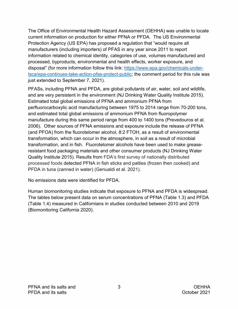
The Office of Environmental Health Hazard Assessment (OEHHA) was unable to locate current information on production for either PFNA or PFDA. The US Environmental Protection Agency (US EPA) has proposed a regulation that “would require all manufacturers (including importers) of PFAS in any year since 2011 to report information related to chemical identity, categories of use, volumes manufactured and processed, byproducts, environmental and health effects, worker exposure, and disposal” (for more information follow this link: https://www.epa.gov/chemicals-under-tsca/epa-continues-take-action-pfas-protect-public; the comment period for this rule was just extended to September 7, 2021).
PFASs, including PFNA and PFDA, are global pollutants of air, water, soil and wildlife, and are very persistent in the environment (NJ Drinking Water Quality Institute 2015). Estimated total global emissions of PFNA and ammonium PFNA from perfluorocarboxylic acid manufacturing between 1975 to 2014 range from 70-200 tons, and estimated total global emissions of ammonium PFNA from fluoropolymer manufacture during this same period range from 400 to 1400 tons (Prevedouros et al. 2006). Other sources of PFNA emissions and exposure include the release of PFNA (and PFOA) from the fluorotelomer alcohol, 8:2 FTOH, as a result of environmental transformation, which can occur in the atmosphere, in soil as a result of microbial transformation, and in fish. Fluorotelomer alcohols have been used to make grease-resistant food packaging materials and other consumer products (NJ Drinking Water Quality Institute 2015). Results from FDA’s first survey of nationally distributed processed foods detected PFNA in fish sticks and patties (frozen then cooked) and PFDA in tuna (canned in water) (Genualdi et al. 2021).
No emissions data were identified for PFDA.
Human biomonitoring studies indicate that exposure to PFNA and PFDA is widespread. The tables below present data on serum concentrations of PFNA (Table 1.3) and PFDA (Table 1.4) measured in Californians in studies conducted between 2010 and 2019 (Biomonitoring California 2020).
PFNA and its salts and 4 OEHHA PFDA and its salts October 2021
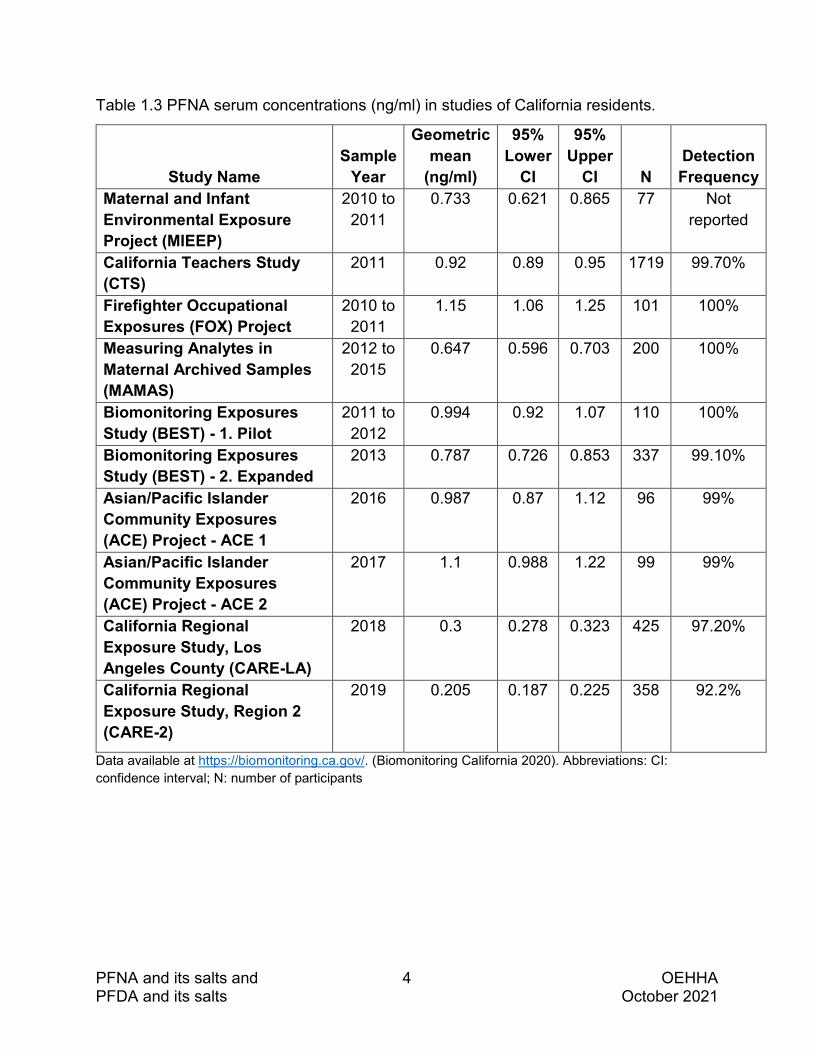
Table 1.3 PFNA serum concentrations (ng/ml) in studies of California residents.
Study Name Sample
Year
Geometric mean
(ng/ml)
95% Lower
CI
95% Upper
CI N Detection Frequency
Maternal and Infant Environmental Exposure Project (MIEEP)
2010 to 2011
0.733 0.621 0.865 77 Not reported
California Teachers Study (CTS)
2011 0.92 0.89 0.95 1719 99.70%
Firefighter Occupational Exposures (FOX) Project
2010 to 2011
1.15 1.06 1.25 101 100%
Measuring Analytes in Maternal Archived Samples (MAMAS)
2012 to 2015
0.647 0.596 0.703 200 100%
Biomonitoring Exposures Study (BEST) - 1. Pilot
2011 to 2012
0.994 0.92 1.07 110 100%
Biomonitoring Exposures Study (BEST) - 2. Expanded
2013 0.787 0.726 0.853 337 99.10%
Asian/Pacific Islander Community Exposures (ACE) Project - ACE 1
2016 0.987 0.87 1.12 96 99%
Asian/Pacific Islander Community Exposures (ACE) Project - ACE 2
2017 1.1 0.988 1.22 99 99%
California Regional Exposure Study, Los Angeles County (CARE-LA)
2018 0.3 0.278 0.323 425 97.20%
California Regional Exposure Study, Region 2 (CARE-2)
2019 0.205 0.187 0.225 358 92.2%
Data available at https://biomonitoring.ca.gov/. (Biomonitoring California 2020). Abbreviations: CI: confidence interval; N: number of participants
PFNA and its salts and 5 OEHHA PFDA and its salts October 2021
Recent findings from other human biomonitoring studies of PFNA include the following:
• The National Health and Nutrition Examination Survey (NHANES) for the years 2009 to 2010 reported serum concentration levels (ng/ml; geometric mean and 95% confidence intervals [CI]) in males aged 20 years or older of 1.40 (1.20, 1.63) (Dobraca et al. 2015).
• NHANES for the years 2013 to 2014 reported serum concentration levels (ng/ml; unadjusted geometric mean and 95% CI) in children aged 3–11 years of 0.79 (0.68−0.93) (Jain 2018).
• A 95% detection frequency of PFNA in the serum (collected in 2009 – 2016) of mothers enrolled in the Northern California CHARGE (CHildhood Autism Risk from Genetics and Environment) case-control study (Kim et al. 2020).

PFNA and its salts and 6 OEHHA PFDA and its salts October 2021
Table 1.4 PFDA serum concentrations (ng/ml) in studies of California residents.
Project Sample
Year
Geometric mean (ng/ml)
95% Lower
CI 95%
Upper CI N Detection Frequency
California Teachers Study (CTS)
2011 0.22 0.21 0.23 1759
94.70%
Firefighter Occupational Exposures (FOX) Project
2010 to 2011
0.899 0.783 1.03 101 100%
Measuring Analytes in Maternal Archived Samples (MAMAS)
2012 to 2015
0.198 0.174 0.226 200 83%
Biomonitoring Exposures Study (BEST) - 1.Pilot
2011 to 2012
0.245 0.216 0.278 110 100%
Biomonitoring Exposures Study (BEST) - 2.Expanded
2013 0.188 0.173 0.205 337 82.50%
Asian/Pacific Islander Community Exposures (ACE) Project - ACE 1
2016 0.477 0.406 0.559 96 80.20%
Asian/Pacific Islander Community Exposures (ACE) Project - ACE 2
2017 0.559 0.49 0.636 99 87.90%
California Regional Exposure Study, Los Angeles County (CARE-LA)
2018 0.0967 0.0894 0.105 425 69.20%
California Regional Exposure Study, Region 2 (CARE-2)
2019 0.0835 0.0776 0.0898 358 65.9%
Data available at https://biomonitoring.ca.gov/. (Biomonitoring California 2020). Abbreviations: CI: confidence interval; N: number of participants
PFNA and its salts and 7 OEHHA PFDA and its salts October 2021
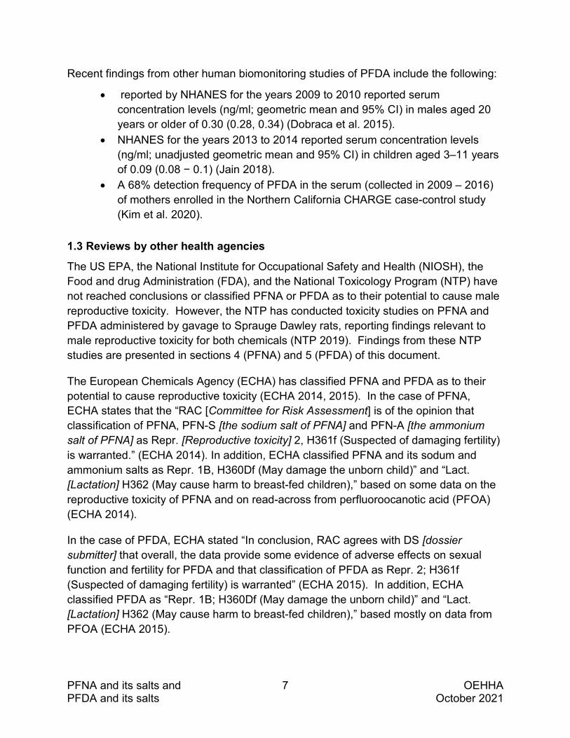
Recent findings from other human biomonitoring studies of PFDA include the following:
• reported by NHANES for the years 2009 to 2010 reported serum concentration levels (ng/ml; geometric mean and 95% CI) in males aged 20 years or older of 0.30 (0.28, 0.34) (Dobraca et al. 2015).
• NHANES for the years 2013 to 2014 reported serum concentration levels (ng/ml; unadjusted geometric mean and 95% CI) in children aged 3–11 years of 0.09 (0.08 − 0.1) (Jain 2018).
• A 68% detection frequency of PFDA in the serum (collected in 2009 – 2016) of mothers enrolled in the Northern California CHARGE case-control study (Kim et al. 2020).
1.3 Reviews by other health agencies
The US EPA, the National Institute for Occupational Safety and Health (NIOSH), the Food and drug Administration (FDA), and the National Toxicology Program (NTP) have not reached conclusions or classified PFNA or PFDA as to their potential to cause male reproductive toxicity. However, the NTP has conducted toxicity studies on PFNA and PFDA administered by gavage to Sprauge Dawley rats, reporting findings relevant to male reproductive toxicity for both chemicals (NTP 2019). Findings from these NTP studies are presented in sections 4 (PFNA) and 5 (PFDA) of this document.
The European Chemicals Agency (ECHA) has classified PFNA and PFDA as to their potential to cause reproductive toxicity (ECHA 2014, 2015). In the case of PFNA, ECHA states that the “RAC [Committee for Risk Assessment] is of the opinion that classification of PFNA, PFN-S [the sodium salt of PFNA] and PFN-A [the ammonium salt of PFNA] as Repr. [Reproductive toxicity] 2, H361f (Suspected of damaging fertility) is warranted.” (ECHA 2014). In addition, ECHA classified PFNA and its sodum and ammonium salts as Repr. 1B, H360Df (May damage the unborn child)” and “Lact. [Lactation] H362 (May cause harm to breast-fed children),” based on some data on the reproductive toxicity of PFNA and on read-across from perfluoroocanotic acid (PFOA) (ECHA 2014).
In the case of PFDA, ECHA stated “In conclusion, RAC agrees with DS [dossier submitter] that overall, the data provide some evidence of adverse effects on sexual function and fertility for PFDA and that classification of PFDA as Repr. 2; H361f (Suspected of damaging fertility) is warranted” (ECHA 2015). In addition, ECHA classified PFDA as “Repr. 1B; H360Df (May damage the unborn child)” and “Lact. [Lactation] H362 (May cause harm to breast-fed children),” based mostly on data from PFOA (ECHA 2015).

PFNA and its salts and 8 OEHHA PFDA and its salts October 2021
1.4 Overview of systematic literature review approach
Searches of the published scientific literature on the developmental and reproductive toxicity (DART) of PFNA and PFDA (and their salts) were conducted in February 2021. The searches sought to identify peer-reviewed open source and proprietary journal articles, print and digital books, reports and gray literature that potentially reported relevant toxicological and epidemiological information on the developmental and reproductive toxicity of these chemicals.
Three types of searches were conducted:
• Primary searches in major biomedical databases, conducted by OEHHA librarian Nancy Firchow, MLS
• Searches in other data sources, including authoritative reviews and reports, and databases or web resources, conducted by OEHHA scientists
• Additional focused searches, conducted by OEHHA scientists
Search process
The US EPA Computational Toxicology (CompTox) Chemicals Dashboard (https://comptox.epa.gov/dashboard) was used to identify synonyms for PFNA and PFDA (and their salts). The PubMed MeSH database (https://www.ncbi.nlm.nih.gov/mesh/) was used to identify subject headings and other index terms related to the chemicals, reproduction and development, and adverse effects on reproduction and development.
Preliminary searches were run and results evaluated to identify additional relevant search terms. The resulting search strategies were then executed in PubMed twice for each chemical (and its salts), limiting the first search to human studies, and the second search to non-human studies. There were no restrictions in the searches on exposure route or duration of exposure, or on publication language. The full DART search strings used in PubMed are included in Appendix A.
The PubMed search strategies were then tailored for use in the additional databases and data sources listed below, according to the search interface and features unique to each resource. For instance, MeSH terms were replaced with Emtree terms for the Embase search strategies.
Data sources:
The following is a list of the major data sources (biomedical literature databases) searched to find information on PFNA and its salts and PFDA and its salts.
• PubMed (National Library of Medicine) (https://www.ncbi.nlm.nih.gov/pubmed)

PFNA and its salts and 9 OEHHA PFDA and its salts October 2021
• Embase (https://www.embase.com) • Scopus (https://www.scopus.com) • SciFinder-n (https://scifinder-n.cas.org/)
In addition to the systematic literature searches, OEHHA asked the public to identify pertinent references through a data call-in that was open from March 26, 2021 to May 10, 2021. No references were submitted for consideration.
Literature screening process
The results of these literature searches were uploaded to EndNote libraries (human and non-human [i.e., experimental animal and cell-free] results were kept separate) and duplicates were removed. A total of 652 and 472 references were identified for PFNA and its salts and for PFDA and its salts, respectively, through this initial literature search process. In addition to the studies identified through this process, other relevant studies were identified from citations in individual articles, and through alert services (e.g., ScienceDirect, Google Scholar, etc.).
The EndNote libraries containing the literature search results (citations) for PFNA and its salts and PFDA and its salts were uploaded to two separate HAWC (Health Assessment Workspace Collaborative, https://hawcproject.org) projects. HAWC is a tool used for multi-level screening of literature search results.
In Level 1 screening, citations were reviewed independently by OEHHA scientists, based solely on study titles and abstracts, using specific inclusion and exclusion criteria to eliminate studies or articles that did not contain information on DART or other key related topics (e.g., pharmacokinetics, mechanisms of action). This initial screen (Level 1) was intended to identify all studies deemed to have a reasonable possibility of containing information relevant to DART that could be useful for the review process, and to further identify (i.e., tag in HAWC) studies relevant to particular aspects of DART (e.g., male reproductive toxicity, female reproductive toxicity, developmental toxicity).
For purposes of identifying the available evidence on the male reproductive toxicity of these chemicals, citations identified as having a reasonable possibility of containing information relevant to male reproductive toxicity underwent Level 2 screening. In the Level 2 screening of this subset of citations, the full text was obtained. These full papers were screened independently by one OEHHA scientist, using similar inclusion/exclusion criteria as was used in the Level 1 screening. However, Level 2 reviewers could make more accurate judgements about the relevance of the articles because they were reviewing the full text in addition to the title and abstract. Following Level 2 screening, the tagging of articles according to key topics was updated in HAWC. Level 1 and 2 screenings were repeated as search results were updated, and with

PFNA and its salts and 10 OEHHA PFDA and its salts October 2021
additional relevant studies identified from citations in individual articles and alert services (e.g., ScienceDirect, Google Scholar). (See Appendix A for additional details).
Literature searches were last updated in July 2021. One hundred and fourteen references were cited in this document.
2. PHARMACOKINETICS OF PFNA AND PFDA
2.1 Absorption
In general, PFASs can be absorbed following either oral, inhalation, or dermal exposures. The principal route of exposure in humans is the oral one followed by inhalation and dermal (Poothong et al. 2020). Studies conducted in animals indicate that about 95% of PFNA and PFDA are absorbed after oral administration (ATSDR 2021). These findings on oral absorption are similar to those for PFOA (92%) and perfluorooctane sulfonic acid (PFOS) (approximately 98%) in gavage studies in male rats (Cui et al. 2010). No quantitative estimates of the fractional absorption of PFDA or PFNA following inhalation or dermal exposure were identified (ATSDR 2021).
It is expected that PFNA and PFDA salts readily dissociate in aqueous media to form the corresponding organic acid, similar to observations that ammonium perfluorooctanoic acid in the presence of water freely forms PFOA (Hundley et al. 2006).
2.2 Distribution
In general, PFASs are present in the blood, and widely distributed in tissues throughout the body. A study measuring a number of PFASs in various human tissues (i.e., liver, kidney, brain, lung, and bone [rib]) reported the highest levels of PFNA in brain and kidney, with lower levels in lung and liver, and no detectable PFNA in bone (Pérez et al. 2013). In this same study the highest levels of PFDA were reported in brain, followed by lung and then kidney, with no detectable PFDA in liver or bone.
In studies in laboratory animals, the highest concentrations of PFASs are generally found in the liver, kidneys, and blood (ATSDR 2021). For example, in studies in male Sprague Dawley (SD) and Wistar rats the order from highest to lowest for maximum concentration (Cmax) in various tissues following a single gavage dose of PFNA was liver> kidney> serum> lungs> heart> spleen> testes> muscle> fat> intestines> brain (Benskin et al. 2009; Iwabuchi et al. 2017). For PFDA, in studies in male Wistar rats after a single gavage dose, the PFDA tissue distribution was about 86% in liver; 13% in serum and 1.4% in brain (Kawabata et al. 2017; Kudo and Kawashima 2003). Wistar rats administered a single intraperitoneal injection of PFDA (20 mg/kg) had a greater proportion of the PFDA in serum (10%) than liver (5%) when assessed seven days after

PFNA and its salts and 11 OEHHA PFDA and its salts October 2021
injection (Ylinen and Auriola 1990). Covalent binding of PFDA was detected in plasma, liver and testes, with a higher relative concentration of bound PFDA in the testis compared to plasma or liver (Vanden Heuvel et al. 1992).
Information on the tissue distribution of PFNA, PFDA, and PFASs is also available from studies in fish, chicks, and the Artic fox. In one study of PFNA in male and female zebrafish, levels were reported to be two times higher in male gonads than in female gonads (Zhang et al. 2016). In a study conducted with one-day-old male chickens exposed orally by gavage to a mixture of three PFASs (including PFDA) for three weeks, the highest levels of PFDA were in the liver, followed by kidney and blood (Yeung et al. 2009). In studies in rainbow trout that measured tissue PFAS concentrations, the highest levels were in liver, followed by blood, kidney, and skin (Falk et al. 2015; Goeritz et al. 2013). And in a study in Artic foxes, blood and liver concentrations of PFNA and PFDA were inversely correlated with levels of body fat, suggesting that body fat composition may be a factor in the tissue distribution and accumulation (Aas et al. 2014).
Transport
PFASs appear to bind to proteins in blood. PFNA and PFDA are primarily transported by albumin in humans (Forsthuber et al. 2020). In an in vitro equilibrium dialysis study, more than 99.9% of PFNA was bound to bovine or human serum albumin (Bischel et al. 2010). In rats PFDA was shown to be bound to serum proteins (Ylinen and Auriola 1990). Covalent binding of PFDA to proteins in plasma was observed in rats following in vivo administration of radiolabeled [1-14C]PFDA (Vanden Heuvel et al. 1992). In in vitro studies these authors showed that PFDA covalently binds to rat albumin and hemoglobin, and that covalent binding of PFDA to albumin was time- and dose- dependent (Vanden Heuvel et al. 1992).
Maternal-fetal transfer
PFASs can be transferred to the human fetus during pregnancy (ATSDR 2021). PFASs were found in the serum of pregnant women and in fetal cord serum in Korean, Chinese and Spanish populations (S-K Kim et al. 2011; S Kim et al. 2011; J Liu et al. 2011; Manzano-Salgado et al. 2015). Several PFASs have been detected in maternal serum, in the placenta and in embryos or fetal tissues. In a Danish population PFNA and PFDA were detected throughout pregnancy in maternal serum samples collected between 2014 and 2015, with a frequency similar to that of PFOA and PFOS; the relative frequency of detection was PFOA>PFOS>PFNA>PFDA (Mamsen et al. 2019). In a small study (N = 32) of placentas collected in a Chinese population in 2010, placental concentrations of PFNA and PFDA were eight times lower than concentrations of PFOS

PFNA and its salts and 12 OEHHA PFDA and its salts October 2021
(T Zhang et al. 2013). PFNA and PFDA appear in fetal liver and lungs with increasing frequency from the second trimester, with PFNA having a higher detection frequency than PFDA (Mamsen et al. 2019).
Several studies have reported that PFNA concentrations in maternal serum are higher than in cord serum (Manzano-Salgado et al. 2015; Needham et al. 2011; T Zhang et al. 2013), and this has also been reported for PFDA (T Zhang et al. 2013). In a recent study in matched maternal−cord serum pairs assessing the transplacental transfer efficiencies of PFNA and PFDA, lower transfer efficiencies were observed in preterm compared to full-term deliveries, with statistically significant lower PFNA and PFDA levels in preterm compared to full-term cord serum (p<0.001) (Jing Li et al. 2020).
Breast milk transfer
PFASs can be transferred to nursing infants via breastmilk (ATSDR 2021). PFNA was detected in 100% and PFDA in 78% of milk samples collected from a population of nursing mothers in China (J Liu et al. 2011). However, PFNA and PFDA levels in breast milk were below the limit of detection in a study conducted in a Korean population (S-K Kim et al. 2011; S Kim et al. 2011) and in a cohort from the Faroe Islands (Needham et al. 2011).
Sex differences in distribution
Studies in humans and animals suggest that there may be differences between males and females in the distribution within the body of PFNA and PFDA. In humans, slightly higher ratios of mean cord to maternal serum concentrations were observed in boys compared to girls for both PFNA and PFDA, although the differences did not reach statistical significance (J Liu et al. 2011). In rats, the concentration of PFNA in the liver was dependent on exposure dose and sex (Kudo and Kawashima 2003). PFNA accumulation in rat liver was greater in males than in females administered PFNA by intraperitoneal injection (Kudo et al. 2000; Kudo et al. 2001; Kudo and Kawashima 2003), and the concentration of PFNA in the serum was also higher in males than females after a single intraperitoneal exposure (Kudo et al. 2001). This higher accumulation of PFNA in the liver of male rats was dependent on testosterone (T), as liver concentrations were reduced in castrated male rats, and supplementation of castrated males treated with exogenous testosterone had PFNA liver concentrations comparable with those seen in intact males (Kudo et al. 2000; Kudo and Kawashima 2003). For PFDA, no differences in the levels of PFDA in rat liver were observed between males and females, following intraperitoneal injection (Kudo et al. 2000; Kudo et al. 2001; Kudo and Kawashima 2003), while females had higher PFDA serum levels

PFNA and its salts and 13 OEHHA PFDA and its salts October 2021
than males following intraperitoneal injection (Kudo et al. 2001) and higher PFDA plasma levels than males following intravenous injection (Dzierlenga et al. 2020).
2.3 Metabolism
Several studies for oral and intraperitoneal exposure as well as in vitro studies suggest that PFASs including PFDA are not metabolized and do not undergo chemical reactions in the body (ATSDR 2021; Ylinen and Auriola 1990). The ATSDR review reported the absence of studies examining metabolism of PFASs following inhalation or dermal exposure then they conclude that “metabolism by these exposure routes is not expected” (ATSDR 2021).
2.4 Excretion
Excretion pathways of PFNA and PFDA include urinary and fecal excretion, and for women, pregnancy (e.g., transfer to fetus), and lactation. PFNA and PFDA may also be eliminated from the body through incorporation into (finger and toe) nails.
In a small biomonitoring study of individuals in China with no known occupational exposures to PFNA or PFDA (N=39), higher detection frequencies of PFNA and PFDA were observed in the nails of study participants (51.2% and 56.4%, respectively) than in the urine (5.13% and 7.7%, respectively) (Wang et al. 2018).
Studies in humans, mice and rats indicate that both PFNA and PFDA are slowly eliminated from the body (Fujii et al. 2015; Kudo et al. 2001). Studies of humans and mice have found that PFNA is excreted to a slightly greater extent in urine compared to feces, while the opposite is the case for PFDA (Fujii et al. 2015). Studies in rats indicate that both PFNA and PFDA are excreted to a greater extent in feces compared to urine (Kudo et al. 2000; Kudo et al. 2001; Vanden Heuvel et al. 1991).
2.5 Species differences in PFNA and PFDA serum half-lives
PFASs, including PFNA and PFDA, demonstrate species differences in half-life, with estimates for serum half-lives of longer-chain PFASs, including PFNA and PFDA, on the order of days for rodents, and years for humans.
PFNA
In a human study (N=86 paired blood and morning urine samples) from healthy volunteers from two cities in China (Shijiazhuang, Handan), the geometric mean serum half-life for PFNA was estimated to be 3.2 years in men (Y Zhang et al. 2013). In male Wistar rats exposed to 22 mg/kg of PFNA by the intravenous route, the half-life was 30 days (NTP 2019). In studies of male SD rats, the plasma half-life for PFNA was 40 days

PFNA and its salts and 14 OEHHA PFDA and its salts October 2021
following intravenous exposure to 3 mg/kg, and ranged from 30 to 55 days following oral exposures (i.e., 10-14 µg/day via diet for 12 days; 3 mg/kg via gavage) (De Silva et al. 2009; Kim et al. 2019).
PFDA
In the same human study described above for PFNA, it was estimated that the geometric mean serum half-life for PFDA was 7.1 years in men (Y Zhang et al. 2013). The serum half-life for PFDA has been investigated in two studies of male SD rats, and estimates ranged from 36 to 109 days following intravenous exposure to 1 or 2 mg/kg PFDA, and from 68 to 80 days following oral exposures to 1 or 2 mg/kg PFDA (Dzierlenga et al. 2020; Kim et al. 2019).
3. ENDOCRINE SYSTEM INVOLVEMENT IN DEVELOPMENT AND FUNCTION OF THE MALE REPRODUCTIVE SYSTEM
Reproductive biology is under close control from the endocrine system. In addition to the intrinsic regulation present in the organism, environmental factors may influence the endocrine system and the reproductive processes controlled by it. Effects of environmental exposures on the endocrine system can occur through a variety of mechanisms, including alterations in hormone synthesis or metabolism, hormone transport and binding to proteins in blood, and target cell receptor mediated events, including transactivation and intracellular signaling. Studies investigating the effect of PFNA or PFDA on aspects of the endocrine system that may be relevant to the development or function of the male reproductive system are discussed in sections 4 (PFNA) and 5 (PFDA). Here we provide a brief overview of the role of the hypothalamus-pituitary-gonad axis and the thyroid hormone system in male reproductive system development and function.
3.1 Hypothalamus-pituitary-gonad axis
The hypothalamus-pituitary-gonad (HPG) axis controls the function of the testes through gonadotropin releasing hormone (GnRH), which is produced in the hypothalamus, and the gonadotropins follicle stimulating hormone (FSH) and luteinizing hormone (LH), which are produced in the pituitary.
The endocrine function of the testes starts early in life, at sexual differentiation during fetal development. Early stages of sexual differentiation involve regression of the Müllerian duct (precursor of the female internal reproductive tract) by action of antimüllerian hormone (AMH), which is produced by fetal testicular Sertoli cells.

PFNA and its salts and 15 OEHHA PFDA and its salts October 2021
Sex steroids also play an important role in male sexual differentiation, as testosterone (produced in the testes and dihydrotestosterone (DHT, produced in the brain, testes, and other organs), the most potent of the androgens, act together in the virilization of the male fetus. In adults, the testes are responsible for producing not only germ cells but also hormones that are important for normal reproductive function. Under the regulatory control of LH, androgens are produced in Leydig cells by a series of enzymatic reactions starting from cholesterol. Secreted testosterone can be further metabolized, primarily in peripheral tissues, to form either DHT or estradiol (E2).
Spermatogenesis requires healthy Sertoli cells and appropriate hormonal control. FSH and testosterone act on specific Sertoli cell receptors (located on the plasma membrane or intracellularly, respectively) in a coordinated manner to facilitate normal sperm production.
There is evidence in animal models that PFNA and PFDA are acting as endocrine disrupting chemicals, i.e., affecting hormone production, metabolism, and receptor mediated effects. Certain aspects of HPG axis disruption have been investigated for PFNA and PFDA, and will be discussed in subsequent sections.
3.2 Thyroid hormones
Thyroid hormones regulate basal metabolic rate, as well as exerting control over growth, development, and differentiation of many cells and organ systems — including the testes (as reviewed by (Rajender et al. 2011)). Despite some inconsistencies among studies, as well as species differences, reviews of thyroid hormones in male reproduction and infertility found indications that short-term hypothyroidism in post-pubertal males can have adverse effects on sperm motility, semen volume, and semen quality (Alahmar et al. 2019; Krajewska-Kulak and Sengupta 2013). The thyroid hormones, tri-iodo-L-thyronine (T3 or triiodothyronine) and tetra-iodo-L-thyronine (T4 or thyroxine), are produced and secreted by the thyroid gland in response to the regulatory hormones thyroid stimulating hormone (TSH) from the pituitary gland, and thyrotropin-releasing hormone (TRH) from the hypothalamus (Chang et al. 2008; Rajender et al. 2011). Once secreted, T4 can be converted to T3 or to reverse triiodothyronine (rT3, an inactive isomer of T3) (Chang et al. 2008; Gutshall et al. 1989).
Thyroid hormone receptors (TRs) have been identified on testicular cells, and T3 binds directly to TRs on Sertoli cells. Binding to Sertoli cell TRs activates gene transcription and protein synthesis, as well as Sertoli cell proliferation and differentiation (Alahmar et al. 2019; Rajender et al. 2011). The binding of T3 to Sertoli cell TRs is suspected to have a role in initiating sperm development. While the Rajender et al. (2011) review notes the existence of contradictory reports as to how thyroid hormone acts on Leydig as well as Sertoli cells, proposed mechanisms suggest a role for T3 in stimulating basal testosterone generation.

PFNA and its salts and 16 OEHHA PFDA and its salts October 2021
Disruption of thyroid homeostasis associated with exposure to PFASs, including PFDA and PFNA, has been reviewed (Xie et al. 2020) and attributed to several potential mechanisms including:
• Competitive displacement of T4 from binding to thyroid hormone transport proteins.
• Activation of TRs and other nuclear receptors. • Effects on expression of genes related to thyroid hormone signaling. • Regulation of enzymatic activities in the thyroid gland.
Experimental treatment of rats with PFOS, for example, was not found to alter thyroid gland histology or produce elevated TSH consistent with primary hypothyroidism (Chang et al. 2008). Observed lowering of serum total thyroxine with PFOS treatment was not attributable to disruption of the hypothalamic-pituitary-thyroid (HPT) axis or interference with release of TSH. The authors suggested that PFOS may compete with T4 for binding with transport proteins, leading to increased turnover and elimination, in turn resulting in reduced serum total T4.
4. PFNA AND ITS SALTS: MALE REPRODUCTIVE TOXICITY
4.1 PFNA: Human studies of male reproductive effects
OEHHA identified 17 epidemiologic studies of possible effects of PFNA on the male reproductive system. These studies examined one or more of the following types of outcomes: anogenital distance (AGD); outcomes related to male reproductive function, including reproductive hormones, semen quality parameters, DNA damage to sperm, and in vitro fertilization (IVF) outcomes; and cancer. The studies are ordered by outcomes, and within outcomes, by publication date, in the following discussion and in Table 4.1. Some studies included multiple outcomes but are included only once in the discussion and table. Statistically significant results in Table 4.1 are in bold type. In the following summary, all results are statistically significant unless otherwise noted.
Anogenital distance
Two prospective cohort studies examined associations between prenatal PFNA exposure and anoscrotal (AGDAS) and anopenile (AGDAP) distance, reporting conflicting findings. Tian et al. (2019), with higher maternal plasma PFNA concentrations (median = 1.75, interquartile range (IQR) = 1.30, 2.47) and larger sample size (n=500), reported non-statistically significant decreases in AGDAS and AGDAP at birth that disappeared by the age of 12 months. In the study by Lind et al. (2017), maternal serum PFNA concentrations were lower: median = 0.7, IQR = 0.5, 0.9 ng/mL. Lind et al. reported an

PFNA and its salts and 17 OEHHA PFDA and its salts October 2021
increase in AGDAS at three months, and no associations with AGDAP (Lind et al. 2017; Tian et al. 2019).
Male reproductive function
Twelve studies examined male reproductive function, including reproductive hormone levels, semen quality parameters, and IVF outcomes, in association with PFNA. Of the seven studies reporting results for associations between PFNA exposure and T, two reported decreases in T for adult men (Cui et al. 2020) and adolescent boys (Zhou et al. 2016), a third reported a nonsignificant decrease in total T in young men (Joensen et al. 2013), three studies reported no associations (Joensen et al. 2009; Joensen et al. 2013; Lewis et al. 2015; Ma et al. 2021) and one reported inconsistent evidence across locations (Specht et al. 2012). For E2, three of four studies reported no associations (Cui et al. 2020; Ma et al. 2021; Specht et al. 2012) and one reported a decrease associated with PFNA (Joensen et al. 2013). No consistent pattern of associations is apparent for other reproductive hormones. Cui et al. (2020) reported decreased sex hormone-binding globulin (SHBG) associated with both semen and serum PFNA and a lower T/LH ratio associated with semen PFNA (Cui et al. 2020). The data suggest that PFNA may be associated with lower T, but not other reproductive hormones and related proteins.
Evidence for associations of PFNA with the percentage of sperm with normal morphology and progressive motility was mixed: Louis et al. 2015 reported an increase in the percentage of normal sperm and a decrease in the percentage with coiled tails, three studies reported non-significant associations with lower proportions of morphologically normal sperm (Ma et al. 2021; Pan et al. 2019; Toft et al. 2012), and one reported no associations (Joensen et al. 2013). Evidence for associations between PFNA and sperm viability, curvilinear velocity (VCL), and straight line velocity (VSL) was sparse (Louis et al. 2015; Pan et al. 2019). Of the six studies that examined effects of PFNA exposure on sperm concentration and count, five reported no associations (Joensen et al. 2013; Louis et al. 2015; Pan et al. 2019; Petersen et al. 2018; Toft et al. 2012), while a study of a cohort of couples undergoing IVF reported that PFNA is associated with a substantial reduction in sperm concentration and a non-statistically significant reduction in sperm count (Ma et al. 2021). IVF outcomes, however, were generally not associated with PFNA (Ma et al. 2021).
Three studies examined possible effects of PFNA on sperm DNA. Specht et al. (2012) reported no associations with sperm DNA fragmentation using the sperm chromatin structure assay (SCSA) and the in situ terminal deoxynucleotidyl transferase dUTP nick-end labeling (TUNEL) assays. However, Pan et al. (2019) also conducted the SCSA and reported that semen PFNA was associated with small increases in DNA

PFNA and its salts and 18 OEHHA PFDA and its salts October 2021
fragmentation index (DFI), and both semen and serum PFNA were associated with small increases in high DNA stainability (HDS) indicative of damage to integrity of DNA (Pan et al. 2019). DNA methylation parameters were generally not associated with PFNA (Leter et al. 2014).
Cancer
Two studies examined associations between PFNA and prostate cancer or prostate-specific antigen (PSA) level. A case-control study in Sweden reported that PFNA exposure above the control median was not associated with prostate cancer (Hardell et al. 2014). In a sample from the C8 Study, PFNA was not associated with PSA level (Ducatman et al. 2015).
A case-control study in Shanghai, China investigating the association between serum PFAS levels and germ cell tumors in children did not report findings by tumor location (e.g., testes vs. ovaries) or sex of the child (Lin et al. 2020). Because of these limitations in reporting, the study is uninformative for purposes of evaluating male reproductive system hazards, and the study was not included in Table 4.1.

PFNA and its salts and 19 OEHHA PFDA and its salts October 2021
Table 4.1 PFNA: Epidemiologic studies of male reproductive toxicity2.
Reference; study location, period, design, sample
Exposure matrix and concentration
(ng/mL) Outcomes Results Covariates, other
PFASs analyzed Comments
Lind et al., 2017 Denmark 2010-2012 Prospective cohort N=299 Odense Child Cohort: sons of mothers who were >16 years old, of Western origin, had no communication barriers, delivered in a reference hospital, and did not use reproductive assistance.
Maternal serum collected at gestational week 5-12 (median 10 weeks) Median (IQR) 0.7 (0.5-0.9)
AGDAS & AGDAP and penile width at 3 months, measured three times by trained study technicians
AGDAS
β (95% CI) per unit increase in ln(PFNA): 1.4 (0.02, 2.9) mm No association when 2nd, 3rd, 4th quartiles of PFNA were compared to 1st AGDAP, penile width: no associations
Adjusted for “post-conceptional age” (sum of GA at birth and age of child at the AGD measurement, in days), z-score for weight at 3 months, parity, maternal smoking, and pre-pregnancy body mass index (BMI) Other PFASs: perfluorohexane sulfonic acid (PFHxS), PFOS, PFDA
Correlations among PFASs not reported. Women who gave birth in 2010 had higher PFOA, PFOS, and PFNA concentrations than women who gave birth in 2011-2012. Women who participated in this study were better educated, smoked less, and were more likely to be of Danish origin than nonparticipants.
Tian et al., 2019 Shanghai, China 2012 Prospective cohort N=500 male singletons born to women who were recruited at gestational weeks 12-16 from one hospital
Maternal plasma (fasting) collected at 12-16 weeks gestation Median (5, 25, 75, 95 percentiles): 1.75 (0.73, 1.30, 2.47, 3.99) Geometric mean (GA; standard deviation or SD): 1.8 (1.68)
AGD [AGDAS & AGDAP] at birth, 6 months, and 12 months Measured by trained examiners with no knowledge of maternal PFAS concentrations
β (95% CI) for unit change in ln(PFNA) (mm) AGDAS Birth: –0.51 (–1.19, 0.17) 6 months: –1.83 (–4.12, 0.45) 12 months: no association AGDAP Birth: –0.34 (–1.14, 0.46) 6 months: –0.87 (–3.17, 1.44) 12 months: no association In longitudinal analyses, no associations with AGD
Selected a priori and adjusted for: maternal age, education, parity, and pre-pregnancy BMI; GA at birth; age at examination (days), infant size (weight at birth, weight-for-length z-score at 6 and 12 months of age) Stratified by breastfeeding duration for AGD at 6 and 12 months 8 PFASs detected in ≥80% of participants: PFHxS, PFOA, PFOS, PFNA, PFDA, PFUnDA, PFDoA, PFTrDA
Pearson correlation coefficient for ln-transformed PFNA and: PFOS r = 0.64 PFOA r = 0.39 PFDA r = 0.79 PFUnDA r = 0.796 PFDoA r = 0.47 PFTrDA r = 0.31 75% of mothers had university-level education or higher
2 Studies are ordered by outcomes (AGD, male reproductive function, prostate cancer), and within outcomes, by publication date. Statistically significant results are in bold type.

PFNA and its salts and 20 OEHHA PFDA and its salts October 2021
Reference; study location, period, design, sample
Exposure matrix and concentration
(ng/mL) Outcomes Results Covariates, other
PFASs analyzed Comments
Joensen et al., 2009 Denmark 2003 Cross-sectional* N=105 Healthy young men from the general population reporting for the military draft, median age 19 years. *Men with highest and lowest T levels were selected. Group 1, n=53, median 31.8 nmol/L Group 2, n=52, median 14.0 nmol/L
Serum Median (5th, 95th percentiles) All 0.8 (0.4, 1.8) High T 0.8 (0.4, 2.0) Low T 0.8 (0.4, 2.0)
Serum reproductive hormones and related proteins: T, E2, sex hormone-binding globulin (SHBG), LH, follicle-stimulating hormone (FSH), inhibin B, free androgen index (FAI) Semen quality: semen volume; sperm concentration, count, % motile, morphology
No associations with T Due to low concentration, PFNA was not included in further analyses
PFNA was included only in bivariate analyses (no adjustments) Other PFASs: PFHxS, PFPA, PFOA, PFOS, PFOSA, PFDA, PFUnDa, PFDoA, PFTrA
Specht et al., 2012 Kharkiv, Ukraine; Greenland; Warsaw, Poland 2002-2004 Cross-sectional N=604 Men who provided semen and blood samples and completed questionnaires, and were partners of pregnant women enrolled in the INUENDO fertility cohort
Serum Median (range) Ukraine 1.0 (0.2, 4) Greenland 1.4 (0.5, 12) Poland 1.2 (0.5, 6)
Percentage of sperm with detectable DNA fragmentation by SCSA DNA fragmentation by in situ TUNEL assay Pro- (Fas) and anti-apoptotic (Bcl-xL) markers on sperm Serum: FSH, LH, E2, T, SHBG Inhibin B
No associations with sperm DNA fragmentation (SCSA and TUNEL assays) (data not reported). No consistent associations with apoptotic markers across study locations or models within locations (data not reported). No consistent relationship with SHBG. Associations with T, E2, and the gonadotropins FSH and LH were not consistent across locations (data not reported). Results for inhibin-B were not reported
Analyses were stratified by study location and adjusted for sexual abstinence period, age, BMI, caffeine consumption, serum cotinine, recent fever, self-reported genital infections, testicular disorders, and semen spillage. Alcohol use was not included due to frequency of missing data. Other PFASs: PFHxS, PFOA, PFOS Spearman’s rank correlations among PFASs ranged from r=0.4 to r=0.9, though correlations with PFNA were not reported.
All men had pregnant partners, thus sterile and highly subfertile men are underrepresented. Men were asked to collect semen after ≥2 days of abstinence High frequency of missing data for Bcl-xL and Fas due to samples lost during shipment and insufficient number of cells for analysis. Many reproductive hormone samples were missing for Greenland (32%) and Poland (41%).

PFNA and its salts and 21 OEHHA PFDA and its salts October 2021
Reference; study location, period, design, sample
Exposure matrix and concentration
(ng/mL) Outcomes Results Covariates, other
PFASs analyzed Comments
Toft et al., 2012 Kharkiv, Ukraine; Greenland; Warsaw, Poland 2002-2004 Cross-sectional N=588 Men who provided semen and blood samples and completed questionnaires, and were partners of pregnant women enrolled in the INUENDO fertility cohort
Serum median (33rd, 67th percentiles) All 1.2 (1.0, 1.5) Ukraine 1.0 (0.8, 1.2) Greenland 1.7 (1.3, 2.4) Poland 1.2 (1.0, 1.3)
Semen quality: sperm concentration, volume, total count, % morphologically normal sperm, % motile sperm
No associations with sperm concentration, volume, total count, % motile sperm (data not reported) NS association with lower % of normal sperm when PFNA was analyzed as a continuous variable (data not reported)
Selected a priori and adjusted for: age, abstinence time, semen spillage, current smoking, history of urogenital infections, BMI, country (combined analyses) Sperm motility analyses were restricted to samples analyzed within 1 hour of collection Other PFASs: PFHxS, PFOA, PFOS
Participation rates: Kharkiv 36% Greenland 79% Warsaw 29% All men had pregnant partners, thus sterile and highly subfertile men are underrepresented. Men were asked to collect semen after ≥2 days of abstinence Volume and total sperm count analyses were restricted to samples with no reported semen spillage

PFNA and its salts and 22 OEHHA PFDA and its salts October 2021
Reference; study location, period, design, sample
Exposure matrix and concentration
(ng/mL) Outcomes Results Covariates, other
PFASs analyzed Comments
Joensen et al., 2013 Denmark 2008-2009 Cross-sectional N=247 Healthy young men randomly selected from the general population, median age 19 years
Serum Median (IQR) 1.07 (0.88, 1.41) Mean ± SD 1.23 ± 0.63
Serum reproductive hormones and related proteins: total T, free T, E2, inhibin B, FAI; T×100/SHBG), FSH, LH, SHBG Semen quality: volume; sperm count, concentration, motility, morphology, total normal sperm Testicular volume
β (CI) per ng/mL increase in PFNA Hormones (ln-transformed) Total T β = –0.059 (–0.118, 0.001) nmol/L Free T β = –0.052 (–0.114, 0.010) nmol/L E2 β = –0.075 (–0.013, –0.019) pmol/L No associations with other reproductive hormones or SHBG Semen quality and testicular volume: no associations
Analyses with hormones and SHBG were adjusted for BMI and smoking Semen volume, concentration, and total count were adjusted for abstinence time % morphologically normal sperm was unadjusted Progressively motile % was adjusted for time to semen analysis Considered but not included: time of day of blood sample, ethnicity, recent alcohol use, prenatal exposure to tobacco smoke, previous or current diseases, recent fever, recent medication use, season, interaction with cigarette smoking Other PFASs: PFHxS, PFHpS, PFOA, PFOS, PFDA
Participation rate was ~30%, which is “higher than other population-based semen quality studies”. Participants were recruited at a compulsory medical examination for consideration for military service Men were asked to abstain for 48 hours; median abstinence period was 62 hours. Analyses were blinded

PFNA and its salts and 23 OEHHA PFDA and its salts October 2021
Reference; study location, period, design, sample
Exposure matrix and concentration
(ng/mL) Outcomes Results Covariates, other
PFASs analyzed Comments
Leter et al., 2014 Kharkiv, Ukraine; Greenland; Warsaw, Poland 2002-2004 Cross-sectional N = 262 from 607 male partners of pregnant women enrolled in the INUENDO fertility cohort (312 samples were randomly selected from those with sufficient semen. Further sample loss is due to “random unpredictable cell loss occurring during the processing of some samples.”
Serum Mean ± SE: Kharkiv 1.1 ± 0.1 Greenland 2.2 ± 0.2 Warsaw 1.4 ± 0.1 Combined 1.6 ± 0.1
Sperm DNA global methylation levels, indicated by a) average DNA methylation in repetitive DNA sequences (Alu, LINE-1, Satα) and b) flow cytometric (FCM) immunodetection of 5-methyl-cytosines using the FCM Sperm DNA Global Methylation (DGML) Assay, performed by blinded investigators
Results were inconsistent across study location β (CI) per unit ln(PFNA): Alu For each study location, the association was negative but ns LINE-1 Kharkiv β = 5.7 (1.4, 10.1) % No associations for other study locations Satα Kharkiv β = 9.3 (1.5, 17.1) % No associations for other study locations FCM DGML Warsaw β = –99.6 (–152.5, –46.8) units No associations for other study locations
Adjusted for age, smoking (selected a priori), study location combined analyses) Other PFASs and correlations: PFHxS r=0.556 PFOA r=0.417 PFOS r=0.571 PFDA*, PFUnDA*, PFDoDA* *excluded due to low detection rate
All men had pregnant partners, thus sterile and highly subfertile men are underrepresented. Men were asked to collect semen after ≥2 days of abstinence
Lewis et al., 2015 United States 2011-2012 Cross-sectional N = 857 males, 12-80 years Subset of the NHANES with data on serum PFASs, T, and thyroid hormones
Serum Median (IQR) by age group: 12 to <20 years: 0.78 (0.56, 1.19) 20 to <40 years: 0.98 (0.67, 1.31) 40 to <60 years: 1.00 (0.67, 1.57) 60 to 80 years: 1.07 (0.77, 1.58)
Serum total T, thyroid stimulating hormone (TSH), and free and total T and free and total thyroxine (T4)
Percent change (95% CI) in hormone concentrations per doubling of PFNA: No associations with T 12 to <20-year-olds: TSH 16.3 (4.0, 30.2), p < 0.05 40 to 60-year-olds: Total T4: –2.5 (–5.2, 0.2), p < 0.1. No other associations with thyroid hormones
Adjusted for age, body mass index (BMI), poverty income ratio, race/ethnicity, serum cotinine Other PFAS: PFHxS, PFOA, PFOS Correlations among PFASs were not reported
The largest association of any PFAS and thyroid hormones was that of PFNA with TSH in 12-20 year olds. Significant reduction in geometric mean levels of the included PFASs compared to previous NHANES studies

PFNA and its salts and 24 OEHHA PFDA and its salts October 2021
Reference; study location, period, design, sample
Exposure matrix and concentration
(ng/mL) Outcomes Results Covariates, other
PFASs analyzed Comments
Louis et al., 2015 Michigan and Texas 2005-2009 Cross-sectional N=462 Population-based sample of couples planning pregnancy, recruited from a marketing database in Michigan and a fishing/hunting license registry in Texas.
Serum Median (IQR); geometric mean (95% CI) Michigan: 1.0 (0.75, 1.35); 0.96 (0.84, 1.11) Texas: 1.65 (1.2, 2.2); 1.68 (1.61, 1.76)
Semen quality: Volume, straw distance, sperm concentration, total count, hypo-osmotic swollen, 8 motility measures, 6 sperm head measures, 12 individual and 2 summary morphology measures, 2 sperm chromatin stability measures
Associations with 1-unit increase in ln(PFNA): No significant differences in semen volume; sperm viability, count, or concentration. % normal sperm, strict criteria, β= 3.897 (0.564, 7.231) % of sperm with coiled tail, β= –4.030 (–7.766, –0.293) No significant associations with other semen parameters
Adjusted for age, BMI, serum cotinine, abstinence, sample age, research site (Texas/Michigan) Other PFASs: PFOA, PFOS, PFOSA, Et- PFOSA-AcOH, Me-PFOSA-AcOH, PFDA
Semen was collected after ≥2 days’ abstinence A 2nd sample collected approximately 1 month later was used to corroborate azoospermia observed in the 1st sample. Semen analysis was performed one day after collection. PFOA, PFOSA, and Me-PFOSA-AcOH were most often associated with semen parameters.
Zhou et al., 2016 Taipei, Taiwan 2009-2010 Cross-sectional N=102 healthy 13-15-year-old boys from 7 public schools From the control cohort of the Genetics and Biomarkers study for Childhood Asthma
Serum sampled after 8-hour fast Median (IQR) 0.8 (0.6, 1.0)
Serum T and E2 (average of 2 values)
β (95% CI) per ng/mL increase in PFNA: Ln(T) –0.4233 (–0.6998, –0.1467) mmol/L Ln(E2) 0.1252 (–0.0758, 0.3263) mmol/L
Adjusted for: age, parental education, BMI, environmental tobacco smoke exposure, regular exercise, month of survey Other PFASs: PFBS, PFHxA, PFHxS, PFOA, PFOS, PFDA, PFDoA, PFTA
More associations between individual PFAS levels and hormone levels were reported in boys than in girls

PFNA and its salts and 25 OEHHA PFDA and its salts October 2021
Reference; study location, period, design, sample
Exposure matrix and concentration
(ng/mL) Outcomes Results Covariates, other
PFASs analyzed Comments
Petersen et al., 2018 Faroe Islands 2007-2009 Cross-sectional N=263 Entire population of 24-26 year old men were invited
Serum Median (range) 0.49 (0.61, 18.10)
Serum reproductive hormones and related proteins: FSH, LH, T, free T, E2, SHBG, T/LH, free T/LH, T/E2, FT/E2), inhibin B, inhibin B/FSH Semen quality: Sperm conc., total sperm count, semen volume, % motile sperm
Specific results for PFNA were not reported. Authors state there were no associations between semen quality and any PFASs. Associations with reproductive hormones were not mentioned for PFNA.
Reproductive hormones (except FSH, LH) were adjusted for BMI, smoking, age, time of day of blood sampling. Sperm conc., total sperm count, and semen volume were adjusted for abstinence (hours). % motile sperm was adjusted for interval between ejaculation and assessment. Other PFASs: PFHxS, PFOA, PFOS, PFDA
49% of those reached by phone (24% of all young men invited by mail) participated.
Pan et al., 2019 Nanjing, China 2015-2016 Cross-sectional N=664 males from couples who visited a reproductive medical center. Some men had fecundity issues and some were partners of women with female factor infertility Exclusions: reproductive tract disease, medication for fertility, insufficient semen volume for analysis
Semen (after 2-day abstinence) Median (IQR) 0.024 (0.013, 0.042) Serum (same day as semen): median (IQR) 1.466 (1.011, 2.216)
Semen quality: semen volume, sperm conc., sperm count, progressively motile (%), VCL, VSL, morphologically normal (%), DNA fragmentation index (DFI), HDS Concentration and motility were measured by CASA
P-values were adjusted for false discovery rate/multiple comparisons Change (95% CI) per unit increase in: Ln(semen PFNA): Progressively motile (%) β=–1.999 (–3.284, –0.714) p=0.02 VCL (µm/s): β=–0.792 (–1.542, –0.042) p=0.1 VSL (µm/s) β=–0.686 (–1.315, –0.058) p=0.1 DFI (%) β=0.106 (0.047, 0.165) p=0.01 HDS (%) β=0.044 (0.002, 0.086) p=0.1 No associations with other semen parameters Ln(serum PFNA): Morphologically normal (%) β=–0.262 (–0.545, 0.020) p=0.2 HDS (%) β=0.119 (0.063, 0.176) p=0.01 No associations with other semen parameters.
Adjusted for: Age, BMI, BMI2, smoking status, alcohol use, abstinence time Also considered: having fathered a pregnancy, occupational hazards, medical history Analyses of associations with semen parameters focused on the most abundant PFASs in serum and semen and those detected in at least 80% of semen samples: PFOA, PFOS, 6:2 Cl-PFESA, PFNA, PFDA, PFUnDA
Correlation between serum and semen PFNA: r=0.72 Correlations between serum PFNA and other PFASs: PFHxS r=0.361 PFOA r=0.570 PFOS r=0.740 6:2 Cl-PFESA r=0.752 8:2 Cl-PFESA r=0.665 PFDA r=0.825 PFUnDA r=0.910 PFDoA r=0.821 PFTrDA r=0.810 Associations of PFNA with semen parameters were similar to those of other PFASs.
Cui et al., 2020 Nanjing, China 2015-2016
Serum Median (IQR) 1.47 (1.03, 2.23)
Serum reproductive hormones and related proteins: total T, free T, E2, FSH, LH, T/LH
% change (95% CI) in hormone level per unit change in ln(serum PFNA), for all men and men < 30 years, with p-trend for analyses by PFNA quartiles: Total T –3.99 (–7.01, –0.87), p-trend = 0.013
Adjusted for: age, BMI, smoking status, time of blood sampling, fasting status
No statistical adjustment for multiple tests

PFNA and its salts and 26 OEHHA PFDA and its salts October 2021
Reference; study location, period, design, sample
Exposure matrix and concentration
(ng/mL) Outcomes Results Covariates, other
PFASs analyzed Comments
Cross-sectional N=651 (see Pan et al., 2019) Additional exclusion: missing reproductive hormone data
Mean (range) 1.82 (0.27, 17.30) Semen sampled after 2-day abstinence: Median (IQR) 0.02 (0.01, 0.04) Mean (range) 0.03 (<LOQ, 0.36)
as an indicator of Leydig cell function SHBG, FAI
< 30 years: –5.54 (–9.09, –1.84), p-trend=0.004
Free T –2.77 (–5.69, 0.25), p-trend = 0.072 < 30 years: –4.54 (–8.90, 0.02), p-trend=0.016
E2 –2.08 (–7.56, 3.73), p-trend=0.475 < 30 years: –5.32 (–11.40, 1.19), p-trend=0.107
SHBG –4.32 (–8.12, –0.37), p-trend = 0.033 < 30 years: –4.50 (–8.00, –0.87), p-trend=0.051
Total T/LH ratio –3.4 (–7.92, 1.34), p-trend = 0.157 < 30 years: –1.65 (–7.18, 4.21), p-trend=0.573
FAI, FSH, LH: no associations
Ln(semen PFNA): Total T –5.27 (–8.27, –2.18), p-trend = 0.001
< 30 years: –7.18 (–10.97, –3.24), p-trend=0.001
Free T –2.76 (–5.71, 0.29), p-trend=0.075 < 30 years: –8.45 (–12.96, 3.71), p-trend=0.062
E2 –2.97 (–8.45, 2.84), p=0.308 < 30 years: –6.49 (–13.02, 0.54), p-trend=0.069
SHBG –6.44 (–10.17, –2.55), p-trend=0.001 < 30 years: –3.82 (–7.67, 0.19), p-trend=0.001
Total T/LH ratio–6.19 (–10.61, –1.56), p-trend=0.009
< 30 years: –6.32 (–12.02, –0.24), p-trend=0.042
FAI, FSH, LH: no associations
Also considered: abstinence time, alcohol use, having fathered a pregnancy Focused on the most abundant PFASs (those that accounted for 70% and 73% % of PFASs in serum and semen samples, respectively): PFOA, PFOS, 6:2 Cl-PFESA, PFNA
Spearman correlation between serum and semen PFNA r=0.716 Associations were stronger in younger men. No SS associations among men >30 years. P-values for associations with serum PFAS were greater than for semen PFAS. Authors note that most toxicological studies demonstrate that PFAS (including PFNA) exposure reduces T secretion. Effect sizes were greater but less likely to reach statistical significance in analyses restricted to men who had fasted.

PFNA and its salts and 27 OEHHA PFDA and its salts October 2021
Reference; study location, period, design, sample
Exposure matrix and concentration
(ng/mL) Outcomes Results Covariates, other
PFASs analyzed Comments
Ma et al., 2021 Hangzhou, Zhejiang, China 2017 Prospective cohort IVF outcomes) and cross-sectional (hormones and semen quality outcomes) N=96 couples undergoing IVF treatment due to tubal factor infertility Men with severe male-factor infertility were excluded
Plasma Median (IQR) [range] 2.3 (1.7,3.9) [0.7, 16.7] Concentrations by tertiles: 1st: 0.7-1.8 2nd: 1.8-3.4 3rd: 3.4-16.7
Plasma reproductive hormones: FSH, LH, E2, T Semen quality: volume; sperm motility, concentration, motility, morphology IVF outcomes
Compared to 1st tertile of PFNA: Hormones No associations with FSH, LH, E2, or T Semen quality Sperm concentration ↓ in 2nd and 3rd (25% reduction) tertiles p-trend = 0.031 Sperm count ns ↓ in 2nd and 3rd tertiles, p-trend = 0.050 % sperm with normal morphology ns ↓ in 2nd and 3rd tertiles, p-trend = 0.109 Progressive motility no association IVF outcomes 2nd tertile exposure was associated with more fertilization, p-trend = 0.667 No associations with number of good quality embryos at day 3, implantation, clinical pregnancy, or live births
Adjusted for age, BMI, smoking status Other PFASs: PFBA, PFHxS, PFHpA, PFOA, PFOS, PFOSA, PFNA, PFUnDA, PFDoA
Each couple contributed one cycle of IVF treatment to the study. Male and female partners’ PFNA levels were highly correlated, r=0.74. PFNA was strongly correlated with: PFDA r = 0.881* PFOS r = 0.736* PFUnDA r = 0.870* *p< 0.05 2nd and 3rd tertile PFUnDA was associated with ↑ T
Hardell et al., 2014 Sweden, 2007-2011 Case-control N=200 cases of newly diagnosed prostate cancer from one hospital N=186 population-based, matched controls with no history of cancer
Whole blood drawn after diagnosis but before treatment with radiation or chemotherapy. Mean, median (range) Cases 0.679, 0.612 (0.0500, 4.6) Controls 0.631, 0.572 (0.0850, 2.1)
Prostate cancer Medical records
OR = 1.2, 95% CI (0.8, 1.8) for exposure above control median Authors state that using 75th percentile exposure cutoff resulted in somewhat higher ORs, but did not show results.
Adjusted for age, BMI, year of sample. Matched on age and geographic area. Other PFASs: PFHxA, PFHxS,PFOA, PFOS, PFDA, PFUnDA, PFDoDA
Participation rates were 60% for controls and 79% for cases. Gleason score, prostate-specific antigen, and a combination of both were not associated with PFASs. Serum was sampled after cancer diagnosis.

PFNA and its salts and 28 OEHHA PFDA and its salts October 2021
Reference; study location, period, design, sample
Exposure matrix and concentration
(ng/mL) Outcomes Results Covariates, other
PFASs analyzed Comments
Ducatman et al., 2015 Mid-Ohio Valley 2005 – 2006 Cross-sectional N = 12,988 males over 20 years of age with measured PSA. Sample from the C8 Study, which initially included those who lived, worked, or went to school in one of the six water districts contaminated with PFOA from a chemical facility; an estimated 81% of eligible residents participated
Serum Mean ± SD 1.47 ± 1.63
Serum PSA concentration PSA ≥ 4.0 was considered clinically significant
Ratio of adjusted geometric mean ln(PFNA) concentrations for PSA ≥ 4.0 vs PSA < 4.0, by age group: 20-49 years: 0.85 (0.69, 1.06) 50-69 years: 1.04 (0.95, 1.13) No relationship with PSA (continuous) in either age group
Stratified by age 20–49 years vs. 50–69 years and adjusted for geometric mean age within age strata (35 or 60 years), smoking status, alcohol intake, and BMI. Other PFASs: PFHxS, PFOA, PFOS
Sample size in analysis is approximately half of original sample. Other than exclusion of men ≥70 years, reasons for the reduced sample size are not stated. Unclear whether stratification and adjustment were appropriate and adequate for addressing confounding by age

PFNA and its salts and 29 OEHHA PFDA and its salts October 2021
4.2 PFNA: Animal studies of male reproductive toxicity
Overview
The male reproductive toxicity of PFNA has been investigated in four studies in rats (Feng et al. 2009; Feng et al. 2010; Hadrup et al. 2016; NTP 2019), five studies in mice (Das et al. 2015; Singh and Singh 2019a, 2019b, 2019c, 2019d) and one study in zebrafish (Zhang et al. 2016) (See Table 4.2.2). Experiments in which PFNA was administered as part of a mixture of chemicals are not included in this review.
Three of the studies conducted in rats were in adult or pubertal male Sprague Dawley (SD) rats and one was in pubertal male Wistar rats. The study by NTP treated nine- to 11-week-old SD rats at five dose levels (ranging from 0.625 to 10.0 mg/kg-day) by daily gavage for 28 days (NTP 2019), 2019 #917}. All animals in the NTP study were subject to histopathological evaluation; however, because of treatment-related mortality in the 5.0 and 10.0 mg/kg-day dose groups (mortality: 8/10 and 10/10, respectively) only animals from the control and three lower dose groups (0.625, 1.25 and 2.5 mg/kg-day) were included in the full toxicity assessment. In the other two studies in SD rats, eight-week-old male rats were treated at three dose levels (1, 3, and 5 mg/kg-day) with PFNA by daily gavage for 14 days (Feng et al. 2009; Feng et al. 2010).
The study by (Hadrup et al. 2016) exposed seven-week-old male Wistar rats to PFNA at three dose levels (0.0125, 0.25, 5 mg/kg-day) by daily gavage for 14 days, and also included additional treatment groups (not discussed further here) where PFNA was administered together with a mixture of 14 other chemicals (Hadrup et al. 2016). Plasma levels of sex steroid hormones and testicular expression of messenger ribonucleic acid (mRNA) of selected genes involved in steroidogenesis were measured; however, differences between “PFNA only” treatment groups and vehicle controls were not always subject to statistical testing and for several of the mRNAs assessed data were not shown.
Three of the studies in mice were in prepubertal male Parkes mice (25 days-old at the start of exposure), with one treating the animals at two dose levels (0.2, 0.5 mg/kg-day) by daily gavage for 90 days (Singh and Singh 2019a) and the other two treating the animals at ten-fold higher doses (2, 5 mg/kg-day) by daily gavage for 14 days (Singh and Singh 2019b, 2019c). Another two studies in mice treated pregnant females during gestation and investigated the effects on male pups (Das et al. 2015; Singh and Singh 2019d). The study by Singh and Singh (2019d) treated pregnant female Parkes mice from gestation day (GD) 12 to parturition at two dose levels (2, 5 mg/kg-day) and assessed male pups on postnatal days (PND) 1 and 3. The study by Das et al. (2015) treated pregnant CD-1 mice from GD 1-17 at four dose levels (1, 3, 5, 10 mg/kg-day), and assessed male fetuses from half of the dams on GD 17 and male offspring from the remaining dams for up to 41 weeks of age. In this study, full litter resorptions occurred in all of the pregnant dams in the 10 mg/kg-day group.

PFNA and its salts and 30 OEHHA PFDA and its salts October 2021
The effects of 180-day exposure of adult zebrafish to three concentration levels of PFNA (0.01, 0.1, 1 mg/L) on gonadosomatic index, measures of fertility, sex steroid hormones, expression of vitellogenin protein and mRNA, and expression of mRNA of selected genes involved in steroidogenesis or associated with the HPG axis were evaluated (Zhang et al. 2016).
Relevant major findings from these studies are discussed below, grouped by the type of effect (reproductive organ weight and histopathology, sperm parameters, hormonal effects, fertility or reproductive performance, and development of the male reproductive system).
Organ Weight and Histopathology
Histopathological changes in the testis have been observed in rats and mice treated by gavage with PFNA.
Key findings from the NTP 28-day rat study are summarized in Table 4.2.1 below (NTP 2019). The animals were approximately 9-11 weeks old at the beginning of dosing. Testis weight was reduced at 1.25 and 2.5 mg/kg-day doses by approximately 7% and 20%, respectively. Epididymal weight was reduced at 0.625, 1.25 and 2.5 mg/kg-day (7%, 13% and 35%, respectively). Increased incidence of germ cell degeneration, spermatid retention (inhibited spermiation), and interstitial cell atrophy were observed in the testes of rats exposed to PFNA at doses of 2.5 mg/kg-day and above. Treatment with Wyeth-14,643, a peroxisome proliferator-activated receptor alpha (PPARα) agonist, at doses up to 25 mg/kg-day did not result in any obvious testicular damage in the same study, even though liver effects were apparent.

PFNA and its salts and 31 OEHHA PFDA and its salts October 2021
Table 4.2.1 PFNA or Wyeth-14,643: Relevant key findings in male rats treated by gavage for 28 days (NTP 2019)
Treatment Dose (mg/kg-day) Wyeth-14,643
PFNA
Doses (mg/kg-day)
0 0.625 1.25 2.5 5.0a 10.0a 25.0b
No. of rats per group
10 10 10 10 10 10 10
Survival rate 10/10 10/10 10/10 10/10 2/10 0/10 10/10
Body weight (g) 344±6 332±6 286±5** 193±9** No data No data 308±5**
Relative liver weight (g)
34.14±0.3 42.12±0.58** 54.47±0.59** 64.37±1.86** No data No data 53.90±1.67**
Hepatocyte cytoplasmic alteration
0/10 10/10** 10/10** 10/10** 9/9** 10/10** 10/10**
Hepatocyte hypertrophy
0/10 7/10** 10/10** 10/10** 9/9** 10/10** 10/10**
Relative thyroid weight (g)
0.07±0.00 0.08±0.00 0.07±0.00 0.09±0.01** No data No data Not reported
TSH (ng/dL) 20.33±2.31 13.70±1.27 10.97±1.22** 10.16±3.35** No data No data 13.07±1.01**
T3 (ng/dL) 78.21±4.54 58.54±2.11 84.93±2.94 111.79±10.16 No data No data Not reported
Free T4 (ng/dL) 216±0.15 0.55±0.02** 0.33±0.01** 0.30±0.00** No data No data 1.31±0.13**
Total T4 (ng/dL) 2.36±0.0.27 0.21±0.07** 0.38±0.07** 1.49±0.13 No data No data 2.78±0.17**
Absolute testis weight (g)
1.885±0.041 1.820±0.035 1.762±0.031* 1.507±0.052** No data No data 1.774±0.036
Absolute epididymal weight (g)
0.555±0.013 0.515±0.010* 0.482±0.005** 0.363±0.022** No data No data 0.512±0.011
Spermatid heads (106/testis)
230.2±10.7 192.5±7.1* 220.8±10.8 205.1±9.5 No data No data 255.8±11.5

PFNA and its salts and 32 OEHHA PFDA and its salts October 2021
Treatment Dose (mg/kg-day) Wyeth-14,643
PFNA
Doses (mg/kg-day)
0 0.625 1.25 2.5 5.0a 10.0a 25.0b
Epididymal sperm count (106/cauda epididymis)
142.3±9.4 136.2±7.9 116.0±6.3* 98.1±9.0** No data No data 116.3±8.4
Epididymal sperm motility
85.2±0.5 85.9±0.4 86.4±0.75 86.4±0.7 No data No data 88.2±0.5
T (ng/mL) 4.48±1.31 4.86±1.33 3.23±1.387 0.85±0.51** No data No data 1.56±0.39*
Interstitial cell atrophy
0/10 0/10 1/10 10/10** 9/9** 10/10** Not reported
Spermatid retention
0/10 0/10 0/10 6/10** 9/9** 10/10** Not reported
Germ cell degeneration
0/10 0/10 0/10 6/10** 9/9** 10/10** 1/10
Epididymal lesion
0/10 0/10 0/10 6/10** 9/9** 10/10** 1/10
a Due to high treatment-related mortality in the 5- and 10-mg/kg-day dosing groups, animals in these groups were subject to histopathology, but were not assessed for other measures. b Only the findings from the high dose of Wyeth-14,643 are presented in this table. * p<0.05 **p<0.01

PFNA and its salts and 33 OEHHA PFDA and its salts October 2021
Treatment with PFNA at 1 or 3 mg/kg-day for 14 days did not cause histopathological changes in the testes of 8-week-old SD rats (Feng et al. 2009). At 5 mg/kg-day, PFNA caused sloughing of germ cells into the lumen of the seminiferous tubules and chromatin condensation and margination in germ cells, characteristic of germ cell apoptosis (Feng et al 2009). In a follow-up study of the same design, ultrastructural changes were observed in the testes of rats treated with PFNA at 3 mg/kg-day for 14-days, with an increased presence of vacuoles between Sertoli cells and spermatogonia, and in the cytoplasm of the Sertoli cells.(Feng et al. 2010). Consistent with the earlier study, germ cell degeneration was observed in rats treated with PFNA at 5 mg/kg-day (Feng et al. 2010).
Prepubertal exposure to PFNA at 2 and 5 mg/kg-day in male Parkes mice from PND 25 to 38 (14 days) caused histopathological damage to the testis (Singh and Singh 2019b, 2019c). The lesions include increased vacuolation in the seminiferous epithelium, marginal chromatin condensation in round spermatids, multinucleated giant cells and sloughing of germ cells into the lumen of seminiferous tubules. In comparison to the controls, the testis weights in the 2 and 5 mg/kg-day dosing groups were reduced by approximately by 14% and 20%, respectively, but the changes in testis weight were not statistically significant. In another study in the same strain of mice by the same authors, degenerative germ cells, intraepithelial vacuolation, and exfoliation of germ cells were observed in mice treated with 0.5 mg/kg-day of PFNA for 90 days, with no effect on the testis weight (Singh and Singh 2019a). No effects on the testis were observed at 0.2 mg/kg-day. In both studies, the number of germ cells with positive staining for proliferating cell nuclear antigen (PCNA) was significantly reduced in the testes of mice treated with PFNA at either 2 or 5 mg/kg-day for 14 days (Singh and Singh 2019b) or 0.5 mg/kg-day for 90 days (Singh and Singh 2019a).
In zebrafish, exposure to PFNA at concentrations as low as 0.01 mg/L for 180 days caused significant reduction in gonadosomatic index, indicative of reduced relative weight of male gonads in zebrafish (Zhang et al. 2016).
PFNA-induced germ cell degeneration by apoptosis was evaluated in the study by (Feng et al. 2009). The authors found increased number of TUNEL-positive germ cells, mainly in spermatocytes and spermatogonia, in the testes of rats treated with PFNA at 3 and 5 mg/kg-day doses. Flow cytometry revealed an increase in apoptotic germ cells at 3 and 5 mg/kg-day. Both mitochondria-dependent and caspase-8-dependent apoptotic pathways may be involved in PFNA-induced germ cell apoptosis, as indicated by changes in the gene expression of Bcl2 associated X (Bax,up-regulated), B-cell lymphoma 2 (Bcl-2,(down-regulated), Fas (up-regulated), and Fas Ligand (FasL,down-regulated), and increased protein levels of active caspase-8. In mice treated with PFNA at 5 mg/kg-day for 14 days, flow cytometry of germ cell populations found decreased relative population size of 4C germ cells, including leptotene, zygotene, pachytene and diplotene primary spermatocytes and decreased spermatogonial cells in G2 phase (Singh and Singh 2019c).

PFNA and its salts and 34 OEHHA PFDA and its salts October 2021
Several studies discussed above found that PFNA at the dosing levels toxic to rat testes also caused reduced food intake and body weight, liver damage, or other toxic effects. As cited in the 2019 NTP study report, Chapin and Creasy had reported that decreased body weight gain and/or decreased food intake may result in decreased T production through decreased GnRH release and cautioned that such effects should not be mistaken for a direct effect of the test material on androgen balance (Chapin and Creasy 2012). Studies investigating the effects of severe food restriction in rats for as long as 17 weeks that resulted in reductions in body weight of up to 30% did not observe effects on fertility, testicular weight, or severe histopathological changes (Chapin et al. 1993; Rehm et al. 2008). Therefore, PFNA-induced testicular effects are unlikely to be secondary to the general toxicity observed in the same animals.
Sperm Parameters
Sperm parameters were evaluated in rats and mice exposed to PFNA. PFNA caused significant reduction in epididymal sperm counts in rats at 1.25 and 2.5 mg/kg-day following 28-day exposure (decreases of 20% and 30%, respectively) (NTP 2019). In mice, exposure to PFNA at 0.5 mg/kg-day for 90 days caused reduction in sperm count (8.5±0.4 million per testis vs. 13.52±1.15 million per testis in controls) and motility (66.8±5.82% vs. 90.8±2.58% in controls) (Singh and Singh 2019a). Sperm viability, measured by optical density (OD) of sperm suspension samples following staining with 3‐(4,5‐dimethylthiazol‐2‐yl)‐2,5diphenyltetrazolium bromide (MTT), was also reduced in these animals by about 65% (0.22±0.02 OD vs. 0.62±0.04 in controls). At 0.2 mg/kg-day, sperm count, motility and viability were also reduced, but the reductions were not statistically significant (11.63±1.18 million per testis, 85.4±4.52% and 0.48±0.09 OD, respectively).
Hormonal Effects
PFNA has been shown to cause reduction in serum or plasma levels of T in rats (Feng et al. 2009; NTP 2019) and mice (Singh and Singh 2019a, 2019b). In rats, reduced serum levels of T were observed in SD rats treated for 28 days at 2.5 mg/kg-day (by about 81%) (NTP 2019) or at 5 mg/kg-day for 14 days (by about 85.4%) (Feng et al. 2009). However, PFNA at 1 mg/kg-day for 14 days caused an 87.5% increase in pubertal rats, with no effect on serum T level at 3 mg/kg-day (Feng et al. 2009). There was no effect on FSH or LH levels in the study by (Feng et al. 2009). The results on hormonal levels from the study in Wistar rats by (Hadrup et al. 2016) are not clear. The authors stated that plasma levels of androstenedione and testosterone were decreased at 5 mg/kg-day, but no statistically significant differences from control levels were noted in Figure 3 of the publication. No effect on plasma dihydrotestosterone (DHT) was reported. The authors did not mention differences from control in plasma LH, FSH or prolactin levels associated with PFNA treatment; however, LH may be reduced and FSH and prolactin may be increased at 5 mg/kg-day. In addition to the effects on

PFNA and its salts and 35 OEHHA PFDA and its salts October 2021
serum levels of T, PFNA treatment for 14 days caused reduced levels of inhibin B at 1, 3 and 5 ma/kg-day doses and increased levels of E2 and AMH (also called Müllerian inhibiting substance or MIS) at 5 mg/kg-day (Feng et al. 2009; Feng et al. 2010).
In mice, treatment with PFNA for 14 days at 2 and 5 mg/kg-day or 90 days at 0.5 mg/kg-day caused reduced serum levels of T when treatment began before puberty (PND 25) (Singh and Singh 2019a, 2019b). The reduction was about 72% (14-day at 2 mg/kg-day), 85% (14-day at 5 mg/kg-day) or 31% (90-day at 0.5 mg/kg-day). Intratesticular T levels were also decreased by 20% and 38% following 14-day exposure at 2 and 5 mg/kg-day, respectively (Singh and Singh 2019b). Similarly, intratesticular levels of T in neonatal (PND 3) mice were reduced following treatment in utero with PFNA at 5 mg/kg-day from GD 12 to parturition) (Singh and Singh 2019b, 2019d).
In zebrafish, increases in serum T were observed in adult male (and female) zebrafish exposed to PFNA at concentrations as low as 0.01 mg/L for 180 days (Zhang et al. 2016). This same study also observed increases in serum E2 in male (and female) zebrafish at PFNA concentrations of 0.1 and 1.0 mg/L (Zhang et al. 2016) and increased liver levels of vitellogenin (VtG) in males at these same concentrations.
There are several in vivo animal studies that investigated the potential gene targets and/or molecular pathways underlying the hormonal effects of PFNA. Detailed discussions on these findings and those from a few in vitro studies are summarized in Section 4.3. Briefly, PFNA treatment has been shown to cause the following changes in enzymes or genes associated with sexual hormone production and/or function in testicular tissues:
• Androgen receptor (AR): reduced mRNA at 2 and 5 mg/kg-day in mice exposed for 14 days (Singh and Singh 2019c).
• Follicle-stimulating hormone receptor (FSHR): reduced mRNA at 0.1 and 1.0 mg/L in zebrafish exposed for 180 days (Zhang et al. 2016).
• Luteinizing hormone receptor (LHR): reduced mRNA at 0.01, 0.1 and 1.0 mg/L in zebrafish exposed for 180 days (Zhang et al. 2016).
• Steroidogenic factor 1 (SF1): reduced protein at 2 and 5 mg/kg-day X 14-day in mice exposed for 14 days (Singh and Singh 2019b); reduced protein at 5 mg/kg-day in mice exposed in utero (Singh and Singh 2019d).
• Steroidogenic acute regulatory protein (StAR); reduced mRNA at 0.5 mg/kg-day in mice exposed for 90 days (Singh and Singh 2019a); reduced protein at 2 and 5 mg/kg-day in mice exposed for 14 days (Singh and Singh 2019b); reduced protein at 2 and 5 mg/kg-day in mice exposed in utero (Singh and Singh 2019d); reduced mRNA at 5 mg/kg-day in rats exposed for 14 days (Hadrup et al. 2016); increased mRNA at 0.1 and 1.0 mg/L in zebrafish exposed for 180 days (Zhang et al. 2016).
• Cytochrome P450 family 11 subfamily A (CYP11a): reduced mRNA at 0.5 mg/kg-day in mice exposed for 90 days (Singh and Singh 2019a); reduced protein at 2 and 5 mg/kg-day in mice exposed for 14 days (Singh and Singh 2019b); reduced protein at 5 mg/kg-

PFNA and its salts and 36 OEHHA PFDA and its salts October 2021
day in mice exposed in utero (Singh and Singh 2019d); reduced mRNA at 5 mg/kg-day in rats exposed for 14 days (Hadrup et al. 2016); increased mRNA at 1.0 mg/L in zebrafish exposed for 180 days (Zhang et al. 2016).
• 3β-Hydroxysteroid dehydrogenase (3β-HSD): reduced mRNA at 0.5 mg/kg-day in mice exposed for 90 days (Singh and Singh 2019a); reduced protein at 2 and 5 mg/kg-day in mice exposed for 14 days (Singh and Singh 2019b); reduced protein at 5 mg/kg-day in mice exposed in utero (Singh and Singh 2019d); reduced mRNA at 0.01, 0.1 and 1.0 mg/L in zebrafish exposed for 180 days (Zhang et al. 2016).
• Cytochrome P450 family 17 (CYP17): reduced mRNA at 5 mg/kg-day in rats exposed for 14 days (Hadrup et al. 2016); no effect on mRNA at 0.01, 0.1 or 1.0 mg/L in zebrafish exposed for 180 days (Zhang et al. 2016).
• 17β-Hydroxysteroid dehydrogenase (17β-HSD): reduced mRNA at 0.5 mg/kg-day in mice exposed for 90 days (Singh and Singh 2019a); reduced protein at 2 and 5 mg/kg-day in mice exposed for 14 days (Singh and Singh 2019b); reduced protein at 5 mg/kg-day in mice exposed in utero (Singh and Singh 2019d); reduced mRNA at 5 mg/kg-day in rats exposed for 14 days (Hadrup et al. 2016); increased mRNA at 0.1 mg/L in zebrafish exposed for 180 days (Zhang et al. 2016).
• Cytochrome P450 family 19 subfamily A (cyp19a): increased mRNA at 0.1 mg/L in zebrafish exposed for 180 days (Zhang et al. 2016).
Fertility or Reproductive Performance
Reductions in fertility index and litter size were observed when unexposed female mice were mated with males exposed to 0.5 mg/kg-day PFNA from PND 25 to 114 (90-day exposure), but the design details for the fertility test in this study were not reported (Singh and Singh 2019a). Exposure of pregnant mice to PFNA at doses of 2 or 5 mg/kg-day from GD 12 until parturition did not cause significant changes in birth rate, number of pups per dam, sex ratio of pups or the birth weight of male pups in mice, though obvious adverse effects were found in neonatal mice following gestational exposure (e.g., survival, body weight gain) (Singh and Singh 2019d).
The weekly fertility assessments conducted throughout the 180-day PFNA exposure period in the adult zebrafish study by Zhang et al. (2016) found that the number of eggs per mated female was significantly reduced at 0.1 mg/L (approximately 210 eggs per female vs. 250 in controls), but not at 0.01 or 1.0 mg/L. In addition, the hatching rate of eggs at the 72-hr time point was reduced at 0.01 mg/L and 1.0 mg/L (approximately 70% vs. 80% in controls), but not at 0.1 mg/L. Since both male and female fish were exposed to PFNA in this study, the reduction in egg production and hatching rate could have resulted from effects in either female or male animals, or in both.

PFNA and its salts and 37 OEHHA PFDA and its salts October 2021
Development of the Male Reproductive System
Developmental exposure to PFNA has been shown to cause adverse effects in the testes of neonatal mice. In the study by (Singh and Singh 2019d), gestational treatment with PFNA at 2 and 5 mg/kg-day reduced testes weights (1.05 ± 0.09 and 0.99 ± 0.13 mg, respectively), in comparison to 1.31 ± 0.11 mg in the controls. The reduction, approximately 20% and 24%, respectively, was not statistically significant, but there were only 10 pups from five litters per group, a relatively small sample size for this parameter. Intratesticular T levels in the two treated groups were also reduced in the 5 mg/kg-day dose group (1.07 ± 0.07 ng/g tissue vs.1.83 ± 0.105 ng/g tissue in the control), but not in the 2-mg/kg-day group (1.70 ± 0.10 ng/g tissue, 3 pooled tissue samples per group). The authors did not find histopathological changes. As shown in Figure 1 of the paper, seminiferous cords of the mouse testes on PND 3 consist mainly of proliferating Sertoli cells with very few large gonocytes in quiescence (Auharek and de França 2010). Therefore, there should be no apparent germ cell degeneration in neonatal testis in mice, assuming the targeting cells in the rodent testis are 4C spermatocytes (Singh and Singh 2019c). In addition, the authors found reduced protein level of PCNA in testicular tissues from neonatal mice exposed gestationally to 5 mg/kg-day, indicative of inhibited proliferation of Sertoli cells. Decreased protein expression of steroidogenic genes (e.g. StAR, CYP11A1, 3β‐HSD, 17β‐HSD and SF1) as well as alteration in the expression of WT1, SF1 and Insl‐3 were also observed in the testes from mice exposed to 5 mg/kg-day gestationally.
In addition to the study by Singh and Singh (2019d), preputial separation was significantly delayed in male mice following gestational exposure of the dams (GD 1- 17) to PFNA at 3.0 and 5.0 mg/kg-day (Das et al. 2015; Singh and Singh 2019d).

PFNA and its salts and 38 OEHHA PFDA and its salts October 2021
Table 4.2.2 PFNA: Evidence on the male reproductive toxicity in animal studies.
Study Design Outcomes Assessed Major Findings Notes
NTP 2019
Male SD rats, 9-11 weeks old at the beginning of dosing, 10 rats per dose group.
Treatment: PFNA (99.5% purity) once daily by gavage in deionized water with 20% Tween® 80 at doses of 0, 0.625, 1.25, 2.5, 5.0 or 10.0 mg/kg-day, respectively, for 28 days. Six other PFASs and Wyeth-14,643 (a PPARα agonist) were alsotested for comparison.
General toxicity: survival, body weights, clinical chemistry, thyroid hormones, liver effects (e.g., gene expression, enzyme activity) and histopathology.
Male reproductive toxicity: testis and epididymis weights; sperm production and motility; histopathology of the testis and epididymis; serum T.
General toxicity: Decreased survival in the 5 and 10 mg/kg-day dose groups (2/10 and 0/10, respectively). All animals in the control and other dose groups survived through the 28-day treatment period. Lower body weights at ≥ 1.25 mg/kg-day. Increased relative weights of liver, kidney, and adrenal gland; increased hepatic expression of PPARα -associated genes (Acox1, Cyp4a1) and constitutive androstane receptor (CAR)-associated genes (Cyp2b1, Cyp2b2) at ≥ 0.625 mg/kg-day.
Male reproductive toxicity:
At 0.625 mg/kg-day: reduction in epididymis weight (7%) and count of spermatid heads per testis (14%); no histopathological lesions in the testis or epididymis.
At 1.25 mg/kg-day: reduction in testis weight (7%), epididymis weight (13%), and cauda epididymal sperm counts. Minor interstitial cell atrophy in one of 10 rats.
At 2.5 mg/kg-day: serum T reduced by 81%; reduction in testis weight (20%), epididymis weight (35%), and cauda epididymal sperm counts (30%).
Of 10 rats in the group, 6 had germ cell degeneration; 6 had exfoliated germ cells in epididymal ducts, and all 10 had interstitial cell atrophy.
At 5 and 10 mg/kg-day: all rats had histopathological lesions in the testis or epididymis.
Treatment period of 28 days is shorter than one full cycle of spermatogenesis in rats (about 56 days).
Similar effects were observed with PFDA treatment. Treatment with Wyeth-14,643 caused significant effects in the liver, but not in the testis, indicating testicular effects of PFNA may not be mediated via the PPARα pathway.

PFNA and its salts and 39 OEHHA PFDA and its salts October 2021
Study Design Outcomes Assessed Major Findings Notes
Feng et al. 2009
Male SD rats, aged 8 weeks at the beginning of treatment; six animals per group.
Treatment: PFNA (97% purity) in water (containing 0.2% Tween-20), daily gavage for two weeks at doses of 0, 1, 3, or 5 mg/kg-day, respectively.
Male reproductive toxicity:
Serum levels of T, E2, FSH and LH.
Testis histopathology; gene expression of Bax, Bcl-2, Fas and FasL; western blotting analysis on caspase-8 and caspase-9 proteins.
Male reproductive toxicity:
Serum T level significantly increased (by 87.5%) at 1 mg/kg-day, significantly reduced at 5 mg/kg-day (by 85.4%). No effect at 3 mg/kg-day.
No effect on serum levels of FSH or LH at any dose.
Increased E2 at 5 mg/kg-day.
Germ cell degeration and sloughing of seminiferous epithelium at 5 mg/kg-day, but not at 1 or 3 mg/kg-day.
Germ cell flow cytometry revealed increased germ cell apoptosis at 3 and 5 mg/kg-day.
Effects observed on the testicular expression of genes and proteins involved in apoptosis pathways:
Increase in Fas and Bax mRNA at 5 mg/kg-day; decrease in FasL at 3 mg/kg-day; decrease in Bcl-2 mRNA at 3 and 5 mg/kg-day; dose dependent increase in active caspase-8 (statistically significant at 3 and 5 mg’kg-day). No effect on active caspase-9.
No data on general toxicity were included in the study report.
The animals were slightly younger at the start of exposure (8 vs. 9-11 weeks of age), and the treatment period was shorter (2 vs 4 weeks), in comparison to the NTP study.
Feng et al. 2010
Male SD rats, 8 weeks old, 6 rats per group.
Treatment: PFNA (97% purity) in water (containing 0.2% Tween-20), daily gavage for two weeks at doses of 0, 1, 3, or 5 mg/kg-day, respectively.
Male reproductive toxicity:
Serum levels of Müllerian inhibiting substance (MIS) and inhibin B.
Testicular ultrastructure by electron microscopy.
Analysis of protein expression by western blotting: Wilms’ tumor gene (WT1) and transferrin.
Male reproductive toxicity:
Serum MIS was significantly increased at 5 mg/kg-day.
Serum inhibin B was significantly reduced at all doses.
Treatment with 3 or 5 mg/kg-day caused increased presence of vacuoles of different sizes either between Seroli cells and spermatogonia or in the cytoplasm of Sertoli cells, indicative of disruption of tight junctions between Sertoli cells and/or degenerative changes in the seminiferous epithelium. No data reported on the ultrastructure of the testes from animals treated with 1.0 mg/kg-day.
Germ cell degeneration shown at 5 mg/kg-day
Increased WT1 protein and reduced transferrin protein in testicular tissues at all doses.
No data on general toxicity were included in the study report.
Study design for the in vivo animal experiment is similar to that used in the Feng et al. 2009 study. The paper also reports results from in vitro experiments conducted with primary Sertoli cells. Findings from the in vitro experiments are summarized in Section 4.3.

PFNA and its salts and 40 OEHHA PFDA and its salts October 2021
Study Design Outcomes Assessed Major Findings Notes
Hadrup et al. 2016
Male Wistar rats, 7 weeks old, 8-10 rats per group.
Treatment: PFNA (purity not stated) in corn oil at doses of 0, 0.0125, 0.25 or 5.0 mg/kg-day by gavage for 14 days. Additional groups of rats were treated with a mixture of 14 chemicals in the presence or absence of PFNA. Findings from exposures to these hecmical mixtures are not discussed further here.
General toxicity: body weight, liver histology (cell size and cell border clearness), plasma levels of corticosterone, ACTH, and brain-deprived neurotrophic factor, mRNA levels of steroidogenesis enzymes in liver and adipose tissues.
Male reproductive toxicity: plasma levels of androstenedione, T, DHT, LH, FSH and prolactin.
mRNA levels of 17β-HSD, StAR, Benzodiazepine receptor (BZRP; i.e., mitochondrial translocator protein or TSPO), CYP11A, and CYP17 in testicular tissues.
General toxicity: Body weight was reduced, and liver cell borders were less apparent at 5.0 mg/kg-day. Nonstatistically significant increase in liver cell size (hypertrophy) with increasing dose. Increased plasma corticosterone at 0.0125 mg/kg-day, but not at higher doses. No effect on plasma levels of ACTH or brain-deprived neurotrophic factor. The authors reported changes in mRNA levels of genes involved in steroidogenesis in liver and adipose tissue at 5 mg/kg-day, but the data were not presented.
Male reproductive toxicity: The authors stated that plasma levels of androstenedione and testosterone were decreased at 5 mg/kg-day, but no statistically significant differences from control levels were noted in Figure 3. No effect on plasma DHT. The authors did not mention differences from control in plasma LH, FSH or prolactin levels associated with PFNA treatment; however, Figure 4 suggests that LH may be reduced and FSH and prolactin may be increased at 5 mg/kg-day.
No effect of 0.0125 or 0.25 mg/kg-day on testicular mRNA levels of 17β-HSD. At 5 mg/kg-day, testicular mRNA levels of 17β-HSD, StAR, BZRP (TSPO), CYP11A, and CYP17 were decreased (data were not shown).
Differences in between “PFNA only” treatment groups and vehicle controls were not subject to statistical testing in many instances, e.g., for plasma LH levels, for tissue-specific mRNA levels.

PFNA and its salts and 41 OEHHA PFDA and its salts October 2021
Study Design Outcomes Assessed Major Findings Notes
Singh and Singh 2019a
Male Parkes mice, 25 days old. 14 animals per group. Seven males per group were randomly selected for fertility test at the end of treatment. One male was mated to one prooestrous female overnight and the mated females were allowed to complete the term for litters. The remaining seven males were sacrificed for testicular weights and tissue samples. Only five from each group were included for sperm evaluation.
Treatment: PFNA (97% purity) in distilled water, daily gavage at doses of 0, 0.2, or 0.5 mg/kg-day, respectively, from PND 25 to 114 (for 90 days). The animals were sacrificed on PND 115.
General toxicity: body weight
Male reproductive toxicity: Epididymal sperm analysis: number, motility, and viability of sperms from cauda epididymidis of five animals per group.
Testis histopathology.
Serum T levels.
Analysis of mRNA expression by semiquantitative RT-PCR: StAR, CYP11A1, 3β‐HSD, 17β‐ HSD in testicular tissues.
Immunoblotting: StAR, CYP11A1, 3β‐HSD, 17β‐ HSD, PCNA & caspase‐3 in testicular tissues.
Immunohistochemical staining: PCNA in testicular tissues.
Activities of superoxide dismutase (SOD), catalase & glutathione-s-transferase (GST) in testicular tissues.
General toxicity: No effect on body weight.
Male reproductive toxicity: No effect on testicular weight at any dose.
At 0.2 mg/kg-day:
Reduced SOD and catalase activity in testes. Sperm number, motility and viability were reduced (17%, 6% & 23%, respectively), but did not reach statistical significance. No effect on other parameters evaluated (serum T level; expression of genes or proteins). No histopathological changes in the testes. No effect on mating index, fertility index or litter size.
At 0.5 mg/kg-day:
Degenerative changes in the seminiferous epithelium: loosening of germinal epithelium, intraepithelial vacuolation, decreased height of germinal epithelium, and exfoliation of germ cells.
Decreased PCNA positive gern cells. Reduced sperm number, motility & viability (by approximately 37%, 26% and 65%, respectively).
Reduced serum T level (31%).
Fertility index was reduced (5/7 mice or 71.42% vs. 7/7 mice or 100.00% in the controls).
Significantly reduced litter size (4.6±1.12 vs. 8.0±1.00 in controls, p<0.05).
Testes: Down-regulation in mRNA levels of StAR, CYP11A1, 3β‐HSD and 17β‐ HSD. Decreased protein levels of StAR, CYP11A1, 3β‐HSD, 17β‐ HSD and PCNA. Increased protein levels of caspase-3. Increased lipid peroxidation and decreased SOD, catalase and GST activity.
Five mice per group were selected for sperm evaluation. Group size is relatively small for these parameters.

PFNA and its salts and 42 OEHHA PFDA and its salts October 2021
Study Design Outcomes Assessed Major Findings Notes
Singh and Singh 2019b
Male Parkes mice, 25 days old (prepubertal). 10 animals per group. Five animals from each group were randomly selected for histology and histochemistry, and remaining five for biochemistry, immunoblotting and measurement of intratesticular testosterone levels.
Treatment: PFNA (97% purity) in distilled water, daily gavage at doses of 0, 2, or 5 mg/kg-day for 14 days (PND 25 to 38). The animals were sacrificed on PND 39.
General toxicity: Body weight.
Male reproductive toxicity: Testis weight.
Testis histopathology.
Germ cell apoptosis by TUNEL.
Germ cell proliferation by PCNA immunostaining
Serum and intra-testicular levels of T.
Testicular protein levels by immunoblotting: SF1, StAR, CYP11A1, 3β-HSD, 17β- HSD, nuclear factor-erythroid-2-related factor-2 (Nrf2), heme oxygenase-1 (HO-1), nuclear factor kappa B (NF-κB), inhibitor of NF-κB kinase (IKKβ), caspase-3, PCNA, and α-tubulin
Testicular lipid peroxidation and SOD, catalase and GST activity.
General toxicity: Reduced body weight gain (24%) at 5 mg/kg-day.
Male reproductive toxicity: Absolute testis weights reduced by 14% and 20% at 2 and 5 mg/kg-day, respectively, but not statistically significant.
Testicular histopathological lesions at both doses: intraepithelial vacuolation, marginal condensation of chromatin in round spermatids, multinucleated giant cells and exfoliation of germ cells.
Increased number of apoptotic germ cells and decreased PCNA positive germ cells at both doses.
Serum T decreased by 72% and 85%, at 2 and 5 mg/kg-day, respectively. Intra-testicular T decreased by 20% and 38%, at 2 and 5 mg/kg-day, respectively.
Significant changes in protein levels in testicular tissues at both doses: Decreased SF1, StAR, CYP11A1, 3β-HSD, 17β-HSD, Nrf2, HO-1, PCNA; increased IKKβ, NF-κB, caspase-3.
Increased testicular lipid peroxidation and decreased SOD, catalase and GST activity.

PFNA and its salts and 43 OEHHA PFDA and its salts October 2021
Study Design Outcomes Assessed Major Findings Notes
Singh and Singh 2019c
Male Parkes mice, 25 days old. 10 animals per group,
Treatment: PFNA (97% purity) in distilled water, daily gavage at doses of 0, 2, or 5 mg/kg-day for 14 days (PND 25 to 38).
General toxicity: Liver weight, histology, lipid peroxidation, PPARα mRNA. Serum cholesterol, blood glucose
Male reproductive toxicity: Germ cell populations by flow cytometry. Germ cells were classified as 1C-round spermatids, 2C spermatogonial cells in G1 phase, S-phase-preleptotene spermatocytes and proliferative spermatogonia, and 4C-leptotene, zygotene, pachytene and diplotene primary spermatocytes and spermatogonial cells in G2 phase.
Testes mRNA levels (qRT-PCR): peroxisome proliferator-activated receptor alpha (PPAR α), scavenger receptor class B type I (SR-B1), 3-hydroxy-3-methylglutaryl coenzyme A synthase 1 (HMG CoA synthase 1), growth hormone receptor (GHR), insulin like growth factor 1 (IGF-1), insulin like growth factor 1 receptor (IGF-1R), AR.
Testicular histology: staining for lactate dehydrogenase type c (LHDC)
Testicular levels of lactate dehydrogenase (LDH) activity, glucose, and lactate.
Testicular protein levels of LDHC, glycose transporter 3 (GLUT-3), and phosphorylated mammalian target of rapamycin (p-mTOR)
General toxicity: Increased absolute and relative liver weight and hypertrophy at both doses; increased liver lipid peroxidation and PPARα mRNA levels at both doses.
Decreased cholesterol at 5 mg/kg-day.
Increased blood glucose at both doses.
Male reproductive toxicity:
Germ cell populations:
decreased relative population size of 4C cells at 5 mg/kg-day;
Increased relative population size of 2C cells at both doses.
No effect on relative population size of 1C or S-phase cells.
Decreased 1C:2C at both doses.
Increased 1C:4C ratio at 5 mg/kg-day.
Reduced mRNA levels of HMG CoA synthase 1, SR-B1, GHR, IGF-1R, AR and PPARα at both doses, and of IGF-1 at 5 mg/kg-day.
Decreased testicular staining for LDHC (expressed primarily in spermatocytes and spermatids) at both doses.
Decreased testicular LDH activity, and glucose and lactate levels at both doses.
Decreased testicular protein levels of LDHC, GLUT-3 and p-mTOR at both doses.
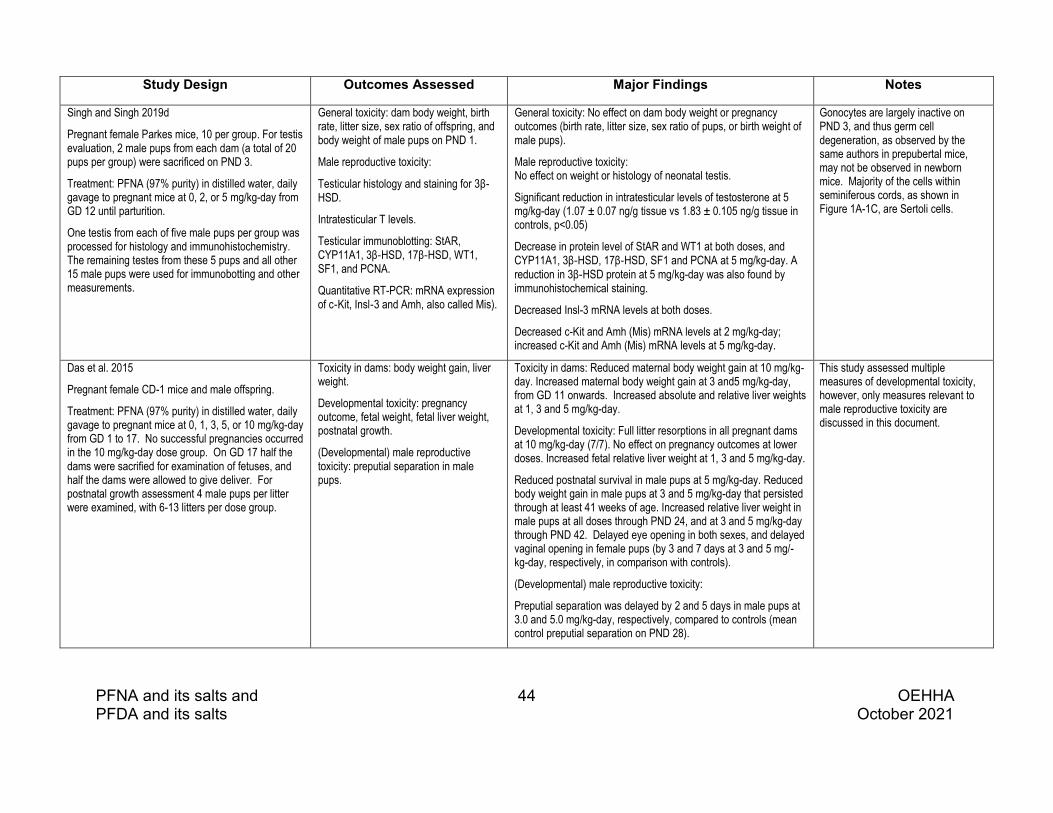
PFNA and its salts and 44 OEHHA PFDA and its salts October 2021
Study Design Outcomes Assessed Major Findings Notes
Singh and Singh 2019d
Pregnant female Parkes mice, 10 per group. For testis evaluation, 2 male pups from each dam (a total of 20 pups per group) were sacrificed on PND 3.
Treatment: PFNA (97% purity) in distilled water, daily gavage to pregnant mice at 0, 2, or 5 mg/kg-day from GD 12 until parturition.
One testis from each of five male pups per group was processed for histology and immunohistochemistry. The remaining testes from these 5 pups and all other 15 male pups were used for immunobotting and other measurements.
General toxicity: dam body weight, birth rate, litter size, sex ratio of offspring, and body weight of male pups on PND 1.
Male reproductive toxicity:
Testicular histology and staining for 3β‐HSD.
Intratesticular T levels.
Testicular immunoblotting: StAR, CYP11A1, 3β‐HSD, 17β‐HSD, WT1, SF1, and PCNA.
Quantitative RT-PCR: mRNA expression of c‐Kit, Insl‐3 and Amh, also called Mis).
General toxicity: No effect on dam body weight or pregnancy outcomes (birth rate, litter size, sex ratio of pups, or birth weight of male pups).
Male reproductive toxicity: No effect on weight or histology of neonatal testis.
Significant reduction in intratesticular levels of testosterone at 5 mg/kg-day (1.07 ± 0.07 ng/g tissue vs 1.83 ± 0.105 ng/g tissue in controls, p<0.05)
Decrease in protein level of StAR and WT1 at both doses, and CYP11A1, 3β‐HSD, 17β‐HSD, SF1 and PCNA at 5 mg/kg-day. A reduction in 3β‐HSD protein at 5 mg/kg-day was also found by immunohistochemical staining.
Decreased Insl-3 mRNA levels at both doses.
Decreased c-Kit and Amh (Mis) mRNA levels at 2 mg/kg-day; increased c-Kit and Amh (Mis) mRNA levels at 5 mg/kg-day.
Gonocytes are largely inactive on PND 3, and thus germ cell degeneration, as observed by the same authors in prepubertal mice, may not be observed in newborn mice. Majority of the cells within seminiferous cords, as shown in Figure 1A-1C, are Sertoli cells.
Das et al. 2015
Pregnant female CD-1 mice and male offspring.
Treatment: PFNA (97% purity) in distilled water, daily gavage to pregnant mice at 0, 1, 3, 5, or 10 mg/kg-day from GD 1 to 17. No successful pregnancies occurred in the 10 mg/kg-day dose group. On GD 17 half the dams were sacrified for examination of fetuses, and half the dams were allowed to give deliver. For postnatal growth assessment 4 male pups per litter were examined, with 6-13 litters per dose group.
Toxicity in dams: body weight gain, liver weight.
Developmental toxicity: pregnancy outcome, fetal weight, fetal liver weight, postnatal growth.
(Developmental) male reproductive toxicity: preputial separation in male pups.
Toxicity in dams: Reduced maternal body weight gain at 10 mg/kg-day. Increased maternal body weight gain at 3 and5 mg/kg-day, from GD 11 onwards. Increased absolute and relative liver weights at 1, 3 and 5 mg/kg-day.
Developmental toxicity: Full litter resorptions in all pregnant dams at 10 mg/kg-day (7/7). No effect on pregnancy outcomes at lower doses. Increased fetal relative liver weight at 1, 3 and 5 mg/kg-day.
Reduced postnatal survival in male pups at 5 mg/kg-day. Reduced body weight gain in male pups at 3 and 5 mg/kg-day that persisted through at least 41 weeks of age. Increased relative liver weight in male pups at all doses through PND 24, and at 3 and 5 mg/kg-day through PND 42. Delayed eye opening in both sexes, and delayed vaginal opening in female pups (by 3 and 7 days at 3 and 5 mg/-kg-day, respectively, in comparison with controls).
(Developmental) male reproductive toxicity:
Preputial separation was delayed by 2 and 5 days in male pups at 3.0 and 5.0 mg/kg-day, respectively, compared to controls (mean control preputial separation on PND 28).
This study assessed multiple measures of developmental toxicity, however, only measures relevant to male reproductive toxicity are discussed in this document.

PFNA and its salts and 45 OEHHA PFDA and its salts October 2021
Study Design Outcomes Assessed Major Findings Notes
Zhang et al. 2016
Zebrafish, wild-type, Tuebingen strain, 5 months old. A total of 480 fish of both sexes were included in these studies. Gonadosomatic index (GSI) assessed in 30 fish/sex/dose. Six pairs of male and female fish from each dose group were randomly selected for fertility assessment (egg production, fertilization rate, hatching rate, and abnormality rate) every week. Serum hormone measurements were performed on 3 pooled samples (pooled from 20 males) per dose group. mRNA analyses were performed on 6 males per dose group.
Treatment: PFNA (97% purity) in water through a flow-through exposure system at concentrations of 0, 0.01, 0.1 and 1.0 mg/L for 180 days.
Male reproductive toxicity: PFNA concentrations in gonads.
GSI: gonad weight x 100/body weight).
Egg production and hatching rate of F1 generation
Serum levels of T and E2.
Vitellogenin mRNA (vtg) and protein (Vtg) levels in male liver.
Transcriptional profiles of genes associated with the synthesis of sex hormones and the hypothalamic-pituitary-gonadal-liver axis (HPGL axis):
Brain: estrogen receptor α (era), estrogen receptor β (erb), ar, cytochrome P450 family 19 subfamily B (cyp19b), luteinizing hormone β (lhb).
Gonad: fshr, lhr, star,
cyp11a, 3β-hsd, cyp17, 17β-hsd, cyp19a.
Liver: era and erb.
Male reproductive toxicity: PFNA concentrations in male spermary were approximately twice as high as in female ovary. Reduced GSI in males at all dosing levels; no effect on GSI in females. Reduced egg production (by approximately 15%) at 0.1 mg/L. Reduced hatching rate at 72-hr time point (by approximately 12%) at 0.01 and 1.0 mg/L. Increased serum T at 0.01 mg/L in males (and females). Increased serum E2 at 0.1 and 1.0 mg/L in males (and females). The T/E2 ratio in males from the 0.01 and 0.1-mg/L groups appeared to be increased (approximately 180 at 0.01 mg/L and 110 at 0.1 mg/L vs. 45 in controls), but statistical significance was not indicated. Increased vtg mRNA levels in male liver at all dosing levels. Increased Vtg protein levels in male liver at 0.1 and 1.0 mg/L. Altered mRNA levels of genes related to the HPGL axis in males: Brain
era: decreased at 0.01 mg/L, increased at 0.1 mg/Lerb: decreased at 0.01 mg/L, increased at 0.1 mg/L ar: decreased at 0.1 and 1.0 mg/L cyp19b: no change fshb: no change lhb: increased at 0.1 mg/L
Gonad fshr: decreased at 0.1 and 1.0 mg/L lhr: decreased at all doses star: increased at 0.1 and 1.0 mg/L cyp11a: increased at 1.0 mg/L 3β-hsd: decreased at all doses cyp17: no change 17β-hsd: increased at 0.1 mg/L cyp19a: increased at 0.1 mg/L
Liver era: increased at 0.1 and 1.0 mg/L erb: increased at 0.1 mg/L
This study evaluated adverse effects of PFNA in zebrafish after chronic exposure and focused on the estrogenic effects of PFNA.
Male zebrafish can affect female egg production and hatching rate. Since both males and females were exposed to PFNA, the findings of reduced egg production and hatching rate could result from effects in males, or females, or both.

PFNA and its salts and 46 OEHHA PFDA and its salts October 2021
4.3 PFNA: Mechanistic considerations and other relevant data
The mechanistic literature regarding PFNA and male reproductive toxicity is reviewed here. First, we will provide a brief overview of general toxicity findings from studies of PFNA, followed by discussion of studies that may be more informative with respect to the effects PFNA may have on the male reproductive system.
General toxicity
In male Sprague-Dawley rats administered PFNA at 0, 0.625, 1.25, 2.5, 5, or 10 mg/kg (once daily by oral gavage, 7 days a week) for 28 days, decreased body weight was observed in all but the lowest dosed group, and survival was significantly reduced in the two highest dose groups (NTP 2019). Other studies have observed similar effects of PFNA on body weight. A study of male Wistar rats administered PFNA orally for 14 days reported a reduction in body weight at 5 mg/kg-d (Hadrup et al. 2016). In prepubertal male Parkes mice administered PFNA for 14 days, a significant reduction in body weight gain was observed at 5 mg/kg-day (Singh and Singh 2019b); however, no effect on body weight gain was observed in male Parkes mice exposed to lower doses of PFNA (0.2, 0.5 or 2 mg/kg-d) for 90 days (Singh and Singh 2019a). Reduced body weight gain was also observed in CD-1 mice exposed in utero to PFNA (maternal doses of 3 and 5 mg/kg-day from GD 1-17), that persisted through at least 41 weeks of age (Das et al. 2015).
In vitro study in testicular cells
In a culture of testicular cells from seminiferous tubules of Wistar rats, exposure to PFNA (300 µM for 48 hours) caused a significant increase in DNA damage (DNA strand breaks, as measured in the comet assay), in the absence of major effects on cell viability (Lindeman et al. 2012).
Effects on the hypothalamic-pituitary-gonadal-(liver) axis
The liver can be considered a component of the HPG axis in fish, as liver production of vitellogenin, a precursor egg yolk protein, in male fish is under estrogenic control (Jo et al. 2014). Studies investigating the effects of PFNA on hormone levels, metabolism and receptor-mediated effects related to the hypothalalamic-pituitary-gonadal-liver (HPGL) axis are summarized in Table 4.3.1.
Effects of PFNA on hormone levels have been studied in vivo in rats, mice and zebrafish and in vitro in cultured primary rat Sertoli cells. PFNA increased serum T levels in one study in male SD rats exposed for two weeks at 1 mg/kg-day (Feng et al. 2009), and reduced serum T levels in two studies in male SD rats exposed to higher doses (i.e., 2.5 mg/kg-day for 28 days

PFNA and its salts and 47 OEHHA PFDA and its salts October 2021
(NTP 2019); 5 mg/kg-day for 2 weeks (Feng et al. 2009). PFNA also reduced serum T levels in two studies in prepubertal male Parkes mice exposed for either 14 days at 2 and 5 mg/kg-day (Singh and Singh 2019b) or 90 days at 0.5 mg/kg-day (Singh and Singh 2019a). Intratesticular T levels were reduced in prepubertal male Parkes mice exposed to PFNA at 2 and 5 mg/kg-day for 14 days (Singh and Singh 2019b) and in PND 3 male Parkes mice exposed to PFNA in utero (Singh and Singh 2019d).
In male SD rats PFNA at 5 mg/kg-day for 14 days increased serum levels of E2 (Feng et al. 2009) and MIS, also known as anti-mullerian hormone or Amh, involved in sex differentiation) following treatment at 5 mg/kg-day for 14 days (Feng et al. 2010). Exposure to PFNA in utero altered testicular gene expression of MIS in male Parkes mice on PND 3, with a decrease in mRNA observed in male pups of dams receiving 2 mg/kg-day (daily from GD 12 to parturition), and an increase in mRNA observed in male pups of dams receiving 5 mg/kg-day (Singh and Singh 2019d). Inhibin B, a glycoprotein produced by Sertoli cells that modulates FSH secretion (via a negative feedback loop), was decreased in male SD rats following treatment at 1, 3, and 5 mg/kg-day for 14 days (Feng et al. 2010).
Data presented in Hadrup et al. (2016) suggest that PFNA may have reduced serum levels of LH and increased serum levels of FSH and prolactin following 14-day exposure of male Wistar rats at 5 mg/kg-day. No effects of PFNA on serum levels of LH or FSH were observed in a 14-day study of male SD rats (Feng et al. 2009).
In male zebrafish, PFNA increased serum T at 0.01 mg/L, increased serum E2 at 0.1 and 1.0 mg/L, and increased the serum T to E2 ratio at 0.01 and 0.1 mg/L. PFNA also increased brain mRNA levels of lutenizing hormone beta polypeptide (lhb), the gene encoding LH, at 0.1 mg/L in male zebrafish (Zhang et al. 2016).
In cultured primary Sertoli cells isolated from 8 week old SD rats, in vitro exposure to PFNA for 24 hours increased MIS mRNA levels at 10, 25, 50, and 75 µM, and decreased inhibin B mRNA levels at 50 and 75 µM (Feng et al. 2010).
Effects of PFNA on hormone synthesis and metabolism have been studied in vivo in rats, mice, and zebrafish and in vitro in a human placental choriocarcinoma cell line and a mouse Leydig tumor cell line.
Exposure of male rats to 5 mg/kg-day PFNA for 14 days was reported to reduce testicular mRNA levels of several steroidogenic enzymes (StAR, TSPO, CYP11A, CYP17, and 17β-HSD), although the data were not shown, and it is unclear whether statistical analyses were performed comparing “PFNA only” treatment groups and vehicle controls (Hadrup et al. 2016).
In a series of studies in male Parkes mice, PFNA was found to reduce testicular gene and protein expression of a number of enzymes and factors involved in steroidogenesis.

PFNA and its salts and 48 OEHHA PFDA and its salts October 2021
• PFNA decreased mRNA levels of HMG Co synthase 1, an enzyme involved in cholesterol synthesis, in prepubertal males exposed for 14 days to 5 mg/kg-day (Singh and Singh, 2019c).
• PFNA decreased protein levels of SF1, a transcription factor involved in sex differentiation that regulates transcription of StAR and CYP11a, in prepubertal males exposed for 14 days to 2 or 5 mg/kg-day (Singh and Singh 2019b) and in neonatal males examined on PND 3 after in utero exposure at 5 mg/kg-day (Singh and Singh 2019d).
• PFNA decreased protein levels of StAR in prepubertal males exposed for 14 days at 2 or 5 mg/kg-day (Singh and Singh 2019b) and in neonatal males examined on PND 3 after in utero exposure at 2 or 5 mg/kg-day (Singh and Singh 2019d), and decreased mRNA levels in prepubertal males exposed for 90 days at 0.5 mg/kg-day (Singh and Singh 2019a).
• PFNA decreased protein levels of CYP11a (P450scc), 3β-HSD, and 17β-HSD in prepubertal males exposed for 14 days at 2 or 5 mg/kg-day (Singh and Singh 2019b) and in neonatal males examined on PND 3 after in utero exposure at 5 mg/kg-day (Singh and Singh 2019d), and decreased mRNA levels of CYP11a and 3β-HSD in prepubertal males exposed for 90 days at 0.5 mg/kg-day (Singh and Singh 2019a).
In male zebrafish, PFNA exposure for 180 days increased gonadal gene expression of star (at 0.1 and 1.0 mg/L), cyp11a (at 1.0 mg/L), 17β-hsd (at 0.1 mg/L), and cyp19a (at 0.1 mg/L), reduced gonadal gene expression of 3β-hsd (at 0.01, 0.1, and 1.0 mg/L), and had no effect on gene expression of cyp17 (Zhang et al. 2016).
No effects on aromatase activity were observed in a human placental choriocarcinoma cell line (JEG-3 cells) following treatment with PFNA (Kjeldsen and Bonefeld-Jørgensen 2013).
In a mouse Leydig tumor cell line (mLTC-1 culture), incubation with PFNA inhibited progesterone production with an IC50 value of 16.6 (Zhao et al. 2017). The authors suggested that PFNA disrupted progesterone production as a result of damage to the mitochondrial membrane and increased production of reactive oxygen species (ROS).
Hormone receptor mediated effects have been studied in vivo in mice and fish, and in vitro in a human embryonic kidney cell line, two human breast adenocarcinoma cell lines, cultured primary mouse Sertoli cells, and a Chinese hamster ovary cell line after exposure to PFNA. In addition, hormone receptor binding studies have been conducted with fish liver cytosol and modeled in silico.
In prepubertal mice, PFNA treatment for 14 days reduced testicular gene expression of several hormone receptors, specifically mRNAs for androgen receptor (AR), growth hormone receptor (GHR), and IGF-1R at doses of 2 and 5 mg/kg-day, and IGF-1 at 5 mg/kg-day (Singh and Singh 2019c).

PFNA and its salts and 49 OEHHA PFDA and its salts October 2021
In male zebrafish, PFNA treatment increased gene and protein expression of vtg, an estrogen-responsive gene, in the liver (Zhang et al. 2016). In this study PFNA treatment also altered gene expression in male zebrafish of certain hormone receptors in the brain (decreased mRNA levels for era, erb, and ar), gonads (decreased mRNA levels for fshr and lhr) and liver (increased mRNA levels for era and erb). In rainbow trout, PFNA treatment via diet resulted in concentration-dependent increases in plasma levels of Vtg in studies with 11-month old juvenile trout (Benninghoff et al. 2011). In other experiments by these authors, PFNA was shown to competitively bind (very weakly) to trout liver Er (in liver cytosol homogenates) (Benninghoff et al. 2011).
In human embryonic kidney (HEK293T) cells transfected with a human ERα reporter gene, PFNA (100-1000 nM) increased human ERα (hERα) reporter activity up to 2.5 fold (Benninghoff et al. 2011).
In MVLN cells (derived from MCF-7 cells, a human breast adenocarcinoma cell line, stably transfected with a luciferase reporter gene and an estrogen-responsive element from the Xenopus vitellogenin A2 gene), exposure to PFNA alone did not induce an estrogenic response. However, PFNA displayed anti-estrogenic activity in MVLN cells pretreated with E2, inhibiting the estrogenic response to E2 (measured as luciferase activity) in a concentration dependent manner. PFNA was a less potent inhibitor of the estrogenic response than PFDA in these cells, with an EC50 of 28.1 µM (Juan Li et al. 2020). In separate experiments in MCF-7 cells (a human breast adenocarcinoma cell line), PFNA also had anti-estrogenic activity, with the addition of PFNA to E2-treated cells resulting in significant down regulation of the E2-responsive genes trefoil factor 1 (TFF1, also known as pS2) and EGR3 (early growth response 3) (Juan Li et al. 2020).
In primary rat Sertoli cell cultures, incubation with PFNA for 24 hours had no effect on mRNA levels of AR at concentrations ranging from 1 to 75 µM, increased levels of androgen binding protein (ABP) at ≥50 µM, reduced levels of transferrin mRNA (involved in spermatogenesis) at ≥25 µM, and reduced mRNA levels of FSH-R at 25 and 50 µM (Feng et al. 2010).
PFNA significantly antagonized DHT-induced AR activity in a concentration-dependent manner in a Chinese hamster ovary cell line (CHO-K1) transfected with AR and a reporter vector (Kjeldsen and Bonefeld-Jørgensen 2013).
Analyses using in silico computational models indicate that PFNA can efficiently dock with human, mouse, and trout ERα in the ligand-binding domain and form a hydrogen bond at residue Arg394 in human ERα in a manner similar to that of estrogens (Benninghoff et al. 2011). Additional in silico modeling studies with the activated form of hERα, in which E2 is bound to the ligand-binding domain, predict that PFNA can bind to sites on the surface of the activated receptor (Juan Li et al. 2020).
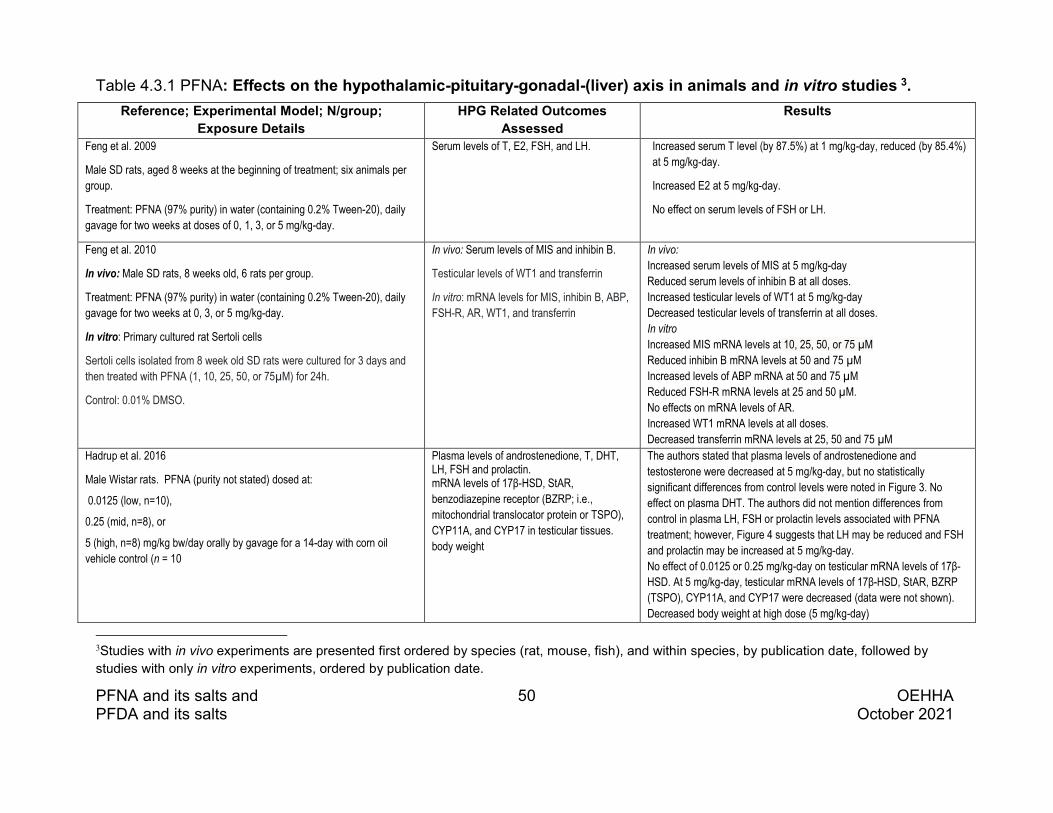
PFNA and its salts and 50 OEHHA PFDA and its salts October 2021
Table 4.3.1 PFNA: Effects on the hypothalamic-pituitary-gonadal-(liver) axis in animals and in vitro studies 3. Reference; Experimental Model; N/group;
Exposure Details HPG Related Outcomes
Assessed Results
Feng et al. 2009
Male SD rats, aged 8 weeks at the beginning of treatment; six animals per group.
Treatment: PFNA (97% purity) in water (containing 0.2% Tween-20), daily gavage for two weeks at doses of 0, 1, 3, or 5 mg/kg-day.
Serum levels of T, E2, FSH, and LH. Increased serum T level (by 87.5%) at 1 mg/kg-day, reduced (by 85.4%) at 5 mg/kg-day.
Increased E2 at 5 mg/kg-day.
No effect on serum levels of FSH or LH.
Feng et al. 2010
In vivo: Male SD rats, 8 weeks old, 6 rats per group.
Treatment: PFNA (97% purity) in water (containing 0.2% Tween-20), daily gavage for two weeks at 0, 3, or 5 mg/kg-day.
In vitro: Primary cultured rat Sertoli cells
Sertoli cells isolated from 8 week old SD rats were cultured for 3 days and then treated with PFNA (1, 10, 25, 50, or 75µM) for 24h.
Control: 0.01% DMSO.
In vivo: Serum levels of MIS and inhibin B.
Testicular levels of WT1 and transferrin
In vitro: mRNA levels for MIS, inhibin B, ABP, FSH-R, AR, WT1, and transferrin
In vivo: Increased serum levels of MIS at 5 mg/kg-day Reduced serum levels of inhibin B at all doses. Increased testicular levels of WT1 at 5 mg/kg-day Decreased testicular levels of transferrin at all doses. In vitro Increased MIS mRNA levels at 10, 25, 50, or 75 µM Reduced inhibin B mRNA levels at 50 and 75 µM Increased levels of ABP mRNA at 50 and 75 µM Reduced FSH-R mRNA levels at 25 and 50 µM. No effects on mRNA levels of AR. Increased WT1 mRNA levels at all doses. Decreased transferrin mRNA levels at 25, 50 and 75 µM
Hadrup et al. 2016
Male Wistar rats. PFNA (purity not stated) dosed at:
0.0125 (low, n=10),
0.25 (mid, n=8), or
5 (high, n=8) mg/kg bw/day orally by gavage for a 14-day with corn oil vehicle control (n = 10
Plasma levels of androstenedione, T, DHT, LH, FSH and prolactin. mRNA levels of 17β-HSD, StAR, benzodiazepine receptor (BZRP; i.e., mitochondrial translocator protein or TSPO), CYP11A, and CYP17 in testicular tissues. body weight
The authors stated that plasma levels of androstenedione and testosterone were decreased at 5 mg/kg-day, but no statistically significant differences from control levels were noted in Figure 3. No effect on plasma DHT. The authors did not mention differences from control in plasma LH, FSH or prolactin levels associated with PFNA treatment; however, Figure 4 suggests that LH may be reduced and FSH and prolactin may be increased at 5 mg/kg-day. No effect of 0.0125 or 0.25 mg/kg-day on testicular mRNA levels of 17β-HSD. At 5 mg/kg-day, testicular mRNA levels of 17β-HSD, StAR, BZRP (TSPO), CYP11A, and CYP17 were decreased (data were not shown). Decreased body weight at high dose (5 mg/kg-day)
3Studies with in vivo experiments are presented first ordered by species (rat, mouse, fish), and within species, by publication date, followed by studies with only in vitro experiments, ordered by publication date.

PFNA and its salts and 51 OEHHA PFDA and its salts October 2021
Reference; Experimental Model; N/group; Exposure Details
HPG Related Outcomes Assessed
Results
NTP 2019
Male SD rats, 9-11 weeks old at the beginning of dosing, 10 rats per dose group.
Treatment: PFNA (99.5% purity) once daily by gavage in deionized water with 20% Tween® 80 at doses of 0, 0.625, 1.25, 2.5, 5.0 or 10.0 mg/kg-day, respectively, for 28 days.
Serum T levels Survival, body weight
Reduced serum T at 2.5 mg/kg-day
Decreased survival in the 5 and 10 mg/kg-day dose groups (2/10 and 0/10, respectively). Lower body weights at ≥ 1.25 mg/kg-day.
Singh and Singh 2019a
Male Parkes mice, 25 days old. 14 animals per group.
Treatment: PFNA (97% purity) in distilled water, daily gavage at 0, 0.2, or 0.5 mg/kg-day, from PND 25 to 114 (for 90 days). The animals were sacrificed on PND 115.
Serum T levels.
Analysis of mRNA expression by semiquantitative RT-PCR: StAR, CYP11A1, 3β‐HSD, 17β‐ HSD.
Immunoblotting: StAR, CYP11A1, 3β‐HSD, 17β‐ HSD.
Body weight
At 0.2 mg/kg-day: No effect on serum T level or expression of genes or proteins assessed. At 0.5 mg/kg-day: Reduced serum T level (31%). Down-regulation in testicular mRNA levels of StAR, CYP11A1, 3β‐HSD and 17β‐ HSD. Decreased testicular protein levels of StAR, CYP11A1, 3β‐HSD, and 17β‐ HSD.
No effect on body weight.
Singh and Singh 2019b
Male Parkes mice, 25 days old. 10 animals per group.
Treatment: PFNA (97% purity) in distilled water, daily gavage at 0, 2, or 5 mg/kg-day from PND 25 to 38. The animals were sacrificed on PND 39.
Serum and intratesticular levels of T.
Protein levels by immunoblotting: SF1, StAR, CYP11A1, 3β-HSD, 17β- HSD.
Body weight.
Decreased serum T level by 72% and 85%, at 2 and 5 mg/kg-day, respectively, and decreased intratesticular T at both doses.
Decreased protein levels of SF1, StAR, CYP11A1, 3β-HSD, and 17β-HSD at both doses.
Reduced body weight gain (24%) at 5 mg/kg-day.
Singh and Singh 2019c
[Study design same as the 2019b study by the same authors.]
Male Parkes mice, 25 days old. 10 animals per group,
Treatment: PFNA (97% purity) in distilled water, daily gavage at 0, 2, or 5 mg/kg-day from PND 25 to 38.
Testicular mRNA levels for AR, GHR, IGF-1, insulin like growth factor 1 receptor (IGF-1R) and HGM Co Synthase 1
Reduced mRNA expression of AR, GHR, and IGF-1R at both doses
Decreased mRNA expression of IGF-1 and HGM Co Synthase 1 at 5 mg/kg-day.

PFNA and its salts and 52 OEHHA PFDA and its salts October 2021
Reference; Experimental Model; N/group; Exposure Details
HPG Related Outcomes Assessed
Results
Singh and Singh 2019d
Female Parkes mice, 10 pregnant mice per group. For testis evaluation, 2 male pups from each dam (a total of 20 pups per group) were sacrificed on PND 3.
Treatment: PFNA (97% purity) in distilled water, daily gavage to pregnant mice at 0, 2, or 5 mg/kg-day from GD 12 until parturition.
One testis from each of five male pups per group was processed for histology and immunohistochemistry. The remaining testes from these 5 pups and all other 15 male pups were used for immunobotting and other measurements.
Evaluation of testicular tissue from male pups: Immunohistochemistry: 3β‐HSD
Immunoblotting: StAR, CYP11A1, 3β‐ HSD, 17β‐HSD
Gene expression of anti-mullerian hormone (Amh, MIS).
Reduction in intratesticular levels of T at 5 mg/kg-day (1.07 ± 0.07 ng/g tissue vs 1.83 ± 0.105 ng/g tissue in controls)
Decreased protein levels of StAR at both doses, and of CYP11A1, 3β‐HSD, 17β‐HSD, and SF1 at 5 mg/kg-day. Reduced 3β‐HSD protein was also found by immunohistochemical staining.
Decreaed MIS mRNA at 2 mg/kg-day, and increased mRNA at 5 mg/kg-day.
Benninghoff et al. 2011 In vivo: Rainbow trout Dietary exposure of 11-month old juvenile rainbow trout to PFNA (highest purity available) at 0, vehicle control (DMSO), 5, 50, or 250 ppm in Oregon Test Diet, OTD) 5 days/week for 14 days (equivalent to 0.1, 1, or 5 mg/kg-d, respectively); animals were sacrificed on day 15. Positive control: 5 ppm E2 N = 6 fish/treated and vehicle control (0.5 ppm DMSO) group. N = 24 fish in untreated control group In vitro: trout liver cytosol, HEK-293T cells Trout liver cytosol ER binding studies in liver cytosol prepared from trout fed 5 ppm E2 (to induce ERα) HEK-293T cells: a human embryonic kidney cell line transfected with a human ERα expression vector and a luciferase reporter plasmid containing an estrogen response element (ERE). PFNA or E2 treatment (1-1000 nM) for 24 hrs; hERα reporter gene activity measured. In Silico Molecular docking studies of PFNA with the active site in human, mouse, and trout ERα.
In Vivo
Vtg levels in plasma
In Vitro
Trout liver cytosol: ERα binding studies
HEK-293T cells: hERα gene reporter activity
In Silico
Molecular docking at the active site in human, mouse and trout ERα
In Vivo Concentration dependent increase in plasma Vtg levels
In Vitro
Trout liver cytosol: PFNA shows weak competitive binding to ERα, completely displacing E2 from the receptor, with more than a 10,000-fold lower relative binding affinity than E2.
HEK-293T cells: PFNA at concentrations of 100-1000 nM induced hERα gene reporter activity up to 2.5 fold
In Silico
PFNA is predicted to bind at the active site of human, mouse, and trout ERα. Predicted hydrogen bond interaction with hERα at Arg394.

PFNA and its salts and 53 OEHHA PFDA and its salts October 2021
Reference; Experimental Model; N/group; Exposure Details
HPG Related Outcomes Assessed
Results
Zhang et al. 2016
Zebrafish (wild type, Tuebingen strain): five-month-old (n = 480)). Animals were separated by sex and randomly assigned to treatment groups
Treatment: PFNA (97% purity) in water through a flow-through exposure system at concentrations of 0, 0.01, 0.1 and 1.0 mg/L for 180 days.
Serum hormone measurements were performed on 3 pooled samples (pooled from 20 males) per dose group. mRNA analyses were performed on 6 males per dose group.
Serum levels of T and E2.
Vitellogenin mRNA (vtg) and protein (Vtg) levels in male liver.
Transcriptional profiles of genes associated with the synthesis of sex hormones and the hypothalamic-pituitary-gonadal-liver axis (HPGL axis):
Brain: estrogen receptor α (era), estrogen receptor β (erb), ar, cytochrome P450 family 19 subfamily B (cyp19b), luteinizing hormone β (lhb).
Gonad: fshr, lhr, star,
cyp11a, 3β-hsd, cyp17, 17β-hsd, cyp19a.
Liver: era and erb.
Increased serum T at 0.01 mg/L in males (and females). Increased serum E2 at 0.1 and 1.0 mg/L in males (and females). The T/E2 ratio in males from the 0.01 and 0.1-mg/L groups appeared to be increased (approximately 180 at 0.01 mg/L and 110 at 0.1 mg/L vs. 45 in controls), but statistical significance was not indicated. Increased vtg mRNA levels in male liver at all dosing levels. Increased Vtg protein levels in male liver at 0.1 and 1.0 mg/L. Altered mRNA levels of genes related to the HPGL axis in males: Brain
era: decreased at 0.01 mg/L, increased at 0.1 mg/Lerb: decreased at 0.01 mg/L, increased at 0.1 mg/L ar: decreased at 0.1 and 1.0 mg/L cyp19b: no change fshb: no change lhb: increased at 0.1 mg/L
Gonad fshr: decreased at 0.1 and 1.0 mg/L lhr: decreased at all doses star: increased at 0.1 and 1.0 mg/L cyp11a: increased at 1.0 mg/L 3β-hsd: decreased at all doses cyp17: no change 17β-hsd: increased at 0.1 mg/L cyp19a: increased at 0.1 mg/L
Liver era: increased at 0.1 and 1.0 mg/L
erb: increased at 0.1 mg/L

PFNA and its salts and 54 OEHHA PFDA and its salts October 2021
Reference; Experimental Model; N/group; Exposure Details
HPG Related Outcomes Assessed
Results
Kjeldsen and Bonefeld-Jørgensen 2013 In vitro: MVLN, CHO-K1 and JEG-3 cell lines PFNA (97% purity) dissolved in dimethyl sulfoxide (DMSO), tested at concentrations of 10-9 – 10-4 M MVLN cells: Derived from MCF-7 cells; stably transfected with a luciferase reporter gene and an estrogen responsive element derived from the Xenopus vitellogein A2 gene. ER transactivation assessed with PFNA alone or co-treated with 25 pM E2 CHO-K1 cells: Derived from CHO cells; transiently co-transfected with the AR plasmid and a luciferase reporter vector AR transactivation assessed with PFNA alone or co-treated with or 25 pM DHT JEG-3 cells: A human choriocarcinoma cell line. Aromatase activity assessed with PFNA treatment
MVLN cells: ER transactivation
CHO-K1 cells: AR transactivation
JEG-3 cells: Aromatase activity
MVLN cells Cytotoxic at 10-4 M ER transactivation: No effects on ER activity CHO-K1cells Cytotoxic at 10-4 M AR transactivation: No AR agonist activity. Concentration-dependent antagonistic effects on DHT-induced AR transactivation (IC50 5.2×10⁻⁵ M). JEG-3 cells Cytotoxic at 10-4 M No effects on aromatase activity.
Zhao et al. 2017
In vitro: Mouse Leydig cell tumor line (mLTC-1)
PFNA (>95% purity)
Experiment 1: PFNA treatment (0-105µM) for 24 hrs; cAMP added and incubated for additional 3 hrs; progesterone quantified via radioimmunoassay
Experiment 2: PFNA treatment (0, 12.5, 25, 50 or 100 µM) for 24 hours; mitochondrial membrane potential measured
Progesterone production
Mitochondrial membrane potential
Experiment 1: Concentration dependent decrease in progesterone production (IC50 16.61 µM)
Experiment 2: Significant decrease in mitochondrial membrane potential at 50 and 100 µM.

PFNA and its salts and 55 OEHHA PFDA and its salts October 2021
Reference; Experimental Model; N/group; Exposure Details
HPG Related Outcomes Assessed
Results
Juan Li et al. 2020
In vitro: MVLN and MCF-7 cell lines
PFNA (97% purity)
MVLN cells: Derived from MCF-7 cells; stably transfected with a luciferase reporter gene and an estrogen responsive element derived from the Xenopus vitellogein A2 gene. PFNA treatment (0, 0.0001-5 mM) for 48 hr in the presence or absence of E2 (1 nM) as positive control or ICI 182,780 as negative control, or both; ER transactivation
MCF-7 cells: human breast adenocarcinoma cell line
PFNA treatment (50 µM) in the presence or absence of E2 for 48 hours; changes in expression of estrogen regulated genes (TFF1/pS2, EGR3)
In silico
Modeling analysis of PFNA interactions with E2-activated hERα
MVLN cells: ER transactivation MCF-7 cells: mRNA gene expression of estrogen regulated genes TFF1/pS2, EGR3
In silico: modeling predictions of interaction with E2-activated hERα
MVLN cells
ER transactivation: No ER agonist activity with PFNA alone; in cells pretreated with E2 PFNA inhibited the estrogenic response to E2 in a concentration dependent manner, with a reported EC50 of 28.1 µM. In cells cotreated with E2 and ICI 182,780 , PFNA caused a significant increase in the inhibition of luciferase activity (attenuated luciferase response) MCF-7 cells
mRNA levels of TFF1 and EGR3: No effect on gene expression with PFNA alone. In cells cotreated with E2, PFNA significantly downregulated expression of both TFF1 and EGR3
In silico
Modeling predicts that PFNA can bind to the surface of the E2 activated form of hERα (at sites distinct from the ER binding pocket)

PFNA and its salts and 56 OEHHA PFDA and its salts October 2021
Effects on the thyroid
PFNA effects on thyroid hormones are summarized in Table 4.3.2 below.
A subset of data from the National Health and Nutrition Examination Survey (NHANES) provided a cross-sectional look at serum levels of PFNA, thyroid hormones, and testosterone in human males aged from 12-80 years (Lewis et al. 2015). Only one whole-animal study in rodents (NTP 2019) provided data on outcomes of both thyroid toxicity and male reproductive toxicity with repeated exposures to PFNA. The only additional whole-animal study of thyroid hormone relevant outcomes involved repeated PFNA dosing of two generations of zebrafish, but did not report on reproductive parameters (Y Liu et al. 2011). An in vitro study using rat pituitary GH3 cells evaluated PFNA for cytotoxicity, effects on cell proliferation, and aryl hydrocarbon receptor (AhR) transactivity (Long et al. 2013). Two studies of PFNA involved cell-free biochemical assays of thyroid binding (Ren et al. 2016; Weiss et al. 2009). The Ren (2016) publication included an in silico molecular docking simulation to model binding of PFNA to thyroid hormone transport proteins.
The Lewis et al. (2015) study analyzed serum samples from a total of 857 human males divided into age groups. Median PFNA levels increased with age from 0.78 ng/mL for 12 to <20 year-olds, to 1.07 ng/mL for individuals aged 60 – 80 years. Samples were also analyzed for levels of testosterone, total TSH, free and total T3 and T4, and several other PFASs. Statistical modeling of percent change in hormone concentration per doubling of PFNA provided only one statistically significant result: a 16.3% increase in TSH for 12 to <20 year-olds. A non-significant increase in total T4 was determined for sampled 40-60 year olds. Adjustments were made for several covariates, but not for other PFASs in the samples.
The NTP (2019) 28-day rat study found that excess mortality at the two highest administered doses of PFNA (5 and 10 mg/kg-day) precluded further assessment of outcomes for those groups. As a result, 2.5 mg/kg-day was the effective high dose for reported outcomes. There was no significant effect on T3. Statistically significant outcomes relevant to thyroid toxicity included:
• Decreased absolute thyroid gland weight at 2.5 mg/kg-day • Increased relative thyroid weight at 2.5 mg/kg-day • Decreased TSH at doses > 1.25 mg/kg-day • Decreased total T4 at 0.625 and 1.25 mg/kg-day (not 2.5 mg/kg-day) • Decreased free T4 at doses > 0.625 mg/kg-day
The same animals showed decreased testosterone levels at 2.5 mg/kg-day. Adverse male reproductive outcomes observed at doses > 1.25 mg/kg-day included reduced testes and epididymis weights, as well as multiple histopathological changes in testes.

PFNA and its salts and 57 OEHHA PFDA and its salts October 2021
Two generations of zebrafish were continuously exposed to PFNA at concentrations of 0, 0.05, 0.1, 0.5, and 1 mg/l (Y Liu et al. 2011). While F0 males and females were sexed at maturity, and then paired for spawning the F1, no mention was made of sex ratios or fertility of these animals.
Reported outcomes included:
• Significantly elevated plasma T3 levels in both F0 and F1 adult males. • T3 + T4 was significantly higher in F0 adult males than in controls. • PFNA-induced histological changes in F0 male zebrafish indicative of
hyperstimulation of thyroid follicles. • Significantly increased hepatic levels of transcript for transthyretin (TTR) in F0
males. • In samples of brain tissue from F0 animals, TRα transcription was down-
regulated at 0.1 and 0.5 mg/l; TRß was down-regulated at 0.1 mg/l. • In homogenized F1 larvae, expression of TRα was induced at concentrations
> 0.1 mg/l; TRß was induced at all PFNA concentrations tested. • TTR expression was increased in F1 larvae following parental exposure to PFNA
at concentrations of 0.05 mg/l or greater. • Overall gene expression profile studies indicated PFNA-induced disruption of
thyroid hormone transport, metabolism, synthesis and function.
Liu et al. (2011) suggested that observed increased TTR transcription in zebrafish could reflect induction due to competitive binding of PFNA. The authors propose that PNFA could induce TTR transcription across species, while producing different results in rats and zebrafish for thyroid hormone levels.
Weiss et al. (2009) tested PFNA, as well as other related compounds, for binding to the human thyroid transport protein TTR in competition with 125I-labeled T4. PFNA inhibited 18% of T4-TTR binding. Binding potency among the series of tested compounds decreased in the order: PFHxS> PFOS/PFOA> perfluoroheptanoic acid (PFHpA)> sodium perfluoro-1-octanesulfinate > PFNA. TTR binding potencies ranged from 12.5–50 times lower than T4 (Weiss et al. 2009).
An in vitro study of PFNA effects on proliferation of T3-dependent rat pituitary GH3 cells found no cytotoxicity under test conditions (Long et al. 2013). Concentration-dependent decreases in GH3 cell proliferation were seen with PFNA alone (1×10⁻⁹–1×10⁻⁴ M) or in combination with 0.5 nM T3 (1x10⁻⁸-1x10⁻⁴). Either alone or in combination with 2,3,7,8-tetrachlorodibenzo-p-dioxin (TCDD), PFNA had no effect on AhR transactivity.
In a cell-free assay, the relative potency for PFNA binding with TTR was 0.016, as compared to T4 representing a relative potency of 1 and 0.083 for PFOA (Ren et al. 2016). In experiments comparing binding to wild type versus mutant thyroxine-binding globulin (TBG), arelative potency was not detectable for PFNA binding to wild type TBG.

PFNA and its salts and 58 OEHHA PFDA and its salts October 2021
In silico molecular docking experiments to TTR and TBG found PFNA to be among the tested perfluoroalkyl acids and sulfonates that fit the binding pockets (Ren et al. 2016).
Overall, experimental evidence from cell-free binding assays and in silico molecular-docking models shows binding of PFNA to TTR (Ren et al. 2016; Weiss et al. 2009) with consequences of PFNA exposure for thyroid hormone levels and function observed in cell culture (Long et al. 2013) and zebrafish (Y Liu et al. 2011). The available human data (Lewis et al. 2015) found no effect of PFNA on testosterone, and a significant effect on TSH levels only for boys in the adolescent age-range. Only the NTP (2019) study in rats reported direct evidence of male reproductive toxicity with exposure to PFNA at doses also associated with decreases in TSH and free T4 (NTP 2019). This co-occurrence of effects is consistent with, but neither establishes nor eliminates, potential for a causal, mechanistic role for thyroid disruption in the male reproductive toxicity of PFNA.

PFNA and its salts and 59 OEHHA PFDA and its salts October 2021
Table 4.3.2 PFNA Effects on thyroid hormones.
Study Design Outcomes Assessed Major Findings Comments NTP 2019 (see Table 4.2.1 for summary of male repro data from this study)
Male SD rats, 10-11 weeks old, 10 rats/dose. PFNA administered once daily by gavage in deionized water with 20% Tween® 80 at doses of 0, 0.625, 1.25, 2.5, 5.0 or 10.0 mg PFDA/kg-day, for 28 days.
At the end of the study, blood was collected from the abdominal aorta for biochemical analysis (including thyroid stimulating hormone (TSH) and thyroid hormones).
Body weights and thyroid gland weights and histology.
Blood levels of TSH, T3, and T4 (total and free).
Due to excess mortality at doses of 5 or 10 mg PFNA/kg-day, further analyses were not presented for outcomes at those doses.
Absolute thyroid gland weight decreased at 2.5 mg/kg-day (p < 0.01).
Increased relative thyroid gland weight at 2.5 mg/kg-day (p < 0.01).
No evidence of follicular cell hypertrophy in thyroid gland.
TSH significantly decreased at 1.25 and 2.5 mg/kg-day (p < 0.01)
No significant effect on T3.
Total T4 significantly decreased at 0.625 and 1.25 mg/kg-day (p < 0.01) but not at 2.5 mg/kg-day.
Free T4 significantly decreased at doses of 0.625, 1.25, and 2.5 mg/kg-day (p < 0.01).
In the same study, direct evidence of male reproductive toxicity was reported at doses > 1.25 mg/kg-day:
Outcomes included reduced testis and epididymis weights, reduced serum testosterone levels, [reduced epididymal sperm counts, increased incidence of interstitial tissue atrophy, germ cell degeneration, and inhibited spermiation.

PFNA and its salts and 60 OEHHA PFDA and its salts October 2021
Study Design Outcomes Assessed Major Findings Comments Ren et al. 2016
Cell-free biochemical assays:
Quantitative assessment of binding affinities of PFASs (including PFNA, purity not specified) to human transthyretin (TTR) and human thyroxine-binding globulin (TBG) by a fluorescent competitive binding assay.
Molecular docking (in silico) was used to simulate interaction of PFNA with TTR.
Binding affinities with thyroid hormone transport proteins: TTR and TBG.
Molecular docking of PFNA with TTR.
The relative potency for PFNA binding with TTR was 0.016, as compared to T4 representing an RP of 1 and 0.083 for PFOA.
For TBG, binding was not detectable for PFNA or PFOA.
PFNA was one of 16 PFASs shown to fit the TTR and TBG binding pockets using a molecular docking model.
The molecular docking model showed that T4 could fill the TTR ligand-binding pocket. PFOA and PFOS could also fill the pocket, with the fluorinated carbon tail extended. PFASs with a medium chain length and a sulfonate acid group were found to be optimal for TTR binding, and PFASs with lengths longer than 12 carbons were optimal for TBG binding.
Lewis et al. 2015
Cross-sectional study of U.S. human subjects
Subset of the National Health and Nutrition Examination Survey (NHANES) with serum samples assayed for PFNA, testosterone, and thyroid hormones.
Data collected in 2011-2012
N = 857 males, 12-80 years
Exclusions: missing data on variables in analyses
Serum PFNA
Serum total T
Total TSH
Free and total T3
Free and total T4
Statistical modeling of associations between PFAS levels and hormone concentrations.
Serum PFNA (ng/mL) expressed as median (IQR) by age group:
12 to <20 years: 0.78 (0.56, 1.19)
20 to <40 years: 0.98 (0.67, 1.31)
40 to <60 years: 1.00 (0.67, 1.57)
60 to 80 years: 1.07 (0.77, 1.58)
Percent change (95% CI) in hormone concentration modeled per doubling of PFNA:
Total T4: 40-60-year-olds: -2.5% (-5.2, 0.2), non-significant apparent association.
TSH: 12 to <20-year-olds: 16.3% (4.0, 30.2), p < 0.05.
No associations for other age groups with these hormones or for any age group with other evaluated hormones.
Adjusted for covariates: age, BMI, PIR,
race/ethnicity, and serum cotinine.
Not adjusted for other PFAS:
PFHxS
PFOA
PFOS
Correlations among PFASs were not reported
Long et al. 2013 Cytotoxicity to rat pituitary GH3 cells and transfected mouse cells
PFNA was not cytotoxic to rat pituitary GH3 cells or transfected mouse cells under test conditions.
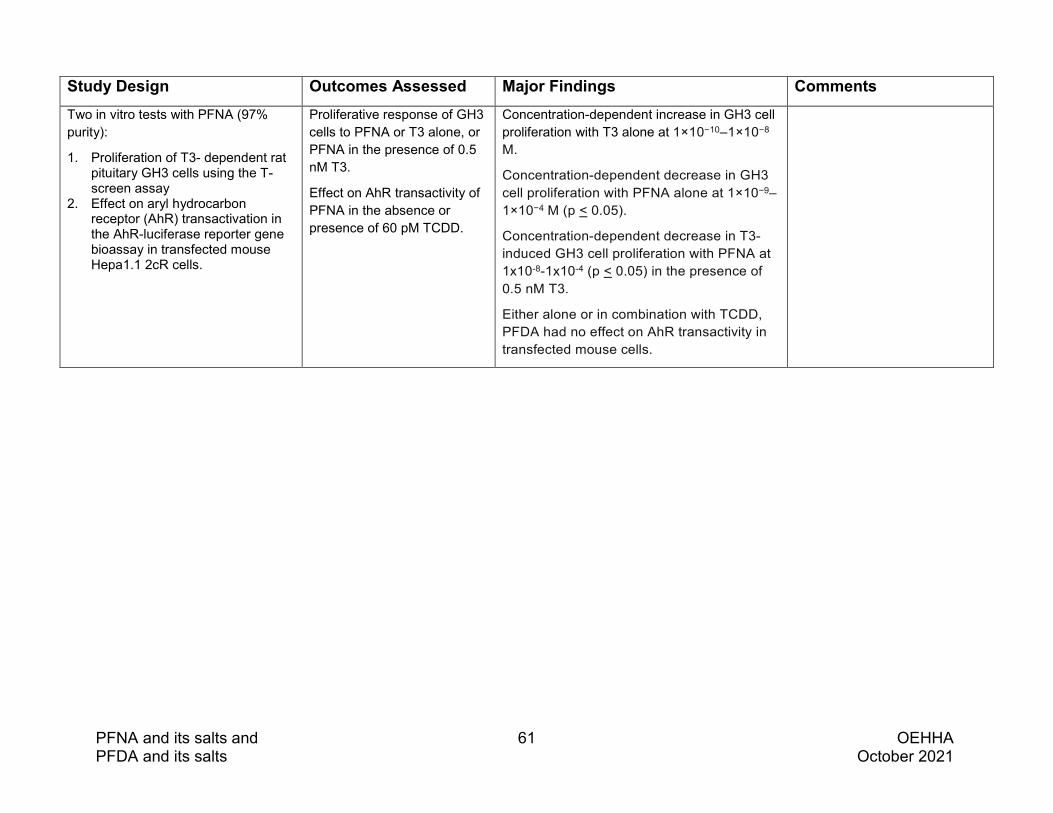
PFNA and its salts and 61 OEHHA PFDA and its salts October 2021
Study Design Outcomes Assessed Major Findings Comments Two in vitro tests with PFNA (97% purity):
1. Proliferation of T3- dependent rat pituitary GH3 cells using the T-screen assay
2. Effect on aryl hydrocarbon receptor (AhR) transactivation in the AhR-luciferase reporter gene bioassay in transfected mouse Hepa1.1 2cR cells.
Proliferative response of GH3 cells to PFNA or T3 alone, or PFNA in the presence of 0.5 nM T3.
Effect on AhR transactivity of PFNA in the absence or presence of 60 pM TCDD.
Concentration-dependent increase in GH3 cell proliferation with T3 alone at 1×10⁻¹⁰–1×10⁻⁸ M.
Concentration-dependent decrease in GH3 cell proliferation with PFNA alone at 1×10⁻⁹–1×10⁻⁴ M (p < 0.05).
Concentration-dependent decrease in T3-induced GH3 cell proliferation with PFNA at 1x10⁻⁸-1x10⁻⁴ (p < 0.05) in the presence of 0.5 nM T3.
Either alone or in combination with TCDD, PFDA had no effect on AhR transactivity in transfected mouse cells.

PFNA and its salts and 62 OEHHA PFDA and its salts October 2021
Study Design Outcomes Assessed Major Findings Comments Y Liu et al. 2011
Zebrafish (Danio rerio) exposed to PFNA (97%) at 0, 0.05, 0.1, 0.5, and 1 mg/l. Treatment of F0 started at 23 days dpf.
N = 50 fish/concentration group.
Exposure continuous for 180 days.
Sex determined at maturity (120 dpf). Males and females from the same dose group were paired for spawning.
F1 offspring raised with or without continued PFNA exposure for an additional 180 days.
Plasma thyroid hormone levels in F0 males after exposure for 180 days, and in F1 males at 180 dpf.
Thyroid follicle histology in F0 males after exposure for 180 days.
Gene expression related to the hypothalamus–pituitary–thyroid (HPT) axis in F0 males (brain and liver) after exposure for 180 days, and in F1 larvae (5 dpf).
Significantly elevated plasma T3 levels in both F0 (p < 0.05 at 0.05, 0.1, and 1 mg/l PFNA for 180 days) and F1 adult males (p < 0.05 for all dose groups).
In F0 males, T4 was elevated above controls at all PFNA concentrations, but did not reach statistical significance; T3 + T4 was significantly higher than controls at 0.1 and 0.5 mg/l (p < 0.05).
PFNA induced histological changes in the thyroid follicles of F0 males: enlarged follicles at 0.05 mg/l, hypertrophy of follicular epithelium and follicular cell hyperplasia at 0.1 and 0.5 mg/l; decreased size of follicle lumens at 1 mg/l.
Increased gene expression of TTR in F0 liver at 0.1, 0.5, and 1 mg/l (p < 0.05), and in F1 5 dpf larvae at 0.05, 0.5, and 1 mg/l (p < 0.05).
Altered expression of additional HPT-associated genes (e.g., thyroid hormone transport, metabolism, synthesis and function) in F0 liver and brain and in F1 5 dpf larvae.
Authors note in discussion evidence that PNFA could induce TTR transcription across species, while producing different results in rats and zebrafish for thyroid hormone levels.
While F0 males and females were sexed at 120 dpf, and paired for spawning the F1, no mention was made of sex ratios or fertility of these animals.
Weiss et al. 2009
Cell-free biochemical assay:
PFNA (97% purity) tested for binding to human thyroid transport protein transthyretin (TTR) in competition with 125I-labeled T4.
% inhibition of T4-TTR binding at maximum concentration, concentration at 50% inhibition (IC50), slope of dose-response curve, relative potency compared to T4 (T4-REP factor).
For PFNA:
18% inhibition of T4-TTR binding at maximum concentration
IC50 = 2737 nM (compared to 61 nM for T4)
Slope = -1.11 (compared to -1.17 for T4)
T4-REP factor = 0.022 (T4 = 1)
Binding potency of PFASs decreased in the order: PFHxS > PFOS/PFOA>PFHpA> sodium perfluoro-1-octanesulfinate >PFNA, with TTR binding potencies 12.5–50 times lower than T4.

PFNA and its salts and 63 OEHHA PFDA and its salts October 2021
Possible involvement of peroxisome proliferator-activated receptors
Peroxisome proliferator-activated receptors (PPARs) are lipid-activated transcription factors that belong to the nuclear hormone receptor family. There are three isotypes of PPARs, each of which are expressed in rodents and humans: PPARα, PPARβ (also known as δ, NUC-1 or fatty acid activated receptor (FAAR)), and PPARγ. They are differentially expressed during rat embryonic development and expression levels can fluctuate during different periods of development (Braissant and Wahli 1998). The three PPAR isoforms are expressed in rat testis, mostly in Leydig and Sertoli cells, and in human Leydig cell tumors (Kotula-Balak et al. 2020; Michalik et al. 2002). Natural ligands of PPARs include some fatty acids and eicosanoids. A number of xenobiotic chemicals are PPAR agonists, including PFNA and other PFASs (e.g. PFDA).
Although PFNA is observed to be an agonist of PPARα (NTP 2019) and PPARγ (C-H Li et al. 2020), there is little evidence to suggest that PPAR activation is involved in the effects of PFNA on the testes. The findings from a series of 28-day studies in male rats conducted by NTP of four perfluoroalkyl carboxylates (perfluorohexanoic acid [PFHxA], PFOA, PFNA, PFDA) and one PPARα agonist (Wyeth-14643) indicate that PPARα activation is unlikely to be involved in the testicular toxicity observed following treatment with PFNA (See Section 4.2 and Tables 4.2.1 and 4.2.2) (NTP 2019). Specifically, liver effects consistent with PPARα activation were observed with Wyeth-14643 and all four perfluoroalkyl carboxylates, however, only treatment with PFNA or PFDA resulted in obvious testicular damage, suggesting PFNA and PFDA target the testis through a mechanism that does not involve PPARα activation (NTP 2019).
4.4 PFNA: Summary of evidence on male reproductive toxicity
Human studies
The levels of exposure to PFNA in the available epidemiologic studies were low, with most serum sample medians near 1 ng/mL. The variability in concentrations was also small, with most IQRs less than 1 ng/mL. These factors would be expected to reduce the ability of the studies to detect effects associated with PFNA.
The epidemiologic evidence for an effect of PFNA on AGD was mixed, with the stronger of two studies reporting a non-significant association with shorter AGDAS and AGDAP that did not remain at 12 months of age (Tian et al. 2019).
Three of seven studies observed exposure-related decreases in serum testosterone levels (two of which were statistically significant) (Cui et al. 2010; Joensen et al. 2013; Zhou et al. 2016), while the other four studies that presented results for reproductive hormones reported inconsistent results across locations (Specht et al. 2012) or no

PFNA and its salts and 64 OEHHA PFDA and its salts October 2021
association between PFNA concentrations and T (Joensen et al. 2009; Lewis et al. 2015; Ma et al. 2021).
Of the six studies that examined effects of PFNA exposure on sperm concentration and count, five reported no associations (Joensen et al. 2013; Louis et al. 2015; Pan et al. 2019; Petersen et al. 2018; Toft et al. 2012), while a small study with relatively high plasma PFNA concentrations and variability reported a substantial reduction in sperm concentration and a non-statistically significant reduction in sperm count; these differences did not appear to affect IVF outcomes, such as pregnancies and live births (Ma et al. 2021). Five of the sperm quality studies examined sperm morphology; of these, one reported no associations (Joensen et al. 2013) and three reported non-statistically significant reductions in the proportion of morphologically normal sperm (Ma et al. 2021; Pan et al. 2019; Toft et al. 2012). The fifth study reported an increase in the proportion of normal sperm (Louis et al. 2015). Pan et al. (2019) also reported that semen PFNA was associated with decreased motility, VCL, and VSL. Sperm DNA integrity was examined in two studies with contrasting population samples: infertile men were underrepresented in one (Specht et al. 2012), and overrepresented in the other (Pan et al. 2019). In the latter study, both serum and semen PFNA were associated with HDS, and semen PFNA was also associated with DFI. The study by Specht et al, (2012) also examined markers of apoptosis and reported no consistent association with PFNA. One study examined and found no association between serum PFNA levels and prostate cancer; another examined and found no association between PFNA levels and PSA concentrations.
Animal studies
PFNA induced dose-dependent reductions in epididymal weight at ≥0.615 mg/kg-day and testis weight at ≥1.25 mg/kg-day in rats exposed for 28 days (NTP 2019), while in prepubertal mice exposed to PFNA at ≥2 mg/kg-day for 14 days reductions in testis weight, which did not reach statistical significance, were observed (Singh and Singh 2019b).
Histopathological changes in the testis (e.g., germ cell degeneration) were observed in rats in three studies (Feng et al. 2009; Feng et al. 2010; NTP 2019) and in mice in three studies (Singh and Singh 2019a, 2019b, 2019c). Testicular lesions observed in rats treated with PFNA by the oral route for 14 days included cell degeneration in spermatocytes and spermatogonia, and cytoplasmic vacuolization in Sertoli cells (Feng et al. 2009; Feng et al. 2010), while in rats exposed to lower doses of PFNA for 28 days dose-related increases in germ cell degeneration (≥2.5 mg/kg-day), interstitial cell

PFNA and its salts and 65 OEHHA PFDA and its salts October 2021
atrophy (≥2.5 mg/kg-day), and spermatid retention (≥2.5 mg/kg-day) were observed (NTP 2019). In prepubertal mice, treatment with PFNA at ≥2 mg/kg-day for 14 days (Singh and Singh 2019b) or 0.5 mg/kg-day for 90 days (Singh and Singh 2019a) resulted in increased germ cell degeneration as well as other changes consistent with these effects (e.g., decreased relative population size of 4C germ cells; decreased spermatogonial cells in G2 phase).
In studies that measured sperm parameters, a dose-dependent reduction in epididymal sperm counts was observed in rats treated with PFNA for 28 days (≥ 1.25 mg/kg-day) (NTP 2019), and in mice dose-related reductions in sperm counts, motility and viability were observed following exposure to PFNA at 0.2 and 0.5 mg/kg-day for 90 days (Singh and Singh 2019a).
Reduced serum levels of T were consistently observed in rats (Feng et al. 2009; NTP 2019) and mice (Singh and Singh 2019a, 2019b) at dosing levels that caused histopathological lesions or changes in sperm parameters, while increased serum T levels were observed in the lowest dose group (but not higher dose groups) in one study in pubertal rats treated for 14 days (at 1 mg/kg-day) (Feng et al. 2009), and in one study in zebrafish treated for 180 days (at 0.01 mg/L) (Zhang et al. 2016).
Gestational exposure to PFNA at 5 mg/kg-day reduced intratesticular levels of T in mice on PND 3 (Singh and Singh 2019d). In this same study gestational exposure to PFNA at 2 and 5 mg/kg-day reduced testicular protein levels of StAR and WT1, and at 5 mg/kg-day reduced testicular protein levels of SF1, CYP11A1, 3β-HSD, 17β-HSD, and PCNA (a marker of cell proliferation). These proteins are critical for steroidogenesis and Sertoli cell function and proliferation in the perinatal period. In another study of male mice, a dose-related delay in preputial separation was observed following gestational exposures to PFNA at 3.0 and 5.0 mg/kg-day (Das et al. 2015).
Data on the male fertility effects of PFNA are limited to one study in mice, and another in zebrafish. In mice, reductions in fertility index and litter size were observed when PFNA-exposed males were mated with unexposed females (Singh and Singh 2019a). The design details for the fertility test in this study were not reported. In the zebrafish study both males and females were exposed to PFNA prior to mating, and reductions in egg production and hatching rate were observed with PFNA treatment (Zhang et al. 2016).
Coherence of results in human and animal studies
In animal studies PFNA exposure reduced sperm counts (rats, mice), sperm motility (mice), and sperm viability (mice), decreased testis (rats, mice) and epididymal (rats) weights, and induced histopathological damage, including germ cell degeneration and apoptosis (rats, mice). Only one of six epidemiologic studies that measured sperm

PFNA and its salts and 66 OEHHA PFDA and its salts October 2021
parameters reported a substantial reduction in sperm concentration and a non-statistically significant reduction in sperm counts associated with PFNA exposure. A study that measured PFNA in semen reported reductions in motility, VSL, and VCL, but other studies reported no associations with sperm motility.
In animal studies, reduced serum levels of T were consistently observed in rats and mice at dosing levels that caused histopathological lesions or changes in sperm parameters. Of the seven epidemiological studies that examined effects on T, higher serum PFNA was significantly associated with lower serum T in three (Cui et al. 2010; Joensen et al. 2013; Zhou et al. 2016).
Effects of prenatal PFNA exposure on the developing male reproductive system were examined in both human and animal studies. In animal studies, gestational treatment with PFNA at 5 mg/kg-day reduced intratesticular levels of T, testicular levels of several proteins associated with steroidogenesis, and testicular levels of PCNA (a marker of cell proliferation in mice on PND 3 (Singh and Singh 2019d). In another study of male mice exposed during gestation, PFNA delayed preputial separation (Das et al. 2015). The findings from two epidemiological studies that examined AGD in boys (assessed at birth and up to 12 months of age) were mixed.
PFNA exposure was associated with decreased fertility index and litter size in the one mouse study that evaluated endpoints related to male fertility, while in zebrafish PFNA exposure of both males and females was associated with reductions in egg production and hatching rate. The one epidemiological study that evaluated endpoints related to male fertility reported no adverse effects of paternal serum PFNA concentration on IVF outcomes.
Mechanistic considerations
While a number of mechanistic pathways could potentially contribute to PFNA-induced male reproductive effects, direct mechanistic relationships to apical outcomes have not been established. Potential mechanistic pathways involved in PFNA mediated male reproductive toxicity include effects on the HPG axis, and effects on thyroid homeostasis.
HPG axis
PFNA-induced germ cell degeneration, poor sperm quality (reduced production, motility and viability) in rats and mice, as well as reduced cell proliferation in neonatal mouse testis following gestational exposure could result from direct insults by PFNA to germ cells and/or Sertoli cells or via endocrine-mediated pathways (including direct effects on Leydig cells).

PFNA and its salts and 67 OEHHA PFDA and its salts October 2021
Whole animal and in vitro studies indicate PFNA affects steroid hormone levels (Feng et al. 2009; NTP 2019; Singh and Singh 2019a, 2019b, 2019d; Zhang et al. 2016). PFNA reduced plasma levels of T in male rats (Feng et al. 2009; NTP 2019) and mice (Singh and Singh 2019a, 2019b), and intratesticular T levels in mice (Singh and Singh 2019b, 2019d). Effects of PFNA exposure on other hormones include increased serum E2 levels in rats (Feng et al. 2009) and zebrafish (Zhang et al. 2016), increased serum MIS in rats (Feng et al. 2010), increased MIS mRNA in primary rat Sertoli cells (Feng et al. 2010), altered testicular gene expression of MIS in mice (Singh and Singh 2019d), and increased brain mRNA levels of the gene encoding LH in male zebrafish (Zhang et al. 2016).
Whole animal studies indicate PFNA affects gene and protein expression of a number of enzymes and factors involved in steroidogenesis (Singh and Singh 2019a, 2019b, 2019c, 2019d; Zhang et al. 2016) and the findings from one in vitro study are consistent with an effect of PFNA on hormone synthesis (Zhao et al. 2017). In mice PFNA decreased testicular mRNA levels of HMG Co synthase 1 (Singh and Singh 2019c), protein levels of SF1 and StAR (Singh and Singh 2019b, 2019d), mRNA levels of StAR (Singh and Singh 2019a), protein levels of CYP11a, 3β-HSD and 17β –HSD (Singh and Singh 2019b, 2019d), and mRNA levels of CYP11a and 3β-HSD (Singh and Singh 2019a). In zebrafish PFNA also decreased gonadal gene expression of 3β-hsd, but for several other steroidogenic enzymes and related factors (i.e., star, cyp11a, 17β-hsd, cyp19a) PFNA increased gonadal gene expression (Zhang et al. 2016). In a mouse Leydig tumor cell line, PFNA decreased steroid hormone production, albeit at concentrations that also reduced mitochondrial membrane potential (Zhao et al. 2017).
There is evidence that PFNA can interact with estrogen receptors from receptor binding studies (Benninghoff et al. 2011) and this is supported by predictions from in silico modeling studies (Benninghoff et al. 2011; Juan Li et al. 2020). Estrogen receptor mediated effects of PFNA have been observed in fish (Benninghoff et al. 2011; Zhang et al. 2016) and human cell lines (Benninghoff et al. 2011; Juan Li et al. 2020). In fish, vtg gene and protein expression was increased in zebrafish (Zhang et al. 2016) and Vtg protein levels were increased in trout (Benninghoff et al. 2011). PFNA induced an estrogenic response in human embryonic kidney cells transfected with a hERα reporter gene (Benninghoff et al. 2011), while anti-estrogenic activity was observed in studies with human breast adenocarcinoma cell lines (Juan Li et al. 2020). PFNA may also interact with androgen receptors, based on observations of anti-AR activity in a Chinese hamster ovary cell line (Kjeldsen and Bonefeld-Jørgensen 2013).
One study each in rats, mice, zebrafish and primary rat Sertoli cell cultures indicate that PFNA may affect gene and protein expression of some hormone receptors, growth factor receptors, and related proteins. In rats, PFNA decreased testicular protein levels of transferrin (Feng et al. 2010). In mice, PFNA reduced testicular mRNA levels for AR,

PFNA and its salts and 68 OEHHA PFDA and its salts October 2021
GHR, IGF-1R, and IGF-1 (Singh and Singh 2019c). In male zebrafish, PFNA decreased brain mRNA levels for era, erb and ar, decreased gonadal mRNA levels for fshr and lhr, and increased liver mRNA levels for era and erb (Zhang et al. 2016). In primary rat Sertoli cell cultures PFNA reduced mRNA levels of FSHR, increased mRNA levels of ABP, and reduced mRNA levels of transferrin (Feng et al. 2010).
Thyroid homeostasis
Binding of PFNA to TTR has been experimentally seen using cell-free binding assays and in silico molecular-docking models (Weiss et al., 2009; Ren et al., 2016). Effects of PFNA exposure on thyroid hormone levels and function have been observed in cell culture (Long et al., 2013) and in zebrafish (Liu et al., 2011). The one available study in humans evaluating effects of PFNA on both TSH and T (Lewis et al., 2015) found no effect of PFNA on T, and a significant effect on TSH levels only for boys in the adolescent age-range. In male rats, evidence of male reproductive toxicity was reported with exposure to PFNA at doses also associated with decreases in TSH and free T4
(NTP, 2019). The co-occurrence of effects on thyroid hormone and male reproductive outcomes following PFNA exposure is consistent with, but cannot confirm, a causal, mechanistic role for thyroid disruption in the male reproductive toxicity of PFNA.
Key characteristics of male reproductive toxicants and endocrine-disrupting chemicals
Recently a set of key characteristics (KCs) that are frequently exhibited by exogenous agents that cause male reproductive toxicity was identified, based on a survey of known male reproductive toxicants and established mechanisms and pathways of toxicity (Arzuaga et al. 2019). The KCs can encompass many types of mechanistic endpoints and are not constrained to previously formulated hypotheses, allowing a broader consideration of multiple mechanistic pathways and hypotheses. The eight KCs of male reproductive toxicants are presented in Table 4.4.1. A chemical need not exhibit all of the KCs, and is not expected to exhibit all of the KCs, in order to be considered a male reproductive toxicant.
Another set of KCs has been developed for endocrine-disrupting chemicals (EDCs), based on knowledge of hormone actions and endocrine-disrupting chemical effects (La Merrill et al. 2020). Because endocrine-disrupting chemicals may be male reproductive toxicants, the set of ten KCs of endocrine-disrupting chemcals are presented in Table 4.4.2. As is true for the KCs of male reproductive toxicity, a chemical need not exhibit all of the KCs, and is not expected to exhibt all of the KCs, in order to be considered an endocrine-disrupting chemical.

PFNA and its salts and 69 OEHHA PFDA and its salts October 2021
Table 4.4.1 Key characteristics of male reproductive toxicants
Key characteristic Example of relevant evidence
1. Alters germ cell development, function, or death
Increased germ cell apoptosis; alterations in sperm acrosome reaction and motility
2. Alters somatic cell development, functions, or death
Increased Sertoli cell apoptosis; alterations in Sertoli cell functions, cytoskeleton, and interactions with germ cells; alterations in Leydig cell development
3. Alters production and levels of reproductive hormones
Decreased Leydig cell steroidogenic functions; increased hepatic metabolism and excretion of sex hormones
4. Alters hormone receptor levels/functions
Androgen receptor antagonism, estrogen receptor activation, decreased LH receptor expression
5. Is genotoxic DNA damage, chromosome fragmentation, altered sperm cell chromosome numbers
6. Induces epigenetic alterations
Altered sperm noncoding RNA (ncRNAs), germ cell DNA methylation patterns, and histone retention sites
7 Induces oxidative stress Reduced tissue antioxidant levels
8. Induces inflammation Increased testicular expression of pro-inflammatory markers and prostaglandin levels
Source: (Arzuaga et al. 2019)

PFNA and its salts and 70 OEHHA PFDA and its salts October 2021
Table 4.4.2 Key characteristics of endocrine-disrupting chemicals
Key characteristic Example of relevant evidence
1. Interacts with or activates hormone receptors
Binding or agonism of hormone receptors
2. Antagonizes hormone receptors Antagonism of nuclear or cell surface hormone receptors
3. Alters hormone receptor expression Abundance, distribution and degradation of hormone receptors
4. Alters signal transduction in hormone- responsive cells factors and transcripts, and activity
Abundance of post- translational modifications, cofactors, transcription
5. Induces epigenetic modifications in hormone- producing or hormone responsive cells
Chromatin modifications, DNA methylation and non- coding RNA expression
6. Alters hormone synthesis Expression or activity of enzymes or substrates in hormone synthesis
7. Alters hormone transport across cell membranes
Intracellular transport, vesicle dynamics or cellular secretion
8. Alters hormone distribution or circulating hormone levels
Blood protein expression and binding capacity , blood levels of pro- hormones and hormones
9. Alters hormone metabolism or clearance of hormones
Inactivation, breakdown, recycling, clearance, excretion or elimination
10. Alters fate of hormone- producing or hormone-responsive cells
Atrophy , hyperplasia, hypertrophy , differentiation, migration, proliferation or apoptosis
Source: (La Merrill et al. 2020)

PFNA and its salts and 71 OEHHA PFDA and its salts October 2021
The available mechanistic information on PFNA is discussed below in relation to the KCs for male reproductive toxicants and the KCs for EDCs.
The available studies on PFNA and its salts provide evidence related to three KCs of male reproductive toxicants, and six KCs of endocrine disrupting chemicals. This information is briefly summarized below. There is clear overlap among KC 3 of male reproductive toxicants (Male KC3) and KC8 of endocrine-disrupting chemicals (EDC KC 8), where both refer to an effect on hormone levels, with Male KC3 being specific to effects on reproductive hormone levels. The data relevant to effects on reproductive hormone levels are therefore presented as being related to both of these KCs.
Male KC1. Alters germ cell development, function, or death
The findings from human studies of PFNA and its potential to cause effects in sperm were not consistent. An association with reduction in sperm concentration and a non-statistically significant reduction in sperm count was reported (Ma et al. 2021); but no associations were noted in other studies (Joensen et al. 2013; Louis et al. 2015; Pan et al. 2019; Petersen et al. 2018; Toft et al. 2012). Reduction in the proportion of morphologically normal sperm with reduced motility was noted (Pan et al. 2019) though a reduction in the proportion of morphologically normal sperm noted in another study was not statistically significant (Toft et al. 2012). An increase in the proportion of normal sperm was also reported (Louis et al. 2015).
A number of animal studies have shown effects of PFNA either on sperm paramenters or testicular histopathology. Reduction in epididymal sperm counts were noted in rats (NTP 2019), and in mice, dose-related reductions in sperm counts, motility and viability were observed following exposure to PFNA (Singh and Singh 2019a). Histopathological changes in the testis with germ cell degeneration were observed in rats (Feng et al. 2009; Feng et al. 2010; NTP 2019) and in mice (Singh and Singh 2019a, 2019b, 2019c).
Male KC2. Alters somatic cell development, functions, or death
Studies in animals have reported inhibited proliferation of Sertoli cells, as evidenced by reduced protein level of PCNA in testicular tissues, in neonatal mice exposed gestationally to PFNA (Singh and Singh 2019d).
Male KC3: Alters production and levels of reproductive hormones; EDC KC8: Alters hormone distribution or circulating hormone levels
Some studies in humans observed exposure-related decreases in serum testosterone levels (two of which were statistically significant) (Cui et al. 2010; Joensen et al. 2013; Zhou et al. 2016), while other studies that presented results for reproductive hormones reported
PFNA and its salts and 72 OEHHA PFDA and its salts October 2021
inconsistent results across locations (Specht et al. 2012) or no association between PFNA concentrations and T (Joensen et al. 2009; Lewis et al. 2015; Ma et al. 2021). One study in humans measured SHBG, and reported that a decrease in SHBG was associated with PFNA levels in semen and serum (Cui et al. 2020). A number of animal studies have shown effects on reproductive hormones. Reduced serum levels of T in rats (NTP, 2019; Feng et al. 2009) and mice (Singh and Singh 2019a, 2019b) were observed at dosing levels that caused histopathological or sperm changes. Increased serum T levels were observed in the lowest dose group (but not higher dose groups) in pubertal rats (Feng et al. 2009), and in zebrafish (Zhang et al. 2016). Gestational exposure to PFNA reduced intratesticular levels of T in mice on PND 3 (Singh and Singh 2019d).
Effects of PFNA exposure on other hormones include increased serum E2 levels in rats (Feng et al. 2009) and zebrafish (Zhang et al 2016), increased serum MIS in rats (Feng et al. 2010) and decreased steroid hormone production, at concentrations that also reduced mitochondrial membrane potential (Zhao et al. 2017).
Male KC4. Alters hormone receptor levels/functions and EDC KC2. Antagonizes hormone receptors and EDC KC4.
Studies are available on PFNA and its potential to affect hormone receptors. PFNA has been shown to increase hERα reporter activity in HEK293T cells (Benninghoff et al. 2011); have antagonistic effects on DHT-induced AR transactivation in CHO-K1 cells (Kjeldsen and Bonefeld-Jørgensen 2013); supress luciferase induction by E2 (in a concentration response manner) in MVLN cells, and cause significant down regulation in levels of estrogen responsive genes in these cells (Juan Li et al. 2020).
Male KC5. Is genotoxic
The literature search identified one study in humans and one in vitro animal study relevant to this key characteristic. In the human study, semen and serum PFNA were associated with small increases in high DNA stainability (Pan et al. 2019). In the in vitro study, PFNA caused an increase in DNA damage, as measured by the comet assay, in testicular cells from seminiferous tubules in Wistar rats (Lindeman et al. 2012).
EDC KC1. Interacts with or activates hormone receptors
Studies suggest that PFNA interacts with the ER. PFNA showed weak competitive binding to the ER in trout liver cytosol homogenates (Benninghoff et al. 2011), and predictions from in silico modeling studies are consistent with PFNA interacting with human, mouse and trout ER (Zhang et al. 2016; Benninghoff et al. 2011). Studies have also reported ER mediated effects of PFNA in fish (Zhang et al. 2016; Benninghoff et al. 2011) and a human cell line (Benninghoff

PFNA and its salts and 73 OEHHA PFDA and its salts October 2021
et al. 2011). In a MVLN cell line, suppression of luciferase induction by E2 in a concentration-response manner was observed, suggesting PFNA interacts with the ER (Juan Li et al. 2020).
EDC KC2. Antagonizes hormone receptors.
In CHO-K1 cells, PFNA caused a concentration dependent antagonistic effect on DHT-induced AR transactivation (Kjeldsen and Bonefeld-Jorgensen 2013) and in MVLN cells, PFNA suppressed luciferase induction by E2 in a concentration-response manner, suggesting that PFNA can inhibit the ER (Juan Li et al. 2020).
EDC KC3. Alters hormone receptor expression
Studies in mice, zebrafish and primary rat Sertoli cell cultures report PFNA-induced changes in gene and protein expression of some hormone receptors, growth factor receptors, and related proteins. In mice, PFNA reduced testicular mRNA levels for AR, GHR, IGF-1R, and IGF-1 (Singh and Singh 2019c). In male zebrafish, PFNA decreased brain mRNA levels for era, erb and ar, decreased gonadal mRNA levels for fshr and lhr, and increased liver mRNA levels for era and erb (Zhang et al. 2016). In primary rat Sertoli cell cultures PFNA reduced mRNA levels of FSH-R, increased mRNA levels of ABP, and reduced mRNA levels of transferrin (Feng et al. 2010).
EDC KC4 Alters signal transduction in hormone- responsive cells factors and transcripts, and activity
In a MVLN cell line, PFNA caused a significant downregulation in ER responsive genes (tff1 and egr3) in presence of E2 (Juan Li et al. 2020).
EDC KC6. Alters hormone synthesis
Gestational exposure to PFNA reduced testicular protein levels of StAR, and at 5 mg/kg-day reduced testicular protein levels of SF1, CYP11A1, 3β-HSD, 17β-HSD and atered testicular gene expression of MIS was reported in mice (Singh and Singh 2019d). Increased brain mRNA levels of the gene encoding LH was observed in male zebrafish (Zhang et al. 2016).
EDC KC8. Alters hormone distribution or circulating hormone levels
In addition to effects on levels of circulating reproductive hormones (discussed above in conjuction with Male KC8), alterations in other hormones reflective of EDCs were noted for PFNA in human studies. These included decreased TSH levels in a cross-sectional study of US subjects (Lewis et al. 2015). In animal studies, decreased TSH, total and free T4 in male

PFNA and its salts and 74 OEHHA PFDA and its salts October 2021
SD rats (NTP 2019); increased T3 and T4 in zebrafish (Y Liu et al. 2011) and inhibition of T4-TTR binding in a cell-free biochemical assay (Weiss et al. 2009) were observed.
EDC KC10. Alters fate of hormone-producing or hormone-responsive cells
Increased relative thyroid gland weight in male SD rats (NTP 2019), concentration-dependent decrease in GH3 cell proliferation with PFNA in vitro (Long et al. 2013); and histological changes in the thyroid follicles of F0 males in zebrafish (Y Liu et al. 2011) have been reported.
5. PFDA AND ITS SALTS: MALE REPRODUCTIVE TOXICITY
5.1 PFDA: Human studies of male reproductive effects
Eleven epidemiologic studies of possible effects of PFDA on the male reproductive system were identified. These studies examined one or more of the following types of outcomes: anogenital distance (AGD); outcomes related to male reproductive function, including reproductive hormones, semen quality parameters, and in vitro fertilization (IVF) outcomes; and cancer. The studies are ordered by type of outcome, and within outcome type, by publication date, in the following discussion and in Table 5.1. Some studies included multiple outcomes but are included only once in the discussion and table. Statistically significant results in Table 5.1 are in bold type. In the following summary, all results are statistically significant unless otherwise noted.
Anogenital distance
The two prospective studies examining the association between early gestational PFDA exposure and AGD in boys reported conflicting findings. The study by Tian et al. (2019) had a larger sample (n=500) and higher maternal PFDA concentrations (plasma median = 2.07, IQR = 1.34, 3.23 ng/mL), and reported an association with shorter anoscrotal distance (AGDAS) and anopenile distance (AGDAP) at birth. However, the differences were smaller and not statistically significant at six months of age, and by 12 months, there were no longer associations (Tian et al. 2019). In the study by Lind et al. (2017), the sample was smaller (n=299), and maternal serum PFDA concentrations were extremely low and had very little variability: median = 0.3, IQR = 0.2, 0.39 ng/mL. Lind et al. reported that prenatal PFDA exposure in the third quartile, compared to the first quartile, was associated with an increase in AGDAS at three months, but no differences for AGDAP (Lind et al. 2017).

PFNA and its salts and 75 OEHHA PFDA and its salts October 2021
Male reproductive function
Five cross-sectional studies examined associations between PFDA exposure and reproductive hormones in adolescent or adult males. PFDA concentrations were low in all of these studies. Zhou et al. (2016) reported that higher serum PFDA was associated with lower serum testosterone (T) in a sample of 102 13- to 15-year-old boys. Other studies included adult male participants and did not report consistent findings regarding reproductive hormones (Joensen et al. 2009; Joensen et al. 2013; Ma et al. 2021; Petersen et al. 2018).
Four cross-sectional studies examined associations between PFDA and a variety of semen quality parameters, including indicators of sperm DNA integrity. The studies in China had the highest PFDA concentrations and reported some associations with poorer semen quality. Pan et al. (2019) had the largest sample (N=664) among the studies and measured both serum and semen PFDA, and reported that higher semen PFDA was associated with decreased progressive motility and straight-line velocity (VSL), and high DNA stainability (HDS) and DNA fragmentation index (DFI), suggesting that PFDA exposure may be harmful to semen quality. Serum PFDA was also associated with HDS (Pan et al. 2019). In a small cohort of couples undergoing IVF due to female factor infertility, plasma PFDA was associated with decreased sperm concentration and possibly reduced count and progressive motility (not statistically significant), but not IVF outcomes (Ma et al. 2021). Other studies did not report results for PFDA due to low exposure (Joensen et al. 2009), or reported few or no adverse effects.
Cancer
Hardell et al. (2014) reported an association between PFDA and prostate cancer among men with a first degree relative with prostate cancer. However, because PFDA was measured after the cancer diagnosis, corroboration of this result is needed.
A case-control study by Lin et al. (2020) in Shanghai, China investigating the association between serum PFDA levels and germ cell tumors in children did not report findings by tumor location (e.g., testes vs. ovaries) or sex of the child (Lin et al. 2020). These limitations in reporting render the study non-informative for purposes of evaluating male reproductive system hazards, and the study was not included in Table 5.1.

PFNA and its salts and 76 OEHHA PFDA and its salts October 2021
Table 5.1 PFDA: Epidemiologic studies of male reproductive toxicity4.
Reference; study location, period, design, sample
Exposure matrix and
concentration (ng/mL)
Outcomes Results Covariates, other PFASs analyzed Comments
Lind et al., 2017
Denmark
2010-2012
Prospective cohort
N=299
Odense Child Cohort: sons of mothers who were >16 years old, of Western origin, had no communication barriers, delivered in a reference hospital, and did not use reproductive assistance.
Maternal serum collected at gestational weeks 5-12 (median 10 weeks)
Median (IQR) 0.3 (0.2-0.39) (from Table 2)
Quartiles
1st 0.1-0.19
2nd 0.2-0.29
3rd 0.3-0.39
4th 0.4-1.8
Anoscrotal distance (AGDAS), anopenile distance (AGDAP), and penile width at 3 months, measured three times by trained study technicians
AGDAS
β (95% CI) by PFDA quartile, mm:
1 Referent
2 0.6 (–1.1, 2.4)
3 2.1 (0.4, 3.9)
4 1.4 (–0.2, 3.1) p-trend = 0.04
Continuous (with ln(PFDA)) 0.2 (–1.1, 1.6)
AGDAP
β (95% CI) by PFDA quartile, mm:
1 Referent
2 0.5 (–1.5, 2.4)
3 1.9 (–0.1, 3.8)
4 –0.4 (–2.3, 1.5) p-trend = 0.97
Continuous (with ln(PFDA)) –0.6 (–2.0, 0.9)
Penile width: no association
Adjusted for “post-conceptional age” (sum of gestational (GA) at birth and age of child at the AGD measurement, in days), z-score for weight at 3 months, parity, maternal smoking, and pre-pregnancy BMI
Other PFASs: PFHxS, PFOS, PFNA
Correlations among PFASs not reported.
Women who gave birth in 2010 had lower PFDA concentrations than women who gave birth in 2011-2012.
Women who participated were better educated, smoked less, and were more likely to be of Danish origin than nonparticipants.
4 Studies are ordered by outcomes (AGD, male reproductive function, prostate cancer), and within outcomes, by publication date. Statistically significant results are in bold type.

PFNA and its salts and 77 OEHHA PFDA and its salts October 2021
Reference; study location, period, design, sample
Exposure matrix and
concentration (ng/mL)
Outcomes Results Covariates, other PFASs analyzed Comments
Tian et al., 2019
Shanghai, China
2012
Prospective cohort
N=500 male singletons born to women who were recruited at gestational weeks 12-16 from one hospital
Maternal plasma (fasting) collected at gestational weeks 12-16
Median (5, 25, 75, 95 percentiles): 2.07 (0.72, 1.34, 3.23, 5.79)
Geometric mean (SD): 2.1 (1.92)
AGD [AGDAS & AGDAP] at birth, 6 months, and 12 months
Measured by trained examiners with no knowledge of maternal PFAS concentrations
β (95% CI) for unit change in ln(PFDA) (mm)
AGDAS
Birth: –0.58 (–1.11, –0.06)
6 months: –1.14 (–2.93, 0.65)
12 months: no association
AGDAP
Birth: –0.63 (–1.24, –0.01)
6 months: –0.60 (–2.41, 1.20)
12 months: no association
Selected a priori and adjusted for:
maternal age, education, parity, and pre-pregnancy BMI; GA at birth, age at examination (days), infant size (weight at birth, weight-for-length z-score at 6 and 12 months of age)
Stratified by breastfeeding duration for AGD at 6 and 12 months
8 PFASs detected in ≥80% of participants: PFHxS, PFOA, PFOS, PFNA, PFDA, PFUnDA, PFDoA, PFTrDA
Pearson correlation coefficient for ln-transformed PFDA and
PFOS r = 0.73
PFOA r = 0.48
PFNA r = 0.79
PFUnDA r = 0.86
PFDoA r = 0.57
PFTrDA r = 0.33
75% of mothers had university- level education or higher
Joensen et al., 2009
Denmark
2003
Cross-sectional*
N=104
Healthy young men from the general population reporting for the military draft, median age 19 years.
*Men were selected for highest and lowest T levels:
Group 1, n=53, median 31.8 nmol/L
Group 2, n=52, median 14.0 nmol/L
Serum
Median (5th, 95th percentiles)
All 0.9 (0.3, 1.1)
High T 0.9 (0.2, 1.1)
Low T 0.8 (0.4, 1.2)
Serum reproductive hormones and related proteins: T, E2, SHBG, LH, follicle-stimulating hormone (FSH), inhibin B, free androgen index (FAI)
Semen quality: semen volume; sperm concentration, count, % motile, morphology
No associations with T
Due to low concentration, PFDA was not included in further analyses
PFDA was included only in bivariate analyses (no adjustments)
Other PFASs: PFHxS, PFPA, PFOA, PFOS, PFOSA, PFNA, PFUnDa, PFDoA, PFTrA
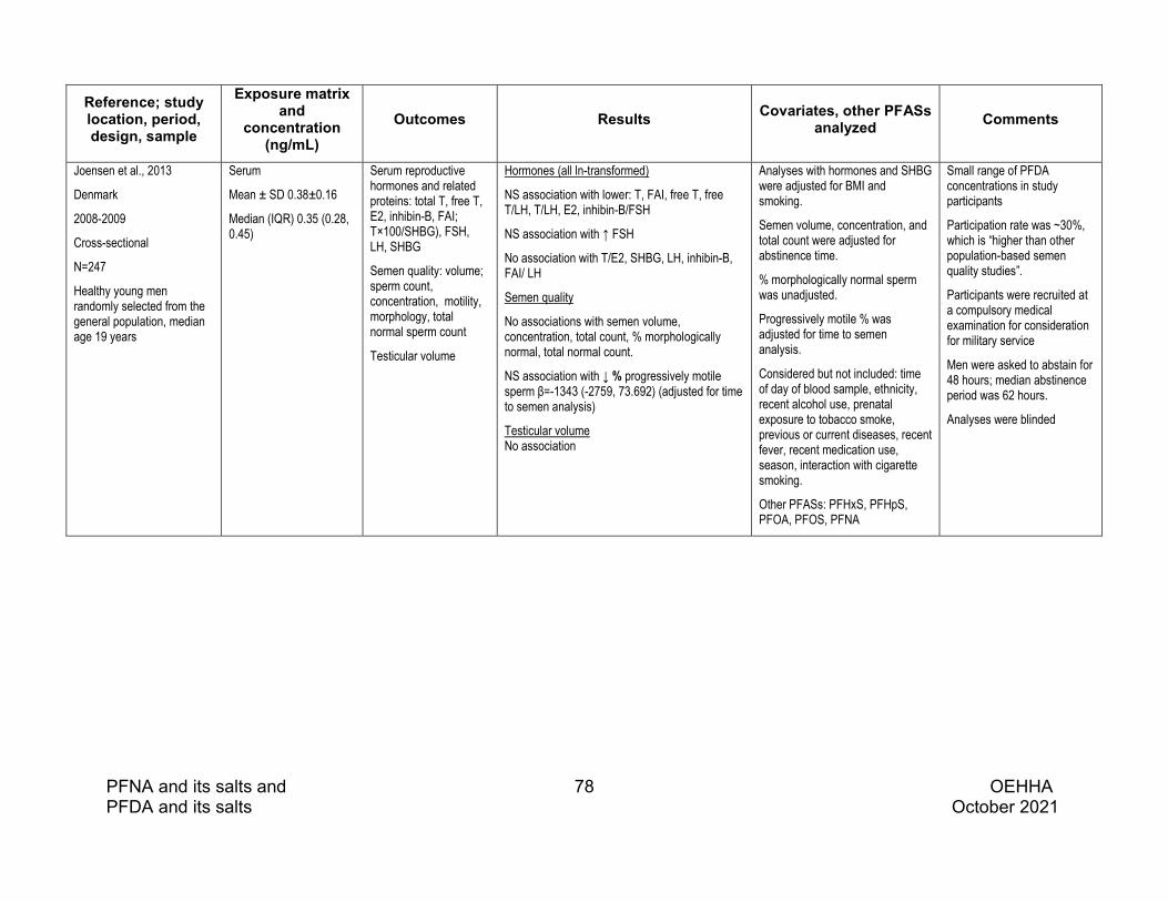
PFNA and its salts and 78 OEHHA PFDA and its salts October 2021
Reference; study location, period, design, sample
Exposure matrix and
concentration (ng/mL)
Outcomes Results Covariates, other PFASs analyzed Comments
Joensen et al., 2013
Denmark
2008-2009
Cross-sectional
N=247
Healthy young men randomly selected from the general population, median age 19 years
Serum
Mean ± SD 0.38±0.16
Median (IQR) 0.35 (0.28, 0.45)
Serum reproductive hormones and related proteins: total T, free T, E2, inhibin-B, FAI; T×100/SHBG), FSH, LH, SHBG
Semen quality: volume; sperm count, concentration, motility, morphology, total normal sperm count
Testicular volume
Hormones (all ln-transformed)
NS association with lower: T, FAI, free T, free T/LH, T/LH, E2, inhibin-B/FSH
NS association with ↑ FSH
No association with T/E2, SHBG, LH, inhibin-B, FAI/ LH
Semen quality
No associations with semen volume, concentration, total count, % morphologically normal, total normal count.
NS association with ↓ % progressively motile sperm β=-1343 (-2759, 73.692) (adjusted for time to semen analysis)
Testicular volume No association
Analyses with hormones and SHBG were adjusted for BMI and smoking.
Semen volume, concentration, and total count were adjusted for abstinence time.
% morphologically normal sperm was unadjusted.
Progressively motile % was adjusted for time to semen analysis.
Considered but not included: time of day of blood sample, ethnicity, recent alcohol use, prenatal exposure to tobacco smoke, previous or current diseases, recent fever, recent medication use, season, interaction with cigarette smoking.
Other PFASs: PFHxS, PFHpS, PFOA, PFOS, PFNA
Small range of PFDA concentrations in study participants
Participation rate was ~30%, which is “higher than other population-based semen quality studies”.
Participants were recruited at a compulsory medical examination for consideration for military service
Men were asked to abstain for 48 hours; median abstinence period was 62 hours.
Analyses were blinded

PFNA and its salts and 79 OEHHA PFDA and its salts October 2021
Reference; study location, period, design, sample
Exposure matrix and
concentration (ng/mL)
Outcomes Results Covariates, other PFASs analyzed Comments
Louis et al., 2015
Michigan and Texas 2005-2009
Cross-sectional
N=501
Population-based sample of couples planning pregnancy, recruited through a marketing database in Michigan and a fishing/hunting license registry in Texas.
Serum
Median (IQR); geometric mean (95% CI)
Michigan 0.3 (0.2, 0.4); 0.31 (0.28, 0.35)
Texas 0.5 (0.3, 0.6); 0.47 (0.45, 0.50)
Semen quality:
Volume, straw distance, sperm concentration, total count, hypo-osmotic swollen,
8 motility measures,
6 sperm head measures,
12 individual and 2 summary morphology measures,
2 sperm chromatin stability measures
Associations with 1-unit increase in ln(PFDA):
No significant differences in semen volume; sperm viability, count, or concentration.
Sperm head length, β=–0.155 (–0.304, –0.006) µm
% of sperm with coiled tail, β=–7.603 (–14.014, –1.193)
No significant associations with other semen parameters
Adjusted for age, BMI, serum cotinine, abstinence, sample age, research site (Texas/Michigan)
Other PFASs: PFOA, PFOS, PFOSA, Et-PFOSA-AcOH, Me-PFOSA-AcOH, PFNA
Semen provided after 2-day abstinence. A 2nd sample collected approximately 1 month later was used to corroborate azoospermia observed in the 1st sample.
Semen analysis was performed one day after collection.
PFOSA, Me-PFOSA-AcOH, and PFOA were most often associated with semen parameters.
Zhou et al., 2016
Taipei, Taiwan
2009-2010
Cross-sectional
N=102 healthy 13-15-year-old boys from 7 public schools
From the control cohort of the Genetics and Biomarkers study for Childhood Asthma
Serum sampled after 8-hour fast
Median (IQR) 0.9 (0.8, 1.1)
Serum T and E2 (average of 2 values)
β (95% CI) per ng/mL increase in PFDA:
Ln(T) –0.2565 (–0.4135, –0.0994) mmol/L
Ln(E2) 0.0734 (–0.1189, 0.2657) pmol/L
Age, parental education, BMI, environmental tobacco smoke exposure, regular exercise, month of survey
Other PFASs: PFBS, PFHxA, PFHxS, PFOA, PFOS, PFNA, PFDoA, PFTA
More associations between individual PFAS levels and hormone levels were reported in boys than in girls

PFNA and its salts and 80 OEHHA PFDA and its salts October 2021
Reference; study location, period, design, sample
Exposure matrix and
concentration (ng/mL)
Outcomes Results Covariates, other PFASs analyzed Comments
Petersen et al., 2018
Faroe Islands
2007-2009
Cross-sectional
N=263
Entire population of 24-26 year old men were invited
Serum
Median (Range) 0.52 (0.16-4.59)
Serum reproductive hormones and related proteins: FSH, inhibin-B, LH, T, FT, estradiol, SHBG, Inhibin-B/FSH, T/LH, FT/LH, T/estradiol, FT/estradiol) Semen quality: Sperm conc., total sperm count, semen volume, % motile sperm
Specific results for PFDA were not reported. Authors state there were no associations between semen quality and any PFASs.
Associations with reproductive hormones were not mentioned for PFDA.
Reproductive hormones (except FSH, LH) were adjusted for BMI, smoking, age, time of day of blood sampling.
Sperm conc., total sperm count, and semen volume were adjusted for abstinence (hours).
% motile sperm was adjusted for interval between ejaculation and assessment.
Other PFASs: PFHxS, PFOA, PFOS, PFNA
49% of those reached by phone (24% of all young men invited by mail) participated.
Pan et al., 2019
Nanjing, China
2015-2016
Cross-sectional
N=664 males from couples visiting a reproductive medical center. Some men had fecundity issues and some were partners of women with female factor infertility.
Exclusions: severe reproductive tract disease
Semen (after 2-day abstinence):
Median (IQR) 0.020 (0.012, 0.034)
Serum (same day as semen)
Median (IQR) 1.240 (0.706, 2.031)
Semen quality:
semen volume,
sperm conc.,
sperm count,
progressively motile (%),
curvilinear velocity (VCL),
straight-line velocity (VSL),
morphologically normal (%),
DNA fragmentation index (DFI),
high DNA stainability (HDS)
Concentration and motility were measured by CASA
P-values were adjusted for false discovery rate/multiple comparisons
Change (95% CI) per unit increase in:
ln(semen PFDA)
Progressively motile (%) β=–1.475 (–2.796, –0.155) p=0.1
VSL (µm/s) β=–0.659 (–1.305, –0.013) p=0.1
DFI (%) β=0.083 (0.021, 0.145) p=0.05
HDS (%) β=0.068 (0.026, 0.110) p=0.02
No associations with other semen parameters
Ln(serum PFDA)
HDS (%) β=0.073 (0.031, 0.115), p=0.02
No associations with other semen parameters
Adjusted for: age, BMI, BMI2, smoking status, alcohol use, abstinence time
Also considered: having fathered a pregnancy, occupational hazards, and medical history
Analyses of associations with semen parameters focused on the most abundant PFASs in serum and semen and those detected in at least 80% of semen samples: PFOA, PFOS, PFNA, PFUnDA, 6:2 Cl-PFESA.
Correlation between serum and semen PFDA: r=0.74
PFDA correlations with other PFASs:
PFHxS r=0.327
PFOA r=0.428
PFOS r=0.688
PFNA r=0.825
PFUnDA r=0.851
Associations of PFDA with semen parameters were similar to those of other PFASs.
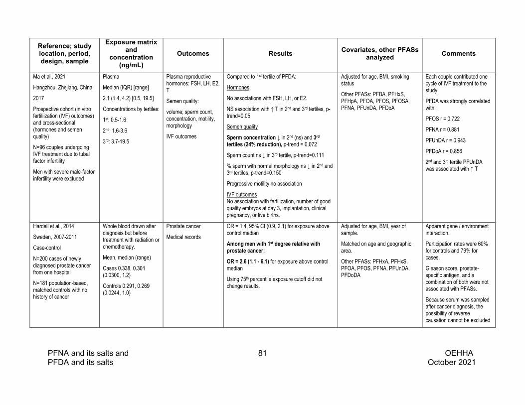
PFNA and its salts and 81 OEHHA PFDA and its salts October 2021
Reference; study location, period, design, sample
Exposure matrix and
concentration (ng/mL)
Outcomes Results Covariates, other PFASs analyzed Comments
Ma et al., 2021
Hangzhou, Zhejiang, China
2017
Prospective cohort (in vitro fertiliization (IVF) outcomes) and cross-sectional (hormones and semen quality)
N=96 couples undergoing IVF treatment due to tubal factor infertility
Men with severe male-factor infertility were excluded
Plasma
Median (IQR) [range]
2.1 (1.4, 4.2) [0.5, 19.5]
Concentrations by tertiles:
1st: 0.5-1.6
2nd: 1.6-3.6
3rd: 3.7-19.5
Plasma reproductive hormones: FSH, LH, E2, T
Semen quality:
volume; sperm count, concentration, motility, morphology
IVF outcomes
Compared to 1st tertile of PFDA:
Hormones
No associations with FSH, LH, or E2.
NS association with ↑ T in 2nd and 3rd tertiles, p-trend=0.05
Semen quality
Sperm concentration ↓ in 2nd (ns) and 3rd tertiles (24% reduction), p-trend = 0.072
Sperm count ns ↓ in 3rd tertile, p-trend=0.111
% sperm with normal morphology ns ↓ in 2nd and 3rd tertiles, p-trend=0.150
Progressive motility no association
IVF outcomes No association with fertilization, number of good quality embryos at day 3, implantation, clinical pregnancy, or live births.
Adjusted for age, BMI, smoking status
Other PFASs: PFBA, PFHxS, PFHpA, PFOA, PFOS, PFOSA, PFNA, PFUnDA, PFDoA
Each couple contributed one cycle of IVF treatment to the study.
PFDA was strongly correlated with:
PFOS r = 0.722
PFNA r = 0.881
PFUnDA r = 0.943
PFDoA r = 0.856
2nd and 3rd tertile PFUnDA was associated with ↑ T
Hardell et al., 2014
Sweden, 2007-2011
Case-control
N=200 cases of newly diagnosed prostate cancer from one hospital
N=181 population-based, matched controls with no history of cancer
Whole blood drawn after diagnosis but before treatment with radiation or chemotherapy.
Mean, median (range)
Cases 0.338, 0.301 (0.0300, 1.2)
Controls 0.291, 0.269 (0.0244, 1.0)
Prostate cancer
Medical records
OR = 1.4, 95% CI (0.9, 2.1) for exposure above control median
Among men with 1st degree relative with prostate cancer:
OR = 2.6 (1.1 - 6.1) for exposure above control median
Using 75th percentile exposure cutoff did not change results.
Adjusted for age, BMI, year of sample.
Matched on age and geographic area.
Other PFASs: PFHxA, PFHxS, PFOA, PFOS, PFNA, PFUnDA, PFDoDA
Apparent gene / environment interaction.
Participation rates were 60% for controls and 79% for cases.
Gleason score, prostate-specific antigen, and a combination of both were not associated with PFASs.
Because serum was sampled after cancer diagnosis, the possibility of reverse causation cannot be excluded

PFNA and its salts and 82 OEHHA PFDA and its salts October 2021
5.2 PFDA: Animal studies of male reproductive toxicity
Several in vivo animal studies investigating the effect of PFDA on outcomes relevant to male reproductive toxicity (e.g., effects on the gonads, reproductive hormone levels) were identified, including multiple studies in male rats (Bookstaff et al. 1990; George and Andersen 1986; NTP 2019; Olson and Andersen 1983; Van Rafelghem et al. 1987b), one study in male mice (Van Rafelghem et al. 1987b), one study in male hamsters (Van Rafelghem et al. 1987b), one study in male guinea pigs (Van Rafelghem et al. 1987b), and one study in zebrafish (Jo et al. 2014) (See Table 5.2.2). Animal studies that administered PFDA as part of a mixture of chemicals were not included in this review.
Of the multiple studies of PFDA conducted in male rats, one study treated male SD rats at five dose levels (0.156-2.5 mg/kg-day) by daily gavage for 28 days (NTP 2019). All other rat studies assessed the effects of PFDA seven to 30 days after administration of a single intraperitoneal (i.p.) injection of relatively high doses (ranging from 20-80 mg/kg). The studies in male mice, hamsters, and guinea pigs also treated the animals with a single i.p. injection of relatively high doses of PFDA and evaluated the toxic effects 14 to 28 days after dosings (Van Rafelghem et al. 1987b). In addition to these studies in rodents, the effects of 120-day exposure to PFDA on sex steroid hormones and expression of mRNA of selected genes in the hypothalamic-pituitary-gonadal-liver (HPGL) axis were evaluated in zebrafish (Jo et al. 2014).
Similar to the studies on PFNA (see Section 4.2), all the studies in rats, mice, hamsters, or guinea pigs used relatively short period of treatment and/or observation. None of the studies in rats or mice treated or observed the animals for one spermatogenic cycle, which is approximately 35 days in mice and 54 days in rats (Clermont 1972; Cordeiro et al. 2021; Johnson et al. 2000). The period of treatment or observation is also shorter than the half-life (>40 days) of PFDA in rats or mice (Kudo et al. 2001; Ohmori et al. 2003). Should the treatment or observation period last more than one spermatogenic cycle, histopathological changes could become more obvious and/or observed at lower doses.
Major findings from these studies are discussed below, grouped by type of effect (reproductive organ weight and histopathology, sperm parameters, hormonal effects, and effects on fertility).
Organ weights & histopathology
PFDA-induced histopathological changes in the testis have been observed in the studies conducted in rats, hamsters, and guinea pigs, but not in the one study conducted in mice, which examined the testes 28 days after a single i.p. injection. In

PFNA and its salts and 83 OEHHA PFDA and its salts October 2021
the NTP 28-day study in SD rats aged 10-14 weeks at the beginning of treatment, PFDA at doses ≥1.25 mg/kg-day reduced epididymal weight by 10-23% and interstitial cell atrophy in 8-10 of 10 rats in each group (NTP 2019). At the high dose of 2.5 mg/kg-day, spermatid retention in seminiferous tubules (inhibited spermiation) was observed in 4 of 10 animals. Minimal germinal epithelium degeneration in the testes and exfoliated germ cells in epididymal ducts were also observed in rats treated with 2.5 mg/kg-day PFDA. Design parameters and key findings from this NTP study are presented in Table 5.2.1.
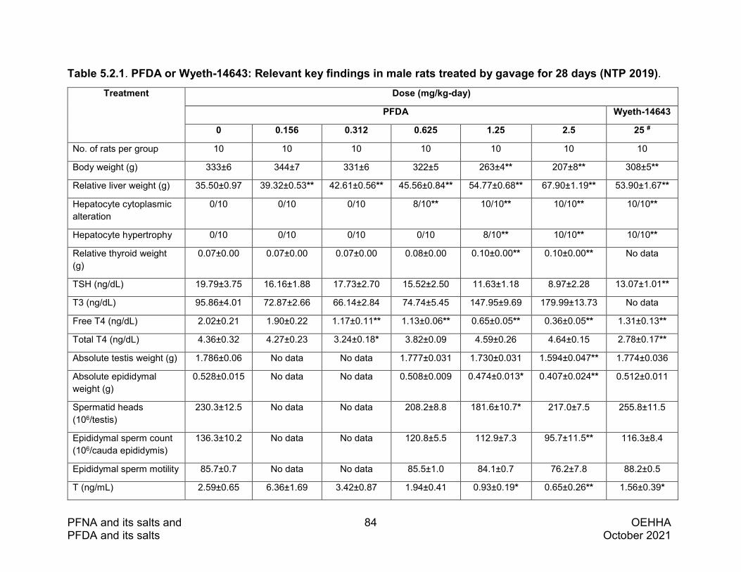
PFNA and its salts and 84 OEHHA PFDA and its salts October 2021
Table 5.2.1. PFDA or Wyeth-14643: Relevant key findings in male rats treated by gavage for 28 days (NTP 2019). Treatment Dose (mg/kg-day)
PFDA Wyeth-14643
0 0.156 0.312 0.625 1.25 2.5 25 #
No. of rats per group 10 10 10 10 10 10 10
Body weight (g) 333±6 344±7 331±6 322±5 263±4** 207±8** 308±5**
Relative liver weight (g) 35.50±0.97 39.32±0.53** 42.61±0.56** 45.56±0.84** 54.77±0.68** 67.90±1.19** 53.90±1.67**
Hepatocyte cytoplasmic alteration
0/10 0/10 0/10 8/10** 10/10** 10/10** 10/10**
Hepatocyte hypertrophy 0/10 0/10 0/10 0/10 8/10** 10/10** 10/10**
Relative thyroid weight (g)
0.07±0.00 0.07±0.00 0.07±0.00 0.08±0.00 0.10±0.00** 0.10±0.00** No data
TSH (ng/dL) 19.79±3.75 16.16±1.88 17.73±2.70 15.52±2.50 11.63±1.18 8.97±2.28 13.07±1.01**
T3 (ng/dL) 95.86±4.01 72.87±2.66 66.14±2.84 74.74±5.45 147.95±9.69 179.99±13.73 No data
Free T4 (ng/dL) 2.02±0.21 1.90±0.22 1.17±0.11** 1.13±0.06** 0.65±0.05** 0.36±0.05** 1.31±0.13**
Total T4 (ng/dL) 4.36±0.32 4.27±0.23 3.24±0.18* 3.82±0.09 4.59±0.26 4.64±0.15 2.78±0.17**
Absolute testis weight (g) 1.786±0.06 No data No data 1.777±0.031 1.730±0.031 1.594±0.047** 1.774±0.036
Absolute epididymal weight (g)
0.528±0.015 No data No data 0.508±0.009 0.474±0.013* 0.407±0.024** 0.512±0.011
Spermatid heads (106/testis)
230.3±12.5 No data No data 208.2±8.8 181.6±10.7* 217.0±7.5 255.8±11.5
Epididymal sperm count (106/cauda epididymis)
136.3±10.2 No data No data 120.8±5.5 112.9±7.3 95.7±11.5** 116.3±8.4
Epididymal sperm motility 85.7±0.7 No data No data 85.5±1.0 84.1±0.7 76.2±7.8 88.2±0.5
T (ng/mL) 2.59±0.65 6.36±1.69 3.42±0.87 1.94±0.41 0.93±0.19* 0.65±0.26** 1.56±0.39*

PFNA and its salts and 85 OEHHA PFDA and its salts October 2021
Treatment Dose (mg/kg-day)
PFDA Wyeth-14643
0 0.156 0.312 0.625 1.25 2.5 25 #
Interstitial cell atrophy 0/10 0/10 0/10 0/10 8/10** 10/10** No data
Spermatid retention 0/10 0/10 0/10 0/10 0/10 4/10* No data
Germ cell degeneration 1/10 0/10 0/10 0/10 0/10 4/10 1/10
Epididymal lesion 1/10 0/10 0/10 0/10 0/10 4/10 1/10
# Only the findings from the high dose of Wyeth presented in this table. *statistically significant, p<0.05. **statistically significant, p<0.01.

PFNA and its salts and 86 OEHHA PFDA and its salts October 2021
In addition to the NTP study in SD rats, degeneration in testicular tissues was observed in male Fischer rats 8 or 16 days after a single i.p. injection of 50 mg/kg of PFDA (George and Andersen 1986; Olson and Andersen 1983; Van Rafelghem et al. 1987b). Bookstaff et al. found no histopathological changes in the testis of adult SD rats seven days after a single i.p. injection of 20, 40 or 80 mg/kg PFDA (Bookstaff et al. 1990). However, the authors found that PFDA at 40 or 80 mg/kg caused significant reduction in plasma levels of T and DHT as well as atrophy of seminal vesicles and prostates. The testis weight was also significantly reduced after PFDA i.p. injection at 80 mg/kg.
In the study comparing the liver and testicular effects of PFDA among rats, mice, hamsters, and guinea pigs, Van Rafelghem et al. treated the animals with a single i.p. injection of PFDA. (Van Rafelghem et al. 1987b). Necropsy was performed on all surviving animals on Day 14 (guinea pigs), Day 16 (rats and hamsters), or Day 28 (mice) after dosing. The authors found degenerative changes in the testis of hamsters (Day 16 at 50 or 100 mg/kg) and guinea pigs (Day 14 at 150 mg/kg) were not as severe as in rats. Single i.p. injections of PFDA at doses up to 250 mg/kg did not cause obvious histopathological lesions in the testis of treated mice on Day 28 after dosing.
Several of the studies discussed above found that PFDA at the dosing levels toxic to rat testes also caused reduced food intake and body weight, liver damage, or other toxic effects. As cited in the 2019 NTP study report, Chapin and Creasy had reported that decreased body weight gain and/or decreased food intake may result in decreased T production through decreased GnRH release and cautioned that such effects should not be mistaken for a direct effect of the test material on androgen balance (Chapin and Creasy 2012). Studies investigating the effects of severe food restriction in rats for as long as 17 weeks that resulted in reductions in body weight of up to 30% did not observe effects on fertility, testicular weight, or severe histopathological changes (Chapin et al. 1993; Rehm et al. 2008). Therefore, PFDA-induced testicular effects are unlikely to be secondary to the general toxicity observed in the same animals.
Sperm parameters
Reduced epididymal sperm count was observed in SD rats following 28-day oral treatment with PFDA at 2.5 mg/kg-day (NTP 2019). There is no other whole animal study that assessed sperm parameters following exposure to PFDA.
Hormonal effects
PFDA-produced reduction in serum or plasma level of T was observed in rats following 28-day oral treatment at ≥1.25 mg/kg-day doses (NTP 2019) or after a single i.p. injection at 40 or 80 mg/kg (Bookstaff et al. 1990). Plasma DHT levels were also decreased by PFDA in the studies by Bookstaff et al. 1990, while no treatment-related

PFNA and its salts and 87 OEHHA PFDA and its salts October 2021
effect was observed on plasma LH. Bookstaff et al. showed PFDA had no effect on plasma T levels in castrated rats supplemented with normal levels of systemic testosterone via testosterone implants, but that PFDA treatment of intact rats decreased the dose-dependent secretion of T in response to human chorionic gonadotropin (hCG) stimulation in studies with decapsulated testes, Based on these studies, Bookstaff et al. suggested that PFDA reduces plasma T in vivo by disrupting testicular response to LH stimulation (Bookstaff et al. 1990).
The effects of PFDA on steroidogenesis were also evaluated in zebrafish (Jo et al. 2014). In this study, freshly fertilized zebrafish eggs were collected and exposed to PFDA at concentrations of 0, 0.01, 0.1 and 1.0 ml/L for 120 days. Compared to controls, the ratios of E2 to T and E2 to 11-ketotestosterone (11-KT) were significantly reduced in the plasma of zebrafish exposed to 1.0 m/L PFDA. Exposure to PFDA at 1.0 mg/L also up regulated transcription levels of the cyp19a gene in male gonads. The authors concluded that PFDA caused estrogenic effects in male zebrafish.
Effects on fertility
There is no study available on the effects of PFDA on male-mediated fertility.

PFNA and its salts and 88 OEHHA PFDA and its salts October 2021
Table 5.2.1 PFDA: Evidence on the male reproductive toxicity in animal studies. Study Design Outcomes Assessed Major Findings Notes
NTP 2019
Male SD rats, 10-11 weeks old at the beginning of dosing, 10 rats per dose group.
Treatment: PFDA (>97% purity, i.e., linear PFDA), once daily by gavage in deionized water with 20% Tween® 80 at doses of 0, 0.156, 0.312, 0.625, 1.25, or 2.5 mg/kg-day, respectively, for 28 days. Six other PFASs and Wyeth-14,643 (a PPARα agonist) were also tested for comparison.
General toxicity: survival, body weights, clinical chemistry, thyroid hormones, liver effects (e.g., gene expression, enzyme activity) and histopathology.
Male reproductive toxicity: testis and epididymis weights; sperm production and motility; histopathology of the testis and epididymis; serum T.
General toxicity:
No effect on survival; lower body weights at ≥ 1.25 mg/-kg-day; reduced free T4 concentrations at ≥ 0.312 mg/kg-day; Increased relative weights of liver, kidney, and adrenal gland; increased hepatic expression of PPARα -associated genes (Acox1, Cyp4a1) and constitutive androstane receptor (CAR)-associated genes (Cyp2b1, Cyp2b2) at ≥ 0.156 mg/kg-day.
Male reproductive toxicity:
Dose-dependent trends in reduction in testis and epididymis weights, serum T level and epididymal sperm count, with statistically significant differences (p < 0.05) at doses of ≥1.25 mg/kg-day (T level and epididymal weights) or 2.5 mg/kg-day (testis weight, epididymal sperm count). Interstitial cell atrophy in 8/10 and 10/10 rats at 1.25 and 2.5 mg/kg-day doses, respectively. At 2.5 mg/kg-day, 4/10 animals had germ cell degeneration, inhibited spermiation, and epididymal lesions.
Treatment period of 28 days is shorter than one full cycle of spermatogenesis in rats (about 56 days).
Similar effects were observed with PFNA treatment.
Treatment with Wyeth-14,643 caused significant effects in the liver, but not in the testis, indicating testicular effects of PFDA may not be mediated via the PPARα pathway.
Olson and Andersen 1983
Male Fischer rats, weighing 200 g. A total of 24 rats per group. Six animals per group were sacrificed on days 2, 4, 8 and 16 after treatment.
Treatment: single i.p. injection of 50 mg/kg PFDA (purity not reported) in propylene glycol-water (1:1) solution, and two control groups: vehicle controls and pair-fed vehicle controls.
General toxicity: survival, food intake, body weight, organ weights, fatty acid level in liver and blood, and general histopathology.
Male reproductive toxicity: testis weight
General toxicity: Deaths occurred in the 2nd and 3rd weeks after dosing with PFDA. Severely reduced food consumption and body weight, beginning one day after dosing, and continuing to study termination. Significantly increased liver weight on days 8 and 16 compared to pair-fed controls; decreased liver weight on days 8 and 16 compared to vehicle controls.
Male reproductive toxicity: Significantly decreased testis weight on day 16 compared to vehicle and pair-fed controls (1.7±0.1 g vs 2.8±0.1 g in vehicle controls and 2.2±0.0 g in pair-fed controls).
The dose used in this study is higher than the acute 30-day LD50 following i.p. injection (41 mg/kg).
The authors noted findings from an earlier study, published only as an abstract, in which a single dose of 100 mg/kg PFDA to male Fischer rats resulted in seminiferous tubule atrophy by day 8 and frank necrosis and calcification by day 16.

PFNA and its salts and 89 OEHHA PFDA and its salts October 2021
Study Design Outcomes Assessed Major Findings Notes George and Andersen 1986
Male Fischer 344 rats, weighing 225-250 g. At least six rats per group weresacrificed on days 4, 8, 12, 16 or 30 aftertreatment.
Treatment: PFDA (96% purity, i.e., linear PFDA) in propylene glycol- water (1:1) solution, single i.p. injection of 50 mg/kg, and pair-fed vehicle controls.
General toxicity: food intake, body weight, organ weights, clinical chemistry, fatty acid level in liver and whole blood, and general histopathology.
Male reproductive toxicity: histopathology of the testis
General toxicity: Severely reduced food consumption and body weight, beginning one day after dosing. Compared to pair-fed controls body weights were significantly decreased, and liver weights were significantly increased. Increase in several fatty acids in liver and in thymic atrophy.
Male reproductive toxicity: Atrophy and degeneration of the seminiferous tubules were first seen at day 16 in treated rats and remained severe at day 30.
Survival not reported. The dose used in this study is higher than the acute 30-day LD50 following i.p. injection (41 mg/kg).
Van Rafelghem et al. 1987b
Male Fischer 344 rats weighing 200-250 g, five rats per group (this study compared the toxic effects of PFDA in rats, mice, hamsters, and guinea pigs).
Treatment: single i.p. injection of 50 mg/kg PFDA (purity not reported) in propylene-water (1:1) solution, and vehicle controls. All animals were sacrificed on day 16.
General toxicity: mortality, food intake, body weight, liver weight
General toxicity: increased mortality by day 16: 0/5 and 1/5 in control and 50 mg/kg dose groups, respectively. Rapid and immediate reduced food intake and body weight. Increased relative liver weight.
Histologic changes in rat liver, thymus, and testes have been described in previous studies:
Liver - heptomegaly and swelling of liver parenchymal cells characterisitic of peroxisome proliferation
Thymus - thymic atrophy
Testes – testicular atrophy.
Van Rafelghem et al. 1987b
Male Syrian golden hamsters weighing 100-120 g, four hamsters per group.
Treatment: single i.p. injection of 50, 100, 200 or 400 mg/kg PFDA (purity not reported) in propylene-water (1:1) solution, and vehicle controls. All animals were sacrificed on day 16.
General toxicity: mortality, food intake, body weight, liver weight, histopathology of liver and thymus.
Male reproductive toxicity: histopathology of the testis
General toxicity: dose-related increase in mortality by day 16: 0/4, 0/4, 2/4, 4/4, and 4/4 in control, 50, 100, 200, and 400 mg/kg dose groups, respectively. Rapid and immediate reduced food intake and body weight. Increased relative liver weight at 50 mg/kg (LD50), heptomegaly and swelling of parenchymal cells characterisitic of peroxisome proliferation, and thymic atrophy.
Male reproductive toxicity: mild seminiferous tubular degeneration with decreased spermatogenesis in surviving hamsters at 100 mg/kg, in 1/4 animals at 50 mg/kg and 0/4 in vehicle controls. Decrease testis weight in treated hamsters (data not shown).

PFNA and its salts and 90 OEHHA PFDA and its salts October 2021
Study Design Outcomes Assessed Major Findings Notes Van Rafelghem et al. 1987b
Male CF-1 mice weighing 30-40 g, ten mice per group.
Treatment: single i.p. injection of 150, 200, or 250 mg/kg PFDA (purity not reported) in propylene-water (1:1) solution, and vehicle controls. All animals were sacrificed on day 28.
General toxicity: mortality, body weight, liver weight, histopathology of liver and thymus.
Male reproductive toxicity: histopathology of the testis
General toxicity: dose-related increase in mortality by day 28: 0/10, 6/10, 10/10, and 10/10 in control, 150, 200, and 250 mg/kg dose groups, respectively. Rapid and immediate reduced body weight. Increased relative weight at 150 mg/kg (LD50), heptomegaly and swelling of parenchymal cells, characterisitic of peroxisome proliferation. No histologic effects on the thymus.
Male reproductive toxicity: no histologic effects on the testes at day 28 in any dose group. A slight decrease in testes weight in treated mice was reported (data not shown).
Van Rafelghem et al. 1987b
Male Hartley guinea pigs weighing 535-660 g, three animals per group.
Treatment: single i.p. injection of 125,150, or 175 mg/kg PFDA (purity not reported) in propylene-water (1:1) solution, and vehicle controls. All animals were sacrificed on day 14.
General toxicity: mortality, body weight, liver weight, histopathology of liver and thymus.
Male reproductive toxicity: histopathology of the testis
General toxicity: increased mortality by day 14: 0/3, 1/3, 0/3, and 2/3 in control, 125, 150, and 175 mg/kg dose groups, respectively. Rapid and immediate reduced body weight. Mild to moderate periportal hepatocellular cytoplasmic alterations at 150 and 175 mg/kg. No clear histologic effects on the thymus.
Male reproductive toxicity: no degenerative changes in the testes of animals at 125 mg/kg; mild to moderate degeneration in the seminiferous epithelia at 150 mg/kg; no effects observed in the testes of the one surviving guinea pig at 175 mg/kg. Decreased testes weight in treated guinea pigs (data not shown).

PFNA and its salts and 91 OEHHA PFDA and its salts October 2021
Study Design Outcomes Assessed Major Findings Notes Bookstaff et al. 1990
Adult male SD rats weighing approximately 295 g. Ten rats per group.
Treatment: PFDA (96% purity) in propylene glycol/water (1:1) solution, a single i.p injection of 20, 40 or 80 mg/kg and two control groups: vehicle controls and pair-fed vehicle controls. All animals were sacrificed on day 7 after dosing.
In a parallel experiment, rats (6 per group) were castrated and implanted with sustained-release testosterone-containing capsules two hours after receiving a single i.p injection of PFDA at 20, 40 or 80 mg/kg, or vehicle (vehicle controls and pair-fed controls). Animals were sacrified on day 7 after dosingand the testes were used in the in vitro experiments (summarized below).
General toxicity: feed consumption and body weight.
Male reproductive toxicity: Plasma levels of T, DHT and LH; histology and organ weights of the testis, seminal vesicles and ventral prostate.
General toxicity: reduced feed consumption and body weight at 80 mg/kg compared to vehicle controls, and reduced body weight at 40 mg/kg compared to vehicle and pair-fed controls (on day 7 post-dosing).
Male reproductive toxicity:
Reduction in plasma levels of T and DHT at 40 and 80 mg/kg compared to vehicle and pair-fed controls. No effect on plasma concentrations of LH.
Reduction in the weight of testis at 80 mg/kg compared to vehicle control, and in the weights of seminal vesicles and ventral prostate at 20, 40 and 80 mg/kg compared to vehicle and pair-fed controls (seminal vesicle weight at 20 mg/kg was decreased compared to vehicle control only). Apparent atrophy of the epithelium of seminal vesicles at 80 mg/kg, and of the ventral prostate at 40 and 80 mg/kg. No histological changes in the testis.
In castrated/testosterone-implanted rats, seminal vesicle weight was decreased at 20 and 40 mg/kg compared to pair-fed controls. PFDA treatment had no effect on ventral prostate weight or plasma T levels.
Depending on the germ cell population targetted by PFDA, testicular lesions may not be obvious on day 7 after a single injection.
The authors concluded that PFDA disrupted the feedback relationship between plasma androgen and LH concentrations in treated animals.
Bookstaff et al. 1990
Experimental Model: in vitro incubation of decapsulated testis from adult rats 7 days after a single i.p. injection of 0, 20, 40, or 80 mg/kg PFDA (96% purity).
Treatment: decapsulated testis was incubated in the absence or presence of 100 mIU human chorionic gonadotropin (hCG)/mlfor 3 hours.
hCG-stimulated T secretion, measured as the difference between basal T secretion (in the absence of hCG) and hCG-stimulated secretion over a 3-hour period
Dose-related decrease in hCG-stimulated T secretion by decapsulated testes from rats treated with PFDA at 40 and 80 mg/kg, compared to testes from vehicle and pair-fed controls.
The authors concluded that PFDA decreases testicular responsiveness to LH-like (hCG) stimulation.

PFNA and its salts and 92 OEHHA PFDA and its salts October 2021
Study Design Outcomes Assessed Major Findings Notes Jo et al. 2014
Zebrafish, four replicates (with 10 fertilized eggs each) per group. Three male fish were randomly sampled from each of four replicate tanks per group for hormone and gene expression measurements
Treatment: PFDA (98% purity) at concentrations of 0, 0.01, 0.1, 1.0 or 10 mg/L in water from 1 day post fertilization (dpf) to 120 dpf.
General toxicity: survival at 17 dpfae), 34 and 61 dpf (juvenile), and 120 dpf (adult); length, wet weight, and condition factor.
Male reproductive toxicity: blood levels of E2, T, and 11-KT.
mRNA expression of genes in the HPGL axis
General toxicity:
Survival at 17 dpf was significantly decreased at concentrations of 0.1 mg/L and above, compared to controls. The no observed effective concentration was 0.01 mg/L. 100% mortality occurred in the 10 mg/L group by 34 dpf. Treatment had no effect on length, weight or condition factor.
Brain: increased mRNA expression of cyp19b, erα, and er2β at 1 mg/L.
Gonad: increased mRNA expression of cyp19a at 1 mg/L.
Liver: increased mRNA expression of vtg1 at 1 mg/L.
Male reproductive toxicity:
No effect on blood levels of E2, T, or 11-KT, compared to controls. Increased ratio of E2/T and E2/11-KT at 1.0 mg/L, compared to controls.
The authors noted that increased ratios of E2/T and E2/11-KT and increased gene expression of vtg1 suggest that PFDA has estrogenic effects in zebrafish.
In an in vitro study reported in the same paper, PFDA had no effect on E2, T, or E2/T in H295R cells, a human adrenocortical carcinoma cell line.

PFNA and its salts and 93 OEHHA PFDA and its salts October 2021
5.3 PFDA: Mechanistic considerations and other relevant data
The mechanistic literature regarding PFDA and male reproductive toxicity is sparse. We provide a brief overview of findings from general toxicity studies of PFDA and studies of general mechanisms of PFDA-induced cytotoxicity, followed by discussion of studies that may be more informative with respect to the effects PFDA may have on the male reproductive system.
General toxicity
In male Sprague-Dawley rats, a single i.p. dose of PFDA resulted in a LD50 of 41 mg/kg (30-days), a LD50 of 64 mg/kg (14 days), and at 50 mg/kg PFDA caused significant reduction in food intake, decreased body weight, and mortality (Olson and Andersen 1983). Other studies have observed similar effects of PFDA on food intake, body weight, and mortality. For example, a study of male rats treated with a single dose of either 40 mg/kg or 80 mg/kg PFDA reported reduced body weight at both doses, as well as reduced food consumption at the higher dose (Bookstaff et al. 1990). Some authors suggested these effects of PFDA are similar to the wasting syndrome observed in rats exposed to TCDD (George and Andersen 1986; Olson and Andersen 1983; Van Rafelghem et al. 1987a; Van Rafelghem et al. 1987b). In other studies, conducted in female Sprague-Dawley rats, Borges et al. 1993 found that PFDA is a noncompetitive and reversible inhibitor of the peroxisomal enzymes fatty acyl-CoA oxidase (HYDRA) and 3-hydroxyacyl-CoA dehydrogenase (DEHYDRO) (the peroxisomal bifunctional protein) (Borges et al. 1993). Borges et al. (1993) suggested that PFDA’s inhibition of these peroxisomal enzymes and the resulting disruption of hepatic lipid cellular metabolism may lead to the observed wasting syndrome.
Cytotoxicity
In HCT116 cells (a human epithelial-like colon carcinoma cell line), PFDA caused cell death (Kleszczyński et al. 2007) and depolarization of plasma membrane potential in a dose- and time-dependent manner (0, 100, 200, 300, or 400 µM, 4-72 hour incubation period) (Kleszczyński and Składanowski 2009). In separate experiments, incubation of HCT116 cells with PFDA resulted in kinase dephosphorylation (200 µM) (Kleszczyński et al. 2009), acidification of the cyptoplasm (0-400 µM) (Kleszczyński and Składanowski 2011), mitochondrial calcium overload (200 µM) (Kleszczyński and Składanowski 2011), and reactive oxygen species (ROS) generation (at 200 µM) (Kleszczyński and Składanowski 2009; Kleszczyński et al. 2009). At 200 µM PFDA, dissipation of mitochondrial membrane potential led to ATP synthesis arrest, cytochrome c release, and activation of caspases associated with apoptosis (Kleszczyński et al. 2009).

PFNA and its salts and 94 OEHHA PFDA and its salts October 2021
Effects on the hypothalamic-pituitary-gonadal-(liver) axis
The liver can be considered another component of the HPG axis in fish, as liver production of vitellogenin (Vtg), a precursor egg yolk protein, in male fish is under estrogenic control (Jo et al. 2014). Studies investigating the effects of PFDA on hormone levels, metabolism and receptor-mediated effects related to the HPGL axis are summarized in Table 5.3.1 below.
Effects of PFDA on hormone levels have been studied in vivo in rats and zebrafish and ex vivo in rats. In male rats treated with a single dose of PFDA there was a reduction in plasma levels of T and DHT at 40 and 80 mg/kg without effect on plasma concentrations of LH (Bookstaff et al. 1990). However, in castrated male rats supplied with T via implant, a single dose of PFDA resulted in a decrease in seminal vesicle weight at 20 and 40 mg/kg without an effect on ventral prostate weight or plasma T levels. In decapsulated testes from rats treated with a single dose of PFDA (40 or 80 mg/kg), there was a dose-related decrease in hCG-stimulated (100 mIU hCG/ml for 3 hours) T secretion (Bookstaff et al. 1990). Decreases in serum levels of T were also observed in male SD rats exposed to PFDA for 28 days at 1.25 and 2.5 mg/kg-day (NTP 2019).
In male zebrafish, PFDA increased the plasma E2 to T ratio and the plasma E2 to 11-ketotestosterone (11KT) ratio (Jo et al. 2014).
Effects of PFDA on hormone synthesis and metabolism have been studied in vivo in zebrafish and in vitro in a human placental choriocarcinoma cell line, in isolated rat Leydig cells, and in a mouse Leydig tumor cell line. One study in zebrafish and one study in a human cell line report that PFDA alters gene expression and enzyme activity, respectively, of aromatase (CYP19A1), a steroidogenic enzyme that converts T to E2. Specifically, a dose-dependent increase in aromatase gene expression (cyp19a) was observed in male gonads of zebrafish exposed to PFDA (Jo et al. 2014), while a decrease in aromatase activity was observed in a human placental choriocarcinoma cell line (JEG-3 cells) following treatment with PFDA (Kjeldsen and Bonefeld-Jørgensen 2013).
In isolated rat Leydig cells exposed in vitro at various concentrations for 48 h, treatment with PFDA resulted in changes in hCG-stimulated T secretion, in the absence of cytotoxicity or major morphological alterations (Boujrad et al. 2000). These changes consisted of an increase in T production (e.g., secretion into the culture medium) at < 10-5 M PFDA, and a decrease in T production at 10-5 M PFDA and above (Boujrad et al. 2000). In separate experiments in MA-10 cells (a mouse Leydig tumor cell line), PFDA exposure decreased hCG-stimulated progesterone production (e.g., secretion into the culture medium) in a concentration- and time-dependent manner, in the absence of cytotoxicity (Boujrad et al. 2000). In MA-10 cells PFDA also inhibited hCG-stimulated

PFNA and its salts and 95 OEHHA PFDA and its salts October 2021
pregnenolone production and cAMP-stimulated progesterone production, decreased mRNA and protein levels of TSPO, also referred to as peripheral-type benzodiazepine receptor [PBR]) located on the outer mitochondrial membrane, and reduced the stability of TSPO mRNA (Boujrad et al. 2000). PFDA had no effect on P450scc (cyp11A1) activity, steroidogenic acute regulatory protein (StAR) protein levels, mitochondrial integrity, total protein synthesis, or DNA damage levels (assessed as poly(ADP-ribose) polymerase activity in MA-10 cells (Boujrad et al. 2000). The authors suggested that decreased Leydig cell steroid synthesis following exposure to PFDA resulted from a decrease in transport of cholesterol from the outer to the inner mitochondrial membrane, perhaps due to decreased outer membrane levels of TSPO (Boujrad et al. 2000).
In a mouse Leydig tumor cell line (mLTC-1 culture), incubation with PFDA inhibited progesterone production with an IC50 value of 11.52 µM (Zhao et al. 2017). The authors suggested that PFDA disrupted progesterone production as a result of damage to the mitochondrial membrane and increased production of ROS.
Hormone receptor mediated effects have been studied in vivo in fish and in vitro in a human embryonic kidney cell line, two human breast adenocarcinoma cell lines, and a Chinese hamster ovary cell line after exposure to PFDA. In addition, hormone receptor binding studies have been conducted with fish liver cytosol and modeled in silico. In zebrafish, PFDA treatment increased liver gene expression of vitellogenin, an estrogen responsive gene, in males (Jo et al. 2014). In rainbow trout, PFDA treatment via diet resulted in concentration-dependent increases in plasma levels of Vtg in studies with 11- and 5-month old juvenile trout (Benninghoff et al. 2011). In other experiments by these authors, PFDA was shown to competitively bind (weakly) to trout liver ER (in liver cytosol homogenates) (Benninghoff et al. 2011).
In human embryonic kidney (HEK293T) cells transfected with a hERα reporter gene, PFDA (100-1000 nM) increased hERα reporter activity up to 2.5 fold (Benninghoff et al. 2011).
In MVLN cells (derived from MCF-7 cells, a human breast adenocarcinoma cell line, stably transfected with a luciferase reporter gene and an estrogen-responsive element from the Xenopus vitellogenin A2 gene), exposure to PFDA alone did not induce an estrogenic response. However, PFDA displayed anti-estrogenic activity in MVLN cells pretreated with E2, inhibiting the estrogenic response to E2 (measured as luciferase activity) in a concentration dependent manner. PFDA was a more potent inhibitor of the estrogenic response than PFNA in these cells, with an EC50 of 20.3 µM (Juan Li et al. 2020). In separate experiments in MCF-7 cells, PFDA also had anti-estrogenic activity, with the addition of PFDA to E2-treated cells resulting in significant down regulation of the E2-responsive genes TFF1 (trefoil factor 1, also known as pS2) and EGR3 (early growth response 3) (Juan Li et al. 2020).

PFNA and its salts and 96 OEHHA PFDA and its salts October 2021
Analyses using in silico computational models indicate that PFDA can efficiently dock with human, mouse, and trout ERα in the ligand-binding domain and form a hydrogen bond at residue Arg394 in human ERα in a manner similar to that of estrogens (Benninghoff et al 2011). Additional in silico modeling studies with the activated form of hERα, in which E2 is bound to the ligand-binding domain, predict that PFDA can bind to sites on the surface of the activated receptor (Juan Li et al. 2020).
PFDA significantly antagonized DHT-induced AR activity in a concentration-dependent manner in a Chinese hamster ovary cell line (CHO-K1) transfected with AR and a reporter vector (Kjeldsen and Bonefeld-Jørgensen 2013).

PFNA and its salts and 97 OEHHA PFDA and its salts October 2021
Table 5.3.1 PFDA: Effects on the hypothalamic-pituitary-gonadal-(liver) axis in animals and in vitro studies5.
Reference; Experimental Model; N/group; Exposure Details HPG-Related Outcomes Assessed
Results
Bookstaff et al. 1990
In vivo
Adult male SD rats weighing approximately 295 g (10 per group).
Treatment: PFDA (96% purity) single i.p injection of 20, 40 or 80 mg/kg; two control groups: vehicle and pair-fed vehicle controls. All animals were sacrificed on day 7 after dosing.
In a parallel experiment, rats (6 per group) were castrated and implanted with sustained-release testosterone-containing capsules two hours after receiving a single i.p injection of PFDA at 20, 40 or 80 mg/kg, or vehicle (vehicle controls and pair-fed controls). Animals were sacrificed on day 7 after dosing and the testes were used in the ex vivo experiments (summarized below).
Ex vivo
Incubation of decapsulated testis from adult rats 7 days after a single i.p. injection of 0, 20, 40, or 80 mg/kg PFDA (96% purity) in the absence or presence of 100 mIU hCG/ml for 3 hours.
In vivo
Plasma levels of T, DHT and LH
Feed consumption and body weight
Ex vivo
hCG-stimulated T secretion, measured as the difference between basal T secretion (in the absence of hCG) and hCG-stimulated secretion over a 3-hour period
In vivo Reduction in plasma levels of T and DHT at 40 and 80 mg/kg compared to vehicle and pair-fed controls. No effect on plasma concentrations of LH.
In castrated/testosterone-implanted rats PFDA treatment had no effect on plasma T levels.
Reduced feed consumption and body weight at 80 mg/kg compared to vehicle controls, and reduced body weight at 40 mg/kg compared to vehicle and pair-fed controls (on day 7 post-dosing).
Ex vivo
Dose-related decrease in hCG-stimulated T secretion by decapsulated testes from rats treated with PFDA at 40 and 80 mg/kg, compared to testes from vehicle and pair-fed controls.
NTP 2019
Male SD rats, 10-11 weeks old at the beginning of dosing, 10 rats per dose group.
Treatment: PFDA (>97% purity, i.e., linear PFDA), once daily by gavage at doses of 0, 0.156, 0.312, 0.625, 1.25, or 2.5 mg/kg-day, respectively, for 28 days.
Serum T levels
Survival, body weight
Reduced serum T at 1.25 and 2.5 mg/kg-day
No effect on survival. Lower body weights at ≥ 1.25 mg/kg-day.
5Studies with in vivo experiments are presented first ordered by species (rat, mouse, fish), and within species, by publication date, followed by studies with only in vitro experiements, ordered by publication date.

PFNA and its salts and 98 OEHHA PFDA and its salts October 2021
Reference; Experimental Model; N/group; Exposure Details HPG-Related Outcomes Assessed
Results
Benninghoff et al. 2011
In vivo: Rainbow trout Experiment 1: Dietary exposure of 11-month old juvenile rainbow trout to PFDA (highest purity available) at 0, vehicle control (DMSO), 5, 50, or 250 ppm in Oregon Test Diet, OTD) 5 days/week for 14 days (equivalent to 0.1, 1, or 5 mg/kg-d, respectively); animals were sacrificed on day 15. Positive control: 5 ppm E2 N = 6 fish/treated and vehicle control (0.5 ppm DMSO) group. N = 24 fish in untreated control group Experiment 2: Dietary exposure of 5-month old juvenile rainbow trout to PFDA (highest purity available) at 0, 0.026, 0.128, 0.64, 3.2,16, 80, 400, or 2000 ppm ppm in Oregon Test Diet, OTD) 5 days/week for 14 days; animals were sacrificed on day 15. Positive control: 5 ppm E2 N = 8 fish/treated group. N = 16 fish/group in untreated controls and positive controls
In vitro: trout liver cytosol, HEK-293T cells Trout liver cytosol ER binding studies in liver cytosol prepared from trout fed 5 ppm E2 (to induce ERα) HEK-293T cells: a human embryonic kidney cell line transfected with a human ERα expression vector and a luciferase reporter plasmid containing an ERE. PFDA or E2 treatment (1-1000 nM) for 24 hrs; hERα reporter gene activity measured. In Silico Molecular docking studies of PFDA with the active site in human, mouse, and trout ERα.
In Vivo
Experiments 1 & 2: Vtg levels in plasma
In Vitro
Trout liver cytosol: ERα binding studies
HEK-293T cells: hERα gene reporter activity
In Silico
Molecular docking at the active site in human, mouse and trout ERα
In Vivo
Exp.1: Concentration dependent increase in Vtg in plasma levels
Exp. 2: Concentration dependent increase in plasma Vtg levels; LOEL: 0.64 ppm; NOEL: 0.128 ppm
In Vitro
Trout liver cytosol: PFDA shows weak competitive binding to ERα, completely displacing E2 from the receptor, with more than a 10,000-fold lower relative binding affinity than E2.
HEK-293T cells: PFDA at concentrations of 100-1000 nM induced hERα gene reporter activity up to 2.5 fold
In Silico
PFDA is predicted to bind at the active site of human, mouse, and trout ERα. Predicted hydrogen bond interaction with hERα at Arg394.

PFNA and its salts and 99 OEHHA PFDA and its salts October 2021
Reference; Experimental Model; N/group; Exposure Details HPG-Related Outcomes Assessed
Results
Jo et al. 2014
PFDA (98% purity); Control dimethyl sulfoxide (DMSO).
In vivo: zebrafish Fertilized eggs exposed to 0, 0.01, 0.1, 1, or 10 mg/L PFDA until 120 dpf.
N=3 /sex per tank were randomly sampled from four replicates.
In vitro: H295R cells, a human adrenocortical carcinoma cell line
H295R cells were treated with PFDA at 0, 0.1, 1, 10, or 100 mg/L for 48 h.
In vivo
Blood levels of E2, T, and 11-KT.
mRNA expression of genes in brain, gonad, liver
In vitro
Levels of E2 and T
In vivo: zebrafish
No effect on blood levels of E2, T, or 11-KT, compared to controls. Increased ratio of E2/T and E2/11-KT at 1.0 mg/L, compared to controls.
Increased mRNA expression of cyp19b, erα, and er2β at 1 mg/L in male fish brain
Increased mRNA of cyp19a in male gonad and vtg1 in male liver, both at 1 mg/L.
In vitro: H295R cells No significant effects on E2, T, or E2/T ratio.
Boujrad et al. 2000
In vitro
Isolated rat Leydig cells:
Cells were isolated from adult SD (300 g) rats; 20–30% of the isolated cells stained positive for the Leydig specific enzyme 3b-hydroxysteroid dehydrogenase. Cells were cultured for 48 h with PFDA (10-7 - 10-3 M) for 48 h, and then washed with serum free medium and stimulated for 2 hr with hCG (1 nM). hCG stimulated T production measured.
MA-10 mouse Leydig tumor cell line:
Cells wereexposed to PFDA (10-7 -3 x 10⁻³ M) for 24, 48, or 72 h. After 24, 48, or 72 hours, cells were washed with serum free medium and stimulated for 2 h with hCG (1 nM).
Exp. 1: hCG stimulated progesterone production measured
Exp. 2: hCG stimulated pregnenlone production measured
Isolated rat Leydig cells Cell viability and morphology T levels weremeasured in culture media after hCG stimulation. MA-10 mouse Leydig tumor cells Exp. 1 Cell viability Progesterone measured in culture media after hCG stimulation Exp. 2: Cell viability Pregnenolone measured in culture media after hCG stimulation Protein determination (Western blot)
mRNA determination (Northern blot)
Isoated rat Leydig cells
PFDA inhibited hCG-stimulated T production, in the absence of effects on cell viablilty or morphology.
MA-10 mouse Leydig tumor cells
Exp. 1:
PFDA inhibited hCG-stimulated progesterone production in a concentration-dependent manner, in the absence of effects on cell viability.
Exp. 2:
PFDA inhibited hCG-stimulated pregnenolone production, in the absence of effects on cell viability. PFDA decreased mRNA and protein levels of TSPO, and reduced the stability of TSPO.
No effects on CYP11A1 (P450scc) activity, StAR protein levels, mitochondrial integrity, total protein synthesis, or DNA damage (assessed as poly(ADP-ribose) polymerase activity

PFNA and its salts and 100 OEHHA PFDA and its salts October 2021
Reference; Experimental Model; N/group; Exposure Details HPG-Related Outcomes Assessed
Results
Kjeldsen and Bonefeld-Jørgensen 2013
In vitro: MVLN, CHO-K1 and JEG-3 cell lines PFDA (97% purity) dissolved in dimethyl sulfoxide (DMSO), tested at concentrations of 10-9 – 10-4 MMVLN cells: Derived from MCF-7 cells; stably transfected with a luciferase reporter gene and an estrogen responsive element derived from the Xenopus vitellogein A2 gene. ER transactivation assessed with PFDA alone or co-treated with 25 pM E2 CHO-K1 cells: Derived from CHO cells; transiently co-transfected with the AR plasmid and a luciferase reporter vector AR transactivation assessed with PFDA alone or co-treated with or 25 pM DHT JEG-3 cells: A human choriocarcinoma cell line. Aromatase activity assessed with PFDA treatment
MVLN cells: ER transactivation CHO-K1 cells: AR transactivation JEG-3 cells: Aromatase activity
MVLN cells Cytotoxic at 10-4 M ER transactivation: No effects on ER activity CHO-K1cells Cytotoxic at 10-4 M AR transactivation: No AR agonist activity. Concentration-dependent antagonistic effects on DHT-induced AR transactivation (IC50 6×10⁻⁵ M). JEG-3 cells Cytotoxic at 10-4 M Decrease in aromatase activity down to 85 % at 10-5 M.
Zhao et al. 2017
In vitro: Mouse Leydig cell tumor line (mLTC-1)
PFDA (>95% purity)
Experiment 1: PFDA treatment (0-105µM) for 24 hrs; cAMP added and incubated for additional 3 hrs; progesterone quantified via radioimmunoassay
Experiment 2: PFDA treatment (0, 6.25, 12.5, 25, or 50 µM) for 24 hours; mitochondrial membrane potential measured
Progesterone production
Mitochondrial membrane potential
Experiment 1: Concentration dependent decrease in progesterone production (IC50 11.52 µM)
Experiment 2: Significant decrease in mitochondrial membrane potential at 25 and 50 µM.

PFNA and its salts and 101 OEHHA PFDA and its salts October 2021
Reference; Experimental Model; N/group; Exposure Details HPG-Related Outcomes Assessed
Results
Juan Li et al. 2020
In vitro: MVLN and MCF-7 cell lines
PFDA (98% purity)
MVLN cells: Derived from MCF-7 cells; stably transfected with a luciferase reporter gene and an estrogen responsive element derived from the Xenopus vitellogein A2 gene. PFDA treatment (0, 0.0001-5 mM) for 48 hr in the presence or absence of E2 (1 nM) as positive control or ICI 182,780 as negative control, or both; ER transactivation
MCF-7 cells: human breast adenocarcinoma cell line
PFDA treatment (50 µM) in the presence or absence of E2 for 48 hours; changes in expression of estrogen regulated genes (TFF1/pS2, EGR3)
In silico
Modeling analysis of PFDA interactions with E2-activated hERα
MVLN cells: ER transactivation MCF-7 cells: mRNA gene expression of estrogen regulated genes TFF1/pS2, EGR3
In silico: modeling predictions of interaction with E2-activated hERα
MVLN cells
ER transactivation: No ER agonist activity with PFDA alone; in cells pretreated with E2 PFDA inhibited the estrogenic response to E2 in a concentration dependent manner, with a reported EC50 of 20.3 µM. In cells cotreated with E2 and ICI 182,780 , PFDA caused a significant increase in the inhibition of luciferase activity (attenuated luciferase response) MCF-7 cells
mRNA levels of TFF1 and EGR3: No effect on gene expression with PFDA alone. In cells cotreated with E2, PFDA significantly downregulated expression of both TFF1 and EGR3
In silico
Modeling predicts that PFDA can bind to the surface of the E2 activated form of hERα (at sites distinct from the ER binding pocket, including the groove of co-activators)
PFNA and its salts and 102 OEHHA PFDA and its salts October 2021
Effects on the thyroid
PFDA effects on thyroid hormones are summarized in Table 5.3. below.
Only one study (NTP 2019) provided data on outcomes of both thyroid toxicity and male reproductive toxicity with repeated exposures to PFDA. Other available whole animal studies (Gutshall et al. 1988; Gutshall et al. 1989; Langley and Pilcher 1985; Van Rafelghem et al. 1987a) looked at aspects of thyroid toxicity following an acute dose of PFDA. An in vitro study using rat pituitary GH3 cells evaluated PFDA for cytotoxicity, effects on cell proliferation, and aryl hydrocarbon receptor (AhR) transactivity (Long et al. 2013). Cell-free biochemical assays and an in silico molecular docking simulation were used to study binding affinities of PFDA to thyroid hormone transport proteins (Ren et al. 2016). Relevant information and data from these studies are summarized in Table 5.3..
The NTP (2019) 28-day study found decreased free thyroxine (T4) with PFDA doses > 0.312 mg/kg-day. Total T4 (free plus bound), T3, and TSH were not affected. The same animals showed effects on outcomes of male reproductive toxicity at doses > 1.25 mg/kg-day. Adverse outcomes included reduced testes and epididymal weights, reduced serum testosterone, reduced epididymal sperm counts, and histopathology indicative of interstitial testicular tissue atrophy, germ cell degeneration, and inhibited spermiation.
Available whole-animal studies on the effects of PFDA on thyroid function used a single dose at much higher levels than tested in the NTP (2019) 28-day study. None of these studies directly evaluated specific outcomes of male reproductive toxicity, but may help interpretation by addressing a potential mechanistic pathway (Gutshall et al. 1988; Gutshall et al. 1989; Langley and Pilcher 1985; Van Rafelghem et al. 1987a).
A single i.p. injection dose of 75 mg PFDA/kg to male rats was associated with reduced serum total levels of T3, T4, and reverse triiodothyronine (rT3) at 12 and 24 hours post-dosing (Gutshall et al. 1989). An in vitro binding experiment described in the same paper reported that PFDA displaced radiolabeled T4 from binding sites on rat serum albumin, which suggested a possible explanation for at least some of the decrease in serum T4 observed in vivo.
An earlier study by the same group (Gutshall et al. 1988) looked at the effects of 75 mg PFDA/kg i.p., given with and without supplemental T4. On its own, 200 µg T4/kg significantly increased serum total T4. When given in combination with PFDA, supplemental T4 only partially restored total serum T4. The authors suggested their results could indicate an inability of PFDA-treated animals to retain T4 due to alterations in plasma protein binding, increased metabolism, or increased excretion.

PFNA and its salts and 103 OEHHA PFDA and its salts October 2021
Compared to ad libitum fed controls, significantly reduced body weight and feed intake were seen in male rats given a single i.p. dose of PFDA at 40 or 80 mg/kg (p < 0.05 at either dose) and evaluated seven days post-dosing (Van Rafelghem et al. 1987a). Each dose group had their own pair-fed group and only those animals pair-fed to the 80 mg/kg dose group also had significantly reduced body weight compared to ad libitum fed controls (p < 0.05). Total serum T4 was significantly reduced compared to ad libitum controls with all three doses of PFDA (p < 0.05 at 20, 40, and 80 mg/kg). T4 levels for the two lower dose PFDA groups were also significantly lower than their pair-fed partners (p < 0.05); only the 80 mg/kg group and their pair-fed partners did not differ from each other. Compared to ad libitum or pair fed controls, the 80 mg PFDA/kg group had significantly higher serum T3 levels (p < 0.05), as well as lower T3 uptake (p < 0.05 for both comparisons). The authors concluded that despite observed effects of PFDA on thyroid hormone levels, treated animals did not show a pattern of effects fully consistent with hypo- or hyper-thyroidism.
The remaining in vivo study on thyroid effects of PFDA on male rats used a single i.p. dose of 75 mg/kg, compared to vehicle and pair-fed controls (Langley and Pilcher 1985). Serum total T4 levels were lower in PFDA-treated rats compared to pair-fed controls at all tested times (p < 0.05 at 0.5, 1, 2, 4, 6, and 8 days post-treatment), and serum total T3 levels were significantly lower than for pair-fed controls at 0.5, 1, and 2 days post-treatment (p < 0.05).
An in vitro study of PFDA effects on proliferation of T3-dependent rat pituitary GH3 cells found no cytotoxicity under test conditions (Long et al. 2013). PFDA at 10−5 M significantly decreased T3-induced cell proliferation (p < 0.05). In the absence of added T3, PFDA alone at 1×10⁻⁸–1×10⁻⁵ M resulted in a concentration-dependent decrease in GH3 cell proliferation (p < 0.05).
An additional experiment reported by Long et al. (2013) found no effect of PFDA alone on aryl hydrocarbon receptor (AhR) transactivity. In the presence of TCDD, 1×10⁻⁹–1×10⁻⁷M PFDA further increased TCDD-induced AhR transactivity. The authors noted consistency of their results with a potential for cross-talk between AhR and peroxisome proliferator-activated receptor alpha (PPARα). PFDA is a known PPARα agonist.
With binding of T4 to the thyroid transport protein transthyretin (TTR) representing a relative potency (RP) of 1, the RP for PFDA binding to TTR was 0.019, as compared to 0.083 for PFOA (Ren et al. 2016). For wild type thyroxine-binding globulin (TBG), an RP was not detectable for PFDA. In the molecular docking experiment with TTR, the longer fluorinated carbon chain structure of PFDA was nearly too large, and required the fluorinated carbon tail to bend so that the ligand could fit into the binding pocket.

PFNA and its salts and 104 OEHHA PFDA and its salts October 2021
Weiss et al. (2009) tested PFDA, as well as other related compounds, for binding to human TTR in competition with 125I-labeled T4. PFDA inhibited 46% of T4-TTR binding.
Overall, studies of the mechanisms by which PFDA interferes with normal thyroid hormone function suggest interference with binding of T4 to transport proteins (Gutshall et al. 1988; Gutshall et al. 1989; Ren et al. 2016). Such interference could result in alterations to normal thyroid hormone transport, metabolism, and feedback systems, but does not necessarily explain all manifestations of PFDA toxicity (Van Rafelghem et al. 1987a). The only study which directly evaluated effects of PFDA on outcomes of male reproductive toxicity as well as on outcomes of thyroid toxicity was the NTP (2019) study. At the two highest doses, reduced levels of free T4 and increased adverse effects on male reproductive outcomes co-occurred. There are some similarities between the types of male reproductive effects reported with PFDA treatment (NTP 2019) and those reported to occur with post-pubertal hypothyroidism (Alahmar et al. 2019; Krajewska-Kulak and Sengupta 2013). However, while the evidence is consistent with a possible mechanistic connection between alterations in thyroid hormones and the male reproductive effects of PFDA, based on the mechanistic evidence alone, a causal connection is far from established.

PFNA and its salts and 105 OEHHA PFDA and its salts October 2021
Table 5.3.2 .PFDA: Effects on thyroid hormones6. Study Design Outcomes Assessed Major Findings Comments
Langley and Pilcher 1985
Male Wistar rats (200-250 g) given single i.p. injection of PFDA at 75 mg/kg (N = 30)
One day after above, 30 weight-matched male rats were given propylene glycol vehicle and pair fed to treated rats.
Additional vehicle controls (N = 8) were pair fed to treated group.
At intervals starting 12 hrs post treatment, treated and control rats sacrificed for blood collection.
Daily food consumption, body weight, and rectal temperature for 8 days.
Resting heart rates measured every other day starting 2 days following treatment.
Measurements of total T4 and total T3 from samples collected at staggered sacrifice (0.5, 1, 2, 4, 6, and 8 days following treatment).
Body weights of PFDA-treated rats fell from pre-treatment average of 250 g to 160 by 8 days post-treatment.
Body weights of pair-fed rats fell to a final weight of 190 g. Ad lib-fed rats gained to a final weight of roughly 275 g.
Compared to pair-fed controls, body temperature in PFDA-treated rats was significantly increased (p < 0.05) on the day following treatment, but significantly decreased (p < 0.05) at 3, 4, 5, 6, 7, and 8 days following treatment.
Resting heart rate progressively slowed in PFDA-treated rats, reaching statistical significance on days 6 and 8 following treatment (p < 0.05). Resting heart rate of pair-fed controls was not affected.
Serum T4 levels were lower in PFDA-treated than pair-fed control at all tested times (p < 0.05). Results for T3 were “similar but less dramatic,” significantly (p < 0.05) lower than for pair-fed controls at 0.5, 1, and 2 days post treatment.
Van Rafelghem et al. 1987a
Young adult male Sprague-Dawley rats given a single dose, i.p. of PFDA at 0, 20, 40, or 80 mg/kg.
Each treated rat had a pair-fed “partner”; pair-fed and ad-lib fed controls given vehicle (1 ml/kg propylene glycol and water, i.p.).
Evaluation at 7 days post-dosing.
Daily body weights.
Body temperature measured at 7 days post PFDA or vehicle treatment.
Absolute and relative total oxygen consumption at 7 days post treatment; measured during periods of both motor activity and rest for 10 out of every 30 minutes over 4 hours. Data were presented as both absolute determinations and as relative to “metabolic body size”.
Reduced feed intake and body weight (p < 0.05 for 40 and 80 mg PFDA/kg for both outcomes).
Body weight was reduced in animals pair fed to the 80 mg/kg dose group (p < 0.05).
Thyroid gland weight for the 80 mg PFDA/kg group was significantly lower than either pair fed or ad lib fed controls (p < 0.05 for both comparisons). Thyroid weight for the group pair fed to the 80 mg/kg group was significantly lower than thyroid weights for ad lib fed controls (p < 0.05).
6 Studies are ordered by species (in vivo followed by in vitro) and within species, by publication date.

PFNA and its salts and 106 OEHHA PFDA and its salts October 2021
Study Design Outcomes Assessed Major Findings Comments
Resting oxygen consumption (total and relative) was also determined for animals inactive for at least 5 minutes prior to measurements.
Basal metabolic rate was determined from oxygen consumption corrected for metabolic body size.
Total T4 and T3 concentration in plasma.
Unsaturated binding capacity of thyroid-binding proteins was assessed by T3 uptake.
Free thyroxine index.
Thyroid gland weights and histology.
Serum total T4 showed a dose-related decrease. Levels were significantly below ad lib controls for all doses (p < 0.05). T4 levels with 20 or 40 mg PFDA/kg were significantly lower than for their corresponding pair-fed group (p < 0.05). The 80 mg/kg group was not significantly different from pair-fed controls.
Serum T3 levels for the 80 mg/kg group were significantly higher than either pair fed or ad lib fed controls (p< 0.05). Pair fed controls for the 80 mg/kg group had significantly lower T3 than the ad-lib controls (p < 0.05).
T3 uptake was significantly lower in the 80 mg/kg group than for either pair fed or ad lib controls (p < 0.05). T3 uptake was not different for pair fed and ad lib controls.
Body temperature of 80 mg/kg group was significantly lower than controls (p < 0.05), while pair fed animals were not affected.
Absolute total oxygen consumption was significantly reduced for both the 80 mg PFDA/kg group and their pair fed controls (p < 0.05). Relative oxygen consumption was not significantly affected for any group.
Resting oxygen consumption was significantly reduced from controls for the 80 mg/kg group and their pair fed controls – for both absolute and relative determinations (p < 0.05).
Basal metabolic rate reduced with PFDA (8% at 80 mg/kg), but even more for pair fed animals (18%).
PFDA caused a dose-dependent decrease in thyroid gland weight (not paralleled by pair feeding alone). No effect on thyroid histology.

PFNA and its salts and 107 OEHHA PFDA and its salts October 2021
Study Design Outcomes Assessed Major Findings Comments
Gutshall et al. 1988
Male Wistar rats (175-225 g; 8-12/group) given:
0, 50, 100, 200, or 250 µg/kg of T4, i.p. daily for 7 days prior to a single acute dose of PFDA i.p. at 75 mg/kg
0 or 200 µg/kg of T4, i.p. daily for 7 days prior to a single acute dose of PFDA i.p. at 75 mg/kg
T4 treatment was continued daily until sacrifice 14 days after PFDA dose.
Body wt, food consumption, rectal body temperature. Total feces dry wt/day.
Blood collected at sacrifice for thyroid hormone collection.
200 µg T4/kg reduced “hypophagia” induced by 75 mg PFDA/kg.
Other doses of T4 less effective or ineffective.
T4 treatment did not prevent PFDA-induced body weight loss, even where food consumption was similar to controls.
PFDA-induced hypothermia was not alleviated by supplemental T4.
PFDA alone: total T4 lower than in control or PFDA + T4 groups (p < 0.05 for both). Serum T3 levels were not affected.
200 µg T4/kg alone caused significant elevations in serum T4 and T3.
T4 levels in the PFDA + T4 group were higher than for PFDA alone, but significantly lower than for untreated controls
PFDA “wasting syndrome” could not be entirely attributed to hypophagia, or to reduced serum thyroid hormones.
PFDA-induced reductions in T4, even when supplemented, may indicate inability to retain T4 due to altered plasma protein binding, or increased metabolism or excretion.
Gutshall et al. 1989 Male Wistar rats (150-250 g); N=5; single i.p. injection dose of 75 mg PFDA/kg, pair fed vehicle controls and untreated ad libitum fed controls. Radioiodine uptake experiment: Rats dosed with PFDA or vehicle, Nine hours later given 14 mg propylthiouracil (PTU) sub cutaneous injection. One-half to one hour after PTU, 5 µCi Na125I. Blood drawn for analysis two hours following radioiodine. 500 µg synthetic thyrotropin-releasing hormone (TRH)/kg-bw by i.p. injection 22 hours following PFDA or vehicle. Sacrifice 24 hours following PFDA. In vitro effect of varying concentrations of PFDA on binding of [125I]T4 to rat albumin at pH 7.4, 30 min, at 37°C.
Food consumption and body weight recorded daily. Radioiodine uptake. Response to TRH challenge. Serum levels of reverse triiodothyronine (rT3) Changes in activity of thyroid hormone-sensitive liver enzymes in response to PFDA Liver mitochondrial α-glycerol phosphate dehydrogenase (GPD) and cytosolic malic enzyme (ME) activities. In vitro albumin binding of thyroxine (T4) in the presence of varying concentrations of PFDA.
Trapping of radiolabeled iodine by thyroid gland unaffected by PFDA. PFDA:
- depressed response to a TRH challenge, suggesting possible function lesions in the thyroid and/or pituitary glands (p < 0.05 for both T3 and T4 at baseline and 2 and 4 hours post challenge). - decreased serum T3, T4, and rT3 (p < 0.05 at 12 and 24 hours post treatment), indicating reduced serum T4 not due to enhanced conversion to T3.displaced radiolabeled T4 from binding sites on serum albumin, which may partially account for decrease in serum T4. - caused significant increases in GPD and ME activity (p < 0.05 for both enzymes at 7 and 14 days, but not 24 hours).
Effects further increased by T4 pretreatment (p < 0.05 for both enzymes at 24 hours, 7 and 14 days post PFDA). T4 + PFDA treatment enhanced the effects of PFDA on these enzymes with both stronger and earlier increases (p < 0.05 at all time-points).
Reduced serum thyroid hormone levels seen in PFDA-treated rats may result from reduced responsiveness of the pituitary and/or thyroid glands to hormonal stimulation, and/or displacement of hormones from plasma protein binding sites.

PFNA and its salts and 108 OEHHA PFDA and its salts October 2021
Study Design Outcomes Assessed Major Findings Comments
NTP 2019 (see Table 5.2.1 for summary of male reproductive toxicity data from this study)
Male SD rats, 10-11 weeks old, 10 rats/dose. PFDA administered once daily by gavage in deionized water with 20% Tween® 80 at doses of 0, 0.156, 0.312, 0.625, 1.25, or 2.5 mg /kg-day, for 28 days.
At the end of the study, blood was collected from the abdominal aorta for biochemical analysis (including thyroid stimulating hormone (TSH) and thyroid hormones.
Body weights and thyroid gland weights and histology.
Blood levels of TSH, T3, and T4 (total and free).
No effect on absolute thyroid gland weight; increased relative thyroid gland weight.
No effect on TSH, T3,or total T4
Decreased free T4 at PFDA doses > 0.312 mg/kg-day (p < 0.01).
In the same study, direct evidence of male reproductive toxicity was reported at doses > 1.25 mg/kg-day. Outcomes included reduced testis and epididymis weights, reduced serum testosterone levels, reduced epididymal sperm counts, increased incidence of interstitial tissue atrophy, germ cell degeneration, and inhibited spermiation.
Weiss et al. 2009
Cell-free biochemical assay:
PFDA (≥98% purity) tested for binding to human TTR in competition with 125I-labeled T4.
% inhibition of T4-TTR binding at maximum concentration, concentration at 50% inhibition (IC50), slope of dose-response curve, relative potency compared to T4 (T4-RP factor).
For PFDA:
46% inhibition of T4-TTR binding at maximum concentration
IC50 = 8954 nM (compared to 61 nM for T4)
Slope = -1.65 (compared to -1.17 for T4)
T4-RP factor = 0.007 (T4 = 1)
Binding potency of PFASs decreased in the order: PFHxS > PFOS/PFOA> PFHpA > sodium perfluoro-1-octanesulfinate > PFNA, with TTR binding potencies 12.5–50 times lower than T4.
Long et al. 2013
Two in vitro tests with PFDA (98% purity):
3. Proliferation of T3- dependent rat pituitary GH3 cells using the T-screen assay
4. Effect on aryl hydrocarbon receptor (AhR) transactivation in the AhR-luciferase reporter gene bioassay in transfected mouse Hepa1.1 2cR cells.
Cytotoxicity to rat pituitary GH3 cells and transfected mouse cells
Proliferative response of GH3 to PFDA or T3 alone, or PFDA in the presence of 0.5 nM T3.
Effect on AhR transactivity of PFDA in the absence or presence of 60 pM 2,3,7,8-tetrachlorodibenzo-p-dioxin (TCDD).
PFDA was not cytotoxic to rat pituitary GH3 cells or transfected mouse cells under test conditions.
Concentration-dependent increase in GH3 cell proliferation with T3 alone at 1×10⁻¹⁰–1×10⁻⁸ M.
Concentration-dependent decrease in GH3 cell proliferation with PFDA alone at 1×10⁻⁸–1×10⁻⁵ M (p < 0.05).
PFDA at 10−5 M significantly decreased T3-induced cell proliferation (p < 0.05).
PFDA alone had no effect on AhR transactivity in transfected mouse cells.
1×10⁻⁹–1×10⁻⁷ M PFDA in the presence of TCDD further increased TCDD-induced AhR transactivity to 124% of 60 pM TCDD.
AhR upregulates phase I metabolizing enzymes such as aryl hydrocarbon hydroxylase.
Study authors note consistency of their results with previous suggestions of a potential for cross-talk between AhR and PPARα. PFDA is a known PPARα agonist in mouse cells.

PFNA and its salts and 109 OEHHA PFDA and its salts October 2021
Study Design Outcomes Assessed Major Findings Comments
Ren et al. 2016
Cell-free biochemical assays:
Quantitative assessment of binding affinities of PFASs (including PFDA, purity not specified) to human TTR and human thyroxine-binding protein (TBG) by a fluorescent competitive binding assay.
Molecular docking (in silico) was used to simulate interaction of PFDA with TTR.
Binding affinities with thyroid hormone transport proteins: TTR and TBG.
Molecular docking of PFDA with TTR.
The relative potency (RP) for PFDA binding with TTR was 0.019, as compared to T4 representing an RP of 1 and 0.083 for PFOA.
For TBG, binding was not detectable for PFDA or PFOA.
In the molecular docking experiment with TTR, the longer fluorinated carbon chain structure of PFDA was nearly too large, and required the fluorinated carbon tail to bend so that the ligand could fit into the binding pocket.
The molecular docking model showed that T4 could fill the TTR ligand-binding pocket. PFOA and PFOS could also fill the pocket, with the fluorinated carbon tail extended. Perfluroalkyl acids with fewer than 8-chained carbons were too small to fill the pocket, while longer fluorinated carbon chains required folding of the tail in order to fit the pocket.
PFNA and its salts and 110 OEHHA PFDA and its salts October 2021
Possible involvement of peroxisome proliferator-activated receptors
PPARs are lipid-activated transcription factors that belong to the nuclear hormone receptor family. There are three isotypes of PPARs, each of which are expressed in rodents and humans: PPARα, PPARβ (also known as δ, NUC-1 or FAAR), and PPARγ. They are differentially expressed during rat embryonic development and expression levels can fluctuate during different periods of development (Braissant and Wahli 1998). The three PPAR isoforms are expressed in rat testis, mostly in Leydig and Sertoli cells, and in human Leydig cell tumor cells (Kotula-Balak et al. 2020; Michalik et al. 2002). Natural ligands of PPARs include some fatty acids and eicosanoids. A number of xenobiotic chemicals are PPAR agonists, including PFDA and other PFASs (e.g. PFNA).
Although PFDA has been demonstrated to be an agonist of PPARα (NTP 2019), 2019), there is little evidence to suggest that PPAR activation is involved in the effects of PFDA on the testes. The findings from a series of 28-day studies in male rats conducted by NTP of four perfluoroalkyl carboxylates (perfluorohexanoic acid [PFHxA], PFOA, PFNA, PFDA) and one PPARα agonist (Wyeth-14643) indicate that PPARα activation is unlikely to be involved in the testicular toxicity observed following treatment with PFDA (See Section 5.2 and Tables 5.2.1 and 5.2.2) (NTP 2019). Specifically, liver effects consistent with PPARα activation were observed with Wyeth-14643 and all four perfluoroalkyl carboxylates, however, only treatment with PFDA or PFNA resulted in obvious testicular damage, suggesting PFDA and PFNA target the testis through a mechanism that does not involve PPARα activation (NTP 2019).
5.4 PFDA: Summary of evidence on male reproductive toxicity
Human studies
The epidemiologic evidence for an effect of PFDA on AGD was mixed, with the stronger of two studies reporting an association with shorter AGDAS and AGDAP that did not remain at 12 months of age (Tian et al. 2019).
Higher serum PFDA was associated with lower serum testosterone in a small sample of adolescent boys (Zhou et al. 2016), while studies of adult males did not report consistent associations with reproductive hormones (Joensen et al. 2009; Joensen et al. 2013; Ma et al. 2021; Petersen et al. 2018).
Semen quality studies with the highest PFDA concentrations reported some associations with poorer semen quality. The sample in the study by Pan et al. (2019) was the largest (N=664), overrepresented infertile men, and measured both serum and

PFNA and its salts and 111 OEHHA PFDA and its salts October 2021
semen PFDA. This study reported that semen PFDA was associated with decreased progressive motility and VSL and with higher HDS and DFI, and that serum PFDA was also associated with HDS (Pan et al. 2019). In a small cohort of couples undergoing IVF due to female factor infertility, plasma PFDA in male partners was associated with decreased sperm concentration and possibly reduced count (Ma et al. 2021).
One study examined PFDA and prostate cancer and reported an association among men with a first-degree relative with prostate cancer; this result needs corroboration (Hardell et al. 2014).
Animal studies
PFDA reduced testis weight in rats (in two studies), hamsters and guinea pigs following a single i.p. injection at relatively high doses ranging from 50-175 mg/kg (George and Andersen 1986; Olson and Andersen 1983; Van Rafelghem et al. 1987b). In an oral study, dose-related reductions in testis and epididymis weights were observed in rats exposed to lower doses for 28 days (≥1.25 mg/kg-d) (NTP 2019).
Histopathological changes in the testis (i.e., germ cell degeneration) were observed in rats (in two studies), hamsters, and guinea pigs that received a single i.p. dose of ≥50 mg/kg (or 150 mg/kg for guinea pigs) (George and Andersen 1986; Olson and Andersen 1983; Van Rafelghem et al. 1987b). Testicular lesions observed in rats exposed to lower doses for 28 days included a dose-dependent increase in interstitial cell atrophy (≥1.25 mg/kg-day), and increased spermatid retention or inhibited spermiation (2.5 mg/kg-day) (NTP 2019). No effects on testis weight or histology were observed in mice on day 28 following a single i.p. dose of 250 mg/kg PFDA.
The one study that measured sperm parameters found a dose-dependent reduction in epididymal sperm counts in rats exposed to PFDA for 28 days, which was significant at 2.5 mg/kg-day (NTP 2019). Reduced serum levels of T were observed in rats exposed to PFDA for 28 days (dose-dependent; statistically significant at ≥1.25 mg/kg-day) (NTP 2019) and in rats receiving a single dose (≥40 mg/kg) (Bookstaff et al. 1990). Plasma DHT levels were also decreased by PFDA in the single dose rat study by Bookstaff et al. 1990. The ratios of blood E2/T and E2/110-KT were increased in zebrafish exposed from 1-120 dpf to PFDA at 1.0 mg/L, but not at higher or lower concentrations (Jo et al. 2014). There are no animal studies available on the male fertility effects of PFDA.
Coherence of results in human and animal studies
Findings from a limited number of epidemiological studies identified associations of increased serum levels of PFDA with reduced sperm count and poor sperm motility. These effects are consistent with reduced epididymal sperm counts observed in rats following 28-day oral treatment with 2.5 mg/kg-day PFDA (NTP 2019), and with germ

PFNA and its salts and 112 OEHHA PFDA and its salts October 2021
cell degeneration observed in rats, hamsters, and guinea pigs treated with a single, relatively high dose of PFDA.
Higher serum PFDA was significantly associated with lower serum T in a small sample of adolescent boys (Zhou et al. 2016), but not in four studies of adult men. Reduced serum T was also observed in rats following 28-day oral treatment with PFDA at doses of ≥1.25 mg/kg-day (NTP 2019) and in rats treated with a single i.p. injection at 40 or 80 mg/kg (Bookstaff et al. 1990).
Mechanistic considerations
While a number of mechanistic pathways could potentially contribute to PFDA-induced male reproductive effects, direct mechanistic relationships to apical outcomes have not been established. Potential mechanistic pathways involved in PFDA mediated male reproductive toxicity include effects on the HPG axis, and effects on thyroid homeostasis.
HPG axis
Whole animal, ex vivo, and in vitro studies indicate PFDA affects steroid hormone levels (Bookstaff et al. 1990; Jo et al. 2014) and hormone synthesis and metabolism (Boujrad et al. 2000; Jo et al. 2014; Kjeldsen and Bonefeld-Jørgensen 2013; Zhao et al. 2017). Reduced serum levels of T were observed in rats exposed to PFDA for 28 days at doses of 1.25 and 2.5 mg/kg-day (NTP 2019). In male rats treated with a single dose of 40 or 80 mg/kg-day, PFDA reduced plasma levels of T and DHT without effects on plasma levels of LH and caused a dose-dependent decrease in hCG-stimulated T secretion in decapsulated testes from PFDA treated animals, indicating disruption of testicular feedback to LH stimulation (Bookstaff et al. 1990). In isolated rat Leydig cells and mouse Leydig tumor cell lines, exposure to PFDA resulted in decreases in steroid hormone production (Boujrad et al. 2000; Zhao et al. 2017). Studies in mouse Leydig tumor cell lines found that PFDA reduced TSPO protein levels and stability of TSPO mRNA and decreased mitochondrial membrane potential, suggesting that impairment of cholesterol transport within the mitochondria, and altered mitochondrial function related to decreased membrane potential may also contribute to decreases in steroidogenesis (Boujrad et al. 2000; Zhao et al. 2017). PFDA may also affect the rate of conversion of T to E2, as it increased gonadal aromatase gene expression in male zebrafish (Jo et al. 2014), while it decreased the level of aromatase activity in a human placental choriocarcinoma cell line (Kjeldsen and Bonefeld-Jørgensen 2013).
There is evidence that PFDA can interact with estrogen receptors from receptor binding studies (Benninghoff et al. 2011), and this is supported by predictions from in silico modeling studies (Benninghoff et al. 2011; Juan Li et al. 2020). Estrogen receptor

PFNA and its salts and 113 OEHHA PFDA and its salts October 2021
mediated effects of PFDA have been observed in fish (Benninghoff et al. 2011; Jo et al. 2014) and human cell lines (Benninghoff et al. 2011; Juan Li et al. 2020). In fish, vtg gene expression was increased in zebrafish (Jo et al. 2014) and Vtg protein levels were increased in trout (Benninghoff et al. 2011). PFDA induced an estrogenic response in human embryonic kidney cells transfected with an hERα reporter gene (Benninghoff et al. 2011), while anti-estrogenic activity was observed in studies with human breast adenocarcinoma cell lines (Juan Li et al. 2020).
PFDA may also interact with androgen receptors, based on observations of anti-AR activity in a Chinese hamster ovary cell line (Kjeldsen and Bonefeld-Jørgensen 2013).
Thyroid homeostasis
PFDA, like many PFASs, can disrupt thyroid homeostasis and this may play a role in male reproductive toxicity (NTP 2019; Rajender et al. 2011; Ren et al. 2016; Weiss et al. 2009; Xie et al. 2020). Whole-animal and in vitro studies indicate PFDA interferes with normal thyroid hormone function (Gutshall et al. 1988; Gutshall et al. 1989; Langley and Pilcher 1985; Long et al. 2013; NTP 2019; Ren et al. 2016; Van Rafelghem et al. 1987a), more specifically with the binding of T4 to transport proteins (Gutshall et al. 1988; Gutshall et al. 1989; Ren et al. 2016). At the two highest PFDA doses tested in the NTP (2019) study, reduced levels of free T4 and increased adverse effects on male reproductive outcomes co-occurred. While there are some similarities between the types of male reproductive effects reported with PFDA treatment in rats (NTP 2019) and those reported to occur in humans with post-pubertal hypothyroidism (Alahmar et al. 2019; Krajewska-Kulak and Sengupta 2013), a clear mechanistic connection between the effects of PFDA on thyroid hormones leading to male reproductive effects has not been established.
Key characteristics of male reproductive toxicants and endocrine-disrupting chemicals
Recently a set of key characteristics (KCs) that are frequently exhibited by exogenous agents that cause male reproductive toxicity was identified, based on a survey of known male reproductive toxicants and established mechanisms and pathways of toxicity (Arzuaga et al. 2019). The KCs can encompass many types of mechanistic endpoints and are not constrained to previously formulated hypotheses, allowing a broader consideration of multiple mechanistic pathways and hypotheses. The eight KCs of male reproductive toxicants, have been presented earlier in this document, in Section 4.4 (See Table 4.4.1). A chemical need not exhibit all of the KCs, and is not expected to exhibt all of the KCs, in order to be considered a male reproductive toxicant.

PFNA and its salts and 114 OEHHA PFDA and its salts October 2021
Another set of KCs has been developed for EDCs, based on knowledge of hormone actions and endocrine-disrupting chemical effects (La Merrill et al. 2020). Because endocrine-disrupting chemicals may be male reproductive toxicants, the set of ten KCs of endocrine-disrupting chemcals has also been presented earlier in this document, in Section 4.4 (See Table 4.4.2). As is true for the KCs of male reproductive toxicity, a chemical need not exhibit all of the KCs, and is not expected to exhibt all of the KCs, in order to be considered an endocrine-disrupting chemical.
The available studies on PFDA and its salts provide evidence related to four KCs of male reproductive toxicants, and four KCs of endocrine disrupting chemicals. This information is briefly summarized below. There is clear overlap among KC3 of male reproductive toxicants (Male KC3) and KC8 of endocrine-disrupting chemicals (EDC KC 8), where both refer to an effect on hormone levels, with Male KC3 being specific to effects on reproductive hormone levels. The data relevant to effects on reproductive hormone levels is therefore presented as being related to both of these KCs.
Male KC1. Alters germ cell development, function, or death
Two studies in humans reported associations of PFDA with effects on sperm function or count. PFDA was associated with decreased progressive motility and VSL in one study (Pan et al. 2019), and with decreased sperm concentration in another (Ma et al. 2021). In the one animal study that assessed parameters related to sperm quality, reduced epididymal sperm count was observed in SD rats exposed to PFDA for 28 days (NTP 2019). Histopathological changes in the testis (i.e., germ cell degeneration) were observed in three studies in rats (NTP; 2019; George and Andersen 1986; Olson and Andersen 1983) and one study each in hamsters, and guinea pigs (Van Rafelghem et al. 1987b).
Male KC2. Alters somatic cell development, functions, or death
A dose-dependent increase in testicular interstitial cell atrophy was reported in a 28-day study in rats (NTP 2019). Other studies in animals have reported atrophy and degeneration of the seminiferous tubules in Fischer rats (George and Andersen 1986), in golden hamster, and guinea pigs (Van Rafelghem et al. 1987b).
Male KC3: Alters production and levels of reproductive hormones; EDC KC8: Alters hormone distribution or circulating hormone levels
In whole animals and in vitro, there are several studies showing effects on hormonal levels. PFDA reduced serum or plasma levels of T in rats (NTP 2019, Bookstaff et al. 1990), and reduced production of T in decapsulated rat testis from PFDA- treated animals (Bookstaff et al. 1990), and in isolated rat Leydig cells (Boujrad et al. 2000). Decreased hCG-stimulated progesterone and pregnenolone production in MA-10 cells

PFNA and its salts and 115 OEHHA PFDA and its salts October 2021
(Boujrad et al. 2000) and in mLTC-1 culture (Zhao et al. 2017) as well as increases in the plasma E2 to T ratio and plasma E2 to 11KT ratio in male zebrafish (Jo et al. 2014) have also been reported.
Male KC4. Alters hormone receptor levels/functions and EDC KC2. Antagonizes hormone receptors
Studies are available on PFDA and its potential to affect hormone receptors. PFDA has been shown to increase hERα reporter activity in HEK293T cells (Benninghoff et al. 2011); show antagonistic effects on DHT-induced AR transactivation in CHO-K1 cells (Kjeldsen and Bonefeld-Jørgensen 2013); cause suppression of luciferase induction by E2 (in a concentration-response manner), and result in significant down regulation in levels of estrogen responseive genes in MVLN cells (Juan Li et al. 2020).
EDC KC1. Interacts with or activates hormone receptors
Interaction with hormone receptors include increased liver gene expression of vtg, an estrogen responsive gene, in male zebrafish (Jo et al. 2014) and plasma levels of Vtg in male rainbow trout (Benninghoff et al. 2011). PFDA showed weak competitive binding to ER in trout liver cytosol homogenates (Benninghoff et al. 2011); and antagonistic effects on DHT-induced AR transactivity in CHO-K1 cells (Kjeldsen and Bonefeld-Jørgensen 2013). In a MVLN cell line, PFDA suppressed luciferase induction by E2 in a concentration-response manner (Juan Li et al. 2020), suggesting interaction with the ER.
EDC KC4 Alters signal transduction in hormone- responsive cells factors and transcripts, and activity
In a MVLN cell line, PFDA caused a significant downregulation in estrogen responsive genes (tff1 and egr3) in the presence of E2 (Juan Li et al. 2020).
EDC KC 6. Alters hormone synthesis
Some in vitro studies have reported effects on hormone synthesis. These include decreased mRNA and protein levels of TSPO located on the outer mitochondrial membrane, reduced the stability of TSPO mRNA In MA-10 cells and a reduction in T production in isolated rat Leydig cells (Boujrad et al. 2000). Additionally, a dose-dependent increase in aromatase gene expression (cyp19a) in male gonads of zebrafish (Jo et al. 2014) and a decrease in aromatase activity in JEG-3 cells (Kjeldsen and Bonefeld-Jørgensen 2013) were reported.
EDC KC8. Alters hormone distribution or circulating hormone levels

PFNA and its salts and 116 OEHHA PFDA and its salts October 2021
In addition to effects on reproductive hormones (discussed above in conjuction with Male KC8), alterations in other hormones reflective of EDCs were noted for PFDA in animal models both in vivo and in vitro. Reduced serum T4 levels in male Wistar rats (Gutshall et al. 1988; Langley and Pilcher 1985) and in male SD rats (NTP 2019); reduced serum T3 and T4 in male SD rats (Van Rafelghem et al. 1987a) and T3, T4, and rT3 in male Wistar rats (Gutshall et al. 1989); as well as inhibition of T4-TTR binding in a cell-free biochemical assay (Weiss et al. 2009) have been noted.
EDC KC10. Alters fate of hormone- producing or hormone-responsive cells
Decreased thyroid gland weight in male SD rats (Van Rafelghem et al. 1987a) and a concentration-dependent decrease in GH3 cell proliferation in vitro (Long et al. 2013) were also observed.

PFNA and its salts and 117 OEHHA PFDA and its salts October 2021
6. REFERENCES Aas CB, Fuglei E, Herzke D, Yoccoz NG, Routti H. 2014. Effect of body condition on tissue distribution of perfluoroalkyl substances (PFASs) in arctic fox (vulpes lagopus). Environ Sci Technol 48:11654-11661.
Alahmar A, Dutta S, Sengupta P. 2019. Thyroid hormones in male reproduction and infertility. Asian Pacific J Repro 8:203-210.
Arzuaga X, Smith MT, Gibbons CF, Skakkebæk NE, Yost EE, Beverly BE, et al. 2019. Proposed key characteristics of male reproductive toxicants as an approach for organizing and evaluating mechanistic evidence in human health hazard assessments. Environ Health Perspect 127:065001.
ATSDR. 2021. Toxicological profile for perfluoroalkyls. Atlanta, GA: Agency for Toxic Substances and Disease Registry. U.S. Department of health and human services, Public Health Service.
Auharek SA, de França LR. 2010. Postnatal testis development, Sertoli cell proliferation and number of different spermatogonial types in c57bl/6j mice made transiently hypo- and hyperthyroidic during the neonatal period. J Anat 216:577-588.
Benninghoff AD, Bisson WH, Koch DC, Ehresman DJ, Kolluri SK, Williams DE. 2011. Estrogen-like activity of perfluoroalkyl acids in vivo and interaction with human and rainbow trout estrogen receptors in vitro. Toxicological Sciences 120:42-58.
Benskin JP, De Silva AO, Martin LJ, Arsenault G, McCrindle R, Riddell N, et al. 2009. Disposition of perfluorinated acid isomers in Sprague-Dawley rats; part 1: Single dose. Environ Toxicol Chem 28:542-554.
Biomonitoring California. 2020. Available: https://biomonitoring.ca.gov/results/chemical/2183?field_chemical_name_target_id_selective%5B%5D=158&field_chemical_name_target_id_selective%5B%5D=161&field_chemical_name_target_id_selective%5B%5D=162&field_chemical_name_target_id_selective%5B%5D=1345 [accessed 8/28/2020].
Bischel HN, Macmanus-Spencer LA, Luthy RG. 2010. Noncovalent interactions of long-chain perfluoroalkyl acids with serum albumin. Environ Sci Technol 44:5263-5269.
Bookstaff RC, Moore RW, Ingall GB, Peterson RE. 1990. Androgenic deficiency in male rats treated with perfluorodecanoic acid. Toxicol Appl Pharmacol 104:322-333.

PFNA and its salts and 118 OEHHA PFDA and its salts October 2021
Borges T, Glauert HP, Robertson LW. 1993. Perfluorodecanoic acid noncompetitively inhibits the peroxisomal enzymes enoyl-coa hydratase and 3-hydroxyacyl-coa dehydrogenase. Toxicol Appl Pharmacol 118:8-15.
Boujrad N, Vidic B, Gazouli M, Culty M, Papadopoulos V. 2000. The peroxisome proliferator perfluorodecanoic acid inhibits the peripheral-type benzodiazepine receptor (pbr) expression and hormone-stimulated mitochondrial cholesterol transport and steroid formation in Leydig cells. Endocrinology 141:3137-3148.
Braissant O, Wahli W. 1998. Differential expression of peroxisome proliferator-activated receptor-α, -β, and -γ during rat embryonic development*. Endocrinology 139:2748-2754.
Chang S-C, Thibodeaux JR, Eastvold ML, Ehresman DJ, Bjork JA, Froehlich JW, et al. 2008. Thyroid hormone status and pituitary function in adult rats given oral doses of perfluorooctanesulfonate (PFOS). Toxicology 243:330-339.
Chapin RE, Gulati DK, Barnes LH, Teague JL. 1993. The effects of feed restriction on reproductive function in Sprague-Dawley rats. Fundam Appl Toxicol 20:23-29.
Chapin RE, Creasy DM. 2012. Assessment of circulating hormones in regulatory toxicity studies II. Male reproductive hormones. Toxicol Pathol 40:1063-1078.
Clermont Y. 1972. Kinetics of spermatogenesis in mammals: Seminiferous epithelium cycle and spermatogonial renewal. Physiol Rev 52:198-236.
Cordeiro DA, Jr., Costa GMJ, França LR. 2021. Testis structure, duration of spermatogenesis and daily sperm production in four wild cricetid rodent species (A. Cursor, A. Montensis, N. Lasiurus, and o. Nigripes). PLoS One 16:e0251256.
Cui L, Liao CY, Zhou QF, Xia TM, Yun ZJ, Jiang GB. 2010. Excretion of PFOA and PFOS in male rats during a subchronic exposure. Arch Environ Contam Toxicol 58:205-213.
Cui Q, Pan Y, Wang J, Liu H, Yao B, Dai J. 2020. Exposure to per- and polyfluoroalkyl substances (PFASs) in serum versus semen and their association with male reproductive hormones. Environ Pollut 266:115330.
Danish EPA (Danish Environmental Protection Agency, Danish Technological Institute). 2018. Risk assessment of fluorinated substances in cosmetic products.
Das KP, Grey BE, Rosen MB, Wood CR, Tatum-Gibbs KR, Zehr RD, et al. 2015. Developmental toxicity of perfluorononanoic acid in mice. Reprod Toxicol 51:133-144.

PFNA and its salts and 119 OEHHA PFDA and its salts October 2021
De Silva AO, Benskin JP, Martin LJ, Arsenault G, McCrindle R, Riddell N, et al. 2009. Disposition of perfluorinated acid isomers in Sprague-Dawley rats; part 2: Subchronic dose. Environ Toxicol Chem 28:555-567.
Dobraca D, Israel L, McNeel S, Voss R, Wang M, Gajek R, et al. 2015. Biomonitoring in California firefighters: Metals and perfluorinated chemicals. J Occup Environ Med 57:88-97.
Ducatman A, Zhang J, Fan H. 2015. Prostate-specific antigen and perfluoroalkyl acids in the c8 health study population. J Occup Environ Med 57:111-114.
Dzierlenga AL, Robinson VG, Waidyanatha S, DeVito MJ, Eifrid MA, Gibbs ST, et al. 2020. Toxicokinetics of perfluorohexanoic acid (PFHxA), perfluorooctanoic acid (PFOA) and perfluorodecanoic acid (PFDA) in male and female hsd:Sprague dawley SD rats following intravenous or gavage administration. Xenobiotica 50:722-732.
ECHA (European Chemicals Agency, Committee for Risk Assessment, RAC). 2014. Opinion proposing harmonised classification and labelling at EU level of perfluoronona-1-oic acid [1]; (2,2,3,3,4,4,5,5,6,6,7,7,8,8,9,9,9-heptadecafluorononanoic acid (PFNA) and its sodium (PFN-S) [2] and ammonium (PFN-A) [3] salts. Helsinki, Finland.
ECHA (European Chemicals Agency, Committee for Risk Assessment RAC). 2015. Opinion proposing harmonised classification and labelling at EU level of nonadecafluorodecanoic acid (PFDA) [1] and its ammonium (PFD-A) [2] and sodium (PFD-S) [3] salts nonadecafluorodecanoic acid [1], ammonium nonadecafluorodecanoate [2], sodium nonadecafluorodecanoate [3]. Helsinki, Finland.
Falk S, Failing K, Georgii S, Brunn H, Stahl T. 2015. Tissue specific uptake and elimination of perfluoroalkyl acids (PFAAs) in adult rainbow trout (oncorhynchus mykiss) after dietary exposure. Chemosphere 129:150-156.
Feng Y, Shi Z, Fang X, Xu M, Dai J. 2009. Perfluorononanoic acid induces apoptosis involving the fas death receptor signaling pathway in rat testis. Toxicol Lett 190:224-230.
Feng Y, Fang X, Shi Z, Xu M, Dai J. 2010. Effects of PFNA exposure on expression of junction-associated molecules and secretory function in rat Sertoli cells. Reprod Toxicol 30:429-437.
Forsthuber M, Kaiser AM, Granitzer S, Hassl I, Hengstschläger M, Stangl H, et al. 2020. Albumin is the major carrier protein for PFOS, PFOA, PFHxS, PFNA and PFDA in human plasma. Environ Int 137:105324.
PFNA and its salts and 120 OEHHA PFDA and its salts October 2021
Fujii Y, Niisoe T, Harada KH, Uemoto S, Ogura Y, Takenaka K, et al. 2015. Toxicokinetics of perfluoroalkyl carboxylic acids with different carbon chain lengths in mice and humans. J Occup Health 57:1-12.
Genualdi S, Beekman J, Carlos K, Fisher CM, Young W, DeJager L, et al. 2021. Analysis of per- and poly-fluoroalkyl substances (PFAS) in processed foods from fda's total diet study. Anal Bioanal Chem.
George ME, Andersen ME. 1986. Toxic effects of nonadecafluoro-n-decanoic acid in rats. Toxicol Appl Pharmacol 85:169-180.
Goeritz I, Falk S, Stahl T, Schäfers C, Schlechtriem C. 2013. Biomagnification and tissue distribution of perfluoroalkyl substances (PFASs) in market-size rainbow trout (oncorhynchus mykiss). Environ Toxicol Chem 32:2078-2088.
Gutshall DM, Pilcher GD, Langley AE. 1988. Effect of thyroxine supplementation on the response to perfluoro‐n‐decanoic acid (PFDA) in rats. J Toxicol Environ Health 24:491-498.
Gutshall DM, Pilcher GD, Langley AE. 1989. Mechanism of the serum thyroid hormone lowering effect of perfluoro‐n‐decanoic acid (PFDA) in rats. J Toxicol Environ Health A 28:53-65.
Hadrup N, Pedersen M, Skov K, Hansen NL, Berthelsen LO, Kongsbak K, et al. 2016. Perfluorononanoic acid in combination with 14 chemicals exerts low-dose mixture effects in rats. Arch Toxicol 90:661-675.
Hardell E, Kärrman A, van Bavel B, Bao J, Carlberg M, Hardell L. 2014. Case-control study on perfluorinated alkyl acids (PFAAs) and the risk of prostate cancer. Environ Int 63:35-39.
Hundley SG, Sarrif AM, Kennedy GL. 2006. Absorption, distribution, and excretion of ammonium perfluorooctanoate (apfo) after oral administration to various species. Drug Chem Toxicol 29:137-145.
Iwabuchi K, Senzaki N, Mazawa D, Sato I, Hara M, Ueda F, et al. 2017. Tissue toxicokinetics of perfluoro compounds with single and chronic low doses in male rats. J Toxicol Sci 42:301-317.
Jain RB. 2018. Contribution of diet and other factors to the observed levels of selected perfluoroalkyl acids in serum among US children aged 3-11 years. Environ Res 161:268-275.

PFNA and its salts and 121 OEHHA PFDA and its salts October 2021
Jo A, Ji K, Choi K. 2014. Endocrine disruption effects of long-term exposure to perfluorodecanoic acid (PFDA) and perfluorotridecanoic acid (PFTrDA) in zebrafish (Danio rerio) and related mechanisms. Chemosphere 108:360-366.
Joensen UN, Bossi R, Leffers H, Jensen AA, Skakkebaek NE, Joergensen N. 2009. Do perfluoroalkyl compounds impair human semen quality? Environ Health Perspect 117:923-927.
Joensen UN, Veyrand B, Antignac J-P, Blomberg Jensen M, Petersen JH, Marchand P, et al. 2013. PFOS (perfluorooctanesulfonate) in serum is negatively associated with testosterone levels, but not with semen quality, in healthy men. Hum Reprod 28:599-608.
Johnson L, Varner DD, Roberts ME, Smith TL, Keillor GE, Scrutchfield WL. 2000. Efficiency of spermatogenesis: A comparative approach. Anim Reprod Sci 60-61:471-480.
Kawabata K, Matsuzaki H, Nukui S, Okazaki M, Sakai A, Kawashima Y, et al. 2017. Perfluorododecanoic acid induces cognitive deficit in adult rats. Toxicol Sci 157:421-428.
Kim K, Bennett DH, Calafat AM, Hertz-Picciotto I, Shin HM. 2020. Temporal trends and determinants of serum concentrations of per- and polyfluoroalkyl substances among northern California mothers with a young child, 2009-2016. Environ Res 186:109491.
Kim S-J, Choi E-J, Choi G-W, Lee Y-B, Cho H-Y. 2019. Exploring sex differences in human health risk assessment for PFNA and PFDA using a pbpk model. Arch Toxicol 93:311-330.
Kim S-K, Lee KT, Kang CS, Tao L, Kannan K, Kim K-R, et al. 2011. Distribution of perfluorochemicals between sera and milk from the same mothers and implications for prenatal and postnatal exposures. Environ Pollut (Oxford, U K) 159:169-174.
Kim S, Choi K, Ji K, Seo J, Kho Y, Park J, et al. 2011. Trans-placental transfer of thirteen perfluorinated compounds and relations with fetal thyroid hormones. Environ Sci Technol 45:7465-7472.
Kjeldsen LS, Bonefeld-Jørgensen EC. 2013. Perfluorinated compounds affect the function of sex hormone receptors. Environ Sci Pollut Res Int 20:8031-8044.
Kleszczyński K, Gardzielewski P, Mulkiewicz E, Stepnowski P, Składanowski AC. 2007. Analysis of structure-cytotoxicity in vitro relationship (sar) for perfluorinated carboxylic acids. Toxicol In Vitro 21:1206-1211.
PFNA and its salts and 122 OEHHA PFDA and its salts October 2021
Kleszczyński K, Składanowski AC. 2009. Mechanism of cytotoxic action of perfluorinated acids.: I. Alteration in plasma membrane potential and intracellular ph level. Toxicol Appl Pharmacol 234:300-305.
Kleszczyński K, Stepnowski P, Składanowski AC. 2009. Mechanism of cytotoxic action of perfluorinated acids: II. Disruption of mitochondrial bioenergetics. Toxicol Appl Pharmacol 235:182-190.
Kleszczyński K, Składanowski AC. 2011. Mechanism of cytotoxic action of perfluorinated acids. III. Disturbance in Ca2+ homeostasis. Toxicol Appl Pharmacol 251:163-168.
Kotula-Balak M, Gorowska-Wojtowicz E, Milon A, Pawlicki P, Tworzydlo W, Płachno BJ, et al. 2020. Towards understanding leydigioma: Do g protein-coupled estrogen receptor and peroxisome proliferator-activated receptor regulate lipid metabolism and steroidogenesis in Leydig cell tumors? Protoplasma 257:1149-1163.
Krajewska-Kulak E, Sengupta P. 2013. Thyroid function in male infertility. Frontiers in Endocrinology: Thyroid Endocrinology 4:1-2.
Kudo N, Bandai N, Suzuki E, Katakura M, Kawashima Y. 2000. Induction by perfluorinated fatty acids with different carbon chain length of peroxisomal β-oxidation in the liver of rats. Chemico-Biological Interactions 124:119-132.
Kudo N, Suzuki E, Katakura M, Ohmori K, Noshiro R, Kawashima Y. 2001. Comparison of the elimination between perfluorinated fatty acids with different carbon chain length in rats. Chem Biol Interact 134:203-216.
Kudo N, Kawashima Y. 2003. Induction of triglyceride accumulation in the liver of rats by perfluorinated fatty acids with different carbon chain lengths: Comparison with induction of peroxisomal beta-oxidation. Biol Pharm Bull 26:47-51.
La Merrill MA, Vandenberg LN, Smith MT, Goodson W, Browne P, Patisaul HB, et al. 2020. Consensus on the key characteristics of endocrine-disrupting chemicals as a basis for hazard identification. Nature Reviews Endocrinology 16:45-57.
Langley AE, Pilcher GD. 1985. Thyroid, bradycardic and hypothermic effects of perfluoro-n-decanoic acid in rats. J Toxicol Environ Health 15:485-491.
Leter G, Consales C, Eleuteri P, Uccelli R, Specht IO, Toft G, et al. 2014. Exposure to perfluoroalkyl substances and sperm DNA global methylation in arctic and european populations. Environ Mol Mutagen 55:591-600.

PFNA and its salts and 123 OEHHA PFDA and its salts October 2021
Lewis RC, Johns LE, Meeker JD. 2015. Serum biomarkers of exposure to perfluoroalkyl substances in relation to serum testosterone and measures of thyroid function among adults and adolescents from NHANES 2011–2012. Int J Environ Res Public Health 12:6098-6114.
Li J, Cai D, Chu C, Li Q, Zhou Y, Hu L, et al. 2020. Transplacental transfer of per- and polyfluoroalkyl substances (PFASs): Differences between preterm and full-term deliveries and associations with placental transporter mRNA expression. Environ Sci Technol 54:5062-5070.
Li Juan, Cao H, Feng H, Xue Q, Zhang A, Fu J. 2020. Evaluation of the estrogenic/antiestrogenic activities of perfluoroalkyl substances and their interactions with the human estrogen receptor by combining in vitro assays and in silico modeling. Environ Sci Tech 54:14514-14524.
Lin HW, Feng HX, Chen L, Yuan XJ, Tan Z. 2020. Maternal exposure to environmental endocrine disruptors during pregnancy is associated with pediatric germ cell tumors. Nagoya J Med Sci 82:323-333.
Lind DV, Priskorn L, Lassen TH, Nielsen F, Kyhl HB, Kristensen DM, et al. 2017. Prenatal exposure to perfluoroalkyl substances and anogenital distance at 3 months of age in a Danish mother-child cohort. Reprod Toxicol 68:200-206.
Lindeman B, Maass C, Duale N, Gützkow KB, Brunborg G, Andreassen Å. 2012. Effects of per- and polyfluorinated compounds on adult rat testicular cells following in vitro exposure. Reprod Toxicol 33:531-537.
Liu J, Li J, Liu Y, Chan HM, Zhao Y, Cai Z, et al. 2011. Comparison on gestation and lactation exposure of perfluorinated compounds for newborns. Environ Int 37:1206-1212.
Liu Y, Wang J, Fang X, Zhang H, Dai J. 2011. The thyroid-disrupting effects of long-term perfluorononanoate exposure on zebrafish (Danio rerio). Ecotoxicology 20:47-55.
Long M, Ghisari M, Bonefeld-Jørgensen EC. 2013. Effects of perfluoroalkyl acids on the function of the thyroid hormone and the aryl hydrocarbon receptor. Environ Sci Pollut Res 20:8045-8056.
Louis GM, Chen Z, Schisterman EF, Kim S, Sweeney AM, Sundaram R, et al. 2015. Perfluorochemicals and human semen quality: The life study. Environ Health Perspect 123:57-63.

PFNA and its salts and 124 OEHHA PFDA and its salts October 2021
Ma X, Cui L, Chen L, Zhang J, Zhang X, Kang Q, et al. 2021. Parental plasma concentrations of perfluoroalkyl substances and in vitro fertilization outcomes. Environ Pollut (Oxford, U K) 269:116159.
Mamsen LS, Björvang RD, Mucs D, Vinnars MT, Papadogiannakis N, Lindh CH, et al. 2019. Concentrations of perfluoroalkyl substances (PFASs) in human embryonic and fetal organs from first, second, and third trimester pregnancies. Environ Int 124:482-492.
Manzano-Salgado CB, Casas M, Lopez-Espinosa MJ, Ballester F, Basterrechea M, Grimalt JO, et al. 2015. Transfer of perfluoroalkyl substances from mother to fetus in a Spanish birth cohort. Environ Res 142:471-478.
Michalik L, Desvergne B, Dreyer C, Gavillet M, Laurini RN, Wahli W. 2002. Ppar expression and function during vertebrate development. Int J Dev Biol 46:105-114.
Needham LL, Grandjean P, Heinzow B, Jorgensen PJ, Nielsen F, Patterson DG, et al. 2011. Partition of environmental chemicals between maternal and fetal blood and tissues. Environ Sci Technol 45:1121-1126.
NJ Drinking Water Quality Institute. 2015. Health-based maximum contaminant level support document: Perfluorononanoic acid (PFNA). New Jersey: Health Effects Subcommittee.
NTP 2019. NTP technical report on the toxicity studies of perfluoroalkyl carboxylates (perfluorohexanoic acid, perfluorooctanoic acid, perfluorononanoic acid, and perfluorodecanoic acid) administered by gavage to Sprague Dawley (hsd:Sprague Dawley SD) rats. Research triangle park, NC: National toxicology program. Toxicity report 97.
Ohmori K, Kudo N, Katayama K, Kawashima Y. 2003. Comparison of the toxicokinetics between perfluorocarboxylic acids with different carbon chain length. Toxicology 184:135-140.
Olson CT, Andersen ME. 1983. The acute toxicity of perfluorooctanoic and perfluorodecanoic acids in male rats and effects on tissue fatty acids. Toxicol Appl Pharmacol 70:362-372.
Pan Y, Cui Q, Wang J, Sheng N, Jing J, Yao B, et al. 2019. Profiles of emerging and legacy per-/polyfluoroalkyl substances in matched serum and semen samples: New implications for human semen quality. Environ Health Perspect 127.

PFNA and its salts and 125 OEHHA PFDA and its salts October 2021
Pérez F, Nadal M, Navarro-Ortega A, Fàbrega F, Domingo JL, Barceló D, et al. 2013. Accumulation of perfluoroalkyl substances in human tissues. Environ Int 59:354-362.
Petersen MS, Halling J, Jorgensen N, Nielsen F, Grandjean P, Jensen TK, et al. 2018. Reproductive function in a population of young faroese men with elevated exposure to polychlorinated biphenyls (pcbs) and perfluorinated alkylate substances (PFAS). Int J Environ Res Public Health 15:1880/1881.
Poothong S, Papadopoulou E, Padilla-Sánchez JA, Thomsen C, Haug LS. 2020. Multiple pathways of human exposure to poly- and perfluoroalkyl substances (PFASs): From external exposure to human blood. Environ Int 134:105244.
Prevedouros K, Cousins IT, Buck RC, Korzeniowski SH. 2006. Sources, fate and transport of perfluorocarboxylates. Environ Sci Tech 40:32-44.
Rajender S, Monica MG, Walter L, Agarwal A. 2011. Thyroid, spermatogenesis, and male infertility. Frontiers in Bioscience E3 June 1, 2011:843-855.
Rehm S, White TE, Zahalka EA, Stanislaus DJ, Boyce RW, Wier PJ. 2008. Effects of food restriction on testis and accessory sex glands in maturing rats. Toxicol Pathol 36:687-694.
Ren X-M, Qin W-P, Cao L-Y, Zhang J, Yang Y, Wan B, et al. 2016. Binding interactions of perfluoroalkyl substances with thyroid hormone transport proteins and potential toxicological implications. Toxicology 366-367:32-42.
Singh S, Singh SK. 2019a. Chronic exposure to perfluorononanoic acid impairs spermatogenesis, steroidogenesis and fertility in male mice. J Appl Toxicol 39:420-431.
Singh S, Singh SK. 2019b. Prepubertal exposure to perfluorononanoic acid interferes with spermatogenesis and steroidogenesis in male mice. Ecotoxicol Environ Saf 170:590-599.
Singh S, Singh SK. 2019c. Acute exposure to perfluorononanoic acid in prepubertal mice: Effect on germ cell dynamics and an insight into the possible mechanisms of its inhibitory action on testicular functions. Ecotoxicol Environ Saf 183:109499.
Singh S, Singh SK. 2019d. Effect of gestational exposure to perfluorononanoic acid on neonatal mice testes. J Appl Toxicol 39:1663-1671.
Specht IO, Hougaard KS, Spanò M, Bizzaro D, Manicardi GC, Lindh CH, et al. 2012. Sperm DNA integrity in relation to exposure to environmental perfluoroalkyl substances

PFNA and its salts and 126 OEHHA PFDA and its salts October 2021
- a study of spouses of pregnant women in three geographical regions. Reprod Toxicol 33:577-583.
Tian Y, Liang H, Miao M, Yang F, Ji H, Cao W, et al. 2019. Maternal plasma concentrations of perfluoroalkyl and polyfluoroalkyl substances during pregnancy and anogenital distance in male infants. Hum Reprod 34:1356-1368.
Toft G, Jönsson BA, Lindh CH, Giwercman A, Spano M, Heederik D, et al. 2012. Exposure to perfluorinated compounds and human semen quality in arctic and european populations. Hum Reprod 27:2532-2540.
Van Rafelghem MJ, Inhorn SL, Peterson RE. 1987a. Effects of perfluorodecanoic acid on thyroid status in rats. Toxicol Appl Pharmacol 87:430-439.
Van Rafelghem MJ, Mattie DR, Bruner RH, Andersen ME. 1987b. Pathological and hepatic ultrastructural effects of a single dose of perfluoro-n-decanoic acid in the rat, hamster, mouse, and guinea pig. Fundam Appl Toxicol 9:522-540.
Vanden Heuvel JP, Kuslikis BI, Van Rafelghem MJ, Peterson RE. 1991. Disposition of perfluorodecanoic acid in male and female rats. Toxicol Appl Pharmacol 107:450-459.
Vanden Heuvel JP, Kuslikis BI, Peterson RE. 1992. Covalent binding of perfluorinated fatty acids to proteins in the plasma, liver and testes of rats. Chem Biol Interact 82:317-328.
Wang Y, Zhong Y, Li J, Zhang J, Lyu B, Zhao Y, et al. 2018. Occurrence of perfluoroalkyl substances in matched human serum, urine, hair and nail. J Environ Sci (China) 67:191-197.
Weiss JM, Andersson PL, Lamoree MH, Leonards PEG, van Leeuwen SPJ, Hamers T. 2009. Competitive binding of poly- and perfluorinated compounds to the thyroid hormone transport protein transthyretin. Toxicological Sciences 109:206-216.
Whitehead HD, Venier M, Wu Y, Eastman E, Urbanik S, Diamond ML, et al. 2021. Fluorinated compounds in north american cosmetics. Environ Sci Tech Letters.
Xie W, Zhong W, Appenzeller BMR, Zhang J, Junaid M, Xu N. 2020. Nexus between perfluoroalkyl compounds (PFCs) and human thyroid dysfunction: A systematic review evidenced from laboratory investigations and epidemiological studies. Crit RevEnviron SciTech:1-15.
Yeung LW, Loi EI, Wong VY, Guruge KS, Yamanaka N, Tanimura N, et al. 2009. Biochemical responses and accumulation properties of long-chain perfluorinated

PFNA and its salts and 127 OEHHA PFDA and its salts October 2021
compounds (PFOS/PFDA/PFOA) in juvenile chickens (gallus gallus). Arch Environ Contam Toxicol 57:377-386.
Ylinen M, Auriola S. 1990. Tissue distribution and elimination of perfluorodecanoic acid in the rat after single intraperitoneal administration. Pharmacol Toxicol 66:45-48.
Zhang T, Sun H, Lin Y, Qin X, Zhang Y, Geng X, et al. 2013. Distribution of poly- and perfluoroalkyl substances in matched samples from pregnant women and carbon chain length related maternal transfer. Environ Sci Technol 47:7974-7981.
Zhang W, Sheng N, Wang M, Zhang H, Dai J. 2016. Zebrafish reproductive toxicity induced by chronic perfluorononanoate exposure. Aquat Toxicol 175:269-276.
Zhang Y, Beesoon S, Zhu L, Martin JW. 2013. Biomonitoring of perfluoroalkyl acids in human urine and estimates of biological half-life. Environ Sci Tech 47:10619-10627.
Zhao W, Cui R, Wang J, Dai J. 2017. Inhibition effects of perfluoroalkyl acids on progesterone production in mltc-1. Journal of Environmental Sciences 56:272-280.
Zhou Y, Hu LW, Qian ZM, Chang JJ, King C, Paul G, et al. 2016. Association of perfluoroalkyl substances exposure with reproductive hormone levels in adolescents: By sex status. Environ Int 94:189-195.

PFNA and its salts and 128 OEHHA PFDA and its salts October 2021
APPENDIX A. LITERATURE SEARCH APPROACH ON THE MALE REPRODUCTIVE TOXICITY OF PFNA AND ITS SALTS AND PFDA AND ITS SALTS Searches of the published scientific literature on the developmental and reproductive toxicity (DART) of PFNA and PFDA (and their salts) were conducted in February 2021. The search sought to identify peer-reviewed open source and proprietary journal articles, print and digital books, reports and gray literature that potentially reported relevant toxicological and epidemiological information on the developmental and reproductive toxicity of these chemicals.
Three types of searches were conducted:
• Primary searches in major biomedical databases, conducted by OEHHA librarian Nancy Firchow, MLS
• Searches in other data sources, including authoritative reviews and reports, and databases or web resources, conducted by OEHHA scientists
• Additional focused searches, conducted by OEHHA scientists
Search process
The US Environmental Protection Agency (US EPA) Computational Toxicology (CompTox) Chemicals Dashboard (https://comptox.epa.gov/dashboard) was used to identify synonyms for PFNA and PFDA (and their salts). The PubMed MeSH database (https://www.ncbi.nlm.nih.gov/mesh/) was used to identify subject headings and other index terms related to the chemicals, reproduction and development, and adverse effects on reproduction and development.
Preliminary searches were conducted and results evaluated to identify additional relevant search terms. The resulting search strategies were then executed in PubMed twice for each chemical (and its salts), limiting the first search to human studies, and the second search to non-human studies. There were no restrictions in the searches on exposure route or duration of exposure, or on publication language. The PubMed search strategies (see Table A.1 and Table A. 2 below) were then tailored for use in the additional databases and data sources listed below, according to the search interface and features unique to each resource. For instance, MeSH terms were replaced with Emtree terms for the Embase search strategies.
Data sources:
The following is a list of the major data sources (biomedical literature databases) searched to find information on PFNA and its salts and PFDA and its salts.
• PubMed (National Library of Medicine) (https://www.ncbi.nlm.nih.gov/pubmed)

PFNA and its salts and 129 OEHHA PFDA and its salts October 2021
• Embase (https://www.embase.com) • Scopus (https://www.scopus.com) • SciFinder-n (https://scifinder-n.cas.org/)
In addition to the systematic literature searches, OEHHA asked the public to identify pertinent references through a data call-in that was open from March 26, 2021 to May 10, 2021. No references were submitted for consideration.
Literature screening process
The results of these literature searches were uploaded to EndNote libraries (human and non-human [i.e., experimental animal and cell-free] results were kept separate) and duplicates were removed. A total of 652 and 472 references were identified for PFNA and its salts and PFDA and its salts respectively, through this initial literature search process. Among these references, 44 were human or animal studies with information directly relevant to male reproductive toxicity. In addition to the studies identified through this initial, primary search process, other relevant studies were identified from citations in individual articles, through alert services (e.g., ScienceDirect, Google Scholar, etc.), and through additional focused searches conducted as needed on mechanistic topics (e.g., role of thyroid system in male reproduction) and other areas (e.g., pharmacokinetics).
Use of Health Assessment Workspace Collaborative
The Health Assessment Workspace Collaborative (HAWC) (https://hawcproject.org/about/) was used as a tool in the systematic review of the literature on the male reproductive toxicity of PFNA and PFDA and their salts, which was a subset of the overall DART literature retrieved.
In using HAWC, the following steps were followed:
• Importing the EndNote libraries into HAWC The citations identified from all literature searches described above for each of the two HAWC projects, (i.e., PFNA and its salts, PFDA and its salts) were uploaded into two corresponding EndNote libraries, and duplicates were removed. Next, the EndNote libraries were imported to HAWC for multi-level screening.
• Defining specific inclusion and exclusion criteria for multi-level literature tags in two HAWC projects.
• Screening and tagging references
In Level 1 screening, the citations were screened independently by OEHHA scientists, based solely on titles and abstracts, to eliminate studies or articles that do not contain information on PFNA and its salts, PFDA and its salts or on any of the key topics such

PFNA and its salts and 130 OEHHA PFDA and its salts October 2021
as studies related to DART in humans and animals, pharmacokinetics, or relevant mechanisms. The initial screen was intended to identify all studies deemed to have a reasonable possibility of containing information that could be useful for the review process. During the screening of each HAWC project, citations were added to the other project when appropriate. Papers identified for inclusion during Level 1 screening were tagged in HAWC according to key topics. A paper can be assigned (or tagged) to one or more of the key topic(s). Citations that were identified as being relevant to male reproductive toxicity during Level 1 sceening were moved to the second level of screening.
In Level 2 screening, the full papers were obtained for all citations that passed the Level 1 screen and were identified as being relevant to male reproductive toxicity. These full papers were screened independently by one OEHHA scientist, using similar inclusion/exclusion criteria as was used in the Level 1 screening. However, Level 2 reviewers could make more accurate judgments about the relevance of the studies because they were reviewing the full text of the articles, in addition to the title and abstract. Following Level 2 screening, the tagging of articles according to key topics was updated.
Level 1 and 2 screenings were repeated as search results were updated, and with additional relevant studies identified from citations in individual articles and alert services (e.g., ScienceDirect, Google Scholar).
The basic search structure used for each of the searches is presented below, in Tables A.1 and A.2. Similar search terms were used in searches of the four biomedical literature databases. The search terms used for the PubMed searches are presented in Table A.1 (Human DART Study Searches) and Table A.2 (Animal DART Study Searches). The same search strategies were used for PFNA and its salts and PFDA and its salts. Only the group of terms used to identify the parent chemical was changed (Set #1 in each search). All searches were conducted in early February 2021.
Literature searches were last updated in July 2021. One hundred fourteen references were cited in this document.

PFNA and its salts and 131 OEHHA PFDA and its salts October 2021
Table A. 1 Human DART Study Searches
PubMed Search
Set #
Search Terms Notes Set #
Search Terms Notes
1 "Perfluorononanoic acid"[nm] OR "Perfluorononanoic acid"[tiab] OR "Heptadecafluorononanoic acid "[tiab] OR "Nonanoic acid, 2,2,3,3,4,4,5,5,6,6,7,7,8,8,9,9,9-heptadecafluoro- "[tiab] OR "PFNA "[tiab] OR "Nonanoic acid, heptadecafluoro- "[tiab] OR "2,2,3,3,4,4,5,5,6,6,7,7,8,8,9,9,9-Heptadecafluorononanoic acid "[tiab] OR "C 1800 "[tiab] OR "Perfluoropelargonic acid "[tiab] OR "perfluorononan-1-oic acid "[tiab] OR "Ammonium perfluorononanoate "[tiab] OR "Ammonium perfluoropelargonate "[tiab] OR "APFN"[tiab] OR "APFNA"[tiab] OR "perfluorononanoate"[tiab] OR "Heptadecafluorononanoate"[tiab] OR "Sodium heptadecafluorononanoate"[tiab] OR "sodium perfluorononanoate"[tiab] OR "potassium perfluorononanoate"[tiab] OR "potassium heptadecafluorononanoate"[tiab] OR 375-95-1[rn] OR 4149-60-4[rn] OR 21049-39-8[rn] OR 21049-38-7[rn]
PFNA Terms
1 "perfluorodecanoic acid"[nm] OR "Perfluorodecanoic acid "[tiab] OR "Nonadecafluorodecanoic acid"[tiab] OR "Decanoic acid, 2,2,3,3,4,4,5,5,6,6,7,7,8,8,9,9,10,10,10-nonadecafluoro-"[tiab] OR "Nonadecafluorodecanoic acid"[tiab] OR "Decanoic acid, nonadecafluoro-"[tiab] OR "2,2,3,3,4,4,5,5,6,6,7,7,8,8,9,9,10,10,10-Nonadecafluorodecanoic acid"[tiab] OR "Perfluoro-1-nonanecarboxylic acid"[tiab] OR "Perfluorocapric acid"[tiab] OR "PFDA"[tiab] OR "nonadecafluorodecanoic acid"[tiab] OR "Nonadecafluoro-n-decanoic acid"[tiab] OR "Perfluoro-n-decanoic acid"[tiab] OR "Ammonium nonadecafluorodecanoate"[tiab] OR "perfluorodecanoate"[tiab] OR "nonadecafluorodecanoate"[tiab] OR "Decanoic acid, 2,2,3,3,4,4,5,5,6,6,7,7,8,8,9,9,10,10,10-nonadecafluoro-, ammonium salt"[tiab] OR "Methyl Perfluorodecanoate"[tiab] OR "Methyl nonadecafluorodecanoate"[tiab] OR "Decanoic acid, 2,2,3,3,4,4,5,5,6,6,7,7,8,8,9,9,10,10,10-nonadecafluoro-, methyl ester "[tiab] OR "methylperfluorodecanoate"[tiab] OR 335-76-2[rn] OR 3108-42-7[rn] OR 307-79-9[rn]
PFDA Terms
Set # Search Terms Notes
2 dart[sb] PubMed DART Subset
3 "abortion, spontaneous"[mh] OR "abortion*"[tiab] OR "Acrosome"[mh] OR "Acrosome"[tiab] OR "Adrenarche"[tiab] OR "androgen antagonists"[mh] OR "androgen*"[tiab] OR "androgens"[mh] OR "Androstenedione"[tiab] OR
Additional DART Terms

PFNA and its salts and 132 OEHHA PFDA and its salts October 2021
Set # Search Terms Notes
"anogenital distance"[tiab] OR "ano genital distance"[tiab] OR "anovulat*"[tiab] OR "Aspermia"[tiab] OR "atretic follicle*"[tiab] OR "Azoospermia"[tiab] OR "birth defect*"[tiab] OR "birth weight"[mh] OR "birth weight"[tiab] OR "breast feed*"[tiab] OR "breast feeding"[mh] OR "breastfeed*"[tiab] OR "chorionic villi"[tiab] OR "conception*"[tiab] OR "congenital abnormalities"[mh] OR "Congenital"[tiab] OR "corpus luteum"[tiab] OR "cumulus cell*"[tiab] OR "cytotrophoblast*"[tiab] OR "decidua"[tiab] OR "deciduum"[tiab] OR "dna damage"[mh] OR "ductus deferens"[tiab] OR "efferent duct*"[tiab] OR "ejaculat*"[tiab] OR "Embryo"[tiab] OR "Embryoes"[tiab] OR "embryonic and fetal development"[mh] OR "embryonic structures"[mh] OR "Embryonic"[tiab] OR "embryotoxic*"[tiab] OR "endometri*"[tiab] OR "Epididymis"[mh] OR "Epididymis"[tiab] OR "erecti*"[tiab] OR "Estradiol"[tiab] OR "estrogen antagonists"[mh] OR "estrogen receptor modulators"[mh] OR "estrogen*"[tiab] OR "estrogens"[mh] OR "Estrus"[tiab] OR "fallopian tube*"[tiab] OR "fallopian tubes"[mh] OR "fecund*"[tiab] OR "Fertility"[mh] OR "Fertility"[tiab] OR "Fertilization"[tiab] OR "Fetal"[tiab] OR "Fetus"[mh] OR "Fetus"[tiab] OR "foetal"[tiab] OR "foetus"[tiab] OR "follicle stimulating hormone"[tiab] OR "FSH"[tiab] OR "genetic diseases, inborn"[mh] OR "genital diseases, female"[mh] OR "genital diseases, male"[mh] OR "genital*"[tiab] OR "genitalia"[mh] OR "germ cell*"[tiab] OR "germ cells"[mh] OR "gestat*"[tiab] OR "gonad*"[tiab] OR "gonadal disorders"[mh] OR "gonadal hormones"[mh] OR "gonadotropins"[mh] OR "gonads"[mh] OR "graafian follicle*"[tiab] OR "granulosa cell*"[tiab] OR "human development"[mh] OR "Implantation"[tiab] OR "in utero"[tiab] OR "infant*"[tiab] OR "infant, newborn"[mh] OR "infertil*"[tiab] OR "Inhibin"[tiab] OR "Intrauterine"[tiab] OR "Lactation"[tiab] OR "lactation disorders"[mh] OR "leydig cell*"[tiab] OR "leydig cells"[mh] OR "LH"[tiab] OR "luteal cell*"[tiab] OR "luteinizing hormone"[tiab] OR "maternal exposure"[mh] OR "Maternal"[tiab] OR "Menses"[tiab] OR "menstrua*"[tiab] OR "miscarriage*"[tiab] OR "neonat*"[tiab] OR "Oligospermia"[tiab] OR "oocyte*"[tiab] OR "Oogonia"[tiab] OR "Ova"[tiab] OR "ovarian follicle*"[tiab] OR "Ovarian"[tiab] OR "Ovaries"[tiab] OR "Ovary"[mh] OR "Ovary"[tiab] OR "oviduct*"[tiab] OR "oviducts"[mh] OR "ovulat*"[tiab] OR "Ovum"[mh] OR "Ovum"[tiab] OR "paternal exposure"[mh] OR "Paternal"[tiab] OR "peripubert*"[tiab] OR "pituitary hormones"[mh] OR "placenta*"[tiab] OR "placenta"[mh] OR "placental hormones"[mh] OR "preconception*"[tiab] OR "pre conception*"[tiab] OR "pregnan*"[tiab] OR "pregnancy complications"[mh] OR "pregnancy"[mh] OR "prenatal exposure delayed effects"[mh] OR "prenatal"[tiab] OR "Pre-natal"[tiab] OR "Preterm"[tiab] OR "Pre-term"[tiab] OR "primary follicle*"[tiab] OR "Progesterone"[tiab] OR "progestin*"[tiab] OR "progestins"[mh] OR "Prostate"[mh] OR "Prostate"[tiab] OR "reproduct*"[tiab] OR "reproductive physiological phenomena"[mh] OR "secondary follicle*"[tiab] OR "Semen"[mh] OR "Semen"[tiab] OR "seminal vesicle*"[tiab] OR "seminal vesicles"[mh] OR "Seminal"[tiab] OR "seminiferous epithelium"[tiab] OR "seminiferous tubule*"[tiab] OR "seminiferous tubules"[mh] OR "Seminiferous"[tiab] OR "sertoli cells"[mh] OR "sertoli cell*"[tiab] OR "sexual development"[mh] OR "Sperm"[tiab] OR "spermatid*"[tiab] OR

PFNA and its salts and 133 OEHHA PFDA and its salts October 2021
Set # Search Terms Notes
"spermatocyte*"[tiab] OR "spermatogenesis"[tiab] OR "Spermatogonia"[tiab] OR "Spermatozoa"[mh] OR "Spermatozoa"[tiab] OR "Sterile"[tiab] OR "Sterility"[tiab] OR "stillbirth*"[tiab] OR "Stillborn"[tiab] OR "syncytiotrophoblast*"[tiab] OR "teratogen*"[tiab] OR "teratogens"[mh] OR "tertiary follicle*"[tiab] OR "Testes"[tiab] OR "testic*"[tiab] OR "Testis"[mh] OR "Testis"[tiab] OR "Testosterone"[tiab] OR "theca cell*"[tiab] OR "thyroid hormones"[mh] OR "trophoblast*"[tiab] OR "urogenital abnormalities"[mh] OR "urogenital*"[tiab] OR "Uterine"[tiab] OR "Uterus"[mh] OR "Uterus"[tiab] OR "vagina*"[tiab] OR "vas deferens"[mh] OR "vas deferens"[tiab] OR "zygote*"[tiab]
4 "spermatozoa"[mh] OR "acrosome reaction"[mh] OR "Sperm Capacitation"[mh] OR "sperm transport"[mh] OR "sperm-ovum interactions"[mh] OR "acrosome"[tiab] OR "spermatozoa"[tiab] OR "sperm"[tiab] OR "spermatogonia"[tiab] OR "spermatophore*"[tiab] OR "spermatocyte*"[tiab] OR "spermatid*"[tiab] OR "spermatogenesis"[tiab] OR "capacitation"[tiab] OR (("Germ cells"[mh] OR "germ cell*"[tiab]) AND ("male"[mh] OR "male"[tiab])) OR "Leydig cells"[mh] OR "leydig cell*"[tiab] OR "sertoli cells"[mh] OR "sertoli cell*"[tiab] OR "cytoskeleton"[tiab] OR "gonadal somatic cells"[tiab] OR "follicle-stimulating hormone"[mh] OR "testosterone congeners"[mh] OR "luteinizing hormone"[mh] OR "androgens"[mh] OR "follicle stimulating hormone*"[tiab] OR "FSH"[tiab] OR "luteinizing hormone"[tiab] OR "LH"[tiab] OR "Inhibin"[tiab] OR "testosterone"[tiab] OR "prolactin"[tiab] OR "androgen*"[tiab] OR "gonadotropin releasing hormone"[tiab] OR "GnRH"[tiab] OR "steroidogenic"[tiab] OR "reproductive hormone*"[tiab] OR "reproductive steroid hormone*"[tiab] OR "sex steroid*"[tiab] OR "hypothalamic pituitary gonadal axis"[tiab] OR "hpg axis"[tiab] OR "hypothalamic pituitary adrenal axis"[tiab] OR "hypothalamic pituitary thyroid axis"[tiab] OR "CYP3A4"[tiab] OR "CYP17A1"[tiab] OR "aromatase"[tiab] OR "receptors, gonadotropin"[mh] OR "receptors, pituitary hormone"[mh] OR "receptors, estrogen"[mh] OR "receptors, oxytocin"[mh] OR "receptors, androgen"[mh] OR "hormone receptor*"[tiab] OR "lh receptor*"[tiab] OR "gonadotropin receptor*"[tiab] OR "estrogen receptor*"[tiab] OR "oestrogen receptor*"[tiab] OR "fsh receptor*"[tiab] OR "androgen receptor*"[tiab] OR "testosterone receptor*"[tiab] OR "prolactin receptor*"[tiab] OR "DNA Adducts"[mh] OR "comet assay"[mh] OR "Germ-line mutation"[mh] OR "Mutagenesis"[mh] OR "Mutagenicity tests"[mh] OR "sister chromatid exchange"[mh] OR "Mutation"[mh] OR "DNA Repair"[mh] OR "genomic instability"[mh] OR "Aneuploidy"[mh] OR "ames assay"[tiab] OR "ames test"[tiab] OR "bacterial reverse mutation assay"[tiab] OR "clastogen*"[tiab] OR "genetic toxicology"[tiab] OR "hyperploid"[tiab] OR "micronucleus test"[tiab] OR "tetraploid"[tiab] OR "chromosome aberrations"[tiab] OR "mutation*"[tiab] OR "chromosome translocation*"[tiab] OR "dna protein crosslink*"[tiab] OR "dna damag*"[tiab] OR "dna inhibit*"[tiab] OR "Micronuclei"[tiab] OR "Micronucleus"[tiab] OR "Mutagens"[tiab] OR "strand break*"[tiab] OR "unscheduled dna synthes*"[tiab] OR "chromosomal aberration*"[tiab] OR "chromosome aberration"[tiab] OR "chromosomal abnormalit*"[tiab] OR
Key Characteristics of Male Reproductive Toxicity

PFNA and its salts and 134 OEHHA PFDA and its salts October 2021
Set # Search Terms Notes
"chromosome abnormalit*"[tiab] OR "chromosome damage*"[tiab] OR "genotoxic*"[tiab] OR "adduct formation"[tiab] OR "dna adduct*"[tiab] OR "dna break*"[tiab] OR "dsdna break*"[tiab] OR ("DNA"[tiab]AND "Crosslink"[tiab]) OR "microsatellite-instability"[tiab] OR "chromosomal-instability"[tiab] OR "binucleation"[tiab] OR "binucleated"[tiab] OR (("comet assay"[tiab] OR "Mutagenic"[tiab] OR "Mutagenicity"[tiab] OR "mutations"[tiab] OR "chromosomal-aberration-test"[tiab] OR "sister chromatid exchange"[tiab] OR "SOS-response"[tiab] OR "polyploid*"[tiab] OR "genomic instability"[tiab] OR "dna repair*"[tiab] OR "aneuploid*"[tiab]) NOT "Medline"[Filter]) OR "epigenesis, genetic"[mh] OR "epigenomics"[mh] OR "DNA methylation"[mh] OR "gene silencing"[mh] OR "histone deacetylases"[mh] OR "RNA Interference"[mh] OR "microRNAs"[mh] OR "rna, small interfering"[mh] OR "cpg islands/genetics"[mh] OR "cpg island methylator"[tiab] OR "cpg island methylation"[tiab] OR "epigenotype"[tiab] OR "epimutation*"[tiab] OR "methylation associated silencing"[tiab] OR "histone tail modifications"[tiab] OR "histone tail modification"[tiab] OR "chromatin organization"[tiab] OR "chromatin packag*"[tiab] OR "histone modification"[tiab] OR "histone retention"[tiab] OR "epigenetic*"[tiab] OR "epigenomic*"[tiab] OR "RNA Interference"[tiab] OR "noncoding rna"[tiab] OR "ncRNA"[tiab] OR "gene activation"[tiab] OR "proteasome"[tiab] OR "DNA methylation"[tiab] OR "Methylation"[Title] OR "CIMP"[tiab] OR "Free Radicals"[mh] OR "Reactive Oxygen Species"[mh] OR "Oxidative stress"[mh] OR "Electron Transport"[mh] OR "free radical*"[tiab] OR "oxygen radical*"[tiab] OR "Oxidative stress"[tiab] OR "redox balance"[tiab] OR "oxidative damage*"[tiab] OR "Reactive Oxygen Species"[tiab] OR "reactive nitrogen species"[tiab] OR "superoxide radical*"[tiab] OR "hydroxyl radical"[tiab] OR "glutathione deplet*"[tiab] OR "oxidative protein damage"[tiab] OR "C-reactive protein"[mh] OR "eosinophils"[mh] OR ("fibrinogen"[tiab]AND "Inflammation"[tiab]) OR "chronicinflammation"[tiab] OR "chronically inflamed"[tiab] OR "acute inflammat*"[tiab] OR "infiltrating leukocyt*"[tiab] OR "inflammatory-leukocyte"[tiab] OR "inflammatory-leukocytes"[tiab] OR "leukocyte infiltrat*"[tiab] OR "pro-inflammatory"[tiab] OR "proinflammatory"[tiab] OR "macrophage-recruitment"[tiab] OR "macrophage inflammatory proteins"[tiab] OR "macrophage colony stimulating factor*"[tiab] OR "urethritis"[tiab] OR "prostatitis"[tiab] OR "seminal vesiculitis"[tiab] OR "epididymitis"[tiab] OR "orchitis"[tiab]
5 ("hypothalamic pituitary ovarian axis"[tiab] OR "hpo axis"[tiab] OR "gonadal hormones"[mh] OR "pituitary hormones"[mh] OR "gonadotropin releasing hormone"[mh] OR "follicle stimulating hormone"[mh] OR "testosterone"[mh] OR "receptors, gonadotropin"[mh] OR "receptors, pituitary hormone"[mh] OR "receptors, estrogen"[mh] OR "receptors, oxytocin"[mh] OR "estrogens"[mh] OR "estradiol"[mh] OR "estriol"[mh] OR "estrone"[mh] OR "luteinizing hormone"[mh] OR "androgens"[mh] OR "hydroxyprogesterones"[mh] OR "Dehydroepiandrosterone"[mh] OR "Androstenedione"[mh] OR "Androstenediol"[mh] OR "Dihydrotestosterone"[mh] OR "androgen
Key Characteristics of Female Reproductive Toxicity

PFNA and its salts and 135 OEHHA PFDA and its salts October 2021
Set # Search Terms Notes
receptor*"[tiab] OR "estradiol receptor*"[tiab] OR "estrogen receptor*"[tiab] OR "follicle stimulating hormone"[tiab] OR "fsh receptor*"[tiab] OR "gonadotropin releasing hormone"[tiab] OR "hormone receptor*"[tiab] OR "lh receptor*"[tiab] OR "luteinizing hormone"[tiab] OR "oestrogen receptor*"[tiab] OR "ovarian hormone*"[tiab] OR "ovarian steroid*"[tiab] OR "oxytocin receptor*"[tiab] OR "plasma membrane receptor*"[tiab] OR "prolactin receptor*"[tiab] OR "reproductive hormone*"[tiab] OR "sex hormone*"[tiab] OR "testosterone receptor*"[tiab] OR "activin"[tiab] OR "estradiol"[tiab] OR "estriol"[tiab] OR "estrogen"[tiab] OR "estrone"[tiab] OR "FSH"[tiab] OR "gnrh"[tiab] OR "gonadotropin*"[tiab] OR "gonadotropin receptor*"[tiab] OR "hcg"[tiab] OR "inhibin"[tiab] OR "LH"[tiab] OR "LHRH"[tiab] OR "oestriol"[tiab] OR "oestradiol"[tiab] OR "oestrogen"[tiab] OR "oestrone"[tiab] OR "Oxytocin"[tiab] OR "progesterone"[tiab] OR "prolactin"[tiab] OR "Steroidogenic"[tiab] OR "testosterone"[tiab] OR "Pregnenolone"[tiab] OR "17alpha hydroxy 6 methylene progesterone"[Supplementary Concept] OR "Dehydroepiandrosterone"[tiab] OR "DHEA"[tiab] OR "DHEAS"[tiab] OR "Androstenedione"[tiab] OR "Androstenediol"[tiab] OR "Dihydrotestosterone"[tiab] OR "steroidogenic acute regulatory protein"[Supplementary Concept] OR "steroidogenic acute regulatory protein"[tiab] OR "star protein"[tiab] OR "cholesterol side chain cleavage enzyme"[mh] OR "cholesterol side chain cleavage enzyme"[tiab] OR "cholesterol desmolase"[tiab] OR "cytochrome p 450 scc"[tiab] OR "P450scc"[tiab] OR "CYP11A"[tiab] OR "CYP11A1"[tiab] OR "17alpha hydroxylase"[tiab] OR "17,20 lyase"[tiab] OR "P450c17"[tiab] OR "CYP17"[tiab] OR "aromatase"[mh] OR "aromatase"[tiab] OR "cytochrome p450 family 19"[mh] OR "cytochrome p450 family 19"[tiab] OR "P450arom"[tiab] OR "CYP19"[tiab] OR "3 or 17 beta hydroxysteroid dehydrogenase"[Supplementary Concept] OR "3beta hydroxysteroid dehydrogenase"[tiab] OR "3beta hsd"[tiab] OR "17beta hydroxysteroid dehydrogenase"[tiab] OR "17beta hsd*"[tiab] OR "5alpha-reductase"[tiab] OR ("DNA Adducts"[mh] OR "Comet Assay"[mh] OR "Germ-line mutation"[mh] OR "Mutagenesis"[mh] OR "Mutagenicity tests"[mh] OR "Sister-chromatid exchange"[mh] OR "Mutation"[mh] OR "Ames-Assay"[tiab] OR "Ames-test"[tiab] OR "Bacterial-Reverse-Mutation-Assay"[tiab] OR "clastogen*"[tiab] OR "dna repair*"[tiab] OR "Genetic-toxicology"[tiab] OR "hyperploid"[tiab] OR "micronucleus-test"[tiab] OR "tetraploid"[tiab] OR "Chromosome-aberrations"[tiab] OR "DNA-damage"[tiab] OR "mutation*"[tiab] OR "chromosome-translocations"[tiab] OR "dna protein crosslink*"[tiab] OR "dna damag*"[tiab] OR "dna inhibit*"[tiab] OR "Micronuclei"[tiab] OR "Micronucleus"[tiab] OR "Mutagens"[tiab] OR "strand break*"[tiab] OR "unscheduled dna synthes*"[tiab] OR "chromosomal aberration*"[tiab] OR "chromosome aberration*"[tiab] OR "chromosomal abnormalit*"[tiab] OR "chromosome abnormalit*"[tiab] OR "genotoxic*"[tiab] OR "adduct-formation"[tiab] OR "dna adduct*"[tiab] OR "dna break*"[tiab] OR "dsdna break*"[tiab]) OR ("epigenesis, genetic"[mh] OR "epigenomics"[mh] OR "DNA methylation"[mh] OR "gene silencing"[mh] OR "histone deacetylases"[mh] OR

PFNA and its salts and 136 OEHHA PFDA and its salts October 2021
Set # Search Terms Notes
"RNA Interference"[mh] OR "microRNAs"[mh] OR "rna, small interfering"[mh] OR "cpg islands/genetics"[mh] OR "cpg island methylator"[tiab] OR "cpg island methylation"[tiab] OR "epigenotype"[tiab] OR "epimutation*"[tiab] OR "methylation associated silencing"[tiab] OR "histone tail modifications"[tiab] OR "histone tail modification"[tiab] OR "chromatin organization"[tiab] OR "chromatin packag*"[tiab] OR "histone modification"[tiab] OR "histone retention"[tiab] OR "epigenetic*"[tiab] OR "epigenomic*"[tiab] OR "RNA Interference"[tiab] OR "noncoding rna"[tiab] OR "ncRNA"[tiab] OR "gene activation"[tiab] OR "proteasome"[tiab] OR "DNA methylation"[tiab] OR "Methylation"[Title] OR "CIMP"[tiab]) OR ("mitochondria"[mh] OR "oxidative phosphorylation"[mh] OR "mitochrondria*"[tiab] OR "oxidative phosphorylation"[tiab] OR "oxidative damage"[tiab] OR "fatty acid beta oxidation"[tiab] OR "calcium buffering"[tiab] OR "ca2 buffering"[tiab] OR "mitochondrial dna mutation*"[tiab] OR "mtdna mutation*"[tiab] OR "mtDNA"[tiab]) OR ("Free Radicals"[mh] OR "Reactive Oxygen Species"[mh] OR "Oxidative stress"[mh] OR "Electron Transport"[mh] OR "free radical*"[tiab] OR "oxygen radical*"[tiab] OR "Oxidative stress"[tiab] OR "redox balance"[tiab] OR "oxidative damage*"[tiab] OR "Reactive Oxygen Species"[tiab] OR "reactive nitrogen species"[tiab] OR "superoxide radical*"[tiab] OR "hydroxyl radical"[tiab] OR "glutathione deplet*"[tiab] OR "oxidative protein damage"[tiab]) OR ("cytotoxicity, immunologic"[mh] OR "Immunologic Factors"[mh] OR "Immunomodulation"[mh] OR "B-Cell Activation Factor Receptor"[mh] OR "Antigenic Modulation"[mh] OR "B-Cell Activating Factor"[mh] OR "Immunologic Factors"[Pharmacological Action] OR "b-cell-activation"[tiab] OR "immune surveillance"[tiab] OR "immune suppress*"[tiab] OR "immunostimulant"[tiab] OR "immune-activation"[tiab] OR "immunodeficien*"[tiab] OR "somatic-hypermutation"[tiab] OR "immune-activation"[tiab] OR "immune-system-activation"[tiab] OR "Chronic-antigenic-stimulation"[tiab] OR "immunosuppress*"[tiab] OR "immune dysregulation"[tiab]) OR ("signal transduction"[mh] OR "signal transduction"[tiab] OR "signal pathway*"[tiab] OR "signaling pathway*"[tiab] OR "ion channel"[tiab] OR "signaling system*"[tiab] OR "cell signal*"[tiab] OR "cellular signal*"[tiab] OR "intracellular signal*"[tiab] OR "signal cascade*"[tiab] OR "signaling cascade*"[tiab] OR "second messenger*"[tiab] OR "calcium signal*"[tiab]) OR ("cell communication"[mh] OR "gap junctions"[mh] OR "connexins"[mh] OR "connexins"[Supplementary Concept] OR "cell communication*"[tiab] OR "cellular communication*"[tiab] OR "intracellular communication*"[tiab] OR "cell interaction*"[tiab] OR "gap junction*"[tiab] OR "connexin*"[tiab]) OR ("Apoptosis"[mh] OR "cytotoxicity, immunologic"[mh] OR "Caspases"[mh] OR "autophagy"[mh] OR "necrosis"[mh] OR "Autolysis"[mh] OR "Angiogenesis Modulating Agents"[mh] OR "Angiogenesis Inducing Agents"[Pharmacological Action] OR "Angiogenesis Inducing Agents"[mh] OR "neovascularization, pathologic"[mh] OR "Cell Proliferation"[mh] OR "homeostasis"[mh] OR "Cyclin-Dependent Kinases"[mh] OR "Cyclin-Dependent Kinase Inhibitor Proteins"[mh] OR "Mitogens"[mh] OR "Mitogens"[Pharmacological Action] OR "cell hypoxia"[mh] OR

PFNA and its salts and 137 OEHHA PFDA and its salts October 2021
Set # Search Terms Notes
"angiogenic"[tiab] OR "Apoptosis"[tiab] OR "autophagy"[tiab] OR "Caspases"[tiab] OR "cell cycle control*"[tiab] OR "cell cycle arrest"[tiab] OR "cell hypoxia"[tiab] OR "Cell Proliferation"[tiab] OR "cellular-energetics"[tiab] OR "cellular-hypoxia"[tiab] OR "cellular proliferation"[tiab] OR "cellular replication*"[tiab] OR "Cytogenesis"[tiab] OR "Cytogenic"[tiab] OR "Cytotoxin"[tiab] OR "hepatocellular-proliferation"[tiab] OR "hyperplasia"[tiab] OR "hypoxic cell*"[tiab] OR "mitogenesis"[tiab] OR "mitotic checkpoint*"[tiab] OR "Neoplasia"[tiab] OR "p53 delet*"[tiab] OR "p53 inactivat*"[tiab] OR "p53 inhibit*"[tiab] OR "prb delet*"[tiab] OR "prb inactivat*"[tiab] OR "prb inhibit*"[tiab] OR "programmed cell death"[tiab] OR ("Rb"[All Fields] AND "p16ink4a inactiv*"[tiab]) OR "retinoblastoma-protein"[tiab] OR "senescence"[tiab] OR "senescent"[tiab] OR "survivin"[tiab]) OR "microtubules"[mh] OR "spindle apparatus"[mh] OR "microtubule organizing center"[mh] OR "microtubule*"[tiab] OR "spindle formation"[tiab] OR "spindle apparatus"[tiab] OR "meiotic spindle*"[tiab] OR "mitotic spindle*"[tiab])
6 #2 OR #3 OR #4 OR #5 Combine DART concept groups
7 #1 AND #6 Combine Chemical & DART
8 #7 NOT (animals[mh] NOT humans[mh]) Remove Animal Studies - FINAL
Table A. 2 Animal DART Study Searches
PubMed Search
Set #
Search Terms Notes Set #
Search Terms Notes
1 "Perfluorononanoic acid"[nm] OR "Perfluorononanoic acid"[tiab] OR "Heptadecafluorononanoic acid "[tiab] OR "Nonanoic acid, 2,2,3,3,4,4,5,5,6,6,7,7,8,8,9,9,9-heptadecafluoro- "[tiab] OR "PFNA "[tiab] OR "Nonanoic acid, heptadecafluoro- "[tiab] OR "2,2,3,3,4,4,5,5,6,6,7,7,8,8,9,9,9-Heptadecafluorononanoic acid "[tiab] OR "C 1800 "[tiab] OR "Perfluoropelargonic acid "[tiab] OR "perfluorononan-1-oic acid
PFNA Terms
1 "perfluorodecanoic acid"[nm] OR "Perfluorodecanoic acid "[tiab] OR "Nonadecafluorodecanoic acid"[tiab] OR "Decanoic acid, 2,2,3,3,4,4,5,5,6,6,7,7,8,8,9,9,10,10,10-nonadecafluoro-"[tiab] OR "Nonadecafluorodecanoic acid"[tiab] OR "Decanoic acid, nonadecafluoro-"[tiab] OR "2,2,3,3,4,4,5,5,6,6,7,7,8,8,9,9,10,10,10-Nonadecafluorodecanoic acid"[tiab] OR "Perfluoro-1-nonanecarboxylic acid"[tiab] OR "Perfluorocapric
PFDA Terms

PFNA and its salts and 138 OEHHA PFDA and its salts October 2021
"[tiab] OR "Ammonium perfluorononanoate "[tiab] OR "Ammonium perfluoropelargonate "[tiab] OR "APFN"[tiab] OR "APFNA"[tiab] OR "perfluorononanoate"[tiab] OR "Heptadecafluorononanoate"[tiab] OR "Sodium heptadecafluorononanoate"[tiab] OR "sodium perfluorononanoate"[tiab] OR "potassium perfluorononanoate"[tiab] OR "potassium heptadecafluorononanoate"[tiab] OR 375-95-1[rn] OR 4149-60-4[rn] OR 21049-39-8[rn] OR 21049-38-7[rn]
acid"[tiab] OR "PFDA"[tiab] OR "nonadecafluorodecanoic acid"[tiab] OR "Nonadecafluoro-n-decanoic acid"[tiab] OR "Perfluoro-n-decanoic acid"[tiab] OR "Ammonium nonadecafluorodecanoate"[tiab] OR "perfluorodecanoate"[tiab] OR "nonadecafluorodecanoate"[tiab] OR "Decanoic acid, 2,2,3,3,4,4,5,5,6,6,7,7,8,8,9,9,10,10,10-nonadecafluoro-, ammonium salt"[tiab] OR "Methyl Perfluorodecanoate"[tiab] OR "Methyl nonadecafluorodecanoate"[tiab] OR "Decanoic acid, 2,2,3,3,4,4,5,5,6,6,7,7,8,8,9,9,10,10,10-nonadecafluoro-, methyl ester "[tiab] OR "methylperfluorodecanoate"[tiab] OR 335-76-2[rn] OR 3108-42-7[rn] OR 307-79-9[rn]
Set # Search Terms Notes
2 dart[sb] PubMed DART Subset
3 "abortion, spontaneous"[mh] OR "abortion*"[tiab] OR "Acrosome"[mh] OR "Acrosome"[tiab] OR "Adrenarche"[tiab] OR "androgen antagonists"[mh] OR "androgen*"[tiab] OR "androgens"[mh] OR "Androstenedione"[tiab] OR "anogenital distance"[tiab] OR "ano genital distance"[tiab] OR "anovulat*"[tiab] OR "Aspermia"[tiab] OR "atretic follicle*"[tiab] OR "Azoospermia"[tiab] OR "birth defect*"[tiab] OR "birth weight"[mh] OR "birth weight"[tiab] OR "breast feed*"[tiab] OR "breast feeding"[mh] OR "breastfeed*"[tiab] OR "chorionic villi"[tiab] OR "conception*"[tiab] OR "congenital abnormalities"[mh] OR "Congenital"[tiab] OR "corpus luteum"[tiab] OR "cumulus cell*"[tiab] OR "cytotrophoblast*"[tiab] OR "decidua"[tiab] OR "deciduum"[tiab] OR "dna damage"[mh] OR "ductus deferens"[tiab] OR "efferent duct*"[tiab] OR "ejaculat*"[tiab] OR "Embryo"[tiab] OR "Embryoes"[tiab] OR "embryonic and fetal development"[mh] OR "embryonic structures"[mh] OR "Embryonic"[tiab] OR "embryotoxic*"[tiab] OR "endometri*"[tiab] OR "Epididymis"[mh] OR "Epididymis"[tiab] OR "erecti*"[tiab] OR "Estradiol"[tiab] OR "estrogen antagonists"[mh] OR "estrogen receptor modulators"[mh] OR "estrogen*"[tiab] OR "estrogens"[mh] OR "Estrus"[tiab] OR "fallopian tube*"[tiab] OR "fallopian tubes"[mh] OR "fecund*"[tiab] OR "Fertility"[mh] OR "Fertility"[tiab] OR "Fertilization"[tiab] OR "Fetal"[tiab] OR "Fetus"[mh] OR "Fetus"[tiab] OR "foetal"[tiab] OR "foetus"[tiab] OR "follicle stimulating hormone"[tiab] OR "FSH"[tiab] OR "genetic diseases, inborn"[mh] OR "genital diseases,
Additional DART Terms

PFNA and its salts and 139 OEHHA PFDA and its salts October 2021
Set # Search Terms Notes
female"[mh] OR "genital diseases, male"[mh] OR "genital*"[tiab] OR "genitalia"[mh] OR "germ cell*"[tiab] OR "germ cells"[mh] OR "gestat*"[tiab] OR "gonad*"[tiab] OR "gonadal disorders"[mh] OR "gonadal hormones"[mh] OR "gonadotropins"[mh] OR "gonads"[mh] OR "graafian follicle*"[tiab] OR "granulosa cell*"[tiab] OR "human development"[mh] OR "Implantation"[tiab] OR "in utero"[tiab] OR "infant*"[tiab] OR "infant, newborn"[mh] OR "infertil*"[tiab] OR "Inhibin"[tiab] OR "Intrauterine"[tiab] OR "Lactation"[tiab] OR "lactation disorders"[mh] OR "leydig cell*"[tiab] OR "leydig cells"[mh] OR "LH"[tiab] OR "luteal cell*"[tiab] OR "luteinizing hormone"[tiab] OR "maternal exposure"[mh] OR "Maternal"[tiab] OR "Menses"[tiab] OR "menstrua*"[tiab] OR "miscarriage*"[tiab] OR "neonat*"[tiab] OR "Oligospermia"[tiab] OR "oocyte*"[tiab] OR "Oogonia"[tiab] OR "Ova"[tiab] OR "ovarian follicle*"[tiab] OR "Ovarian"[tiab] OR "Ovaries"[tiab] OR "Ovary"[mh] OR "Ovary"[tiab] OR "oviduct*"[tiab] OR "oviducts"[mh] OR "ovulat*"[tiab] OR "Ovum"[mh] OR "Ovum"[tiab] OR "paternal exposure"[mh] OR "Paternal"[tiab] OR "peripubert*"[tiab] OR "pituitary hormones"[mh] OR "placenta*"[tiab] OR "placenta"[mh] OR "placental hormones"[mh] OR "preconception*"[tiab] OR "pre conception*"[tiab] OR "pregnan*"[tiab] OR "pregnancy complications"[mh] OR "pregnancy"[mh] OR "prenatal exposure delayed effects"[mh] OR "prenatal"[tiab] OR "Pre-natal"[tiab] OR "Preterm"[tiab] OR "Pre-term"[tiab] OR "primary follicle*"[tiab] OR "Progesterone"[tiab] OR "progestin*"[tiab] OR "progestins"[mh] OR "Prostate"[mh] OR "Prostate"[tiab] OR "reproduct*"[tiab] OR "reproductive physiological phenomena"[mh] OR "secondary follicle*"[tiab] OR "Semen"[mh] OR "Semen"[tiab] OR "seminal vesicle*"[tiab] OR "seminal vesicles"[mh] OR "Seminal"[tiab] OR "seminiferous epithelium"[tiab] OR "seminiferous tubule*"[tiab] OR "seminiferous tubules"[mh] OR "Seminiferous"[tiab] OR "sertoli cells"[mh] OR "sertoli cell*"[tiab] OR "sexual development"[mh] OR "Sperm"[tiab] OR "spermatid*"[tiab] OR "spermatocyte*"[tiab] OR "spermatogenesis"[tiab] OR "Spermatogonia"[tiab] OR "Spermatozoa"[mh] OR "Spermatozoa"[tiab] OR "Sterile"[tiab] OR "Sterility"[tiab] OR "stillbirth*"[tiab] OR "Stillborn"[tiab] OR "syncytiotrophoblast*"[tiab] OR "teratogen*"[tiab] OR "teratogens"[mh] OR "tertiary follicle*"[tiab] OR "Testes"[tiab] OR "testic*"[tiab] OR "Testis"[mh] OR "Testis"[tiab] OR "Testosterone"[tiab] OR "theca cell*"[tiab] OR "thyroid hormones"[mh] OR "trophoblast*"[tiab] OR "urogenital abnormalities"[mh] OR "urogenital*"[tiab] OR "Uterine"[tiab] OR "Uterus"[mh] OR "Uterus"[tiab] OR "vagina*"[tiab] OR "vas deferens"[mh] OR "vas deferens"[tiab] OR "zygote*"[tiab]
4
"spermatozoa"[mh] OR "acrosome reaction"[mh] OR "Sperm Capacitation"[mh] OR "sperm transport"[mh] OR "sperm-ovum interactions"[mh] OR "acrosome"[tiab] OR "spermatozoa"[tiab] OR "sperm"[tiab] OR "spermatogonia"[tiab] OR "spermatophore*"[tiab] OR "spermatocyte*"[tiab] OR "spermatid*"[tiab] OR "spermatogenesis"[tiab] OR "capacitation"[tiab] OR (("Germ cells"[mh] OR "germ cell*"[tiab]) AND ("male"[mh] OR "male"[tiab])) OR "Leydig cells"[mh] OR "leydig cell*"[tiab] OR
Key Characteristics of Male Reproductive Toxicity

PFNA and its salts and 140 OEHHA PFDA and its salts October 2021
Set # Search Terms Notes
"sertoli cells"[mh] OR "sertoli cell*"[tiab] OR "cytoskeleton"[tiab] OR "gonadal somatic cells"[tiab] OR "follicle-stimulating hormone"[mh] OR "testosterone congeners"[mh] OR "luteinizing hormone"[mh] OR "androgens"[mh] OR "follicle stimulating hormone*"[tiab] OR "FSH"[tiab] OR "luteinizing hormone"[tiab] OR "LH"[tiab] OR "Inhibin"[tiab] OR "testosterone"[tiab] OR "prolactin"[tiab] OR "androgen*"[tiab] OR "gonadotropin releasing hormone"[tiab] OR "GnRH"[tiab] OR "steroidogenic"[tiab] OR "reproductive hormone*"[tiab] OR "reproductive steroid hormone*"[tiab] OR "sex steroid*"[tiab] OR "hypothalamic pituitary gonadal axis"[tiab] OR "hpg axis"[tiab] OR "hypothalamic pituitary adrenal axis"[tiab] OR "hypothalamic pituitary thyroid axis"[tiab] OR "CYP3A4"[tiab] OR "CYP17A1"[tiab] OR "aromatase"[tiab] OR "receptors, gonadotropin"[mh] OR "receptors, pituitary hormone"[mh] OR "receptors, estrogen"[mh] OR "receptors, oxytocin"[mh] OR "receptors, androgen"[mh] OR "hormone receptor*"[tiab] OR "lh receptor*"[tiab] OR "gonadotropin receptor*"[tiab] OR "estrogen receptor*"[tiab] OR "oestrogen receptor*"[tiab] OR "fsh receptor*"[tiab] OR "androgen receptor*"[tiab] OR "testosterone receptor*"[tiab] OR "prolactin receptor*"[tiab] OR "DNA Adducts"[mh] OR "comet assay"[mh] OR "Germ-line mutation"[mh] OR "Mutagenesis"[mh] OR "Mutagenicity tests"[mh] OR "sister chromatid exchange"[mh] OR "Mutation"[mh] OR "DNA Repair"[mh] OR "genomic instability"[mh] OR "Aneuploidy"[mh] OR "ames assay"[tiab] OR "ames test"[tiab] OR "bacterial reverse mutation assay"[tiab] OR "clastogen*"[tiab] OR "genetic toxicology"[tiab] OR "hyperploid"[tiab] OR "micronucleus test"[tiab] OR "tetraploid"[tiab] OR "chromosome aberrations"[tiab] OR "mutation*"[tiab] OR "chromosome translocation*"[tiab] OR "dna protein crosslink*"[tiab] OR "dna damag*"[tiab] OR "dna inhibit*"[tiab] OR "Micronuclei"[tiab] OR "Micronucleus"[tiab] OR "Mutagens"[tiab] OR "strand break*"[tiab] OR "unscheduled dna synthes*"[tiab] OR "chromosomal aberration*"[tiab] OR "chromosome aberration"[tiab] OR "chromosomal abnormalit*"[tiab] OR "chromosome abnormalit*"[tiab] OR "chromosome damage*"[tiab] OR "genotoxic*"[tiab] OR "adduct formation"[tiab] OR "dna adduct*"[tiab] OR "dna break*"[tiab] OR "dsdna break*"[tiab] OR ("DNA"[tiab]AND "Crosslink"[tiab]) OR "microsatellite-instability"[tiab] OR "chromosomal-instability"[tiab] OR "binucleation"[tiab] OR "binucleated"[tiab] OR (("comet assay"[tiab] OR "Mutagenic"[tiab] OR "Mutagenicity"[tiab] OR "mutations"[tiab] OR "chromosomal-aberration-test"[tiab] OR "sister chromatid exchange"[tiab] OR "SOS-response"[tiab] OR "polyploid*"[tiab] OR "genomic instability"[tiab] OR "dna repair*"[tiab] OR "aneuploid*"[tiab]) NOT "Medline"[Filter]) OR "epigenesis, genetic"[mh] OR "epigenomics"[mh] OR "DNA methylation"[mh] OR "gene silencing"[mh] OR "histone deacetylases"[mh] OR "RNA Interference"[mh] OR "microRNAs"[mh] OR "rna, small interfering"[mh] OR "cpg islands/genetics"[mh] OR "cpg island methylator"[tiab] OR "cpg island methylation"[tiab] OR "epigenotype"[tiab] OR "epimutation*"[tiab] OR "methylation associated silencing"[tiab] OR "histone tail modifications"[tiab] OR "histone tail modification"[tiab] OR "chromatin organization"[tiab] OR "chromatin packag*"[tiab] OR "histone
PFNA and its salts and 141 OEHHA PFDA and its salts October 2021
Set # Search Terms Notes
modification"[tiab] OR "histone retention"[tiab] OR "epigenetic*"[tiab] OR "epigenomic*"[tiab] OR "RNA Interference"[tiab] OR "noncoding rna"[tiab] OR "ncRNA"[tiab] OR "gene activation"[tiab] OR "proteasome"[tiab] OR "DNA methylation"[tiab] OR "Methylation"[Title] OR "CIMP"[tiab] OR "Free Radicals"[mh] OR "Reactive Oxygen Species"[mh] OR "Oxidative stress"[mh] OR "Electron Transport"[mh] OR "free radical*"[tiab] OR "oxygen radical*"[tiab] OR "Oxidative stress"[tiab] OR "redox balance"[tiab] OR "oxidative damage*"[tiab] OR "Reactive Oxygen Species"[tiab] OR "reactive nitrogen species"[tiab] OR "superoxide radical*"[tiab] OR "hydroxyl radical"[tiab] OR "glutathione deplet*"[tiab] OR "oxidative protein damage"[tiab] OR "C-reactive protein"[mh] OR "eosinophils"[mh] OR ("fibrinogen"[tiab]AND "Inflammation"[tiab]) OR "chronicinflammation"[tiab] OR "chronically inflamed"[tiab] OR "acute inflammat*"[tiab] OR "infiltrating leukocyt*"[tiab] OR "inflammatory-leukocyte"[tiab] OR "inflammatory-leukocytes"[tiab] OR "leukocyte infiltrat*"[tiab] OR "pro-inflammatory"[tiab] OR "proinflammatory"[tiab] OR "macrophage-recruitment"[tiab] OR "macrophage inflammatory proteins"[tiab] OR "macrophage colony stimulating factor*"[tiab] OR "urethritis"[tiab] OR "prostatitis"[tiab] OR "seminal vesiculitis"[tiab] OR "epididymitis"[tiab] OR "orchitis"[tiab]
5 ("hypothalamic pituitary ovarian axis"[tiab] OR "hpo axis"[tiab] OR "gonadal hormones"[mh] OR "pituitary hormones"[mh] OR "gonadotropin releasing hormone"[mh] OR "follicle stimulating hormone"[mh] OR "testosterone"[mh] OR "receptors, gonadotropin"[mh] OR "receptors, pituitary hormone"[mh] OR "receptors, estrogen"[mh] OR "receptors, oxytocin"[mh] OR "estrogens"[mh] OR "estradiol"[mh] OR "estriol"[mh] OR "estrone"[mh] OR "luteinizing hormone"[mh] OR "androgens"[mh] OR "hydroxyprogesterones"[mh] OR "Dehydroepiandrosterone"[mh] OR "Androstenedione"[mh] OR "Androstenediol"[mh] OR "Dihydrotestosterone"[mh] OR "androgen receptor*"[tiab] OR "estradiol receptor*"[tiab] OR "estrogen receptor*"[tiab] OR "follicle stimulating hormone"[tiab] OR "fsh receptor*"[tiab] OR "gonadotropin releasing hormone"[tiab] OR "hormone receptor*"[tiab] OR "lh receptor*"[tiab] OR "luteinizing hormone"[tiab] OR "oestrogen receptor*"[tiab] OR "ovarian hormone*"[tiab] OR "ovarian steroid*"[tiab] OR "oxytocin receptor*"[tiab] OR "plasma membrane receptor*"[tiab] OR "prolactin receptor*"[tiab] OR "reproductive hormone*"[tiab] OR "sex hormone*"[tiab] OR "testosterone receptor*"[tiab] OR "activin"[tiab] OR "estradiol"[tiab] OR "estriol"[tiab] OR "estrogen"[tiab] OR "estrone"[tiab] OR "FSH"[tiab] OR "gnrh"[tiab] OR "gonadotropin*"[tiab] OR "gonadotropin receptor*"[tiab] OR "hcg"[tiab] OR "inhibin"[tiab] OR "LH"[tiab] OR "LHRH"[tiab] OR "oestriol"[tiab] OR "oestradiol"[tiab] OR "oestrogen"[tiab] OR "oestrone"[tiab] OR "Oxytocin"[tiab] OR "progesterone"[tiab] OR "prolactin"[tiab] OR "Steroidogenic"[tiab] OR "testosterone"[tiab] OR "Pregnenolone"[tiab] OR "17alpha hydroxy 6 methylene progesterone"[Supplementary Concept] OR "Dehydroepiandrosterone"[tiab] OR "DHEA"[tiab] OR "DHEAS"[tiab] OR "Androstenedione"[tiab] OR "Androstenediol"[tiab] OR
Key Characteristics of Female Reproductive Toxicity
PFNA and its salts and 142 OEHHA PFDA and its salts October 2021
Set # Search Terms Notes
"Dihydrotestosterone"[tiab] OR "steroidogenic acute regulatory protein"[Supplementary Concept] OR "steroidogenic acute regulatory protein"[tiab] OR "star protein"[tiab] OR "cholesterol side chain cleavage enzyme"[mh] OR "cholesterol side chain cleavage enzyme"[tiab] OR "cholesterol desmolase"[tiab] OR "cytochrome p 450 scc"[tiab] OR "P450scc"[tiab] OR "CYP11A"[tiab] OR "CYP11A1"[tiab] OR "17alpha hydroxylase"[tiab] OR "17,20 lyase"[tiab] OR "P450c17"[tiab] OR "CYP17"[tiab] OR "aromatase"[mh] OR "aromatase"[tiab] OR "cytochrome p450 family 19"[mh] OR "cytochrome p450 family 19"[tiab] OR "P450arom"[tiab] OR "CYP19"[tiab] OR "3 or 17 beta hydroxysteroid dehydrogenase"[Supplementary Concept] OR "3beta hydroxysteroid dehydrogenase"[tiab] OR "3beta hsd"[tiab] OR "17beta hydroxysteroid dehydrogenase"[tiab] OR "17beta hsd*"[tiab] OR "5alpha-reductase"[tiab] OR ("DNA Adducts"[mh] OR "Comet Assay"[mh] OR "Germ-line mutation"[mh] OR "Mutagenesis"[mh] OR "Mutagenicity tests"[mh] OR "Sister-chromatid exchange"[mh] OR "Mutation"[mh] OR "Ames-Assay"[tiab] OR "Ames-test"[tiab] OR "Bacterial-Reverse-Mutation-Assay"[tiab] OR "clastogen*"[tiab] OR "dna repair*"[tiab] OR "Genetic-toxicology"[tiab] OR "hyperploid"[tiab] OR "micronucleus-test"[tiab] OR "tetraploid"[tiab] OR "Chromosome-aberrations"[tiab] OR "DNA-damage"[tiab] OR "mutation*"[tiab] OR "chromosome-translocations"[tiab] OR "dna protein crosslink*"[tiab] OR "dna damag*"[tiab] OR "dna inhibit*"[tiab] OR "Micronuclei"[tiab] OR "Micronucleus"[tiab] OR "Mutagens"[tiab] OR "strand break*"[tiab] OR "unscheduled dna synthes*"[tiab] OR "chromosomal aberration*"[tiab] OR "chromosome aberration*"[tiab] OR "chromosomal abnormalit*"[tiab] OR "chromosome abnormalit*"[tiab] OR "genotoxic*"[tiab] OR "adduct-formation"[tiab] OR "dna adduct*"[tiab] OR "dna break*"[tiab] OR "dsdna break*"[tiab]) OR ("epigenesis, genetic"[mh] OR "epigenomics"[mh] OR "DNA methylation"[mh] OR "gene silencing"[mh] OR "histone deacetylases"[mh] OR "RNA Interference"[mh] OR "microRNAs"[mh] OR "rna, small interfering"[mh] OR "cpg islands/genetics"[mh] OR "cpg island methylator"[tiab] OR "cpg island methylation"[tiab] OR "epigenotype"[tiab] OR "epimutation*"[tiab] OR "methylation associated silencing"[tiab] OR "histone tail modifications"[tiab] OR "histone tail modification"[tiab] OR "chromatin organization"[tiab] OR "chromatin packag*"[tiab] OR "histone modification"[tiab] OR "histone retention"[tiab] OR "epigenetic*"[tiab] OR "epigenomic*"[tiab] OR "RNA Interference"[tiab] OR "noncoding rna"[tiab] OR "ncRNA"[tiab] OR "gene activation"[tiab] OR "proteasome"[tiab] OR "DNA methylation"[tiab] OR "Methylation"[Title] OR "CIMP"[tiab]) OR ("mitochondria"[mh] OR "oxidative phosphorylation"[mh] OR "mitochrondria*"[tiab] OR "oxidative phosphorylation"[tiab] OR "oxidative damage"[tiab] OR "fatty acid beta oxidation"[tiab] OR "calcium buffering"[tiab] OR "ca2 buffering"[tiab] OR "mitochondrial dna mutation*"[tiab] OR "mtdna mutation*"[tiab] OR "mtDNA"[tiab]) OR ("Free Radicals"[mh] OR "Reactive Oxygen Species"[mh] OR "Oxidative stress"[mh] OR "Electron Transport"[mh] OR "free radical*"[tiab] OR "oxygen radical*"[tiab] OR "Oxidative stress"[tiab] OR

PFNA and its salts and 143 OEHHA PFDA and its salts October 2021
Set # Search Terms Notes
"redox balance"[tiab] OR "oxidative damage*"[tiab] OR "Reactive Oxygen Species"[tiab] OR "reactive nitrogen species"[tiab] OR "superoxide radical*"[tiab] OR "hydroxyl radical"[tiab] OR "glutathione deplet*"[tiab] OR "oxidative protein damage"[tiab]) OR ("cytotoxicity, immunologic"[mh] OR "Immunologic Factors"[mh] OR "Immunomodulation"[mh] OR "B-Cell Activation Factor Receptor"[mh] OR "Antigenic Modulation"[mh] OR "B-Cell Activating Factor"[mh] OR "Immunologic Factors"[Pharmacological Action] OR "b-cell-activation"[tiab] OR "immune surveillance"[tiab] OR "immune suppress*"[tiab] OR "immunostimulant"[tiab] OR "immune-activation"[tiab] OR "immunodeficien*"[tiab] OR "somatic-hypermutation"[tiab] OR "immune-activation"[tiab] OR "immune-system-activation"[tiab] OR "Chronic-antigenic-stimulation"[tiab] OR "immunosuppress*"[tiab] OR "immune dysregulation"[tiab]) OR ("signal transduction"[mh] OR "signal transduction"[tiab] OR "signal pathway*"[tiab] OR "signaling pathway*"[tiab] OR "ion channel"[tiab] OR "signaling system*"[tiab] OR "cell signal*"[tiab] OR "cellular signal*"[tiab] OR "intracellular signal*"[tiab] OR "signal cascade*"[tiab] OR "signaling cascade*"[tiab] OR "second messenger*"[tiab] OR "calcium signal*"[tiab]) OR ("cell communication"[mh] OR "gap junctions"[mh] OR "connexins"[mh] OR "connexins"[Supplementary Concept] OR "cell communication*"[tiab] OR "cellular communication*"[tiab] OR "intracellular communication*"[tiab] OR "cell interaction*"[tiab] OR "gap junction*"[tiab] OR "connexin*"[tiab]) OR ("Apoptosis"[mh] OR "cytotoxicity, immunologic"[mh] OR "Caspases"[mh] OR "autophagy"[mh] OR "necrosis"[mh] OR "Autolysis"[mh] OR "Angiogenesis Modulating Agents"[mh] OR "Angiogenesis Inducing Agents"[Pharmacological Action] OR "Angiogenesis Inducing Agents"[mh] OR "neovascularization, pathologic"[mh] OR "Cell Proliferation"[mh] OR "homeostasis"[mh] OR "Cyclin-Dependent Kinases"[mh] OR "Cyclin-Dependent Kinase Inhibitor Proteins"[mh] OR "Mitogens"[mh] OR "Mitogens"[Pharmacological Action] OR "cell hypoxia"[mh] OR "angiogenic"[tiab] OR "Apoptosis"[tiab] OR "autophagy"[tiab] OR "Caspases"[tiab] OR "cell cycle control*"[tiab] OR "cell cycle arrest"[tiab] OR "cell hypoxia"[tiab] OR "Cell Proliferation"[tiab] OR "cellular-energetics"[tiab] OR "cellular-hypoxia"[tiab] OR "cellular proliferation"[tiab] OR "cellular replication*"[tiab] OR "Cytogenesis"[tiab] OR "Cytogenic"[tiab] OR "Cytotoxin"[tiab] OR "hepatocellular-proliferation"[tiab] OR "hyperplasia"[tiab] OR "hypoxic cell*"[tiab] OR "mitogenesis"[tiab] OR "mitotic checkpoint*"[tiab] OR "Neoplasia"[tiab] OR "p53 delet*"[tiab] OR "p53 inactivat*"[tiab] OR "p53 inhibit*"[tiab] OR "prb delet*"[tiab] OR "prb inactivat*"[tiab] OR "prb inhibit*"[tiab] OR "programmed cell death"[tiab] OR ("Rb"[All Fields] AND "p16ink4a inactiv*"[tiab]) OR "retinoblastoma-protein"[tiab] OR "senescence"[tiab] OR "senescent"[tiab] OR "survivin"[tiab]) OR "microtubules"[mh] OR "spindle apparatus"[mh] OR "microtubule organizing center"[mh] OR "microtubule*"[tiab] OR "spindle formation"[tiab] OR "spindle apparatus"[tiab] OR "meiotic spindle*"[tiab] OR "mitotic spindle*"[tiab])

PFNA and its salts and 144 OEHHA PFDA and its salts October 2021
Set # Search Terms Notes
6 #2 OR #3 OR #4 OR #5 Combine DART concept groups
7 #1 AND #6 Combine Chemical & DART
8 ("Animals, Genetically Modified"[mh] OR "Animals, Inbred Strains"[mh] OR "Chimera"[mh] OR "Animals, Laboratory"[mh] OR animals[mh:noexp]) OR (animal-stud*[tiab] OR wood-mouse[tiab] OR murinae[tiab] OR muridae[tiab] OR cricetinae[tiab] OR rodentia[tiab] OR rodent[tiab] OR rodents[tiab] OR ferrets[tiab] OR ferret[tiab] OR polecat*[tiab] OR mustela-putorius[tiab] OR cavia[tiab] OR callithrix[tiab] OR marmoset*[tiab] OR chinchilla*[tiab] OR jird[tiab] OR jirds[tiab] OR merione[tiab] OR meriones[tiab] OR cats[tiab] OR cat[tiab] OR felis[tiab] OR canis[tiab] OR sheep[tiab] OR sheeps[tiab] OR goats[tiab] OR goat[tiab] OR capra[tiab] OR saguinus[tiab] OR tamarin*[tiab] OR leontopithecus[tiab] OR ape[tiab] OR apes[tiab] OR pan-paniscus[tiab] OR bonobo*[tiab] OR pan-troglodytes[tiab] OR gibbon*[tiab] OR siamang*[tiab] OR nomascus[tiab] OR symphalangus[tiab] OR chimpanzee*[tiab] OR orangutan*[tiab] OR horse[tiab] OR horses[tiab] OR equus[tiab] OR cow[tiab] OR cows[tiab] OR chicken[tiab] OR chickens[tiab] OR wistar[tiab] OR balb[tiab] OR C57[tiab] OR C57bl[tiab] OR quail[tiab] OR long-evans[tiab] OR guppy[tiab] OR medaka[tiab] OR zebrafish[tiab] OR flying-fox[tiab] OR Fruit-bat[tiab] OR non-human-primate*[tiab] OR capuchin*[tiab] OR rhesus[tiab] OR macaque*[tiab] OR cattle[tiab] OR bovine[tiab] OR pigs[tiab] OR pig[tiab] OR swine[tiab] OR swines[tiab] OR piglet*[tiab] OR Sprague-Dawley[tiab] OR vervet*[tiab]) OR ((mice[tiab] OR mouse[tiab] OR murine[tiab] OR rats[tiab] OR rat[tiab] OR hamster[tiab] OR hamsters[tiab] OR guinea-pig*[tiab] OR gerbil*[tiab] OR rabbits[tiab] OR rabbit[tiab] OR dogs[tiab] OR dog[tiab] OR monkey[tiab] OR monkeys[tiab] OR pongo-pygmaeus[tiab] OR sow[tiab] OR sows[tiab] OR boar[tiab] OR boars[tiab]) NOT medline[sb]) OR ((in vitro[tiab] OR in vitro techniques[mh] OR cell line*[tiab]) AND animals[mh:noexp])
Experimental animal terms (Modified from RoC)
9 #7 AND #8
Combine Chemical + DART + Animals
FINAL