evaluation of recurrent infections and primary antibody deficiencies · 2016-10-03 · evaluation...
TRANSCRIPT

Evaluation of Recurrent
Infections and Primary
Antibody Deficiencies
Ewa Schafer, MD
October 1, 2016

Disclosure InformationNorthshore Pediatric Specialty Symposium
Ewa Schafer, MD
I have no financial relationships to disclose.
I will not discuss off label use and/or
investigational use in my presentation.
Learning Objectives
• Identify who should be screened for
antibody disorders.
• Describe basic laboratory evaluation of the
Immune system.
• Understand how to evaluate for qualitative
antibody function.
• Describe treatments available for common
antibody immunodeficiencies
Case Presentation
• 12 year old girl referred from ENT for
recurrent sinus infections. She is treated for
4-5 sinus infections a year. She also had a
history of recurrent ear infections as a
young child. She was treated for a
pneumonia once in the last year.

Case Presentation
• Medications:
–Saline rinses rinses daily *
–Nasonex 1 spray each nostril
daily
*not using regularly
Case Presentation- PMH
• Born at 34 weeks gestation.
• Triplet birth
• History of wheezing
• Excision of skin tumor –
granulomatous on pathology

Case Presentation- FH
• Paternal Uncles with recurrent sinus infections.
• Brother with autism
• No death from infections
• No lymphomas
• No autoimmune disease.

Case Presentation- Evaluation
• The patient underwent allergy testing which
was negative.
• Medications were adjusted.
– Rinses twice a day
– Nasonex 1 spray each nostril twice a day
– Need for compliance reviewed
• Cough, nasal congestion, post nasal drip
continued.
• Need for antibiotics continued
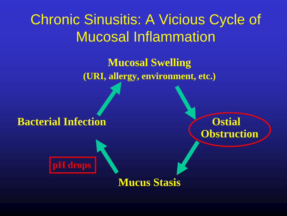
Chronic Sinusitis: A Vicious Cycle of
Mucosal Inflammation
Mucosal Swelling
(URI, allergy, environment, etc.)
Bacterial Infection Ostial Obstruction
Mucus Stasis
pH drops

ANATOMIC
GENETIC/
IMMUNOLOGIC
ENVIRONMENTAL
Chronic Rhinosinusitis - Pathophysiology
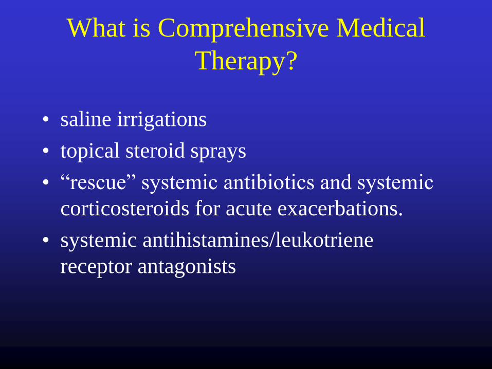
What is Comprehensive Medical
Therapy?
• saline irrigations
• topical steroid sprays
• “rescue” systemic antibiotics and systemic
corticosteroids for acute exacerbations.
• systemic antihistamines/leukotriene
receptor antagonists

Which Antibiotic?
• First line: Amoxicillin +/- clavulanate
• Second line:• Doxycycline*
• Fluoroquinolone*
• Trimethoprim-sulfamethoxazole**
• Macrolides**
• Duration: 10-14 days
*American Academy of Otolaryngology- Head and Neck Surgery,
Infectious Diseases Society of America
**Canadian Clinical Practice Guidelines

Topical Antibiotics
• Particularly helpful after
sinus ostia have been
opened.
• Alternative for patients
with poor tolerance for PO
antibiotics.
• Multiple delivery methods
– Dissolved in irrigation
solution
– Nebulized
– Directly instilled under
endoscopic guidance
Desrosiers et al., AJR 2007

Etiologies of Recurrent Infections
• Anatomic/ Physiologic
• Secondary Immunodeficiency
• Primary Immunodeficiency

Etiologies of Recurrent Infections –
Anatomic/Physiologic• Recurrent otitis
– Cholesteoma
– TM perforation
– ETD due to allergy or viral infection
• Sinusitis
– OMU obstruction
– Nasal polyps
– Mucocele
– Allergic rhinitis
– Viral infection
– Inadequate therapy for acute sinusitis
• Lung
– Smoking
– Intrinsic airway disorders
– Recurrent aspiration
– Esophageal disease
– Bronchial obstruction
– Unrecognized CF or ciliary dyskinesia
• GU tract
– Urinary stasis
– Compromised hygiene
– Diaphragm use
– Instrumentation
– Renal calculi, ureteral obstruction, ureteral
reflux
• Skin
– Trauma
– Lymphedema, lymphatic dysfunction,
venous insufficiency, chronic edema
– Prior cellulitis
– Obesity
– Poor hygiene
• Meningitis
– Cranial vault defects
– Chronic mastoid/sinus infection
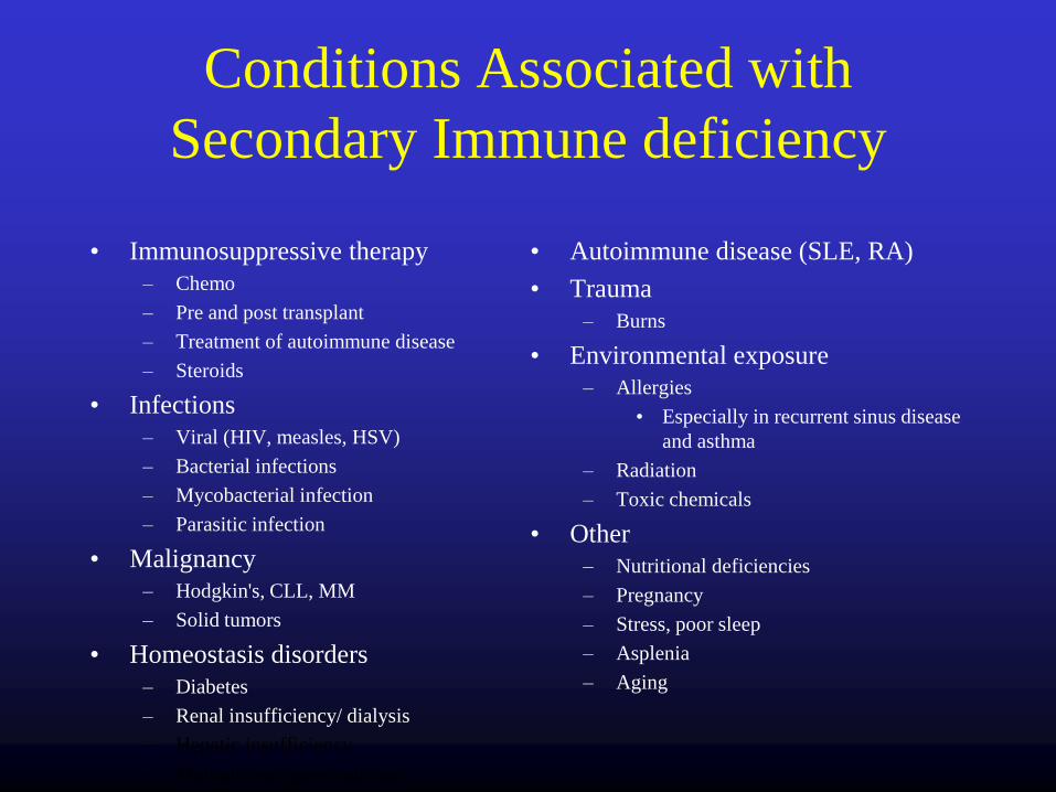
Conditions Associated with
Secondary Immune deficiency
• Immunosuppressive therapy
– Chemo
– Pre and post transplant
– Treatment of autoimmune disease
– Steroids
• Infections
– Viral (HIV, measles, HSV)
– Bacterial infections
– Mycobacterial infection
– Parasitic infection
• Malignancy
– Hodgkin's, CLL, MM
– Solid tumors
• Homeostasis disorders
– Diabetes
– Renal insufficiency/ dialysis
– Hepatic insufficiency
– Malnutrition, poor nutrition
• Autoimmune disease (SLE, RA)
• Trauma
– Burns
• Environmental exposure
– Allergies
• Especially in recurrent sinus disease
and asthma
– Radiation
– Toxic chemicals
• Other
– Nutritional deficiencies
– Pregnancy
– Stress, poor sleep
– Asplenia
– Aging
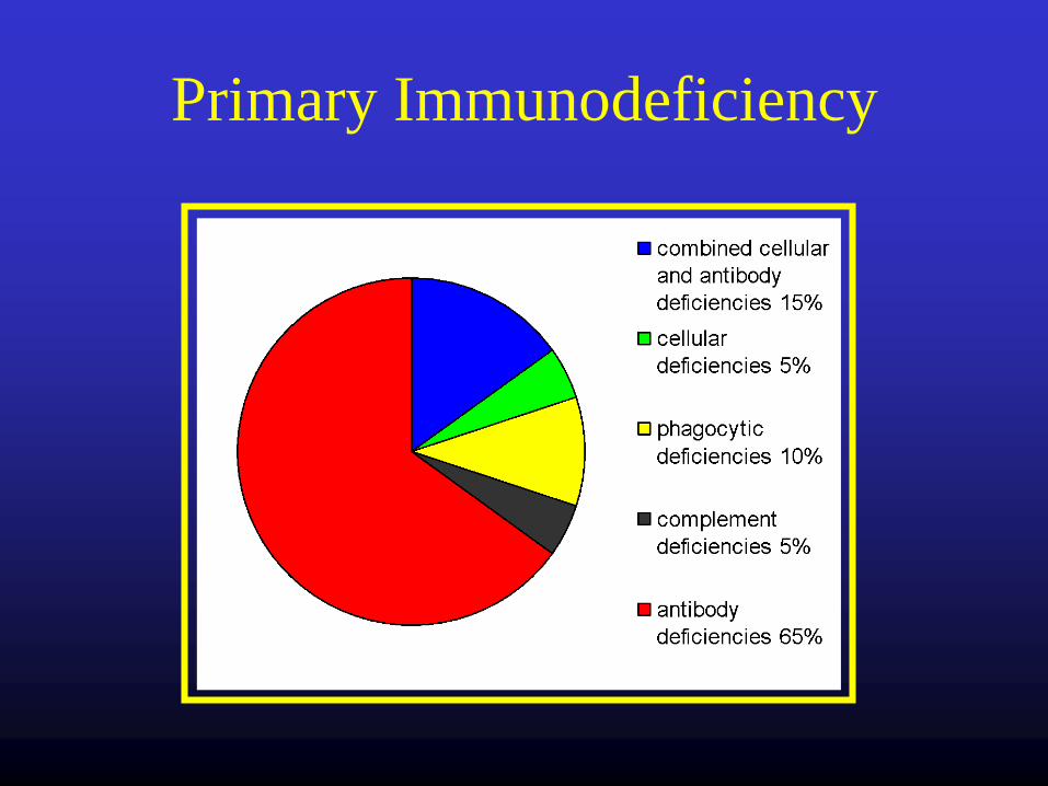
Primary Immunodeficiency

Warning Signs of
Primary
Immunodeficiency
*The Jeffrey Modell
Foundation Medical
Advisory Board
Immune effector mechanisms
B T
PMN
Μ
+ Complement
T cell help
Cytotoxicity
Intracellular
killing
Antibody
productio
n
Extracellular
Killing
Antibody

Laboratory Evaluation of the Immune
System
• Screening:
– CBC with diff
– Comprehensive Chemistry
– IgG, IgM, IgA, IgE
– HIV (consider)
– CH50 (consider)

Laboratory Evaluation of the Immune
System
• More specific testing
– Pneumococcal, Hib, Tetanus titers (i.e.
specific antibody titers)
– Flow cytometry for B/ T cells
– Antigen/mitogen stimulation studies
– +/- IgG subclasses (consider)s

Case Presentation- Evaluation
• An Immune workup was ordered.

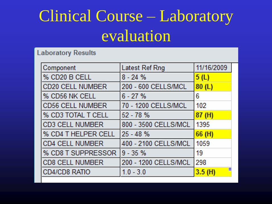
Clinical Course – Laboratory
evaluation
Repeat Testing confirmed abnormal values.
Clinical Course – Laboratory
evaluation

Clinical Course – Laboratory
evaluation
• Pneumococcal titers 0/23 protective
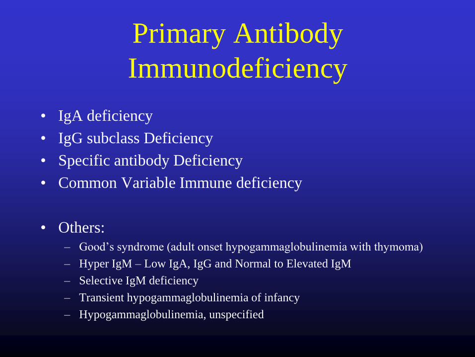
Primary Antibody
Immunodeficiency
• IgA deficiency
• IgG subclass Deficiency
• Specific antibody Deficiency
• Common Variable Immune deficiency
• Others:– Good’s syndrome (adult onset hypogammaglobulinemia with thymoma)
– Hyper IgM – Low IgA, IgG and Normal to Elevated IgM
– Selective IgM deficiency
– Transient hypogammaglobulinemia of infancy
– Hypogammaglobulinemia, unspecified

Clinical Presentation
Antibody Deficiency
Increased susceptibility to infections:– Encapsulated bacterial organisms
• H. influenzae
• S. pneumoniae
– Infection with GNR (pseudomonas and others) may occur
especially in patients treated repeatedly with broad spectrum
antibiotics
– Mycoplasma, Ureaplasma
– Viruses (enteroviruses)
– Protozoa (giardia, cryptosporidium)

Clinical Presentation
Antibody Deficiency
• Recurrent upper/lower respiratory tract infections
are the most common presenting symptom
– Think about antibody deficiency in patients with > 1
pneumonia, intractable sinusitis, recurrent otitis media
• GI tract infections also common
– Intractable or recurrent giardia, enteroviruses
• Less common– Meningitis, septicemia, osteomyelitis

Laboratory Evaluation of the
Immune System
IgA• Secretory component
• Found in saliva, colostrum and breast milk,
tears, mucosal secretions from the respiratory
tract, GU tract and prostate
Low IgA leads to more infections in the sinuses,
lungs and gut
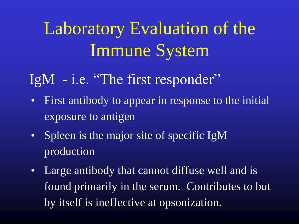
Laboratory Evaluation of the
Immune System
IgM - i.e. “The first responder”
• First antibody to appear in response to the initial
exposure to antigen
• Spleen is the major site of specific IgM
production
• Large antibody that cannot diffuse well and is
found primarily in the serum. Contributes to but
by itself is ineffective at opsonization.

Laboratory Evaluation of the
Immune System
• IgG – Represents 75% of serum immunoglobulins
in humans
– Crosses the placenta, secreted in breast milk,
high percentage found in colostrum
– Involved in many pathways (opsonization,
complement pathway, antibody dependent
cell-mediated toxicity (ADCC), Type II and
III hypersensitivity

Laboratory Evaluation of the
Immune System
IgG
• Appears 24-48 hours after
antigenic stimulation
• On repeat exposure to the antigen
IgG will proliferate
– I.e. MEMORY
– Induced by vaccines

Laboratory Evaluation of the
Immune System - Qualitative• Look at specific antibodies to evaluate B cell function
– Tetanus (protein function)
– H. influenzae
– S. pneumoniae (polysaccharide function)
If levels are low (nonprotective) then vaccinate and assess
response to vaccination.
• T cell function
– Antigen/mitogen stimulation

Laboratory Evaluation of the
Immune System - Qualitative
Interpretation of S. pneumoniae titers
– A minimum of 14 pneumococcal serotypes
should be assessed (At Northshore we check
23)
– A titer of 1.3 mcg/ml or greater is protective,
regardless of prevaccination titer.
– A normal response is defined as the generation
of protective titers in more than 70% of
serotypes (50% in those age 5 and younger)
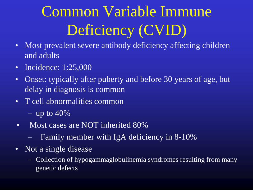
Common Variable Immune
Deficiency (CVID)• Most prevalent severe antibody deficiency affecting children
and adults
• Incidence: 1:25,000
• Onset: typically after puberty and before 30 years of age, but
delay in diagnosis is common
• T cell abnormalities common
– up to 40%
• Most cases are NOT inherited 80%
– Family member with IgA deficiency in 8-10%
• Not a single disease
– Collection of hypogammaglobulinemia syndromes resulting from many
genetic defects
CVID
• Age of onset and clinical course is variable
• Delay in diagnosis is common
• Usual presenting symptoms:– Recurrent/ chronic upper and lower respiratory tract infections
– GI infections
• With the use of high dose IgG replacement therapy
invasive infections have decreased
• Causes of death– Pulmonary disease
– Malignancy (B cell lymphomas > gastric cancer)
– Autoimmune complications
– Liver disease
– Infection

CVID - diagnosis
• Low IgG AND low IgA and/or IgM– > 2 SD below the mean
• Poor response to vaccines
• Exclusion of other causes of hypogammaglobulinemia
– Medications (rituximab, steroids)
– Protein loss (GI, lymphatics, renal, burns)
– Malignancies (B cell lymphomas, myeloma)
– Bone marrow failure

CVID Noninfectious Complications
• Lung Disease (29%)
– Diffuse (restrictive)
• Granulomatous
lymphocytic ILD
• Bronchiolitis obliterans
organizing pneumonia
(BOOP)
• Malignancy
– Obstructive
• Bronchiectasis
• Asthma
• Bronchiolitis obliterans
• Autoimmunity (29%)
– Hematologic most common
(ITP, hemolytic anemia,
Evan’s syndrome)
– RA or RA like
– Thyroid disease
– vitiligo

CVID Noninfectious Complications
• GI disease (15%)
– Enteropathy
– IBD
– Focal nodular hyperplasia
– Atrophic gastritis
• Granulomatous disease (8-
20%)
– Noncaseating
– Lymphoid or solid organs
• Liver disease (9%)
– Nodular regenerative
hyperplasia
– Autoimmune hepatitis
• Neoplasia
– Lymphoma (8.2%)
• Non-Hodgkin’s more
common
– Other (7%)
• Gastric more common

Treatment of CVID
• IgG replacement therapy
– IV every 4 weeks (400mg/kg)
• At infusion center or at home with home health
– SQ every week
• can be given by the patient
• Early evaluation and recognition of
infections

Effects of Ig Replacement
• Decreased infections
– Still have susceptible to sinopulmonary
and gut infections
– Decreased antibiotic use
• Decreased hospitalizations
• May slow the progression of chronic
lung disease in patients with CVID
Risks of IVIG
• Chills/fever/flushing/myalgias/malaise/nausea and
vomiting (often related to rate)
• Neurologic
– Headache (common)
– Migraine and aseptic meningitis
• Renal injury (more so in older preparations in sucrose)
• Hematologic complications (hemolysis, neutropenia)
• Thrombotic complications including CVA, MI, DVT/PE
(increased risk with higher doses > 1g/kg)
• Anaphylaxis
• Transmission of blood borne pathogens *
Clinical Course
• Patient was started on IVIG replacement
therapy with marked improvement for
several years.
• Sinus infections returned.
• Patient underwent limited ESSS
• Compliance with topical therapy addressed.
• Sinus infections now occurring 1-2 times
per year at age 18.

Questions?