evaluation and management of the patient with a neck mass melanie giesler, do
TRANSCRIPT

Evaluation and Management Evaluation and Management of the Patient with a Neck of the Patient with a Neck
MassMass
Melanie Giesler, DOMelanie Giesler, DO
IntroductionIntroduction
• Common clinical findingCommon clinical finding
• All age groupsAll age groups
• Very complex differential Very complex differential diagnosisdiagnosis
• Systematic approach Systematic approach essentialessential

Differential Diagnosis Differential Diagnosis

Anatomical ConsiderationsAnatomical Considerations
• Prominent Prominent landmarkslandmarks
• Triangles of the Triangles of the neckneck
• Carotid bulbCarotid bulb
• Lymphatic levelsLymphatic levels

Anatomical ConsiderationsAnatomical Considerations
• Prominent Prominent landmarkslandmarks
• Triangles of the Triangles of the neckneck
• Carotid bulbCarotid bulb
• Lymphatic levelsLymphatic levels

Anatomical ConsiderationsAnatomical Considerations
• Prominent Prominent landmarkslandmarks
• Triangles of the Triangles of the neckneck
• Carotid bulbCarotid bulb
• Lymphatic levelsLymphatic levels

Anatomical ConsiderationsAnatomical Considerations
• Prominent Prominent landmarkslandmarks
• Triangles of the Triangles of the neckneck
• Carotid bulbCarotid bulb
• Lymphatic levelsLymphatic levels

Surgical LevelsSurgical Levels

General ConsiderationsGeneral Considerations
• Patient agePatient age– Pediatric (0 – 15 years): 90% benignPediatric (0 – 15 years): 90% benign– Young adult (16 – 40 years): similar to pediatricYoung adult (16 – 40 years): similar to pediatric– Late adult (>40 years): “rule of 80s” Late adult (>40 years): “rule of 80s”
• 80% of masses are malignant of which 80% are metastatic. 80% of masses are malignant of which 80% are metastatic.
• 80% of these metastatic masses arise from sites above the clavicle 80% of these metastatic masses arise from sites above the clavicle
• 80% of these are metastatic squamous cell carcinomas.80% of these are metastatic squamous cell carcinomas.
• LocationLocation– Congenital masses: consistent in locationCongenital masses: consistent in location– Metastatic masses: key to primary lesionMetastatic masses: key to primary lesion

Metastasis Location Metastasis Location according to Various according to Various Primary LesionsPrimary Lesions

Diagnostic StepsDiagnostic Steps
• HistoryHistory– Developmental time courseDevelopmental time course– Associated symptoms (dysphagia, otalgia, Associated symptoms (dysphagia, otalgia,
voice)voice)– Personal habits (tobacco, alcohol)Personal habits (tobacco, alcohol)– Previous irradiation or surgeryPrevious irradiation or surgery
• Physical ExaminationPhysical Examination– Complete head and neck exam (visualize & Complete head and neck exam (visualize &
palpate)palpate)– Emphasis on location, mobility and consistencyEmphasis on location, mobility and consistency

Empirical AntibioticsEmpirical Antibiotics
• Inflammatory mass suspectedInflammatory mass suspected
• Two week trial of antibioticsTwo week trial of antibiotics
• Follow-up for further investigationFollow-up for further investigation

Diagnostic TestsDiagnostic Tests
• Fine needle aspiration biopsy (FNAB)Fine needle aspiration biopsy (FNAB)
• Computed tomography (CT)Computed tomography (CT)
• Magnetic resonance imaging (MRI)Magnetic resonance imaging (MRI)
• Ultrasonography Ultrasonography

Nodal Mass Workup in the Nodal Mass Workup in the AdultAdult
• Any solid asymmetric mass MUST be Any solid asymmetric mass MUST be considered a metastatic neoplastic considered a metastatic neoplastic lesion until proven otherwiselesion until proven otherwise
• Asymptomatic cervical mass – 12% Asymptomatic cervical mass – 12% of cancerof cancer
• ~ 80% of these are SCCa~ 80% of these are SCCa

Nodal Mass Workup in the Nodal Mass Workup in the AdultAdult
• Ipsilateral otalgia with normal Ipsilateral otalgia with normal otoscopy – direct attention to tonsil, otoscopy – direct attention to tonsil, tongue base, supraglottis and tongue base, supraglottis and hypopharynxhypopharynx
• Unilateral serous otitis – direct Unilateral serous otitis – direct examination of nasopharynxexamination of nasopharynx

Nodal Mass Workup in the Nodal Mass Workup in the AdultAdult
• Panendoscopy Panendoscopy – FNAB positive with no primary on repeat examFNAB positive with no primary on repeat exam– FNAB equivocal/negative in high risk patientFNAB equivocal/negative in high risk patient
• Directed BiopsyDirected Biopsy– All suspicious mucosal lesions All suspicious mucosal lesions – Areas of concern on CT/MRIAreas of concern on CT/MRI– None observed – nasopharynx, tonsil None observed – nasopharynx, tonsil
(ipsilateral tonsillectomy for jugulodigastric (ipsilateral tonsillectomy for jugulodigastric nodes), base of tongue and piriformsnodes), base of tongue and piriforms
• Synchronous primaries (10 to 20%)Synchronous primaries (10 to 20%)

Primary TumorsPrimary Tumors
• Thyroid massThyroid mass
• LymphomaLymphoma
• Salivary tumorsSalivary tumors
• LipomaLipoma
• Carotid body and Carotid body and glomus tumorsglomus tumors
• Neurogenic Neurogenic tumorstumors

Thyroid MassesThyroid Masses
• Leading cause of anterior neck massesLeading cause of anterior neck masses• ChildrenChildren
– Most common neoplastic conditionMost common neoplastic condition– Male predominanceMale predominance– Higher incidence of malignancyHigher incidence of malignancy
• AdultsAdults– Female predominanceFemale predominance– Mostly benignMostly benign

Thyroid MassesThyroid Masses
LymphomaLymphoma
• More common in children and young adultsMore common in children and young adults
• Up to 80% of children with Hodgkin’s have a Up to 80% of children with Hodgkin’s have a neck massneck mass
• Signs and symptomsSigns and symptoms– Lateral neck mass only (discrete, rubbery, Lateral neck mass only (discrete, rubbery,
nontender)nontender)– FeverFever– HepatosplenomegalyHepatosplenomegaly– Diffuse adenopathyDiffuse adenopathy

LymphomaLymphoma
• FNAB – first line diagnostic testFNAB – first line diagnostic test
• If suggestive of lymphoma – open If suggestive of lymphoma – open biopsybiopsy
• Full workup – CT scans of chest, Full workup – CT scans of chest, abdomen, head and neck; bone abdomen, head and neck; bone marrow biopsymarrow biopsy

LymphomaLymphoma

Salivary Gland TumorsSalivary Gland Tumors
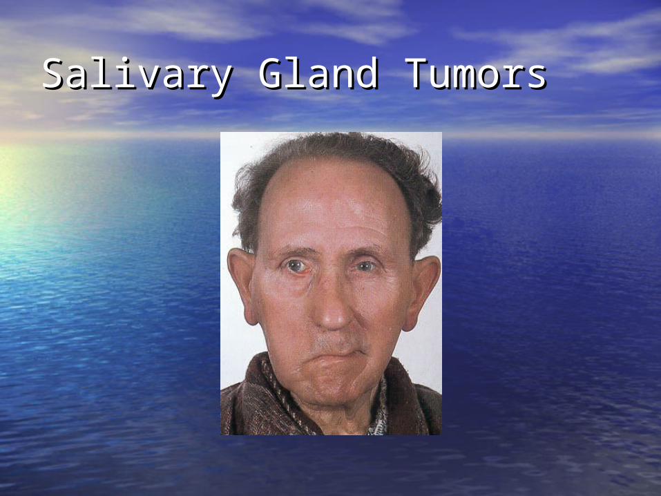
• Enlarging mass anterior/inferior to Enlarging mass anterior/inferior to ear or at the mandible angle is ear or at the mandible angle is suspectsuspect
• Benign Benign – Asymptomatic except for massAsymptomatic except for mass
• MalignantMalignant– Rapid growth, skin fixation, cranial Rapid growth, skin fixation, cranial
nerve palsiesnerve palsies
Salivary Gland TumorsSalivary Gland Tumors• Diagnostic testsDiagnostic tests
– Open excisional biopsy (submandibulectomy or Open excisional biopsy (submandibulectomy or parotidectomy) preferredparotidectomy) preferred
– FNAB FNAB • Shown to reduce surgery by 1/3 in some studiesShown to reduce surgery by 1/3 in some studies• Delineates intra-glandular lymph node, localized Delineates intra-glandular lymph node, localized
sialadenitis or benign lymphoepithelial cystssialadenitis or benign lymphoepithelial cysts• May facilitate surgical planning and patient counselingMay facilitate surgical planning and patient counseling• Accuracy >90% (sensitivity: ~90%; specificity: ~80%)Accuracy >90% (sensitivity: ~90%; specificity: ~80%)
– CT/MRI – deep lobe tumors, intra vs. extra-parotidCT/MRI – deep lobe tumors, intra vs. extra-parotid
• Be prepared for total parotidectomy Be prepared for total parotidectomy with possible facial nerve sacrificewith possible facial nerve sacrifice
Salivary Gland TumorsSalivary Gland Tumors

Carotid Body TumorCarotid Body Tumor
• Rare in childrenRare in children• Pulsatile, compressible massPulsatile, compressible mass• Mobile medial/lateral Mobile medial/lateral notnot superior/inferior superior/inferior• Clinical diagnosis, confirmed by angiogram or CTClinical diagnosis, confirmed by angiogram or CT• TreatmentTreatment
– Irradiation or close observation in the elderlyIrradiation or close observation in the elderly– Surgical resection for small tumors in young patientsSurgical resection for small tumors in young patients
• Hypotensive anesthesiaHypotensive anesthesia• Preoperative measurement of catecholaminesPreoperative measurement of catecholamines

Carotid Body TumorCarotid Body Tumor

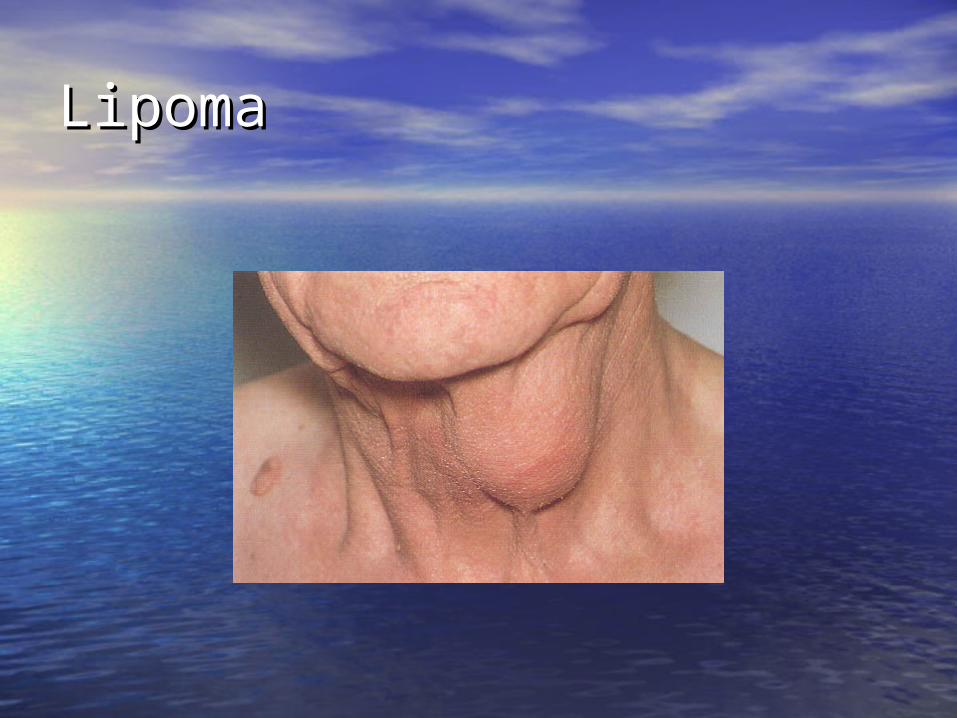
LipomaLipoma
• Soft, ill-defined massSoft, ill-defined mass
• Usually >35 years of ageUsually >35 years of age
• AsymptomaticAsymptomatic
• Clinical diagnosis – confirmed by Clinical diagnosis – confirmed by excisionexcision
LipomaLipoma

Neurogenic TumorsNeurogenic Tumors
• Arise from neural crest derivativesArise from neural crest derivatives
• Include schwannoma, neurofibroma, Include schwannoma, neurofibroma, and malignant peripheral nerve and malignant peripheral nerve sheath tumorsheath tumor
• Increased incidence in NF syndromesIncreased incidence in NF syndromes
• Schwannoma most common in head Schwannoma most common in head & neck& neck

SchwannomaSchwannoma
• Sporadic cases mostlySporadic cases mostly• 25 to 45% in neck when extracranial25 to 45% in neck when extracranial• Most commonly between 20 and 50 yearsMost commonly between 20 and 50 years• Usually mid-neck in poststyloid Usually mid-neck in poststyloid
compartmentcompartment• Signs and symptomsSigns and symptoms
– Medial tonsillar displacementMedial tonsillar displacement– Hoarseness (vagus nerve)Hoarseness (vagus nerve)– Horner’s syndrome (sympathetic chain)Horner’s syndrome (sympathetic chain)

Congenital and Congenital and Developmental MassDevelopmental Mass
• Epidermal and sebaceous cystsEpidermal and sebaceous cysts
• Branchial cleft cystsBranchial cleft cysts
• Thyroglossal duct cystThyroglossal duct cyst
• Vascular tumorsVascular tumors

Epidermal and Sebaceous Epidermal and Sebaceous CystsCysts
• Most common Most common congenital/developmental masscongenital/developmental mass
• Older age groupsOlder age groups
• Clinical diagnosisClinical diagnosis– Elevation and movement of overlying Elevation and movement of overlying
skinskin– Skin dimple or pore Skin dimple or pore
• Excisional biopsy confirmsExcisional biopsy confirms

Epidermal and Sebaceous Epidermal and Sebaceous CystsCysts

Branchial Cleft CystsBranchial Cleft Cysts
• Branchial cleft anomaliesBranchial cleft anomalies• 22ndnd cleft most common (95%) – tract cleft most common (95%) – tract
medial to CN XII between internal and medial to CN XII between internal and external carotidsexternal carotids
• 11stst cleft less common – close cleft less common – close association with facial nerve possibleassociation with facial nerve possible
• 33rdrd and 4 and 4thth clefts rarely reported clefts rarely reported• Present in older children or young Present in older children or young
adults often following URIadults often following URI

Branchial Cleft CystsBranchial Cleft Cysts
• Most common as smooth, fluctuant mass Most common as smooth, fluctuant mass underlying the SCMunderlying the SCM
• Skin erythema and tenderness if infectedSkin erythema and tenderness if infected
• TreatmentTreatment– Initial control of infectionInitial control of infection– Surgical excision, including tractSurgical excision, including tract
• May necessitate a total parotidectomy May necessitate a total parotidectomy (1(1stst cleft) cleft)

Branchial Cleft CystsBranchial Cleft Cysts
Thyroglossal Duct CystThyroglossal Duct Cyst
• Most common congenital neck mass (70%)Most common congenital neck mass (70%)
• 50% present before age 2050% present before age 20
• Midline (75%) or near midline (25%)Midline (75%) or near midline (25%)
• Usually just inferior to hyoid bone (65%)Usually just inferior to hyoid bone (65%)
• Elevates on swallowing/protrusion of tongueElevates on swallowing/protrusion of tongue
• Treatment is surgical removal Treatment is surgical removal (Sistrunk/Schlange) after resolution of any (Sistrunk/Schlange) after resolution of any infectioninfection

Thyroglossal Duct CystThyroglossal Duct Cyst
Vascular TumorsVascular Tumors
• Lymphangiomas and hemangiomasLymphangiomas and hemangiomas
• Usually within 1Usually within 1stst year of life year of life
• Hemangiomas often resolve Hemangiomas often resolve spontaneously, while lymphangiomas spontaneously, while lymphangiomas remain unchangedremain unchanged
• CT/MRI may help define extent of CT/MRI may help define extent of diseasedisease
Vascular TumorsVascular Tumors
• TreatmentTreatment– Lymphangioma – surgical excision for Lymphangioma – surgical excision for
easily accessible or lesions affecting easily accessible or lesions affecting vital functions; recurrence is commonvital functions; recurrence is common
– Hemangiomas – surgical excision Hemangiomas – surgical excision reserved for those with rapid growth reserved for those with rapid growth involving vital structures or associated involving vital structures or associated thrombocytopenia that fails medical thrombocytopenia that fails medical therapy (steroids, interferon)therapy (steroids, interferon)

Vascular Tumors Vascular Tumors (lymphangioma)(lymphangioma)

Vascular Tumors Vascular Tumors (hemangioma)(hemangioma)

Inflammatory DisordersInflammatory Disorders
• LymphadenitisLymphadenitis
• Granulomatous lymphadenitisGranulomatous lymphadenitis

LymphadenitisLymphadenitis
• Very common, especially within 1Very common, especially within 1stst decade decade
• Tender node with signs of systemic infectionTender node with signs of systemic infection
• Directed antibiotic therapy with follow-upDirected antibiotic therapy with follow-up
• FNAB indications (pediatric)FNAB indications (pediatric)– Actively infectious condition with no responseActively infectious condition with no response– Progressively enlargingProgressively enlarging– Solitary and asymmetric nodal massSolitary and asymmetric nodal mass– Supraclavicular mass (60% malignancy)Supraclavicular mass (60% malignancy)– Persistent nodal mass without active infectionPersistent nodal mass without active infection

LymphadenopathyLymphadenopathy
• Equivocal or suspicious FNAB in the Equivocal or suspicious FNAB in the pediatric nodal mass requires open pediatric nodal mass requires open excisional biopsy to rule out excisional biopsy to rule out malignant or granulomatous diseasemalignant or granulomatous disease

Granulomatous Granulomatous lymphadenitislymphadenitis• Infection develops over weeks to Infection develops over weeks to
monthsmonths
• Minimal systemic complaints or findingsMinimal systemic complaints or findings
• Common etiologiesCommon etiologies– TB, atypical TB, cat-scratch fever, TB, atypical TB, cat-scratch fever,
actinomycosis, sarcoidosisactinomycosis, sarcoidosis
• Firm, relatively fixed node with Firm, relatively fixed node with injection of skininjection of skin

Granulomatous Granulomatous lymphadenitislymphadenitis

SummarySummary
• Extensive differential diagnosisExtensive differential diagnosis
• Age of patient is importantAge of patient is important
• Accurate history and complete exam Accurate history and complete exam essentialessential
• FNAB – invaluable diagnostic toolFNAB – invaluable diagnostic tool
• Possibility for malignancy in any age groupPossibility for malignancy in any age group
• Close follow-up and aggressive approach Close follow-up and aggressive approach is best for favorable outcomesis best for favorable outcomes