epithelial anion transport in health and...
TRANSCRIPT

Novartis Foundation Symposium 273
EPITHELIAL ANION TRANSPORTIN HEALTH AND DISEASE:
THE ROLE OF THE SLC26TRANSPORTERS FAMILY
2006


EPITHELIAL ANION TRANSPORTIN HEALTH AND DISEASE:
THE ROLE OF THE SLC26TRANSPORTERS FAMILY

The Novartis Foundation is an international scientifi c and educational charity (UK Registered Charity No. 313574). Known until September 1997 as the Ciba Foundation, it was established in 1947 by the CIBA company of Basle, which merged with Sandoz in 1996, to form Novartis. The Foundation operates independently in London under English trust law. It was formally opened on 22 June 1949.
The Foundation promotes the study and general knowledge of science and in particular encourages international co-operation in scientifi c research. To this end, it organizes internationally acclaimed meetings (typically eight symposia and allied open meetings and 15–20 discussion meetings each year) and publishes eight books per year featuring the presented papers and discussions from the symposia. Although primarily an operational rather than a grant-making foundation, it awards bursaries to young scientists to attend the symposia and afterwards work with one of the other participants.
The Foundation’s headquarters at 41 Portland Place, London W1B 1BN, provide library facilities, open to graduates in science and allied disciplines. Media relations are fostered by regular press conferences and by articles prepared by the Foundation’s Science Writer in Residence. The Foundation offers accommodation and meeting facilities to visiting scientists and their societies.
Information on all Foundation activities can be found athttp://www.novartisfound.org.uk

Novartis Foundation Symposium 273
EPITHELIAL ANION TRANSPORTIN HEALTH AND DISEASE:
THE ROLE OF THE SLC26TRANSPORTERS FAMILY
2006

Copyright © Novartis Foundation 2006Published in 2006 by John Wiley & Sons Ltd, The Atrium, Southern Gate, Chichester PO19 8SQ, UK
National 01243 779777 International (+44) 1243 779777 e-mail (for orders and customer service enquiries): [email protected] Visit our Home Page on http://www.wileyeurope.com or http://www.wiley.com
All Rights Reserved. No part of this book may be reproduced, stored in a retrieval system or transmitted in any form or by any means, electronic, mechanical, photocopying, recording, scanning or otherwise, except under the terms of the Copyright, Designs and Patents Act 1988 or under the terms of a licence issued by the Copyright Licensing Agency Ltd, 90 Tottenham Court Road, London W1T 4LP, UK, without the permission in writing of the Publisher. Requests to the Publisher should be addressed to the Permissions Department, John Wiley & Sons Ltd, The Atrium, Southern Gate, Chichester, West Sussex PO19 8SQ, England, or emailed to [email protected], or faxed to (+44) 1243 770620.
This publication is designed to provide accurate and authoritative information in regard to the subject matter covered. It is sold on the understanding that the Publisher is not engaged in rendering professional services. If professional advice or other expert assistance is required, the services of a competent professional should be sought.
Other Wiley Editorial Offi ces
John Wiley & Sons Inc., 111 River Street, Hoboken, NJ 07030, USA
Jossey-Bass, 989 Market Street, San Francisco, CA 94103-1741, USA
Wiley-VCH Verlag GmbH, Boschstr. 12, D-69469 Weinheim, Germany
John Wiley & Sons Australia Ltd, 33 Park Road, Milton, Queensland 4064, Australia
John Wiley & Sons (Asia) Pte Ltd, 2 Clementi Loop #02-01, Jin Xing Distripark, Singapore 129809
John Wiley & Sons Canada Ltd, 6045 Freemont Boulevard Mississauga, Ontario, Canada L5R 4J3
Wiley also publishes its books in a variety of electronic formats. Some content that appears in print may not be available in electronic books.
Novartis Foundation Symposium 273x + 273 pages, 49 fi gures, 3 tables
British Library Cataloguing in Publication Data
A catalogue record for this book is available from the British Library
ISBN-13 978-0-470-01624-4 (HB)ISBN-10 0-470-01624-8 (HB)
Typeset in 10½ on 12½ pt Garamond by SNP Best-set Typesetter Ltd., Hong KongPrinted and bound in Great Britain by T. J. International Ltd, Padstow, Cornwall.This book is printed on acid-free paper responsibly manufactured from sustainable forestry, in which at least two trees are planted for each one used for paper production.

Contents
v
Symposium on Epithelial anion transport in health and disease: the role of the SLC26
transporters family, held at the Novartis Foundation, London 1–3 March 2005
Editors: Derek J. Chadwick (Organizer) and Jamie Goode
This symposium is based on a proposal made by Shmuel Muallem
Michael J. Welsh Chair’s introduction 1
Juha Kere Overview of the SLC26 family and associated diseases 2 Discussion 11
Marlies Knipper, Thomas Weber, Harald Winter, Claudia Braig, Jelka Cimerman, Juergen T. Fraenzer and Ulrike Zimmermann Individual characteristics of members of the SLC26 family in vertebrates and their homologues in insects 19
Discussion 30
Daniel Markovich Sulfate transport by SLC26 transporters 42 Discussion 51
Jonathan Ashmore and Jean-Marie Chambard Sugar transport by members of the SLC26 superfamily 59
Discussion 68
Pia Höglund SLC26A3 and congenital chloride diarrhoea 74 Discussion 86
Manoocher Soleimani Expression, regulation and the role of SLC26 Cl−/HCO3
− exchangers in kidney and gastrointestinal tract 91 Discussion 103

vi CONTENTS
Seth L. Alper, Andrew K. Stewart, Marina N. Chernova, Alexander S. Zolotarev, Jeffrey S. Clark and David H. Vandorpe Anion exchangers in fl ux: functional differences between human and mouse SLC26A6 polypeptides 107
Discussion 119
Michael F. Romero, Min-Hwang Chang, Consuelo Plata, Kambiz Zandi-Nejad, Adriana Mercado, Vadjista Broumand, Caroline R. Sussman and David B. Mount Physiology of electrogenic SLC26 paralogues 126
Discussion 138
Peter S. Aronson Role of SLC26-mediated Cl−/base exchange in proximal tubule NaCl transport 148
Discussion 158
Péter Hegyi, Zoltán Rakonczay Jr., László Tiszlavica, András Varró, András Tóth, Gábor Rácz, Gábor Varga, Michael A. Gray and Barry E. Argent SLC26 transporters and the inhibitory control of pancreatic ductal bicarbonate secretion 164
Discussion 173
Nikolay Shcheynikov, Shigeru B. H. Ko, Weizhong Zeng, Joo Young Choi, Michael R. Dorwart, Philip J. Thomas and Shmuel Muallem Regulatory interaction between CFTR and the SLC26 transporters 177
Discussion 186
Antonella Forlino, Benedetta Gualeni, Fabio Pecora, Sara Della Torre, Rocco Piazza, Cecilia Tiveron, Laura Tatangelo, Andrea Superti-Furga, Giuseppe Cetta and Antonio Rossi Insights from a transgenic mouse model on the role of SLC26A2 in health and disease 193
Discussion 206
Lorraine A. Everett New insights into the role of pendrin (SLC26A4) in inner ear fl uid homeostasis 213
Discussion 225
Susan M. Wall The renal physiology of pendrin (SLC26A4) and its role in hypertension 231
Discussion 239

CONTENTS vii
Dominik Oliver, Thorsten Schächinger and Bernd Fakler Interaction of prestin (SLC26A5) with monovalent intracellular anions 244
Discussion 253
Final discussion 261
Index of contributors 265
Subject index 267

Participants
viii
Seth L. Alper Molecular and Vascular Medicine and Renal Units, Beth Israel Deaconess Medical Center, Department of Medicine, Harvard Medical School, Boston, MA 02215, USA
Barry E. Argent Institute for Cell and Molecular Biosciences, University of Newcastle upon Tyne, Catherine Cookson Building, Framlington Place, Newcastle upon Tyne NE2 4HH, UK
Peter S. Aronson Section of Nephrology, Department of Medicine, Yale Uni-versity School of Medicine, 1 Gilbert Street, TAC S-255, PO Box 208029, New Haven, CT 06520-8029, USA
Jonathan Ashmore Department of Physiology, University College London, Gower Street, London WC1E 6BT, UK
Maynard Case Faculty of Life Sciences, University of Manchester, Floor 2, Core Technology Facility, 46 Grafton St, Manchester M13 9NT, UK
Hsiao Chang Chan Epithelial Cell Biology Research Centre, Department of Physiology, The Chinese University of Hong Kong, Shatin, Hong Kong
Lorraine A. Everett Audiovestibular Genomics Group, The Wellcome Trust Sanger Institute, Wellcome Trust Genome Campus, Hinxton, Cambridge CB10 1SA, UK
Michael A. Gray Institute for Cell and Molecular Biosciences, University of Newcastle upon Tyne, Catherine Cookson Building, Framlington Place, Newcastle upon Tyne NE2 4HH, UK
Pia Höglund Hospital for Children and Adolescents, PO Box 281 (Stenbäckinkatu 11), 00029 Hus, Finland
Hiroshi Ishiguro Internal Medicine and Human Nutrition, Nagoya University School of Medicine, Showa-ku, Nagoya 466-8550, Japan
PARTICIPANTS ix
Juha Kere Department of Biosciences at Novum and Clinical Research Centre, Karolinska Institutet, 14157 Huddinge, Sweden
Marlies Knipper Department of Otolaryngology, Tübingen Hearing Research Centre, Molecular Neurobiology, Elfriede-Aulhorn-Strasse 5, D-72076 Tübingen, Germany
Min Goo Lee Department of Pharmacology, Yonsei University College of Medicine, 134 Sinchon-Dong, Seoul 120-752, Korea
Daniel Markovich School of Biomedical Sciences, Department of Physiology and Pharmacology, University of Queensland, Brisbane, Queensland 4072, Australia
David B. Mount Renal Division, Brigham and Women’s Hospital and VA Boston Healthcare System, Room 542, Harvard Institutes of Medicine, 4 Black-fan Circle, Boston, MA 02115, USA
Shmuel Muallem Department of Physiology, Room K4-120, UT South Western Medical Centre, 5323 Harry Hines Blvd, Dallas, TX 75390 9040, USA
Dominik Oliver Institute of Physiology, University of Freiburg, Hermann-Herder-Straße 7, 79104 Freiburg, Germany
Paul M. Quinton Department of Pediatrics, University of California San Diego School of Medicine, 9500 Gilman Drive—0831, La Jolla, CA 92093-0831, USA
Michael F. Romero Department of Physiology & Biophysics, Room #SOM-E563, Case Western University School of Medicine, 2119 Abington Rd, Cleveland, OH 44106-4970, USA
Antonio Rossi Department of Biochemistry, University of Pavia, Via Taramelli, 3/B, I-27100 Pavia, Italy
Ursula Seidler Medizinische Hochshule Hannover, Carl-Neuberg-Strasse 1, 30625 Hannover, Germany
Manoocher Soleimani Division of Nephrology and Hypertension, University of Cincinnati Medical Center, 231 Albert Sabin Way, MSB G259, Cincinnati, OH 45267 0585, USA

x PARTICIPANTS
Andrew K. Stewart (Novartis Foundation Bursar) RW 773, Molecular Medicine and Renal Units, Beth Israel Deaconess Medical Center, East Campus, 330 Brookline Avenue, Boston, MA 02215, USA
Philip J. Thomas The University of Texas, Southwestern Medical Center at Dallas, Department of Physiology, Room K4.140A, 5323 Harry Hines Blvd, Dallas, TX 75390-9040, USA
R. James Turner Gene Therapy and Therapeutics Branch, National Institute of Dental and Craniofacial Research, Building 10, Room 1A01, National Institutes of Health, Bethesda, MD 20892-1190, USA
Susan M. Wall Renal Division, Department of Medicine, Emory University, Mailstop: 1930/001/1AG (Medicine-Renal), Atlanta, GA 30322, USA
Michael J. Welsh (Chair) Howard Hughes Medical Institute/500 EMRB, Carver College of Medicine, University of Iowa, Iowa City, IO 52242, USA

Chair’s introductionMichael J. Welsh
Howard Hughes Medical Institute/500 EMRB, University of Iowa, Iowa City, IO 52242, USA
In introducing this symposium, I am expecting that this will be an interesting meeting for three reasons. First, I have much to learn. Secondly, this is an interest-ing family of transporters, because there are multiple members, there is rich diver-sity, and there are some common themes. Thirdly, this is a small meeting which should permit some excellent discussion.
I am not an expert on the SLC26 family, so what I can say by way of introduc-tion is quite limited. Instead, I’ll highlight what I would like to learn from this meeting. First, how do these channels work? Are they electrically conductive? Or are they neutral transporters? If they are conductive, what is their relationship to ion channels? I would like to know how selectivity is determined. As I look at some of the literature, the issues of selectivity should prove very interesting. I would also like to learn something about how this family has evolved. There are multiple members and we may learn something if we look at these transporters through evolution.
The second main question I would like to address is how they contribute to normal physiology, in many different epithelia. What are the common threads and what is unique? If you transplanted one of these transporters into a different place, would it adopt new functions depending on its new home? Or are the functions all intrinsic to the protein?
Third, I hope to learn more about how loss of their function disrupts physiology. What is the pathophysiology associated with loss or mutation? If we understand this better, might it allow us to do something about disease? These are the main things I would like to learn from this meeting.
As I looked through the history of the Novartis Foundation, I came across the following quote from Lord Beveridge, made at the inauguration of the Foundation in 1949. He said, ‘This place itself is not a laboratory for mixing compounds, but we do mean to make it a laboratory for mixing scientists’. So I hope we’ll mix it up, have a good time, and learn much over the next few days.
1

Overview of the SLC26 family and associated diseasesJuha Kere
Department of Biosciences at Novum, Karolinska Institutet, 14157 Huddinge, Sweden and Department of Medical Genetics, University of Helsinki, 00014 Helsinki, Finland
Abstract. In the late 1990s the SLC26 family of anion exchangers emerged as the second, structurally distinct gene family capable of similar transport functions as the classical SLC4 or anion exchanger (AE) gene family. The observations leading to the characteri-zation of the SLC26 family were fi rmly based on research on rare human diseases and aided by comparison to Caenorhabditis elegans. SLC26A1, or rat sulphate/anion transporter 1 (Sat1), was the fi rst gene cloned in mammals, but not characterized in humans until the year 2000. Three rare recessive diseases in humans, namely diastrophic dysplasia (cartilage disorder resulting in growth retardation), congenital chloride diarrhoea (anion exchange disorder of the intestine) and Pendred syndrome (deafness with thyroid disor-der) turned out to be caused by the highly related genes SLC26A2 (fi rst called DTDST ), SLC26A3 (fi rst called CLD or DRA) and SLC26A4 (fi rst called PDS ), respectively. Subsequently, others and our laboratory cloned prestin, a cochlear motor protein gene (SLC26A5), a putative pancreatic anion transporter (SLC26A6), and SLC26A7–SLC26A11. Some SLC26 family members show highly specifi c tissue expression pat-terns, others are widely expressed. The SLC26 exchangers are capable of transporting, with different affi nities, at least the chloride, iodide, sulfate, bicarbonate, hydroxyl, oxalate and formate anions, and have distinct anion specifi city profi les.
2006 Epithelial anion transport in health and disease: the role of the SLC26 transporters family. Wiley, Chichester (Novartis Foundation Symposium 273) p 2–18
Transport of small molecules across lipid membranes is a fundamental function of all cellular organisms. Hundreds of proteins with specialized transport capabili-ties are expressed in different tissues of multicellular organisms, and indeed in different domains of membranes in individual cells. The transporter proteins come in families, with different members sharing structural similarities but often with distinct properties and physiological functions that may or may not be interchange-able. Distinct expression patterns in different tissues also suggest that the corre-sponding genes have highly specialized regulatory elements, in spite of high similarity of coding sequences. Finally, just a few changes in protein sequences may cause radical differences in the transport properties. The SLC26 family of anion exchangers provides examples of a wide spectrum of all these features, and
2

OVERVIEW 3
is largely uncharacterized. This is not surprising, considering that most members of the whole gene and protein family were described only a few years ago. Many transporter proteins were fi rst isolated based on their functional properties, the discovery of the SLC26 gene family has been driven by human disease gene cloning and thereafter genomic approaches, based on the homology of the gene family and availability of whole genome sequences. At the beginning of this odyssey, only one gene belonging to the SLC26 gene family was known in mammals, the rat sulphate anion transporter 1 (Sat1) gene. The next three SLC26 family members were identifi ed by positional cloning of rare recessive human disease genes (Hästbacka et al 1994, Höglund et al 1996, Everett et al 1997) even though one of them, SLC26A3, had been fi rst cloned as a suggested tumour suppressor gene (Schweinfest et al 1993). One gene was fi rst characterized in gerbil rather than human based on its function, motor activity in cochlear cells of the inner ear (Zheng et al 2000). Finally, all the remaining fi ve currently known SLC26 genes were identifi ed by a genomic homology-driven approach in human (Lohi et al 2000, 2002) and in parallel, by other approaches (Waldegger et al 2001, Toure et al 2001, Vincourt et al 2002, 2003, Mount & Romero 2004). The nomenclature of this gene family follows the convention of other solute carrier genes, starting with SLC, followed by the family number and an A separating the individual gene number. The individual members of the SLC26 family got their number identities in July 2000 (for SLC26A1 to A6) and in January 2001 (for SLC26A7 to A11, based on the full or partial human cDNA sequences AF331521 to AF331526 sub-mitted from our laboratory) after exchange of email messages between the author of this review, Dr Elspeth Bruford of the HUGO Gene Nomenclature Committee, and nomenclature reviewers, including Dr Matthias A. Hediger. The entire human SLC26 gene family with references to the earliest GenBank sequence database entries and diseases associated with them are presented in Table 1. In the following paragraphs, I will briefl y describe the discovery of the different SLC26 genes. I will only discuss the molecular cloning of each gene, even though the existence of such transporters had been demonstrated earlier by functional studies, and for considerations of space, I have omitted most of the literature related to their func-tions. Much additional information has already been revealed about their specifi c functional properties and physiological roles, and some of the fi rst mouse knock-out models are also available. More detailed reviews of these studies will be pre-sented by other papers in this book.
SLC26A1
The rat liver canalicular sulfate transporter was the fi rst gene of the SLC26 family to be molecularly characterized in mammals, cloned by Bissig et al (1994) and characterized by Markovich et al (1994). Curiously, the human gene remained

4 KERE
TABLE 1 The human SLC26 family of anion transporters
References toGene Diseases, molecularsymbol Chromosome Main sites of OMIM Sequence cloning and(aliases) location expression numbers accession diseases
SLC26A1 4p16 Liver, kidney, Not known AF297659, Lohi et al (SAT1) pancreas, brain AY124771 2000, Regeer et al 2003SLC26A2 5q32-q33.1 Ubiquitous, DTD, U14528 Hästbacka et al (DTDST) cartilage 222600 1994, 1996 ACGIB, 600972 AOG2, 256050 EDM4, 226900SLC26A3 7q31 Ileum, colon, CLD, L02785 Schweinfest et al (CLD, seminal vesicle, 214700 1993, Höglund DRA) eccrine sweat et al 1996 glandSLC26A4 7q31 Thyroid, PDS, AF030880 Everett et al 1997, (PDS) kidney, cochlea 274600 Haila et al 1998, DFNB4, Li et al 1998 600791 EVA, 603545SLC26A5 7q22.1 Outer hair cells DFNB61, AF523354, Zheng et al 2000, (PRES) of the cochlea 604943 AY289133 Liu et al 2003SLC26A6 3p21 Kidney, Not known AF279265, Lohi et al 2000, (PAT1) pancreas, AF288410 Waldegger et al skeletal muscle 2001SLC26A7 8q21 Kidney, Not known AF331521, Lohi et al 2000, placenta, testis AJ413228- 2002, Vincourt AJ413230 et al 2002SLC26A8 6p21 Testis Not known AF314959, Lohi et al 2000, (TAT1) AF331522 2002, Toure et al 2001SLC26A9 1q32 Lung Not known AF314958, Lohi et al 2000, AF331525 2002SLC26A10 12q14 Brain, ubiquitous Not known AF331523, Lohi et al 2000 AL050358SLC26A11 17q25 Ubiquitous Not known AF331524, Lohi et al 2000, AF345195 Vincourt et al 2003
For disease designations, please see text. Many of the genes were fi rst published as GenBank database entries, many independently by two research groups within a few months. Sequence accession numbers are given for the earliest full or partial cDNA sequences.

OVERVIEW 5
uncharacterized until several years later, when Hannes Lohi in our group de -termined and submitted the human sequence to GenBank in August 2000 (AF297659; Lohi et al 2000). The gene structure was confi rmed in June 2002 by Regeer, Lee and Markovich (AY124771; Regeer et al 2003). The mouse gene was cloned and characterized by Lee et al (2003).
Diastrophic dysplasia and SLC26A2
Diastrophic dysplasia (DTD) is an autosomal recessive cartilage disorder, leading to a disproportionate growth disorder. DTD is one of those about 30 rare recessive disorders that have been observed at much higher frequencies in Finland than in most other countries, a phenomenon that has been well explained by genetic founder effects; curiously, congenital chloride diarrhoea (CLD) is another. Johanna Hästbacka, a doctoral student of Dr Albert de la Chapelle, fi rst mapped the gene to chromosome 5q31 and later, working as a postdoc in Dr Eric Lander’s labora-tory, positionally cloned it (Hästbacka et al 1994; sequence AF345195 submitted to GenBank in September 1994). The gene was named DTD sulfate transporter (DTDST ) and its homology to the rat Sat1 gene was immediately noticed, suggest-ing a pathogenetic mechanism for DTD. Cartilage is dependent on high amounts of sulfate and Hästbacka et al (1994) suggested that impaired function of DTDST may lead to undersulfation of proteoglycans in cartilage matrix. Later, mutations in SLC26A2 were found also in achondrogenesis type IB (ACGIB), atelosteogen-esis type II (AOG2), and multiple epiphyseal dysplasia type 4 (EDM4) (for OMIM numbers, see Table 1). Even though SLC26A2 mRNA is quite abundantly expressed and SLC26A2 protein has been detected in colon, sweat glands, pancreas and placenta in addition to cartilage, symptoms of recessive SLC26A2 mutations seem to be confi ned to problems in cartilage development (Hästbacka et al 1996, Haila et al 2001).
Congenital chloride diarrhoea and SLC26A3
With the aim of identifying potential tumour suppressor genes in colon cancer, Schweinfest et al (1993) discovered a gene that was abundantly expressed in normal colon epithelium, but strongly down-regulated in malignant cells; they named the gene Down-Regulated in Adenoma (DRA; L02785). The same year that DRA was cloned, we published the mapping of the gene for congenital chloride diarrhoea (CLD) to chromosome 7q22-q31 (Kere et al 1993). The recessively inherited intes-tinal Cl− absorption disorder CLD is unusually common, though still rare (1 : 20 000) in Finland (Kere et al 1999, Mäkelä et al 2002). The likely mutational homogeneity caused by a genetic founder effect suggested a way to its positional cloning by a similar linkage disequilibrium strategy that Johanna Hästbacka used to clone

6 KERE
DTDST. Pia Höglund undertook the cloning work as her PhD thesis project, and we reported the identifi cation of DRA as the CLD gene based on mutations in patients from Finland and Poland (Höglund et al 1996). This discovery suggested a new function for the now emerging family of genes, namely Cl−/HCO3
− exchange, implicated by the body of physiological information of the defect in CLD (Holmberg 1986). So far, all families with diagnosed CLD have revealed mutations in SLC26A3, and no diseases other than CLD have been associated with SLC26A3 mutations (Mäkelä et al 2002).
Pendred syndrome and SLC26A4
Pendred syndrome is characterized by congenital sensorineural deafness and goitre. Everett et al (1997) cloned positionally the SLC26A4 gene and observed that it is both highly homologous to the SLC26A3 gene and located less than 50 kb away from it on chromosome 7 (the sequence AF030880 was submitted in October 1997). The gene had been identifi ed also by Dr Stephen W. Scherer’s group as mentioned in a report of the genomic structure of SLC26A3 (Haila et al 1998). In addition to Pendred syndrome, another recessive, non-syndromic deafness gene DFNB4 had been mapped to the same general region, and Li et al (1998) found mutations in SLC26A4 in these patients. Interestingly, despite their close proxim-ity and structural relationship, SLC26A4 is expressed in the thyroid gland, kidney and cochlea, whereas SLC26A3 is abundantly expressed only in the colon, sug-gesting that the genes possess highly specialized regulatory regions. SLC26A4 was studied as a candidate gene in autoimmune thyroiditis, including Graves’ disease, Hashimoto thyroiditis and primary idiopathic myxedema, and genetic association of microsatellite alleles suggested that variation in SLC26A4 might modify sus-ceptibility to some forms of autoimmune thyroiditis (Kacem et al 2003).
Hearing and SLC26A5
The gene for prestin, or SLC26A5, is the second SLC26 family gene fi rst cloned in a species other than human. It was identifi ed based on subtractive hybridization and differential screening in outer hair cells of the gerbil cochlea (Zheng et al 2000). As a molecular motor protein, its functional properties are distinct from the other SLC26 family members (Dallos & Fakler 2002). The human gene sequence was deposited to GenBank by Liu and co-workers in June 2002 (AF523354) and by David Mount in May 2003 (AY289133), and cloning of the human SLC26A5 gene and mutations in non-syndromic recessive deafness in at least two families were then described by Liu et al (2003). Even though both of the genes SLC26A4 and A5 cause deafness when mutated, they have different functional properties (Oliver et al 2001).

OVERVIEW 7
SLC26A6
The fact that three very different recessive diseases were caused by the related genes SLC26A2, A3 and A4 led us to look into the possibility that several genes of the same structural family might remain unknown, with possibly new dis-eases to be explained by such genes. The publication of the genomic sequence of the fi rst multicellular organism, Caenorhabditis elegans, allowed us to have a comprehensive look at a genomic level. To our pleasant surprise, C. elegans pos-sessed no fewer than seven genes homologous to the three disease genes, and this observation suggested that the gene family might also be much larger in human (Kere et al 1999). We then searched all publicly available human expressed sequence tag and high-throughput genomic sequences for homologues of SLC26A2 to A4 and identifi ed fragments of new genes that were distinguished by their genomic positions and that were subsequently named SLC26A6 to A11 (Lohi et al 2000). SLC26A6 was the fi rst new gene that we characterized in detail (AF279265, submitted in June 2000). SLC26A6 sequence was also sub-mitted to GenBank by Waldegger and co-workers a month later (AF288410; Waldegger et al 2001). Polyclonal antibodies revealed that SLC26A6 protein resided on the luminal membrane of pancreatic ductal cells, prompting us to suggest that it might function as the luminal Cl−/HCO3
− exchanger of pancreatic ducts (Lohi et al 2000). Wang et al (2002) determined that in the intestine SLC26A6 is expressed most abundantly in the duodenum, and elsewhere in small intestine but not in colon, a pattern that is opposite to that of SLC26A3 expression.
SLC26A7
The remaining human SLC26 family genes were cloned using standard genome-driven approaches, and the work was greatly aided by the rapidly cumulating genome sequences as well as direct PCR cloning using cDNA from relevant tissues. We submitted to GenBank the coding sequence for SLC26A7 in Decem-ber 2000 (AF331521) and it was confi rmed by J. Girard in September 2001 (AJ413228). In accordance with initial RT-PCR experiments that revealed its abun-dant expression in the kidney (Lohi et al 2000), we used human kidney cDNA to amplify overlapping fragments of SLC26A7. Initial characterization of its trans-port properties in a frog oocyte expression system supported the concept that SLC26A7 has anion transport properties (Lohi et al 2002). Independently, Vincourt et al (2002) cloned SLC26A7 from endothelial cells of high endothelial venules, confi rming also its sequence and most abundant expression in the kidney. Both groups found that SLC26A7 has alternative polyadenylation signals affecting the 3′ UTR, but their physiological signifi cance has so far remained poorly
8 KERE
characterized. In addition, SLC26A7 appears also to have alternative splicing that leads to a change in the C-terminal tail, where the last 11 amino acids of the major isoform may be replaced by an 18 amino acids polypeptide in the minor isoform variant (Vincourt et al 2002).
SLC26A8
The testis-specifi c SLC26A8 gene was sequenced independently by three groups. David Mount submitted its sequence to GenBank in October 2000 (AF314959) and Lohi and Kere in December 2000 (AF331522; Lohi et al 2000). SLC26A8 was also characterized by Toure et al (2001) who used yeast two-hybrid affi nity cloning to fi nd proteins binding to the MgcRacGAP gene, a new male germ cell specifi c GTPase activating protein that they had previously characterized in sper-matocytes. This family of proteins is known for their pleiotropic properties, includ-ing effects on cell motility, adhesion, cell cycle progression and cell division. They determined that the SLC26A8 protein binds through its C-terminal part to the N-terminal domain of MgcRacGAP protein, and that SLC26A7 can act as a sulfate transporter (Toure et al 2001). SLC26A8 and its transport properties were also described by Lohi et al (2002) as part of our systematic approach to characterize new members of the SLC26 family. We predicted the exons of SLC26A8 from genomic sequences at 6p21 where we had fi rst mapped the gene, verifi ed the exon-intron boundaries by PCR from human testis cDNA, and determined that SLC26A8 protein appeared to transport both chloride and sulfate anions, possibly also oxalate (Lohi et al 2000, 2002). Both groups raised polyclonal antibodies against SLC26A8 protein and determined by immunohistochemistry that SLC26A8 is almost exclusively expressed by spermatocytes and spermatids (Toure et al 2001, Lohi et al 2002).
Because the Drosophila RotundRacGAP protein, close homologue of MgcRac-GAP is essential for male fertility in fruit fl y, and because SLC26A8 expression is strictly testis-specifi c in human, it became plausible to hypothesize that SLC26A8 might be occasionally mutated in human male infertility. This possibility was further indirectly supported by the occurrence of a male-specifi c infertility-associated chromosome translocation breakpoint on chromosome 6p21 (Paoloni-Giacobino et al 2000). To explore the possibility that SLC26A8 might be mutated in some forms of male infertility, Mäkelä et al (2005) sequenced the coding parts of SLC26A8 in 116 infertile men and detected altogether fi ve amino acid substitu-tions. However, these polymorphisms appeared also in population controls at similar frequencies. Assuming that male infertility would be caused by a recessive mechanism involving SLC26A8, Mäkelä et al (2005) concluded that SLC26A8 mutations are not common in male infertility caused by primary spermatogenic failure.
OVERVIEW 9
SLC26A9
The sequence of the SLC26A9 gene was submitted to GenBank by David Mount in October 2000 (AF314958) and by Lohi and Kere in December 2000 (AF331525). Its expression pattern and fi rst characteristics were described by Lohi et al (2002). Again, we used the genomic sequence as a basis for exon prediction, verifi ed the exon–intron boundaries with PCR on lung cDNA, and expanded the 5′ and 3′ sequences by rapid amplifi cation of cDNA ends (RACE). Immunohistochemistry using polyclonal antibodies revealed SLC26A9 protein in both bronchial and alveolar epithelial cells. Expression of SLC26A9 in frog oocytes led to a signifi cant increase of chloride, sulfate and oxalate transport above background (Lohi et al 2002).
SLC26A10
Lohi and Kere identifi ed an unspliced cDNA sequence homologous to the SLC26 family members and submitted a putative coding sequence to GenBank in Decem-ber 2000 (AF331523). An mRNA sequence for SLC26A10 has also been submitted by Koehrer and co-workers (AL050358). More detailed characterization of SLC26A10 has not yet been published, and it is presently unsettled whether this gene is functional or not.
SLC26A11
A partial sequence for SLC26A11 was submitted to GenBank by Lohi and Kere in December 2000 (AF331524) and its complete coding sequence by David Mount in February 2001 (AF345195). Subsequently, cloning of SLC26A11 and initial characterization of its sulfate transport ability were published by Vincourt et al (2003). SLC26A11 was found expressed by all tissues on a multiple tissue cDNA panel, and its sequence was determined based on an adrenal gland clone (Lohi et al 2000); Vincourt et al (2003) used endothelial venule and kidney cells to verify its sequence. Vincourt et al (2003) suggested that SLC26A11 might be a candidate gene for congenital deafness (DFNA20) and Usher syndrome (USH1G), but these hypotheses remain untested.
Conclusions
The history of the discovery of the SLC26 gene family is unique in that it was mostly driven by positional cloning of rare human diseases and by genomic approaches. Nevertheless, it has interested physiologists, because some of these genes may turn out to be responsible for transporter functions long known to exist but remaining unidentifi ed. Remarkably, a PubMed search in May 2005 found

10 KERE
nearly 250 citations with the keyword “slc26a*”, when the number of citations for the classical anion exchanger family “slc4a*” was about 140. Such a rapid publica-tion burst testifi es to the exciting scientifi c questions that the SLC26 gene family pose. As an example, we still know very little about the control of tissue-specifi c expression of these genes.
Acknowledgements
The studies conducted in our laboratory were carefully performed by my former students Pia Höglund, Siru Mäkelä (née Haila), Hannes Lohi and presently, Minna Kujala. I wish to thank them and our many collaborators. We have enjoyed long-term fi nancial support for work in our laboratory by the Sigrid Jusélius Foundation and Academy of Finland. The SLC26 gene project has been specifi cally supported by the Ulla Hjelt Fund and Helsinki University Hospital Research funds.
References
Bissig M, Hagenbuch B, Stieger B, Koller T, Meier PJ 1994 Functional expression cloning of the canalicular sulfate transport system of rat hepatocytes. J Biol Chem 269:3017–3021
Dallos P, Fakler B 2002 Prestin, a new type of molecular motor. Nat Rev Mol Cell Biol 3:104–111
Everett LA, Glaser B, Beck JC et al 1997 Pendred syndrome is caused by mutations in a putative sulphate transporter gene (PDS). Nat Genet 17:411–422
Haila S, Höglund P, Scherer SW et al 1998 Genomic structure of the human congenital chloride diarrhea (CLD) gene. Gene 214:87–93
Haila S, Hästbacka J, Böhling T, Karjalainen-Lindsberg M-L, Kere J, Saarialho-Kere U 2001 SLC26A2 (diastrophic dysplasia sulfate transporter) is expressed in developing and mature cartilage but also in other tissues and cell types. J Histochem Cytochem 49:973–982
Hästbacka J, de la Chapelle A, Mahtani MM et al 1994 The diastrophic dysplasia gene encodes a novel sulfate transporter: positional cloning by fi ne-structure linkage disequilibrium mapping. Cell 78:1073–1087
Hästbacka J, Superti-Furga A, Wilcox WR, Rimoin DL, Cohn DH, Lander ES 1996 Atelosteo-genesis type II is caused by mutations in the diastrophic dysplasia sulfate-transporter gene (DTDST): evidence for a phenotypic series involving three chondrodysplasias. Am J Hum Genet 58:255–262
Höglund P, Haila S, Socha J et al 1996 Mutations in the down-regulated in adenoma (DRA) gene cause congenital chloride diarrhoea. Nat Genet 14:316–319
Holmberg C 1986 Congenital chloride diarrhea. Clin Gastroenterol 3:583–602Kacem HH, Rebai A, Kaffel N, Masmoudi S, Abid M, Ayadi H 2003 PDS is a new susceptibility
gene to autoimmune thyroid diseases: association and linkage study. J Clin Endocr Metabol 88:2274–2280
Kere J, Sistonen P, Holmberg C, de la Chapelle A 1993 The gene for congenital chloride diarrhea maps close to but is distinct from the gene for cystic fi brosis transmembrane conductance regulator. Proc Natl Acad Sci USA 90:10686–10689
Kere J, Lohi H, Höglund P 1999 Genetic disorders of membrane transport III. Congenital chloride diarrhea. Am J Physiol 276:G7–13
Lee A, Beck L, Markovich D 2003 The mouse sulfate anion transporter gene Sat1 (Slc26a1): cloning, tissue distribution, gene structure, functional characterization, and transcriptional regulation thyroid hormone. DNA Cell Biol 22:19–31

OVERVIEW 11
Li XC, Everett LA, Lalwani AK et al 1998 A mutation in PDS causes non-syndromic recessive deafness. Nat Genet 18:215–217
Liu XZ, Ouyang XM, Xia XJ et al 2003 Prestin, a cochlear motor protein, is defective in non-syndromic hearing loss. Hum Mol Genet 12:1155–1162
Lohi H, Kujala M, Kerkelä E, Saarialho-Kere U, Kestilä M, Kere J 2000 Mapping of fi ve new putative anion transporter genes in human and characterization of SLC26A6, a candidate for pancreatic anion exchanger. Genomics 70:102–112
Lohi H, Kujala M, Makela S et al 2002 Functional characterization of three novel tissue-specifi c anion exchangers SLC26A7, -A8, and -A9. J Biol Chem 277:14246–14254
Mäkelä S, Kere J, Holmberg C, Höglund P 2002 SLC26A3 mutations in congenital chloride diarrhea. Hum Mut 20:425–438
Mäkelä S, Eklund R, Lähdetie J, Mikkola M, Hovatta O, Kere J 2005 Mutational analysis of the human SLC26A8 gene: exclusion as a candidate for male infertility due to primary sper-matogenic failure. Mol Hum Reprod 11:129–132
Markovich D, Bissig M, Sorribas V, Hagenbuch B, Meier PJ, Murer H 1994 Expression of rat renal sulfate transport systems in Xenopus laevis oocytes. Functional characterization and molecular identifi cation. J Biol Chem 269:3022–3026
Mount DB, Romero MF 2004 The SLC26 gene family of multifunctional anion exchangers. Pfl ugers Arch 447:710–721
Oliver D, He DZZ, Klocker N et al 2001 Intracellular anions as the voltage sensor of prestin, the outer hair cell motor protein. Science 292:2340–2343
Paoloni-Giacobino A, Kern I, Rumpler Y, Djlelati R, Morris MA, Dahoun SP 2000 Familial t(6;21)(p21.1;p13) translocation associated with male-only sterility. Clin Genet 58:324–328
Regeer RR, Lee A, Markovich D 2003 Characterization of the human sulfate anion transporter (hsat-1) protein and gene (SAT1; SLC26A1). DNA Cell Biol 22:107–117
Schweinfest CW, Henderson KW, Suster S, Kondoh N, Papas TS 1993 Identifi cation of a colon mucosa gene that is down-regulated in colon adenomas and adenocarcinomas. Proc Natl Acad Sci USA 90:4166–4170
Toure A, Morin L, Pineau C, Becq F, Dorseuil O, Gacon G 2001 Tat1, a novel sulfate trans-porter specifi cally expressed in human male germ cells and potentially linked to RhoGTPase signaling. J Biol Chem 276:20309–20315
Vincourt JB, Jullien D, Kossida S, Amalric F, Girard JP 2002 Molecular cloning of SLC26A7, a novel member of the SLC26 sulfate/anion transporter family, from high endothelial venules and kidney. Genomics 79:249–256
Vincourt JB, Jullien D, Amalric F, Girard JP 2003 Molecular and functional characterization of SLC26A11, a sodium-independent sulfate transporter from high endothelial venules. FASEB J 17:890–892
Waldegger S, Moschen I, Ramirez A et al 2001 Cloning and characterization of SLC26A6, a novel member of the solute carrier 26 gene family. Genomics 72:43–50. Erratum in: Genom-ics 2001 77:115
Wang Z, Petrovic S, Mann E, Soleimani M 2002 Identifi cation of an apical Cl/HCO3 exchanger in the small intestine. Am J Physiol Gastrointest Liver Physiol 282:G573–579
Zheng J, Shen W, He DZ, Long KB, Madison LD, Dallos P 2000 Prestin is the motor protein of cochlear outer hair cells. Nature 405:149–155
DISCUSSION
Welsh: As you described the congenital chloride diarrhoea (CLD), it sounds like a complete loss of function, on the basis of the mutations you have and looking at families. With other diseases, is it always a loss of function? Is there

12 KERE
ever an autosomal dominant? Is there heterogeneity in the loci for the clinical phenotype?
Kere: I don’t think there is any example of a dominant inheritance for these dis-eases. With regard to heterogeneity for hearing disorders, there are a couple of hundred genes for different hearing defects. The non-syndromic type that goes with the Pendred syndrome is just one of those. Clinically, it is diffi cult to distin-guish among these different hearing disorder types. When we look at the mutation spectrum, as we start with patients we are trying to fi nd those mutations that cause loss of function. Other kinds of mutations might occur in the populations, but they would not cause any problem. With CLD we have an example of this kind of mutation that changes the coding region but occurs as a common polymorphism in populations. This is C307W.
Welsh: In thinking about loss of function, some of these are in sites accessible to biopsy such as the colon and rectum. Is what is found there consistent with what you fi nd, for example, in some of your nonsense mutations and splice site variations where you would expect a decrease in the amount of message?
Kere: We have done very little with these patients. The Finnish patients show normal levels of the message, as expected, because it is just a change in the protein.
Knipper: You mentioned the close neighbourhood of SLC26A3 and A4. They are localized close together on chromosome 7. A6 is on chromosome 3. Do you think A3 and A4 came from gene duplication?
Kere: It would be interesting to look at A3 and A4 now that the full genomic sequence is available. In human, their coexistence close to each other with such high homology is indicative of a very recent duplication event.
Knipper: Is there homology with C. elegans sequences?Kere: Yes, indeed, but C. elegans has fewer members, with just seven genes.Everett: A3 and A4 split after divergence of mammals from fi sh. A3 and A4 are
conserved in mammals. It may be interesting to look at other vertebrates though, for example chicken.
Markovich: David Mount suggested that A10 is a pseudogene. What is the closest gene to A10, and do you agree that it is a pseudogene?
Kere: I have no idea.Mount: From sequencing expressed sequence tags (ESTs) we gave up on A10
relatively quickly; in mouse and human there are some ESTs that have stop codons within the putative coding region. To my understanding it is an expressed pseu-dogene, I haven’t seen any evidence to the contrary.
Markovich: What is it closest related to?Mount: We can’t answer that because we weren’t able to parse together a coherent
open reading frame. Just from the evolutionary perspective, it is interesting that there are 10 genes in Drosophila, but except for prestin orthologues and some

OVERVIEW 13
A6-like homology, you can’t fi nd clear orthologues of the mammalian genes in lower vertebrates. This is in contrast with most other transporters. You can fi nd something close to the CLC2 gene in C. elegans (Rutledge et al 2001); in the cation chloride transporter family there is clearly an NKCC1-like gene and a KCC-like gene in this species. When did the mammalian-type SLC26 orthologues appear? What does this tell us about the role of the human SLC genes? An intriguing aspect of this family is that there are so many of them and they are not particularly con-served between invertebrates and mammals.
Kere: One of the interesting features that deserves greater study is that even though the structures are so well conserved (the exons are the same size and so on) there must be big differences in the regulatory sequences. I have seen relatively little published work looking at the promoters and what makes some of these transporters so tissue specifi c and others broadly expressed.
Welsh: The multi-tissue Northern blots you showed are interesting, but tissues are of course made of many different types of cells. Has the location of transport-ers been examined within different organs? I am particularly interested in the brain, where A1 was expressed. In the brain I’m thinking about the choroid plexus, of course. But there was quite heavy expression there: is it in neurons, astrocytes or endothelium, for example?
Kere: We don’t know about brain. For us, one of the striking features has been this chloride diarrhoea. When we look at the colonic epithelium, it is not expressed in the crypts, which is consistent with the notion that it is down-regulated in adenoma. The least differentiated cell types do not express it, but as the colon epithelial cells climb up on the surface epithelium, they start to express this. So there is a cell differentiation stage-specifi c pattern of expression for some of these transporters, which is highly interesting. The same is true for the testis-specifi c expression, where it is expressed by the germ cell lineage, but not by all types.
Soleimani: Many of the SCL26 family members show distinct expression patterns in various cell types both in the kidney and gastrointestinal (GI) tract. As an example, in the kidney, A1 is expressed on the basolateral membrane of the proxi-mal tubule whereas A4 is detected on the apical membrane of certain intercalated cells in cortical collecting duct. A6 is located on the apical membrane of the kidney proximal tubule whereas A7 is detected on the basolateral membrane of alpha intercalated cells in outer medullary collecting. These results indicate that even within the same tissue, SLC26 isoforms show cell-specifi c expression patterns. The same observation is true in the GI tract, where A6, A7 and A9 are demonstrated in the stomach by tissue blot, but each is targeted to different cell types in the same tissue.
Muallem: The same transporter can either go to the apical membrane or the basolateral membrane.

14 KERE
Aronson: With A7, we have some antibodies that show apical staining in the proximal tubule, as well as the basolateral staining that Mannoocher Soleimani has seen. This is still a preliminary fi nding.
Muallem: No one knows how these transporters are targeted in different tissues.Case: We talk about tissue specifi city. It reminds me of drugs: when they are
invented they are always ‘tissue specifi c’, but the more we know about them the looser the specifi city becomes. Are we going to fi nd that these channels have a lot wider distribution than we’ve been led to believe?
Kere: Clearly, some of them are more widely distributed than we fi rst thought. I showed them as they were in the papers originally. Now we know that A6, for example, is present in kidney. Those that still show rather specifi c expression pat-terns are chloride diarrhoea, Pendred syndrome, prestin and TAT1 or SLC26A8.
Case: Have thorough searches been made?Soleimani: The tissue distribution of SLC26 family members in human and
experimental animals may be different. The case in point is SLC26A8. The original studies by Juha Kere in human tissues detected A8 only in testis whereas in mouse, A8 expression is widespread. The story with A11 is also similar to A8 with regard to its distribution in human vs. experimental animals.
Mount: It is like a housekeeping gene. We have not as yet achieved convincing functional expression for either the human or mouse orthologue, despite numerous attempts with chloride, sulphate and oxalate uptakes.
Case: Isn’t that typical of scientists? If it is everywhere, we don’t fi nd it interest-ing, but if it is in your left toe it suddenly becomes the most important thing around!
Alper: Housekeeping may not have the same meaning in every tissue or cell type.
Seidler: I am interested in the PDZ domain consensus sites that you fi nd in four of the 11. Whenever I look for PDZ domain proteins I fi nd them near the apical region, not the basolateral membrane. One of the four was A7: my understanding what that it is basolaterally localized. I hear there is a discussion about this.
Mount: There are PDZ domain Lin7 homologues in the kidney that are basola-teral (Straight et al 2000). As epithelial physiologists we appear to be biased towards thinking that PDZ domains are all apical, but there clearly are basolateral PDZ-domain proteins.
Alper: There are two cloned isoforms of A7 in the original paper. They differ in the C-terminus, and I remember a PDZ recognition motif in only one of them. Therein might be a more conventional explanation.
Chan: With regard to the tissue distribution of the gene family, I want to add that A6 is also found in the uterus, in the endometrium to be exact. There seems to be interaction between this and cystic fi brosis transmembrane regulator protein (CFTR).

OVERVIEW 15
Markovich: We published a paper last year (Reeger & Markovich 2004) looking at A1 traffi cking. We found it contains a PDZ domain. It is traffi cked to the baso-lateral membrane and proximal tubule. If we mutate a dileucine motif in its C-terminus, this stops it getting to the basolateral membrane. When we mutate the PDZ domain it still gets to the basolateral membrane. We think the dileucine motif might be important for traffi cking, which was shown for the sodium phosphate co-transporters as well.
Ashmore: I want to jump back to the discussion on evolution. What is the conclu-sion about the nature of the ancestral member of this SLC26 family? What do the dendrograms show?
Welsh: Are these in prokaryotes?Alper: SLC26 genes are found widely among prokaryotes. Their number only
proliferates.Welsh: What do we know about their function in prokaryotes?Alper: The fi rst paper on function appeared in December 2004 (Price et al 2004).
This was done by a [14C]bicarbonate fl ux assay. It appears to be a bicarbonate transporter, but it is likely that there will be many substrates.
Kere: In human the gene structures fall into two classes. A1 and A2 are related, with four coding exons, and the remaining genes have 21 exons.
Ashmore: So the conclusion is that A1 and A2 are the earliest members?Kere: I’m not sure this is true: introns may have been added or lost.Mount: The interesting question is, when did some of the mammalian ortho-
logues emerge? It is not the primordial ancestor that interests me, but where in the middle of evolution did the event happen. This may be different for different branches of the gene family.
Alper: How do you decide that? The lower metazoans have many genes, but they appear to be equidistant from all the mammalian SLC26 genes.
Mount: In Xenopus and Ciona intestinalis, there are some that look almost like A2 and A6, but not completely. This is based on blast alignments in large part, and not a rigorous assessment.
Everett: I have actually performed a careful phylogenetic study of all fully sequenced model organisms, so perhaps I can clear this up a little. Basically, I used the most conserved regions of these proteins to perform a TBLASTN search on the full genomic sequence for each organism, rather than using cDNA or other expressed sequences, so that I know I included the full set of SLC26 paralogues for each organism in the analysis. From my results, it’s probably most relevant to mention the comparison of the set of mammalian paralogues with those of fi sh such as Fugu. We see that the duplication of some paralogues occurred before the divergence of mammals and fi sh (i.e. there is direct Fugu orthologue for a mam-malian transporter), and some afterwards (i.e. there is no direct orthologue, or there are two fi sh genes for one mammalian or vice versa). On the other hand, if

16 KERE
you go further back, looking at non-vertebrates (for example C. elegans, Drosophila and Arabidopsis), you will fi nd that usually, just one ancestral gene was present (or retained) upon the divergence of each lineage, which has later duplicated such that each organism has its own set of paralogues.
Alper: Juha Kere, I’d like to come back to the point you made about the other allele in some of the patients with recessive disease in whom only a single mutation was found on one allele. Has anyone had the other allele completely sequenced at a genomic level?
Kere: This is in our plan. Conceivably, there may be something in the promoter. There could be something in the 5′ introns. It could be something downstream of the last exon. The sequencing effort required is not that bad. The problem is that it may be a single nucleotide change in one of the introns. There are lots of SNPs in introns, so fi nding the right one is going to be tough.
Soleimani: You referred to DRA (or A3) as a chloride–bicarbonate exchanger, and indeed some functional studies in certain in vitro expression systems show that DRA mediates chloride bicarbonate exchange. However, one can not conclude that DRA is a chloride bicarbonate exchanger just based on the presence of chloride losing diarrhoea and metabolic alkalosis in patients with a mutation in DRA. Even if DRA mediates a base other than bicarbonate in exchange for chloride, still its inactivation, as in the case of congenital chloride diarrhoea, should lead to hypochloraemia and as a consequence metabolic alkalosis.
Seidler: There are several expression studies for the human sequence that clearly show a preference for chloride–bicarbonate exchange over chloride–base exchange. The same is true when the brush border membranes are isolated and chloride–base versus chloride–bicarbonate exchange is looked for. The data show that bicarbo-nate is transported preferentially.
Soleimani: The point is that this is similar to A6; one can get multiple exchange modes with DRA depending on many factors, including the use of different expression systems or imposition of certain ionic gradients.
Mount: There are two issues. One is that the volume depletion is why you get the hypokalemic alkalosis. The other question becomes one of specifi city, which I fi nd the most puzzling in this area.
Alper: Physiologically, they are all operating in the presence of 10–25 mM bicar-bonate and sub-micromolar hydroxyl. The buffer capacity says that if they show in vitro preference for bicarbonate, then it is bicarbonate exchange.
Aronson: Henry Binder’s group (Rajendran & Binder 1994) has shown that chlo-ride–butyrate exchange and the recycling of butyrate can accomplish the same thing. Has this specifi cally been tested?
Alper: Yes, at least for some.Mount: We have looked at butyrate uptake and have not had convincing transport
for A6 and other SLC26s, with SLC5A8 as a positive control.

OVERVIEW 17
Soleimani: I wanted to emphasize that congenital chloride diarrhoea by itself doesn’t mean that we are missing chloride–bicarbonate, but rather chloride–base exchange.
Markovich: My understanding of evolution is that genes that cause diseases evolve faster.
Welsh: Is that true?Markovich: Chris Ponting from Oxford University had a Genome Research paper
last year in which he looked at the evolution of genes in mammals (Winter et al 2004). He found that disease-causing genes evolve faster. I would suggest that SLC26 members that may not be linked to diseases could be older.
Welsh: It seems surprising.Alper: Disease-causing genes are often those with vital functions in tissues
which have no alternate back-up mechanisms that can be relied upon to compen-sate for the loss-of-function.
Welsh: You did a nice job explaining how we have learned about this family. Most of the information has come from humans, which is in contrast to some other fi elds. In this regard, it seems like this is a wonderful opportunity to look at people with mutations in, say, CLD, and see whether they have phenotypes in other organs. It is interesting to consider whether there might be propensities for dis-eases associated with haploinsuffi ciency.
Kere: Going back to the gut situation, perhaps it is physiologically interesting that even though the DTDST (SLC26A2) and many other members are present in the gut, they don’t replace the missing function arising from mutations in the chloride diarrhoea gene.
Soleimani: One specifi c way to test this issue is to overexpress A6 in the colon of an animal model of congenital chloride diarrhoea (A3 deletion) and see whether the diarrhoea stops.
Kere: Then, for the heterozygous state, the chloride diarrhoea gene was originally suggested as a gene down-regulated in colon cancer. There was a study done by Pia Höglund and her husband Dr Aaltonen who is a cancer geneticist, looking at cancer incidence increase among the heterozygous carriers of the mutation. The result showed no relevant increase in any type of cancer (Hemminki et al 1998). The heterozygous state seems to be normal for cancer. We are all carriers of half a dozen or so recessive disease alleles, so this may contribute in combination with other genes to some disease susceptibility. The other gene that we have studied for disease associations in human is A8. In human there is a testis-specifi c expres-sion pattern. In a recent study we picked males with infertility hoping to see muta-tions in A8 (Mäkelä et al 2005). The only things that we could pick up were a couple of polymorphisms in a fairly large crowd of infertile men.
Markovich: Are they in the coding region?Kere: Yes, some were (fi ve lead to amino acid substitutions).

18 KERE
References
Hemminki A, Hoglund P, Pukkala E et al 1998 Intestinal cancer in patients with a germline mutation in the down-regulated in adenoma (DRA) gene. Oncogene 16:681–684
Price GD, Woodger FJ, Badger MR, Howitt SM, Tucker L 2004 Identifi cation of a SulP-type bicarbonate transporter in marine cyanobacteria. Proc Natl Acad Sci USA 101:18228–18233
Makela S, Eklund R, Lahdetie J, Mikkola M, Hovatta O, Kere J 2005 Mutational analysis of the human SLC26A8 gene: exclusion as a candidate for male infertility due to primary spermatogenic failure. Mol Hum Reprod 11:129–132
Regeer RR, Markovich D 2004 A dileucine motif targets the sulfate anion transporter (sat-1) to the basolateral membrane in renal cell lines. Am J Physiol Cell Physiol 287:C365–372
Rutledge E, Bianchi L, Christensen M et al 2001 CLH-3, a ClC-2 anion channel ortholog activated during meiotic maturation in C. elegans oocytes. Curr Biol 11:161–170
Straight SW, Karnak D, Borg JP, Kamberov E, Dare H, Margolis B, Wade JB 2000 mLin-7 is localized to the basolateral surface of renal epithelia via its NH(2) terminus. Am J Physiol Renal Physiol 278:F464–475
Winter EE, Goodstadt L, Ponting CP 2004 Elevated rates of protein secretion, evolution, and disease among tissue-specifi c genes. Genome Res 14:54–61

Individual characteristics of members of the SLC26 family in vertebrates and their homologues in insectsMarlies Knipper, Thomas Weber*, Harald Winter, Claudia Braig, Jelka Cimerman, Juergen T. Fraenzer and Ulrike Zimmermann
University of Tübingen, Department of Otolaryngolog y, Tübingen Hearing Research Center, Molecular Neurobiolog y, Elfriede-Aulhorn-Strasse 5, D-72076 Tübingen, Germany and *St. Jude Children’s Research Hospital, Department of Developmental Neurobiolog y, 332 North Lauderdale, Memphis, TN 38105–2794, USA
Abstract. The 10-member SLC26 gene family encodes anion exchangers of which SLC26A5 appears to be restricted to the outer hair cells of the inner ear. Here, the so-called prestin protein acts as a molecular motor, thought to be responsible for active mechanical amplifi cation in the mammalian cochlea. We introduce special characteris-tics of SLC26A5 which may have relevance for other members of the family as well. As such, data point to a characteristic transcriptional control mechanism of which thyroid hormone surprisingly takes a role not only as an enhancer of expression, but also as a regulator of the subcellular redistribution of the prestin protein. Of signifi cance for other members of the SLC26 family may be the observation that the failure of the subcellular redistribution of prestin protein prior to the onset of hearing leads to severe defi cit of mature prestin function. Data will furthermore be argued in the context that prestin-related SLC26 proteins in the auditory organs of non-mammalian vertebrates and insects are widespread, possibly ancestral constituents of auditory organs and are likely to serve salient roles in mammals and across taxa.
2006 Epithelial anion transport in health and disease: the role of the SLC26 transporters family. Wiley, Chichester (Novartis Foundation Symposium 273) p 19–41
The fi fth member of the SLC26 family, SLC26A5 (also known as prestin), was recently identifi ed due to amino acid sequence and gene structure and its high homology within the sulfate transport region (Zheng et al 2000). While no doubt exists anymore about prestin’s function as the motor protein of outer hair cells (OHCs) in the inner ear (Liberman et al 2002, Zheng et al 2002), its function as a solute carrier (SLC) with anion transport capability is still a matter of debate (Chambard & Ashmore 2003, Zheng et al 2002). Nevertheless, despite high tissue specifi city of the distinct members of the SLC26 family, the homologous structure of all SLC26 members, well conserved across different species, may indicate com-parable features of regulation and function. As such, preliminary data on the
19

20 KNIPPER ET AL
transcriptional regulation and alteration of subcellular distribution of SLC26A5, as well as its homologues in fi sh and insects, may have some relevance for other members of the SLC26 family.
Materials and methods
All the material and methods have already been described elsewhere and will be mentioned in brief. Tissue preparation, immunohistochemistry, riboprobe synthe-sis and in situ hybridization were performed as previously described in Knipper et al (1999, 2000) and Weber et al (2003). Zebrafi sh riboprobe synthesis was per-formed as described in Weber et al (2003). The quantity of electrically functional prestin molecules present in the OHC membrane was assessed by the magnitude of the voltage-dependent capacitance (Cnonlin) resulting from translocation of its voltage sensor (Santos-Sacchi 1991). Voltage-dependent capacitance was measured by using a software-based lock-in technique (phase tracking) as described (Oliver & Fakler 1999). The amount of axial, electromechanical force produced by an isolated OHC against a calibrated mechanical load at its apical end was measured as described previously (Gummer et al 2002).
Results
Considering the intriguing roles of the SLC26 anion exchangers in normal physiol-ogy and human pathophysiology, an understanding of the principles of transcrip-tional regulation is of fundamental interest. SLC26A5 (prestin) (Zheng et al 2000, Oliver et al 2001) exhibits a restricted expression pattern in the lateral wall of OHCs in the cochlea (Belyantseva et al 2000).
Prestin expression in the inner ear
Initially we studied prestin expression during development at the protein and mRNA level. Prestin mRNA is up-regulated during a critical developmental period of the inner ear, when most genetic and acquired hearing defi cits are manifested. Prestin mRNA was noted from P2 onwards, fi rst distributed around the nucleus in the cytoplasm of the OHC, shown for the midbasal turn at P7 (Fig. 1), while from approximately P8 onwards mRNA appeared to move towards the apical part of the hair cells, where it was restricted beyond P10, shown for P12 (Fig. 1). Using antibodies directed to the C-terminal part of SLC26A5 (Weber et al 2002) from approximately P3 onwards prestin protein was distributed across the entire OHC membrane (shown for P7, Fig. 1), while coincidentally to the redistribution of mRNA, the prestin protein became redistributed to the lateral membrane of the OHC membrane (shown for P12, Fig. 1).

SLC26A5 21
Transcriptional control of prestin
As described (Weber et al 2002), we identifi ed a putative response element (TREPrest) for nuclear receptors starting at position −416 relative to the ATG codon (Weber et al 2002) which has a human prestin gene homologue in approximately the same position. To determine the relative position of TREPrest we identifi ed the transcrip-tional start site of the prestin gene performing a 5′-rapid amplifi cation of cDNA ends and detected so far unknown fragments, the sequence of which covered a presumptive fi rst and second exon, consistent with the presence of two untrans-lated exons (exons I and II) upstream of the exon containing the ATG codon (exon III) (Fig. 2A). Using electrophoretic mobility shift assay (EMSA) and reporter gene studies (Weber et al 2002), we learned that TREPrest specifi cally binds thyroid hormone receptors (TRs) and that TRa-T3-dependent and TRb -T3-dependent increase in reporter gene activation could be further enhanced by RXRa and all-trans-retinoic acid. These data indicate that thyroid hormone (TH) through TRb/RXR positively modulates prestin expression, ensuring its timely up-regulation prior to the onset of hearing function (Weber et al 2002).
While it needs to be clarifi ed whether a TH/TR transcriptional modulation is a cell-specifi c feature of SLC26A5 in OHCs or also acts on other members of the
FIG. 1. Localization of prestin mRNA and protein in cross-sections of rat cochleae at P7 and P12 using in situ hybridization and immunohistochemistry. Note the restricted localization of mRNA to the apical part of OHC, whereas prestin protein becomes redistributed from the entire OHC membrane (P7) to the lateral plasma membrane (P12). OHC, outer hair cell; D, Deiters cell.

SLC26 family, data claim that further trans-acting proteins act on prestin expres-sion. We screened the 5′-untranslated region of the SLC26A5 gene in humans and mice including the two untranslated exon I and II for cis-acting elements and focused in a fi rst approach on the identifi ed GC and TATA boxes upstream and within exon I. Co-transfection of either SP1 or TFIID, the trans-acting pro-teins of GC or TATA motifs respectively, revealed specifi c binding in humans and mice for SP1 (Fig. 2B) while no specifi c binding could be achieved with any of the TATA motifs (data not shown). Various gene constructs within or upstream of exon I displayed multi-fold enhancement of luciferase activity in reporter gene studies (Fig. 2C), indicating a critical role for cis-acting elements within this upstream region.
In conclusion, TR/RXR acting on TRE, probably in addition to cis-acting ele-ments in the 5′-upstream regions covering exon I and an upstream region from exon I, plays a crucial role in the transcriptional control of prestin in humans and rodents.
Alteration of subcellular distribution
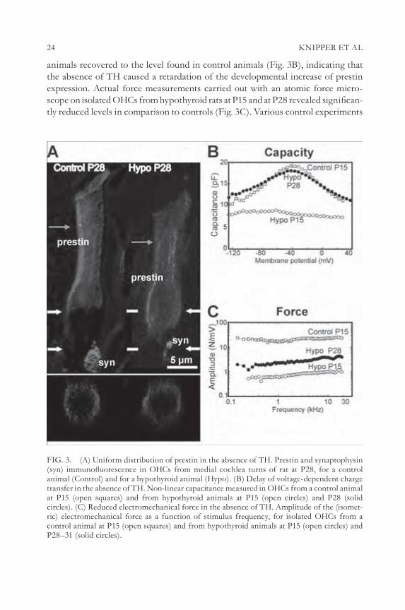
We analysed prestin protein distribution immunohistochemically in the cochlea of hypothyroid animals at P28 using double-labelling of anti-prestin and anti- synaptophysin. An immature prestin protein distribution across the entire OHC membrane was still visible in the absence of TH (Fig. 3A, Hypo P28), which allowed us to examine the role of subcellular prestin distribution in hair cell function.
Motile responses of OHCs, approaching 0.2 mm, as well as prestin protein density can be measured by the analysis of non-linear capacitance (Ashmore 1990, Oliver & Fakler 1999) and the force produced by motile responses is detectable by atomic force microscopy (Gummer et al 2002). In the absence of TH non-linear capacitance (Cnonlin) (Fig. 3B) and charge density (data not shown) were still reduced at P15 corresponding to the prestin density found at P8 in normally developing rats (Oliver & Fakler 1999). At a later developmental stage (P28), Cnonlin of hypothyroid
22 KNIPPER ET AL
FIG. 2. (A) Position of TREPrest within the prestin gene. Based on homologous human genomic sequence, a model was designed proposing the position of TREPrest within the exon–intron map of the prestin gene. Note its position downstream of exons I and II and upstream of exon III. (B) Position of GC- and TATA-elements within the prestin gene. GC- and TATA-elements are noted in the 5′-upstream region of exon I or within the exon I. GC-boxes in humans (B, position 1–6) and mice (B, position 7, 8) but not TATA-boxes neither in humans nor mice (data not shown) bind specifi cally their correspondent trans-active protein SP1 or TFIID. (C) Analysis of 5′-upstream regions of SLC26A5 in reporter gene studies. A presump-tive minimal promoter function of the 5′-region upstream of exon II is supported upon differ-ential use of distinct sequences of this region within luciferase reporter gene studies.

24 KNIPPER ET AL
animals recovered to the level found in control animals (Fig. 3B), indicating that the absence of TH caused a retardation of the developmental increase of prestin expression. Actual force measurements carried out with an atomic force micro-scope on isolated OHCs from hypothyroid rats at P15 and at P28 revealed signifi can-tly reduced levels in comparison to controls (Fig. 3C). Various control experiments
FIG. 3. (A) Uniform distribution of prestin in the absence of TH. Prestin and synaptophysin (syn) immunofl uorescence in OHCs from medial cochlea turns of rat at P28, for a control animal (Control) and for a hypothyroid animal (Hypo). (B) Delay of voltage-dependent charge transfer in the absence of TH. Non-linear capacitance measured in OHCs from a control animal at P15 (open squares) and from hypothyroid animals at P15 (open circles) and P28 (solid circles). (C) Reduced electromechanical force in the absence of TH. Amplitude of the (isomet-ric) electromechanical force as a function of stimulus frequency, for isolated OHCs from a control animal at P15 (open squares) and from hypothyroid animals at P15 (open circles) and P28–31 (solid circles).

SLC26A5 25
indicated furthermore that the reduction in force production under hypothyroid conditions was not due to structural parameters (Zimmermann et al 2006, in preparation).
In conclusion, TH affects the resonance behaviour of the basilar membrane of the inner ear by infl uencing the subcellular distribution of prestin protein in OHCs and thereby the generation of mechanical force.
Prestin homologues in non-mammalian vertebrates and insects
When we questioned evolutionary novelty of prestin motor in mammalian OHCs we were surprised to get positive hybridization with the rat prestin riboprobe in the auditory organ of mosquitoes, the Johnston’s organ, which consists of thou-sands of radially arranged multi-cellular mechanoreceptor units (Fig. 4A).
By using the deduced amino acid sequence 465–704 of the rat prestin gene for BLAST algorithms of the NCBI, Ensembl and FlyBase genome browsers (Weber et al 2003) alignments were performed and extended to the other members of the SLC26 family (A1–A9). As described (Weber et al 2003) Zebrafi sh I gene product was revealed as most closely related to prestin (SLC26A5) of rat, mice and gerbil (Fig. 4B, Zebrafi sh I). In Drosophila, the FlyBase BDGB BLAST server identifi ed a predicted gene with highest similarity to rodent prestin, CG5485 (Weber et al 2003). Zebrafi sh I and Drosophila CG5485 were cloned and again specifi c ribo-probes showed hybridization with the chordotonal sensilla of Johnston’s organ (Fig. 5A), and in the region of the auditory organs respectively (Fig. 5B) (Zebrafi sh I Genbank submission accession number AY278118).
In conclusion, data point to the existence of SLC26A5 homologues in zebrafi sh and Drosophila which are expressed in sensory organs, challenging the question to their function as either anion transporters and/or active amplifi ers.
Discussion
Transcriptional regulation
Our data suggest TH/TR as a fi rst transcriptional regulator of one of the members of the SLC26 family, the inner ear-specifi c SLC26A5 (prestin) (Weber et al 2002). Thus, TH/TR might regulate prestin expression in a cell-specifi c manner together with other as yet unknown trans-acting elements to defi ne its cell-specifi c tempo-rally precise expression (Weber et al 2002).
SLC26A6 proteins show best sequence similarity to SLC26A5 (Lohi et al 2000). Two alternative 5′ non-coding regions and transcriptional start sites with Kozak sequences have been predicted for SLC26 members with start codons either in exon 1 or exon 2 (Lohi et al 2000). However, information about upstream sequences displaying promoter function within the SLC26 family is rare. Cloning of the

26 KNIPPER ET AL
FIG. 4. (A) mRNA expression in cross-sections of the mosquito Toxorhynchites brevipalpis using in situ hybridization with rat prestin-specifi c riboprobes. In cross-sections of T. brevipalpis anten-nae, hybridization signals occur in the second antennal segment (pedicel), in the chordotonal sensilla, the auditory mechanosensory units of Johnston’s organ ( JO). No signals were observed with the sense probe (inset). (B) Phylogenetic tree. The relationship between prestin homolo-gous genes from different species and from a selection of representative members of the SLC26 family. For the generation of the phylogenetic tree the alignment from Fig. 2A in Weber et al (2003) was used.

SLC26A5 27
human prestin cDNA revealed a gene with 21 exons including two untranslated exons (Liu et al 2003), confi rming our recent fi ndings (Weber et al 2002). The fi rst two exons were suggested to be important for prestin expression (Zheng et al 2003). Here, we report a functional role of TREs within the fi rst 600 bp of the homologous intron of the human prestin gene and describe specifi c binding of GC elements but not TATA within a 5′-untranslated region downstream and within exon I. Further studies are required to verify the function of other cis-acting elements within the 5′-upstream region of SLC26A5 and challenge its comparison with other members of the SLC26 family.
FIG. 5. (A) mRNA expression in cross-sections of the fruit fl y Drosophila melanogaster using in situ hybridization with Drosophila CG5485-specifi c riboprobes. Reactive signals were detected in cells within the Johnston’s organ in the second segment of antennae (pedicel) of Drosophila melanogaster and (B) in the utriculus, sacculus and lagena of the hearing organ of the zebrafi sh.

28 KNIPPER ET AL
Regulation of subcellular distribution
The coordinated transport of ions and water across intact epithelia requires selec-tive sorting of receptors, ion channels and transporters to apical or basolateral cell surfaces. PDZ (PSD-95/Disc-large/ZO-1) domains exhibit protein–protein inter-action domains that play an essential role for determining cell polarity or mem-brane targeting (Songyang et al 1997). From all members of the SLC26 family, SLC26A6 is the fi rst described to exhibit PDZ-interaction pathways and its organi-zation in membrane micro-domains together with PDZ domain proteins such as NHERF and E3KARP (Lohi et al 2003).
SLC26A5 does not contain a PDZ-motif in its C-terminal part, and data presented here strongly suggest a mechanism of subcellular distribution of SLC26A5 different from that of other members of the family (Zimmermann et al 2006, in preparation). Indeed, the fact that a correct subcellular distribution is not achieved in the absence of TH, despite normal bell-shaped non-linear capacity curves with equal V1/2 values, indicates that at least structural maturation or distinct prestin protein phosphorylation steps, known to affect membrane targeting as well as V1/2 values of non-linear capacitance (Deak et al 2005) do not play a role for the regulation of (polarized) subcellular distribution of SLC26A5.
Expression in non-mammalian vertebrates and insects
Beside SLC26A5, no further members of the SLC26 family have been identifi ed in mechanosensitive organs in mammals, non-mammalian vertebrates or insects. This study establishes the presence of SLC26 members for the auditory sense organs of non-mammalian vertebrates, i.e. teleost fi sh, and insects, documenting that SLC26 proteins occur in a vast variety of ears (Weber et al 2003). Although the ears of insects and vertebrates have evolved independently, their auditory mechanosensors (chordotonal sensilla and hair cells, respectively) may share a common evolutionary origin (Gillespie & Walker 2001). Such a scenario is sup-ported by the fact that homologous genes control the development of these sensors (Hassan & Bellen 2000) and may explain the presence of closely related SLC26 members in insect and vertebrate ears. While non-linear amplifi cation has been described for tetrapods and insects (Gopfert & Robert 2001, 2003), fi sh are not known to improve audition by active mechanical amplifi cation, although physio-logical evidence exists that their hair bundles can move in response to electrical stimulation (Rusch & Thurm 1990). Molecular and functional studies are in progress to clarify the evolutionary origin of motile function of SLC26A5 orthologues.

SLC26A5 29
References
Ashmore JF 1990 Forward and reverse transduction in the mammalian cochlea. Neurosci Res 12 (suppl):S39–50
Belyantseva IA, Adler HJ, Curi R, Frolenkov GI, Kachar B 2000 Expression and localization of prestin and the sugar transporter GLUT-5 during development of electromotility in cochlear outer hair cells. J Neurosci 20:RC116
Chambard JM, Ashmore JF 2003 Sugar transport by members of the SLC26 superfamily of anion-bicarbonate exchangers. J Physiol 550:667–677
Deak L, Zheng J, Orem A et al 2005 Effects of cyclic nucleotides on prestin’s function. J Physiol 563:483–496
Gillespie PG, Walker RG 2001 Molecular basis of mechanosensory transduction. Nature 413:194–202
Gopfert MC, Robert D 2001 Active auditory mechanics in mosquitoes. Proc R Soc Lond B Biol Sci 268:333–339
Gopfert MC, Robert D 2003 Motion generation by Drosophila mechanosensory neurons. Proc Natl Acad Sci USA 100:5514–5519
Gummer AW, Meyer J, Frank G, Scherer MP, Preyer S 2002 Mechanical transduction in outer hair cells. Audiol Neurootol 7:13–16
Hassan BA, Bellen HJ 2000 Doing the MATH: is the mouse a good model for fl y development? Genes Dev 14:1852–1865
Knipper M, Gestwa L, Ten Cate WJ et al 1999 Distinct thyroid hormone-dependent expression of TrKB and p75NGFR in nonneuronal cells during the critical TH-dependent period of the cochlea. J Neurobiol 38:338–356
Knipper M, Zinn C, Maier H et al 2000 Thyroid hormone defi ciency before the onset of hearing causes irreversible damage to peripheral and central auditory systems. J Neurophysiol 83:3101–3112
Liberman MC, Gao J, He DZ et al 2002 Prestin is required for electromotility of the outer hair cell and for the cochlear amplifi er. Nature 419:300–304
Liu XZ, Ouyang XM, Xia XJ et al 2003 Prestin, a cochlear motor protein, is defective in non-syndromic hearing loss. Hum Mol Genet 12:1155–1162
Lohi H, Kujala M, Kerkela E et al 2000 Mapping of fi ve new putative anion transporter genes in human and characterization of SLC26A6, a candidate gene for pancreatic anion exchanger. Genomics 70:102–112
Lohi H, Lamprecht G, Markovich D et al 2003 Isoforms of SLC26A6 mediate anion transport and have functional PDZ interaction domains. Am J Physiol Cell Physiol 284:C769–779
Oliver D, Fakler B 1999 Expression density and functional characteristics of the outer hair cell motor protein are regulated during postnatal development in rat. J Physiol 519 Pt 3:791–800
Oliver D, He DZ, Klocker N et al 2001 Intracellular anions as the voltage sensor of prestin, the outer hair cell motor protein. Science 292:2340–2343
Rusch A, Thurm U 1990 Spontaneous and electrically induced movements of ampullary kino-cilia and stereovilli. Hear Res 48:247–263
Santos-Sacchi J 1991 Reversible inhibition of voltage-dependent outer hair cell motility and capacitance. J Neurosci 11:3096–3110
Songyang Z, Fanning AS, Fu C et al 1997 Recognition of unique carboxyl-terminal motifs by distinct PDZ domains. Science 275:73–77
Weber T, Zimmermann U, Winter H et al 2002 Thyroid hormone is a critical determinant for the regulation of the cochlear motor protein prestin. Proc Natl Acad Sci USA 99:2901–2906

30 KNIPPER ET AL
Weber T, Gopfert MC, Winter H et al 2003 Expression of prestin-homologous solute carrier (SLC26) in auditory organs of nonmammalian vertebrates and insects. Proc Natl Acad Sci USA 100:7690–7695
Zheng J, Shen W, He DZ et al 2000 Prestin is the motor protein of cochlear outer hair cells. Nature 405:149–155
Zheng J, Madison LD, Oliver D, Fakler B, Dallos P 2002 Prestin, the motor protein of outer hair cells. Audiol Neurootol 7:9–12
Zheng J, Long KB, Matsuda KB et al 2003 Genomic characterization and expression of mouse prestin, the motor protein of outer hair cells. Mamm Genome 14:87–96
Zimmermann U, Frank G, Oliver D et al 2006 Expression of prestin and cellular force genera-tion by auditory outer hair cells are differentially affected by thyroid hormone depletion during development. In preparation
DISCUSSION
Welsh: How does prestin work? Do we know?Knipper: I can only say a little bit; Dominic Oliver will say more in his
paper. Currently, the concept is that a voltage-dependent conformational change of prestin is discussed as the molecular basis for motile outer hair cell (OHC) responses.
Muallem: Do you have any information about the splice variants? Are there any differences among them?
Knipper: The data are preliminary. We need to have the proper function informa-tion if at all possible. We are currently looking for a presumptive role of PDZ domains, and we are planning to test whether the splice variants, when coex-pressed, change the charge behaviour.
Muallem: So you don’t know that it is even functional?Knipper: Not at the moment. The nicest thing would be if it has no impact on
the motility, but it has an impact on the force.Alper: Is the boundary between the lateral and basal membranes, which is
defi ned in the mature state by the redistribution of prestin, defi ned by the Deiter’s cell contact? And is it the Deiter’s cell interface that is the infl uence driving prestin out?
Knipper: You have good information about the OHC, because most people here don’t even know what and where Deiter’s cells are in relation to outer hair cells. The Deiter’s cells are the supporting cells apposing outer hair. Indeed, there seems to be a striking temporal parallel between the change in the sub cellular distribution of SLC26A5 from a wide one to a restricted membrane distribution, and the enlargement of the contact zone of Deiter’s cell and OHCs. While we do not have any functional evidence for this, we have to consider such a concept.
Alper: What happens to the Deiter’s cells in hypothyroid state? What happens developmentally?

SLC26A5 31
Knipper: They look nice. There is an inward rectifying K+ channel in Deiter’s cells that is strongly expressed in the apical part, just opposite the KCNQ4 ion channel in the base of OHCs that leads to the repolarization of the cell. In the absence of thyroid hormone (TH) we have current evidence that it looks not so much different. On the level of ion channels, we do not have anatomical or func-tional evidence that Deiter’s cells are affected during hypothyroidism.
Alper: How about cytoskeletal candidate protein redistribution paralleling prestin distribution?
Knipper: No one knows.Lee: How does TH change the distribution of prestin variants?Knipper: We performed studies with RT-PCR and found a profound reduction
of the splice variants in the absence of TH. This fi ts with our model. But it is not a total absence.
Lee: In the case of Pendred syndrome, there is an association with hypo-thyroidism. Is prestin somehow responsible for the deafness in Pendred syndrome?
Knipper: I don’t think anyone has looked for prestin in Pendred knockouts. The typical feature or phenotype is the loss of the endocochlear potential as a fi rst sign in the PDS knockout. In other knockouts that we work in we have looked at pheno-types where we have a loss of endocochlear potential. We noticed that in these mice the prestin expression appears normal. Thus the prestin expression is extremely robust. We still can observe it when other outer hair cell ion channels have gone already, indicating a low protein turnover rate which is not that much infl uenced by metabolic energy.
Everett: I think the fact that Pendred syndrome has features of both inner ear and thyroid disease leads to a lot of confusion. People have long thought that the deafness is thyroid hormone related but I do not believe that hypothyroidism plays any part in the deafness of Pendred syndrome. If you look at people or mice with mutations in thyroid peroxidase, which causes a more severe iodide organifi cation defect and overt congenital hypothyroidism, they don’t have a severe hearing defect. I am sure that the deafness in Pendred syndrome has nothing to do with any mild hypothyroidism. The hair cell defects seen in hypothyroidism are very different from the defects of endolymphatic hydrops and swelling of the inner ear in the Pendred knockout. It is just a coincidence that there is also a defect in the thyroid in Pendred syndrome.
Kere: One of the things we haven’t discussed yet is the topology of the proteins on the membrane. You showed a model with 10 transmembrane domains. We have tended to favour the 12-transmembrane domain model for the other members.
Knipper: This might be a misunderstanding. We also favour a model with 12 transmembrane domains. I showed a picture which I found in the recent review

32 KNIPPER ET AL
by Dallos (Dallos & Fakler 2002) in which they analysed the glycosylation/phos-phorylation steps. Due to their results a further experimental basis for a 12 trans-membrane domain topology was generated.
Turner: There are a number of theoretical methods for predicting membrane protein topology, and these proteins are a mess! Especially at the N-termini where many of the predictive algorithms give quite different results.
Kere: We have some data to show that the C-terminal tail is inside the cell for a few SLC26 proteins.
Knipper: We can confi rm this for the A5 members.Everett: We took all the SLC26 transporters known at the time and analysed
them together using a topology program, to give a more robust conclusion that, for all these transporters collectively, the C-terminus is in the cell. This is just a computational prediction though; there is better wet-bench based evidence to support this.
Turner: A number of the recently crystallized membrane proteins have been shown to have hydrophobic regions that don’t completely cross the membrane. I think all bets are off for now.
Kere: Does anyone have evidence for an odd number of transmembrane domains?
Ashmore: Historically, prestin was predicted to have 11 domains, and then this was revised to 12. I am surprised that you say that the mess is at the N-terminal rather than the C-terminal end. Or do all of these prediction programs make a mess of both ends?
Turner: There is mess at the C-terminal end as well. But at least there are some clearly defi ned hydrophobic peaks. The predictive algorithms tend to correspond very well when there is a sharp peak. There are also a couple of not such sharp peaks that might be either one or two membrane-spanning domains.
Welsh: I take it from this discussion no one has mapped the topology.Mount: And we assume that the topology is going to be the same for all the
members, which is probably an oversimplifi cation. In some programs A1 seems to have an extra transmembrane domain early on in the N-terminus. I think there will be common transmembrane domains, but there may be other regions in the protein where there may be some divergence.
Kere: The splice variation produces different numbers.Muallem: There is probably something in the number 12. It is not only cystic
fi brosis transmembrane regulator protein (CFTR), but also almost all the other epithelial chloride channels that have 12 trans membrane domains.
Alper: Except for 18 or 14 for CLC. How do you defi ne precisely what is trans-membrane, even with the crystal structure?
Welsh: The structure of CLC threw predictions into disarray.Alper: I think you can be confi dent about 12 for the ABC family.

SLC26A5 33
Muallem: When you look at the CLC structure, you can kind of count 12 trans-membrane domains, even though there are some additional helices that protrude into the membrane.
Case: Is this effect of TH on the hair cell something seen with other trans-porters, or is this a unique situation?
Knipper: Yes, we see an effect of TH not only on transporters such as prestin, but also on ion channels, that are expressed during the critical time period prior to hearing function in hair cells and spiral ganglia neurons. Before this time and after this time TH does not display any effect on the inner ear according to our studies. During this critical period, however, TH indeed appears to affect particu-lar ion channels and transporters, those proteins, which are up-regulated during the most fi nal differentiation of hair cells and spiral ganglia neurons. We also presume that this is the period when in the body, the lung, heart, bones and intestines other fi nal differentiation processes occur under the control of TH, including differentiation of functionally relevant ion channels. Thus TH may have a most critical role for synchronous running of fi nal differentiation processes in multiple organs and cells.
Mount: We were able to PCR a fair amount of A5 from brain. There is some expression in heart as well. Have you looked with your antibody outside of the inner ear?
Knipper: Yes. We cannot fi nd anything.Oliver: We were also able to PCR A5 from brain of rat and mouse.Mount: So you don’t see prominent expression in rare cell types.Oliver: No.Knipper: The promoter of prestin was used to drive Cre recombinase expression
at a time when promoter sequences were still presumed to be upstream of exon III. The result was a extraordinarily low expression also in inner hair cell in addi-tion to outer hair cells (Tian et al 2004). Thus it can be concluded that there is a very low expression in the inner hair cell. We however, could never detect prestin protein at the inner hair cell level.
Ashmore: Even so, it is tempting to think that prestin is present throughout subsystems of the brain, and not just the auditory and ascending auditory pathways. It would be interesting to know specifi cally where it is in the brain.
Alper: If so, do you think it would serve the same electromechanical function?
Ashmore: I don’t know.Knipper: It would be interesting if we knew how this protein functions, if we do
not have a dense expression pattern. Despite the fact that a variable density of the protein in transfected cells may nevertheless result in conformational change leading to detectable movement or better non-linear capacity, we might not neces-sarily pick it up if prestin isn’t expressed apart from in OHC.

34 KNIPPER ET AL
Welsh: How dense is its expression?Knipper: It is extraordinarily dense. This is comparable to what has been described
for A1 in erythrocytes.Ashmore: There are 10 million copies per cell. About 40% of the surface mem-
brane is prestin.Soleimani: Are there other chloride transporters in OHC?Knipper: We have strong evidence that SLCA2 that is expressed over the entire
OHC membrane is partially overlapping with prestin.Soleimani: What about NKCC1 or others? The reason for this question is that
we did some experiments on prestin in collaboration with Dr Dallos. We couldn’t detect any chloride–bicarbonate exchange mediated by prestin, but he later showed that if cells were depleted of chloride, the motility that is generated by prestin stops, even though prestin does not transport chloride. These results suggest that intracellular chloride is essential for the activity of prestin. Is it possible to inhibit another chloride transporter and therefore deplete intracellular chloride and as a result inhibit prestin?
Oliver: A similar idea was proposed by Joe Santos-Sacchi (Rybalchenko & Santos Sacchi 2003). He showed that there is a chloride conductance in the lateral mem-brane of the outer hair cell, which seems to be stretch sensitive. It is not clear what molecule provides this conductance. Chloride fl ux through this conductance may modulate the intracellular chloride concentration and modify the function of prestin.
Soleimani: What is the effect of furosemide on this chloride conductance? This question can be rephrased into whether prestin is the fi nal pathway for a good number of diseases that cause deafness.
Oliver: Furosemide impairs inner ear function mainly by abrogating the endo-cochlear potential, the electrical driving force that is crucial for the electrical activity of the hair cells. This then also blocks OHC motility. There is chloride transport in other parts of the inner ear too, namely in the stria vascularis and the supporting cells, which is absolutely critical for inner ear function without directly pertaining to prestin function. Transport in the stria vascularis generates the endocochlear potential and involves the Na-K-2Cl co-transporter (NKCC1/Scl12a2) which explains the furosemide sensitivity (Delpire et al 2003).
Knipper: Every protein that would change the turgor of the outer hair cell would lead to deafness. From this point of view, we can hardly understand why prestin has become so famous. For the hair cell, the consequences are the same.
Welsh: Are people with prestin mutations completely deaf?Knipper: Liu et al (2003) found a mutation in a splice acceptor site to be respon-
sible for deafness. This is one of the fi rst human diseases related to prestin.Oliver: Carriers in whom both alleles are affected are completely deaf (Liu et al
2003), but this might also have to do with the degeneration of hair cells. In the

SLC26A5 35
prestin knockout mouse, hair cells also tend to degenerate (Liberman et al 2002).
Welsh: You would predict, therefore, that if the hair cell didn’t degenerate but you took away prestin, people would still hear, although the acuity might decrease.
Oliver: Yes, I predict that there would be a drop-off in sensitivity of 50–60 db. This is actually what was observed in cochlear regions of prestin knockout animals where the hair cells were fully intact.
Markovich: Prestin seems to be the odd gene out in this family. It doesn’t seem to have any anion transport. Why? Has it lost certain sequences?
Oliver: I don’t know whether or not it has anion transport activity. As far as I know, people haven’t looked very carefully.
Soleimani: We tested both mouse and human prestin, generous gifts from Peter Dallos. However we did not detect any chloride–bicarbonate exchange by either clone.
Knipper: So the answer is no?Soleimani: Dr Karniski examined radiolabelled chloride uptake in oocytes
expressing prestin. My understanding was prestin did not show any measurable chloride uptake (personal communication via Dr Dallos).
Ashmore: I still think it transports, but at a very low rate!Soleimani: Perhaps the activity of prestin requires the right environment in terms
of substrates, osmolarity, pH or the expression system (oocytes, mammalian cells, etc.).
Markovich: So it could be an anion exchanger?Thomas: Along these lines, we heard about hypothyroidism and the fact that
those patients don’t exhibit this localization in OHC and, yet, there is charge move-ment without mechanical function. Let’s just presume that under these expression levels you get some paracrystalline array. My guess is that no one is testing function under those conditions where we know in the native cell it is actually performing its mechanical function.
Welsh: You may get the same kind of array in an oocyte. I don’t know whether it has a particular characteristic that you can recognize, for example, by scanning EM.
Knipper: No one has done a crystal array.Thomas: At the higher density, is it functional?Oliver: Basically, we don’t get these high densities in our expression systems.Alper: In transiently transfected cells we can get about a million copies per
cell.Oliver: Looking at the charge transfer density normalized to the linear mem-
brane capacitance (which is a measure for the membrane area), we are about a factor of 10 lower than in OHCs.

36 KNIPPER ET AL
Mount: Aquaporin 4 gives these orthogonal arrays in the proximal tubule of the kidney, which are lost in knockout mice (Verbavatz et al 1997, Yang et al 1996). You would think that it might be a characteristic of the protein itself, to make some kind of crystalline structure in the membrane, which may be dependent on the concentration.
Knipper: This is the reason why we have to get it out. What is the reason for this high expression of prestin in the membrane, a feature clearly different from other SLC26 members? This comes back to the question of whether prestin might have a physiological function as a transporter. Both high expression and loss of trans-porter function point to SLC26A5 as an exception among the other members. The question raised by this is whether at the time of the divergence of other vertebrates from fi sh it was necessary to invent something auditory-specifi c, since there is a protein which has only this particular characteristic and function in outer hair cells of the inner ear. If this is the case, we also have the solution for the striking cell-specifi c expression pattern of the other SLC26 members. The auditory hearing organ can be traced back at the molecular level to a common origin as might other distinct organs as well. Thus, prestin might indeed be an exception. It had 100 000 years to develop into a motile protein with molecular characteristics different from the other members.
Aronson: At least in principle, you don’t need a large structural difference. It is a subtle kinetic change to let it do a transport hemi-cycle. You say there is an inside chloride-dependent conformational change; if you just had an anion exchanger with a very slow off-rate, it could have a reorientation step and a low rate of anion exchange. It really is an anion exchanger, just a very sluggish one, so it mainly has conformational changes with a low fl ux rate.
Muallem: Before we can answer these questions, someone needs to take the hair cell itself and measure something in it. It wouldn’t be too diffi cult to measure pH and current. The cell milieu might provide some kind of regulation in addition to the structure.
Mount: A broader point is that we are biased that these are anion exchangers. In the CLC literature Chris Miller (Accardi & Miller 2004) and others (Picollo & Pusch 2005, Scheel et al 2005) have shown that the CLC chloride ‘channels’ can function as exchangers, exchanging chloride with a proton. The level of homology between SLC26 paralogues is not that high. It is not surprising that some of these are channel-like, and some of them have different stoichiometries. It doesn’t bother me that it is not an exchanger; it may be just some variant of a global transport characteristic which all of them have at varying levels. There is this tendency to think that they are all anion exchangers. If anything comes out of this meeting, it will be that this is probably not the case.
Muallem: I agree with you, obviously! Not only that, but also as we learn more about them we know very little about what the true substrate specifi city is. I would

SLC26A5 37
not be surprised to fi nd out that this transporter is very good at transporting some other molecule.
Mount: I am not happy with negative results, such that I no longer have faith that if I can’t fi nd something transported by a given member of this family that it isn’t transported. There are phosphorylation-dependent effects of chloride that we may be forgetting about, such that the conditions we use might not lead to suc-cessful transport. For example, intracellular chloride activates or inactivates the various modes of cation-chloride co-transport (Lytle & McManus 2002).
Alper: In this respect, with this question of conformational change without transport and conformational change with slippage to make for a leaky kind of transport, it is of note that that GFP-tagged prestin still localizes normally and gives the same electromechanical non-linear capacitance under conditions of hypothyroidism. With the non-linear capacitance you have protein which on a single molecule basis is moving at 50 000 Hz or so. Could you see that with your GFP-tagged prestin? Are there optical tools that would allow you to resolve that movement, especially in the case where that movement is uncoupled from your measured whole cell contractile force?
Knipper: This brings us back to a basic question concerning in vitro expression systems. Can we mimic hair cells in a culture system? In a culture system we can produce a situation where the SLC26A5 can produce motility with a voltage charge comparable to transfected cells, but we can never produce force.
Mount: Can you defi ne the difference between the capacitance changes and the force?
Knipper: If we isolate hair cells we mimic the in vivo situation, because people put the hair cells in a kind of chamber that mimics somehow cell turgor. The hair cells are soaked in a pipette and thereby only motile responses and force can be investigated.
Oliver: We should distinguish between the microscopic force generated by the single molecule and the macroscopic force that is measurable from the whole cell. The second will depend on structural things like the cytoskeleton and so on, but most probably not the microscopic force produced by the conformational change of a single molecule.
Mount: So in the hypothyroid state in the outer hair cells you see capacitance changes but you don’t see force.
Knipper: Yes, this is measured in in vivo situations. It is reduced force. We need to know more about the normal in vivo situation, including details of pho sphorylation and glycosylation. The specifi city of the ion channel may be infl uenced by these kinds of biochemical and structural changes. In in vitro expression systems these are different from the in vivo system. Therefore I presume we have a gap to fi ll out. We cannot mimic the in vivo system if we do not know how much infl uence the biochemical changes have on a specifi c motile cell response. If I give you a 3D

38 KNIPPER ET AL
culture system where the cochlea remains intact, and someone tries to convince you that you could mimic the in vivo system, I would say they are wrong. This is because in the in vitro situation some ion channels are missing which are important for normal outer hair cell function. This is often disregarded. For example, KCNQ4 is missing in any in vitro situation, em phasizing the artifi cial nature of an in vitro situation. I presume that in every in vitro expression system that others are looking at, the situation is similar. Within hours, and even minutes, distinct ion channels go down and change. We can’t disregard this fact. The phenotype is different, and we need to bear this in mind.
Welsh: What happens to people who have hypothyroidism? The changes you saw were quite dramatic. Do you see changes consistent with hypothyroidism?
Knipper: The kind of deafness I described was as a result of a hypothyroid state during embryonal development and not in adults. We learned from animal experi-ments that TH affects the ear only during a limited window, which in humans would be between the second and sixth embryonal months. We furthermore learned that every period of TH defi ciency during this critical time period, even if only transient, leads to deafness, and thus would lead to different degrees of hearing loss in newborn children. You would not be able to diagnose the child as hypothyroid and would not be able to link the different degrees of deafness to TH defi ciency. In adults experiencing a defi ciency of thyroid hormone, the hearing would not be expected to be dramatically effected.
Welsh: You can measure the movement of the outer hair cells. Does that change?
Ashmore: There are also distortion product oto-acoustic emissions (DPOAEs) which are an indication of outer hair cell function.
Knipper: There seems to be some controversial evidence as to whether DPOAEs change when adults suffer from hypothyroidism. I asked medical doctors working with hypothyroid patients, and got various controversial answers. Some said that over a long period of hypothyroidism the patients have a hearing defi cit, but with compensation therapy this is reversed.
Quinton: Does anything occur with ageing?Knipper: We work on two relevant ageing animal models, Fischer rats and gerbils.
These get profound human-like hearing defi cits, comparable to what is known in humans. We have one phenotype that has smaller efferents, and when we measure the DPOAE, concomitant with the hearing loss there is a loss of active cochlear mechanics in both of these animals, even though their hair cells are present and prestin is present. In age-dependent hearing loss in humans, the mechanism of loss of active cochlear mechanics is also controversial. So for humans it is an open question, but for animal models we have some evidence that the active cochlear mechanics may be slightly affected.
Quinton: Is there any difference in expression?

SLC26A5 39
Knipper: We don’t see any.Ashmore: In the cochlea there is no turnover of cells. There may be turnover of
protein, but this is inadequately documented. You are born with a fi xed number of cells in your cochlear so you need to look after them.
Kere: What do we know about the mutation spectrum in humans?Knipper: The only study currently published is by Liu et al (2003) who saw a
mutation in the splice acceptor site.Kere: Are there any coding short nucleotide polymorphisms (SNPs) in public
databases?Knipper: People in the hearing centre in Tuebingen and probably also in other
places are currently looking for it.Kere: Has anyone screened deaf individuals?Knipper: This is ongoing; we have a database of 2000 people in our hearing
research centre.Alper: What is known about the lack of replacement of cochlear hair cells in
Rb−/− or Rb+/− mice? There is new work about the regeneration of hair cells with the retinoblastoma gene product. Is prestin working similarly in the different tonotopic sections of the cochlear, with equal abundance?
Chan: There is literature on the regeneration of outer hair cells after damage by antibiotics, indicating that these cells do have the ability to regenerate. Whether these cells can regenerate could affect the interpretation of some of the data presented.
Knipper: In our hearing research centre Professor Gummer works on motile responses in isolated OHCs. The message is that if you take OHCs from either the apical or basal part, they appear to exhibit the same voltage charge responses.
Ashmore: There is a battle going on at the moment between researchers in the fi eld as to whether amplifi cation in the ear is to do with prestin, or—perish the thought—nothing to do with prestin but rather something to do with the hairy end of the cell.
Oliver: There is one paper that describes that the density of the charge movement via prestin increases towards higher frequencies (Santos-Sacchi et al 1998).
Knipper: Jonathan Ashmore, isn’t it true that force production is dramatically infl uenced by the basilar membrane structure? Along the tonotopic axis, the ana-tomical structure determines the extent of motile responses? In the apical part the larger outer hair cells are not embedded in such a fi xed structural environment as is the case in the basal cochlear turns.
Ashmore: The complication in the cochlear mechanics arises because at the high-frequency end of the basilar membrane the structure gets stiffer. Therefore you need larger forces to compensate for this increased stiffness and presumably an increase in associated viscosity. At the low frequency end the problem is not so extreme. This is why the observation that the density of prestin increases at the

40 KNIPPER ET AL
high frequency end is working in the right direction. The current debates in the fi eld surround whether it is helping enough.
Welsh: You mentioned that you see A5 in mosquito and Drosophila. It looked like it was in the proboscis.
Knipper: In the mosquito I showed the SLC25A5 homologue expression in the thousands of radially oriented sensory receptor units in the chordotonalorgan, which is the hearing organ.
Welsh: Where else do you see it?Knipper: The brain. There are ongoing studies for detection of prestin expression
outside the ear in Drosophila.Welsh: Has anyone else looked at analogues of this in non-vertebrates?Knipper: I presume one of the challenging questions is has this kind of homo-
logue in the inner ear undergone a special evolution or not? In other organs it might be an ion channel, but in the inner ear it could be a protein which somehow has progressed and developed to become fi nally a motile protein. The striking question thus is, does the homologue move or not? Does it show non-linear capaci-tance or not? These are critical questions. Does A6, which is closely related, have the same properties?
References
Accardi A, Miller C 2004 Secondary active transport mediated by a prokaryotic homologue of ClC Cl− channels. Nature 427:803–807
Dallos P, Fakler B 2002 Prestin, a new type of motor protein. Nat Rev Mol Cell Biol 3:104–111
Delpire E, Lu J, England R, Dull C, Thorne T 2003 Deafness and imbalance associated with inactivation of the secretory Na-K-2Cl co-transporter. Nat Genet 22:192–195
Liberman MC, Gao J, He DZ, Wu X, Jia S, Zuo J 2002 Prestin is required for electromotility of the outer hair cell and for the cochlear amplifi er. Nature 419:300–304
Lytle C, McManus T 2002 Coordinate modulation of Na-K-2Cl cotransport and K-Cl cotrans-port by cell volume and chloride. Am J Physiol Cell Physiol 283: C1422–1431
Liu XZ, Ouyang XM, Xia XJ et al 2003 Prestin, a cochlear motor protein, is defective in non-syndromic hearing loss. Hum Mol Genet 12:1155–1162
Picollo A, Pusch M 2005 Chloride/proton antiporter activity of mammalian CLC proteins ClC-4 and ClC-5. Nature 436:420–423
Rybalchenko V, Santos-Sacchi J 2003 Cl− fl ux through a non-selective, stretch-sensitive con-ductance infl uences the outer hair cell motor of the guinea-pig. J Physiol 547:873–891
Santos-Sacchi J, Kakehata S, Kikuchi T, Katori Y, Takasaka T 1998 Density of motility-related charge in the outer hair cell of the guinea pig is inversely related to best frequency. Neurosci Lett 256:155–158
Scheel O, Zdebik AA, Lourdel S, Jentsch TJ 2005 Voltage-dependent electrogenic chloride/proton exchange by endosomal CLC proteins. Nature 436:424–427
Tian Y, Li M, Fritzsch B, Zuo J 2004 Creation of a transgenic mouse for hair-cell gene targeting by using a modifi ed bacterial artifi cial chromosome containing prestin. Develop Dynamics 230:199–203

SLC26A5 41
Verbavatz JM, Ma T, Gobin R, Verkman AS 1997 Absence of orthogonal arrays in kidney, brain and muscle from transgenic knockout mice lacking water channel aquaporin-4. J Cell Sci 110:2855–2860
Yang B, Brown D, Verkman AS 1996 The mercurial insensitive water channel (AQP-4) forms orthogonal arrays in stably transfected Chinese hamster ovary cells. J Biol Chem 271:4577–4580

Sulfate transport by SLC26 transportersDaniel Markovich
Department of Physiolog y and Pharmacolog y, School of Biomedical Sciences, University of Queensland, Brisbane, St. Lucia, QLD 4072, Australia
Abstract. Sulfate is the fourth most abundant anion in human plasma that is essential for numerous physiological functions, including biotransformation of xenobiotics, steroids, non-steroidal anti-infl ammatory drugs (NSAIDs), adrenergic stimulants/blockers and analgesics. Sulfate is also required for activation of many endogenous compounds (heparin, heparan sulfate, dermatan sulfate, bile acids) and utilized in the metabolism of neurotransmitters. Sulfation of structural components, including glycosaminoglycans and cerebroside sulfate, is essential for the maintenance of normal structure and function of tissues. Due to its hydrophilic nature, sulfate cannot readily cross the lipid bilayer of cells, thus plasma membrane proteins, known as sulfate transporters, are required for the movement of sulfate into/out of cells. Sulfate transporters can be divided into two distinct groups: Na+ -dependent sulfate transporters belonging to the SLC13 gene family and Na+ -independent sulfate transporters (antiporters, exchangers) belonging to SLC26 gene family. There are 11 members of the SLC26 family (including Sat1, DTDST, CLD, pendrin, prestin, cfex) whose structures and functions have been only partially charac-terized. In this presentation, the current information on the structures and functions of the sulfate transporters in the SLC26 gene family will be described and the issue that certain members of this family are unable to transport sulfate, will be addressed.
2006 Epithelial anion transport in health and disease: the role of the SLC26 transporters family. Wiley, Chichester (Novartis Foundation Symposium 273) p 42–58
SLC26 genes encode sulfate transporters
SLC26A1 (Sat1)
The fi rst member of the SLC26 gene family was identifi ed by expression cloning using a Xenopus oocyte system to screen a rat liver cDNA library by radiotracer sulfate uptake (Bissig et al 1994). A single functional clone was isolated, which became known as Sat1 (sulfate anion transporter 1). Functional characterization in Xenopus oocytes using radiotracer sulfate measurements, revealed Sat1 to encode a Na+ -independent sulfate transporter (see Table 1), with a high affi nity (Km = 136 mM) for sulfate, that was strongly inhibited by a stilbene derivative DIDS (4,4′-diisothiocyanato-2,2′-disulfonate) (IC50 DIDS = 28 mM) and oxalate, but not by succinate or cholate (Bissig et al 1994). These properties correlated closely with the functional activities of the sulfate/bicarbonate exchanger in liver canalicular
42

SULFATE TRANSPORT 43
membrane vesicles (Meier et al 1987). The coding region of the rat Sat1 (rSat1) cDNA predicts a protein of 703 amino acids with a calculated molecular mass of 75.4 kDa. Hydrophobicity analysis of rSat1 protein suggests 12 putative transmem-brane domains (TMDs)(Markovich 2001). Northern blot analysis detected a single mRNA (3.8 kb) transcript for Sat1 very strongly in liver and kidney, with weaker signals in skeletal muscle and brain (Bissig et al 1994). The abundance of Sat1 mRNA in the kidney was identical to its expression in the liver (the tissue from which it was cloned), prompting us to examine the role of Sat1 in the kidney. When rat kidney mRNA was injected into Xenopus oocytes it induced a Na+ -independent sulfate transport activity which was inhibitable by DIDS, probenecid and phenol red (Markovich et al 1994). The degree of inhibition by these compounds was closely correlated with the inhibition pattern of the renal basolateral membrane (BLM) sulfate anion exchanger (David & Ullrich 1992), as well as with Sat1 cRNA-induced activity in Xenopus oocytes (Bissig et al 1994). Furthermore, using a hybrid depletion strategy, Sat1 antisense oligonucleotides led to a complete abolition of the kidney mRNA-induced Na+ -independent sulfate transport activity (in Xenopus oocytes), confi rming that Sat1 encodes the Na+ -independent sulfate transporter, whose function most closely correlates with the proximal tubular BLM sulfate/bicarbonate anion exchanger. This fi nding was later confi rmed by immunocyto-chemistry using Sat1-specifi c peptide polyclonal antibodies showing the location
TABLE 1 Sulfate transport by SLC26 transporters
Gene/Protein Transports DIDSHGNC Name Name Tissue distribution Sulfate Sensitive2
SLC26A1 Sat1 widespread Yes1 YesSLC26A2 DTDST ubiquitous Yes YesSLC26A3 DRA, CLD widespread Yes YesSLC26A4 PDS, Pendrin inner ear, kidney, No Yes thyroidSLC26A5 Prestin widespread NDSLC26A6 PAT1, CFEX widespread Yes YesSLC26A7 — kidney Yes YesSLC26A8 Tat1 sperm, brain Yes YesSLC26A9 — lung Yes YesSLC26A10 pseudogene ND ND NDSLC26A11 — widespread Yes Yes
HGNC, Human Genome Nomenclature Committee; ND, not determined; widespread, four or more tissues.1Km for sulfate has been determined at 0.1–0.3 mM.2Sulfate transport is signifi cantly inhibited by anion exchange inhibitor DIDS, a stilbene-derivative.

44 MARKOVICH
of the Sat1 protein in the kidney to be restricted to the BLM of kidney proximal tubules (Karniski et al 1998).
We have also identifi ed Sat1 expression in the brain (Lee et al 1999). Injection of rat brain mRNA into Xenopus oocytes induced a saturable Na+ -independent sulfate uptake, which was inhibited by DIDS (IC50 DIDS = 150 ± 30 mM) and oxalate, with properties identical to Sat1 anion exchange activity. The involvement of a Sat1-like transporter responsible for the sulfate transport in rat brain was con-fi rmed by hybrid depletion using Sat1 antisense oligonucleotides, which completely abolished the brain mRNA-induced sulfate transport activity in Xenopus oocytes (Lee et al 1999). Furthermore, in situ hybridization using Sat1 probes detected Sat1 mRNA expression most prominently in the hippocampus and cerebellum, in par-ticular in the neuronal-rich cell layer, suggesting that Sat1 may play an important role in sulfate transport in neuronal or glial cells (Lee et al 1999).
Recently, this laboratory isolated the mouse (Lee et al 2003) and human (Regeer et al 2003) Sat1 orthologues (named mSat1 and hSAT1, respectively) of the rat Sat1 cDNA, using a homology screening approach. We also identifi ed their gene struc-tures, determined their protein function in Xenopus oocytes and characterised promoter activities in mammalian renal cell lines. mSat1 encodes a 704 amino acid protein (75.4 kDa) with 12 putative transmembrane domains (TMDs) that induces sulfate, oxalate and chloride transport in Xenopus oocytes (Lee et al 2003). By RT-PCR, mSat1 mRNA expression was found to be strongest in the kidney and liver, with lower levels observed in caecum, calvaria, brain, heart and skeletal muscle. Two distinct transcripts were expressed in kidney and liver due to alternative utilization of the fi rst intron, corresponding to an internal portion of the 5′-untranslat ed region. The mouse Sat1 gene (Slc26a1) is approximately ∼6 kb in length and consists of 4 exons, with an optional intron 1. The Sat1 promoter is ∼52% G + C rich and contains a number of well-characterized cis-acting elements, including sequences resembling responsive elements to thyroid hormone (T3REs) and vitamin D (VDREs). We demonstrated that Sat1 promoter driven basal transcrip-tion in renal opossum kidney (OK) cells was stimulated by tri-iodothyronine (T3), but not by 1,25-(OH)2 vitamin D3 (vitamin D). Site-directed mutagenesis of an imperfect T3RE sequence at −454 bp led to a loss of Sat1 promoter inducibility by T3. This suggests that position −454 in the Sat1 promoter contains a functional T3RE that is responsible for transcriptional activation of Sat1 by T3.
By radiotracer measurements in Xenopus oocytes, mSat1 led to a ∼20-fold induction in Na+ -independent sulfate transport, a ∼sixfold induction in oxalate and chloride transport, but was unable to induce any formate, L-leucine or succinate uptakes, when compared to water injected (control) oocytes. This sug-gests that mSat1 transport was restricted to the anions SO4
2−, oxalate and chloride. As shown for rSat1 (Satoh et al 1998), mSat1-induced SO4
2− transport occurred only in the presence of extracellular chloride, suggesting its requirement for chlo-

SULFATE TRANSPORT 45
ride. Typical Michaelis-Menten saturation kinetics were observed for mSat1-induced SO4
2− uptake, with a calculated Km = 0.31 ± 0.05 mM and Vmax = 143.90
± 5.32 pmol/h for SO42− interaction. These values are in close agreement with
rSat1-induced SO42−
transport kinetics (Km = 0.14 mM) (Bissig et al 1994). mSat1-induced SO4
2− transport was signifi cantly inhibited by molybdate, selenate, tung-state, DIDS, thiosulfate, phenol red and probenecid, whereas citrate and glucose had no effect. These data suggest that mSat1 encodes a functional SO4
2−/chloride/oxalate anion exchanger, whose activity is blocked by anion exchange inhibitors DIDS and phenol red, tetra-oxyanions (selenate, molybdate and tungstate) and thiosulfate.
Using a similar strategy, we characterized the human SAT1 (hSAT1) trans-porter. hSAT1 encodes a 701 amino acid protein (75 kDa), with 12 putative TMDs, that induces sulfate, chloride and oxalate transport in Xenopus oocytes (Regeer et al 2003). By RT-PCR, hSAT1 mRNA expression was found most abundantly in the kidney and liver, with lower levels in the pancreas, testis, brain, small intestine, colon and lung. The human SAT1 gene (SLC26A1) is comprised of four exons stretching approximately 6 kb in length, with an alternative splice site formed from an optional exon (exon II). The SAT1 promoter is ∼60% G + C rich, contains a number of well-characterized cis-acting elements, but lacks any canonical TATA- or CAAT-boxes. The SAT1 5′ fl anking region led to basal promoter activity in renal OK and LLC-PK1 cells. The use of SAT1 5′ fl anking region truncations has shown the fi rst 135 bp to be suffi cient for basal promoter activity in both cell lines. Unlike in the mouse Sat1 promoter, neither tri-iodothyronine (T3), nor 1,25-(OH)2 vitamin D3 (vitamin D) could trans-stimulate SAT1 promoter activity, despite the presence of sequences in its promoter resembling responsive elements to thyroid hormone (T3REs) and vitamin D (VDREs). Mutation of the activator protein 1 (AP-1) site at position −52 in the SAT1 promoter led to loss of transcriptional activity, suggesting the requirement of AP-1 for SAT1 basal transcription.
By radiotracer measurements in Xenopus oocytes, hSAT1 led to ∼40-fold induc-tion in Na+ -independent sulfate uptake, ∼fi vefold induction in chloride uptake and ∼sixfold induction in oxalate uptake, but was unable to induce formate uptake, when compared to the water injected (control) oocytes. This is in agreement with the transport activities for rSat1 and mSat1, both of which transport sulfate, oxalate and chloride, but not formate (Bissig et al 1994, Lee et al 2003). Typical Michaelis-Menten saturation kinetics were observed for hSAT1-induced sulfate uptake in Xenopus oocytes, with a calculated Km for SO4
2− of 0.19 ± 0.066 mM and a Vmax of 52.35 ± 3.30 pmol/oocyte/hour (Lee et al 2003). Various compounds were tested as possible inhibitors of hSAT1-induced sulfate uptake in Xenopus oocytes: the stilbene-derivative DIDS, phenol red, thiosulfate, molybdate, tung-state and selenate all signifi cantly inhibited hSAT1-induced SO4
2− transport, whereas citrate and glucose had no effect.

46 MARKOVICH
mSat1 and hSat1 proteins share 94% and 77% amino acid identities, respectively, with the rSat1, suggesting that they encode orthologues of the same protein and belong to the same gene family (SLC26). Interestingly, there are two consensus regions which are highly conserved between the Sat1 proteins, as well as other members of the SLC26 gene family. These are the ‘phosphopantetheine attach-ment site’ (PROSITE PS00012) motif at residues 415–430 of rSat1 (QTQLSSV-VSAAVVLLV) and the ‘sulfate transporter signature’ (PROSITE PS01130) motif at residues 98–119 of both rSat1 and mSat1 (PIYSLYTSFFANLIYFLMGTSR). Furthermore, there is a ‘sulfate transporter and anti-sigma antagonist’ (STAS) domain found in the C-terminal cytoplasmic domain of the Sat1 protein, which appears conserved in all members of the SLC26 members (Mount & Romero 2004). The functional signifi cance of these motifs is yet unknown.
SLC26A2 (DTDST)
By positional cloning, a gene was identifi ed on human chromosome 5q to be linked to an autosomal recessive osteochondrodysplasia called diastrophic dysplasia (DTD; OMIM 222600), with clinical features including dwarfi sm, spinal deforma-tion and abnormalities of the joints (Hastbacka et al 1994). The human DTD gene (SLC26A2) is comprised of four exons and three introns spanning over 40 kb. DTD shares 48% and 33% amino acid identities with rSat1 (Slc26a1) and DRA (Slc26a3), respectively (Hastbacka et al 1994). Due to its high sequence similarity with rSat1, the exact function of DTD was sought. Primary skin fi broblasts obtained from normal, carrier and DTD patients (with mutations in the DTD gene) were cultured and assayed for sulfate uptake (Hastbacka et al 1994). Cells from normal and carrier samples showed saturable sulfate transport, whereas cells from the DTD patients showed a greatly diminished sulfate uptake (Hastbacka et al 1994), suggesting that the normal (wild-type) DTD gene encodes a functional sulfate transporter, which loses its transport activity when mutated (as in DTD patients). Thus the cDNA encoding the DTD gene became known as the diastrophic dys-plasia sulfate transporter (DTDST ) (Hastbacka et al 1994). Translation of DTDST cDNA produced a protein of 739 amino acids, with a predicted molecular mass of 82 kDa, containing 12 putative TMDs (Hastbacka et al 1994). DTDST mRNA expression was ubiquitous, detected by Northern blot analysis in all tissues of the body (Hastbacka et al 1994). At least 30 different mutations have been identifi ed in the human DTDST gene (Dawson & Markovich 2005), which are responsible for a variety of recessively inherited chondrodysplasias, including diastrophic dys-plasia (DTD), atelosteogenesis type 2 (AO2) and a lethal condition known as achondrogenesis 1B (ACG1B) (Hastbacka et al 1994). For a recent review on DTDST mutations and their contributions to these pathogenic states, see Dawson & Markovich (2005).

SULFATE TRANSPORT 47
Subsequently, the rat DTDST gene and cDNA (rDtdst) were cloned and its function was characterised in Xenopus oocytes (Satoh et al 1998). rDtdst induced Na+ -independent sulfate transport, which was inhibited by chloride, unlike rSat1 which required extracellular chloride for sulfate transport (Satoh et al 1998). Furthermore, rDtdst-induced sulfate uptake was inhibited by thiosulfate, oxalate and DIDS, with a cis-inhibition pattern identical to rSat1 and DRA. No informa-tion is presently available on sulfate transport kinetics of DTDST. The mouse Dtdst (mDtdst) cDNA (initially named st-ob for sulfate transporter in osteoblasts) was isolated from mouse fi broblastic cells C3H10T1/2 (Kobayashi et al 1997). The mDtdst protein shares 80% amino acid identity with the human DTDST protein. Tissue distribution showed the mDtdst mRNA strongly expressed in the thymus, testis, calvaria and the osteoblastic MC3T3-E1 cells, having a reduced expression in the undifferentiated C3H10T1/2 cells. The expression of mDtdst mRNA in C3H10T1/2 cells was shown to be increased by transforming growth factor b1 (TGFb1), retinoic acid and dexamethasone, as well as BMP2 (Kobayashi et al 1997). BMP2 also led to a twofold increase in sulfate uptake in C3H10T1/2 cells when compared to untreated (control) cells, suggesting that st-ob encodes a functional sulfate transporter, whose activity was up-regulated by BMP2. Since osteoblasts actively take up sulfate to synthesize proteoglycans (being major com-ponents of the extracellular matrix of bone and cartilage), the functional activity of mDtdst protein as a sulfate transporter may be important for osteoblastic differentiation.
SLC26A3 (DRA/CLD)
By subtractive hybridization using cDNA libraries from normal colon and adenocarcinoma tissues, a human cDNA which was down-regulated in adenomas (DRA) was isolated (Schweinfest et al 1993). Human DRA (hDRA) mRNA was found to be exclusively expressed in normal colon tissues, with its expression signifi cantly decreased in colonic adenomas (polyps) and adenocarcinomas. hDRA mRNA was detected throughout the intestinal tract (duodenum, ileum, caecum, distal colon), but not in the oesophagus or stomach (Byeon et al 1996). The hDRA gene (SLC26A3) was mapped to human chromosome 7q22-q31.1 (Taguchi et al 1994). The predicted topology of this polypeptide was a 84.5 kDa protein, having either 10, 12 or 14 TMDs, with charged clusters of amino acids at its N- and C-termini, potential nuclear targeting motifs, an acidic transcriptional activation domain and a homeobox domain (Schweinfest et al 1993). These putative domains in the hDRA protein suggest it could interact with a transcription factor, which would be consistent with hDRA having a role in tissue-specifi c gene expression and/or as a candidate tumour suppressor gene. By use of the baculovirus expres-sion system, hDRA was expressed in insect Sf9 cells where it led to greater than

48 MARKOVICH
a threefold increase in sulfate uptake when compared to control cells (Byeon et al 1998). In Xenopus oocytes, hDRA-induced Na+ -independent sulfate, chloride and oxalate transport, which was DIDS sensitive (Silberg et al 1995, Moseley et al 1999, Lohi et al 2003), suggesting that hDRA encodes a functional sulfate/chloride/oxalate anion exchanger. No information is presently available on the transport kinetics of DRA. The hDRA gene (SLC26A3) was found to be defective in patients with congenital chloride diarrhoea (CLD; OMIM 214700) syndrome (Moseley et al 1999), a recessively inherited defect of intestinal chlo-ride/bicarbonate exchange. CLD is a potentially fatal diarrhoea characterized by a high chloride content (Holmberg 1986, Kere et al 1999). To date, over 30 dif-ferent mutations have been identifi ed in CLD patients, from various ethnic popu-lations (Kere et al 1999). For a recent review on hDRA mutations and their contributions to pathogenic states, see Dawson & Markovich (2005). DRA was implicated as a positional and functional candidate for CLD (Höglund et al 1995, 1996). Due to its link with congenital chloride diarrhoea, hDRA became known as the CLD gene. The CLD gene spans ≈39 kb and is comprised of 21 exons (Haila et al 1998). Analysis of the putative CLD promoter region (570 bp) showed puta-tive TATA and CCAAT boxes and multiple tran scription factor binding sites for AP-1 and GATA-1 (Haila et al 1998). However, the functional signifi cance of these sites is unknown. The mouse DRA (mDRA) cDNA orthologue was cloned using a combination of RT-PCR and RACE techniques (Melvin et al 1999). When expressed in human embryonic kidney HEK293 cells, mDRA protein conferred Na+ -independent, electroneutral chloride/bicarbonate exchange activity. North-ern blot analysis showed mDRA mRNA expressed at high levels in caecum and colon, and at lower levels in small intestine (Melvin et al 1999).
SLC26A4 (pendrin) and SLC26A5 (prestin)
Due to signifi cant structural similarities with members of the SLC26 gene family, the pendrin (SLC26A4) (Everett et al 1997) and prestin (SLC26A5) (Zheng et al 2000) genes were initially thought to encode sulfate transporters. However, pendrin was unable to induce any radiotracer sulfate uptake, but rather led to the transport of iodide, chloride (Scott et al 1999) and formate (Scott & Karniski 2000) when expressed in Xenopus oocytes and insect Sf9 cells, suggesting that pendrin had anion preferences for chloride, iodide and formate, rather than sulfate. Similarly, upon its isolation, prestin was suggested to encode a motor protein of the cochlear outer hair cells (Zheng et al 2000) and no evidence to date has been presented that it functions as a sulfate transporter. This broke down the ‘central dogma’ that all SLC26 proteins encode functional sulfate transporters and due to this incongruence, the signifi cance of these proteins to

SULFATE TRANSPORT 49
this discussion is inappropriate, but will form the focus of other articles in this volume.
SLC26A6-A10
Using bioinformatics/genomic approaches, the last remaining six members of the SLC26 gene family (SLC26A6-A11) were recently identifi ed by screening databases using sequence information of the fi rst fi ve SLC26 members (Lohi et al 2000, Vincourt et al 2003). Each gene encoding SLC26A6-A11 was found to reside on a different human chromosome, with a unique tissue distribution (Lohi et al 2000, Mount & Romero 2004). Functional expression of SLC26A6, A7, A8 and A9 individually in Xenopus oocytes demonstrated signifi cant radi-otracer uptake of sulfate, chloride, oxalate and formate, which was inhibited by DIDS (Lohi et al 2002, Jiang et al 2002). SLC26A10 was found to be expressed in the brain (Lohi et al 2000). However, its function is yet unknown and has been suggested to be an expressed pseudogene (Mount & Romero 2004). The SLC26A11 cDNA was isolated from human high endothelial venule endothe-lial cells (HEVECs) and when expressed in Xenopus oocytes led to Na+ -independen t sulfate transport that was DIDS-sensitive (Vincourt et al 2003). No information is presently available on the transport kinetics of SLC26A6–A11 proteins.
Summary
The SLC26 gene family represents a group of structurally related proteins that encode anion exchangers, with two exceptions, SLC26A5 and SLC26A10. Most members of this family can mediate sulfate transport, with one exception, SLC26A4, which prefers other anions. The localizations of the SLC26 proteins in various tissues, highlights the importance of sulfate in mammalian physiology. However, the overall functional signifi cance of these transporters and the fact that they can transport other anions in addition to sulfate, still needs to be further characterized. Future studies with these unique proteins, will be aimed at accu-rately determining their substrate preferences and transport kinetics, following the example of the fi rst member (Sat1; SLC26A1) being the most characterized. Simi-larly, the signifi cance of the STAS and the ‘sulfate transporter signature’ domains conserved in all members of this family, will yield more insights on the structure/function relationships of these proteins. Finally, the big picture of how these pro-teins contribute to sulfate homeostasis and the pathophysiological consequences of their absence in our genomes can only be determined when knockout animals of these transporters will be generated and their phenotype(s) characterized in the near future.

50 MARKOVICH
Acknowledgements
This work was supported in part by the Australian Research Council, the National Health and Medical Research Council of Australia and the University of Queensland Foundation.
References
Bissig M, Hagenbuch B, Stieger B, Koller T, Meier PJ 1994 Functional expression cloning of the canalicular sulfate transport system of rat hepatocytes. J Biol Chem 269:3017–3021
Byeon MK, Westerman MA, Maroulakou IG et al 1996 The down-regulated in adenoma (DRA) gene encodes an intestine-specifi c membrane glycoprotein. Oncogene 12:387–396
Byeon MK, Frankel A, Papas TS et al 1998 Human DRA functions as a sulfate transporter in Sf9 insect cells. Protein Expr Purif 12:67–74
David C, Ullrich KJ 1992 Substrate specifi city of the luminal Na+ -dependent sulphate transport system in the proximal renal tubule as compared to the contraluminal sulphate exchange system. Pfl ugers Archiv 421:455–465
Dawson PA, Markovich D 2005 Pathogenetics of the Human SLC26 Transporters. Curr Med Chem 12:385–396
Everett LA, Glaser B, Beck JC et al 1997 Pendred syndrome is caused by mutations in a putative sulphate transporter gene (PDS). Nat Genet 17:411–422
Haila S, Höglund P, Scherer SW et al 1998 Genomic structure of the human congenital chloride diarrhea (CLD) gene. Gene 214:87–93
Hastbacka J, de la Chapelle A, Mahtani MM et al 1994 The diastrophic dysplasia gene encodes a novel sulfate transporter: positional cloning by fi ne-structure linkage disequilibrium mapping. Cell 78:1073–1087
Holmberg C 1986 Congenital chloride diarrhoea. Clin Gastroenterol 15:583–602Höglund P, Sistonen P, Norio R et al 1995 Fine mapping of the congenital chloride diarrhea
gene by linkage disequilibrium. Am J Hum Genet 57:95–102Höglund P, Haila S, Scherer SW et al 1996 Positional candidate genes for congenital chloride
diarrhea suggested by high-resolution physical mapping in chromosome region 7q31. Genome Res 6:202–210
Jiang Z, Grichtchenko II, Boron WF, Aronson PS 2002 Specifi city of anion exchange mediated by mouse Slc26a6. J Biol Chem 277:33963–33967
Karniski LP, Lotscher M, Fucentese M et al 1998 Immunolocalization of sat-1 sulfate/oxalate/bicarbonate anion exchanger in the rat kidney. Am J Physiol 275:F79–87
Kere J, Lohi H, Höglund P 1999 Genetic disorders of membrane transport III. Congenital chloride diarrhea. Am J Physiol 276:G7–13
Kobayashi T, Sugimoto T, Saijoh K, Fujii M, Chihara K 1997 Cloning and characterization of the 5′-fl anking region of the mouse diastrophic dysplasia sulfate transporter gene. Biochem Biophys Res Commun 238:738–743
Lee A, Beck L, Brown RJ, Markovich D 1999 Identifi cation of a mammalian brain sulfate transporter. Biochem Biophys Res Commun 263:123–129
Lee A, Beck L, Markovich D 2003 The mouse sulfate anion transporter gene Sat1 (Slc26a1): cloning, tissue distribution, gene structure, functional characterization, and transcriptional regulation thyroid hormone. DNA & Cell Biol 22:19–31
Lohi H, Kujala M, Kerkela E et al 2000 Mapping of fi ve new putative anion transporter genes in human and characterization of SLC26A6, A candidate gene for pancreatic anion exchanger. Genomics 70:102–112
Lohi H, Kujala M, Makela S et al 2002 Functional characterization of three novel tissue-specifi c anion exchangers SLC26A7, -A8, and -A9. J Biol Chem 277:14246–14254

SULFATE TRANSPORT 51
Lohi H, Lamprecht G, Markovich D et al 2003 Isoforms of SLC26A6 mediate anion transport and have functional PDZ interaction domains. Am J Physiol Cell Physiol 284:C769–779
Markovich D 2001 The physiological roles and regulation of mammalian sulfate transporters. Physiol Rev 81:1499–1534
Markovich D, Bissig M, Sorribas V et al 1994 Expression of rat renal sulfate transport systems in Xenopus laevis oocytes. Functional characterization and molecular identifi cation. J Biol Chem 269:3022–3026
Meier P, Valantinas J, Hugentobler G, Rahm I 1987 Bicarbonate sulfate exchange in canalicular rat liver plasma membrane vesicles. Am J Physiol 253:461–468
Melvin JE, Park K, Richardson L et al 1999 Mouse down-regulated in adenoma (DRA) is an intestinal Cl(−)/HCO(3)(−) exchanger and is up-regulated in colon of mice lacking the NHE3 Na(+)/H(+) exchanger. J Biol Chem 274:22855–22861
Moseley RH, Höglund P, Wu GD et al 1999 Downregulated in adenoma gene encodes a chloride transporter defective in congenital chloride diarrhea. Am J Physiol 276:G185–192
Mount DB, Romero MF 2004 The SLC26 gene family of multifunctional anion exchangers. Pfl ugers Arch 447:710–721
Regeer RR, Lee A, Markovich D 2003 Characterization of the human sulfate anion transporter (hsat-1) protein and gene (SAT1; SLC26A1). DNA & Cell Biol 22:107–117
Satoh H, Susaki M, Shukunami C et al 1998 Functional analysis of diastrophic dysplasia sulfate transporter. Its involvement in growth regulation of chondrocytes mediated by sulfated proteoglycans. J Biol Chem 273:12307–12315
Schweinfest C, Henderson K, Suster S, Kondoh N, Papas T 1993 Identifi cation of a colon mucosa gene that is down-regulated in colon adenomas and adenocarcinomas. Proc Natl Acad Sci USA 90:4166–4170
Scott DA, Karniski LP 2000 Human pendrin expressed in Xenopus laevis oocytes mediates chloride/formate exchange. Am J Physiol Cell Physiol 278:C207–211
Scott DA, Wang R, Kreman TM, Sheffi eld VC, Karnishki LP 1999 The Pendred syndrome gene encodes a chloride-iodide transport protein. Nat Genet 21:440–443
Silberg DG, Wang W, Moseley RH, Traber PT 1995 The down regulated in adenoma (dra) gene encodes an intestine-specifi c membrane sulfate transport protein. J Biol Chem 270:11897–11902
Taguchi T, Testa RT, Papas TS, Schweinfest C 1994 Localization of a candidate colon tumor suppressor gene (DRA) to 7q22-q31.1 by fl uorescence in situ hybridization. Genomics 20:146–147
Vincourt JB, Jullien D, Amalric F, Girard JP 2003 Molecular and functional characterization of SLC26A11, a sodium-independent sulfate transporter from high endothelial venules. FASEB J 17:890–892
Zheng J, Shen W, He DZ et al 2000 Prestin is the motor protein of cochlear outer hair cells. Nature 405:149–155
DISCUSSION
Welsh: What would happen if you knocked A2 out and in its place expressed A4 at the same site? Would there be any phenotype?
Markovich: It hasn’t been tried.Muallem: If you look at these transporters, their properties are completely dif-
ferent: none of them work the same. We don’t know, of course, how all of them function. In our hands A3 transports sulfate about as well as gluconate, which is

52 MARKOVICH
close to nil. A6 seems to behave the same. For A7, there isn’t much sulfate transport by this transporter. For all of these that you are describing as sulfate transporters, the assay that you are using is based on isotope fl uxes. You are incubating oocytes for about an hour to get something. You end up with some counts, but I’m not sure these are physiologically signifi cant fl uxes.
Welsh: Shall we make a bet about the consequences of replacing one transporter with another? How about a bottle of wine?
Muallem: Look at the diseases resulting from loss of the specifi c transporters: they are very different.
Welsh: I will bet that you could make a knockout A2 or A6, express a different one at the same location, and rescue the disease. The results might prove informative.
Soleimani: One has to take into consideration the membrane targeting (apical vs. basolateral) as well as the electrogenicity of these anion exchangers when designing experiments of this nature.
Muallem: That is my whole point. The transport properties of A6 and A3 are completely different. If you look at pendrin, its substrate specifi city is very different from that of A3 and A6. If we look closely the differences are very prominent.
Welsh: I don’t argue with that. But are the differences physiologically important? And might the site of expression affect their physiological function?
Soleimani: There is another aspect that is worth mentioning. All these tissues have their own unique milieu. We need to think about the effect of the indigenous milieu on the transport activity. A3 (DRA) is expressed in colon, where there is abundant concentration of ammonium, somewhere in excess of 25–50 mM, which is generated locally and absorbed. No other tissue has a comparable level of ammo-nium. I am not sure that anyone has looked at whether ammonium has the same effect on A3 as it does on A6 in chloride–chloride, chloride–bicarbonate or chlo-ride–oxalate exchange mode. Even the milieu of proximal tubule differs versus duodenum where A6 is expressed. It seems that milieu is another unknown variable.
Alper: We have looked at ammonium, and it differs by species among the same orthologues.
Soleimani: I am not sure that one could substitute with the other one. We don’t know the impact of indigenous milieu on the function of each of these genes, if they are going to be expressed in another tissue.
Muallem: I am addressing a problem that is a lot more basic than that: what are the transport properties and the physiological substrates of these proteins? The more we look at them, they appear very different. It is like asking the Ca2+ -activated chloride channel to substitute for cystic fi brosis transmembrane regulator protein (CFTR). It is no coincidence that it doesn’t because these proteins do more than one thing, and the transport pro perties are different enough that even when

SULFATE TRANSPORT 53
they both function as chloride channels we can still not substitute one for the other. In this case we are dealing with transporters that have the same function of Cl− channel activity.
Welsh: I stand duly chastized!Muallem: It is a good experiment, though: rescue experiments may tell you some-
thing about the other functions of the protein or the degree to which you can spare Cl−/HCO3
− exchange by DRA or sulfate transporters by A1, for example.Seidler: I think Daniel Markovich has data that show that the transport properties
of sulfate, for example, will be very different on the basis of the individual protein, when he showed the competition experiments. He stimulates sulfate uptake of Sat1 by adding extracellular chloride. The speed of sulfate transport will be very dif-ferent based on the individual protein expressed in the same cell type. On top of that, different organ contexts will have very different conditions. One transporter expressed in one organ might not transport sulfate at all, although it has some sulfate transport capability. The next one might have a higher rate.
Markovich: The mice who have some of these transporters knocked out could be useful here. You could use them and look at whether you get changes in expres-sion of mRNAs for these different transporters.
Romero: The only thing that mRNA expression patterns for other transporters in knockout animals will tell you is that in the tissues where those genes are nor-mally expressed, whether the expression of the transporter genes has changed? What Mike was proposing is if you were to take one of these homologue transport-ers and put it into the tissue where it was knocked out, would it replace function? So for the SLC26A6 knockout, if you put DRA into that same tissue, or you take A6 and put it into the colon under a colon-specifi c promoter, this would be the more appropriate experiment. But a gene array of the knockouts will just tell you the systemic response of the absence of this one gene.
Soleimani: That is an excellent point. Dr Gary Shull examined the differentially regulated genes in the small intestine of the NHE3 knockout mouse by DNA microarray. The NHE3 knockout mice are dehydrated as a result of decreased absorption of salt and fl uid in the small intestine. He found changes in the expres-sion of certain specifi c genes involved with the regulation of salt and water absorp-tion. This suggests compensatory alteration in the expression of genes that could affect the absorption of NaCl in this animal model.
Seidler: I disagree. I think the NHE3 knockout model was a good example of what goes on in terms of adaptation in the human body to loss of proximal intes-tinal Na+ uptake. There is a very strong transcriptional up-regulation of the epi-thelial sodium channel, as well as the colonic H+/K+ ATPase, under the regulatory infl uence of elevated aldosterone concentration (Schultheis et al 1998). Similar fi ndings have been seen in patients with chronic diarrhoea. It appears that the organism can, or cannot, adapt to the changes one creates by knocking out a
54 MARKOVICH
particular gene, based on whether those changes somehow occur in a physiological or pathophysiological situation.
Romero: It needs to be under the control of the right promoter to respond in the right way.
Mount: We fi nd sulfate to be a much more discriminatory substrate than oxalate in the oocyte. We fi nd human DRA/A3 can mediate signifi cant chloride trans-port. It is very robust, at the level of mouse A6. We reproducibly cannot fi nd any sulfate transport with the appropriate positive controls. With the mouse ortho-logue of A7 and A9, we get very good modest chloride uptake for A7 but no sulfate transport whatsoever. This is where I get frustrated: it is the absence of positive data.
Markovich: You are using a different species: your mouse clones.Mount: I am saying that for human DRA we get no sulfate transport. It may be a
latitude–longitude effect, because across the street Seth Alper gets similar results!Markovich: Previous studies by Silberg et al (1995) have done that.Mount: Not for human A3.Kere: For DRA it has been shown by others that SLC26A3 transports sulfate
and we have reproduced this together with Richard Moseley and Gary Wu (Moseley et al 1999).
Mount: The Moseley paper had a twofold increase in uptake, versus the several hundred-fold increase we see for chloride transport by SLC26A3. It went from 1 pM/oocyte per hour sulfate uptake to two or three. I grew up in a fi eld of cation chloride transport, where Xenopus oocytes are pretty much the only place we can express NKCC2, for example. We became sensitized to the fact that there is tre-mendous variability among oocyte groups. On a given day we get water uptakes of 200–500 pM per oocyte in chloride. Statistically, if we use anything less than 15–20 oocytes per group we see differences between injected groups. If we use 25–30 oocytes, for human A3 we do not see sulfate transport. It is the same for Sat1.
Alper: It is the same for us. Mouse A3 mediates very low levels of sulfate trans-port in our hands, but human A3-mediated sulfate transport is below our threshold of detection.
Mount: Part of this is because of species differences. But we see this reproducibly for human A3 where we have that difference. We also don’t see chloride uptake for mouse Sat1. In a classical anion exchanger, if we have a substrate and do radio-active uptake, and then add cold substrate and see cis-inhibition, this is consistent with anion exchange. We see some sulfate uptake in A1 that is markedly potenti-ated by any other extracellular monovalent anion (Xie et al 2002). I don’t know how to reconcile this with anion exchange, or the lack of effect of basolateral chloride on sulfate exchange in the proximal tubule. We are not all fi nding the same things.
SULFATE TRANSPORT 55
Alper: Could you review the proximal tubule data on basolateral sulfate. Has this experiment been done?
Aronson: It has been done in basolateral vesicles, which is probably the cleanest system, because you can have no chloride. You get perfectly fi ne bicarbonate-sulfate exchange (Pritchard & Renfro 1983, Kuo & Aronson 1988).
Alper: What happens when you are looking in the presence of some chloride?Aronson: When we looked we didn’t see any inhibition by chloride (Kuo &
Aronson 1988). That doesn’t mean that chloride doesn’t do something in an intact living cell if it has some type of indirect effect.
Mount: Also, this gets back to the issue that we are assuming that these are all simple anion exchangers. There may be some chloride conductance that we don’t see. I don’t think A1 is a sulfate–chloride exchanger. I fi nd this hard to reconcile with this cis activation.
Aronson: There may be a fi nite rate of sulfate transport through several of these transporters when studied in expression systems. The issue is the relative affi nities of the anions compared to the physiological concentrations. Extracel-lular chloride is 100 times greater than sulfate. Even if they had equal affi nity, the outside site would mainly be occupied by chloride. If you take a tracer and look at the uptake in the absence of competing anions, this doesn’t tell you what the function would be under physiological conditions. For example, A3 may be capable of transporting sulfate but may not do so under physiological conditions.
Muallem: This is the power of the disease. This is why we know that A2 is a sulfate transporter. But we don’t know why it is a sulfate transporter.
Mount: It is a potent chloride and oxalate transporter, as well.Soleimani: Dr Muallem, your analogy is only applicable to disease states. There
are at least six other members of this family that are not associated with any disease. What would be the best way to obtain that information? It seems that it has to be based on in vitro expression studies and then on studies in mice lacking those genes.
Alper: You are also following the squeaky wheel hypothesis. The phenotype of the A2 defi ciency is achondrodysplasia. There is a real defi ciency in sulfated pro-teoglycan biosynthesis as a result. There may be other phenotypes. Our attention is drawn to the major phenotype. When you evoke phenotypes with some other provocative stimulus, you may uncover other phenotypes.
Muallem: That is true. The fact that A2 is so widespread means that it would be surprising if it doesn’t have other phenotypes. I was waiting to see the lung pheno-type in this case to see whether there was any problem in synthesis or sulfation of mucins. It is a sulfate transporter, whatever else it is. It may be able to transport other anions, but our job is to fi nd out why it is a sulfate transporter, how it does it and why this is a problem in the disease. In this case there is a clear-cut answer:

56 MARKOVICH
that is what it does. The same thing is true for DRA: it is a chloride transporter. Pendrin is the same.
Romero: What keeps being repeated is that the context of these cells and trans-porters is critical. In the colon the environment is wildly different from that seen by other cells in the body. The same is true of chondrocytes, which have a different environment from cells of the proximal tubule. Notwithstanding just the ion concentrations, we all know that there are different ways that these transporters are predicted to be regulated. We have to remember that these are three-dimensional proteins that have three-dimensional surfaces. It is the three-dimen-sional structure and what is around that is going to determine what these proteins do in the cells that they are in. Even if you could do the gene replacement experi-ment with the right promoter, you would then have to fi x all the other factors. You have to get it talking to the right intracellular molecules and interacting with other proteins, such as CFTR and NHERF, to get it to respond the way the native pro-teins work. This is assuming that the ion gradients, Km and Vmax are all the same. Electrochemical potentials matter.
Muallem: Remember Drosophila. Rescues teach us a lot, even if they are only partial. The only problem with the experiment is that it is too expensive.
Alper: I’d like to return to this cis requirement of chloride for Sat1-mediated sulfate transport. Has anyone tried to defi ne the concentration dependence of that requirement and the anion selectivity?
Mount: We published that other monovalent ions could substitute for chloride (Xie et al 2002). Other halides could also activate it. Lactate did as well. All the monovalent ions we looked at would cis activate, whereas oxalate and cold sulfate dramatically cis inhibited.
Alper: Did cis-gluconate serve to potentiate sulfate uptake?Mount: All our uptakes are done in gluconate.Ashmore: Could you explain the phenomenon for non-transporter people?Mount: This was doing a hot sulfate uptake in the presence or absence of
chloride. The Japanese group (Satoh et al 1998) found very little if any sulfate uptake. We get statistically signifi cant and reproducible sulfate transport activity for A1 in the absence of chloride. But we get a signifi cant activation in the presence of chloride. It is more or less the same at both 10 and 100 mM. It is completely different from the way any of the others behave. We also looked at chloride uptake in the presence of extracellular sulfate and didn’t see any. We looked at oxalate uptake in the presence of extracellular chloride and got a more modest effect, but we did see an activation. Both divalent substrates that in our hands are reproducible for A1 seem to be potentiated by extracellular chloride.
Seidler: Did you clamp the intracellular chloride concentration?

SULFATE TRANSPORT 57
Mount: NoSeidler: Could the extracellular chloride be serving as an intracellular exchange
substrate? In mammalian cells there is a rapid leakage of chloride into cells.Mount: The oocytes are pretty full of chloride.Muallem: The oocytes are highly impermeable to Cl− and lose very little Cl− after
2 h incubation in a Cl−-free medium. We had a diffi cult time depleting oocytes of Cl−.
Soleimani: Is it possible that Sat1 or A1 can also mediate chloride bicarbonate exchange? If so, then this changes the intracellular pH of the oocyte which can secondarily activate the sulfate uptake.
Knipper: Did I understand correctly that there is no structural correlate between those SLC26 channels that have a preference for either mono of bivalent anions?
Mount: For the one where we can compare the three groups, human A3, we don’t fi nd any sulfate transport whereas Daniel does. I fi nd that oxalate is a more permissive anion: some of the things that don’t transport sulfate do transport oxalate. I don’t know that it is going to be a straightforward issue of divalent charge.
Knipper: A brief comment on the knock-in and knockout experiments. I presume it would work if you delete the gene and put it under a cell-specifi c promoter, leading to deletion restrictively in that distinct organ. Then all the problems which we discussed with the cellular context should be solved.
Romero: No, because the transport rates and the kinetics of the various transport-ers vary dramatically for these SLC26 transporters. The regulation of these trans-porters also varies.
Knipper: But if you put it in the correct genetic context it should work.Romero: If, for example, the transporter doesn’t have the ability to be regulated
by PKC or PKA, and the regulation is at that level, then this ‘proper genetic context’ won’t work.
References
Kuo SM, Aronson PS 1988 Oxalate transport via the sulfate/HCO3 exchanger in rabbit renal basolateral membrane vesicles. J Biol Chem 263:9710–9717
Moseley RH, Hoglund P, Wu GD et al 1999 Downregulated in adenoma gene encodes a chloride transporter defective in congenital chloride diarrhea. Am J Physiol 276:G185–192
Pritchard JB, Renfro JL 1983 Renal sulfate transport at the basolateral membrane is mediated by anion exchange. Proc Natl Acad Sci USA 80:2603–2607
Satoh H, Susaki M, Shukunami C, Iyama K, Negoro T, Hiraki Y 1998 Functional analysis of diastrophic dysplasia sulfate transporter. Its involvement in growth regulation of chondro-cytes mediated by sulfated proteoglycans. J Biol Chem 273:12307–12315
Schultheis PJ, Clarke LL, Meneton P et al 1998 Renal and intestinal absorptive defects in mice lacking the NHE3 Na+/H+ exchanger. Nat Genet 19:282–285
58 MARKOVICH
Silberg DG, Wang W, Moseley RH, Traber PG 1995 The down regulated in adenoma (dra) gene encodes an intestine-specifi c membrane sulfate transport protein. J Biol Chem 270:11897–11902
Xie Q, Welch R, Mercado A, Romero MF, Mount DB 2002 Molecular characterization of the murine Slc26a6 anion exchanger, functional comparison to Slc26a1. Am J Physiol 283:F826–838

Sugar transport by members of the SLC26 superfamilyJonathan Ashmore and Jean-Marie Chambard*
Department of Physiolog y, University College London, Gower Street, WC1E 6BT London, UK and the UCL Ear Institute, 332 Gray’s Inn Road, London WC1X 8EE, and *Assay Development & Compound Profi ling, GlaxoSmithKline, Gunnels Wood Road, Stevenage, Hertfordshire SG1 2NY, UK
Abstract. Two members of the SLC26 superfamily of transport proteins are expressed in the mammalian inner ear. These are pendrin (SLC26A4) and prestin (SLC26A5). Muta-tions in either lead to deafness but for different reasons. Prestin forms the molecular underpinning of the mechanical feedback step in cochlear amplifi cation. The outer hair cell (OHC) motor basolateral membrane contains a high density of prestin (more than 107 copies per cell). OHCs can thus act as a laboratory to study this transporter. Before prestin was identifi ed by a subtractive cDNA strategy, it was recognized that OHCs selectively transport fructose. Cylindrical OHCs swell reversibly and therefore shorten on exposure to iso-osmotic replacement of bath glucose with fructose. The uptake is saturable with Km = 16 mM. We studied such transport in a heterologous expression system. Prestin expression levels, assayed by electrophysiology, were at least 30 times lower than in OHCs. Radioactive fl ux measurements show that fructose was transported into transfected cells. Measuring fl uxes by cell swelling suggested that about 300 water molecules are co-transported per fructose, probably through the same salicylate-sensi-tive pore. Pendrin shares the same properties. The phenomenon reveals features of the extracellular surface of SLC26 while charge translocation reveals anion binding features of intracellular surface.
2006 Epithelial anion transport in health and disease: the role of the SLC26 transporters family. Wiley, Chichester (Novartis Foundation Symposium 273) p 59–73
Two members of the SLC26 family have featured in the hearing literature. These are SLC26A4 (pendrin) and SLC26A5 (prestin) both of which will be the topics of other chapters in this volume. Pendrin is expressed in the transport epithelium, the stria vascularis of the cochlea, and mutations lead to developmental abnormali-ties of the inner ear and deafness. Prestin is expressed at very high levels in the outer hair cells of the cochlea but for more subtle reasons. Mutations in prestin also lead to deafness (Liberman et al 2002). The exceptionally high expression density of this protein in this class of hair cells was a key feature leading to its identifi cation from subtractive library for genes expressed preferentially in outer hair cells (OHCs) (Zheng et al 2000).
59

60 ASHMORE & CHAMBARD
The mammalian cochlea, the organ of hearing, contains a variety of different cell types, all of which are precisely regulated in number and integral to its correct functioning. The sensory hair cells have been the subject of intense study, not least because the cellular structures are very striking but also because alterations in hair cell function lead to deafness. There are two distinctive hair cell classes arranged along the length of the cochlea, inner (IHCs) and outer (OHCs) hair cells but of these, only OHCs exhibit a phenomenon known as electromotility. It has been argued that electromotility underpins auditory sensitivity (see e.g. Ashmore et al 2000). Over 40 genes have been identifi ed which when mutated lead to deafness and of these about half affect some feature of the hair cell function.
Why look for sugar transport in SLC26 family members?
In seeking hair cell-specifi c markers, it was reported a decade ago that antibodies raised against GLUT5, the fructose transporter (SLC2A5) found in gut, also labelled cells in the cochlea (Nakazawa et al 1995). The immunochemical label was strongest on the basolateral membrane of OHCs and it was suggested then that sugar transport could be there to maintain the metabolic demands of these cells. With hindsight, it seems likely that the GLUT5 antibody was cross-reacting with other components of the basolateral membrane. There is indeed weak sequence similarity between GLUT5 and SLC26A5 in the C-terminal region against which the antibody was raised. Nevertheless, to a physiologist this was a challenge and we tested the idea that sugars could be transported across the basolateral mem-brane. We found that fructose was indeed preferentially transported into hair cells (Géléoc et al 1999).
The experiments are simple. They involve exposing isolated OHCs to iso-osmotic solution changes, replacing the glucose in the cell bathing solution for fructose. Cells swelled in volume by up to about 10% when 30 mM fructose replaced the normal external 30 mM glucose. We interpreted this observation as water entering together with the sugar to maintain osmotic balance. We also found that the initial swelling rates depended on the fructose concentration in a saturable manner, fi tted with a Km = 16 mM, and a Hill coeffi cient of 1. The changes depended on the type of sugar (fructose was preferentially transported over galac-tose, mannose, sucrose and glucose).
OHCs exhibit a gating charge movement when the membrane potential is stepped. The charge movement is closely related to the OHC electromotile length change both in time course and voltage dependence. The associated voltage dependent capacitance of the OHC membrane thus becomes an electrophysiologi-cal fi ngerprint for the length change. The motility can be driven experimentally at frequencies approaching 80 kHz. This is too fast to be described by water move-ments across the membrane or even by conformational changes as in ionic chan-

SUGAR TRANSPORT 61
nels. It can be explained by mechanical area changes of the OHC motor in the plane of the membrane, but without any associated bulk fl ow. Lateral conforma-tional changes can be fast. The speed of the motor suggested to us that the motor protein had to be a transporter, not an ion channel (Gale & Ashmore 1997). Prestin, SLC26A5, found 3 years later, has many properties of this transporter except that it appears to be a quite ineffi cient anion exchanger (Oliver et al 2001).
Sugar transport in an expression system
Thus OHCs are cells where electrophysiology and transport mechanisms meet. The advantage of using OHCs is that the prestin density is very high (about 107 copies per cell), and so small currents passing through prestin can be measured. Our experiments with OHCs show that these cells could be expressing further membrane transporters. By turning to an expression system, it can be shown that SLC26A5 takes up sugars when transfected into several distinct cell lines.
Direct measurements of radioactively labelled fructose transport into trans-fected COS-7 cells have been carried out. COS-7 cells were used because approxi-mately 70% of these cells become transfected. We used [14C]fructose as a probe, loading the cells by replacing glucose with fructose. These experiments show that fructose is taken up into prestin-transfected, but not untransfected, cells. A stand-ard volume of cells transfected with GFP and prestin yielded a count (in dpm, disintegrations per minute) of 35907 ± 4146 dpm (n = 3), whereas as control for background, cells transfected with GFP alone yielded a count of 6083 ± 424 dpm (n = 3). Thus fructose uptake increased by approximately sixfold in cells expressing prestin.
It is easier, although indirect, to measure transport by imaging the cell swelling. As with OHCs, it is straightforward to show that transfected cells undergo revers-ible volume changes when exposed to iso-osmotic glucose/fructose exchange. By carrying out the electrophysiology on the cells, quantitative measurements of the copy number, at least of SLC26A5 where there is quantifi able charge movement, can be obtained.
Human embryonic kidney (HEK-293), COS-7 and Chinese hamster ovary (CHO) -K1 cells were used for these experiments. They were transfected directly using ‘LipofectAmine’ with either GFP, GFP-prestin (a gift from B. Fakler), GFP-pendrin (a gift from B. Trembath) and as a control for the expression, by a plasmid coding for the 6 transmembrane KCNQ4 potassium channel (a gift from A. Tinker). The C-terminal GFP-tagged rat prestin construct was expressed in HEK-293, COS-7 and CHO cells, with prestin-associated fl uorescence appearing pre-dominantly in the membrane. When the GFP was expressed alone it was found in the cytoplasm and not in the membrane. There are no native fructose transporters in these cells, and this was checked by carrying out RT-PCR experiments.

62 ASHMORE & CHAMBARD
For a spherical cell, the relation between cell diameter D, cell area A, and cell volume V is:
δ δV
V
A
A
L
D= =3
23
δ
so that in principle cell volume changes can be determined from dynamic cell diameter changes. We determined D using special purpose software to an accuracy of better than 1%. Changes can also be measured from the cross sectional area A, although the image processing is slightly more elaborate. Figure 1 shows the change in cell diameter of a typical prestin-expressing HEK-293 cell when the external glucose was replaced iso-osmotically with fructose. For a nearly spherical cell about 20 mm in diameter, the cell increased by 0.5–0.8 mm in diameter, or equivalently by 2.9 ± 1.7% (mean ± SD, n = 20).
The observations are consistent with selective transport of fructose. A similar transport was observed using a C-terminal GFP-tagged pendrin (SLC26A4) con-struct. Cells transfected with GFP alone did not respond to sugars. However, the
Cha
nge
indi
amet
er (
µm)
0.5
0 100 200 300 400
1
0
500
Time (s)
FIG. 1. Diameter change during sugar application on HEK-293 cells expressing the rat prestin. The exchange of glucose for fructose is indicated by the line. When HEK-293 cells were transfected with the GFP construct alone, there was no change in diameter or area when 30 mM fructose was substituted for 30 mM glucose in the external media (data not shown). In order to eliminate the possibility that transport occurred via a pathway activated by expression of membrane proteins, a membrane protein not related to the SLC26 family, a potassium channel, was used as a control expressed protein. In such cells there was no cell swelling, even though independent whole cell recording showed that this protein was functionally present in the membrane.

SUGAR TRANSPORT 63
fl uorescence intensity in cells co-transfected with GFP and pendrin was measured in the cytoplasm before, during and after sugar application. The reduction of the cytoplasmic GFP signal was entirely consistent with water moving in to establish osmotic balance and diluting the fl uorescent signal.
There are two additional advantages of using imaging methods rather than radioactive fl uxes. First the temporal behaviour of the system can be measured easily. The initial rate of the volume change of the cell (Vi ) could be fi tted by a Michaelis-Menten kinetics for a saturable uptake mechanism
V V S K Si m= +( )max[ ] [ ]
with Vmax the maximum rate, [S ] the sugar concentration, and Km the half-saturat-ing concentration. The data are consistent with fructose being transported by prestin with an apparent Km = 24 mM. Vmax was 0.04%/s−1. These values show that these cells reached a steady state diameter in approximately 2.9/0.04 = 75 s after sugar exposure. For comparison, the length of OHCs (where Km = 16 mM for fructose) reached a steady state in approximately 5 s. There was no evidence for a baseline substantial exchange of glucose across the cell membrane as no change in area was detectable when 30 mM sucrose was replaced by 30 mM glucose in the bathing medium. Replacing 30 mM glucose with 30 mM mannose produced a ninefold increase in cell volume. Thus mannose transport occurs in HEK-293 cells even though there is still a substrate specifi city for fructose.
The second advantage of this system is that the copy number of prestin can be determined by electrophysiology. Voltage-dependent capacitance changes, although small in transfected cells, are measurable by using a lock-in amplifi er (Neher & Marty 1982, Gale & Ashmore 1997). From these measurements, the number of copies of prestin protein per cell can be estimated from the maximum non-linear capacitance (Cmax) and given by the number N of charges moved through the membrane fi eld
NC
e= 4α max
where a = (ze/kB T )−1 is the slope factor of the voltage dependence of charge transfer (here kB is Boltzmann’s constant, T is absolute temperature and ze is frac-tional electronic charge moving through the fi eld). Such measurements suggest that N = 250 000, signifi cantly less (by about 50 times) than found in OHCs.
Blocking sugar transport
Salicylate blocks charge movement in OHCs (Tunstall et al 1995). It is thought to behave as a competitive inhibitor of chloride binding in the prestin molecule (Oliver et al 2001) and also inhibits fructose uptake in OHCs (Ashmore et al 2000).
64 ASHMORE & CHAMBARD
To test the effect of salicylate on prestin-transfected HEK-293 cells, iso-osmotic replacement of glucose with fructose was carried out in the presence of 10 mM salicylate. The cell medium was replaced by 30 mM glucose solution containing 10 mM salicylate and allowed to stand for 5 minutes with the result that there was no longer a response to 30 mM fructose. Thus salicylate blocked the fructose-induced cell volume change and may indicate that the anion movement and fruc-tose share a common pathway.
Estimation of the sugar transport rate per prestin molecule
The initial rate of cell volume change when exposed to 30 mM fructose was
d dt V V( )( ) = × = −δ 3 0 03 100 0 001 1. . s
This value is about two orders of magnitude less than found in adult OHCs (Ashmore et al 2000). Since the experiment used iso-osmotic replacement of 30 mM of a non-transported sugar (glucose) with a transported sugar (fructose), we can calculate the number of fructose molecules transported on the assumption that the fructose entering the cell neither concentrated nor diluted the cell con-tents. For a typical HEK-293 cell (volume = 4 pl), the initial transport rate was
0.001 × 4 × 10−12 × 0.320 = 1.3 × 10⁻¹⁵ moles.s−1
or 3080 fructose/prestin.s−1. The maximum transport rate is thus about 6000
fructose molecules per prestin per second.
Water co-transport with fructose
Coupled water transport has been described for other systems (e.g. Zeuthen et al 2001). We carried out experiments which show that water was carried with fructose through the prestin transport pathway.
Figure 2 shows the reversible swelling of a prestin-transfected cell produced by a hypo-osmotic solution. Transfected cells were bathed in standard glucose con-taining solution which was then made hypo-osmotic by removing the glucose. After 200 s the cells had increased in volume by 5.2 ± 1.0% but, unlike sugar replacement experiments, only showed a partial volume recovery on return to 30 mM glucose. Prolonged application of the hypo-osmotic solution produced a fi nal volume increase (after 480 s, dashed lines) of 10 ± 1.6% (n = 3), which is consistent with a fi nal equilibrium determined by osmotic balance.
The initial slope of these data (3.0 ± 0.5 × 10⁻⁴ s−1, n = 4) can be used to deter-mine an apparent water permeability, Lp. The value was 2.0 ± 0.3 × 10⁻⁴ cm.s−1 (when Dp = 30 mOsm/kg), consistent with water diffusion through lipid bilayers but not through an aquaporin in the membrane (Verkman & Mitra 2000). The

SUGAR TRANSPORT 65
volume increase produced by fructose was 3.3 times greater than by hypo-osmotic stimulation. The fi nal steady volume was 108% of that with the control glucose solution, and could be reversed at almost the same rate. The data are consistent with a rapid movement of fructose across the cell membrane bringing water with it to balance osmotic pressure.
It is possible to calculate the volume fl ow of water through prestin. It is for a typical HEK-293 cell,
4 pl × 0.001 s/250 000 = 16 zl H2O.prestin−1.s−1
where 1 zl = 10−21 l. This is a number close to that found for water movement through SGLT1 in oocytes (3.8 zl. transporter−1.s−1; Loo et al 1999) where a differ-ent method of estimating transporter number was used. Thus the number of water molecules transported with each fructose is
16 zl × N/VW, = 180 H2O.fructose−1.s−1
(where N is Avogadro’s number and Vw = 8 ml/mol the partial molar volume of water).
0
2
4
6
8
10%
cha
nge
in v
olum
e
-6
-4
-2
0 200 400 600Time (s)
Iso-osmotic 30 mM glucose/fructosereplacement
hypo-osmotic fructose application
hyper-osmotic fructose application
FIG. 2. Water movement in HEK-293 cells transfected with rat prestin. Cell swelling caused by iso-osmotic 30 mM glucose/fructose replacement (open circles) and 30 mOsm/Kg hypo-osmotic glucose solution application (solid circles). Exchange indicated by line. With prolonged hypo-osmotic glucose solution exposure (dotted line) the cell reached a steady state. Cell shrink-ing caused by hyper-osmotic fructose solution application (solid squares). Cell equilibrated in 20 mM fructose solution and then exposed to 10 mOsm/Kg hyper-osmotic solution containing 30 mM fructose. Although the cell volume decreased, the water permeability (Lp) is estimated to be the same as in the iso-osmotic fructose replacement.

66 ASHMORE & CHAMBARD
Sugar transport by other SLC26 transporters
We tested pendrin (SLC26A4) for sugar transport by similar experiments but using a GFP tagged pendrin construct in HEK-293 cells. Transfected cells also produced a reversible swelling. On replacement of glucose with fructose and mannose, pendrin-transfected cells swelled by 7.6 ± 4.8% (n = 12) and 5.4 ± 3.7% (n = 4) respectively. This was more than that found with prestin-transfected cells. It does show however that pendrin is also capable of using either mannose or fructose as a substrate for transport. We have not investigated other members of the SLC26 family.
Signifi cance of sugar transport by SLC26 members
These experiments indicate that members of the SLC26 family may be broader spectrum transporters than originally thought. Both prestin and pendrin are expressed in the cochlea. Prestin’s main role is that of the motor for OHC elec-tromotility whereas pendrin (SLC26A4) is present in the stria vascularis of the cochlea and may function as a transporter regulating endolymphatic concentra-tions during development. Fructose transport by prestin does not necessarily imply that fructose is used as the substrate in the cochlea, but it does indicate that with these two transporters a wide spectrum of substrates, both charged and uncharged, are transported. Either transporter could be involved in osmoregulation of the cells, a role for prestin important in OHCs. Likewise, pendrin may well act in the osmoregulation of the fl uids of the cochlear endolymph. There is not evidence that fructose is present in measurable quantities in the cochlear fl uids whereas glucose is present at milllimolar concentrations.
The transport rate for mannose for both prestin and pendrin in expression systems is close to the rate for fructose. In OHCs these two sugars differ in their ability to produce shape changes (Géléoc et al 1999). The present data suggest that the interaction domain of the sugar in SLC26A5 needs to be identifi ed. It is still possible that the transport of sugars depends on a protein complex of prestin with several other as yet unidentifi ed sub-units. In this case each sub-unit could act as a cofactor for the motor protein, sensitive to the intracellular anion concentration and in such multimeric structures a central pore could always allow transport of non-charged molecules.
Some indications as to why SLC26 transporters also transport sugars may be suggested by the recent structural modelling of the archetypal sugar transporter GLUT1/SLC2A1 (Salas-Burgos et al 2004). The similarity in hydrophobicity plots between the SLC2 and SLC26 is striking and could indicate that there are a number of structural motifs in common between these families, both of which contain 12 transmembrane a helices. Indeed, modelling considerations suggest that the specifi city of the glucose transporters may reside in the shape of the extra-
SUGAR TRANSPORT 67
cellular vestibule which can bind the smaller fi ve-membered ring of the ketose sugars but not the six-membered ring of glucose. Modelling also suggests that, in SLC2A1, the cytoplasmic vestibule appears to be considerably larger than the exofacial pocket and may form the structure in which the charge movement is possible.
Finally, it has been observed that sugar dependent swelling rates in OHCs depend on membrane voltage (Géléoc et al 1999, Belyantseva et al 2000). It seems most probable that this effect is a consequence of the voltage dependent transport cycle of SLC26A5. It is more problematic to investigate in detail this question in transfected cells as the expression level is low although in principle the experiment is feasible. The transport in such systems would be predicted to be voltage depend-ent. Nevertheless, the movement of solutes and water controlled by membrane potential could be a route by which OHCs control their internal pressure and hence the forces which they exert in cochlear mechanics. Thus this member of the SLC26 family fulfi ls multiple roles in hearing: it is a motor, it is a mechanism through which the cell controls its internal pH and fi nally through the transport of a hexose sugar, it controls the osmotic balance of a hair cell.
Acknowledgements
We thank H. Dorricott for technical assistance and G. Géléoc and L. Harbott who carried out the early experiments on outer hair cell transport. B. Fakler, R. Trembath and A. Tinker kindly gave us gifts of constructs. We also thank B. Perez-Mansilla and G. Thomas for their help with radioactive transport experiments and D. Muallem for discussion about mechanisms of prestin transport. This work was supported by the Wellcome Trust.
References
Ashmore JF, Géléoc GS, Harbott L 2000 Molecular mechanisms of sound amplifi cation in the mammalian cochlea. Proc Natl Acad Sci USA 97:11759–11764
Belyantseva IA, Frolenkov GI, Wade JB, Mammano F, Kachar B 2000 Water permeability of cochlear outer hair cells: characterisation and relationship to electromotility. J Neurosci 20:8996–9003
Gale JE, Ashmore JF 1997 An intrinsic frequency limit to the cochlear amplifi er. Nature 389:63–66
Géléoc GS, Casalotti SO, Forge A, Ashmore JF 1999 A sugar transporter as a candidate for the outer hair cell motor. Nat Neurosci 2:713–719
Liberman MC, Gao J, He DZZ, Wu X, Jia S, Zuo J 2002 Prestin is required for electromotility of the outer hair cell and for the cochlear amplifi er. Nature 419:300–304
Loo DDF, Hirayama BA, Meinild A-K, Chandy G, Zeuthen T, Wright EM 1999 Passive water and ion transport by cotransporters. J Physiol 518:195–202
Nakazawa K, Spicer SS, Schulte BA 1995 Postnatal expression of the facilitated glucose trans-porter GLUT 5 in gerbil outer hair cells. Hear Res 82:93–99
Neher E, Marty A 1982 Discrete changes of cell membrane capacitance observed under condi-tions of enhanced secretion in bovine adrenal chromaffi n cells. Proc Natl Acad Sci USA 79:6712–6716

68 ASHMORE & CHAMBARD
Oliver D, He DZ, Klocker N et al 2001 Intracellular anions as the voltage sensor of prestin, the outer hair cell motor protein. Science 292:2340–2343
Salas-Burgos A, Iserovich P, Zuniga F, Vera JC, Fischbarg J 2004 Predicting the three-dimensional structure of the human facilitative glucose transporter Glut1 by a novel evolu-tionary homology strategy: insights on the molecular mechanism of substrate migration, and binding sites for glucose and inhibitory molecules. Biophys J 87:2990–2999
Tunstall MJ, Gale JE, Ashmore JF 1995 Action of salicylate on membrane capacitance of outer hair cells from the guinea-pig cochlea. J Physiol 485:739–752
Verkman AS, Mitra AK 2000 Structure and function of aquaporin water channels. Am J Physiol Renal Physiol 278:F13–28
Zeuthen T, Meinild AK, Loo DD, Wright EM, Klaerke DA 2001 Isotonic transport by the Na+-glucose cotransporter SGLT1 from humans and rabbit. J Physiol 531:631–644
Zheng J, Shen W, He DZ, Long KB, Madison LD, Dallos P 2000 Prestin is the motor protein of cochlear outer hair cells. Nature 405:149–155
DISCUSSION
Muallem: Is there actually a Cl− fl ux associated with sugar transport?Ashmore: This is on our list of things to study. We have an electrophysiological
way of measuring Cl−. We think there is evidence for a Cl− current that is modulated by bicarbonate. We haven’t yet done the experiment where we exchange external glucose for fructose. This would be the other way round: looking for a Cl− current under the infl uence of fructose.
Muallem: This means that the Cl− is needed to move the fructose. Is it some kind of coupling?
Ashmore: If pushed, I would say that Cl− and fructose are sharing the same pathway through that complex. Does one exclude the other? I can’t say.
Muallem: It is diffi cult for me to understand how you can move 150 molecules of water by the movement of one fructose molecule.
Ashmore: It is because the space around the fructose molecule is large.Muallem: But can you fi t 150 molecules of water around it?Quinton: The water doesn’t necessarily have to go through that pathway. It can
go anywhere across the membrane. The number ‘150’ that he gives for water mol-ecules is equivalent to the number of water molecules (about 200) moved iso-osmotically per molecule of solute.
Alper: Ernie Wright proposes that the water indeed traverses the solute trans-porter polypeptide.
Quinton: I know that is what Ernie wants, but are you taking the same tack that this is an aquaporin?
Ashmore: No. On the basis of the hydraulic permeability of the cells, it appears that there aren’t any aquaporins in the basolateral membrane of these cells. The water is not going through an aquaporin.
Muallem: You just showed us that you can swell the cells by putting them in hypotonic media.

SUGAR TRANSPORT 69
Ashmore: This is with HEK cells, not hair cells. But we can also swell hair cells with hypotonic media.
Muallem: It is interesting that the fructose doesn’t come faster, which means that whatever water movement you have, it has to be mediated by some water channels which are activated by fructose. The fact that you are getting swelling that is much faster by adding the fructose than that obtained by osmotic shock alone, means that the water must be moving by some route that is associated with fructose transport.
Ashmore: Why can’t it be moving through A5?Muallem: I am not saying that it can’t; I am trying to conceptualize how 150
water molecules move for one fructose molecule.Gray: It could be that the sugar is opening up a conductive pathway for ions.Muallem: I want to know how you can take one fructose molecule and get move-
ment of 150 water molecules.Gray: What if fructose was opening up the structure of the protein?Muallem: It would become a water channel; that is fi ne. I don’t have a problem
with that.Turner: If you calculate the volume of 150 water molecules is it reasonable com-
pared with the size of these proteins?Ashmore: It is minute, isn’t it?Thomas: I want to talk further about scale here. If we think about the expression
of 250 000 molecules per HEK293 cell, we can make a conservative estimate that this represents 5% of the total membrane. Are you dealing with a 6% change in surface area? If so, these are dramatic changes in the size/conformation of this protein.
Romero: You are comparing apples and oranges. The estimate of 250 000 was based on the density in hair cells.
Ashmore: No, this is the number in HEK cells. I am told by people who know that this is a typical expression level for most proteins with transient transfection.
Turner: Let’s say it is less than 5% of the total membrane surface area. You are getting a 6% change in the membrane surface area.
Welsh: No, he is getting a 6% change in volume. This is a much smaller change in the area.
Turner: All I am trying to say is that the cartoons may under-represent the kinds of conformational changes.
Mount: Have you looked at Glut5-transfected HEK cells? Do they differ in behaviour from prestin-transfected cells? This would help you separate the dose effect from the prestin-specifi c effect.
Ashmore: No, we haven’t looked.Lee: You showed that fructose can be transported by the prestin. Do you think
this is related to the mode of transducing activity of prestin?

70 ASHMORE & CHAMBARD
Ashmore: I don’t think so. I think prestin is behaving as a conduit for fructose plus the water. There is a voltage dependence to the uptake of fructose which could be explained by when you depolarize the cells you somehow modify the shape of that pore. This is consistent with the idea that the pore closes down when it is depolarized.
Knipper: If you think that prestin takes the job of the fructose transporter, what is the job of the glucose-5 transporter? Do you know anything about the knockout of Glut5?
Ashmore: All we know is what the immunohistochemistry tells us. The jury is still out as to whether there is Glut5, or whether it is bad immunohistochemistry which explains the Glut5 presence there. It led us down a rather strange route that essentially got us sitting in this meeting here. There has been talk of a Glut5 knockout, but I haven’t seen anyone doing it.
Soleimani: Have you examined the effect of amiloride or amiloride analogues in the experiments testing the effects of fructose?
Ashmore: We only tried DIDS on the prestin-transfected cells, not on the pendrin-transfected cells. We haven’t tried amiloride.
Alper: What are the metabolic effects of such high fructose concentrations in terms of changing NADP/NADP balance? Could you be triggering such down-stream effects by activating a pentose shunt?
Ashmore: Yes. The fact that we get the uptake graded and described by a simple Michaelis–Menten equation sort of argues against that suggestion, but not com-pletely. We thought of using this as an assay for fructose uptake at one stage. Each time the hexokinase acts on the fructose a proton is liberated, so pH could be used as an assay. We were certainly worried about the possibility that additional osmo-lytes were being generated when the fructose was taken up.
Alper: Is there a physiological source of fructose in the inner ear that would suggest that this has something to do with the physiological function of the outer hair cells (OHC)?
Ashmore: I don’t think it has any physiological function; it is just a way of probing what the prestin complex can do. There is certainly glucose present surrounding the basolateral membrane, but not fructose. You don’t suddenly start hearing better if you eat an apple!
Aronson: In terms of the water pathway that is prestin independent, and whether the enhanced water permeability is parallel or co-transport, co-transport would predict that there should be coupling of the water fl ux and fructose fl ux, so if you made an osmotic gradient when you were measuring [14C]fructose uptake, you should actually see water-driven fructose transport. This wouldn’t be true with the parallel pathway. Have you ever tried this?
Ashmore: No.

SUGAR TRANSPORT 71
Welsh: Prestins are regulated as the hairs on the OHCs move. Is this by the voltage?
Ashmore: The sequence of events is that mechanical distortions of the cochlea defl ect the stereocilia of hair cells and this opens ion channels at the tips. Current fl ows in and changes the membrane potential and then the conformational change in prestin is driven by potential. This explanation brings in other electrophysiologi-cal problems which researchers are still fi ghting over.
Welsh: If you change the volume of that OHC, does that then modify what is seen in terms of the movement of the cell? Does it change stiffness, for example?
Ashmore: The actual voltage dependence of that anion movement depends on the lateral tension in the membrane of the OHC. There is a coupling. The biologi-cal piezoelectricity of the membrane prestin sheet goes both ways: voltage pro-duces area changes in the membrane and the area changes produce changes in the membrane potential. The molecular basis for this might be that one is changing the structure of the vestibule.
Romero: The fl ip way of thinking about this is that if you do a voltage clamp experiment where the voltage is controlled, can you control the amount of fructose that is moved in and out, either in the hair or HEK cells? The prediction would be that you should be able to have a voltage-dependent fructose transport, if in fact you are causing this conformational change that you are supposing leads then to water conductivity.
Ashmore: The only experiment that addresses this question is one where we looked at the fructose-induced swelling at different membrane potentials. As we depolarized the cell more there was less fructose uptake—if you hold at +20 mV you get a lower initial rate of change of swelling than if you hold at −50 mV. The interpretation is that the fructose uptake decreases as the cell is depolarized.
Romero: What are the relevant voltage ranges for the hair cell?Ashmore: The cell normally sits at −70 mV. This is way out of the range of poten-
tials where we saw an effect on fructose transport.Romero: Under the normal transduction pathway, you open up the channels and
see membrane voltage changes, and this sets about a cascade. Presumably these changes have been measured by tweaking of the stereocilia and other things. What is the range that an OHC membrane might expect to see?
Ashmore: Everything in the cochlea takes place on a very small scale. The dis-placement changes that are critical for hearing are measured in nanometers; the changes in membrane potential may be no more than a few millivolts.
Alper: After your fi rst paper on the fructose appeared, one of the experiments we did was to ask whether fructose could block pendrin-mediated isotopic chloride transport. In our hands there was no effect of 30 mM fructose, mannitol or glucose.

72 ASHMORE & CHAMBARD
Ashmore: Did you see fructose uptake?Alper: We didn’t have labelled fructose. We asked whether the excess sugar
would alter the isotopic chloride movement. It didn’t.Mount: We tried [14C]fructose uptake. Without prestin as a positive control we
didn’t see it for A6 or a Xenopus orthologue of pendrin. But we did this with our standard uptake, using gluconate as our chloride substitution.
Muallem: It is not clear to me why we are completely dismissing this mechanism as having no physiological signifi cance. It doesn’t have to be fructose, but the fact that you can see such rapid changes in cell volume might be very important as a mechanism for regulating the volume of the OHCs.
Ashmore: I agree. There are several different time scales involved in hearing. There is a microsecond timescale, which is the one involved in fast amplifi cation, and there is one on a 10 s to 1000 s timescale important for overall gain setting which could well be mediated by movements of solutes such as this. It is clearly important that the cell maintains an internal turgor pressure so that when prestin does gener-ate a force in the basolateral membrane it is transmitted all around on the cell.
Mount: Aren’t these cells also anatomically circumscribed? How much can they swell on an anatomical basis? I always thought they were held.
Ashmore: They are held tightly in the organ of Corti. The thinking is that you need to generate the forces along the axis of the cell. This is the force that pushes down onto the basilar membrane.
Welsh: It could go either direction. Neighbouring cells could move up and down also.
Ashmore: The effi ciency is less in that situation.Mount: If you had aquaporins in there, wouldn’t this impair the ability of the
cells to do that?Ashmore: Yes, you would end up with a less stiff OHC.Mount: You don’t want something fl oppy. This kind of mechanism where you
don’t have aquaporins but instead solute-driven volume changes that are related to prestin are advantageous.
Ashmore: There are also other mechanisms that regulate the stiffness of OHCs. There are clear indications that there are signal transduction pathways that control cytoskeletal stiffness. The internal structure of these cells is pretty complicated. There is an actin–spectrin spring, an endoplasmic reticulum that runs up and down the side, and there are mitochondria everywhere. One could provide a semi-classical model where pumping water is the most important stiffness regulator, but this is clearly not the only cell biology involved.
Muallem: Unless I’ve got this wrong, it was striking that the water movement with prestin is much faster than that possible with aquaporins. HEK293 cells do have aquaporins, and you are still getting a rate that is faster than swelling without fructose.
SUGAR TRANSPORT 73
Ashmore: You increase the rate by increasing the number of prestin molecules. This is exactly what the hair cell has done.
Romero: How fast is the red cell going through the inner medulla and out of the inner medulla?
Alper: Our experience with 293 cell volume subjected to a 60 milliosmolar shift is that the initial osmotic volume change appears complete within tens of seconds.
Turner: You are thinking about recovery from osmotic shock, which is on a second time scale. The initial shrinkage takes place very quickly.
Alper: It is limited by the time of the bath change.Romero: In the vesicle studies there are insanely fast water transfer rates (4–18 ×
10−3 cm/s).Muallem: But in intact cells, when they are exposed to a hypotonic solution, the
process of swelling takes about half a minute. It is defi nitely not instantaneous.Case: But what about red cells on their journey through the renal circulation?Romero: That’s for a red cell, which has fewer aquaporins per surface area than
prestin molecules per hair cell.Muallem: Red cells are rather unique, but average secretory cells don’t swell and
sink that fast. Their volume regulates over minutes, but the swelling and shrinkage takes about 30 s. Cotton et al (1989) calculated carefully the time of water move-ment and came to the same conclusion. What you are showing us is close to 50–100 times faster.
Ashmore: The fructose experiments have the cells swelling on a timescale of 10 s.
Muallem: If another molecule is the physiological substrate, not fructose, which can do what fructose is doing, coupled with the level of prestin in the hair cell, we have a mechanism to swell and shrink the cells in less than a second. This is much faster than we are used to seeing.
Reference
Cotton CU, Weinstein AM, Reuss L 1989 Osmotic water permeability of Necturus gallbladder epithelium. J Gen Physiol 93:649–679

SLC26A3 and congenital chloride diarrhoeaPia Höglund
Hospital for Children and Adolescents, University of Helsinki, FIN-00029 HUS, Helsinki, Finland
Abstract. Congenital chloride diarrhoea (CLD, OMIM214700) is a rare genetic disease caused by mutations in a plasma membrane protein, the solute-linked carrier family 26 member A3 (SLC26A3) protein, which encodes for an epithelial anion exchanger for Cl− and HCO3
−. The main clinical symptom is a lifetime watery diarrhoea with a high Cl− content and low pH, causing dehydration and hypochloremic metabolic alkalosis. CLD may be fatal, if not adequately treated by substitution of NaCl, KCl and fl uid lost in the faeces. Long-term prognosis is generally favourable, but complications such as renal disease, infl ammatory bowel disease, hyperuricemia, inguinal hernias, spermatoce-les and male subfertility are possible. The role of dysfunctional SLC26A3 in the patho-genesis of these complications is poorly known. Altogether 30 different mutations of the SLC26A3 gene are currently known among patients with CLD; the most common of them being the three founder mutations present among Finns (V317del), Polish (I675-676ins) and Arabic (G187X) populations. Individual variation in the clinical picture of CLD is common, but not known to associate with the genotype.
2005 Epithelial anion transport in health and disease: the role of the SLC26 transporters family. Wiley, Chichester (Novartis Foundation Symposium 273) p 74–90
Clinical picture of CLD is characterized by a lifetime, severe diarrhoea
Congenital chloride diarrhoea (CLD, OMIM 214700) is a rare recessively inherited disease with approximately 270 known cases worldwide (reviewed in Mäkelä et al 2002). CLD is caused by a loss/reduced function of a chloride–bicarbonate exchange mechanism located in the ileal and colonic epithelium. As a result, massive loss of Cl− in the stools occurs, together with metabolic alkalosis because of a failure in the excretion of HCO3
− into the intestinal lumen. Acidic bowel content subsequently also limits the absorption of Na+ through a decreased func-tion of a Na+/H+ exchanger (Holmberg et al 1975, 1977a). Defective reabsorption of both Cl− and Na+ is followed by water, manifesting as voluminous watery diarrhoea.
Diarrhoea begins in utero, leading to intestinal dilation and polyhydramnios, and often premature birth. Neonates with CLD usually present with abdominal distention, absence of meconium and electrolyte abnormalities, including
74

CLD 75
hyponatremic, hypochloremic metabolic alkalosis and often hyperbilirubinemia (Holmberg et al 1977a, Holmberg 1986). The diagnosis can be confi rmed by meas-uring faecal concentration of Cl−, which always exceeds 90 mmol/l in patients with corrected water and electrolyte balance (Holmberg et al 1975). DNA analyses are also available. The diagnosis can be delayed, because the watery diarrhoea may be mistaken for urine.
The treatment consists of supplementation of NaCl, KCl and fl uid lost in the faeces to maintain normal serum levels of Cl−, Na+, K+ and bicarbonate (Holmberg 1986). Adequate replacement therapy from birth is life-saving and allows normal growth and developmental milestones. Maintenance of normal fl uid- and electro-lyte-balance is also likely to prevent contraction-induced renal impairment and mortality during acute gastroenteritis (Holmberg et al 1977a). If untreated, dehy-dration is often fatal and hyponatremic attacks may lead to mental and psychomo-tor impairment. Those who survive the fi rst few critical months and remain undiagnosed may adopt a less aggressive, chronic course of the disease presenting with retarded growth, and later renal impairment (Holmberg et al 1977a,b) and gout (Nuki et al 1991).
The replacement therapy, if correctly administered, has no effect on the diar-rhoea. Omeprazol has been shown to reduce stool volume and Cl− content in one patient (Aichbichler et al 1997), whereas limited effectiveness have been reported in others (Höglund et al 2001a). A recently published case report suggested that a short-chain fatty acid (SCFA) butyrate might have a benefi cial effect on CLD-related diarrhoea (Canani et al 2004). Most experience is available of cholesty-ramin, shown to reduce the amount of diarrhoea for at least a few weeks (Holmberg 1986). In future, efforts to fi nd therapies that correct defective SLC26A3 directly will hopefully result in an introduction of an easy, permanent and safe treatment to achieve symptomatic relief, as well.
With a combination of optimal substitution therapy and aggressive treatment of acute illnesses, the survival of these patients has dramatically improved. A recent Finnish survey for the long-term prognosis among a large cohort of adolescent and adult patients revealed that when diagnosed early and adequately treated, the long-term prognosis of CLD is excellent, and an increasing number of adult patients live close to normal life despite CLD. Since the treatment does not reduce diarrhoea, soiling and enuresis are common in early life, but usually subside later, and a great majority (92%) of adult patients rate their quality of life as excellent or good. Renal complications have resulted in end-stage renal disease in a few patients, usually associated with suboptimal treatment and follow-up. Other asso-ciations have been infl ammatory bowel diseases (rare), hyperuricemia (common), inguinal hernias and enamel hypoplasia of the teeth. Spermatoceles and male subfertility were described as novel and relatively common manifestations of CLD (Hihnala et al 2006, Höglund et al 2006).

76 HÖGLUND
Incidence of CLD: three clusters and solitary cases throughout the world
CLD is particularly frequent in Finland, affecting approximately 1/40 000 people (Holmberg 1986, Höglund et al 1998a). In Poland, the disease is fi vefold less fre-quent affecting only 1 per 200 000 live births (Höglund et al 1998a), higher inci-dences between 1 in 5000 and 1 in 3200 live births have been reported among Arabic people consanguineous. Thus two thirds of all known CLD cases come from these three geographical areas, the rest being solitary cases originating throughout the world. Some of the isolated cases are likely to be missed, since diarrhoea is a very common symptom and an important contributor to the mortal-ity of infants especially in developing countries.
The SLC26A3 gene
Human genetics studies have shown that CLD is caused by mutations in a trans-membranic solute linked carrier gene family 26 (SLC26) member A3 gene, SLC26A3 (OMIM 126650), previously known as DRA (for down-regulated in adenomas; Schweinfest et al 1993) or CLD (Höglund et al 1996). The gene has been located on chromosome 7q31 and encodes a single 3.7 kb transcript spanning 39 kb of genomic DNA arranged into 21 exons. It resides only 49 kb away from the SLC26A4 gene (pendrin), which also shows closest homology with SLC26A3 (44% identity). The protein is a 764 amino acid hydrophobic glycoprotein with a molecular weight of approximately 82 kDa. Its predicted membrane topology is similar to other members of the SLC26 family, the exact number of transmem-brane segments are yet to be characterized, but varies from 10 to 14 times, so that both the N- and C-terminal domains remain intracellularly.
Expression profi le and functional properties of the SLC26A3 protein
Immunohistochemical and in situ hybridization studies have demonstrated SLC26A3 expression in the apical membranic and apical cytoplasm of the ileal and colonic surface epithelium (Haila et al 2000), in the brush-border membrane of duodenal epithelium (Jacob et al 2002), in the epithelia of seminal vesicles and sweat glands (Haila et al 2000), in mouse heart myocardium (Alvarez et al 2004), and in cultured cells derived from the pancreatic duct (Greeley et al 2001) and trachea (Wheat et al 2000).
Much has been learned about functional properties of the SLC26A3 protein since the initial report indicated SLC26A3 as a transmembranic sulfate transporter (Silberg et al 1995). Functional properties and anion selectivity of both human and mouse SLC26A3, which share 75% amino acid identity, have been widely studied, e.g. in Xenopus laevis oocytes. Current models support the idea that SLC26A3 func-

CLD 77
tions mainly as an exchanger for both Cl−/HCO3− and perhaps Cl−/OH−, whereas
substrate specifi city to oxalate, butyrate and sulfate remain of low or close to undetectable magnitude (Moseley et al 1999, Melvin et al 1999, Ko et al 2002, Chernova et al 2003, Alvarez et al 2004). This is in accordance with the major phenotypical manifestation of CLD, characterized by a loss of chloride in acidic stools due to SLC26A3 defi ciency. Detailed discussion about functional properties and regulation of SLC26A3 and other members of the SLC26 transporter family is presented elsewhere.
Spectrum of mutations in the SLC26A3 gene in patients with CLD
Altogether 30 different mutations in the SLC26A3 gene have been identifi ed among more than 100 individuals with CLD originating from several different geographical areas and having a wide spectrum of ethnic backgrounds (Table 1; Etani et al 1998, Höglund et al 1996, 1998a,b, 2001a,b, Mäkelä et al 2002). Of all the mutations found, approximately 1/3 are microdeletions, 1/3 single nucleotide missense and nonsense substitutions, 20% are splicing mutations and 10% micro-insertions. The remaining two mutations are a more complex deletion combined with an insertion (2104-2105delGGins29bp), and a 3.5 kb genomic deletion remov-ing exons 7 and 8 (Höglund et al 2001b). Thus far, no whole gene deletions, pro-moter mutations or de novo mutations have been identifi ed.
Altogether four polymorphic nucleotide changes have been characterized in the coding region of SLC26A3, two of them are silent single nucleotide polymor-phisms and two are polymorphic both at the DNA and protein level (Höglund et al 2001b). The overall paucity of polymorphisms on the coding sequence of SLC26A3 may refl ect an overall high sensitivity of SLC26A3 to any sequence changes.
Genetic epidemiology
Distinct founder mutations have been demonstrated in all three regions with higher than average incidence of CLD. The V317del-mutation is enriched in the Finnish population due to genetic founder effect, and to date, 98% of all CLD-associated chromosomes carry this specifi c mutation. In Arab countries (Saudi Arabia and Kuwait), the consanguinity has led to the enrichment of the G187X mutation in some tribes, and the mutation is found in 94% of disease-associated chromosomes. In Poland, the major insertion mutation (I675-676ins) segregates in 47% of the CLD-chromosomes unmasking a set of the rarer/solitary mutations as compound heterozygotes that would result in disease very rarely alone, and also emerges as true homozygotes, which together have given rise to an apparently high incidence of CLD in Poland (Höglund et al 1998a).

TA
BL
E 1
Su
mm
ary
of a
ll m
utat
ion
s re
por
ted
in t
he
SL
C26
A3 g
ene
wor
ldw
ide
Mut
atio
n
Nuc
leot
ide
Eff
ect
on c
odin
g
Pro
tein
P
atie
nt
Nam
e E
xon
ch
ange
Se
quen
ce
Cod
on
prod
uct
orig
in
Com
men
t
MIS
SEN
SE
G12
0S
EX
4
358G
> A
G
lyci
ne →
Ser
ine
at 1
20
120
Con
serv
ed r
esid
ue
Pola
nd, S
wed
en,
recu
rren
t
Nor
way
H
124L
E
X 4
37
1A >
T
His
tidi
ne →
Leu
cine
at
124
124
Con
serv
ed r
esid
ue
Pola
nd, S
wed
en
Polis
h m
inor
mut
.
(Pol
ish
ance
stry
)
P13
1R
EX
5
392C
> G
P
rolin
e →
Arg
inin
e at
131
13
1 pa
rtia
lly c
onse
rved
U
.S.A
.
S206
P E
X 6
61
6T >
C
Seri
ne →
Pro
line
at 2
06
206
Con
serv
ed r
esid
ue
Hol
land
(Mor
occo
anc
estr
y)
D46
8V
EX
12
1403
A >
T
Asp
arti
c ac
id →
Val
ine
at 4
68
468
Con
serv
ed r
esid
ue
Pola
nd
L49
6R
EX
13
1487
T >
G
Leu
cine
→ A
rgin
ine
at 4
96
496
mou
se: l
oss
of
Hon
g K
ong
func
tion
I5
44N
E
X 1
5 16
31T
> A
Is
oleu
cine
→ A
spar
agin
e at
544
54
4 L
oss
of f
unct
ion
Vie
tnam
NO
NSE
NSE
G
187X
E
X 5
55
9G >
T
Gly
cine
→ S
TO
P at
187
18
7 T
runc
ated
Sa
udi A
rabi
a, K
uwai
t,
Ara
bic
foun
der
U
.K.
m
utat
ion
in 9
0% o
f
C
LD
-ass
ocia
ted
chr.
Y
305X
E
X 8
91
5C >
A
Tyro
sine
→ S
TO
P at
305
30
5 T
runc
ated
Po
land
Po
lish
min
or m
ut.
Q
436X
E
X 1
1 13
06C
> T
G
luta
min
e →
ST
OP
at 4
36
436
Tru
ncat
ed
Hol
land
W
462X
E
X 1
2 13
86G
> A
T
rypt
opha
n →
ST
OP
at 4
62
462
Tru
ncat
ed
U.K
.
DE
LE
TIO
N
145-
157d
el13
bp
EX
3
Fr
ames
hift
4
9 T
runc
ated
B
elgi
um
(Tog
o an
cest
ry)
34
4del
T
EX
4
Fr
ames
hift
11
5 T
runc
ated
Po
land
Po
lish
min
or m
utat
ion
3.
5 kb
del
etio
n IN
6–8
Los
s of
exo
ns 7
and
8,
D
elet
ed
Japa
n
fr
ames
hift
V
317d
el
EX
8
In
-fra
me
loss
of
a
317
mou
se: l
oss
of
Fin
land
, Sw
eden
F
inni
sh f
ound
er
V
alin
e at
317
fu
ncti
on
(F
inni
sh a
nces
try)
mut
atio
n in
97%
of
13
42–1
343d
elT
T
EX
12
Fr
ames
hift
44
8 T
runc
ated
Ja
pan
C
LD
-ass
ocia
ted
chr.

15
16de
lC
EX
14
Fr
ames
hift
50
5 T
runc
ated
Po
land
15
48–1
551d
elA
AC
C
EX
14
Fr
ames
hift
51
6 T
runc
ated
Po
land
Y
527d
el
EX
14
In
-fra
me
loss
of
a
527
Del
eted
Po
land
Tyro
sine
at
527
16
09de
lA
EX
15
Fr
ames
hift
53
7 T
runc
ated
C
anad
a
2116
delA
E
X 1
9
Fram
eshi
ft
706
Los
s of
fun
ctio
n F
inla
nd
INSE
RT
ION
17
7–17
8ins
C
EX
3
Fr
ames
hift
6
0 T
runc
ated
U
.S.A
.
268–
269i
nsA
A
EX
3
Fr
ames
hift
9
0 T
runc
ated
H
ong
Kon
g
I675
–67
6ins
AT
C
EX
18
In
-fra
me
addi
tion
of
67
6 m
ouse
: los
s of
Po
land
Po
lish
foun
der
mut
atio
n
an
Iso
leuc
ine
func
tion
in
47%
of
CL
D
-a
ssoc
iate
d ch
r.
RE
PL
AC
EM
EN
T
2104
–210
5del
E
X 1
9 re
plac
emen
t of
In
-fra
me
subs
titut
ion
70
3
Nor
way
G
Gin
s29b
p
G
G a
t
G
702T
and
add
ition
of
nine
21
04–2
105
am
ino
acid
s
with
29
bp
inse
rtio
n
SPL
ICE
DE
FE
CT
IV
S5-2
A >
G
IN 5
Des
truc
tion
of
the
intr
on
Can
ada
acce
ptor
site
AG
IV
S5-1
G >
T
IN 5
Des
truc
tion
of
the
intr
on
U.S
.A.
acce
ptor
site
AG
IV
S11-
1G >
A
IN 1
1
Des
truc
tion
of
the
intr
on
Pola
nd
ac
cept
or s
ite A
G
IVS1
2-1G
> C
IN
12
D
estr
ucti
on o
f th
e in
tron
G
erm
any
(Pal
esti
ne
ac
cept
or s
ite A
G
an
cest
ry)
IV
S13-
2del
A
IN 1
3
Des
truc
tion
of
the
intr
on
Kuw
ait
acce
ptor
site
AG
PO
LYM
OR
PHIS
MS
C
/W
307
EX
8
921T
/G
Cys
tein
e/T
rypt
opha
n at
307
30
7 fu
ncti
on n
orm
al
Fin
land
, U.S
.A, S
wed
en
1299
G/A
E
X 1
1 12
99G
/A
No
chan
ge
433
Nor
mal
U
.S.A
., K
uwai
t, U
.K.,
Ja
pan,
Sw
eden
13
14C
/T
EX
12
1314
C/T
N
o ch
ange
43
8 N
orm
al
Kuw
ait
R
554Q
E
X 1
5 16
61G
> A
A
rgin
ine/
Glu
tam
ine
at 5
54
554
Non
cons
erve
d Ja
pan

80 HÖGLUND
Functional consequences of mutations in the SLC26A3 gene and relevance for the phenotype
When looking at the distribution of all the mutations along the coding region of the SLC26A3 gene, there resides a putative mutation hotspot region between nucleotides 1299 and 1661 harbouring more than half of all sequence changes demonstrated to date (Fig. 1). This region encodes the proximal part of the intra-cellular tail of the predicted SLC26A3 membrane protein and overlaps with the sulfate transporter anti-sigma factor antagonist (STAS) domain, homologous to bacterial anti-sigma antagonists. Biochemical consequences of both naturally occurring sequence changes (e.g. I544N and 2116delA) and artifi cial mutations studied in detail in Xenopus oocyte systems suggest that preservation of the STAS domain is a prerequisite for the normal function, regulation and interactive proper-ties of SLC26A3 (Chernova et al 2003, Ko et al 2004).
1 2 3 4 5 6 7 8 9 10 11 12 14 15 16 17 18 19 20 2113
NH2 COOHSTSLC26 STAS-like PDZ
193 503102-123 525 720 762-764
11 15
CFTR-R-domain
E3KARP adapter
8
DeletionInsertion
SubstitutionSplice site
ReplacementPolymorphism
TRUNCATING
NO
FRAMESHIFT
-NH2 12 ? transmembranic domains -COOH
FIG. 1. A schematic presentation of the SLC26A3 gene showing its genomic structure, with all the known CLD-associated mutations and functionally important domains. The SLC26A3 gene consists of 21 exons (drawn to scale), of which 20 are coding (marked in grey). Mutations that are shown above the graph are deletions, insertions and splice site mutations, whereas those below are all single nucleotide substitutions. Truncating mutations are marked with black and non-truncating with white symbols, polymorphisms are marked with gray. The three founder mutations have been marked with asterisks. A mutation hotspot region between exons 11 and 15 resides partially on the STAS-like domain, whereas no point mutations have been found to reside on the PDZ-domain at the very C-terminus of the protein.

CLD 81
Artifi cial truncations of both the early N-terminal (52 aa) or up to 44 C-terminal amino acids from the putatively cytoplasmic, hydrophilic domain have been shown to decrease but not abolish transport function (Chernova et al 2003), which would also be consistent with our fi nding that no CLD-associated missense mutations have been found in the very proximal N-terminus of the SLC26A3, nor in the most distal C-terminus of the SLC26A3 (Höglund et al 1998b, Mäkelä et al 2002).
Another potentially important region with regulatory properties is a consensus binding site for the type I PDZ domain. It resides in the very C-terminus of the SLC26A3 protein (Lamprecht et al 2000) and has a potential to recruit SLC26A3 through regulatory proteins, such as E3KARP (NHE3 protein kinase A regulatory protein) with other transporters, such as NHE3 and CFTR (cystic fi brosis transmembrane regulator protein). These large protein complexes are thought to function in close interaction with each other both in the plasma membranic micro-environment and also may form cross-links with the underlying actin cytoskeleton. For example, linked functions of SLC26A3 and CFTR have been suggested to be mediated partially through this pathway (Ko et al 2004), and the same pathway is also likely to connect SLC26A3 and NHE3 (Lamprecht et al 2002).
It is, however, of interest that no diverse or more severe clinical manifestations of CLD are observed in patients, who lack both copies of their SLC26A3-associated PDZ-motifs due to homozygosity for truncating mutations, the G187X ‘Arabic’ founder mutation being the most common (Höglund et al 1998a). Some compensa-tion is likely to occur, e.g. through an increase in amounts of interactive proteins with concomitant use of alternative binding sites, as proposed for CFTR. High level of expression of SLC26A3 in a cell system has been found to allow coimmunopre-cipitation with a mutant CFTR that lacks the PDZ motif (Ko et al 2002).
To date, no clear evidence for a genotype-phenotype variation has been pre-sented, whereas it is not uncommon that even affected siblings with identical mutations present with somewhat diverging phenotypes. More important con-tributors to the phenotype may be variation in the treatment and individual com-pensatory mechanisms, such as dietary salt consumption (Höglund et al 2001a). A recent identifi cation of functionally important domains provides novel insights for further studies of possible phenotypic subseries.
SLC26A3 functions as an exchanger for Cl-/HCO3- with distinct
physiological roles in different tissues
The basic function of SLC26A3, exchange of intracellular HCO3− for extracellular
Cl−, has been found to be modulated through the surrounding biological micro-environment, providing diverse and intriguing roles for SLC26A3 in normal physiology.

82 HÖGLUND
Intestine
Under physiological conditions prevailing in the apical membrane of the ileal and colonic surface epithelium, SLC26A3 is likely to mediate the major reuptake of luminal chloride. Joint function of SLC26A3 and a Na+/H+ exchanger 3 (NHE3) has a central role in the transepithelial absorption of NaCl (Holmberg et al 1975, Melvin et al 1999, Moseley et al 1999, Chernova et al 2003, Lamprecht et al 2002). Accordingly, defective absorption of ileal and colonic Na+ in addition to Cl− is a constant feature in CLD, which can be demonstrated by showing high faecal sodium and chloride content in these patients (Holmberg et al 1975).
In the upper intestine, the brush border membrane of duodenal epithelial cells present with secretive rather than absorptive functions, thus the physiological role of SLC26A3 in duodenum has been proposed to be involved in the secretion of HCO3
−, which has a central role in the neutralization of intermittent pulses of gastric acid ( Jacob et al 2002). In patients with CLD, no clinical consequences, such as duodenal ulcers have been seen, thus loss of putative SLC26A3-mediated neutralizing functions may not have clinical importance in the upper intestine.
Pancreas, sweat gland and lung
The importance of expression of the SLC26A3 protein (or its mouse homologue) in cultured cells derived from pancreas (Greeley et al 2001) in sweat gland (Haila et al 2000) and in lung tissues (Wheat et al 2000) is emphasized by the observation that many of these tissues are most severely affected in cystic fi brosis. Indeed, interaction between CFTR and SLC26A3 is a target of active research, and several molecular mechanisms and novel functional properties (e.g. electrogenic transport activities in ductal systems that concentrate bicarbonate) for SLC26A3 have recently been proposed (Wheat 2000, Greeley et al 2001, Ko et al 2002, 2004, Chernova et al 2003). Detailed discussion about these studies is presented else-where. Clinically it is evident that patients with SLC26A3 defi ciency do not present with symptomatic pancreatic insuffi ciency, and children with CLD grow and gain weight normally, when on adequate salt substitution (Holmberg 1986). Thus it seems that the function of SLC26A3 both in the pancreas and in the upper intes-tine may not be of major importance, or, if defective, can be highly compensated through the action of other transporters.
Similarly, a role for SLC26A3 in sweat formation has been proposed. Using immunohistochemistry, Haila et al (2000) demonstrated that the SLC26A3 is expressed in the coiled secretory part of eccrine sweat glands. The fi nal composi-tion of sweat is modulated through a dual action of coiled secretory part and straight ductular portion of sweat gland, thus the fi nal composition of sweat is dependent on e.g. sweat rate in addition to characteristics of primary sweat (Sato 1993). We have found solitary patients with V317del/V317del genotype and CLD,

CLD 83
whose sweat test values are above 60 mmol/L, but the signifi cance of this fi nding is unknown (Hihnala et al 2006). Minor or occasional (e.g. during excessive perspiration) loss of salt through sweat glands may be possible in CLD, refl ected also by the observation that some adult patients with CLD report to benefi t from temporary addition of salt substitution after physical activity (Hihnala et al 2006).
Male reproductive tract
In contrast to many other tissues with shown expression of SLC26A3, the male reproductive tract appears to be clearly affected by SLC26A3 defi ciency. After the observation that adult male patients with CLD have very few offspring (Hihnala et al 2006), the putative role of SLC26A3 in male reproduction has been evaluated by investigating fertility potential among Finnish adult males with CLD who are homozygous for the V317del mutation (Höglund et al 2006). By history, none of them had naturally conceived children. Their sperm parameters were highly abnor-mal, and a higher content of chloride and lower pH were found in their seminal plasma when compared with fertile-proven control individuals. In addition, more than 1/3 of these males had spermatoceles. Reproductive hormones were normal and spermatogenesis in testicular biopsies appeared normal. Dysfunction of SLC26A3 in several tissues in the male reproductive tract with shown expression of SLC26A3 (seminal vesicle, efferent duct of the testis) was thus proposed to be responsible for altered water, pH and electrolyte homeostasis in these tissues, with subsequent disruption in normal sperm maturation (Höglund et al 2006).
These fi ndings are important for diagnosis and adequate treatment of infertility in males with CLD. In vitro fertilization is likely to give most infertile men with CLD the chance of biological fatherhood. Furthermore, these fi ndings may provide novel insights into cystic fi brosis (CF)-related infertility. Although infertility in males with CF is mainly due to an opposite defect, lack of water in the male genital tract, some of the features, such as low seminal plasma pH is common to both males with CF or CLD. As a recent study implicated a role for defective regulation of SLC26A3 in the CF, it is possible that at least the observed dysfunction of seminal vesicles found in males with CF could be mediated, through a loss-of-action of SLC26A3.
Other tissues
A recent study by Alvarez et al (2004) demonstrated that mouse Slc2a3 mRNA is abundantly expressed in adult mouse myocardium. In the plasma membrane of cardiomyocytes, SLC26A3 might act as a potential acid-loader together with the isoform SLC26A6, an important function for both normal pH regulation, recovery

84 HÖGLUND
from ischaemic acidosis and in the development of cardiac hypertrophy (Alvarez et al 2004). A putative role for SLC26A3 in human heart is yet to be clarifi ed. To date, cardiac complications observed among patients with CLD are rare, and related to hypokalemia.
Relevance to IBD and intestinal cancer
Observation of an altered expression of the SLC26A3 mRNA in the colonic epi-thelium in infl ammatory bowel disease has raised a question, whether dysfunction of the colonic SLC26A3 might be associated with infl ammatory bowel disease (IBD)-related diarrhoea (Lohi et al 2002). On the other hand, a slight increase in the fi gures of incidence of IBD among adult patients with CLD has been observed (Hihnala et al 2006). So far, identifi cation of a role, if any, for dysfunctional SLC26A3 in the pathogenesis of IBD-related diarrhoea is uncertain, and other features, such as acidic intestinal content, subsequent alteration in the intestinal microbial fl ora and increased fl ux of bile acids into the colon, may all be factors that generate and maintain chronic infl ammation in those few patients with CLD and IBD.
Even more arguable is a possible role of SLC26A3 in the formation of malignan-cies. The undeniable fi nding is that the expression of SLC26A3 is lost during neoplastic evolution, initially interpreted to be due to tumour suppressor proper-ties (Schweinfest et al 1993). Indeed, patients and carriers of the V317del-mutation have been found to have a subtle increase in the fi gures of colonic cancer (SIR 3.4, 95% CI 1.4–7.0, Hemminki et al 1998), although later transfection studies utilizing mammalian cells indicated that the V317del mutant cells presented growth sup-pression properties similar to that observed in the wild-type cells (Chapman et al 2002). The same study proposed a role for the STAS domain in the control of growth (Chapman et al 2002), thus it remains to be clarifi ed, whether a loss of the putative tumour suppressor activity of SLC26A3 with concomitant formation of malignancies could be rather associated with mutations disrupting the STAS domain. Hence, minimally increased fi gures of colonic cancer in the patients and carriers of the V317del-mutation could simply refl ect consequences of altered intestinal physiology, such as a chronic infl ammatory response due to low fecal pH. In that case, loss of the expression of SLC26A3 in malignancies could just refl ect general dedifferentiation, rather than a primary role in the formation of cancer (Haila et al 2000).
References
Aichbichler BW, Zerr CH, Santa Ana CA, Porter JL, Fordtran JS 1997 Proton-pump inhibition of gastric chloride secretion in congenital chloridorrhea. N Engl J Med 336:106–109

CLD 85
Alvarez BV, Kieller DM, Quon AL, Markovich D, Casey JR 2004 Slc26a6: a cardiac chloride/hydroxyl exchanger and predominant chloride/bicarbonate exchanger of the mouse heart. J Physiol 561:721–734
Canani RB, Terrin G, Cirillo P et al 2004 Butyrate as an effective treatment of congenital chloride diarrhea. Gastroenterology 127:630–634
Chapman JM, Knoepp SM, Byeon MK, Henderson KW, Schweinfest CW 2002 The colon anion transporter, down-regulated in adenoma, induces growth suppression that is abrogated by E1A. Cancer Res 62:5083–5088
Chernova MN, Jiang L, Shmukler BE et al 2003 Acute regulation of the SLC26A3 congenital chloride diarrhoea anion exchanger (DRA) expressed in Xenopus oocytes. J Physiol 549:3–19
Etani Y, Mushiake S, Tajiri H et al 1998 novel mutation of the down-regulated in adenoma gene in a Japanese case with congential chloride diarrhea. Mutations in brief no. 198. Hum Mutat 12:362
Greeley T, Shumaker H, Wang Z, Schweinfest CW, Soleimani M 2001 Downregulated in adenoma and putative anion transporter are regulated by CFTR in cultured pancreatic duct cells. Am J Physiol Gastrointest Liver Physiol 281:G1301–1308
Haila S, Saarialho-Kere U, Karjalainen-Lindsberg ML et al 2000 The congenital chloride diarrhea gene is expressed in seminal vesicle, sweat gland, infl ammatory colon epithelium, and in some dysplastic colon cells. Histochem Cell Biol 113:279–286
Hemminki A, Höglund P, Pukkala E et al 1998 Intestinal cancer in patients with a germline mutation in the down-regulated in adenoma (DRA) gene. Oncogene 16:681–684
Hihnala S, Höglund P, Lammi L, Kokkonen J, Örmälä T, Holmberg C 2006 Long-term clinical outcome in patients with congenital chloride diarrhea. J Pediatr Gastroenterol Nutr, in press
Holmberg C 1986 Congenital chloride diarrhoea. Clin Gastroenterol 15:583–602Holmberg C, Perheentupa J, Launiala K 1975 Colonic electrolyte transport in health and in
congenital chloride diarrhea. J Clin Invest 56:302–310Holmberg C, Perheentupa J, Launiala K, Hallman N 1977a Congenital chloride diarrhoea.
clinical analysis of 21 Finnish patients. Arch Dis Child 52:255–267Holmberg C, Perheentupa J, Pasternack A 1977b The renal lesion in congenital chloride
diarrhea. J Pediatr 91:738–743Höglund P, Haila S, Socha J et al 1996 Mutations of the down-regulated in adenoma (DRA)
gene cause congenital chloride diarrhoea. Nat Genet 14:316–319Höglund P, Auranen M, Socha J et al 1998a Genetic background of congenital chloride diarrhea
in high-incidence populations: Finland, Poland, and Saudi Arabia and Kuwait. Am J Hum Genet 63:760–768
Höglund P, Haila S, Gustavson KH et al 1998b Clustering of private mutations in the congenital chloride diarrhea/down-regulated in adenoma gene. Hum Mutat 11:321–327
Höglund P, Holmberg C, Sherman P, Kere J 2001a Distinct outcomes of chloride diarrhoea in two siblings with identical genetic background of the disease: implications for early diagnosis and treatment. Gut 48:724–727
Höglund P, Sormaala M, Haila S et al 2001b Identifi cation of seven novel mutations including the fi rst two genomic rearrangements in SLC26A3 mutated in congenital chloride diarrhea. Hum Mutat 18:233–242
Höglund P, Hihnala S, Kujala M, Tiitinen A, Dunkel L, Holmberg C 2006 Disruption of the SLC26A3-mediated anion transport is associated with male subfertility. Fertil Steril 85:232–235
Jacob P, Rossmann R, Lamprecht G et al 2002 Down-regulated in adenoma mediates apical Cl-/HCO3- exchange in rabbit, rat, and human duodenum. Gastroenterology 122:709–724
Ko SB, Shcheynikov N, Choi JY et al 2002 A molecular mechanism for aberrant CFTR-dependent HCO(3)(-) transport in cystic fi brosis. EMBO J 21:5662–5672

86 HÖGLUND
Ko SB, Zeng W, Dorwart MR et al 2004 Gating of CFTR by the STAS domain of SLC26 transporters. Nat Cell Biol 6:343–350
Lamprecht G, Heil A, Baisch S et al 2002 The down regulated in adenoma (dra) gene product binds to the second PDZ domain of the NHE3 kinase A regulatory protein (E3KARP), potentially linking intestinal Cl-/HCO3- exchange to Na+/H+ exchange. Biochemistry 41:12336–12342
Lohi H, Makela S, Pulkkinen K et al 2002 Upregulation of CFTR expression but not SLC26A3 and SLC9A3 in ulcerative colitis. Am J Physiol Gastrointest Liver Physiol 283:G567–575
Melvin JE, Park K, Richardson L, Schultheis PJ, Shull GE 1999 Mouse down-regulated in adenoma (DRA) is an intestinal Cl(-)/HCO(3)(-) exchanger and is up-regulated in colon of mice lacking the NHE3 Na(+)/H(+) exchanger. J Biol Chem 274:22855–22861
Moseley RH, Höglund P, Wu GD et al 1999 Downregulated in adenoma gene encodes a chloride transporter defective in congenital chloride diarrhea. Am J Physiol 276:G185–192
Mäkelä S, Kere J, Holmberg C, Höglund P 2002 SLC26A3 mutations in congenital chloride diarrhea. Hum Mutat 20:425–438
Nuki G, Watson ML, Williams BC, Simmonds HA, Wallace R 1991 Congenital chloride losing enteropathy associated with tophaceous gouty arthritis. Adv Exp Med Biol 309A:203–208
Sato H 1993 Biology of the eccrine and apocrine sweat gland. In: Fitzpatrick T, Eisen AZ, Wolff K et al (eds) Dermatology in general medicine. McGraw-Hill, New York, p 154–164
Schweinfest CW, Henderson KW, Suster S, Kondoh N, Papas TS 1993 Identifi cation of a colon mucosa gene that is down-regulated in colon adenomas and adenocarcinomas. Proc Natl Acad Sci USA 90:4166–4170
Silberg DG, Wang W, Moseley RH, Traber PG 1995 The Down Regulated in Adenoma (dra) gene encodes an intestine-specifi c membrane sulfate transport protein. J Biol Chem 270:11897–11902
Wheat VJ, Shumaker H, Burnham C, Shull GE, Yankaskas JR, Soleimani M 2000 CFTR induces the expression of DRA along with Cl(-)/HCO(3)(-) exchange activity in tracheal epithelial cells. Am J Physiol Cell Physiol 279:C62–71
DISCUSSION
Muallem: It doesn’t surprise me that there isn’t much effect on pancreatic func-tion. We published a paper showing that when 95% of exocrine pancreas is absent the mice show no phenotype (Luo et al 2005). The pancreas has an enormous spare capacity.
Case: It is the same in humans. 85% loss of tissue has no obvious effects on digestion.
Muallem: I have a question relating to the cognitive functions of these patients. There is one patient in Dallas who has some cognitive problems. Have you seen this?
Höglund: Solitary cases with impaired cognitive function are known, but not in Finland. We believe that this is secondary and associated with severe electrolyte disturbances, especially hyponatremic crisis during the early phases of the disease.

CLD 87
Quinton: I was struck with the localization of this protein in the sweat gland. I don’t know whether you can do this experiment in cold Finland, but is there an abnormality in sweating?
Höglund: Solitary patients show abnormally high concentration of chloride in the sweat test.
Quinton: But your localization is in the secretory coil while there is very little localization in the reabsorptive duct, which would not account for the higher Cl− values in fi nal sweat. I would have anticipated that there would have been a decreased secretory response in these patients.
Höglund: Usually the induced secretion appears totally normal.Quinton: Did they have any problems with thermal regulation?Höglund: It is not known whether they have troubles with thermal regulation,
however some of the patients may lose salt during excessive sweating, although we don’t have any measurements to prove this. The patients are telling us that during sport they feel better if they take extra doses of salt.
Kere: Finland is the best place to do these experiments because people go to saunas frequently! We would hear of complaints of non-sweating and dry skin if there was a problem.
Quinton: Is the protein expressed in the sweat glands of these patients?Kere: We have not done biopsies. In chloride diarrhoea patients we see normal
cDNA in the gut, and the protein should also be there, because it is an amino acid change.
Lee: Have you tried to look at the protein expression of the Finnish mutation? Since it is a single amino acid deletion, it reminds me of the case of the delta F508 in the cystic fi brosis transmembrane regulator protein (CFTR).
Höglund: In cell systems the mouse equivalent has been tested and it doesn’t go to the membrane; it is retained in the cytoplasm.
Lee: Is it degraded by the proteasome?Höglund: This isn’t known.Thomas: We have looked at this as well. Almost all these mutations are traffi cking
mutations. However, they don’t seem to be turned over by the proteasome.Mount: Is this the A3 mutation in congenital chloride diarrhoea (CLD)?Thomas: All the mutations we have looked at are those within the STAT
domains.Seidler: A few years ago we looked at various gastrointestinal organs in the rabbit.
We found very high expression of DRA in the gall bladder, which seemed to cor-relate with the very high capacity for resorbing fl uid from the lumen. Do the patients exhibit large gall bladders when examined by ultrasonography?
Höglund: By ultrasound, we have found no evidence for enlarged gall bladders among children or adolescents with CLD.

88 HÖGLUND
Alper: In the past, I think it was Fordtran who proposed omeprazole as a treat-ment for cases that couldn’t be supported by salt supplementation (Aichbichler et al 1997). Have you had any experience with omeprazole use? Or have you tried to use drugs that are designed to be antidiarrhoeals?
Höglund: The Aichbichler et al (1997) paper was a single report that showed that omeprazole reduced the amount of diarrhoea in an adult patient with CLD. We have heard several reports against its effi cacy and believe that salt replacement therapy would be as effective as omeprazole, if the dose were adjusted optimally.
Alper: What about anti-diarrhoeals based on either K+ channel blockers or anti-CFTR drugs?
Höglund: These haven’t been tested.Alper: Blocking secretion should cause less salt to be lost.Soleimani: There are some claims in the literature that the symptoms of diarrhoea
may diminish with age in CLD. An investigator is currently examining the expres-sion of A6 in the small intestine of patients with CLD. Is there any evidence indicating the up-regulation of A6 in the small intestine of patients with A3 mutation?
Höglund: That’s an interesting question but we haven’t done any studies on this.
Soleimani: She is doing duodenal biopsy and we are giving her the antibody to do staining.
Seidler: Does the diarrhoea really diminish with age? I have read this also, but the basic defect does not change with age.
Höglund: It is perhaps because of the change in relative volume of the intestine: it’s smaller in babies, and increases with age. Thus the total intestinal volume may increase more than general growth in proportion, so with age this loss is less disabling than it is in infancy. The amount of fl uid passed doesn’t diminish with age.
Mount: One common misconception is that the public single nucleotide poly-morphism (SNP) databases are complete for every gene in the genome. For A2, 3 and 4 there should be enough collective experience and chromosome sequence that we would have a pretty good idea of the full spectrum of coding polymor-phisms. It seems surprising that you only see four coding SNPs. Is that because you are looking mostly in restricted populations? How global is the racial coverage of the patients that you have screened over the years?
Höglund: We have screened patients primarily by SSCP, and then went along with sequencing. The sequence changes found have been tested in the control population. So no systematic sequencing among control populations has been performed.
Mount: But do you get patients from all over the world?Höglund: Yes.

CLD 89
Mount: How many chromosomes have you sequenced of diseased patients in the last decade? For how many patients have you screened the entire gene for coding disease mutations?
Höglund: In the earlier phase of the study we screened using single-strand con-formation polymorphism (SCCP). When we found the aberration we sequenced that exon only. Of course, there were cases where the SCCP results were normal. In those instances we went to direct sequencing. But I can’t give fi gures of how many patients have been completely sequenced, however; every patient has been screened by SSCP at least.
Mount: How many different disease-associated mutations have been found in CLD?
Höglund: 30.Chan: Your fi nding on male infertility from A3 knockout mice is interesting
and puzzling. The fi nding in the seminal vesicle is consistent with sperm physiol-ogy since after going through the epididymis and vas deferens, sperm require bicarbonate to stimulate motility. However, I don’t understand that you have the defect in A3 knockout epididymis because the epididymis is not supposed to have bicarbonate since the sperm need to be kept quiescent for their storage before ejaculation. This means that either the same transporter would act differently in the seminal vesicle and the epididymis, or its interaction with other proteins would be different at these two sites. It is possible that in the epididymis there is NHE working together with A3 to bring about NaCl and fl uid reabsorption and in the meantime the NHE ejects protons that neutralize the luminal bicarbonate secreted by A3. However, in the seminal vesicle there may be no NHE and there-fore the primary role of A3 is to secret bicarbonate required for stimulation of sperm motility. You said that in A3 knockout epididymis, there is large fl uid accumulation in the lumen. Have you seen the same phenomenon in the seminal vesicle?
Höglund: By ultrasound, there has not been any evidence for swelling of the seminal vesicle, in contrast to the efferent duct of the testis.
Chan: This is consistent with a different role of A3 in the seminal vesicle from that in the epididymis.
Quinton: Does this involve a swelling of the vas deferens?Höglund: As far as we know, A3 is not expressed in the vas deferens; there is no
effect there.Kere: That is where the CFTR is.Seidler: I recently took apart the colon of an NHE3 knockout mouse. I was sur-
prised at the enormous hypertrophy of the colonic crypts. Do you see anything similar in the CLD patients? Is there a change in morphology of the intestinal epithelium?
Höglund: Their colonic histology is completely normal.

90 HÖGLUND
Welsh: When the CFTR gene was discovered, people made the association with male infertility. Researchers then went to infertility clinics and screened people for mutations in the CFTR gene, and they discovered that CFTR gene variations are associated with infertility. Have you or anyone else done something similar with A3?
Höglund: We haven’t, but we have been thinking about this.Mount: There was a specifi c genetic interaction: there was a splice polymorphism
and patients with this, along with other polymorphisms, would have infertility. The genetics is quite complicated (Cuppens et al 1998).
Welsh: What you are talking about is a polythymidine track in an intron just upstream of a splice acceptor site. The effi ciency of splicing varies depending on the number of thymidines. There could be a disease-causing mutation on one chromosome; on the other chromosome you could have variations in the polythy-midine track, along with a mild mutation in one haplotype downstream. What is occurring is a titration of the amount of CFTR. And because the vas deferens is quite sensitive to the loss of CFTR, you see disease there. It could be of some interest to do a similar study with A3. There is a lot of unexplained infertility.
Quinton: In cystic fi brosis patients the ejaculate is very acidic, with a pH of around 6. Normally it is about 8.
Höglund: The seminal vesicle has a central role in the neutralization of the acidic seminal plasma.
References
Aichbichler BW, Zerr CH, Santa Ana CA, Porter JL, Fordtran JS 1997 Proton-pump inhibition of gastric chloride secretion in congenital chloridorrhea. N Engl J Med 336:106–109
Cuppens H, Lin W, Jaspers M et al 1998 Polyvariant mutant cystic fi brosis transmembrane conductance regulator genes. The polymorphic (Tg)m locus explains the partial penetrance of the T5 polymorphism as a disease mutation. J Clin Invest 101:487–496
Luo X, Shin DM, Wang X, Konieczny SF, Muallem S 2005 Aberrant localization of intra cellular organelles, Ca2+ signaling and exocytosis in Mist1 null mice. J Biol Chem 280:12668–12675

Expression, regulation and the role of SLC26 Cl-/HCO3
- exchangers in kidney and gastrointestinal tractManoocher Soleimani
Department of Medicine, University of Cincinnati and Veterans Affair Medical Center, Cincinnati, OH, USA
Abstract. SLC26 isoforms are members of a large, conserved family of anion exchangers that display highly restricted and distinct tissue distribution. Cloning experiments have identifi ed the existence of 10 SLC26 genes or isoforms (SLC26A1–11). The products of all, excepting SLC26A5 (prestin), function as anion exchangers with versatility with respect to transported anions. Modes of transport mediated by SLC26 members include the exchange of chloride for bicarbonate, hydroxyl, sulfate, formate, iodide, or oxalate with variable specifi city. Several members of SLC26 family mediate chloride–bicarbonate exchange and display expression in a limited number of tissues including the gastrointestinal tract and/or kidney, with distinct subcellular (apical or basolateral) localization. These include SLC26A3 (DRA), SLC26A4 (pendrin), SLC26A6 (PAT1 or CFEX), SLC26A7 and SLC26A9. SLC26A3 and A9 are not expressed in the kidney but SLC26A4, A6 and A7 are. Genetically engineered null mice have highlighted the impor-tant role of two members of the SLC26 family, SLC26A4 and SLC26A6, in homeostatic function in kidney and/or intestine. In conjunction with expression studies, the evolving picture points to important roles for SLC26 family in chloride, bicarbonate, oxalate or sulfate transport and homeostasis in gastrointestinal tract, kidney and several other tissues. This review in particular focuses on the role and regulation of SLC26A6, A7 and A9 in the kidney and/or gastrointestinal tract.
2006 Epithelial anion transport in health and disease: the role of the SLC26 transporters family. Wiley, Chichester (Novartis Foundation Symposium 273) p 91–106
Cloning and characterization of SLC26 family members
Recent studies have identifi ed a highly conserved family of anion exchangers, referred to as SLC26, which include at least 10 distinct genes (Bissig et al 1994, Hastabacka et al 1994, Everett et al 1997, Zheng et al 2000, Lohi et al 2000, 2002, Vincourt et al 2002, 2003). All excepting prestin (SLC26A5) show versatility in mediating multiple anion exchange modes, with several members showing capabil-ity to function as Cl−/HCO3
− exchanger and displaying a unique expression pattern in kidney and/or gastrointestinal tract. DRA (SLC26A3) can function as a
91

92 SOLEIMANI
Cl−/HCO3−, Cl−/OH− and sulfate/OH− exchanger (Melvin et al 1999, Wheat et al
2000, Ko et al 2002, Mount & Romero 2002, Chernova et al 2003). Pendrin can function in Cl−/HCO3
−, Cl−/OH− and Cl−/formate exchange modes (Soleimani et al 2001, Soleimani 2001, Royaux et al 2001, Ko et al 2002, Scott & Karniski 2000). SLC26A7 and SLC26A9 mediate Cl−/HCO3
− exchange as well as oxalate and sulfate (Lohi et al 2002, Petrovic et al 2003a, 2004, Xu et al 2005b). Modes of transport mediated by SLC26A6 (PAT1 or CFEX) include Cl−/formate exchange, Cl−/HCO3
− exchange and Cl−/oxalate exchange (Knauf et al 2001, Wang et al 2002, Xie et al 2002, Ko et al 2004, Chernova et al 2005). The Cl−/HCO3
− exchange modes of DRA and PAT1 have been shown to be electro-neutral by some investi-gators but electrogenic by others (Melvin et al 1999, Ko et al 2004, Chernova et al 2003, 2005). The Cl−/oxalate exchange mediated via PAT1 has been shown to be electrogenic by all investigators (Xie et al 2002, Jiang et al 2002, Chernova et al 2005).
SLC26A6 expression
SLC26A3 (DRA) and SLC26A6 (PAT1) are expressed in the intestine (Schweinfest et al 1993, Melvin et al 1999, Wang et al 2002, 2005, Jacob et al 2002). The expres-sion pattern of PAT1 in the intestine, however, is completely opposite to that of DRA (Wang et al 2002). While DRA is predominantly expressed in the large intestine, the expression of PAT1 is very high in the small intestine and stomach but very low in the large intestine (Melvin et al 1999, Wang et al 2002). Both DRA and PAT1 are expressed on the apical membrane in the intestine and the pancreatic duct (Hoglund et al 1996, Melvin et al 1999, Greeley et al 2001, Wang et al 2002, Jacob et al 2002). In addition to the small intestine and pancreatic duct, PAT1 is also expressed on the apical membrane of kidney proximal tubule cells and in gastric parietal cells whereas DRA shows no expression in the kidney (Knauf et al 2001, Petrovic et al 2002, 2003b).
SLC26A6 null mice
To study its role in kidney and intestine physiology, gene targeting was used to prepare mice lacking Slc26a6 (Wang et al 2005). Homozygous mutant Slc26a6−/− mice appeared healthy and exhibited normal blood pressure, kidney function and plasma electrolyte profi le (Wang et al 2005). In proximal tubules microperfused with a low bicarbonate-high chloride solution, the baseline rate of fl uid absorption ( Jv), an index of NaCl transport under these conditions, was the same in wild-type and null mice (Wang et al 2005). However, the stimulation of Jv by oxalate observed in wild-type mice was completely abolished in Slc26a6 null mice (Wang et al 2005). Apical membrane chloride-base exchange activity, assayed with the pH-sensitive dye

SLC26 CHLORIDE–BICARBONATE EXCHANGE 93
BCPCF in microperfused proximal tubules, was decreased by ∼60% in Slc26a6−/− animals (Wang et al 2005). In the duodenum, the baseline rate of bicarbonate secre-tion, measured in mucosal tissue mounted in Ussing chambers, was decreased by ∼30%, whereas the forskolin-stimulated component of bicarbonate secretion was the same in wild-type and Slc26a6−/− mice (Wang et al 2005). It was concluded that SLC26A6 mediates oxalate-stimulated NaCl absorption and also contributes to apical membrane Cl− base exchange in the kidney proximal tubule; it also plays an important role in bicarbonate secretion in the duodenum (Wang et al 2005). Recent studies demonstrate that PGE2-stimulated bicarbonate secretion in the duodenum is predominantly mediated via PAT1 (Seidler, Soleimani, personal obser-vation). The schematic diagram in Fig. 1 depicts the localization of SLC26A6 (PAT1) in the villi of the duodenum and its role as the main apical Cl−/HCO3
− exchanger and mediator of chloride absorption and bicarbonate secretion in the small intestine.
FIG. 1. Schematic diagram depicting the localization of SLC26A6 (PAT1) in the villi of the duodenum. PAT1 is the major PGE2-activated apical anion exchanger, with DRA being a minor contributor to chloride absorption and bicarbonate secretion in the duodenum.

94 SOLEIMANI
SLC26A7 expression
Cloning, functional expression and localization of SLC26A7 in the stomach
A recently cloned member of the SLC26A family is SLC26A7, which has been shown to be expressed in kidney and testis (Lohi et al 2002). More recent studies indicate that SLC26A7 is highly expressed in the stomach (Petrovic et al 2003a), with the stomach expression predominantly limited to parietal cells. Functional studies demonstrated that SLC26A7 mediates Cl−/HCO3
− exchange (Petrovic et al 2003a, 2004) and immunofl uorescent studies localized A7 to the basolateral mem-brane of gastric parietal cells (Petrovic et al 2003a). Due to its unique expression in the stomach and its functional mode, it was postulated that SLC26A7 plays an important role in gastric acid secretion by exchanging the intracellular HCO3
− for the extracellular Cl− across the basolateral membrane of parietal cells, therefore providing H+ for secretion via the apical gastric H-K-ATPase (Petrovic et al 2003a).
Basolateral Cl −/HCO3− exchangers in acid secreting gastric parietal cells
The acid (HCl)-secreting parietal cells of the human, rat, rabbit and mouse stomach have been shown to express a basolateral Cl−/HCO3
− exchanger. This exchanger is essential for acid secretion by transporting the intracellular HCO3
− into the peri-tubular space, therefore generating intracellular H+ for secretion across the apical membrane via H-K-ATPase (Seidler et al 1989, Thomas & Machen 1991). Based on the localization of AE2 to the basolateral membrane of gastric parietal cells ( Jons 1994 et al, Cox et al 1996) it was concluded that this anion exchanger is the main basolateral Cl−/HCO3
− exchanger in gastric parietal cells.However, a more detailed analysis of the functional properties of the basolateral
Cl−/HCO3− exchanger in parietal cells shows signifi cant differences vs. the known
functional properties of AE2. For example, the basolateral Cl−/HCO3− exchanger
in gastric parietal cells is active at acidic pHi and helps with intracellular pH recov-ery from acidosis, a property not demonstrated by AE2 (Seidler et al 1992, 1994, Paradiso et al 1989). Further, the basolateral Cl−/HCO3
− exchanger in parietal cells requires an inhibitory concentration of disulfonic stilbene DIDS that is more than ∼10–100-fold higher than the concentration needed to inhibit AE2 (Seidler et al 1992, 1994). Taken together, these observations suggest that basolateral Cl−/HCO3
− exchanger(s) distinct from AE2 may play a major role in basolateral HCO3
− extru-sion in exchange for Cl− entry into the gastric parietal cells. Given the localization of SLC26A7 on the basolateral membrane of the parietal cells and its function as a Cl−/HCO3
− exchanger we suggest that SLC26A7 plays an important role in bicar-bonate transport across the basolateral membrane and acid secretion across the apical membrane (Petrovic et al 2003a). A recent study demonstrated that the deletion of AE2 resulted in achlorhydria (Gawenis et al 2004). One however

SLC26 CHLORIDE–BICARBONATE EXCHANGE 95
should be cautious with linking the achlorhydria directly to AE2 deletion since AE2 null mice also showed more than 80% reduction in the expression of SLC26A7 (Gawenis et al 2004), the other basolateral Cl−/HCO3
− exchanger in parietal cells (Petrovic et al 2003a). The schematic diagram in Fig. 2 depicts the localization of AE2 and SLC26A7 on the basolateral membrane of gastric parietal cells and their contribution to basolateral bicarbonate exit and luminal acid secretion.
Expression and subcellular localization of SLC26A7 in the kidney
Northern hybridizations demonstrated the predominant expression of SLC26A7 in the outer medulla, with lower levels in the inner medulla and faint expression in the cortex (Petrovic et al 2004, Barone et al 2004). Western blotting confi rmed the results of Northern hybridization (Petrovic et al 2004). Immunocytochemical staining demonstrated the localization of SLC26A7 on the basolateral membrane of a subset of outer medullary collecting duct cells (Petrovic et al 2004, Barone et al 2004). No labelling was detected in the cortex. Double immunofl uorescence labelling with the AQP2 and SLC26A7 antibodies identifi ed the SLC26A7-expressing cells as acid secreting a intercalated cells (Petrovic et al 2004, Barone et al 2004). Immunofl uorescent labelling demonstrated the co-localization of AE1 and SLC26A7 on the basolateral membrane of a intercalated cells (Petrovic et al 2004, Barone et al 2004). Functional studies in oocytes demonstrated that increas-ing the osmolarity of the media (to simulate the physiologic milieu in the medulla) increased the Cl−/HCO3
− exchanger activity mediated via SLC26A7 (Petrovic et al 2004). It was postulated that SLC26A7 is a basolateral Cl−/HCO3
− exchanger in
FIG. 2. Schematic diagram depicting the localization of AE2 and SLC26A7 on the basolateral membrane in gastric parietal cells and their contribution to bicarbonate exit and luminal acid secretion.

96 SOLEIMANI
intercalated cells of the outer medullary collecting duct (OMCD) and may play an important role in bicarbonate reabsorption in the medullary collecting duct (Petrovic 2004 et al, Barone et al 2004).
Basolateral Cl −/HCO3− exchangers in acid secreting a intercalated cells in the collecting duct
Functional studies in microperfused kidney collecting duct have identifi ed the presence of a basolateral Cl−/HCO3
− exchanger in a intercalated cells of the cortical and outer medullary collecting tubules (Alpern et al 1991, Schuster 1991). Using antibodies specifi c for the membrane-spanning domain of the human erythrocyte band 3, Wagner et al (1987) have demonstrated that a highly hom-ologous exchanger is expressed on the basolateral membranes of the collecting duct of human kidney. Immunohistochemical studies have localized AE-1 to the basolateral membrane of the a intercalated cells in cortical collecting duct (CCD) and OMCD (Alper et al 1989). In support of a major role for AE1 as the main basolateral Cl−/HCO3
− exchanger in a intercalated cells in CCD, studies in human indicate that mutation in AE1 results in classical distal renal tubular acidosis, suggesting a major role for AE1 in bicarbonate reabsorption in the CCD (Karet et al 1998, Tanphaichitr et al 1998). However, deletion of AE1 in mouse does not completely abrogate the basolateral Cl−/HCO3
− exchanger activity in a intercalated cells in OMCD (Wagner et al 2004), strongly supporting the existence of another Cl−/HCO3
− exchanger on the basolateral membrane of these cells. Given its basolateral localization in a intercalated cells and its function as a Cl−/HCO3
− exchanger we suggest that SLC26A7 is a major basolateral Cl−/HCO3−
exchanger in a intercalated cells and plays an important role in bicarbonate absorption in OMCD (Petrovic et al 2004, Barone et al 2004). The schematic diagram in Fig. 3 depicts the localization of AE1 and SLC26A7 on the basolateral membrane of a intercalated cells and their role in bicarbonate absorption in OMCD.
Differential regulation of SLC26A7 and AE1 in the kidney
Two recent studies have examined the regulation of SLC26A7 and AE1 in the kidney in pathophysiological states. In the fi rst series of studies, rats were subjected to water deprivation, which is known to increase medullary interstitial osmolarity, and then examined. The results demonstrated that the expression of SLC26A7 increased whereas the expression of AE1 decreased in water deprivation, indicating differential regulation of these two anion exchangers in water deprivation (Barone et al 2004). In the second series of studies, Brattleboro rats which lack endogenous vasopressin (AVP) were treated with the synthetic AVP analogue (dDAVP) for 8 days and examined. The results demonstrated that AVP treatment, which increases

SLC26 CHLORIDE–BICARBONATE EXCHANGE 97
the interstitial osmolarity and normalizes the urine out, osmolarity and AQP2 expression, heavily up-regulated the expression of SLC26A7 with no signifi cant effect on AE1 expression (Petrovic et al 2005). Taken together, these results dem-onstrate differential regulation of SLC26A7 and AE1 in OMCD in pathophysio-logical states.
SLC26A7 traffi cking in cultured kidney cells
Immunocytochemical staining demonstrated that in addition to the membrane, SLC26A7 shows signifi cant intracellular, sub-membrane localization in both kidney and stomach cells (Petrovic et al 2003a, 2004, Barone et al 2004), raising the possibility of additional functional regulation by altering its abundance at the cell surface by changes in traffi c between cell membrane and intracellular compart-ments. To analyse this possibility, we cloned full length human SLC26A7 cDNA, which was then fused in frame with N-terminally-tagged GFP, transiently expressed in Madin-Darby canine kidney (MDCK) epithelial cells and visualized by confocal microscopy (Xu et al 2006). In monolayer cells grown to polarity in isotonic media and at pH 7.4, SLC26A7 showed punctate distribution through the cell cytoplasm, with no expression on the membrane. However, in media made hypertonic (50 mM NaCl added to the isotonic media, pH 7.4) for 16 h, GFP-SLC26A7 was detected exclusively in the plasma membrane (Xu et al 2005b). The presence of MAP kinase inhibitors completely blocked the traffi cking of SLC26A7 to the plasma membrane. Double labelling with trans ferrin receptor in isotonic and hypertonic
FIG. 3. Schematic diagram depicting the localization of AE1 and SLC26A7 on the basolateral membrane of a intercalated cells and their role in bicarbonate absorption in OMCD.
98 SOLEIMANI
media demonstrated that SLC26A7 is targeted to the transferrin receptor positive recycling endosomes (Xu et al 2005b). Deletion of the last 16 amino acids on the C-terminal end prevented the targeting of SLC26A7 to the recycling endosomes in isotonic media and to the membrane in hypertonic media. Further, while SLC26A1 (SAT1) is targeted to the membrane in isotonic and hypertonic media, SLC26A7/SLC26A1 chimera encoding the N-terminal fragment of A7 and the C-terminal tail of A1 failed to localize to the recycling endosomes in isotonic media and to the membrane in hypertonic media, confi rming that the carboxyl terminal end of SLC26A7 is responsible for its targeting to the endosomes and traffi cking to the membrane.
In separate studies, SLC26A7 showed predominant localization in plasma membrane in potassium depleted isotonic media (0.5 mM/L KCl in isotonic media, pH 7.4) vs. cytoplasmic distribution in normal potassium isotonic media (4 mEq/L in isotonic media, pH 7.4). Taken together, these results demonstrate that SLC26A7 is present in recycling endosomes and its targeting to the basolateral membrane is increased in hypertonicity and potassium depletion. The traffi cking to the cell surface demonstrates novel functional regulation of SLC26A7 in states associated with increased medullary tonicity or hypokalemia, and serves to maintain or enhance bicarbonate absorption across the basolateral membrane of OMCD cells, thereby regulating or dysregulating, respectively, the acid base balance.
SLC26A9 expression
Cloning, functional expression and localization of SLC26A9 in the stomach
Recently, SLC26A9 was cloned based on homology to other SLC26 transporters (Lohi et al 2002). SLC26A9 was detected in the lung and facilitated the uptake of chloride, sulfate and oxalate in oocytes (Lohi et al 2002). A very recent study demonstrated that A9 is abundantly expressed in the stomach (Xu et al 2005b). In
situ hybridization and immunofl uorescence labelling studies demonstrated that SLC26A9 is expressed on the apical membrane of gastric surface epithelial cells and cells of the gastric gland which seemed to be parietal cells (Xu et al 2005b). Functional studies in cultured cells demonstrated that SLC26A9 mediates Cl−/HCO3
− exchange and chloride-independent bicarbonate transport (Xu et al 2005b).
Apical Cl −/HCO3− exchange, Cl −-independent HCO3
− transport and HCO3− secretion in
gastric surface epithelial cells
The acidic solution that is secreted from the gastric parietal cells can achieve a pH as low as 1.5 (Feldman 1998). To protect itself against the corrosive effects

SLC26 CHLORIDE–BICARBONATE EXCHANGE 99
of luminal acid, the gastric surface epithelium secretes a HCO3−-rich fl uid into
the mucus gel layer (Phillipson et al 2002, Engel et al 1995). HCO3− secretion by
surface epithelial cells was reported to be dependent on luminal Cl−, suggesting that Cl−/HCO3
− exchange was involved (Feldman 1998). These results have been disputed (Takeuchi et al 1982), however, and there is evidence for a HCO3
− con-ductance pathway (Curci et al 1994). Both the Cl−/HCO3
− exchange and HCO3−
conductance pathway remain attractive and relevant possibilities as major bicarbonate secretory pathways in neutralizing the H+ diffusion into the mucus layer. In particular, Cl−/HCO3
− exchange remains an attractive possibility because the diffusion of H+ and Cl− into the mucus layer would both neutralize outwardly transported HCO3
− and provide Cl− for inward transport, thereby maintaining a strong driving force for Cl−/HCO3
− exchange. This hypothesis has received support by the recent fi nding of a HCO3
− secretory mechanism in rat gastric mucosa that is sensitive to DIDS (Phillipson et al 2002), an inhibitor of some of the electroneutral Cl−/HCO3
− exchangers and related transporters. Recent studies from our laboratories ide ntifi ed AE4 as a Cl−/HCO3
− exchanger expressed on the apical membrane of gastric surface epithelial cells and villus cells in the duodenum (Xu et al 2003). AE4, however, is signifi cantly resistant to inhibition by DIDS (Tsuganezawa et al 2001), whereas acid-stimulated bicarbonate secretion in gastric surface epithelial cells is signifi cantly inhibited by DIDS (Phillipson et al 2002). This suggests that bicarbonate transporter(s) other than AE4 play a major role in bicarbonate secretion in the stomach. Given the apical localization and func-tional studies, as well as inhibition by ammonium (Xu et al 2005b) we suggest that SLC26A9 plays an important role in bicarbonate secretion by gastric surface epithelial cells.
In conclusion, SLC26 Cl−/HCO3− exchangers play important roles in the normal
physiology and homeostatic functions of kidney and gastrointestinal tract. Coupled with expression studies, the evolving picture points to important roles for the SLC26 family in chloride, bicarbonate, oxalate or sulfate transport and homeosta-sis in gastrointestinal tract, kidney and several other tissues.
Acknowledgements
These studies were supported by the National Institute of Health Grant DK 62809, a Merit Review Grant, a Cystic Fibrosis Foundation grant, and grants from Dialysis Clinic Incorporated (to MS).
References
Alper SL, Natale J, Gluck S, Lodish HF, Brown D 1989 Subtypes of intercalated cells in rat kidney collecting duct defi ned by antibodies against erythroid band 3 and renal vacuolar H+ -ATPase. Proc Natl Acad Sci USA 86:5429–5433

100 SOLEIMANI
Alpern RJ, Stone DK, Rector FC 1991 Jr Renal acidifi cation mechanisms. In: Brenner BM, Rector FC, Saunders WB Jr (eds) The Kidney. WB Saunders Co, Philadelphia p 318–378
Barone S, Amlal H, Xu J et al 2004 Differential regulation of basolateral Cl−/HCO3− exchang-ers SLC26A7 and AE1 in kidney outer medullary collecting duct. J Am Soc Nephrol 15:2002–2011
Bissig M, Hagenbuch B, Stieger B, Koller T, Meier PJ 1994 Functional expression cloning of the canalicular sulfate transport system of rat hepatocytes. J Biol Chem 269:3017–3021
Chernova MN, Jiang L, Shmukler BE et al 2003 Acute regulation of the SLC26A3 congenital chloride diarrhoea anion exchanger (DRA) expressed in Xenopus oocytes. J Physiol 549:3–19
Chernova MN, Jiang L, Friedman DJ 2005 Functional comparison of mouse slc26a6 anion exchanger with human SLC26A6 polypeptide variants: differences in anion selectivity, regu-lation, and electrogenicity. J Biol Chem 280:8564–8580
Cox KH, Adair-Kirk TL, Cox JV 1996 Variant AE2 anion exchanger transcripts accumulate in multiple cell types in the chicken gastric epithelium. J Biol Chem 271:8895–8902
Coyle B, Reardon W, Herbrick J-A et al 1998 Molecular analysis of the PDS gene in Pendred syndrome (sensorineural hearing loss and goitre). Human Mol Gen 7:1105–1112
Curci S, Debellis L, Caroppo R, Fromter E 1994 Model of bicarbonate secretion by resting frog stomach fundus mucosa. I. Transepithelial measurements. Pfl uegers Arch 428:648–654
Engel E, Guth PH, Nishizaki Y, Kaunitz JD 1995 Barrier function of the gastric mucus gel. Am J Physiol 269:G994–999
Everett LA, Glaser B, Beck JC et al 1997 Pendred syndrome is caused by mutations in a putative sulphate transporter gene (PDS). Nat Genet 17:411–422
Feldman M 1998 Gastric secretion: normal and abnormal. In: Feldman MD, Scharschmidt BF, Sleisenger MH, Klein SWB (eds) Gastrointestinal and liver disease: pathophysiology/diagnosis/management (2nd ed), Saunders, Philadelphia p 587–603
Gawenis LR, Ledoussal C, Judd LM et al 2004 Mice with a targeted disruption of the AE2 Cl −/HCO3
− exchanger are achlorhydric. J Biol Chem 279:30531–30539Greeley T, Shumaker H, Wang Z, Schweinfest CW, Soleimani M 2001 Downregulated in
adenoma and putative anion transporter are regulated by CFTR in cultured pancreatic duct cell. Am J Physiol 281:G1301–1308
Hastabacka J, de la Chapelle A, Mahtani MM et al 1994 The diastrophic dysplasia gene encodes a novel sulfate transporter: positional cloning by fi ne-structure linkage disequilibrium mapping. Cell 78:1073–1087
Höglund P, Haila S, Socha J et al 1996 Mutations of the down-regulated in adenoma (DRA) gene cause congenital chloride diarrhea. Nat Genet 14:316–319
Jacob P, Rossmann H, Lamprecht G et al 2002 Down-regulated in adenoma mediates apical Cl−/HCO3− exchange in rabbit, rat, and human duodenum. Gastroenterology 122:709–724
Jiang Z, Grichtchenko II, Boron WF, Aronson PS 2002 Specifi city of anion exchange mediated by mouse Slc26a6. J Biol Chem 277:33963–33967
Jons T, Warrings B, Jons A, Drenckhahn D 1994 Basolateral localization of anion exchanger 2 (AE2) and actin in acid-secreting (parietal) cells of the human stomach. Histochemistry 102:255–263
Karet FE, Gainza FJ, Gyory AZ et al 1998 Mutations in the chloride-bicarbonate exchanger gene AE1 cause autosomal dominant but not autosomal recessive distal renal tubular acido-sis. Proc Natl Acad Sci USA 95:6337–6342

SLC26 CHLORIDE–BICARBONATE EXCHANGE 101
Knauf F, Yang CL, Thomson RB, Mentone SA, Giebisch G, Aronson PS 2001 Identifi cation of a chloride-formate exchanger expressed on the brush border membrane of renal proximal tubule cells. Proc Natl Acad Sci USA 98:9425–9430
Ko SB, Shcheynikov N, Choi JY et al 2002 A molecular mechanism for aberrant CFTR-dependent HCO(3)(−) transport in cystic fi brosis. EMBO J 21:5662–5672
Ko SB, Zeng W, Dorwart MR et al 2004 Gating of CFTR by the STAS domain of SLC26 transporters. Nat Cell Biol 6:343–350
Lohi H, Kujala M, Kerkela E, Saarialho-Kere U, Kestila M, Kere J 2000 Mapping of fi ve new putative anion transporter genes in human and characterization of SLC26A6, a candidate gene for pancreatic anion exchanger. Genomics 70:102–112
Lohi H, Kujala M, Makela S et al 2002 Functional characterization of three novel tissue-specifi c anion exchangers SLC26A7, -A8, and -A9. J Biol Chem 277:14246–14254
Melvin JE, Park K, Richardson L, Schultheis PJ, Shull GE 1999 Mouse down-regulated in adenoma (DRA) is an intestinal Cl−/HCO3
− exchanger and is upregulated in colon of mice lacking the NHE-3 Na+/H+ exchanger. J Biol Chem 274:22855–22861
Mount DB, Romero MF 2004 The SLC26 gene family of multifunctional anion exchangers. Pfl ugers Arch 447:710–721
Paradiso AM, Townsley MC, Wenzl E, Machen TE 1989 Regulation of intracellular pH in resting and in stimulated parietal cells. Am J Physiol 257:C554–561
Petrovic S, Wang Z, Ma L et al 2002 Colocalization of the apical Cl−/HCO3− exchanger PAT1
and gastric H-K-ATPase in stomach parietal cells. Am J Physiol Gastrointest Liver Physiol 283:G1207–1216
Petrovic S, Ju X, Barone S et al 2003a Identifi cation of a basolateral Cl−/HCO3− exchanger
specifi c to gastric parietal cells. Am J Physiol Gastrointest Liver Physiol 284:G1093–1103
Petrovic S, Ma L, Wang Z, Soleimani M 2003b Identifi cation of an apical Cl−/HCO3− exchanger in rat kidney proximal tubule. Am J Physiol Cell Physiol 285:C608–617
Petrovic S, Barone S, Xu J et al 2004 SLC26A7: a basolateral Cl−/HCO3− exchanger specifi c to
intercalated cells of the outer medullary collecting duct. Am J Physiol Renal Physiol 286:F161–169
Petrovic S, Amlal H, Ma L et al 2005 Vasopressin induces the expression of the Cl−/HCO3−
exchanger SLC26A7 in the kidney medullary collecting duct of Brattleboro rats. Am J Physiol Renal Physiol Dec 13 [Epub ahead of print]
Phillipson M, Atuma C, Henriksnas J, Holm L 2002 The importance of mucus layers and bicarbonate transport in preservation of gastric juxtamucosal pH. Am J Physiol Gastrointest Liver Physiol 282:G211–219
Royaux IE, Wall SM, Karniski LP et al 2001 Pendrin, encoded by the Pendred syndrome gene, resides in the apical region of renal intercalated cells and mediates bicarbonate secretion. Proc Natl Acad Sci USA 98:4221–4226
Schuster VL 1991 Cortical collecting duct bicarbonate secretion. Kid Int (suppl) 33:S47–50Schweinfest CW, Henderson KW, Suster S, Kondoh N, Papas TS 1993 Identifi cation of a colon
mucosa gene that is down-regulated in colon adenomas and adenocarcinomas. Proc Natl Acad Sci USA 90:4166–4170
Scott DA, Karniski LP 2000 Human pendrin expressed in Xenopus laevis oocytes mediates chloride/formate exchange. Am J Physiol Cell Physiol 278:C207–211
Seidler U, Carter K, Ito S, Silen W 1989 Effect of CO2 on pHi in rabbit parietal, chief, and surface cells. Am J Physiol 256(3 Pt 1):G466–475
Seidler U, Roithmaier S, Classen M, Silen W 1992 Infl uence of acid secretory state on Cl(−)-base and Na(+)-H+ exchange and pHi in isolated rabbit parietal cells. Am J Physiol 262(1 Pt 1):G81–91

102 SOLEIMANI
Seidler U, Hubner M, Roithmaier S, Classen M 1994 pHi and HCO3− dependence of proton extrusion and Cl(−)-base exchange rates in isolated rabbit parietal cells. Am J Physiol 266(5 Pt 1):G759–776
Soleimani M 2001 Molecular physiology of the renal chloride-formate exchanger. Curr Opin Nephrol Hypertens 10:677–683
Soleimani M, Greeley T, Petrovic S et al 2001 Pendrin: an apical Cl−/OH−/HCO3− exchanger
in the kidney cortex. Am J Physiol 280:F356–364Takeuchi K, Merhav A, Silen W 1982 Mechanism of luminal alkalinization by bullfrog fundic
mucosa. Am J Physiol 243:G377–388Tanphaichitr VS, Sumboonnanonda A, Ideguchi H et al 1998 Novel AE1 mutations in recessive
distal renal tubular acidosis. Loss-of-function is rescued by glycophorin A. J Clin Invest 102:2173–2179
Thomas HA, Machen TE 1991 Regulation of Cl/HCO3 exchange in gastric parietal cells. Cell Regul 2:727–737
Tsuganezawa H, Kobayashi K, Iyori M et al 2001 A new member of the HCO3-transporter superfamily is an apical anion exchanger of beta-intercalated cells in the kidney. J Biol Chem 276:8180–8189
Vincourt JB, Jullien D, Kossida S, Amalric F, Girard JP 2002 Molecular cloning of SLC26A7, a novel member of the SLC26 sulfate/anion transporter family, from high endothelial venules and kidney. Genomics 79:249–256
Vincourt JB, Jullien D, Amalric F, Girard JP 2003 Molecular and functional characterization of SLC26A11, a sodium-independent sulfate transporter from high endothelial venules. FASEB J 17:890–892
Wagner S, Vogel R, Lietzke R, Koob R, Drenckhahn D 1987 Immunochemical characterization of a band 3-like anion exchanger in collecting duct of human kidney. Am J Physiol 253:F213–221
Wagner S, Stehberger P, Stuart-Tilley AK, Shumkler BS, Alper SL, Wagner CA 2004 Impaired distal renal acidifi cation in a mouse model for distal renal tubular acidosis lacking the AE1 Cl/HCO3− exchanger (Slc4a1). J Am Soc Nephrol 15:70A
Wang Z, Petrovic S, Mann E, Soleimani M 2002 Identifi cation of an apical Cl(−)/HCO3(−) exchanger in the small intestine. Am J Physiol Gastrointest Liver Physiol 282:G573–579
Wang Z, Wang T, Petrovic S et al 2005 Kidney and intestine transport defects in Slc26a6 null mice. Am J Physiol Cell Physiol 288:C957–965
Wheat VJ, Shumaker H, Burnham C, Shull GE, Yankaskas JR, Soleimani M 2000 Cystic fi brosis transmembrane conductance regulator induces the expression of DRA (Down-Regulated in Adenoma) along with Cl−/HCO3
− exchange activity in tracheal epithelial cells. Am J Physiol:Cell 279:62–70
Xie Q, Welch R, Mercado A, Romero MF, Mount DB 2002 Molecular characterization of the murine Slc26a6 anion exchanger: functional comparison with Slc26a1. Am J Physiol Renal Physiol 283:F826–838
Xu J, Barone S, Petrovic S et al 2003 Identifi cation of an apical Cl−/HCO3− exchanger in gastric surface mucous and duodenal villus cells. Am J Physiol 285:G1225–1234
Xu J, Worrell RT, Li HC, Petrovic S, Barone SL, Soleimani M 2006 The Cl−/HCO3− exchanger
SLC26A7 is localized in recycling endosomes in kidney medullary collecting cells and is targeted to the basolateral membrane in hypertonicity and potassium depletion. J Am Soc Neph, in press
Xu J, Henriksnas J, Barone S et al 2005b SLC26A9 is expressed in gastric surface epithelial cells, mediates Cl−/HCO3
− exchange and is inhibited by NH4+ Am J Physiol: Cell Physiol 289:
C493–505Zheng J, Shen W, He DZ, Long KB, Madison LD, Dallos P 2000 Prestin is the motor protein
of cochlear outer hair cells. Nature 405:149–155

SLC26 CHLORIDE–BICARBONATE EXCHANGE 103
DISCUSSION
Muallem: We published recently a paper on the properties of A7 (Kim et al 2005). It is a pure Cl− channel as far as we can see. The reason I mention this is that it raises the question of what will a Cl− channel be doing in the basolateral membrane of the parietal cells? I have a question for Ursula Seidler who did this work: isn’t prostaglandin E2 (PGE2) supposed to work through cAMP?
Seidler: It is a complicated story in the duodenum. The acid-activated bicarbonate secretory response in vivo is mediated by an axonal refl ex, so it can only be studied in vivo. This acid-activation of bicarbonate secretion is gone in the PGE2/EP3 receptor knockout mouse. It is the EP3 receptor that would be linked to inhibition of cAMP generation. In the in vitro situation, in which we have studied these epi-thelia, we put TTX on and indomethacin, and the response of PGE2 on bicarbo-nate secretion is probably mediated by the EP4 receptor subtype, which is located in the surface epithelium. The EP4 receptor has a lower affi nity to PGE2 than the EP3 receptor, and it is linked to the generation of cAMP in the cell. This may not be its only second messenger system. When we look at all the data there is some debate about whether PGE2 has any additional signal transduction pathways. We would think from the accumulated data in the literature that this is the case. The problem in the duodenum is that there are so many different PGE2 receptors, and there is a difference in the endothelial layer, so when you accumulate the data you can’t be sure that you are seeing a pure cAMP response. Interestingly, the PGE2-mediated short circuit current response is completely gone in the CFTR knockout mouse. This suggests that PGE2 opens the CFTR conductance to chloride. PGE2 can stimulate a bicarbonate secretory response, which looks electroneutral, but this is in intact tissue so we can’t be sure about the second messenger systems that are involved. There are two very strange observations here. First, why can PGE2 open the chloride conductance of CFTR, but not the bicarbonate conductance? This could be related to a very rapid rise and decline in cAMP, which has been shown to happen with PGE2 and not forskolin. Second, why can forskolin not stimulate this chloride-dependent electroneutral PAT1-mediated, bicarbonate secretory response? These data were unexpected and they are still puzzling.
Muallem: You are using two modalities to increase cAMP, one of which (stimula-tion by PGE2) is completely abolished by A6 (as one would have expected) and the other which is forskolin. I would tend to believe the results with PGE2 because this is a hormone, and forskolin is a drug. I am aware that PGE2 does induce [Ca2+]i increase but not in the duodenum and not the type of receptors that work in the intestine. Did you measure short-circuit current stimulated by PGE2 in those A6 knockout mice?
Seidler: The stimulation of short-circuit current by PGE2 was the same in the knockout; only bicarbonate secretion was inhibited.

104 SOLEIMANI
Soleimani: This would be explainable if A6 were working as an electroneutral chloride–bicarbonate exchanger.
Muallem: It is absolutely not.Soleimani: There are two conclusions from these studies. One is that activation
of A6 is independent of CFTR. The other is that it is most likely electroneutral.Alper: In terms of independence, we have data that A6 can be activated by cAMP
in the absence of exogenous or heterologous CFTR.Welsh: How is this thing controlled? I would like to understand better the regu-
latory processes.Alper: The important thing to remember is that although AE2 is inhibited by
acid pH inside or outside the cell, in the presence of the ambient ammonia con-centrations found in gut and renal medulla we might expect not to see acid inhibi-tion. Also, in the context of the hypotonicity of the medulla, that portion of AE2 which is presumably active in principle cells will still be active. It is another issue in the context of AE1. There the regulation is lacking. The other regulation you are showing with A7 is likely to be very interesting and important. The other thing about AE2 in the intestine and stomach is that there are isoforms and variants that we are characterizing that have shifted pH dependences. Expressed in ensem-ble, these will very much extend the range over which pH regulates chloride–bicarbonate exchange.
Soleimani: In the parietal cells, based on our experiments, A7 and AE2 are present; the question is, what do they do? If you look at the developmental regula-tion of AE2, from day 1 after birth to the adulthood it is abundantly present. A7, on the other hand, shows no detectable expression at birth and on day 3 after birth, but it shows detectable expression on day 17 after birth, which is around weaning time, which corresponds to the initiation of secretion of the gastric acid. An AE2 knockout mouse has been made and A7 knockout mouse is in the process of being generated. The question for us is whether AE2 is more important than A7 or vice versa when it comes to gastric acid secretion. I’d like to believe that A7 is more important for acid secretion. AE2 knockouts are very small—they display devel-opmental arrest—and it seems that the same thing happens with their parietal cells, which are immature. Perhaps AE2 plays more of a housekeeping role, and is important developmentally. A7, on the other hand, may be more important with regard to acid secretion in response to stomach stimuli.
Alper: The components of acid secretion are present in those parietal cells. The number of parietal cells is just about normal. The cytological immaturity is a soft call. The achlorhydria is not a soft call.
Soleimani: If you look at the stomach of AE2 knockout, you will see that A7 is signifi cantly down-regulated, by 60–70%. This raises the question of the role of AE2 and A7 in decreased acid secretion by gastric parietal cells. What is the cause of decreased gastric acid secretion in the AE2 knockout? How much of that is due

SLC26 CHLORIDE–BICARBONATE EXCHANGE 105
to AE2 defi ciency and how much is due to the down-regulation of A7? Why is A7 down-regulated?
Alper: With respect to acid activation, as you defi ne it by recovery from the CO2 pulse, it turns out that AE1, EAE1, KAE1 and AE2 do exactly the same things. Your recoveries were done with elevated, out-of-equilibrium bicarbonate. When we do this in equilibrated CO2 bicarbonate, A7, A1 and A2 have fairly comparable rates of recovery. When we use your conditions of 33–35 mM bicarbonate, indeed we see accelerated rates of recovery. It is not a unique property of SLC26.
Muallem: Have you ever measured Cl−/HCO3− exchange with A7 using microe-
lectrode as we did and recorded exactly how the oocytes respond to it?Alper: In my paper I will show data on chloride–bicarbonate exchange measured
without microelectrodes, and I’ll show data on currents and fl uxes.Welsh: Is all regulation controlled solely by insertion and retrieval? Is the impor-
tant variable simply the amount of transporter in the membrane? Or is there phosphorylation of this transporter? Is it modifi ed in some other way while it is in the membrane?
Soleimani: We haven’t looked at the regulation of the A7 transporter in detail. These ideas all need to be tested. We have been trying to fi nd the membrane tar-geting domain of A7, and we have made A7/A3 chimeras as well as A7/A1 chi-meras. We are also trying to fi nd out the domain or motif that senses the osmolarity. Our data suggest that it is likely to be located in the C-terminal end.
Romero: I think the regulation by osmolarity is intriguing. When you stimulate the duodenal cells and look at A7, in the short-circuit chronic measurements, in your set-up can you dissect out the apical or basolateral membrane conduct-ances? The model that you are proposing (assuming that under some conditions it is a chloride–bicarbonate exchanger and under other conditions it acts perhaps as an anion channel) predicts that in addition to bicarbonate secretion you would also see a specifi c change in the basolateral membrane conductance, if in fact this is being traffi cked to the basolateral membrane. This could account for some of the effects. With a number of these members, I am wrestling with whether these functions coexist at the same time, or is there some modality of regulation? For example, with A6, sometimes it does chloride–formate exchange but it can also exchange chloride for oxalate or bicarbonate. Many of the others have similar switching of characteristics. Is the regulation through second mes-sengers or is it cell type specifi c? There are two questions: is there a change in the membrane conductance of these basolateral membranes that A7 has been traffi cked to? And do you have any handle on whether there are switches in modalities that happen physiologically, either in transfected cells or in an intact system?
Seidler: The presented functional data on A7 were not performed in intact tissues, like the stomach. For the stomach, it would be very diffi cult to look at basolateral
106 SOLEIMANI
membrane conductance. What we do in intact gastric mucosa in Ussing-chambers is look at transepithelial potential measurements. In the stomach this is unbeliev-ably complex and no one has yet fi gured out the changes in potential difference and what this relates to. To look at traffi cking of A7, it would be possible to isolate parietal cells and take them into culture or use them in isolated gland situation, and then isolate various membrane fractions. You can actually look at traffi cking that way, to see translocation of K+ channels into either the apical or basolateral membrane, for example. This hasn’t been done for A7. Functional data for this transporter from the stomach are lacking.
Wall: What happens to A7 expression with changes in acid–base balance?Soleimani: That is a good question. In collaboration with Dr Petrovic, we are
examining these questions. It seems that in acidosis there may be an enhancement of traffi cking of A7 to the membrane. If we look at the AE1 knockout there is a residual chloride–bicarbonate exchange activity on the basolateral membrane of a intercalated cells in outer medullary collecting duct (OMCD), which we believe is mediated via A7 since AE1 and A7 colocalize to this membrane domain. Moreover, in the presence of increased osmolarity in microperfused tubules, we observe that the chloride–bicarbonate exchanger activity across the basolateral membrane of OMCD cells becomes more DIDS resistant than in isotonic envi-ronment. We interpret this to indicate that increasing the osmolarity activates A7 (and maybe down regulates AE1). Studies in OMCD of A7 knockout mice should clarify the role of A7 in kidney tubules.
Gray: Can you comment on the pancreatic duct work you have done on the A6 knock out animals?
Soleimani: We have collaborated with Dr Min Goo Lee on this issue. He sees that in perfused pancreatic duct, apical chloride–bicarbonate exchanger activity is signifi cantly reduced in A6 knockout mice. cAMP-stimulated bicarbonate secre-tion, however, is not reduced. We are planning to do the same experiment with the double A3/A6 knockout to make sure that A3 is not compensating for A6 deletion.
Reference
Kim KH, Shcheynikov N, Wang Y, Muallem S 2005 SLC26A7 is a Cl− channel regulated by intracellular pH. J Biol Chem 280:6463–6470

Anion exchangers in fl ux: functional differences between human and mouse SLC26A6 polypeptidesSeth L. Alper, Andrew K. Stewart, Marina N. Chernova, Alexander S. Zolotarev, Jeffrey S. Clark and David H. Vandorpe
Molecular and Vascular Medicine and Renal Units, Beth Israel Deaconess Medical Center, Department of Medicine, Harvard Medical School, Boston, MA 02215, USA
Abstract. The SLC26 anion transporter polypeptides exhibit considerably greater sequence diversity among near-species orthologues than is found among the SLC4 bicarbonate transporters, and among SLC26 transporters is most marked among SLC26A6 orthologues. This observation prompted systematic functional comparison in Xenopus oocytes of mouse Slc26a6 with several human SLC26A6 polypeptide variants. Mouse and human polypeptides exhibited similar rates of bidirectional [14C]oxalate fl ux, Cl−/HCO3
− exchange, and Cl−/OH− exchange, and similar cAMP-stimulation and enhancement of that stimulation by wild-type but not DF508 CFTR. However, high rates of 36Cl− and 35S-sulfate transport by mouse Slc26a6 contrasted with low transport rates of the human proteins. The high 36Cl− transport phenotype cosegregated with the trans-membrane domain of mouse Slc26a6 in chimera studies. Mouse Slc26a6 and human SLC26A6 each mediated electroneutral Cl−/HCO3
− and Cl−/OH− exchange. But, whereas Cl−/oxalate exchange by mouse Slc26a6 was electrogenic, that mediated by human SLC26A6 appeared electroneutral. Oocyte expression of either mouse or human ortho-logue elicited currents that were pharmacologically distinct from the monovalent anion exchange activities measured in the same lots of oocytes. The human SLC26A6 polypep-tide variants SLC26A6c and SLC26A6d were inactive in isotopic fl ux assays. Under-standing of SLC26 transport mechanisms and pathophysiology will benefi t from recognition of substantial differences in transport properties among orthologues.
2006 Epithelial anion transport in health and disease: the role of the SLC26 transporters family. Wiley, Chichester (Novartis Foundation Symposium 273) p 107–125
Electroneutral Cl−/HCO3− exchange fulfi ls several major functions in eukaryotic
cells (Alper 2002, Romero et al 2004). Cl−/HCO3− exchange regulates intracellular
pH in the presence of usual prevailing transmembrane gradients of Cl− and HCO3−
by mediating homeostatic pHi recovery from an alkaline load. In polarized epi-thelial cells, basolateral Cl−/HCO3
− exchange can extrude HCO3− from cell to
inter stitial fl uid to support apical H+ secretion, and/or can load Cl− into the cell
107

108 ALPER ET AL
for transepithelial Cl− secretion. Apical Cl−/HCO3− exchange can also mediate
transepithelial HCO3− secretion in the presence of basolateral HCO3
− loading of the cell by other transporters, or can load luminal Cl− into the cell to accomplish transepithelial Cl− reabsorption. Since cellular and organismic demands for acid/base balance and for volume homeostasis can confl ict, anion exchangers must be regulated coordinately with additional cellular transport systems to allow at least independent control of intracellular volume and pH.
Cellular Cl−/HCO3− exchangers are encoded by two major gene superfamilies,
SLC4 and SLC26 (Alper 2002, Romero et al 2004, Mount & Romero 2004). The fi rst SLC26 genes identifi ed encoded fungal and plant sulfate transporters. The polypeptide products of the 10 human SLC26 genes transport a wide variety of anions, including Cl−, HCO3
−, OH−, sulfate, formate, iodide and oxalate. Mutations in four of the SLC26 genes, A2, A3, A4 and A5, cause familial disease, with pheno-types replicated or approximated by mouse knockouts (Dawson & Markovich 2005, Mount & Romero 2004). Mouse knockouts of the SLC26 genes not yet implicated in human genetic disease are beginning to reveal in vivo roles for these anion transporters (Wang et al 2005).
Several SLC26 polypeptides are localized to the apical membrane of polarized epithelial cells, where they colocalize with the cystic fi brosis transmembrane regu-lator Cl− channel, CFTR. SLC26 polypeptide-mediated Cl−/HCO3
− exchange activ-ity is up-regulated by cAMP-stimulated CFTR (Ko et al 2002, 2004, Chernova et al 2005), likely by direct interaction with the R domain (Ko et al 2004). In contrast, SLC4 polypeptide-mediated Cl−/HCO3
− exchange is insensitive to CFTR activity. Moreover, the C-terminal cytoplasmic STAS domain of SLC26A3 can directly activate CFTR channel function (Ko et al 2004). SLC26A6 and SLC26A3, in particular, have been proposed to serve as major pathways of HCO3
− secretion by pancreatic and duodenal epithelia. CFTR mutations associated with ‘pancreatic suffi ciency’ have also been speculated to represent mutations which allow contin-ued stimulation of SLC26 mediated Cl−/HCO3
− exchange by CFTR (Choi et al 2001, Ko et al 2002, Chernova et al 2003, 2005) and/or continued STAS domain stimulation of CFTR (Ko et al 2004).
Human SLC4 polypeptide sequences share >90% amino acid identity with their orthologous rodent sequences, and no functional differences between human and rodent SLC4 polypeptides have been described. In contrast, with the notable excep-tion of the highly conserved putative mechanotransducer of the cochlear outer hair cell, SLC26A5/prestin, human SLC26 polypeptide sequences exhibit compara-tively low amino acid identity with orthologous rodent sequences. Most remarkably, human SLC26A6/PAT1 and mouse Slc26a6/CFEX share only 78% amino acid identity, raising questions about extrapolation from mouse physiology to the human. This concern was intensifi ed by earlier reports that human SLC26A6 was function-ally inactive (Waldegger et al 2001) or minimally active (Lohi et al 2003), and by

FUNCTIONAL DIFFERENCES BETWEEN HUMAN AND MOUSE SLC26A6 109
restriction of detailed functional studies of mammalian Slc26a6 polypeptides to the rodent (Knauf et al 2001, Xie et al 2002, Ko et al 2002, 2004, Wang et al 2002).
These concerns along with the important role proposed for SLC26A6 in the pathophysiology of cystic fi brosis, prompted our hypothesis that the interspecies divergences in amino acid sequence might reveal differences in substrate selectiv-ity, mechanism, or regulation of anion transport. If true, these differences might then reveal regions of the SLC26 protein contributing to regulation and to anion selectivity. We found that four human SLC26A6 polypeptide variants are active anion transporters when expressed in Xenopus oocytes. Human SLC26A6 trans-ports oxalate in exchange for Cl− at rates 40–80% those of mouse Slc26a6. Human SLC26A6 mediates nominal Cl−/HCO3
− exchange at a rate 80% that of mouse Slc26a6. Curiously, however, human SLC26A6 transports 36Cl− at barely detectable levels, and [35S]sulfate at rates of 5–10% those of mouse Slc26a6. The high 36Cl− transport phenotype segregates with the Slc26a6 transmembrane domain in studies of chimeras. The orthologous mouse and human polypeptides also differ in their acute regulation.
Human and mouse SLC26A6 differ in their transport of 36Cl- and of 35SO42-
We compared expression in Xenopus oocytes of four polypeptide variants of human SLC26A6 (L+Q, L−Q, S+Q, S−Q; Chernova et al 2005) with mouse Slc26a6(S−Q)/CFEX (Knauf et al 2001). All polypeptides accumulated and reached the oocyte surface at roughly equivalent levels, but without equivalence of function (Fig. 1). In contrast to robust Cl−/Cl− exchange mediated by mouse Slc26a6 and measured as bidirectional 36Cl− fl uxes (Fig. 1a–c), all human SLC26A6 polypeptides exhibited 36Cl− infl ux at rates only 5% those of mouse Slc26a6 (Fig. 1a). Rates of 36Cl− effl ux by human SLC26A6 polypeptides into Cl− bath were less that 5% those of mouse, and (as evident in Fig. 1b and 1c) often undetectable. Cl−
/sulfate exchange by human SLC26A6 measured as [35S]sulfate uptake was only 10% the rate exhibited by mouse Slc26a6 (Fig. 1d). Cl−/formate exchange by human SLC26A6 was undetectable as measured by [14C]formate effl ux in contrast to sub-stantial effl ux by mouse Slc26a6 at pH 7.4 and 9.0 (Clark & Alper, unpublished). The substantial Cl−/nitrate and Cl−/Br− exchange activities of mouse Slc26a6 were similarly minimal or undetectable in oocytes expressing human SLC26A6 (Chernova et al 2005). In contrast, Cl−/oxalate exchange by human SLC26A6 measured as [14C]oxalate uptake from 0.1 mM oxalate bath was 40% that of mouse Slc26a6 (Fig. 1e), and [14C]oxalate effl ux into Cl− bath was 80% that of mouse Slc26a6 (Fig. 1f). Thus, in contrast to the ‘multi-anion exchanger’ properties of mouse Slc26a6, human SLC26A6 behaves in isotopic fl ux experiments as though it is a specifi c Cl−/oxalate exchanger, with little or no capacity for Cl−/Cl− homo-exchange or for hetero-exchange of Cl− with sulfate, formate, nitrate or bromide.

110 ALPER ET AL
0
0.2
0.4
0.6
0.8
1
Oxa
late
upta
ke(n
mol
/30
min
)
L+Q L-Q S+Q S-Q CFEXH20
(135)
(24)
(17) (64)(15)
(19)
L+Q L-Q S+Q S-Q CFEX
(16)
(15)
(8)
(9)
0.05
0.1
0.15
0.2
0.25
0
(8)
(into Cl-)
k(m
in-1
)1 4
C-o
x al a
t eef
flux
A
0
2
4
6
8
10
12
L+Q L-Q S+Q S-Q CFEX
human mouse
Cl- u
ptak
e( n
mol
/30
mi n
)
(10)
(12) (11)(16)(14)
B
C D
0
0.1
0.2
0.3
36C
l- eff
lux
k(m
in-1
)
(27) (39) (14) (11) (6)
(4)
H20 L+Q L-Q S+Q S-Q CFEX
(into Cl-)
Cl- Cl--free
0
1
2
3
4
5
5
10 15 20
Time (min)
ln(%
36C
l-re
mai
ning
) L+QL-QS+QS-Q
CFEX
H2O
H20 L+Q L-Q S+Q S-Q CFEX0
40
80
120
(26)(6) (5) (33) (5)
(4)su
lfate
upta
ke(p
mo l
/30
min
)
E F
FIG. 1. Mouse Slc26a6 (CFEX) and human SLC26A6 variant polypeptides exhibit different anion selectivities. (A) Unidirectional uptake of 36Cl−. Values are corrected for uptake by water-injected oocytes. (B) Time course of unidirectional 36Cl− effl ux by representative oocytes expressing mouse Slc26a6 (CFEX) or the indicated human SLC26A6 variant polypeptides, and by one water-injected oocyte. (C) Summary of 36Cl− effl ux rate constants. (D) Unidirectional [35S]sulfate uptake by oocytes expressing mouse Slc26a6(CFEX) or human SLC26A6 variants. (E) Unidirectional [14C]oxalate uptake by oocytes expressing mouse Slc26a6(CFEX) or human SLC26A6 variants. (F) [14C]oxalate effl ux rate constants of oocytes expressing mouse Slc26a6(CFEX) or human SLC26A6 variants. (Modifi ed from Chernova et al 2005.)

FUNCTIONAL DIFFERENCES BETWEEN HUMAN AND MOUSE SLC26A6 111
Human and mouse SLC26A6 differ little in their Cl-/HCO3-
exchange activities
The oxalate/Cl− exchange activity of human SLC26A6 shows that earlier reports of lack of function were not defi nitive. However, the central role suggested for human SLC26A6 in pancreatic and gastrointestinal bicarbonate secretion and in the defective bicarbonate secretion of cystic fi brosis has been based upon studies with rodent Slc26a6. Extrapolation of this role to human SLC26A6 requires activ-ity as a Cl−/HCO3
− exchanger. Figure 2 demonstrates that human SLC26A6 expressed in Xenopus oocytes exhibits Cl−/HCO3
− exchange activity measured by BCECF excitation ratio fl uorimetry, in marked contrast to its minimal Cl−/Cl− exchange activity. The rate of both Cl−/HCO3
− exchange and of Cl−/OH− exchange exhibited by human SLC26A6 were ∼80% those exhibited by mouse Slc26a6 (Fig. 2). The rate of Cl−/HCO3
− exchange measured by BCECF fl uorescence ratio (6.2 mM/s) and that from 36Cl− effl ux into 5% CO2/24 mM HCO3
−/72 mM gluco-nate (>8.6 mM/s) are in reasonable agreement. However, 36Cl effl ux from oocytes expressing human SLC26A6 could not be reliably measured for a similar compari-son. Extensive control experiments excluded DNA sequence, expression vector, infl ux conditions, source, lot, or toxic contamination of isotope as sources of the very different Cl−/Cl− exchange rates of mouse Slc26a6 and human SLC26A6 (Chernova et al 2005). Exchange of intracellular HCO3
− for extracellular oxalate rapidly slowed within 5 min (Fig. 2), perhaps refl ecting combined replacement of extracellular Cl− and competition by the accumulation of intracellular oxalate.
Cl−/HCO3− exchange by human SLC26A6 and by mouse Slc26a6 each exhibited
modest cAMP-stimulation in Xenopus oocytes, and that stimulation was for each enhanced by coexpression with CFTR. However, coexpression with mutant CFTR DF508 abolished stimulation by cAMP (Chernova et al 2005). The enhancement by CFTR of co-expressed Slc26a6-mediated Cl−/HCO3
− exchange activity in oocytes is consistent with observations in HEK-293 cells (Ko et al 2002). [14C]oxalate transport by human SLC26A6 was strongly inhibited by butyrate-induced intracellular acidifi cation and activated by NH4
+. In contrast, the murine protein was minimally inhibited by butyrate and unaffected by NH4
+.
The ability to mediate Cl−/HCO3− exchange in the absence of Cl−/Cl−
exchange is not shared with human SLC26A3, but is not unique
The dichotomy between Cl−/Cl− exchange and Cl−/HCO3− exchange shown above
for human SLC26A6 is unprecedented in our previous studies of SLC4-mediated Cl−/Cl− and Cl−/HCO3
− exchange. However, Fig. 3 shows that not all human SLC26 anion exchangers exhibit this dichotomy. Human SLC26A3/DRA medi-ates Cl−/Cl− exchange as evidenced by unidirectional 36Cl− infl ux and effl ux, as well as Cl−/HCO3
− exchange as measured by BCECF ratio fl uorimetry and by

112 ALPER ET AL
FIG. 2. Transport of HCO3− and (nominal) OH− by human SLC26 variants. (A) HCO3
− exchange with Cl− (fi rst two bath change cycles) and with 5 mM oxalate (fi nal bath change) in representative oocytes expressing mouse Slc26a6 (CFEX, black diamonds) or SLC26A6(L+Q) (grey squares). (B) Initial rates of Cl−/HCO3
− exchange (white bars, measured upon Cl− readdi-tion) and of oxalate/HCO3
− exchange (black bars, measured upon oxalate addition) by oocytes injected with water or expressing the indicated human SLC26A6 variant. *, P < 0.001 vs. same assay in water-injected oocytes; #, P < 0.005 vs. oxalate/HCO3
− exchange by oocytes expressing same SLC26A6 variant. Mean initial pHi values for all groups were statistically indistinguisha-ble. (C) Nominal Cl−/OH− exchange by representative oocytes expressing mouse Slc26a6/CFEX (triangles) or human SLC26A6(L+Q) (squares). (D) Rates of nominal Cl−/HCO3
− exchange (white bars) and Cl−/OH− exchange (black bars) in oocytes injected with water or expressing SLC26A6(L+Q) or mouse Slc26a6/CFEX. Values in bar graphs are mean + SEM for (n) oocytes. *, P < 0.001 vs. same assay in water-injected oocytes; #, P < 0.005 vs. Cl−/OH− exchange by same polypeptide. Mean initial resting pHi values for all groups were indistinguish-able in the presence of CO2/HCO3
−. Mean initial resting pHi values in room air were similarly indistinguishable among groups. (Reproduced from Chernova et al 2005.)
pH-sensitive microelectrode recording (Chernova et al 2003, see also Melvin et al 1999, Lamprecht et al 2005). However, preliminary studies of human SLC26A7, A8, A9 and A11 indicate a pattern similar to that of human SLC26A6: oocytes injected with 10 ng cRNA express easily detectable Cl−/HCO3
− exchange accom-panied by minimal or undetectable Cl−/Cl− exchange (bidirectional 36Cl− fl ux).

FUNCTIONAL DIFFERENCES BETWEEN HUMAN AND MOUSE SLC26A6 113
The marked difference in rates of Cl-/Cl- and Cl-/sulfate exchange rates between human and mouse SLC26A6 segregates with the polypeptides’ polytopic transmembrane domains
We constructed chimeric proteins to test whether species-specifi c anion selectivity could be attributed to the transmembrane domain or to the C-terminal cytoplasmic region containing the STAS domain and the far C-terminal PDZ domain recogni-tion motif (Fig. 4). All constructs were functional as oxalate transporters, but the presence of either transmembrane or cytoplasmic domain of the human conferred
ND-96
3
4
0 10 20 30 40Time (min)
0
10
20
30
40
50
60
5% CO2/HCO3-
10 min6.6
6.8
7.0
7.2
7.4
7.6Cl-- free
HCO3-
HCO3- ND-96
A B
C D7.3
7.2
7.1
7.00 10 20 30 40
Time (min)
+Cl- -Cl- +Cl-
Cl-- free
Cl-- free
(4)
(4)
(4)
FIG. 3. Human SLC26A3/DRA mediates Cl−/Cl− and Cl−/HCO3− exchange. (A) [36Cl]effl ux
traces from water-injected oocytes (upper three traces) and from SLC26A3-expressing oocytes (lower four traces) during sequential incubations in baths without Cl− containing 24 mM HCO3
−, and containing 96 mM Cl− (ND-96). (B) Fold-stimulation of 36Cl− effl ux upon changing from Cl−-free baths to baths containing the indicated anion (n = 4). (C) pHi of representative oocytes previously injected with water (open squares) or with hDRA cRNA (fi lled diamonds) during bath Cl− removal and restoration. pHi was measured by BCECF fl uorescence ratio imaging. (D) pHi of a representative oocyte previously injected with water (lower trace) or with human SLC26A3 cRNA (upper trace) during bath Cl− removal and restoration. pHi was measured by pH-sensitive microelectrode. (Modifi ed from Chernova et al 2003.)
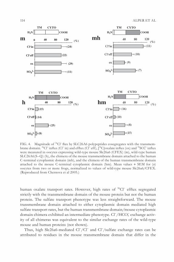
114 ALPER ET AL
human oxalate transport rates. However, high rates of 36Cl− effl ux segregated strictly with the transmembrane domain of the mouse protein but not the human protein. The sulfate transport phenotype was less straightforward. The mouse transmembrane domain attached to either cytoplasmic domain mediated high sulfate transport rates, but the human transmembrane domain/mouse cytoplasmic domain chimera exhibited an intermediate phenotype. Cl−/HCO3
− exchange activ-ity of all chimeras was equivalent to the similar exchange rates of the wild-type mouse and human proteins (not shown).
Thus, high Slc26a6-mediated Cl−/Cl− and Cl−/sulfate exchange rates can be attributed to residues in the mouse transmembrane domain that differ in the
Cl-in
Cl-eff
ox
12040 80
(15)
(14)
(25)
(8)SO42-
0 40 80 120
Cl-in
Cl-eff
ox
(24)
(22)
(29)
(11)SO42-
mH2N COOH
TM CYTO
h
(%)
(%)
(%)
SO42-
mh
hm
SO42-
H2N COOH
TM CYTOH2N COOH
TM CYTO
H2N COOH
TM CYTO
FIG. 4. Magnitude of 36Cl− fl ux by SLC26A6 polypeptides cosegregates with the transmem-brane domain. 36Cl− infl ux (Cl− in) and effl ux (Cl− eff ), [14C]oxalate infl ux (ox) and 35SO4
2− infl ux were measured in oocytes expressing wild-type mouse Slc26a6 (CFEX) (m), wild-type human SLC26A6(S–Q) (h), the chimera of the mouse transmembrane domain attached to the human C-terminal cytoplasmic domain (mh), and the chimera of the human transmembrane domain attached to the mouse C-terminal cytoplasmic domain (hm). Mean values + SEM for (n) oocytes from two or more frogs, normalized to values of wild-type mouse Slc26a6/CFEX. (Reproduced from Chernova et al 2005.)

FUNCTIONAL DIFFERENCES BETWEEN HUMAN AND MOUSE SLC26A6 115
human transmembrane domain. The transmembrane domain sequences of both orthologous polypeptides are permissive for robust bidirectional transport of Cl− in exchange for HCO3
− or for oxalate.
Electroneutral monovalent anion exchange and species-specifi c electrogenic oxalate/Cl− exchange by Slc26a6
Two groups have observed that Slc26a6-expressing oocytes reversibly hyperpolar-ize in response to bath Cl− removal (Ko et al 2002, Xie et al 2002), whereas Slc26a3-expressing oocytes reversibly depolarize (Ko et al 2002). Ko et al (2002) also reported that oocytes expressing either Slc26a3 or Slc26a6 exhibit two elec-trode voltage clamp currents attributable to Cl−/OH− and to Cl−/HCO3
− exchange. These data were interpreted as refl ecting electrogenic anion exchange. The impact of inclusion of apical electrogenic anion exchange of appropriate stoichiometry into models of stimulated pancreatic bicarbonate secretion has remained uncertain (Steward et al 2005).
In contrast, we have observed much smaller currents in voltage-clamped oocytes expressing either human SLC26A6 or mouse Slc26a6 in Cl− media equilibrated with either room air or CO2/HCO3
− (Fig. 5). Oocytes from the same lots exhibited robust [14C]oxalate fl ux and Cl−/HCO3
− exchange (not shown). Several observa-tions suggest that these currents do not refl ect electrogenic anion exchange by either Slc26a6 orthologue. First, the currents represented charge movement that could account for <5% of measured 36Cl− infl ux or 36Cl− effl ux. Second, the reversal potential of the measured current was insensitive to replacement of bath Cl− with gluconate, whether in the absence or presence of CO2/HCO3
−. Third, the small currents did not exhibit the pharmacological profi le of the 36Cl− and oxalate fl uxes (DIDS, NS3623, tenidap). Fourth, high K+ depolarization did not alter rates of Cl−/HCO3
− exchange or of effl ux of 36Cl− effl ux mediated by mouse Slc26a6 ortho-logue (Chernova et al 2005). These results suggested electroneutrality of Cl−/OH− and Cl−/HCO3
− exchanges mediated by mouse Slc26a6 and human SLC26A6. Similar results supporting electroneutral Cl−/OH− and Cl−/HCO3
− exchange were recorded in voltage-clamped oocytes expressing mouse Slc26a3, human SLC26A3 and rat Slc26a4/pendrin (not shown) and in BCECF-loaded HEK-293 cells express-ing mouse Slc26a3 (Melvin et al 1999) or human SLC26A3 (Lamprecht 2005).
Our two electrode voltage clamp current measurements are validated by two additional observations. First, addition of 1 mM oxalate to Slc26a6-expressing oocytes in gluconate bath greatly increased outward current and shifted the reversal potential by −24 mV (Fig. 5) and in a second series of oocytes by −35 mV (Chernova et al 2005). This current refl ects inward negative charge movement predicted by exchange of extracellular divalent oxalate with intracellular monovalent Cl− in agreement with Jiang et al (2002). Second, this oxalate-induced current was DIDS-sensitive (Chernova et al 2005, Jiang et al 2002).
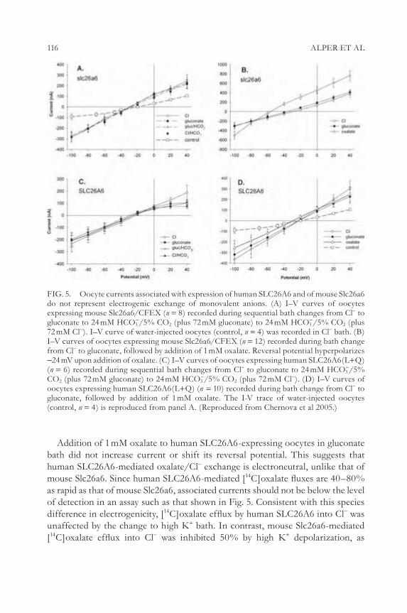
116 ALPER ET AL
Addition of 1 mM oxalate to human SLC26A6-expressing oocytes in gluconate bath did not increase current or shift its reversal potential. This suggests that human SLC26A6-mediated oxalate/Cl− exchange is electroneutral, unlike that of mouse Slc26a6. Since human SLC26A6-mediated [14C]oxalate fl uxes are 40–80% as rapid as that of mouse Slc26a6, associated currents should not be below the level of detection in an assay such as that shown in Fig. 5. Consistent with this species difference in electrogenicity, [14C]oxalate effl ux by human SLC26A6 into Cl− was unaffected by the change to high K+ bath. In contrast, mouse Slc26a6-mediated [14C]oxalate effl ux into Cl− was inhibited 50% by high K+ depolarization, as
FIG. 5. Oocyte currents associated with expression of human SLC26A6 and of mouse Slc26a6 do not represent electrogenic exchange of monovalent anions. (A) I–V curves of oocytes expressing mouse Slc26a6/CFEX (n = 8) recorded during sequential bath changes from Cl− to gluconate to 24 mM HCO3
−/5% CO2 (plus 72 mM gluconate) to 24 mM HCO3−/5% CO2 (plus
72 mM Cl−). I–V curve of water-injected oocytes (control, n = 4) was recorded in Cl− bath. (B) I–V curves of oocytes expressing mouse Slc26a6/CFEX (n = 12) recorded during bath change from Cl− to gluconate, followed by addition of 1 mM oxalate. Reversal potential hyperpolarizes −24 mV upon addition of oxalate. (C) I–V curves of oocytes expressing human SLC26A6(L+Q) (n = 6) recorded during sequential bath changes from Cl− to gluconate to 24 mM HCO3
−/5% CO2 (plus 72 mM gluconate) to 24 mM HCO3
−/5% CO2 (plus 72 mM Cl−). (D) I–V curves of oocytes expressing human SLC26A6(L+Q) (n = 10) recorded during bath change from Cl− to gluconate, followed by addition of 1 mM oxalate. The I-V trace of water-injected oocytes (control, n = 4) is reproduced from panel A. (Reproduced from Chernova et al 2005.)

FUNCTIONAL DIFFERENCES BETWEEN HUMAN AND MOUSE SLC26A6 117
expected for electrogenic transport of one negative charge per transport cycle (not shown). These fi ndings suggest that the oocyte depolarization produced by high K+ bath change is of suffi cient degree to alter the rates of electrogenic anion exchange exposed to steady-state anion gradients. The insensitivity of rates of 36Cl−/Cl− exchange by Slc26a6 or by Slc26a3 thus supports an electroneutral mech-anism of monovalent anion exchange.
Initial characterization of SLC26-related polypeptides in lower organisms
The Caenorhabditis elegans genome includes four SLC4-related abts genes encoding at least fi ve transcripts and eight SLC26-related sulp genes encoding at least 14 transcripts. Sherman et al (2005) have localized GFP promoter fusion products from each of these genes, and have localized translational GFP polypeptide fusions of these anion transporter genes for half of them. The SULP4 polypeptide expressed in Xenopus oocytes increased uptake of 36Cl− by 2.5-fold, and uptake of [35S]sulfate by eightfold (Sherman et al 2005).
The SLC26-related Sulp sulfate permease gene family is found widely among sequenced unicellular eukaryotic, bacterial and archaebacterial genomes. In marine cyanobacteria, a Sulp gene with a carbonic anhydrase domain in place of a STAS domain mediates Na-dependent HCO3
− uptake with a postulated role in carbon fi xation (Price et al 2004). Similar SulP–carbonic anhydrase open reading frame fusions are present in several other bacterial genomes (Felce & Saier 2005).
We have cloned and inducibly overexpressed in Escherichia coli a 6-His-tagged version of the Sulp gene product Rv1739c from Mycobacterium tuberculosis. Rv1739c overexpression increased uptake of [35S]sulfate, but not 36Cl− or [14C]oxalate. Induced [35S]sulfate uptake is activated by acid pH, exhibits a Km (app) of 4.1 mM at pH 6, and is inhibited by N-ethylmaleimide and CCCP. Uptake is also inhibited by cold sulfate, thiosulfate, selenate, poorly inhibited by molybdate, and is not affected by Cl− or HCO3
−. Rv1739c associated sulfate uptake is maintained in a protein in which all three Cys residues are changed to Ser. The Rv1739c polypep-tide has solubilized and affi nity purifi ed in dodecylmaltoside, with a yield of 1 mg/l of induced culture (not shown).
Conclusion
The SLC26 family gene products include the apical membrane anion exchanger polypeptides which appear to mediate much of the long sought CFTR-stimulated luminal Cl−/HCO3
− activity of epithelia. This is confi rmed in the case of the human congenital disease chloride-losing diarrhoea, secondary to loss-of-function muta-tions in SLC26A3/DRA. In the absence of a human SLC26A6 defi ciency disease, the role of apical SLC26A6 in epithelial bicarbonate secretion remains less clear.

118 ALPER ET AL
Further study of the physiology of the recently described Slc26a6−/− mouse will be central to this understanding. However, as summarized in this report, the amino acid sequence divergence between mouse Slc26a6 and human SLC26A6 polypep-tides refl ects signifi cant differences in anion selectivity, acute regulation, and pos-sibly also mechanism of divalent/monovalent anion exchange. Human SLC26A6, in particular, exhibits an unusual dichotomy between very low rates of Cl−/Cl− exchange and robust Cl−/HCO3
− exchange activity. Although both human and mouse SLC26 polypeptides mediate Cl−/HCO3
− exchange sensitive to activation of coexpressed CFTR, the consequences to our extrapolation of fi ndings in the genetically modifi ed mouse to pathophysiology in the human remain to be seen. The reported electrogenicity of Cl−/HCO3
− exchange by Slc26a3 and Slc26a6 remains controversial. Indeed, the importance of electrogenic vs. electroneutral apical Cl−/HCO3
− exchange (or any Cl−/HCO3− exchange) during maximal HCO3
− secretion by the human pancreatic duct is still being debated (Sohma et al 2000, Ko et al 2002, Whitcomb & Ermentrout 2004, Steward et al 2005). Whatever the electrogenicity of Cl−/HCO3
− exchange, the species specifi city of SLC26-mediated anion transport properties may help explain the difference between the <70 mM peak pancreatic [HCO3
−] secretion of rat (and likely mouse), and up to 145 mM or more in human, dog, cat and guinea pig.
Acknowledgement
This work was supported by NIH grant DK43495.
References
Alper SL 2002 Genetic diseases of acid-base transporters. Annu Rev Physiol 64:899–823Chernova MN, Jiang L, Shmukler BE et al 2003 Acute regulation of the SLC26A3 congenital
chloride diarrhea anion exchanger (DRA) expressed in Xenopus oocytes. J Physiol 549:3–19
Chernova MN, Jiang L, Friedman DJ et al 2005 Functional comparison of mouse slc26a6 anion exchanger with human SLC26A6 polypeptide variants. J Biol Chem 280:8564–8580
Choi JY, Muallem D, Kiselyov K, Lee MG, Thomas PJ, Muallem S 2001 Aberrant CFTR–dependent HCO3− transport in mutations associated with cystic fi brosis. Nature 410:94–97
Dawson PA, Markovich D 2005 Pathogenetics of the human SLC26 transporters. Curr Med Chem 12:385–396
Felce J, Saier MH 2005 Carbonic anhydrases fused to anion transporters of the SulP family: evidence for a novel type of bicarbonate transporter. J Mol Microbiol Biotechnol 8:169–176
Jiang Z, Grichtchenko II, Boron WF, Aronson PS 2002 Specifi city of anion exchange mediated by mouse Slc26a6. J Biol Chem 277:33963–33967
Knauf F, Yang CL, Thomson RB, Mentone SA, Giebisch G, Aronson PS 2001 Identifi cation of a chloride-formate exchanger expressed on the brush border membrane of renal proximal tubule cells. Proc Natl Acad Sci USA 98:9425–9430

FUNCTIONAL DIFFERENCES BETWEEN HUMAN AND MOUSE SLC26A6 119
Ko SB, Shcheynikov N, Choi JY et al 2002 A molecular mechanism for aberrant CFTR-dependent HCO3− transport in cystic fi brosis. EMBO J 21:5662–5672
Ko SB, Zeng W, Dorwart MR et al 2004 Gating of CFTR by the STAS domain of SLC26 transporters. Nat Cell Biol 6:343–350
Lamprecht G, Baisch S, Schoenleber E, Gregor M 2005 Transport properties of the human intestinal anion exchanger DRA (down-regulated in adenoma) in transfected HEK293 cells. Pfl ugers Arch 449:479–490
Lohi H, Lamprecht G, Markovich D et al 2003 Isoforms of SLC26A6 mediate anion transport and have functional PDZ interaction domains. Am J Physiol Cell Physiol 284:C769–779
Melvin JE, Park K, Richardson L, Schultheis PJ, Shull GE 1999 Mouse down-regulated in adenoma (DRA) is an intestinal Cl−/HCO3− exchanger and is upregulated in colon of mice lacking the NHE3 Na+/H+ exchanger. J Biol Chem 274:22855–22861
Mount DB, Romero MF 2004 The SLC26 gene family of multifunctional anion exchangers. Pfl ugers Arch 447:710–721
Price GD, Woodger FJ, Badger MR, Howitt SM, Tucker L 2004 Identifi cation of a SulP-type bicarbonate transporter in marine cyanobacteria. Proc Natl Acad Sci USA 101:18228–18233
Romero MF, Fulton CM, Boron WF 2004 The SLC4 family of HCO3− transporters. Pfl ugers Arch 447:495–509
Sherman T, Chernova MN, Clark JS, Jiang L, Alper SL, Nehrke KE 2005 The abts and sulp families of anion transporters from Caenorhabditis elegans. Am J Physiol Cell Physiol 289:C341–351
Sohma Y, Gray MA, Imai Y et al 2000 HCO3 transport in a mathematical model of the pan-creatic ductal equilibrium. J Membr Biol 176:77–100
Steward MC, Ishiguro H, Case RM 2005 Mechanisms of bicarbonate secretion in the pancreatic duct. Annu Rev Physiol 67:377–409
Waldegger S, Moschen I, Ramirez A et al 2001 Cloning and characterization of SLC26A6, a novel member of the solute carrier 26 gene family. Genomics 72:102–112
Wang Z, Petrovic S, Mann E, Soleimani M 2002 Identifi cation of an apical Cl/HCO3 exchanger in the small intestine. Am J Physiol Gastroenterol Physiol 282:G573–579
Wang Z, Wang T, Petrovic S et al 2005 Renal and intestinal transport defects in slc26a6-null mice. Am J Physiol Cell Physiol 288:C957–965
Whitcomb DC, Ermentrout GB 2004 A mathematical model of the pancreatic duct cell gen-erating high bicarbonate concentrations in pancreatic juice. Pancreas 29:e30–40
Xie Q, Welch R, Mercado A, Romero MF, Mount DB 2002 Molecular characterization of the murine Slc26a6 anion exchanger: functional comparison with Slc26a1. Am J Physiol Renal Physiol 283:F826–838
DISCUSSION
Welsh: Has anyone purifi ed any member of this family and studied it in a recon-stitution system, such as a vesicle or lipid membrane? This might be one approach to resolving some of the differences.
Alper: We have purifi ed one of the Mycobacterium tuberculosis proteins and we are now trying to reconstitute it.
Muallem: It is mind boggling to me that you can see such an incredible difference in 36Cl− fl uxes between the human and mouse isoforms, and yet still see the same Cl−/HCO3
− exchange activity in the various clones that you are using. We have

120 ALPER ET AL
turned the human exchangers upside down and we can see absolutely no activity. This is also showing very well with the oxalate transporters: we see no change in reversal potential. We see no oxalate transport by the human transporter and we have no idea why or how you can see all this Cl−/HCO3
− exchange activity and it does not bother you.
Alper: What you are saying is that you see no oxalate transport electrophysiologically.
Muallem: Yes, but we do everything electrophysiologically. We measure the pH and we take into account the enormous buffer capacity of the entire oocyte volume. We don’t measure just the pH under the plasma membrane of the oocytes.
Alper: Nor do we.Muallem: But the currents you are measuring can only be described as puny
compared with ours.Alper: Yes, I agree, they are small. But we get CFTR currents that are the same
magnitude that everyone else gets.Muallem: I would suggest that something is wrong when you get huge Cl− fl uxes
with one isoform and almost none with the other. Yet you get the same Cl−/HCO3−
exchange activity with both isoforms. This should have raised a huge red fl ag.Alper: It did, and is why we have waited a long time to publish.Seidler: When you express CFTR in an oocyte and it enhances the rate of
chloride–bicarbonate exchange for all the transporters, what is the CFTR doing? Is it a chloride shunt pathway through the membrane?
Alper: I glossed over the fact that in our hands, expression of CFTR alone actu-ally increases some nominal chloride–bicarbonate exchange activity in oocytes. These are oocytes with injection of only CFTR cRNA. This enhanced activity is inhibited by the CFTR inhibitor. When it is co-expressed with A7 the elevated activity is inhibited by the CFTR inhibitor only to the extent that it would inhibit the activity contributed by CFTR expression in the absence of A7. This is an indi-cation that this enhancement of activity may not just be direct stimulation. It is conceivable that it only represents addition of two activities which are not phar-macologically identical, and may not even be the same mechanistically. So when we do chloride removal and restoration assays, what are the possible interpretations of the result? It may not be only coupled chloride–bicarbonate exchange in the context in which we have been thinking of it in the past.
Mount: If you look at KCl co-transport, there is endogenous oocyte activity and oocytes express a Xenopus KCC (Mercado et al 2001). In terms of anion fl uxes, if you telescope down we do see changes in sulfate transport in response to cis-inhibition, for example, that are consistent with endogenous SLC26 transporters in the oocyte. We have the Xenopus A6 orthologue that is highly functional. As a brief comment: we tend to think of the oocyte as a bland cell, but these cells do have their own low repertoire of other transporters.

FUNCTIONAL DIFFERENCES BETWEEN HUMAN AND MOUSE SLC26A6 121
Alper: One way to look at this is that CFTR is activating an endogenous SLC26, although the sensitivity to the CFTR inhibitor 172 raises a potential question mark about this.
Aronson: With respect to the discordance between the 36Cl− fl ux and chloride–bicarbonate exchange, when you look at 36Cl− uptake you don’t really know what it is exchanging for. At least when outside chloride is removed, you are looking at apparent chloride–bicarbonate exchange, and chloride is effl uxing. In this case you would think that there should be some correspondence between the chloride fl ux and the acid base equivalence. You didn’t show that calculation.
Alper: For mouse A6, the rate constant for 36Cl− effl ux into bicarbonate/gluco-nate is 10% of that into ND96. JH+ measured from dpHi/dt and buffer capacity comes within 1.5-fold of the effl ux rate constant of 36Cl− into bicarbonate. This effl ux rate constant is only 1/10 of what is seen effl uxing into chloride. This is a modestly satisfying calculation, but this is only for mouse. In human A6, isotopic chloride effl ux is too low to account for measured nominal chloride/bicarbonate exchange.
Aronson: So there is far too little chloride effl ux to account for the bicarbonate effect. What is the explanation? Is it possible that outside chloride is stimulating bicarbonate exit, but there is a chloride-dependent bicarbonate transport that is not coupled to the movement of chloride?
Alper: I doubt that is true when we are looking at chloride restoration, but it could be true. When you are looking at the nominal (MQAE) chloride measure-ment itself, it could represent another anion. There may be some important depend-ence for activity on bicarbonate, and chloride–chloride exchange is one function but chloride–bicarbonate exchange is another. The latter is conserved among all of these homologues.
Aronson: How can you have chloride–bicarbonate exchange bringing bicarbo-nate in without the chloride leaving?
Alper: It is in the presence of bicarbonate. It is in the nature of the isotopic effl ux assay.
Aronson: A different internal anion might be effl uxing.Alper: Yes, or it might be that bicarbonate is there, and for reasons we don’t
understand we can’t get that 36Cl− assay to work to validate this hypothesis. But if so, only for human A6. The various measured modalities of transport are consist-ent for mouse a6.
Quinton: All these studies were taking place in the oocyte. What kind of carbonic anhydrase is present?
Alper: We can immuno-detect carbonic anhydrase 2 (CA2). We know that acetazolamide treatment of the oocyte produces the same 50% inhibition of recombinant AE1-mediated chloride–bicarbonate exchange as it produces in 293

122 ALPER ET AL
cells expressing human AE1. We know that Xenopus laevis expresses CA4 mRNA, but we don’t know if CA4 activity is present in the oocyte.
Quinton: Since the carbonic anhydrases appear to have such a signifi cant effect on the anion exchangers, I can’t help but wonder whether there must be something unique about each of the 14 or more carbonic anhydrase isoforms that exist relative to different HCO3
− transporters.Romero: When I was in Walter Boron’s lab, we did a lot of work with carbonic
anhydrases when we were studying aquaporins. The endogenous oocyte has a very little bit of acetozolamide and ethoxolamide inhibitable pH decay or recovery when it is treated. The oocyte by itself doesn’t seem to have a lot of carbonic anhydrase activity as detected by the more soluble of the carbonic anhydrase inhibitors at pretty industrial concentrations.
Case: I’d like to refer back to fl uxes and stoichiometry. We are interested in the pancreatic duct which secretes large quantities of bicarbonate. We were excited by the fact that the luminal anion exchanger might have a 2 : 1 stoichiometry, because we are always faced with the problem of how we get this high bicarbonate con-centration. We postulated that if it was 2 : 1 this helps us get a bit further towards the 140 mM bicarbonate seen in pancreatic secretion; but what would really help is if there were two bicarbonates going out and one bicarbonate going in. Then it would effectively be a sort of bicarbonate channel (Steward et al 2005). In your oocyte expression systems you said you incubated in nitrate overnight, to get a low intracellular chloride concentration. So when you add bicarbonate, what happens to intracellular pH?
Alper: We haven’t yet measured that. It’s a good question.Markovich: I am not surprised by the different transport rates among different
species. We see the same.Alper: It is not the difference between species that is surprising, but rather the
selective modality differences.Markovich: It is, because your chimera data showed that there is the ability to
change the transport capability. The big change is due to the sequence of the transporter, and also the functional signifi cance of that protein in that species. You see different tissues express A6 between human and mouse.
Alper: I am not sure this is ultimately going to be functionally important. If it is chloride–bicarbonate exchange, the human and the mouse are quite similar. All of those chimeras have now been tested for chloride–bicarbonate exchange in BCEF-loaded oocytes, and they are all pretty comparable. All that is changed is unidirectional isotopic fl ux rate constants.
Welsh: When you measure pH with the fl uorophores are you measuring pH just beneath the membrane?
Alper: We are measuring a crescent-like area of interest that extends in 10–20% of the cell diameter, and we are doing this in two areas, taking the average of these.
FUNCTIONAL DIFFERENCES BETWEEN HUMAN AND MOUSE SLC26A6 123
When we have watched regions of interest in towards the centre of the cell we haven’t seen great changes. We don’t have defi nitive kinetics on the spatial gradient of pH versus time after solution changes. When we have done preliminary micro-electrode experiments we have been able to show that distance in affects the pH measured.
Romero: Joachim Deitmer’s lab (Kaiserslautern, Germany) has shown that with multiple pH electrodes (Bröer et al 1998). The further in the cell you are, the slower the pH changes for monocarboxylate transporters (MCT1).
Alper: The region of interest we select is a strip under the membrane.Welsh: Are you doing this with conventional epifl uorescence?Alper: Yes, we have not yet used laser confocal microscopy for submembrane
pHi measurements, but we hope to validate this in the future.Seidler: For the mouse and human SLC26 analogue you showed different Km
values for chloride, but similar ones for bicarbonate. Couldn’t this explain the dif-ferences in 36Cl− effl ux rates versus your chloride–bicarbonate exchange where you have relatively high concentrations of both ions? If you do a 36Cl− fl ux study you probably use low concentrations.
Alper: No, our infl ux studies are done with 104 mM extracellular chloride. Among the things we varied to make sure that we weren’t crazy was the concentra-tions: we replicated our assays with every published concentration condition.
Muallem: I say it again: if one transporter is not active, that’s what you get.Alper: It is active, because it is active as a bicarbonate mover. With A6 it is active
as a bicarbonate mover: do you see this?Muallem: With the human clone we see no Cl−/HCO3
− exchange, no Cl− trans-port, no oxalate transport and no change in current.
Alper: Oxalate fl uxes by mouse a6 are electrogenic, but with human A6 appear to be electroneutral.
Soleimani: I wanted to raise two questions. First, when you express a transporter in oocytes, if you don’t observe any function it does not mean that that protein is inactive. Some transporters show better expression or display activity in mammalian expression systems such as cultured cells. We observe this with regard to A9 that failed to give a robust chloride–bicarbonate exchange in oocytes, but showed a pronounced function in cultured HEK cells. The same is true with Pendrin, at least in our hands. Contrary to these examples, both A6 and A7 function very well in oocytes. There is something about the structure of the oocyte membrane and the accessory proteins related to this that may be at work here.
Alper: The temperature dependence of the transport allows us to accelerate or slow the rate, but it doesn’t change the ratio of mouse to human.
Soleimani: Yes, and the osmolarity of the oocyte is around 200 mM, whereas cultured cells have an osmolarity of ∼300 mM. There are a good number of

124 ALPER ET AL
differences, and one has to use an array of expression systems to fi nd the answers to these questions.
Welsh: The oxalate seems to be a good test to try to fi gure out what is different here.
Muallem: In the mouse with oxalate we see a transient but clear alkalinization, which disappears as oxalate accumulates in the oocytes. We see a hyperpolarizing signal, from about −30 mV to −70 mV.
Welsh: What is oxalate doing?Muallem: That’s a good question: we aren’t sure how it is working. To hyperpo-
larize the membrane there needs to be more than just Cl−/oxalate exchange. It does do this, but the fi rst hyperpolarization seems the result of a channel-like activity because the Cl− almost doesn’t change in the cell.
Welsh: What is different about what you are doing?Alper: There seems to be nothing different. The only difference is that one of
us has performed one assay that the other hasn’t, which is [14C]oxalate fl ux. By this assay, human SLC26A6 is 40–80% as robust as the mouse. Shmuel wouldn’t see this electrophysiologically for apparently electroneutral human A6, but he would for electrogenic mouse a6.
Muallem: This doesn’t make sense to me.Mount: Look at analogous experiments with analogous orthologues. One example
is comparing our paper on mouse A6 with Peter Aronson’s, where in chloride–bicarbonate exchange, you saw a modest hyperpolarization but nowhere near the amount that we reported. This was with very different amounts of extracellular CO2 and bicarbonate. When you do mouse A6 and look at bicarbonate infl ux in the presence of extracellular CO2 and bicarbonate, do you see the hyperpolariza-tion that Michael sees?
Alper: We have not done pH and voltage recording simultaneously.Mount: What are comparable experiments?Alper: We have only done voltage clamp. We have not done the voltage-sensitive
microelectrode versus time with solution changes.Muallem: In one of your fi gures you showed zero Cl− and no change in membrane
potential.Alper: That’s right. This is under voltage clamp.Aronson: To get back to the issue of pH and bicarbonate, I’d point out that SAT1,
which most people agree is mainly a divalent anion exchanger, exchanges sulfate for oxalate and allegedly sulfate for two bicarbonate. It is possible that it is not really changing sulfate for two bicarbonates, but for carbonate. Suppose that car-bonate can actually exchange, and assume that A6 can exchange monovalents for each other and certain divalents for monovalents. Then under more alkaline pH conditions favouring the formation of carbonate, you can get carbonate exchange for chloride which would be electrogenic, whereas under less alkaline pH condi-

FUNCTIONAL DIFFERENCES BETWEEN HUMAN AND MOUSE SLC26A6 125
tions where the carbonate is very low, chloride would exchange for bicarbonate which would be electroneutral. So whether apparent chloride-bicarbonate exchange mediated by A6 is electrogenic would depend on the pH.
Mount: You are getting ahead of my question, which is a completely naïve techni-cal question: why, with the same clone and vectors in oocytes, are other people not seeing the same hyperpolarizations with mouse A6?
Soleimani: You can go a step further. We see A7 as a chloride–bicarbonate exchanger in the same oocytes, and Dr Muallem doesn’t see this. It is the same set-up.
Mount: That’s the heart of my question: it is not the same set-up. If we just look at the CO2 and bicarbonate concentrations, they are not the same.
Alper: Peter’s results are coincident with ours, despite the fact that he used 1.5% CO2 and 10 mM bicarbonate, and we used 5% CO2 and 25 mM bicarbonate. Michael uses 33 mM bicarbonate and 5% CO2.
Mount: Part of the problem is that no one is using exactly the same conditions.Alper: Why do you choose to use a non-physiological solution, which might
evoke a more robust response?Romero: Because that’s what gives a pH that is 7.4–7.5 at room temperature with
an oocyte. That is, for pH 7.5 and 5% CO2, the [HCO3−] is 33 mM.
References
Broer S, Schneider HP, Broer A, Rahman B, Hamprecht B, Deitmer JW 1998 Characterization of the monocarboxylate transporter 1 expressed in Xenopus laevis oocytes by changes in cytosolic pH. Biochem J 333:167–174
Mercado A, de Los Heros P, Vazquez N, Meade P, Mount DB, Gamba G 2001 Functional and molecular characterization of the K–Cl cotransporter of Xenopus laevis oocytes. Am J Physiol Cell Physiol 281:C670–680
Steward MC, Ishiguro H, Case RM 2005 Mechanisms of bicarbonate secretion in the pancreatic duct. Annu Rev Physiol 67:377–409

Physiology of electrogenic SLC26 paraloguesMichael F. Romero, Min-Hwang Chang, Consuelo Plata, Kambiz Zandi-Nejad*, Adriana Mercado*, Vadjista Broumand*, Caroline R. Sussman and David B Mount*
Physiolog y & Biophysics, Case Western Reserve University, School of Medicine, Cleveland, OH 44106-4970 and *Renal Division, Brigham and Women Hospital and Division of General Internal Medicine, VA Boston Healthcare System, Harvard Medical School, Boston, MA 02115, USA
Abstract. SLC26 anion exchangers transport monovalent and divalent anions, with a diversity of anion specifi city and stoichiometry. Our microelectrode studies indicate that several SLC26 members are electrogenic. We reported that Slc26a6 functions as a Cl−/formate, Cl−/oxalate, Cl−/OH− and electrogenic Cl−/nHCO3
− exchanger. Recently, we have also confi rmed that Slc26a7 does not behave as a Cl−/HCO3
− exchanger but does function as an electrogenic anion conductance, perhaps a channel. We have also cloned murine Slc26a9, which is strongly expressed in the respiratory tract and stomach. Radioisotope uptakes in Xenopus oocytes indicate that Slc26a9 is a highly selective anion exchanger, transporting Cl− but neither formate, oxalate, nor SO4
2−. We also uti-lized electrophysiology to voltage clamp (VC) and/or measure intracellular pH (pHi), Cl− ([Cl−]i) and Na+ ([Na+]i), in response to various ion replacements. Cl− removal in HCO3
− depolarizes oocytes (to > +60 mV), alkalinizes oocytes, and decreases aCli
−. Slc26a9 thus functions as an electrogenic nCl−/HCO3− exchanger, suggesting a role
in pulmonary and gastric HCO3− secretion and/or CO2 transport. VC experiments
revealed channel-like currents (>10 mA at −60 mV and >80 mA at +60 mV) mediated by Slc26a9 in the presence and absence of HCO3
−. Our experiments and those of others continue to reveal additional characteristics and unique roles for this new class of electrogenic anion transporters.
2006 Epithelial anion transport in health and disease: the role of the SLC26 transporters family. Wiley, Chichester (Novartis Foundation Symposium 273) p 126–147
SLC26 transporters: multifunctional anion transporters
The rapidly growing SLC26 gene family encodes moderately homologous anion exchangers with a diversity of physiological roles. The fi rst family member, SLC26A1 (Sat1), encodes an SO4
2−/oxalate exchanger identifi ed by expression cloning (Bissig et al 1994). Other family members were subsequently identifi ed through positional cloning of the disease genes for diastrophic dysplasia (SLC26A2 or DTDST) (Hastbacka et al 1992), congenital chloride diarrhoea (SLC26A3/DRA) (Hoglund et al 1996), and Pendred syndrome (SLC26A4/pendrin) (Everett et al 1997). Subtractive cloning of the ‘molecular motor’ of cochlear outer hair
126

ELECTROGENIC SLC26 PARALOGUES 127
cells subsequently yielded SLC26A5 (prestin) (Zheng et al 2000), and SLC26A6 was identifi ed as a candidate gene for both the apical bicarbonate transporter of the exocrine pancreas (Lohi et al 2000) and the Cl−-formate exchanger of the proximal tubule (Knauf et al 2001). Further work has completed the identifi cation of the entire gene family, which now numbers 10; an eleventh family member (SLC26A10) appears to be a transcribed pseudogene.
The anions transported by the SLC26 gene family include SO42−, Cl−, I−, formate,
oxalate, OH− and HCO3− (Scott & Karniski 2000, Bissig et al 1994, Soleimani et al
2001, Satoh et al 1998, Karniski et al 1998, Melvin et al 1999). The evolving func-tional characterizations of this gene family have revealed distinctive patterns of anion specifi city and cis-inhibition. SLC26A3 and SLC26A6 are thus capable of transporting most if not all of the substrates above (Melvin et al 1999, Xie et al 2002). In contrast, SLC26A1 does not transport Cl− or formate, but does transport SO4
2− and oxalate (Bissig et al 1994, Satoh et al 1998, Karniski et al 1998, Xie et al 2002). SLC26A4 in turn transports monovalent anions such as Cl−, I− and formate, but not divalent anions such as SO4
2− and oxalate (Scott et al 1999, Scott & Karniski 2000). Several paralogues function in Cl−/OH− and Cl−/HCO3
− exchange modes, with increasingly important roles in transepithelial Na+/Cl− and HCO3
− cotrans-port. Where direct comparisons have been made, the SLC26 proteins appear to be more potent exchangers of Cl− with HCO3
− than with OH− (Xie et al 2002, Ko et al 2002, Wang et al 2002). The transport characteristics and anion specifi cities of the individual SLC26 paralogues and how these paralogues might contribute to physiology has recently been reviewed (Mount & Romero 2004).
The physiological role(s) of individual SLC26 anion exchangers is presumably a function of both substrate specifi city and expression pattern, and the diverse physiology of this gene family is illustrated by the phenotypes associated with loss-of-function. The SLC26A3 protein is expressed at the brush border membrane of differentiated colonic epithelial cells (Haila et al 2000), and Cl−-base exchange via SLC26A3 is thought to function in transepithelial salt transport by the colon. Accordingly, recessive loss-of-function mutations in the SLC26A3 gene result in severe congenital diarrhoea (Hoglund et al 1996). In contrast, mutations in SLC26A2 are associated with disordered skeletal development in diastrophic dys-plasia and related disorders. In these patients, loss-of-function mutations in SLC26A2 impair SO4
2− uptake by chondrocytes, and the resulting decrease in the sulfation of extracellular matrix decreases the response to matrix-dependent devel-opmental cues such as fi broblast growth factor (Hastbacka et al 1992, Satoh et al 1998). Loss-of-function in SLC26A4 (pendrin) is in turn characterized by deafness with or without congenital goitre. SLC26A4 appears to function as a Cl−/I− exchanger at the apical membrane of thyroid follicles (Scott et al 1999, Royaux et al 2000) and to thus participate in the organifi cation of thyroglobulin. Although defects in SLC26A4 frequently result in goitre in patients with the complete

128 ROMERO ET AL
Pendred syndrome, mice defi cient in Slc26a4 do not have abnormalities in thyroid structure or function (Everett et al 2001). Slc26a4 null mice do however have deafness and variable vestibular dysfunction, with endolymphatic dilatation in the inner ear (Everett et al 2001). Within the kidney, Slc26a4 is expressed at the apical membrane of type B intercalated cells, where it functions in renal bicarbonate secretion (Royaux et al 2001). Slc26a6 is at the apical membrane of gastric glands, colocalizing with the H+/K+ pump (Petrovic et al 2002). Recently Slc26a7 has been found to act as a Cl−/HCO3
− exchanger by one group (Petrovic et al 2003) and a ‘Cl− channel’ by another (Kim et al 2005). Regardless, Slc26a7 is found in basolateral membranes of parietal cells (Petrovic et al 2003) and intercalated cells of the renal outer medullary collecting duct (Petrovic et al 2004).
Respiratory acid–base compensation SLC4 transporters
Systemic acid-base homeostasis (blood pH) is a balance between renal function (control of HCO3
− and H+ absorption and secretion) and the lungs (control of PCO2
via ventilation). NBCe1 mutations cause a severe pRTA: blood pH ≥ 7.1, blood [HCO3
−] 3–11 mM. Yet, PCO2 levels in these NBCe1 patients reveal that minimal respiratory compensation occurs (PCO2
≥ 32 mmHg rather than <25 mmHg calcu-lated by Henderson-Haselbalch). The patient with the Q29X-NBCe1 mutation has this uncompensated phenotype, implicating the kNBCe1 as a ‘controller protein’ in both renal and respiratory control of systemic acid–base homeostasis. Simplisti-cally, this compensation could be in (a) the central chemoreceptors (pre-Bötzinger complex), (b) the peripheral chemoreceptors (the glomus cells), (c) the lung epi-thelia and/or (d) nerves/muscles controlling breathing (e.g. diaphragm and phrenic nerve). We know that NBCe1 mRNA is found in lung (Romero et al 1998), yet thus far NBCe1 has only been localized to basolateral membranes of Calu-3 cells (Devor et al 1999). Likewise an electroneutral NBC (NBCn1) has been cloned from pulmonary arteries (Choi et al 2000). A report using ducks as a model indi-cates that pulmonary chemoreceptor discharge is stimulated by the stilbene inhibi-tor DIDS (Shoemaker & Hempleman 2001), which can block Na+ independent and Na+ dependent SLC4 HCO3
− transporters.The AE2 anion exchanger (Slc4a2) is found in the lung (Kemp & Boyd 1993)
and appears important for mucous cells of the gut and lung (Rossmann et al 2001). AE2 is thought to mediate the apical exit of HCO3
− from epithelia of larger airways. Bronchi treated with anion inhibitors with ACh stimulation are blocked with mucin, presumably from lack of proper fl uid clearance. Transport of anionic oxi-dants (superoxides) can cause changes in pulmonary vascular tone which is attrib-uted to AE2 (Nozik-Grayck et al 2003). Anion exchange activity is also found in alveolar type II cells (Kemp & Boyd 1993), though the molecular entity is unclear. Recently, AE1–3 (Vince & Reithmeier 2000), NBCe1 and NBCn1 have been shown to interact with carbonic anhydrases (CA)(see for review Romero

ELECTROGENIC SLC26 PARALOGUES 129
et al 2004). It is also known that mice or human null for CA-II have respiratory acidosis. Thus, interaction of HCO3
− transporters with carbonic anhydrases may also be involved in altering gas exchange at the level of the small airways.
SLC26 transporters in the lung
Slc26a6 (Xie et al 2002) and Slc26a9 mRNAs are found in lung tissue. Clearly, anion exchange as indicated above is also important, but Slc26 paralogues have not pre-viously been implicated in this process. As with the Slc4 transporters, a recent report indicates that CA-II can have a stimulatory interaction with DRA (SLC26A3) (Sterling et al 2002). We identifi ed and cloned Slc26a9 from mouse and human lung.
Results and discussion
Slc26a9 cloning and Mrna
Given the existence of nine Slc26 anion exchangers in the Drosophila genome (not shown), the likelihood was high that the mammalian gene family included more than the published six members. Indeed, the existence of mammalian expressed sequence tags (ESTs) derived from novel family members was noted some time ago (Everett & Green 1999). To clone these novel family members we used genomic database mining, gene annotation, RT-PCR and EST characterization. Human cDNAs are noted with all capitals, while other species are lower case.
Human SLC26A9 exons were initially identifi ed in the draft sequences of the BAC clones RP11-196022 and RP11-370I5. The fi rst coding exon was identifi ed in these contigs using GENSCAN, neither gene annotation programs nor homology to other SLC26 proteins were useful for the identifi cation of the last coding exon. Therefore, 3′ sequences from the SLC26A9 genomic contigs were used to search the human EST database for potential 3′-UTR ESTs. One such EST (#2915384) was sequenced in its entirety; this insert extends from bp 2502 to 4526 of the fi nal cDNA sequence. The open reading frame was then cloned from human lung using Takara LAPCR polymerase and a sense primer in exon 1 an antisense primer derived from the sequence of the 3′-EST. A 500 000 bp contig containing the entire mouse Slc26a9 gene was identifi ed using a blastn search of the Celera mouse genomic database with the human SLC26A9 cDNA. A 3049 bp cDNA encompassing the entire open reading frame was then amplifi ed by RT-PCR from mouse lung, using Takara LA polymerase and the sense primer with the antisense primer. Northern analysis revealed that robust SLC26A9 and Slc26a9 expression in lung.
Cl − transport by Slc26a9
The hallmark of Slc26 transporters is anion transport, particularly Cl−. We have measured Cl− uptake by several Slc26 paralogues (Fig. 1A). Using isotopes, we

130 ROMERO ET AL
Chlo
ride
Upta
ke (p
mol
/ooc
yte/
h)
0
500
1000
1500
2000
Water
ControlIodideBrom
ideNitrateSulfate
LactateForm
ateO
xalate
E
Sulfa
te U
ptak
e (p
mol
/ooc
yte/
h)
0
500
1000
1500
2000
a2 a7 A8 a9 a11W
BA
*
0
100
200
300
400
500
600
Oxa
late
upt
ake
(pm
ol/o
ocyt
e/h)
a6 a7 a9W
0
10
20
30
40
50
60
Form
ate
upta
ke (p
mol
/ooc
yte/
h)
a6 a7 a9W
DC * *
Sulfa
te U
ptak
e (p
mol
/ooc
yte/
h)Ch
lorid
e Up
take
(pm
ol/o
ocyt
e/h)
a2 a7 A8 a9 a11+- +- +- +- +- +-
W
0
1000
2000
3000
4000
*
Chlo
ride
Upta
ke (p
mol
/ooc
yte/
h)
W Slc26a9
0
500
1000
1500
2000H†
* ‡
Chlo
ride
Upta
ke (p
mol
/ooc
yte/
h)
W iso hypo
0
hypohyper
0
500
1000
1500GF
7.4 6 6D 7.4 6 6D 7.4 6 6D
Slc26a7
hyper
†
*
†
‡‡
a2 a7 A8 a9 a11W0
1000
2000
3000
4000
*
**
†
FIG. 1. Functional characterization of SLC26/Slc26 paralogues: chloride, sulfate, oxalate and formate uptakes. (A) 36Cl− uptake (pmol/oocyte/h) in injected Xenopus oocytes, with extracellular pH of both 7.4 (open bars) and 6.0 (black fi ll). Xenopus oocytes injected as noted with water (W), Slc26a2 (a2), Slc26a7 (a7), human SLC26A8 (A8), Slc26a9 (a9) and Slc26a11 (a11). Uptakes were performed over one hour, in the absence of extracellular Cl−. Slc26a2-, Slc26a7- and Slc26a9-injected oocytes demonstrate uptake of 36Cl− that is signifi cantly higher than that of water-injected controls. (B) [35S]sulfate uptake (pmol/oocyte/h) in Xenopus oocytes injected as noted with W, a2, a7, A8, a9 and a11. Uptakes were performed over one hour, in the absence of extracellular Cl−, with extracellular pH of both 7.4 (open bars) and 6.0 (black fi ll). (C) Oxalate uptake in oocytes injected with water, a6, a7 or a9. (D) Formate uptake in oocytes injected with water, a6, a7 or a9. (E) Lack of cis-inhibition of 36Cl− uptake in Slc26a9-injected oocytes by various anionic substrates. Oocytes were incubated in 36Cl− uptake medium for one hour, in the absence (control) or presence of 25 mM concentrations of the indicated anions. (F) Effect of extracellular Cl− on 35SO4
2− uptake. Oocytes injected as noted were incu-bated in 35SO4
2− uptake medium for one hour, in the absence (−) or presence (+) of 25 mM Cl−. Sulfate uptake by a2-injected oocytes is cis-inhibited by extracellular Cl−; unlike Slc26a1 (Xie et al 2002), latent 35SO4
2− uptake in Slc26a7/a8/a9 was not elicited by extracellular Cl−. (G) 36Cl− uptake (pmol/oocyte/h) in water (W) and mouse a7-injected oocytes, as a function of extracellular osmolality. Oocytes were incubated during the pre-uptake period and uptake period at the indicated osmolality—120 mOsm/kg (hypotonic, ‘−’), 210 mOsm/kg (isotonic, ‘0’) and 300 mOsm/kg (hypertonic, ‘+’)—at either pH 7.4 (open bars), pH 6.0 (black bars), or in the presence of 1 mM DIDS at pH 6.0 (grey bars). (H) DIDS inhibition of 36Cl− uptake (pmol/oocyte/h) in Slc26a9-injected oocytes. Oocytes were incubated during the preuptake period and uptake period at either pH 7.4 (open bars), pH 6.0 (black bars), or in the presence of 1 mM DIDS at pH 6.0 (grey bars). The uptake experiments included 12–18 oocytes in each experimental group, and results were reported as means ± SEM. The * symbol denotes a statistically signifi cant difference (p < 0.05) from water-injected control oocytes; ‘†’ denotes P < 0.05 from pH 7.4; ‘‡’ denotes p < 0.05 from pH 6.0 (Xie et al 2002, used with permission from The American Physiological Society).

ELECTROGENIC SLC26 PARALOGUES 131
showed that Slc26a9 had larger Cl− uptake at pH 6 than 7.4, but uptake was higher for Slc26a3 and Slc26a6 (not shown). With Slc26a9 there was a 40% dependence on extracellular Na+ (not shown). Cl− uptake by Slc26a9 cannot be cis-inhibited by other halides or monovalent anions (Fig. 1E), suggesting that it is highly specifi c for Cl−. Slc26a2 transports sulfate whereas this activity is reproducibly absent in Slc26a7, Slc26a8, Slc26a9 or Slc26a11 (Fig. 1B). We previously found that Slc26a1-mediated sulfate transport is activated considerably in the presence of extracellular Cl− (Xie et al 2002); however, 25 mM Cl− in the extracellular medium did not bring out a latent sulfate transport activity by Slc26a7/a8/a9, and cis-inhibited Slc26a2 (Fig. 1F). Likewise, though Slc26a6-expressing oocytes transport oxalate (Fig. 1C) and formate (Fig. 1D), these anions are not substrates for Slc26a7 or Slc26a9. Cl− uptake by Slc26a7 is moderately regulated by osmolarity and extracellular pH (Fig. 1G). Slc26a7-mediated Cl− transport (pH 6) can be inhibited by ∼50% by DIDS (Fig. 1G). While we have not been able to show that Slc26a9 is regulated by osmolarity, Cl− uptake (pH 6) is partially blocked by DIDS (Fig. 1H). In summary, Slc26a9 functions as a highly selective Cl− transporter. Unlike Slc26a6 (Xie et al 2002), Cl− uptake in Slc26a9-expressing cells is reproducibly increased by an acid-outside uptake solution.
To better understand the ion transport mediated by Slc26a9, we used micro-electrodes to monitor intracellular Cl− ([Cl−]i) and membrane potential (Vm). These experiments revealed that resting [Cl−]i for H2O-oocytes was 43 ± 6 mM, while Slc26a9 [Cl−]i was lower (27 ± 4 mM). Addition of 5% CO2/33 mM HCO3
− increases [Cl−]i about the same amount for both groups (H2O, 7.3 ± 3.1 vs. Slc26a9, 7.4 ± 1.4 mM). Removal of bath Cl− (‘0Cl−’ gluconate replacement), results in an immediate, rapid fall of [Cl−]i for Slc26a9 (∼20 mM) vs. H2O (2.0 mM). 0Cl− also elicits a robust depolarization (>100 mV for Slc26a9 vs. ± 2 mV for H2O). This was our fi rst indication that Slc26a9 was electrogenic. Removal of Na+ (0Na+, choline or N-methyl-D-glucamine, NMDG), did not cause measurable differences in [Cl−]i (−0.5 mM for Slc26a9 vs. −0.7 mM for H2O).
Slc26a9 is a novel, electrogenic Cl −/HCO3− exchanger
To determine whether Slc26a9 functions as a Cl−/HCO3− exchanger, we measured
intracellular pH (pHi) in response to the manipulation of both HCO3− and Cl− (Fig.
2C) as previously described (Romero et al 1998, Xie et al 2002). The addition of CO2/HCO3
− to the bath solution causes oocyte pHi to decrease due to CO2 plasma membrane diffusion followed by intracellular hydration and dissociation forming intracellular H+ and HCO3
−. Water-injected oocytes exposed to 5% CO2/33 mM HCO3
− (pH 7.5) acidify by ∼0.44 pH units at 460 × 10−5 pH units/s similar to oocytes expressing AE2 (Fig. 2A), Slc26a6 (Fig. 2B) (Xie et al 2002), Slc26a9 (Fig. 2C) and SLC26A7 (Fig. 2D). Unlike AE2-oocytes, HCO3
− caused a slight but

132 ROMERO ET AL
abrupt depolarization in Slc26a6, Slc26a9 and SLC26A7-injected oocytes, reminis-cent of the anion conductance observed in oocytes expressing NDAE1 (Romero et al 2000), not observed in AE2-expressing cells. Replacement of Cl− (gluconate) did not change the pHi or Vm observed in the water control. However, Figs 2A–D show that Cl− removal increased pHi in AE2, Slc26a6 and Slc26a9 oocytes but not SLC26A7 oocytes. Surprisingly, while gluconate replacement evoked no Vm change for AE2, Slc26a6 hyperpolarized (−22.7 ± 2.9 mV, n = 9), and Slc26a9 (+60–100 mV) and SLC26A7 (+35–40 mV) depolarized. Cl− removal in solutions bubbled with 100% O2, reveal a similar, yet less robust, alkalinization (+23–30 × 10−5 pH units/s) and depolarization (>100 mV). Thus, Slc26a9 appears to have an electro-genic Cl−/HCO3
− exchange, i.e. nCl−:1HCO3− (see Fig. 2C, Fig. 5).
C Slc26a9
Vm
-80
-40
0
pHi
6.9
7.1
7.3
7.5
7.7
7.9CO2 / HCO3
-
0Na+
10 min
010726md
0Cl-
B Slc26a6
-40-90
-5Vm
0
-10
CO2 / HCO3-
10 min
960222mc
0Na+
0Cl-
6.8
7.2
7.6
pHi
A AE2
7.0
7.2
7.4
pHi
-20
0
20
Vm
0Na+
CO2 / HCO3
0Cl-
5 mi
040831cd
D SLC26A7
HCO3-
Cl-
nHCO3-
Cl-
HCO3-
nCl-
Cl-
6.8
60
-40
-20
0
20
40
Vm
10 min
020115ma
7.0
7.2
7.4
7.6
7.8
pHi
0Cl-
CO2 / HCO3-
0Na+
FIG. 2. Functional expression electrogenic Slc26 paralogues. Using ion-selective micro-elec-trodes, we measured intracellular pH (pHi) and membrane potential (Vm) of oocytes injected with human AE2/SLC4A2 (A), mouse Slc26a6 (B), mouse Slc26a9 (C) or human SLC26A7 (D). Addition of 5% CO2/33 mM HCO3
− (pH 7.5) to the bath elicits a slight depolarization and results in cell acidifi cation in oocytes injected with water (controls, not shown). That is, CO2 diffuses across the membrane, forms H2CO3, and then subsequently generates H+ and HCO3
− from fast dissociation. AE2 (A) is not electrogenic yet alkalinizes during Cl− removal (Cl−/HCO3
− exchange) but not during Na+ removal. While both Slc26a6 and Slc26a9 alkalinize with Cl− removal, Slc26a6 hyperpolarizes (B) and Slc26a9 depolarizes (C). SLC26A7 depolarizes (D) with Cl− removal, yet fails to alkalinize. Panel A was modifi ed from Romero (2001) and B (Slc26a6) (Xie et al 2002, used with permission from The American Physiological Society).

ELECTROGENIC SLC26 PARALOGUES 133
We also replaced Na+ with choline to test the cation dependence of Slc26a9; Na+ removal reduced pHi (−15–30 × 10−5 pH units/s) and depolarized the oocytes (>25 mV). This activity is similar to that observed with an electrogenic Na+/HCO3
− cotransporter like NBCe1 (Dinour et al 2004, Romero et al 1998) and is distinct from known Na+ driven Cl−-HCO3
− exchangers like NDAE1 (Romero et al 2000). Prior to CO2 removal, pHi rises to 7.2 (almost the non-HCO3
− level). Removal of CO2/HCO3
− elicits a robust alkalinization and pHi overshoot to 7.9 (7.79 ± 0.11, n = 7), indicative of cellular HCO3
− loading (+0.37 ± 0.06 pH units above starting pHi) (Romero et al 2000, 1998). This overshoot was not observed in controls (not shown). The Slc26a9 oocytes also showed an abrupt hyperpolarization (∼5–7 mV) with removal of CO2/HCO3
−.
Na+ transport by Slc26a9
To determine whether Na+ was a cofactor or actually transported, we made Na+ electrodes to measure intracellular [Na+] ([Na+]i) in Xenopus oocytes. Resting [Na+]i was similar for water (3.4 ± 0.2 mM) and Slc26a9 (3.8 ± 0.6 mM) as previously reported. Removal of Cl− or Na+ did not alter [Na+]i for water controls. However, Na+ removal for Slc26a9 oocytes decreased [Na+]i (∼1 mM) while also depolarizing the cell, similar to NBCe1 (Romero et al 1998). Likewise, readdition of Na+ in the absence of Cl− raised [Na+]i which could be further increased by readdition of Cl−. Vm changes were similar to those seen in Fig. 2. Xu and co-workers found that Cl−/HCO3
− exchange activity of Slc26a9 was inhibited by the NH4
+ cation (Xu et al 2005), implying more general cation transport or regulation of Slc26a9. Our [Na+]i and Vm data indicate that Slc26a9 transports Na+ and has an activity consistent with electrogenic Na+/nCl− or Na+/nHCO3
− cotransport.
Voltage clamp studies with Slc26a9
We performed two-electrode voltage clamp (VC) experiments to directly examine the currents mediated by Slc26a9 (Ia9; Fig. 3A,C) and compare them to those of the SLC26A7 ‘channel/transporter’ (IA7; Fig. 3B,D) (Kim et al 2005). Initial clamping in ND96 (NaCl ringer) resulted in a huge negative current (−8 to −20 mA) which decayed over ∼5 min (not shown). Interestingly, though SLC26A7 has been characterized as an anion channel (Kim et al 2005), the Slc26a9 currents are much larger than SLC26A7 (Fig. 3B). Figure 3C shows the current sweeps resulting from clamping at −60mV and using a pulse protocol. For these VC experiments, we cannot inject more than 0.5 ng Slc26a9 cRNA/oocyte or we often exceed the capacity of our clamping amplifi er. Current at +60 mV is often > +60 mA and there appear to be tail-currents (not shown). Removal of Cl− (Fig. 3A,C; inverted

134 ROMERO ET AL
triangle) dramatically reduced Ia9 but currents were ± 5 mAs whereas control oocytes had currents ±300 nA (not shown). Na+ removal elicited obvious currents for Slc26a9 (Fig. 3A,C) but not SLC26A7 (Fig. 3B,D). Removal of Na+ and Cl− in a CO2/HCO3
− solution has similar though reduced currents (not shown).
Immunolocalization of Slc26a9 in stomach
After cloning SLC26A9 and Slc26a9, we generated rabbit Slc26a9 peptide antibod-ies. Antibodies were immunopurifi ed and then tested on Western blots from oocytes expressing Slc26a1, Slc26a3, Slc26a6, Slc26a8 and Slc26a9. All of the
Cl-
-3
-2
-1
0
1
2
3
Ia9µ A
050107pc
2 min
0Cl-
0Na+
0Cl-
Vh=-60mV
050107pc
-150 -100 -50 50
-20
-10
10
20
30
mV
µ A
ND960Na+
0Cl-(a)0Cl- (b)
A
C
Slc26a9
ND96
B
-3
-2
-1
0
1
2
3
050107pa
2 min
IA7µ A
0Cl-
0Na+
0Cl-
Vh=-60mV
-150 -100 -50 50
-20
-10
10
20
30
mV
µ A
050107pa
D
SLC26A7
FIG. 3. Whole oocyte currents evoked by Slc26a9 vs. SLC26A7. Two electrode voltage clamp experiment on an oocyte injected with 0.5 ng/oocyte Slc26a9 and 10 ng/oocyte of human SLC26A7. SLC26A7 has been reported by Muallem’s group to be an anion channel without Cl−/HCO3
− exchanger activity (Kim et al 2005). For these experiments, oocytes were pre-incubated in 0Cl− ND96 (triangle) for 3–5 min prior to clamping to reduce the steady-state currents. (A) The Slc26a9 currents at −60 mV with the indicated solution changes. (B) The same experiment for SLC26A7. (C) Current-voltage (IV) relationship for Slc26a9. (D) SLC26A7-IV. Oocytes were held at −60 mV and stepped from −160 to +60 mV in 20 mV steps. The currents for ND96 (squares), 0Na+ ND96 (circle), and 0Cl− ND96 (inverted triangle) are plotted.

ELECTROGENIC SLC26 PARALOGUES 135
antibodies were specifi c for their target transporter (not shown). One Slc26a9 antibody specifi cally labels on mouse and rat tissues. Figure 4 shows phase contrast (A) and epifl uorescent images of rat stomach (B,C).
Colocalization of Slc26a9 with the b -HK pump in mouse stomach showed only slight overlap between Slc26a9 and the HK pump (Fig. 4) as previously reported (Xu et al 2005). That is, cells secreting acid do not appear to contain much if any Slc26a9. Along the gastric gland axis, Slc26a9 expression is high at the base (not shown) and surface cells but is conspicuously absent from the middle part of the gland. Given that Slc26a9 can have several different functional modes, this distri-bution may refl ect Na+ transport but not Cl−/HCO3
− exchange or anion conduct-
ance at the gastric gland base, and anion conductance or Cl−/HCO3− exchange
activity by the surface cells.
Potential cellular physiolog y of electrogenic SLC26 transporters
Our understanding of the SLC26 family has increased greatly over the past several years (see (Mount & Romero 2004) for review). Our group has focused primarily on newer Slc26 members (Slc26a6–a11). We have found that several of these members (Slc26a6, SLC26A7 and Slc26a9) are electrogenic. Our recent studies have also shown that some members (Slc26a6 and Slc26a9) can exchange Cl− for HCO3
− (Fig. 5). Other members have anion conductances (SLC26A7 and Slc26a9).
We also have some indications that there may be yet other activities that these
FIG. 4. Immunolocalization of Slc26a9 in stomach. Adult mouse stomach was fi xed in PLP, cryosectioned, and double-labelled (A) with a mouse monoclonal antibody against the b -HK pump (CalBiochem, used 1:1000) visualized with secondary antibody conjugated to Cy2 (B) and a rabbit antibody against Slc26a9 (used 1:2000) visualized with secondary antibody conju-gated to Cy3 (C). Nuclei were stained with DAPI (not shown). A phase contrast image of the same fi eld of view is shown (A). Note predominant labelling of Slc26a9 at the tips of microvilli and of the HK pump deeper in stomach crypts. No labelling was observed in sections exposed to secondary antibodies conjugated to Cy3 and Cy2 alone or when the Slc26a9 primary antibody was competed with peptide immunogen (not shown).
B -HK C a950 µ MA
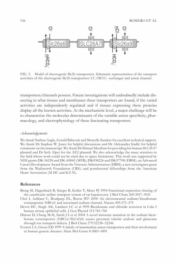
136 ROMERO ET AL
transporters/channels possess. Future investigations will undoubtedly include dis-secting in what tissues and membranes these transporters are found, if the varied activities are independently regulated and if tissues expressing these proteins display all the known activities. At the mechanistic level, a major challenge will be to characterize the molecular determinants of the variable anion specifi city, phar-macology, and electrophysiology of these fascinating transporters.
Acknowledgements
We thank Nathan Angle, Gerald Babcock and Montelle Sanders for excellent technical support. We thank Dr Stephan W. Jones for helpful discussions and Dr Aleksandra Sindic for helpful comments on the manuscript. We thank Dr Shmuel Muallem for providing his human SLC26A7 plasmid and Dr Seth Alper for the AE2 plasmid. We also acknowledge the many scientists in the fi eld whose work could not be cited due to space limitations. This work was supported by NIH grants DK-56218 and DK-60845 (MFR); DK038226 and DK57708 (DBM), an Advanced Career Development Award from the Veterans Administration (DBM); a new investigator grant from the Wadsworth Foundation (CRS); and postdoctoral fellowships from the American Heart Association (M-HC and KZ-N).
References
Bissig M, Hagenbuch B, Stieger B, Koller T, Meier PJ 1994 Functional expression cloning of the canalicular sulfate transport system of rat hepatocytes. J Biol Chem 269:3017–3021
Choi I, Aalkjaer C, Boulpaep EL, Boron WF 2000 An electroneutral sodium/bicarbonate cotransporter NBCn1 and associated sodium channel. Nature 405:571–575
Devor DC, Singh AK, Lambert LC et al 1999 Bicarbonate and chloride secretion in Calu-3 human airway epithelial cells. J Gen Physiol 113:743–760
Dinour D, Chang M-H, Satoh J-I et al 2004 A novel missense mutation in the sodium bicar-bonate cotransporter (NBCe1/SLC4A4) causes proximal tubular acidosis and glaucoma through ion transport defects. J Biol Chem 279:52238–52246
Everett LA, Green ED 1999 A family of mammalian anion transporters and their involvement in human genetic diseases. Hum Mol Genet 8:1883–1891
Cl -nHCO
3 -
Slc26a6
Cl -
SLC26A7
nCl -HCO
3 -
Cl -
Slc26a9
FIG. 5. Model of electrogenic Slc26 transporters. Schematic representation of the transport activities of the electrogenic Slc26 transporters: Cl−/HCO3
− exchanger and anion channel.

ELECTROGENIC SLC26 PARALOGUES 137
Everett LA, Glaser B, Beck JC et al 1997 Pendred syndrome is caused by mutations in a putative sulphate transporter gene (PDS). Nat Genet 17:411–422
Everett LA, Belyantseva IA, Noben-Trauth K et al 2001 Targeted disruption of mouse Pds provides insight about the inner-ear defects encountered in Pendred syndrome. Hum Mol Genet 10:153–161
Haila S, Saarialho-Kere U, Karjalainen-Lindsberg ML et al 2000 The congenital chloride diarrhea gene is expressed in seminal vesicle, sweat gland, infl ammatory colon epithelium, and in some dysplastic colon cells. Histochem Cell Biol 113:279–286
Hastbacka J, de la Chapelle A, Kaitila I et al 1992 Linkage disequilibrium mapping in isolated founder populations: diastrophic dysplasia in Finland. Nat Genet 2:204–211
Hoglund P, Haila S, Socha J et al 1996 Mutations of the down-regulated in adenoma (DRA) gene cause congenital chloride diarrhoea. Nat Genet 14:316–319
Karniski LP, Lotscher M, Fucentese M et al 1998 Immunolocalization of sat-1 sulfate/oxalate/bicarbonate anion exchanger in the rat kidney. Am J Physiol 275:F79–87
Kemp PJ, Boyd CA 1993 Anion exchange in type II pneumocytes freshly isolated from adult guinea-pig lung. Pfl ugers Arch 425:28–33
Kim KH, Shcheynikov N, Wang Y, Muallem S 2005 SLC26A7 Is a Cl− Channel regulated by intracellular pH. J Biol Chem 280:6463–6470
Knauf F, Yang CL, Thomson RB et al 2001 Identifi cation of a chloride-formate exchanger expressed on the brush border membrane of renal proximal tubule cells. Proc Natl Acad Sci USA 98:9425–9430
Ko SB, Shcheynikov N, Choi JY et al 2002 A molecular mechanism for aberrant CFTR-dependent HCO3
− transport in cystic fi brosis. EMBO J 21:5662–5672Lohi H, Kujala M, Kerkela E et al 2000 Mapping of fi ve new putative anion transporter genes
in human and characterization of SLC26A6, a candidate gene for pancreatic anion exchanger. Genomics 70:102–112
Melvin JE, Park K, Richardson L, Schultheis PJ, Shull GE 1999 Mouse down-regulated in adenoma (DRA) is an intestinal Cl−/HCO3
− exchanger and is up-regulated in colon of mice lacking the NHE3 Na+/H+ exchanger. J Biol Chem 274:22855–22861
Mount DB, Romero MF 2004 The SLC26 gene family of multifunctional anion exchangers. Pfl ügers Arch 447:710–721
Nozik-Grayck E, Huang YC, Carraway MS, Piantadosi CA 2003 Bicarbonate-dependent superoxide release and pulmonary artery tone. Am J Physiol Heart Circ Physiol 285:H2327–2335
Petrovic S, Wang Z, Ma L et al 2002 Colocalization of the apical Cl−/HCO3− exchanger PAT1
and gastric H-K-ATPase in stomach parietal cells. Am J Physiol Gastrointest Liver Physiol 283:G1207–1216
Petrovic S, Ju X, Barone S et al 2003 Identifi cation of a basolateral Cl−/HCO3− exchanger
specifi c to gastric parietal cells. Am J Physiol Gastrointest Liver Physiol 284:G1093–1103
Petrovic S, Barone S, Xu J et al 2004 SLC26A7: a basolateral Cl−/HCO3− exchanger specifi c to
intercalated cells of the outer medullary collecting duct. Am J Physiol Renal Physiol 286:F161–169
Romero MF 2001 The electrogenic Na+/HCO3− cotransporter, NBC. J Pancreas 2:182–191
Romero MF, Fong P, Berger UV, Hediger MA, Boron WF 1998 Cloning and functional expres-sion of rNBC, an electrogenic Na+ -HCO3
− cotransporter from rat kidney. Am J Physiol 274:F425–432
Romero MF, Henry D, Nelson S et al 2000 Cloning and characterization of a Na+ driven anion exchanger (NDAE1): a new bicarbonate transporter. J Biol Chem 275:24552–24559
Romero MF, Fulton CM, Boron WF 2004 The SLC4 gene family of HCO3− transporters.
Pfl ügers Arch 447:495–509

138 ROMERO ET AL
Rossmann H, Bachmann O, Wang Z et al 2001 Differential expression and regulation of AE2 anion exchanger subtypes in rabbit parietal and mucous cells. J Physiol 534:837–848
Royaux IE, Suzuki K, Mori A et al 2000 Pendrin, the protein encoded by the Pendred syndrome gene (PDS), is an apical porter of iodide in the thyroid and is regulated by thyroglobulin in FRTL-5 cells. Endocrinology 141:839–845
Royaux IE, Wall SM, Karniski LP et al 2001 Pendrin, encoded by the Pendred syndrome gene, resides in the apical region of renal intercalated cells and mediates bicarbonate secretion. Proc Natl Acad Sci USA 98:4221–4226
Satoh H, Susaki M, Shukunami C et al 1998 Functional analysis of diastrophic dysplasia sulfate transporter. Its involvement in growth regulation of chondrocytes mediated by sulfated proteoglycans. J Biol Chem 273:12307–12315
Scott DA, Karniski LP 2000 Human pendrin expressed in Xenopus laevis oocytes mediates chloride/formate exchange. Am J Physiol Cell Physiol 278:C207–211
Scott DA, Wang R, Kreman TM, Sheffi eld VC, Karniski LP 1999 The Pendred syndrome gene encodes a chloride-iodide transport protein. Nat Genet 21:440–443
Shoemaker JM, Hempleman SC 2001 Avian intrapulmonary chemoreceptor discharge rate is increased by anion exchange blocker ‘DIDS’. Respir Physiol 128:195–204
Soleimani M, Greeley T, Petrovic S et al 2001 Pendrin: an apical Cl−/OH−/HCO3− exchanger
in the kidney cortex. Am J Physiol Renal Physiol 280:F356–364Sterling D, Brown NJ, Supuran CT, Casey JR 2002 The functional and physical relationship
between the DRA bicarbonate transporter and carbonic anhydrase II. Am J Physiol Cell Physiol 283:C1522–1529
Vince JW, Reithmeier RA 2000 Identifi cation of the carbonic anhydrase II binding site in the Cl−/HCO3
− anion exchanger AE1. Biochemistry 39:5527–5533Wang Z, Petrovic S, Mann E, Soleimani M 2002 Identifi cation of an apical Cl(−)/HCO3(−)
exchanger in the small intestine. Am J Physiol Gastrointest Liver Physiol 282:G573–579Xie Q, Welch R, Mercado A, Romero MF, Mount DB 2002 Molecular and functional charac-
terization of the Slc26A6 anion exchanger, functional comparison to Slc26a1. Am J Physiol Renal Physiol 283:F826–838
Xu J, Henriksnas J, Barone S et al 2005 SLC26A9 is expressed in gastric surface epithelial cells, mediates Cl−/HCO3
− exchange and is inhibited by NH4+. Am J Physiol Cell Physiol 289:
C493–505Zheng J, Shen W, He DZ et al 2000 Prestin is the motor protein of cochlear outer hair cells.
Nature 405:149–155
DISCUSSION
Ashmore: The huge chloride leak seen with A9 is reminiscent of something that besets the hair cell people. This is the leak current that appears in outer hair cells and whose origin is unknown. Is there any evidence that A9 is stretch sensitive?
Romero: That’s an interesting point; I haven’t really thought of this. The one caveat is that we haven’t looked in the ear to see whether there is an A9 message or protein. If Slc26a9 were something that were stretch sensitive, clearly with prop-erties like this you wouldn’t need a lot of Slc26a9 to cause a signifi cant change.
Ashmore: The suggestion seems to be that A9 and A5 may both act as a chloride channels.
Welsh: Did you only see those currents after you removed extracellular chloride?

ELECTROGENIC SLC26 PARALOGUES 139
Romero: No. Readdition of Cl− and removal or readdition of Na+ also elicits cur-rents in oocytes expressing Slc26a9.
Oliver: Was the reversal potential of this huge current consistent with it being a pure chloride conductance?
Romero: If you remove all the chloride, yes. If you go from full chloride to zero chloride it looks like a pure anion conductance. I can also tell you that bromide will substitute for the chloride current and may even increase the current a little bit.
Case: What about bicarbonate, in the absence of chloride?Romero: The currents are a bit diminished in the presence of chloride with
bicarbonate.Case: It is quite important for us in the pancreas. There is very little chloride in
the cells and we still have to get the bicarbonate out.Romero: This is not in the pancreas!Muallem: In our hands it is not an HCO3
− conductance.Romero: There is a bit of effect on the current, but we did do the experiment
where we reduced chloride levels initially to the level of our bicarbonate replace-ment. When we do this, there is not much or any of a residual voltage change or current going from 71 mM chloride plus 33 mM gluconate to 71 mM chloride, 33 mM bicarbonate. This issue of stoichiometry is one that I don’t want to tackle because it is all over the place.
Welsh: You showed that the current is not very rectifi ed. When you take away chloride from the outside, intracellular chloride falls. Yet when you replace the extracellular chloride, it doesn’t come back.
Romero: The current comes back.Welsh: How can that be?Romero: That’s a good question.Alper: What happens to Shmuel Muallem’s bicarbonate switch in the absence of
extracellular chloride?Romero: There doesn’t seem to be an obvious bicarbonate conductance.Muallem: We don’t see it in the presence or absence of Cl−. This is one of the
things I don’t understand. We remove Cl−, lose the current, add Cl− and see the current come back completely, but unfortunately we never thought of measuring Cl− in oocytes expressing A9.
Welsh: You would have to have an enormous shift in pH for those currents.Muallem: In our hands, the current is pH dependent. I’m referring to both A7
and A9.Welsh: I was talking about A9.Romero: Part of the reason for showing the chloride uptake data is that at lower
extracellular pH, chloride uptake is increased with A9.Mount: When we started doing chloride uptake experiments, we did simple
experiments with pH 7.4 outside versus pH 6. Like Peter Aronson showed with

140 ROMERO ET AL
A6, there was no change. So we thought, simplistically, that increasing the gradient for hydroxyl exit would increase it. This had been shown with vesicle data. For A9 over an embarrassingly long time, there is a reproducible 40% increase in uptake. We see the same level with adding Na+ for chloride uptake. All our other chloride uptakes are done in the nominal absence of extracellular Na+.
Muallem: But you do acidify the oocytes when you reduce the external pH to 6.
Welsh: You remove extracellular chloride. Intracellular chloride has gone way down, yet you see large inward and outward currents. What is carrying the current? Does your reversal potential change? If you remove chloride and then wait a while, what is the reversal potential after intracellular chloride has depleted?
Romero: This is a good point, and I am not sure we have looked at this. I have been perplexed by this feature as well. It is clearly an anion conductance. For example, when we are looking to see whether other anions would replace chloride, we replace chloride with gluconate and go back to normal chloride, and then replace chloride with bromate, and then go back to normal chloride and so on. We haven’t overlaid the chloride removals that were spread apart far in time.
Muallem: I can give you an answer. The membrane depolarized to about +45 mV, and then adding chloride takes this back to about −30 mV. Add it back and it comes back to exactly what it is supposed to. Which cells in the lung express A9?
Soleimani: The tracheal epithelial cells. We have looked at this.Muallem: Is it the same as CFTR?Soleimani: Yes, exactly the same. This is one reason A9 knockout is important.
If you do antibody staining in nasal epithelia A9 is localized exactly where CFTR is. We have not done double staining with CFTR and A9 but based on published reports on CFTR, it seems that A9 and CFTR may co-localize. Further, there is signifi cant amount of A9 labelling from nasal epithelium to the tracheal epithe-lium, both in human as well as mouse.
Muallem: CFTR activates A9.Alper: One further factor is that of book-keeping. I’m glad that you mentioned
that the chloride electrode measurements were done under open circuit conditions. This would compute to about 5 nM chloride per minute, which is 300 nM per hour. How does this compare with the magnitude of your infl ux measurements? Have you ever done an effl ux measurement with isotope?
Mount: We have found with the KCCs that even under conditions that are linear we get dramatically different uptake of chloride versus rubidium. I am not comfortable making those kinds of calculations. For a transporter we accept as electroneutral, which transports 1 K+ for 1 Cl−, we never get concordance of chloride uptake for rubidium uptake. Partly because of this I have stayed away from making these kinds of calculations.
Alper: Those are pretty low fl uxes, against a background of chloride.

ELECTROGENIC SLC26 PARALOGUES 141
Mount: For us, it is a lot.Alper: What is the magnitude of the chloride fl ux you have shown here?Mount: This is one thing that we have been puzzling about. With human A3, we
get 1000–2000 picomoles per oocyte per hour compared with 1000 picomoles per oocyte per hour for a9, which has such gargantuan chloride currents.
Alper: So that is 15 picomoles per minute versus 5 nmoles per minute in open circuit recording.
Romero: We would really like to look at chloride effl ux.Alper: There could be bidirectional currents.Romero: The chloride electrode data show that chloride falls dramatically when
you are forcing it to go out, but when you give chloride back there is modest if not almost non-existent recovery of intracellular chloride.
Alper: If you start in zero chloride conditions, recording under voltage clamp, by addition of chloride you do see some signifi cant changes.
Romero: We see a current. We haven’t done the chloride electrode and clamping at the same time.
Welsh: It would be interesting to do the clamping.Seidler: I am trying to reconcile your data on A9 with what is known in stomach
physiology, particularly in relation to surface cells. People have looked unsuccess-fully for chloride–bicarbonate exchange in these cells. One thing that is known is that if you put a stomach into an Ussing chamber there is a gradual build up of a dramatic negative potential difference. This is attributed to an as-yet uncharacter-ized cation conductance. It could be anything, though, even A9. Has anyone looked at amiloride sensitivity of A9?
Romero: No, we haven’t. I have been thinking in anion mode. From our work with the Na+ bicarbonate transporters in the SLC4 family we know that they are not particularly sensitive to industrial doses of amiloride and its variant compounds.
Seidler: Your transporter would be able to mediate chloride and Na+ uptake or secretion. Have you looked at the change in membrane potential to see the relative movements of anion and cations?
Romero: The stoichiometry would indicate that it is more anions than Na+.Soleimani: We have looked at A9 activity in cultured HEK cells. We haven’t
looked in detail at the membrane potential, but one of the things that goes along with what you said is that when we lower the extracellular pH in the presence of bicarbonate there is more rapid intracellular acidifi cation. This happens even when we do not have lumenal chloride on the outside. We have interpreted this to suggest that this transporter can transport bicarbonate in a chloride-independent fashion. We have referred to it as chloride-independent bicarbonate transport. We were not able to replicate these experiments in oocytes, because when we did similar studies and lowered extracellular pH in oocytes in the presence of bicarbonate we saw a

142 ROMERO ET AL
huge intracellular acidifi cation in water-injected oocytes. This suggests that there is an endogenous process in oocytes which is responsible for acidifi cation under these circumstances. In HEK cells, the magnitude of acidifi cation is faster in A9-expressing cells vs. non-transfected cells. Even though this pathway (chloride-independent bicarbonate transport) is very similar to what has been observed in apical membrane of tracheal epithelium and gastric surface epithelial cells one can not link A9 to these processes in vivo yet. Interestingly, in the presence of 4 mM ammonium the activity of this transporter is signifi cantly decreased.
Romero: Is this in HEK cells?Soleimani: Yes, HEK cells.Romero: So this is alkalinization.Soleimani: Yes, when we remove chloride the alkalinization is signifi cantly blunted
in A9.Romero: For the benefi t of others, the reason I am making this clarifi cation is
because when we give ammonium chloride to an oocyte, we don’t see an alkalini-zation, we only see acidifi cation. In contrast, in HEK cells we see an alkalinization in the presence of ammonium.
Soleimani: Lena Holm, our collaborator at Uppsala University, Sweden, has applied this information to gastric bicarbonate secretion in vivo. When she adds ammonium at the same concentration (4 mM) on the lumenal side of the stomach and stimulates acid secretion, bicarbonate secretion by surface cells is signifi cantly inhibited. This is far fetched, but we like to suggest that this fi nding is relevant to the pathophysiology of peptic ulcer by Helicobacter pylori. H. pylori generates ammo-nium under its enzyme urease, which splits the urea to ammonium. If you calculate the concentration of ammonium in the stomach of people infected with H. pylori, it comes up to more than 7–10 mM, well above the concentration needed to inhibit gastric bicarbonate secretion.
Romero: This would fi t with the extracellular pH dependency of the chloride uptakes, as well. Even though we haven’t done the experiments, you would predict that if you raise extracellular pH the activity of Slc26a9 would go down signifi cantly.
Quinton: Have you estimated what the single channel conductance of this thing might be?
Romero: No, but we have done excised patch experiments. With these sorts of currents we should be able to see something. In fact, in a normal patch electrode we can see currents at −60 mV ranging from 20–50 picoamps coming through a patch of cells that is expressing this transporter. We are setting up to look to see whether there is real channel activity here. With currents like these we should be able to see channel activity. Certainly the wave forms, and what seem to be gating currents, are very reminiscent of what many people see with chloride channels.
Quinton: Why do you want to hook this up to CFTR?

ELECTROGENIC SLC26 PARALOGUES 143
Romero: We haven’t done the colocalization experiments. If there is a regula-tion, as with other members of the family, between CFTR and Slc26a9, this alters the picture dramatically of what might be going on in the tissue. In the absence of having done the experiments myself I don’t know exactly what the distribution is. I was interested to see that A9 is seen down at the base of the glands and in the surface cells, with very little in the middle. The gradient of CFTR goes down in the stomach as we get higher up. There may be a titra-tion effect in different types of cells along the gastric gland, assuming that there is interaction.
Welsh: Are any of these SLC26 members intracellular nucleotide dependent?Mount: There is an assumption that they are because of the STAS domain
(Aravind & Koonin 2000), which in bacteria binds GTP. They don’t have a Walker motif, but they have something that could behave like that.
Muallem: The Na+/H+ exchanger NHE1 is dead when the cells are depleted of ATP and it is not necessary to completely deplete the cells of ATP to completely shut off NHE1. I wouldn’t be surprised if the SLC26 transporters are also regu-lated by phosphorylation and by ATP in a mechanism that is more than simply phosphorylation.
Welsh: Phosophorylation requires only very low levels of ATP.Alper: Nifl umate inhibits 36Cl− fl ux by several SLC26 transporters. Does it also
inhibit the current you observe?Romero: We haven’t tried this. Nifl umic acid does not do anything to chloride
uptake.Alper: Have you noticed, while you are dumping the oocyte chloride within a
matter of minutes, what happens to the volume of the oocyte?Romero: I haven’t looked at the oocytes while these experiments have been done.
That’s a good point.Alper: It is such a rapid shrinkage you could imagine the thing crinkling.Ashmore: That has a bearing on your chloride measurements, doesn’t it? There
is a microenvironment around your chloride electrode.Mount: You can’t necessarily make the calculation to the whole volume of the
oocyte. That is a good point.Gray: I wanted to return to the stoichiometry of anion transport by SLC26A6.
You showed that chloride/hydroxyl exchange was occurring. I thought from the membrane potential measurements that you showed, the degree of hyperpolariza-tion in the low chloride solution was different (less) from what was observed during low chloride substitution in a bicarbonate containing solution?
Romero: It is less. The pH changes are also less. It fi ts.Gray: So you think the stoichiometry is the same?Romero: We see alkalinization with removal of chloride, with and without bicar-
bonate, and we see hyperpolarization in both cases. In a non-bicarbonate condition

144 ROMERO ET AL
the alkalinization is decreased and the hyperpolarization is decreased. They are moving in the same direction.
Gray: I wondered if there was perhaps some chance that carbonate was being transported.
Romero: We haven’t looked at that.Muallem: It seems that when you look at the Na+ effect, the only thing that Na+
blocks is the time-dependent current.Romero: No, it is also the magnitude.Muallem: Is this another mode of transport? What is going on here?Romero: You are right; it does block a time-dependent current.Gray: Presumably you could have two independent conductances here. If you
take the Na+ away, this just leaves the instantaneous activated current, which is very similar to what Shmuel has published for SLC26A7.
Muallem: Yes. Just look at the instantaneous current. It is about 20 mA.Romero: What I am referring to is the steady-state current. There is still a little
bit of increase of current here, but the timescale is low: we only hold these test pulses for 30 ms. If we do it longer there are other issues.
Muallem: The only thing that seems to disappear is the time-dependent activa-tion of the Na+ current.
Romero: You would argue that the time-dependent current is Na+ dependent; that’s an interesting idea.
Muallem: Do you see the tail currents when you hold membrane potential for shorter periods?
Romero: We need to start thinking of channel protocols. Let’s hold at different voltages and look for tail currents and things like this, to get at potential differ-ences. This is part of the direction that we are going to go with this.
Muallem: The tail current disappears in the absence of Na+. This leads me to think that you are changing the transporter from one mode to another mode in a voltage-dependent manner.
Gray: You are not getting rectifi cation of that current. If you are only taking external chloride away acutely, and you have a chloride conductance, you would predict some inward current rectifi cation. You are actually shutting down a con-ductance when chloride is removed.
Romero: If we look at the IV curves, we see large currents when chloride is present and smaller currents, yet non-zero currents, when there is no chloride.
Gray: The reversal potential is different as well.Muallem: The instantaneous current is still the same. The time-dependent current
has disappeared.Romero: That is a good point.Argent: Can you enlarge on the ammonium effect? How many of these trans-
porters demonstrate this?

ELECTROGENIC SLC26 PARALOGUES 145
Romero: We haven’t looked at this. The reason I mentioned it is that the effect that Manoocher brought up was consistent with the pH effect that David has on chloride uptake. If you lower extracellular pH, chloride uptake goes up. In Manoocher’s experiment, when they add ammonium they see a decrease in activity. It may be a general cation effect on the activity of Slc26a9.
Alper: I’ll speak to that question with respect to A3 and A6. We haven’t yet done this with A7–11. With A3 and A6 intracellular acidifi cation by butyrate will sup-press activity, while intracellular acidifi cation by ammonium activates. We believe it is not the acidifi cation but ammonium that is responsible. The same phenomenon applies to AE2 but not to AE1, and it applies to a certain degree to AE3.
Soleimani: There are two different processes here. We are looking at the chloride–bicarbonate exchange mode and you are looking at chloride–chloride exchange mode.
Alper: We have also shown that these things apply to chloride–bicarbonate exchange (36Cl− effl ux into HCO3
−/gluconate bath). Activity seems to increase. It is happening independent of acidifi cation.
Romero: If we haven’t learned anything else, we should know by now that it is dangerous to make generalizations about how all the members of the family work. We need to look one-by-one at ion dependencies, effects of pH versus ammonia and so on.
Case: You started off by saying how different they were among different species, and then you go and do all your experiments in a frog egg! The question is not only how different are they all, but what are they doing where they are?
Welsh: I was thinking along the same lines. Can studies in animals help us, or moving back, studies in disease? We have had a lot of discussion about the mecha-nisms by which these transporters work. Are there any insights to be gained from going back to the physiology?
Soleimani: Some of the work, such as that done by Dr Seidler with PGE2 stimula-tion in animals is very relevant to human disease. The idea is that you use an alternative to stimulate bicarbonate in cystic fi brosis patients. This would be a reasonable approach. When CFTR is absent there are a number of defects, not only in the intestine or trachea, but also in the stomach. This is the reason why there has been interest in Ca2+ -activated chloride channels in the cystic fi brosis fi eld.
Seidler: We know quite a bit about the gastric surface and the environment around it. Assume that A9 sits in the apical membrane of the gastric surface cells: it won’t have much contact with the lumenal acid because it is protected, and it will have an apical membrane voltage of around −60 mV. What would A9 do in this situation? Would it resorb Na+ and chloride, or would it secrete bicarbonate.
Romero: At −60 mV the anions would be moving in.Seidler: Would the sodium drive some amount of chloride uptake?

146 ROMERO ET AL
Case: There won’t be any sodium there, will there?Seidler: Yes, there will be. Around the surface cell, intricate studies using the
surface electrode have measured the pH during stimulation of acid secretion. There seems to be a rather neutral pH around 7, and the Na+ concentration will be isotonic. The only thing really known about these surface cells is that they have a huge cation conductance. While the tissue is doing this it secretes a low level of bicarbonate. I wonder whether Na+ and chloride uptake, a phenomenon described 20 years ago when people commonly used frog stomach, could be mediated, at least in part, via A9.
Soleimani: There is a good amount of lumenal chloride in the stomach. Independ-ent of membrane potential, several groups have shown that A9 is a chloride–bicar-bonate exchanger.
Alper: There is chloride-dependent bicarbonate movement.Soleimani: If there is abundant chloride in the lumen, then it is possible that A9
can operate in chloride–bicarbonate exchange mode.Aronson: The problem is that you have a stoichiometry that is more than one
chloride per bicarbonate. Intracellular chloride is above equilibrium and intracel-lular bicarbonate is above equilibrium. If the stoichiometry of chloride/bicarbo-nate exchange is high enough, then the chloride gradient will have a larger effect than the bicarbonate gradient as a driving force and chloride will go out and bring bicarbonate in.
Mount: Whatever cation effect there may be, it may be buried by this.Muallem: The HCO3
− movement seen is very small. We see it only when there is zero Cl− outside. If you have 100 mM Cl− on the surface of epithelial cells and a membrane potential of −60 mV, I can’t imagine that you will move much HCO3
− through these transporters.
Case: But it must go.Muallem: I don’t have an obvious explanation for this. I have the same problem
with CFTR.Romero: With A9, it is only when you take away chloride that you see
alkalinization.Muallem: When you measure the rate of HCO3
− transport at zero Cl−, we see very slow transport compared with any of the classical Cl−/HCO3
− exchangers. Even CFTR transports more HCO3
− under zero Cl− conditions.Seidler: That is exactly what is found in the surface cell. There is very sluggish
chloride–bicarbonate exchange. Bicarbonate secretion of the isolated tissue is very low.
Muallem: Many of these anion channels will allow anions through them. As far as I know, HCO3
− is an anion that will go through a channel. The question is, is this a functional HCO3
− channel under physiological conditions? From the fl uxes and currents that we see it is almost inconceivable to see much HCO3
− moving

ELECTROGENIC SLC26 PARALOGUES 147
through it under physiological conditions. It seems to me to operate as a very nice Cl− channel. If it is a stretch activated channel I can give you a good reason why it should be in the surface epithelial cells. As acid is secreted and the gland lumen acidifi es you can activate an acid activated Cl− channel and recirculate Cl− across the luminal membrane to support Cl−/HCO3
− exchange and protect the surface cells in the gland from the acid secreted by parietal cells.
Soleimani: Is that parietal cells or surface epithelial cells?Muallem: The surface cells. In the lung, if the glands start expanding and apply
stretch you can activate A9 to circulate Cl−.Soleimani: We haven’t yet proven that A9 is the main apical bicarbonate trans-
porter in surface epithelial cells. If our studies prove that to be the case then one way to activate A9 is when the lumen becomes acidic in response to acid secretion by parietal cells, which means that bicarbonate concentration drops to negligible levels. Coupled to high luminal chloride, that sets the chemical gradients for exchange of luminal chloride for intracellular bicarbonate.
Reference
Aravind L, Koonin EV 2000 The STAS domain—a link between anion transporters and antisigma-factor antagonists. Curr Biol 10:R53–55

Role of SLC26-mediated Cl-/base exchange in proximal tubule NaCl transportPeter S. Aronson
Departments of Medicine and of Cellular and Molecular Physiolog y, Yale University School of Medicine, New Haven, CT 06520-8029, USA
Abstract. The majority of the Na+ and Cl− fi ltered by the kidney is reabsorbed in the proximal tubule. In this nephron segment, a signifi cant fraction of Cl− is transported via apical membrane Cl−/base exchange: Cl−/formate exchange in parallel with Na+/H+ exchange and H+/formate cotransport, and Cl−/oxalate exchange in parallel with oxalate/sulfate exchange and Na+/sulfate cotransport. Apical membrane Cl−–OH− or Cl−/HCO3
− exchange has also been observed. NHE3 mediates most if not all apical membrane Na+/H+ exchange in the proximal tubule. We evaluated SLC26 family members as candidates to mediate proximal tubule Cl−/base exchange. We could not detect pendrin (SLC26A4) expression in the proximal tubule, and found no change in transtubular NaCl absorption in pendrin null mice. We did fi nd expression of SLC26A6 (CFEX, PAT1) on the apical membrane of proximal tubule cells, and demonstrated that SLC26A6 is capable of mediating the Cl−/base exchange activities described to take place across the brush border membrane. Microperfusion studies on SLC26A6 null mice demonstrated that SLC26A6 is essential for oxalate-dependent NaCl absorption but does not contribute to baseline transport, suggesting it primarily mediates Cl−/oxalate exchange rather than Cl−–OH− or Cl−/HCO3
− exchange in the proximal tubule. Expres-sion of SLC26A7 was also detected on the brush border membrane of proximal tubule cells. Finally, we demonstrated an essential role for the scaffolding protein PDZK1 in apical membrane expression of SLC26A6.
2006 Epithelial anion transport in health and disease: the role of the SLC26 transporters family. Wiley, Chichester (Novartis Foundation Symposium 273) p 148–163
The majority of the Na+ and Cl− fi ltered by the kidney is reabsorbed in the proximal tubule. Our initial studies to elucidate the underlying molecular mechanisms began over 20 years ago and used isolated brush border membrane vesicles as an experi-mental system to defi ne transport activities physiologically. In recent years, we have sought the molecular identities of the apical membrane transporters responsible for these transport activities, and we have attempted to assess the contributions of these molecularly defi ned pathways to mediating net NaCl transport in the
148

SLC26-MEDIATED Cl−/BASE EXCHANGE IN PROXIMAL TUBULE 149
proximal tubule under physiological conditions. These recent studies have identi-fi ed an essential role for SLC26A6 (CFEX, PAT1) in mediating modes of apical membrane Cl−/base exchange that contribute importantly to NaCl absorption in the proximal tubule.
Apical membrane ion exchange activities in the proximal tubule
The presence of Na+/H+ exchange activity in brush border vesicles isolated from rat renal cortex was fi rst demonstrated by Murer and colleagues over 25 years ago (Murer et al 1976). We used a similar preparation of microvillus membrane vesicles isolated from rabbit renal cortex to confi rm and extend these fi ndings (Kinsella & Aronson 1980). We demonstrated that this transporter is sensitive to inhibition by amiloride and that it can function in additional modes such as to mediate Na+/NH4
+ exchange (Kinsella & Aronson 1981a, 1981b).By immunofl uorescence microscopy and immunoblotting of membrane frac-
tions, we identifi ed expression of at least three NHE isoforms in proximal tubule cells. Expression of NHE1 was detected along the basolateral membrane in the proximal tubule and in multiple other nephron segments (Biemesderfer et al 1992). In contrast, NHE3 was observed on the apical surface in the proximal tubule as well as the loop of Henle (Biemesderfer et al 1993, 1997). Recently, we demon-strated that NHE8 is also expressed on the brush border membrane of proximal tubule cells (Goyal et al 2003, 2005).
We used several different approaches to assess the contribution of NHE3 to mediating apical membrane Na+/H+ exchange and transtubular HCO3
− absorption in this segment of the nephron. The profi le of observed inhibitor sensitivity sug-gested that NHE3 accounts for virtually all of the measured Na+/H+ exchange activity in isolated brush border membrane vesicles (Wu et al 1996). Similarly, the profi le of sensitivity to inhibitors suggested a major role for NHE3 in mediating volume and HCO3
− absorption in microperfused proximal tubules (Wang et al 2001b). In addition, we found that the rates of fl uid and HCO3
− absorption are reduced by about 60% in microperfused tubules of NHE3 null compared to wild-type mice (Schultheis et al 1998), confi rming a major role for NHE3 in mediating NaHCO3 reabsorption in the proximal tubule.
Although a major component of Cl− reabsorption in the proximal tubule is passive and paracellular, several lines of evidence indicated an important contribu-tion of transcellular mechanisms (Aronson & Giebisch 1997). We therefore used renal brush border membrane vesicles to test for the presence of mechanisms for apical Cl− entry that had previously been found in other nephron segments and in other epithelia. We could not detect signifi cant Na+/Cl− or Na+/K+/2Cl− cotrans-port, or Cl−/OH− or Cl−/HCO3
− exchange (Seifter et al 1984). But we did fi nd appreciable Cl−/formate and Cl−/oxalate exchange activities in renal brush border

150 ARONSON
membrane vesicles (Karniski & Aronson 1985, 1987). We demonstrated that Cl−/oxalate exchange is electrogenic, whereas Cl−/formate exchange is electroneutral (Karniski & Aronson 1987).
Given the submillimolar concentrations of formate and oxalate in biological fl uids, we reasoned that Cl−/formate exchange and Cl−/oxalate exchange could only mediate substantial quantities of Cl− absorption across the apical membrane of proximal tubule cells if there were mechanisms available to recycle these organic anions from the lumen back into the cell and thereby to continuously replenish their intracellular concentrations.
When we screened for possible mechanisms of lumen to cell formate transport, we could not demonstrate direct Na+/formate cotransport, but instead found that imposing an inward H+ gradient markedly stimulated formate uptake and caused its transient uphill accumulation (Karniski & Aronson 1985, Saleh et al 1996). The kinetics of this pH-dependent formate transport suggested a mediated process of H+/formate cotransport or OH−/formate exchange rather than simple nonionic diffusion (Saleh et al 1996). These fi ndings suggested a model by which formate might facilitate NaCl entry across the apical membrane of proximal tubule cells (Karniski & Aronson 1985), as illustrated in Fig. 1. The inward H+ gradient gener-ated by apical membrane Na+/H+ exchange would drive fi ltered formate into the
Cl--FormateExchange
Cl--OxalateExchange
Cl--OH-
Exchange
Cl--HCO3-
Exchange
FIG. 1. Models of apical membrane NaCl entry mediated by different modes of Cl−/base exchange.

SLC26-MEDIATED Cl−/BASE EXCHANGE IN PROXIMAL TUBULE 151
cell by H+ -coupled transport, shown in Fig. 1 as H+/formate cotransport. The outward formate gradient would then drive Cl− entry by Cl−/formate exchange.
We similarly evaluated possible mechanisms to mediate lumen to cell transport of oxalate. Although we did not detect signifi cant Na+/oxalate cotransport, we did fi nd that oxalate is capable of exchanging for sulfate in renal brush border vesicles (Kuo & Aronson 1996). These fi ndings suggested a model by which oxalate might facilitate NaCl entry across the apical membrane of proximal tubule cells (Kuo & Aronson 1996), as also illustrated in Fig. 1. Uphill sulfate absorption by Na+/sulfate cotransport would generate an outward sulfate gradient that could then drive oxalate uptake by sulfate/oxalate exchange. The outward oxalate gradient would then drive Cl− entry by Cl−/oxalate exchange.
Although we did not detect appreciable Cl−/OH− or Cl−/HCO3− exchange activ-
ity in renal brush border membrane vesicles, measurements of intracellular pH have indicated that Cl−/OH− or Cl−/HCO3
− exchange activity is present on the apical membrane of intact proximal tubule cells (Kurtz et al 1994, Sheu et al 1995), although there is nephron heterogeneity in this regard (Sheu et al 1995). As illus-trated in Fig. 1, operation of Cl−/OH− or Cl−/HCO3
− exchange in parallel with Na+/H+ exchange is yet another potential mechanism for NaCl entry across the apical membrane of proximal tubule cells. This mechanism would not require the presence of formate or oxalate.
It was important to test the models of NaCl transport shown in Fig. 1 and to determine whether formate and oxalate actually promote transtubular NaCl reab-sorption. Indeed, we found that addition of formate or oxalate strongly stimulated the rate of transtubular NaCl absorption when tubules were microperfused with a low HCO3
−, high Cl− solution as occurs physiologically in the late proximal tubule (Schild et al 1987, Wang et al 1992). Shown in Fig. 2, the increments in NaCl absorption induced by formate and oxalate were not additive and were completely inhibited by addition of an anion exchange inhibitor, DIDS, to the lumen perfusate or a Cl− channel blocker, DPC, to the capillary perfusate (Wang et al 1992). These fi ndings were consistent with the proposed roles of Cl−/formate and Cl−/oxalate exchange in mediating NaCl transport as shown in Fig. 1. Importantly, as also illustrated in Fig. 2, the baseline rate of NaCl absorption measured in the absence of formate and oxalate was completely insensitive to DIDS and DPC. This fi nding suggested that the transcellular Cl− transport pathways operating in the presence of formate and oxalate did not operate in their absence, arguing against a signifi -cant role for Cl−/OH− or Cl−/HCO3
− exchange in mediating NaCl transport in the proximal tubule under these conditions.
We also conducted experiments to test the proposed mechanisms of formate and oxalate recycling that were illustrated in Fig. 1 (Wang et al 1996). Luminal application of the Na+/H+ exchange inhibitor EIPA abolished stimulation of Cl− absorption by formate, but had no effect on stimulation by oxalate. Conversely,
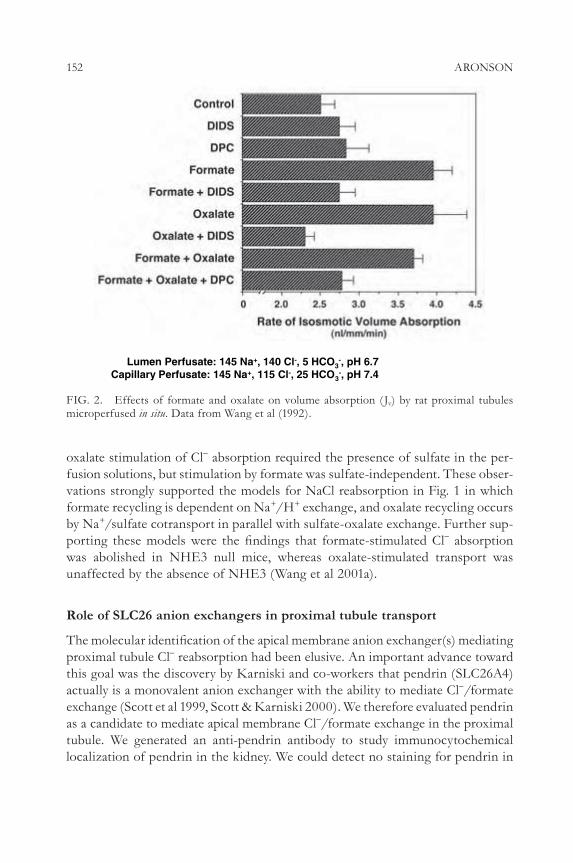
152 ARONSON
oxalate stimulation of Cl− absorption required the presence of sulfate in the per-fusion solutions, but stimulation by formate was sulfate-independent. These obser-vations strongly supported the models for NaCl reabsorption in Fig. 1 in which formate recycling is dependent on Na+/H+ exchange, and oxalate recycling occurs by Na+/sulfate cotransport in parallel with sulfate-oxalate exchange. Further sup-porting these models were the fi ndings that formate-stimulated Cl− absorption was abolished in NHE3 null mice, whereas oxalate-stimulated transport was unaffected by the absence of NHE3 (Wang et al 2001a).
Role of SLC26 anion exchangers in proximal tubule transport
The molecular identifi cation of the apical membrane anion exchanger(s) mediating proximal tubule Cl− reabsorption had been elusive. An important advance toward this goal was the discovery by Karniski and co-workers that pendrin (SLC26A4) actually is a monovalent anion exchanger with the ability to mediate Cl−/formate exchange (Scott et al 1999, Scott & Karniski 2000). We therefore evaluated pendrin as a candidate to mediate apical membrane Cl−/formate exchange in the proximal tubule. We generated an anti-pendrin antibody to study immunocytochemical localization of pendrin in the kidney. We could detect no staining for pendrin in
Lumen Perfusate: 145 Na+, 140 Cl-, 5 HCO3-, pH 6.7
Capillary Perfusate: 145 Na+, 115 Cl-, 25 HCO3-, pH 7.4
FIG. 2. Effects of formate and oxalate on volume absorption ( Jv) by rat proximal tubules microperfused in situ. Data from Wang et al (1992).

SLC26-MEDIATED Cl−/BASE EXCHANGE IN PROXIMAL TUBULE 153
proximal tubule cells, but found pendrin expression on the apical membrane of a subpopulation of cells in the collecting tubule (Knauf et al 2001). A more detailed analysis of pendrin kidney expression was performed by Royaux et al (2001) who identifi ed this cell population as non-a-intercalated cells mediating HCO3
− secre-tion in the cortical collecting tubule. A signifi cant functional role for pendrin in mediating proximal tubule Cl− absorption was effectively ruled out by our studies with Karniski indicating that there is no reduction in either brush border mem-brane Cl−/formate exchange or formate-stimulated NaCl absorption in pendrin null mice that had been generated by Everett and colleagues (Karniski et al 2002).
As a strategy to identify additional candidate anion exchangers capable of medi-ating Cl− reabsorption in the proximal tubule, we screened the expressed sequence tag (EST) database for homologues of pendrin (Knauf et al 2001). We isolated a cDNA encoding a pendrin homologue expressed in kidney that appeared to be the mouse orthologue of human SLC26A6, a putative anion exchanger of then unknown function (Lohi et al 2000, Waldegger et al 2001). We raised antibodies against the encoded protein and demonstrated its expression in the kidney by Western blot analysis and on the brush border membrane of proximal tubule cells by immunocytochemistry (Knauf et al 2001).
We functionally expressed the transporter successfully in Xenopus oocytes (Knauf et al 2001). When we found that it can mediate DIDS-sensitive Cl−/formate exchange we tentatively named it CFEX. But our subsequent studies demonstrated that CFEX (SLC26A6) can mediate additional anion exchange activities previously described in renal brush border membrane vesicles including electrogenic Cl−/oxalate exchange, and oxalate/sulfate exchange ( Jiang et al 2002). We ( Jiang et al 2002) and other groups (Wang et al 2002, Xie et al 2002) also found that SLC26A6 can mediate Cl−/HCO3
− exchange and Cl−/OH− exchange. We found that affi nity of SLC26A6 for oxalate was the highest among the sub-strates tested and that a concentration of oxalate in the range found in urine (100 mM) can signifi cantly convert the function of SLC26A6 from Cl−/HCO3
− exchange to Cl−/oxalate exchange ( Jiang et al 2002).
Taken together, these functional expression studies clearly indicated that SLC26A6 is at least capable of operating in all four of the Cl−/base exchange modes previously illustrated in Fig. 1 that could contribute to NaCl entry across the apical membrane of proximal tubule cells. But a key issue was which if any of these modes of anion exchange are actually mediated by SLC26A6 in proximal tubule cells under physiological conditions. To address this issue, tubule microp-erfusion studies were performed in Slc26a6 null mice generated by Soleimani’s group (Wang et al 2005).
Proximal tubules were microperfused in situ with a low bicarbonate (5 mM), low pH (6.7) solution simulating conditions in the later portions of the proximal tubule

154 ARONSON
after substantial secondary active reabsorption of bicarbonate has taken place. Under these conditions, the rate of volume absorption ( Jv) is an index of ‘isosmotic’ NaCl absorption (Wang et al 1996). As shown in Fig. 3, the increment in volume absorption induced by the addition of oxalate to the perfusion solution was com-pletely abolished in Slc26a6 null mice. The increment in volume absorption induced by formate was also at least partially inhibited. Interestingly, the baseline rate of volume reabsorption measured in the absence of added formate and oxalate was not measurably decreased in the Slc26a6 null mice.
The fi ndings in Fig. 3 indicated that SLC26A6 primarily functions to mediate Cl− absorption occurring by Cl−/oxalate exchange and probably Cl−/formate exchange in the proximal tubule, but does not mediate Cl− absorption by Cl−/HCO3
− or Cl−/OH− exchange. These latter modes of Cl−/base exchange, if opera-ble, should have contributed to the baseline rate of NaCl absorption in tubules perfused with a low bicarbonate, low pH luminal solution. The lack of contribution of SLC26A6 to the baseline rate of NaCl transport is consistent with the observa-tions shown earlier in Fig. 2 that the disulfonic stilbene DIDS, an inhibitor of SLC26A6 activity (Knauf et al 2001, Jiang et al 2002), completely abolished the
Lumen Perfusate: 145 Na+, 140 Cl-, 5 HCO3-, pH 6.7
FIG. 3. Effects of formate and oxalate on volume absorption ( Jv) by proximal tubules micro-perfused in situ in wild-type and SLC26A6 null mice. Data from Wang et al (2005).

SLC26-MEDIATED Cl−/BASE EXCHANGE IN PROXIMAL TUBULE 155
increments in proximal tubule volume reabsorption induced by formate or oxalate but did not affect the baseline rate of transport (Wang et al 1992). Thus, although SLC26A6 had been shown in functional expression studies to be capable of mediating all of the modes of Cl−/base exchange illustrated in Fig. 1, in the intact proximal tubule under physiological conditions it appeared to primarily mediate Cl−/oxalate exchange and probably Cl−/formate exchange, but not Cl−/HCO3
− or Cl−/OH− exchange. Taken together, our results support the model of transcellular NaCl absorption shown in Fig. 4 in which SLC26A6, operating in parallel with Na+/H+ exchanger NHE3 and Na+/sulfate cotransporter NaSi, represents the principal pathway for entry of Cl− across the apical membrane of proximal tubule cells.
The reasons why some modes of SLC26A6-mediated Cl−/base exchange might be favoured over others in a given epithelium are not yet known. One possible explanation derives from the prediction that the function of SLC26A6 should depend on the relative concentrations and affi nities for substrates and inhibitors. For example, we found that oxalate is a relatively high affi nity substrate for SLC26A6 and that para-aminohippurate, a prototypic organic anion secreted by proximal tubule cells, is a non-transported inhibitor ( Jiang et al 2002). Perhaps the intracellular concentration of inhibitor anions like hippurates accumulated by proximal tubule cells is suffi cient to block activity of SLC26A6. If the binding of
NHE3
NaSi
SLC26A6
SLC26A6
FIG. 4. Model for role of SLC26A6 in mediating transcellular NaCl absorption in the proxi-mal tubule.

156 ARONSON
hippurates can be competitively released by the high affi nity substrate oxalate but not by bicarbonate it might then explain why SLC26A6 functions principally as a Cl−/oxalate exchanger in the proximal tubule. Another possibility to explain tissue-specifi c differences in modes of transport mediated by SLC26A6 is that they result from tissue-specifi c differences in interactions with neighbouring membrane proteins. For example, NaCl absorption by Cl−/formate or Cl−/oxalate exchange may require clustering of SLC26A6 with the transporters mediating formate or oxalate recycling across the apical membrane as illustrated in the models of Fig. 1 and Fig. 4.
As an initial strategy to investigate the role of protein interactions in governing membrane expression or function of SLC26A6, we evaluated its interaction with the brush border scaffolding protein PDZK1. Sequence analysis of SLC26A6 had revealed the presence of a predicted PDZ-interaction motif at its extreme c-termi-nus (Lohi et al 2003), raising the possibility that it may interact with a PDZ domain-containing protein in the brush border. Indeed, by use of yeast two-hybrid and fusion protein interaction assays, Murer and colleagues identifi ed PDZK1 as a potential binding partner for SLC26A6 (CFEX) (Gisler et al 2003). PDZK1 has four predicted PDZ domains and is expressed in pancreas, liver, gastrointestinal tract, adrenal cortex and kidney, where it is localized exclusively in the brush border of the proximal tubule (Kocher et al 1998).
We performed a series of pulldown assays from detergent-solubilized brush border membranes and confi rmed that a fusion protein containing the C-terminal 185 amino acids of SLC26A6 was capable of interacting with native PDZK1. We then performed overlay assays to test the ability of SLC26A6 fusion proteins with or without truncation of the putative C-terminal PDZ-interaction motif to bind to a full-length PDZK1 fusion protein. These studies demonstrated that the interaction of SLC26A6 with PDZK1 was due to direct binding medi-ated through the putative PDZ-interaction motif located at the C-terminus of SLC26A6.
Finally, to determine the role of interaction with PDZK1 in governing the brush border membrane expression of SLC26A6, we assessed the abundance and mem-brane localization of SLC26A6 in PDZK1 null mice. Western blot analysis of renal membranes revealed that expression of CFEX was dramatically reduced in the kidneys of PDZK1 null mice. Immunofl uorescence microscopy confi rmed that expression of SLC26A6 on the brush border membrane of proximal tubule cells was markedly reduced in PDZK1 null mice. These fi ndings indicated that the PDZ domain scaffolding protein PDZK1 plays an essential role in brush border mem-brane expression of SLC26A6 (Thomson et al 2005). We have recently observed that another SLC26 isoform, SLC26A7, is also expressed on the apical membrane of proximal tubule cells (Dudas et al 2006). Whether PDZK1 similarly affects the membrane expression of SLC26A7 in the proximal tubule remains to be determined.

SLC26-MEDIATED Cl−/BASE EXCHANGE IN PROXIMAL TUBULE 157
Acknowledgements
Work in the author’s laboratory has been supported by National Institutes of Health grants R01-DK37933 and PO1-DK17433.
References
Aronson PS, Giebisch G 1997 Mechanisms of chloride transport in the proximal tubule. Am J Physiol 273:F179–192
Biemesderfer D, Reilly RF, Exner M et al 1992 Immunocytochemical characterization of Na+ -H+ exchanger isoform NHE-1 in rabbit kidney. Am J Physiol 263:F833–840
Biemesderfer D, Pizzonia J, Abu-Alfa A et al 1993 NHE3: A Na+/H+ exchanger isoform of renal brush border. Am J Physiol 265:F736–742
Biemesderfer D, Rutherford PA, Nagy T et al 1997 Monoclonal antibodies for high-resolution localization of NHE3 in adult and neonatal rat kidney. Am J Physiol 273:F289–299
Dudas PL, Mentone S, Greineder CF, Biemesderfer D, Aronson PS 2006 Immunolocalization of anion transporter Slc26a7 in mouse kidney. Am J Physiol Renal Physiol 290:F937–945
Gisler SM, Pribanic S, Bacic D et al 2003 PDZK1: I. A major scaffolder in brush borders of proximal tubular cells. Kidney Int 64:1733–1745
Goyal S, Vanden Heuvel G, Aronson PS 2003 Renal expression of novel Na+/H+ exchanger isoform NHE8. Am J Physiol Renal Physiol 284:F467–473
Goyal S, Mentone S, Aronson PS 2005 Immunolocalization of NHE8 in rat kidney. Am J Physiol Renal Physiol 288:F530–538
Jiang Z, Grichtchenko II, Boron WF, Aronson PS 2002 Specifi city of anion exchange mediated by mouse Slc26a6. J Biol Chem 277:33963–33967
Karniski LP, Aronson PS 1985 Chloride/formate exchange with formic acid recycling: A mechanism of active chloride transport across epithelial membranes. Proc Natl Acad Sci USA 82:6362–6365
Karniski LP, Aronson PS 1987 Anion exchange pathways for Cl− transport in rabbit renal microvillus membranes. Am J Physiol 253:F513–521
Karniski LP, Wang T, Everett LA et al 2002 Formate-stimulated NaCl absorption in the proxi-mal tubule is independent of the pendrin protein. Am J Physiol Renal Physiol 283:F952–956
Kinsella JL, Aronson PS 1980 Properties of the Na+ -H+ exchanger in renal microvillus mem-brane vesicles. Am J Physiol 238:F461–469
Kinsella JL, Aronson PS 1981a Amiloride inhibition of the Na+ -H+ exchanger in renal micro-villus membrane vesicles. Am J Physiol 241:F374–379
Kinsella JL, Aronson PS 1981b Interaction of NH4+ and Li+ with the renal microvillus mem-
brane Na+ -H+ exchanger. Am J Physiol 241:C220–226Knauf F, Yang CL, Thomson RB et al 2001 Identifi cation of a chloride-formate exchanger
expressed on the brush border membrane of renal proximal tubule cells. Proc Natl Acad Sci USA 98:9425–9430
Kocher O, Comella N, Tognazzi K, Brown LF 1998 Identifi cation and partial characterization of PDZK1: A novel protein containing PDZ interaction domains. Lab Invest 78:117–125
Kuo SM, Aronson PS 1996 Pathways for oxalate transport in rabbit renal microvillus membrane vesicles. J Biol Chem 271:15491–15497
Kurtz I, Nagami G, Yanagawa N et al 1994 Mechanism of apical and basolateral Na+ -independent Cl−/base exchange in the rabbit superfi cial proximal straight tubule. J Clin Invest 94:173–183
Lohi H, Kujala M, Kerkela E et al 2000 Mapping of fi ve new putative anion transporter genes in human and characterization of SLC26A6, a candidate gene for pancreatic anion exchanger. Genomics 70:102–112
158 ARONSON
Lohi H, Lamprecht G, Markovich D et al 2003 Isoforms of SLC26A6 mediate anion transport and have functional PDZ interaction domains. Am J Physiol Cell Physiol 284:C769–779
Murer H, Hopfer U, Kinne R 1976 Sodium/proton antiport in brush-border-membrane vesicles isolated from rat small intestine and kidney. Biochem J 154:597–604
Royaux IE, Wall SM, Karniski LP et al 2001 Pendrin, encoded by the pendred syndrome gene, resides in the apical region of renal intercalated cells and mediates bicarbonate secretion. Proc Natl Acad Sci USA 98:4221–4226
Saleh AM, Rudnick H, Aronson PS 1996 Mechanism of H+ -coupled formate transport in rabbit renal microvillus membranes. Am J Physiol 271:F401–407
Schild L, Giebisch G, Karniski LP, Aronson PS 1987 Effect of formate on volume reabsorption in the rabbit proximal tubule. J Clin Invest 79:32–38
Schultheis PJ, Clarke LL, Meneton P et al 1998 Renal and intestinal absorptive defects in mice lacking the NHE3 Na+/H+ exchanger. Nat Genet 19:282–285
Scott DA, Karniski LP 2000 Human pendrin expressed in Xenopus laevis oocytes mediates chloride/formate exchange. Am J Physiol Cell Physiol 278:C207–211
Scott DA, Wang R, Kreman TM et al 1999 The pendred syndrome gene encodes a chloride-iodide transport protein. Nat Genet 21:440–443
Seifter JL, Knickelbein R, Aronson PS 1984 Absence of Cl-OH exchange and NaCl cotransport in rabbit renal microvillus membrane vesicles. Am J Physiol 247:F753–759
Sheu JN, Quigley R, Baum M 1995 Heterogeneity of chloride/base exchange in rabbit superfi -cial and juxtamedullary proximal convoluted tubules. Am J Physiol 268:F847–853
Thomson RB, Wang T, Thomson BR et al 2005 Role of PDZK1 in membrane expression of renal brush border ion exchangers. Proc Natl Acad Sci USA 102:13331–13336
Waldegger S, Moschen I, Ramirez A et al 2001 Cloning and characterization of SLC26A6, a novel member of the solute carrier 26 gene family. Genomics 72:43–50
Wang T, Giebisch G, Aronson PS 1992 Effects of formate and oxalate on volume absorption in rat proximal tubule. Am J Physiol 263:F37–42
Wang T, Egbert AL Jr, Abbiati T et al 1996 Mechanisms of stimulation of proximal tubule chloride transport by formate and oxalate. Am J Physiol 271:F446–450
Wang T, Yang CL, Abbiati T et al 2001a Essential role of NHE3 in facilitating formate-dependent NaCl absorption in the proximal tubule. Am J Physiol Renal Physiol 281:F288–292
Wang T, Hropot M, Aronson PS, Giebisch G 2001b Role of NHE isoforms in mediating bicarbonate reabsorption along the nephron. Am J Physiol Renal Physiol 281:F1117–1122
Wang Z, Petrovic S, Mann E, Soleimani M 2002 Identifi cation of an apical Cl−/HCO3− exchanger
in the small intestine. Am J Physiol Gastrointest Liver Physiol 282:G573–579Wang Z, Wang T, Petrovic S et al 2005 Renal and intestinal transport defects in Slc26a6-null
mice. Am J Physiol Cell Physiol 288:C957–965Wu MS, Biemesderfer D, Giebisch G, Aronson PS 1996 Role of NHE3 in mediating renal
brush border Na+ -H+ exchange. Adaptation to metabolic acidosis. J Biol Chem 271:32749–32752
Xie Q, Welch R, Mercado A et al 2002 Molecular characterization of the murine Slc26a6 anion exchanger: functional comparison with Slc26a1. Am J Physiol Renal Physiol 283:F826–838
DISCUSSION
Markovich: Have you tested any other PDZ proteins to see whether they interact?

SLC26-MEDIATED Cl−/BASE EXCHANGE IN PROXIMAL TUBULE 159
Aronson: No.Soleimani: What is the renal phenotype of the PDZK1 knockout?Aronson: We are trying to look at this. There aren’t many labs that can micro-
perfuse proximal tubules any more. Tong Wang at Yale has done a few studies. She started with oxalate-stimulated chloride transport, and this looked as if it was abolished. Also, Brent Thomson, who is the main person doing the work on PDZK1 in our lab, has preliminary data that Cl−/oxalate exchange in brush border membranes is greatly reduced in PDZK1 null mice.
Lee: What are the underlying molecular mechanisms of the decrease of the A6 expression in the PDZK1 knockout? Is it defects in protein sorting or in the plasma membrane half-life?
Aronson: We did things backwards. Most people would start with the association and go to transfected cells. We started with the end result and then worked back-wards, to make sure there was an effect in the real animal. We did try this in oocytes, and this is an illustration of the difference between oocytes and renal proximal tubules. Coexpressing A6 with PDZK1 in oocytes made no difference, nor did truncating the C-terminus of A6. We now need to study this in a proximal tubule cell line. My guess is that it plays a role in retention, in the absence of which it is endocytosed and degraded. But this is just a postulate.
Mount: One issue with PDZK1, which is supported in CFTR, is that in some papers it seems that the NHERF1 or PDZK1 activate CFTR by tethering mono-mers together (Wang et al 2000, Raghuram et al 2001). It is surprising in this sense that they wouldn’t function that way. I don’t know how controversial this phenom-enon is, the activation of channels by PDZ domain multimerization.
Aronson: That is possible, certainly. We haven’t looked at NHERF1. The fact that NHE3 protein expression wasn’t affected in the PDZK1 null animals doesn’t mean that there is not an effect on its function. The NHERF1 knockout had per-fectly normal localization of NHE3 (Shenolikar et al 2002), and NHERF1 was originally described as an NHE3-associated protein. PDZK1 had the most atten-tion in the proximal tubule because it was found to be a major associated protein for NaPi-IIa. Yet in PDZK1 null animals there was no change in NaPi-II surface expression, at least on a control diet (Capuano et al 2005). This emphasizes that there might be multiple proteins that are interacting and competing with each other.
Alper: You have described regulation of NHE3 activity by GP330/600/megalin. Is it a stretch to say that megalin is part of a LRP family that includes the scavenger receptor? The scavenger receptor is grossly dysfunctional in the PDZK1 knockout mouse. Is megalin regulation of NHE3 altered?
Aronson: We haven’t looked at that, and so far we haven’t stained for megalin in the PDZK1 null mice. The phenotype that was originally found by Kocher in PDZK1 null animals was elevated cholesterol and high density lipoprotein (HDL)

160 ARONSON
levels. These were attributed to loss of membrane expression of HDL receptor scavenger receptor class B type I (SR-BI) that interacts with PDZK1 in the liver (Kocher et al 2003). For those studying other SLC26s, PDZK1 is known to be expressed in liver, pancreas and intestine (Kocher et al 1998).
Stewart: Have you checked the A6 knockout for up-regulation of A7, given that they are both probably expressed in the same place?
Aronson: No.Turner: Do you think the basal activity is just passive?Aronson: Our assumption over the years had been that since the baseline rate
of NaCl absorption measured in the absence of formate and oxalate was DIDS insensitive, and also insensitive to an inhibitor on the basolateral side that could abolish the oxalate and formate-stimulated transport, perhaps it was entirely passive and paracellular. This may be the case, particularly if A7 is not a chloride/bicarbonate exchanger. We were intrigued by the possibility that A7 had been reported to be signifi cantly less DIDS sensitive than A6 (Petrovic et al 2003). Our assumption had been that if transtubular transport is DIDS insensitive that there is probably no anion exchange and it is passive. Perhaps this assumption is wrong if some members of this family have considerably less DIDS sensitivity. In particular, where these experiments are being done under physiological conditions with high substrate concentration, the DIDS sensitivity could be skewed even further. Our assumption has always been that it is passive, but now we are enter-taining the idea that perhaps a DIDS insensitive chloride/base exchanger could play a role.
Turner: In your perfusion studies, do you always perfuse the same segment?Aronson: These are proximal tubule loops that are accessible on the surface. This
is in situ work, not in isolated perfused tubules.Markovich: Do the A6 knockout mice develop oxalate kidney stones?Soleimani: We have collaborated on this issue with Dr Marguerite Hatch at the
University of Florida. We know that up to one year of age the A6 knockout animals do not develop kidney stones. What we know is that in wild-type animals there is a net secretion of oxalate, but in the knockout there is a complete reversal, resulting in net absorption of oxalate. This raises the question as to where this absorbed oxalate goes.
Markovich: Do you see a lot of oxalate in the urine?Soleimani: We haven’t looked at the crystals, but we are currently measuring
this.Alper: Has there been a mouse model of kidney stones? Aren’t mice extremely
resistant to oxalate nephrolithiasis?Mount: There is calcium phosphate nephrolithiasis in mice but not oxalate.Soleimani: We are examining the S3 segment of the proximal tubule in A6 knock-
out mice, as this segment is the main site of chloride reabsorption in the proximal

SLC26-MEDIATED Cl−/BASE EXCHANGE IN PROXIMAL TUBULE 161
tubule. Dr Aronson’s lab is predominantly examining the S2 segment of the proxi-mal tubule. There is a possibility that one can get a different phenotype in S3 vs. S2 segment.
Romero: Getting back to the animal models, I think they are quite useful. It is nice to see the fi eld come back to real physiology. It is also worth mentioning that in the context of funding times getting diffi cult, Larry Karniski was the only person in the world who would have thought to test formate. This speaks to the fact that when people are trained in a particular arena they bring things with them to other fi elds that give unique insights. It is important to keep our eyes open, to use what we know from other systems for application in new systems, otherwise insights like this are going to be lost. 20 years down the road a very controversial observation that fi tted well for the model of sodium chloride absorption is starting to come full circle, and we now have animal models that can be applied to disease.
Aronson: It is gratifying to see the molecular observation for some of the things we observed. But there is a larger question that we haven’t tested yet: how much does this really contribute to proximal tubule reabsorption with native glomerular fi ltration going on?
Welsh: You can do these studies in the animals.Aronson: Yes, we are on the waiting list for a micropuncture study.Welsh: What about the whole organs?Aronson: One problem with the proximal tubule is that it is easily buffered by
the loop of Henle with respect to NaCl transport. In terms of having a major impact on Na+ balance, it almost certainly doesn’t. Manoocher’s original descrip-tion of the animals was that they had normal blood pressure. In the steady state they are going to come into Na+ balance; the question is, at what blood pressure? This was normal. We monitored blood pressure on a low salt diet and it was also not different. We are currently evaluating effects of loop diuretics and thiazides: if we give such agents to block downstream NaCl reabsorption, there may be a bigger fall in blood pressure.
Mount: The corollary to that is that we don’t know that up-regulation of A6 doesn’t play a role in hypertension. There are data from Pedro Jose’s lab suggesting that dopamine doesn’t inhibit A6 or A6-like activity in the same way that it doesn’t inhibit NHE3 activity in hypertensive rats (Pedrosa et al 2004). Does this explain why NaC1 doesn’t collaborate? Does NaSi-1 or NaSi-2 interact with PDZ domain proteins?
Markovich: It does, apparently, according to a postdoc called Serge Gisler in Heini Murer’s lab at the University of Zurich, Switzerland. But there isn’t a con-sensus PDZ domain in the C-terminus. It seems to be somewhere else.
Aronson: It should be emphasized that NHE3 is associated with NHERF2 through an internal sequence rather than through its predicted C-terminal PDZ

162 ARONSON
binding motif (Yun et al 1998). A protein that doesn’t necessarily have a classical PDZ motif can still associate with a PDZ domain protein.
Mount: The whole clustering doesn’t explain the collaboration of NHE3 for formate exchange, does it?
Aronson: We are trying to determine which domains of PDZK1 associate with SLC26A6 and NHE3. If it is one and the same domain, you could still have networks with multimers, but it would be nicer if different domains were involved.
Welsh: One of the things I thought particularly interesting was the lack of effect of knockout on Cl− and bicarbonate. I don’t know the signifi cance of this. Perhaps it has to do with the ion concentrations or gradients.
Aronson: Manoocher Soleimani’s lab has shown that we can make a distinction between chloride–bicarbonate exchange activity on the apical membrane and its contribution to transtubular transport.
Soleimani: In perfused S3 segment of the proximal tubule the apical chloride/bicarbonate exchange is decreased by more than 60% compared with the wild-type. This is tested by measuring the rate of intracellular pH alteration in response to changing the luminal chloride concentration. What it means in terms of net chloride absorption needs further studies. Peter Aronson’s studies are focusing on the S2 segment.
Aronson: We are not ruling out that later on in the proximal tubule perhaps the chloride–bicarbonate exchange isn’t working. But at least in the section of the proximal tubule being microperfused in our studies there is a mode, namely Cl−/HCO3
− exchange, that is perfectly demonstrable in an oocyte but doesn’t seem to be participating in transport. In terms of explanations, we think oxalate has high affi nity. It is possible that when oxalate is present, or other endogenous anions that might be in a proximal tubule, these anions are binding with high affi nity and keeping bicarbonate off. For chloride/bicarbonate exchange to participate with Na+/H+ exchange to give transtubular transport, carbonic anhydrase needs to be in the complex and perhaps in this epithelium it is not in the complex.
Soleimani: What happened with the oxalate in the S3 segment?Aronson: We haven’t done this study.Soleimani: You could look at volume absorption.Aronson: Years ago when we did studies in rabbit, not mouse, we could stimulate
reabsorption in S1 and S2 segments of proximal tubule by adding formate but we didn’t study S3 (Schild et al 1987). Baum and colleagues have reported that there is heterogeneity in the proximal tubule and that certain segments have formate-independent chloride/base exchange that contributes to NaCl absorption (Sheu et al 1995).
Soleimani: Our studies show that intracellular pH is decreased in the S3 segment of the proximal tubule in A6 knockout mouse. One has to be careful that this
SLC26-MEDIATED Cl−/BASE EXCHANGE IN PROXIMAL TUBULE 163
happens in S3. We have looked at the apical Na+/H+ exchanger NHE3 activity in isolated perfused S3 segment and found it to be signifi cantly reduced. I don’t know whether NHE3 activity is also reduced in the S2 segment. It is possible that depending on the ion gradients or other, as yet to be discovered, properties a transporter can work in two different functional modes in two different segments in the kidney.
Muallem: I like the fact that you don’t see much effect on HCO3− transport. But
if you are ever going to learn anything from the in vitro system that is relevant to in vivo we need to agree what these transporters do. How can we get transporters to function in one way in the kidney and another in the pancreas? Do NHE3 and A6 co-immunoprecipitate? Do they exist in the same complex?
Aronson: We haven’t been able to demonstrate that.
References
Capuano P, Bacic D, Stange G et al 2005 Expression and regulation of the renal Na/phosphate cotransporter NaPi-IIa in a mouse model defi cient for the PDZ protein PDZK1. Pfl ugers Arch 449:392–402
Kocher O, Comella N, Tognazzi K Brown LF 1998 Identifi cation and partial characterization of PDZK1: A novel protein containing PDZ interaction domains. Lab Invest 78:117–125
Kocher O, Yesilaltay A, Cirovic C et al 2003 Targeted disruption of the PDZK1 gene in mice causes tissue-specifi c depletion of the high density lipoprotein receptor scavenger receptor class B type I and altered lipoprotein metabolism. J Biol Chem 278:52820–52825
Pedrosa R, Jose PA, Soares-da-Silva P 2004 Defective D1-like receptor-mediated inhibition of the Cl−/HCO3
− exchanger in immortalized SHR proximal tubular epithelial cells. Am J Physiol Renal Physiol 286:F1120–1126
Petrovic S, Ju X, Barone S et al 2003 Identifi cation of a basolateral Cl−/HCO3− exchanger
specifi c to gastric parietal cells. Am J Physiol Gastrointest Liver Physiol 284:G1093–1103Raghuram V, Mak DD, Foskett JK 2001 Regulation of cystic fi brosis transmembrane conduc-
tance regulator single-channel gating by bivalent PDZ-domain-mediated interaction. Proc Natl Acad Sci USA 98:1300–1305
Shenolikar S, Voltz JW, Minkoff CM et al 2002 Targeted disruption of the mouse NHERF-1 gene promotes internalization of proximal tubule sodium-phosphate cotransporter type IIa and renal phosphate wasting. Proc Natl Acad Sci USA 99:11470–11475
Wang S, Yue H, Derin RB, Guggino WB, Li M 2000 Accessory protein facilitated CFTR-CFTR interaction, a molecular mechanism to potentiate the chloride channel activity. Cell 103:169–179
SLC26 transporters and the inhibitory control of pancreatic ductal bicarbonate secretionPéter Hegyi*†, Zoltán Rakonczay Jr.*†, László Tiszlavicz‡, András Varró§, András Tóth§, Gábor Rácz¶, Gábor Varga¶, Michael A. Gray* and Barry E. Argent*1
* Institute for Cell and Molecular Biosciences, University of Newcastle upon Tyne, NE2 4HH, UK, †First Department of Medicine, ‡Department of Patholog y and §Department of Pharmacolog y and Pharmacotherapy, Faculty of Medicine, University of Szeged, H-6720, Szeged, Hungary, and ¶Molecular Oral Biolog y Research Group, Department of Oral Biolog y, Hungarian Academy of Sciences and Semmelweis University, H-1089 Budapest, Hungary
Abstract. SLC26 anion exchangers (probably SLC26A3 and SLC26A6) are expressed on the apical membrane of pancreatic duct cells and play a key role in HCO3
− secretion; a process that is inhibited by the neuropeptide, substance P (SP). SP had no effect on basolateral HCO3
− transporters in the duct cell or on CFTR Cl− channels, but inhibited a Cl−-dependent HCO3
− effl ux step on the apical membrane. In microperfused ducts, luminal H2DIDS (0.5 mM) caused intracellular pH to alkalinize (consistent with inhibi-tion of HCO3
− effl ux) and, like SP, inhibited HCO3− secretion. SP did not reduce HCO3
− secretion further when H2DIDS was applied to the duct lumen, suggesting that SP and H2DIDS inhibit the same transporter on the apical membrane. As SLC26A6 is DIDS-sensitive, this isoform is the likely target for SP. The inhibitory effect of SP was mimicked by phorbol 12,13-dibutyrate (PDBu), an activator of protein kinase C (PKC). Moreover, bisindolylmaleimide, a blocker of PKC, relieved the inhibitory effect of both SP and PDBu on HCO3
− secretion. Western blot analysis revealed that guinea pig pancreatic ducts express the a , b1, d , e , h, q, z and m isoforms of PKC. We conclude that PKC is a negative regulator of SLC26 activity in pancreatic duct cells.
2006 Epithelial anion transport in health and disease: the role of the SLC26 transporters family. Wiley, Chichester (Novartis Foundation Symposium 273) p 164–176
SLC26 anion exchangers play a key role in the mechanism by which pancreatic ducts secrete a fl uid containing up to 140 mM NaHCO3 (Fig. 1) (for reviews see Argent et al 2006, Steward et al 2005). SLC26A3 (down regulated in adenoma, DRA) and SLC26A6 (putative anion transporter 1, PAT1) are the most likely
164
1 This paper was presented at the symposium by Barry E. Argent, to whom correspondence should be addressed.

PKC INHIBITS SLC26 TRANSPORTERS IN PANCREATIC DUCT 165
candidates for the apical anion exchanger in the duct cell (Fig. 1). Importantly, SLC26A6 has been localized to the apical membrane of human pancreatic duct cells by immunocytochemistry (Lohi et al 2000).
How the SLC26 exchangers and apical Cl− channels interact to produce a high HCO3
− secretion is controversial (Argent et al 2006, Steward et al 2005). One hypothesis is that HCO3
− is secreted on the SLC26 exchanger until the luminal concentration reaches about 70 mM, after which the additional HCO3
− required to raise the luminal concentration to ∼140 mM is transported via either cystic fi brosis transmembrane conductance regulator (CFTR) or Ca2+ -activated Cl− channels (CACC), depending on whether the cAMP or the [Ca2+]i intracellular signalling pathway has been activated (Sohma et al 2000, Ishiguro et al 2002). A second hypothesis is that all of the HCO3
− is secreted on the exchangers (Ko et al 2004). In this case CFTR and CACC act to dissipate the intracellular Cl− that accumulates as the exchangers cycle. The SLC26 exchangers are activated by cyclic AMP-dependent phosphorylation of CFTR (Ko et al 2002), via an interaction between the R-domain of CFTR and the STAS domain of the exchanger (Gray 2004,
HCO3-
Na+
H+
H+
H+
HCO3- HCO3
-
Cl-
Na+ CFTR(cyclic AMP)
BLOOD LUMENDUCT CELL
HCO3-
Cl-
H2CO3
CO2 + H2O
Cl-
Cl-
Ca2+-activatedCl- channel
Na+, K+
SLC26 anion exchangers
DIDS(-)
DIDS(-)
Amiloride(-)
DIDS(-)
CA
FIG. 1. Cellular mechanism of HCO3− secretion by pancreatic duct cells. HCO3
− ions are fi rst accumulated across the basolateral membrane and then exit across the apical membrane by a mechanism that involves Cl− channels and SLC26 anion exchangers. The basolateral Cl−/HCO3
− exchanger (probably AE2), which should oppose HCO3
− secretion, is probably down-regulated following stimulation. CA, carbonic anhydrase; DIDS, diisothiocyanatostilbene-disulfonic acid; (−), inhibitor. For reviews see Argent et al (2006), Steward et al (2005).

166 HEGYI ET AL
Ko et al 2004). A rise in [Ca2+]i also activates SLC26 exchangers, provided the cells express wild-type CFTR (Namkung et al 2003). There is evidence that SLC26A3 and A6 are electrogenic (Ko et al 2002, Xie et al 2002), although this fi nding is controversial (Chernova et al 2003). However, with electrogenic exchang-ers it is possible to envisage a secretory model in which HCO3
− transport occurs via SLC26A6 (2 HCO3
−/1 Cl−) in the proximal ducts where luminal [Cl−] should be high and via SLC26A3 (1 HCO3
−/2 Cl−) in the more distal ducts in which [Cl−] will be lower (Ko et al 2004). However, calculations based on the electrochemical gradients for Cl− and HCO3
− across the luminal membrane of guinea-pig duct cells suggest that the maximum luminal HCO3
− concentration that could be achieved by this model is rather less than the 140 mM found in pancreatic juice (Steward et al 2005).
Figure 2 summarizes the control of pancreatic ductal HCO3− secretion. Here we
focus on inhibitory pathways, which may be physiologically important in terms of
FIG. 2. Regulation of pancreatic ductal HCO3− secretion. Stimulation of ductal HCO3
− secre-tion can occur by activation of cyclic AMP, cyclic GMP and Ca2+ intracellular signalling path-ways. Inhibitory control includes somatostatin and peptide YY (both of which probably work by interfering with the hormonal/neural mechanisms activating secretion) plus a number of other agents that have a direct effect on the ductal epithelium. L and B, receptors located on the luminal and basolateral sides of the duct cell respectively.
STIMULATORY
Cyclic AMP CalciumSecretin AChVIP CCK ß-Agonists Neurotensin
BombesinCyclic GMP TrypsinGuanylin (L) Calcium (L)
ATP (L)
+
INHIBITORY
SomatostatinPeptide YY
-----------------------5-HTAVP
ATP (B)Substance P
-

PKC INHIBITS SLC26 TRANSPORTERS IN PANCREATIC DUCT 167
limiting the hydrostatic pressure that develops within the lumen of the duct and in terms of switching off pancreatic secretion after a meal. The neuropeptide SP has been identifi ed in periductal nerves of the pancreas (Hegyi et al 2005), and has been shown to inhibit fl uid secretion from the intact gland in vivo (Konturek et al 1981, Katoh et al 1984, Iwatsuki et al 1986) and in vitro (Kirkwood et al 1999). Furthermore, we have shown that SP inhibits HCO3
− secretion from isolated pan-creatic ducts, providing evidence for a direct effect of SP on the ductal epithelium (Ashton et al 1990, Hegyi et al 2003, 2005). The inhibitory effect of SP on isolated ducts is dose-dependent and reversed by spantide, a SP receptor antagonist (Ashton et al 1990, Hegyi et al 2003). The aim of this study was to identify the cellular mechanism by which SP exerts its inhibitory effect on pancreatic ductal HCO3
− secretion.
Experimental procedures
The experiments were performed on small inter- and intralobular ducts isolated from the guinea-pig pancreas (Argent et al 1986). The ducts were maintained in culture overnight, during which time their ends seal and they infl ate due to con-tinued secretion into the closed luminal space (Argent et al 1986). HCO3
− secre-tion in these sealed ducts was measured using changes in intracellular pH (pHi) (Hegyi et al 2003, 2004, 2005). The ‘inhibitor stop’ method involves suddenly stopping HCO3
− accumulation by the basolateral base loaders with amiloride (0.2 mM) and diisothiocyanatostilbene-disulfonic acid (DIDS, 0.1 mM) and then following the decline in pHi as HCO3
− secretion continues across the apical mem-brane (Szalmay et al 2001). The ‘alkali load’ method involves alkali loading cells by exposure to NH4Cl (20 mM) and then following the recovery of pHi. In some experiments the sealed ends of the ducts were cut off and the ducts microperfused allowing access to both the basolateral and luminal membranes (Hegyi et al 2005).
Results and discussion
SP inhibits an anion exchanger on the apical membrane of the duct cell
Initial studies on sealed ducts ruled out an effect of SP on the basolateral NaHCO3 cotransporters and Na+/H+ exchangers and indicated that SP inhibits a Cl−-dependent HCO3
− transport step on the apical membrane of the duct cell (Hegyi et al 2003). As SP had no effect on whole-cell CFTR currents, we concluded that the peptide inhibits an SLC26 anion exchanger (Hegyi et al 2003). Support for this idea came from studies on microperfused ducts. As with sealed ducts, SP caused a marked inhibition of both basal and secretin-stimulated HCO3
− secretion

168 HEGYI ET AL
(Fig. 3). Furthermore, exposing the apical membrane to H2DIDS caused pHi to alkalinize (consistent with inhibition of a HCO3
− exit step on the apical membrane), and also reduced HCO3
− secretion to a similar extent to the inhibition observed with SP (Fig. 3). As extracellular DIDS has no effect on CFTR (Gray et al 1993), we concluded that DIDS inhibits HCO3
− secretion by inhibiting the apical anion exchanger. Importantly, SP did not further inhibit HCO3
− secretion in the presence of H2DIDS, consistent with SP and the disulfonic stilbene inhibiting the same transporter (Fig. 3). As SLC26A6 is DIDS sensitive (Ko et al 2002, Wang et al 2002), whereas SLC26A3 is only weakly inhibited by DIDS (Ko et al 2002, Chernova et al 2003), we conclude that A6 is the important isoform in guinea-pig ducts and that this transporter is inhibited following exposure to SP (Hegyi et al
FIG. 3. Effect of secretin, substance P and H2DIDS on HCO3− secretion in microperfused
ducts. Microperfused ducts were exposed to basolateral 0.2 mM amiloride and 0.5 mM H2DIDS for 5 minutes (‘inhibitor stop’ method). The initial rates of intracellular acidifi cation were measured and, knowing the buffering capacity of the cell, converted to a base fl ux, −J(B−) (Hegyi et al 2003, 2005). 1% albumin (control, and the vehicle for secretin and SP), 20 nM SP and/or 10 nM secretin were administered basolaterally with/without luminal H2DIDS (0.5 mM) for 10 minutes before the test. Each column is the mean ± SEM for 5 ducts, except for the secretin + H2DIDS experiments where n was 4. *P < 0.05 vs. the control, **P < 0.001 vs. the control; oP < 0.05 vs. secretin, ooP ≤ 0.05 vs. H2DIDS alone and secretin alone (ANOVA). (Hegyi et al 2005, used with permission from The American Physiological Society.)
0
2
4
6
8
10
- J (m
M B
- /min
)
SPControl Secretin SP+H2DIDS
*** *
*
Secretin +SP
Secretin+H2DIDS
OOO
H2DIDS
Inhibitor stop method

PKC INHIBITS SLC26 TRANSPORTERS IN PANCREATIC DUCT 169
2005). Furthermore, as H2DIDS and SP virtually abolished the stimulatory effect of secretin on HCO3
− secretion (Fig. 3), our data support a secretory model in which the SLC26 anion exchanger is the major pathway for HCO3
− transport across the apical membrane of the duct cell.
Involvement of protein kinase C in the inhibitory effect of SP
Previous work in our laboratory showed that SP had no effect on intracellular cAMP concentration in isolated ducts (Ashton et al 1993), and also inhibited fl uid secretion in response to dibutyryl cAMP (Ashton et al 1990). These data indicated that the inhibitory effect of SP must occur downstream in the intracellular signal-ling pathway from the generation of cAMP. SP interacts with tachykinin receptors, which are coupled via the Gq/G11 family of G proteins to the Ca2+ and protein kinase C (PKC) intracellular signalling pathways. We considered that the involve-ment of intracellular Ca2+ in the inhibitory effect of SP was unlikely as the peptide has either no effect on [Ca2+]i (Evans et al 1990, Hug et al 1994), or causes only a small increase at high doses (Hug et al 1994). As it was known that activation of PKC with phorbol esters inhibits pancreatic secretion in vivo (Salido et al 1990), we tested the possibility that PKC was involved in the inhibitory effect of SP observed in isolated ducts. Activation of PKC with phorbol 12,13-dibutyrate (PDBu) inhibited HCO3
− secretion from isolated ducts (Hegyi et al 2005) (Fig. 4). In contrast, the inactive phorbol ester, PDD, had no effect (Hegyi et al 2005). Furthermore, bisindolylmaleimide (BIS), a highly selective, cell-permeable, PKC inhibitor, caused a dose-dependent reversal of the inhibitory effects of both SP and PDBu on HCO3
− secretion (Hegyi et al 2005) (Fig. 5). Finally, using Western blotting we showed that guinea-pig pancreatic ducts expressed PKCa and b1 of the classic isoforms, PKCd , -e , -h and -q of the novel isoforms and PKCz and -m of the atypical isoforms (Hegyi et al 2005).
Inhibitory effects of PKC on Cl−/HCO3− and Cl−/OH− exchange processes have
previously been reported in hepatocytes (Benedetti et al 1994) and in the human Caco-2 intestinal cell line (Saksena et al 2002, 2005). In Caco-2 cells the effect of PKC on Cl−/OH− exchange was blocked by a specifi c inhibitor of the novel, Ca2+ -independent, PKCe (Saksena et al 2002). The same group has recently shown that c-Src and PKCd are involved in the inhibition of Cl−/OH− exchange in Caco-2 cells by 5-hydroxytryptamine (Saksena et al 2005). So how might PKC activation down-regulate SLC26 exchangers? One possibility is that PKC phosphorylates the exchanger itself or an accessory protein (e.g. CFTR). Another possibility, for which some evidence exists, is that PKC stimulates endocytosis so reducing the number of transporters in the apical membrane. Activation of PKC in MDCK and Caco-2 cells has been shown to increase apical membrane endocytosis (Holm et al 1995), and PKC-mediated internalization of the human amino acid transporter hCAT-1

170 HEGYI ET AL
underlies the inhibitory effect of PKC on amino acid transport in Caco-2 cells (Rotmann et al 2004).
In conclusion, our data suggest that SP and PKC are negative regulators of SLC26 anion exchangers in pancreatic duct cells. The PKC isoform that is acti-vated by SP, the temporal and spatial aspects of PKC translocation in the duct cell, and whether PKC stimulates apical endocytosis are questions that remain to be answered.
FIG. 4. Effect of protein kinase C activation on pancreatic ductal HCO3− secretion measured
in sealed ducts using the ‘alkali load’ method. (A) Basal secretion. (B) Secretin-stimulated secretion. The additions, alone or in combination, were: 1% albumin (control, and the vehicle for secretin and SP), 20 nM SP, 100 nM phorbol 12,13-dibutyrate and 10 nM secretin. Base effl ux (−J[B−]) was calculated as described in the legend to Fig. 3. Each column is the mean ± SEM for 6–8 ducts. *P < 0.05 vs. the control (ANOVA). (Used with permission from The American Physiological Society, Hegyi et al 2005.)
0
10
20
30
40
- J
(mM
B- /m
in)
0
25
50
75
- J
(mM
B- /m
in)
*
Control
A
*
Control
BSP
Secretin
*
PDBu
Secretinand SP
Secretinand PDBu
Alkali load method

FIG. 5. Effect of protein kinase C inhibition on pancreatic ductal HCO3− secretion measured
using the ‘alkali load’ method. (A) Effect of 1000 nM bisindolylmaleimide (BIS) on HCO3−
secretion in the presence of secretin and SP. The additions, alone or in combination, were: 1% albumin (control, and the vehicle for secretin and SP), 20 nM SP, 1000 nM bisindolylmaleimide I (BIS) and 10 nM secretin. Base effl ux (−J[B−]) was calculated as described in the legend to Fig. 3. Each column is the mean ± SEM of data for 6 ducts. (B) Dose–response curve for the effect of BIS on HCO3
− secretion in the presence of secretin and SP. Each point is the mean ± SEM for fi ve ducts. *P < 0.05 vs. the control (ANOVA). (Used with permission from The American Physiological Society, Hegyi et al 2005.)
0
25
50
75-
J (m
M B
- /min
)
25
40
55
- J
(mM
B- /m
in) *
**
10nM Secretin and 20 nM SP
0 1 10 100 1000
concentration of BIS (nM)
Control BIS Secretin Secretinand SP
SecretinSP + BIS
A
B
**
Alkali load method

172 HEGYI ET AL
Acknowledgements
Funded by a Wellcome Trust International Research Development Award (Grant No. 022618) and a Hungarian Scientifi c Research Fund Postdoctoral (Grant No. D42188) to PH, and by a Wellcome Trust Travelling Fellowship (Grant No. 069470) to ZR.
References
Argent BE, Arkle S, Cullen MJ, Green R 1986 Morphological, biochemical and secretory studies on rat pancreatic ducts maintained in tissue culture. Q J Exp Physiol 71:633–648
Argent BE, Gray MA, Steward MC, Case RM 2006 Cell physiology of pancreatic ducts. In: Johnson LR (eds) Physiology of the gastrointestinal tract, 4th edn. Elsevier, San Diego, in press
Ashton N, Argent BE, Green R 1990 Effect of vasoactive intestinal peptide, bombesin and substance P on fl uid secretion by isolated rat pancreatic ducts. J Physiol (Lond) 427:471–482
Ashton N, Evans RL, Elliott AC, Green R, Argent BE 1993 Regulation of fl uid secretion and intracellular messengers in isolated rat pancreatic ducts by acetylcholine. J Physiol (Lond) 471:549–562
Benedetti A, Strazzabosco M, Ng OC, Boyer JL 1994 Regulation of activity and apical targeting of the Cl− HCO3
− exchanger in rat hepatocytes. Proc Natl Acad Sci USA 91:792–796Chernova MN, Jiang L, Shmukler BE et al 2003 Acute regulation of the SLC26A3 congenital
chloride diarrhoea anion exchanger (DRA) expressed in Xenopus oocytes. J Physiol (Lond) 549:3–19
Evans RL, Ashton N, Elliot AC 1990 Intracellular free [Ca2+] in rat pancreatic ducts is increased by acetylcholine but not by bombesin or substance P. J Physiol (Lond) 429:90P
Gray MA 2004 Bicarbonate secretion: it takes two to tango. Nat Cell Biol 6:292–294Gray MA, Plant S, Argent BE 1993 cAMP-regulated whole cell chloride currents in pancreatic
duct cells. Am J Physiol 264:C591–602Hegyi P, Gray MA, Argent BE 2003 Substance P inhibits bicarbonate secretion from guinea
pig pancreatic ducts by modulating an anion exchanger. Am J Physiol 285:C268–276Hegyi P, Rakonczay Z, Gray MA, Argent BE 2004 Measurement of intracellular pH in
pancreatic duct cells—a new method for calibrating the fl uorescence data. Pancreas 28:427–434
Hegyi P, Rakonczay Jr Z, Tiszlavicz L et al 2005 Protein kinase C mediates the inhibitory effect of substance P on HCO3
− secretion from guinea-pig pancreatic ducts. Am J Physiol 288:C1030–1041
Holm PK, Eker P, Sandvig K, Vandeurs B 1995 Phorbol-myristate acetate selectively stimulates apical endocytosis via protein-kinase-C in polarized MDCK cells. Exp Cell Res 217:157–168
Hug H, Pahl C, Novak I 1994 Effect of ATP, carbachol and other agonists on intracellular calcium activity and membrane voltage of pancreatic ducts. Pfl ügers Arch 426:412–418
Ishiguro H, Steward MC, Sohma Y et al 2002 Membrane potential and bicarbonate secretion in isolated interlobular ducts from guinea-pig pancreas. J Gen Physiol 120:617–628
Iwatsuki N, Yamgishi F, Chiba S 1986 Effects of substance P on pancreatic exocrine secretion stimulated by secretin, dopamine and cholecystokinin in dogs. Clin Exp Pharmacol Physiol 13:663–670
Katoh K, Murai K, Nonoyama T 1984 Effects of substance-P on fl uid and amylase secretion in exocrine pancreas of rat and mouse. Res Vet Sci 36:147–152
Kirkwood KS, Kim EH, He XD et al 1999 Substance P inhibits pancreatic exocrine secretion via a neural mechanism. Am J Physiol 277:G314–320

PKC INHIBITS SLC26 TRANSPORTERS IN PANCREATIC DUCT 173
Ko SBH, Shcheynikov N, Choi JY et al 2002 A molecular mechanism for aberrant CFTR-dependent HCO3
− transport in cystic fi brosis. EMBO J 21:5662–5672Ko SBH, Zeng WZ, Dorwart MR et al 2004 Gating of CFTR by the STAS domain of SLC26
transporters. Nat Cell Biol 6:343–350Konturek SJ, Jaworek J, Tasler J, Cieszkowski M, Pawlik W 1981 Effect of substance P and its
C-terminal hexapeptide on gastric and pancreatic secretion in the dog. Am J Physiol 241:G74–81
Lohi H, Kujala M, Kerkela E, Saarialho-Kere U, Kestila M, Kere J 2000 Mapping of fi ve new putative anion transporter genes in human and characterization of SLC26A6, a candidate gene for pancreatic anion exchanger. Genomics 70:102–112
Namkung W, Lee JA, Ahn WI et al 2003 Ca2+ activates cystic fi brosis transmembrane conduc-tance regulator- and Cl−-dependent HCO3
− transport in pancreatic duct cells. J Biol Chem 278:200–207
Rotmann A, Strand D, Martine U, Closs EI 2004 Protein kinase C activation promotes the internalization of the human cationic amino acid transporter hCAT-1: a new regulatory mechanism for hCAT-1 activity. J Biol Chem 279:54185–54192
Saksena S, Gill RK, Syed IA et al 2002 Inhibition of apical Cl−/OH− exchange activity in Caco-2 cells by phorbol esters is mediated by PKCe . Am J Physiol 283:C1492–1500
Saksena S, Gill RK, Tyagi S et al 2005 Involvement of c-Src and PKCd in the inhibition of Cl−/OH− exchange activity in Caco-2 cells by serotonin. J Biol Chem 280:11859–11868
Salido GM, Francis LP, Camello PJ, Singh J, Madrid JA, Pariente JA 1990 Effects of phorbol esters and secretin on pancreatic juice secretion in the anaesthetized rat. Gen Pharmacol 21:465–469
Sohma Y, Gray MA, Imai Y, Argent BE 2000 HCO3− transport in a mathematical model of the
pancreatic ductal epithelium. J Membr Biol 176:77–100Steward MC, Ishiguro H, Case RM 2005 Mechanisms of bicarbonate secretion in the pancreatic
duct. Annu Rev Physiol 67:377–409Szalmay G, Varga G, Kajiyama F et al 2001 Bicarbonate and fl uid secretion by cholecystokinin,
bombesin and acetylcholine in isolated guinea-pig pancreatic ducts. J Physiol (Lond) 535.3:795–807
Wang Z, Petrovic S, Mann E, Soleimani M 2002 Identifi cation of an apical Cl−/HCO3− exchanger
in the small intestine. Am J Physiol 282:G573–579Xie Q, Welch R, Mercado A, Romero MF, Mount DB 2002 Molecular characterization of the
murine Slc26a6 anion exchanger: functional comparison with Slc26a1. Am J Physiol 283:F826–838
DISCUSSION
Case: Have you superimposed any acetazolamide on these experiments to see whether there were any additional effects?
Argent: No.Knipper: Now we have at least two hints that endocytosis might be involved. In
Peter Aronson’s experiments, the PDZK1 knockout and the missing SLC26A6 (CFEX) may also point to this. Do you have any evidence that the phenotype of those knockouts that exist have lysosomal defects, such as defects in lyosomal membrane proteins such as LIMP?
Argent: No.

174 HEGYI ET AL
Aronson: For what it is worth, Hatim Hassan in my lab has found that activation of protein kinase C causes endocytosis of A6 in oocytes resulting in inhibition of transport activity, whereas it had no effect on pendrin.
Argent: This is interesting: there might be a specifi c effect of PKC on A6. A6 expression has been detected in the apical membrane of human pancreatic duct cells using immunocytochemistry, so A6 is the strongest candidate for involvement in ductal HCO3
− secretion.Markovich: Is that a direct effect by PKC?Aronson: Hassan then screened a bunch of PKC inhibitors and found it was
blocked by rottlerin, which is a relatively specifi c PKCd inhibitor. There was one putative PKCd site which was mutated but didn’t affect the inhibition. We also looked at phosphorylation and couldn’t detect a change. My guess is that it is an indirect effect.
Argent: What is the timescale of the PKC effect?Aronson: 15–30 min.Argent: We exposed the ducts to substance P for 10 min.Case: Why did you choose 10 min?Argent: It was the time we were using for secretin stimulation, and we observed
an effect of substance P after a 10 min preincubation.Ishiguro: Obviously apical HCO3
− transport via anion exchange is dependent on the gradients of HCO3
− and Cl− across the apical membrane. I want to ask about the data from the alkaline load method by NH4
+ application. During the alkaline load, is the HCO3
− concentration in the cell high?Argent: I would assume that the intracellular HCO3
− concentration is high because the pH is high.
Ihsiguro: So perhaps there is some outward gradient of HCO3− and inward gradi-
ent of Cl− across the apical membrane. If there is an anion exchanger in the apical membrane, it must work for HCO3
− effl ux in this situation. Many years ago I tried alkaline load with a different method. We superfused the isolated pancreatic ducts with a HCO3
−-CO2-buffered solution and then changed to a HCO3−-CO2-free solu-
tion. We saw a big alkalinization and then recovery from alkaline load. When I removed bath Cl− at the same time, intracellular pH went up and didn’t come down again.
Argent: That would point to a Cl−-dependent HCO3− effl ux process, which could
be an SLC26 anion exchanger.Ishiguro: An apical anion exchanger would work for HCO3
− effl ux during the alkaline load (a special experimental condition) but not necessarily in a physiological condition. In physiological conditions, intracellular pH never goes up above 7.4 even with high HCO3
− (125–145 mM) in the lumen after secretin stimulation. The gradients of HCO3
− and Cl− across the apical membrane are not large enough for any anion exchanger to work for HCO3
− effl ux.

PKC INHIBITS SLC26 TRANSPORTERS IN PANCREATIC DUCT 175
Argent: I appreciate your point about the alkali load method being unphysio-logical, and that is why we repeated the experiments using the inhibitor stop method as well. Using that technique the intracellular pH and HCO3
− concentra-tion should be physiological at the start of the experiment. We got the same results. So regardless of whether the intracellular HCO3
− concentration is normal (inhibitor stop method) or high (alkali load method), substance P inhibits a Cl− dependent HCO3
− effl ux mechanism. I also appreciate the fact that your careful measurements of the Cl− and HCO3
− electrochemical gradients across the luminal membrane of the duct cell indicate that they are not large enough to drive secretion of ∼140 mM HCO3
− on an anion exchanger. Nevertheless, in microper-fusion experiments, we found that luminal H2DIDS inhibited HCO3
− secretion consistent with SLC26A6, rather than CFTR, being the major pathway for HCO3
− secretion.Alper: I wanted to follow up on what happens to Na+/K+ ATPase and membrane
capacitance during the 15–30 min period with phorbol ester.Aronson: We have been slow about publishing these results for that reason. For
us, pendrin was a control which didn’t change under the same conditions.Muallem: Does SP activate the WNK4 kinase?Aronson: I don’t know. We have collaborated with Rick Lifton’s lab, and they
found that WNK4 was present in pancreatic duct at the site of A6 (Kahle et al 2004). Coexpressing WNK4 with A6 in oocytes markedly inhibits A6 activity (Kahle et al 2004).
Muallem: Stimulation of that kinase markedly induces endocytosis of A6. This might explain its inhibition by SP.
Argent: Is there any evidence that A3 and A6 are actually phosphorylated and that phosphorylation can modulate activity?
Markovich: I don’t think this has been studied.Argent: When we ran A3 and A6 through the programs for looking at phos-
phorylation sites there were six or seven PKC consensus sites.Markovich: Whether they use them or not is unknown.Argent: That is why I was asking the question. Is direct phosphorylation going
to be a regulatory phenomenon, or are we always going to be talking about regula-tion via accessory proteins (if I can include CFTR in that category!)?
Seidler: The alkaline load method that you describe is also used for assessing the activity of NKCCs when you take the fall in intracellular pH after ammonium chloride exposure that is sensitive to loop diuretics. Do you have NKCC1 expres-sion in your pancreatic ducts?
Argent: No, NKCC1 is not present in guinea-pig duct cells. However, NKCC1 is expressed on the basolateral membrane of rat duct cells and this probably accounts for the rat having a low bicarbonate concentration, about 70 mM, in its pancreatic juice. It seems that rat duct cells secrete a mixture of NaCl and NaHCO3,

176 HEGYI ET AL
whereas the duct cells of guinea-pigs, cats, dogs and humans secrete only NaHCO3 up to a concentration of about 140 mM.
Reference
Kahle KT, Gimenez I, Hassan H et al 2004 WNK4 regulates apical and basolateral Cl− fl ux in extrarenal epithelia. Proc Natl Acad Sci USA 101:2064–2069

Regulatory interaction between CFTR and the SLC26 transportersNikolay Shcheynikov*, Shigeru B. H. Ko*†2, Weizhong Zeng*, Joo Young Choi*, Michael R. Dorwart*, Philip J. Thomas*2 and Shmuel Muallem*1
* Department of Physiolog y, University of Texas Southwestern Medical Center at Dallas, Dallas, TX 75390-9040, USA and †Division of Gastroenterolog y, Department of Medicine, Nagoya University Graduate School of Medicine, Nagoya 466-8550, Japan
Abstract. Most epithelia that express CFTR secrete fl uid rich in HCO3− and poor in Cl−
that is generated by a CFTR-dependent Cl− absorption and HCO3− secretion process that
when aberrant leads to human diseases such as cystic fi brosis and congenital chloride diarrhoea. Epithelial Cl− absorption and HCO3
− secretion require expression of CFTR and other Cl− and HCO3
− transporters in the luminal membrane of the secreting cells. Recent advances in understanding this critical epithelial function revealed that the luminal Cl− and HCO3
− transporters are members of the SLC26 family. Characterization of several members of the family reveals that all characterized thus far are electrogenic with an isoform specifi c Cl−/HCO3
− transport stoichiometry. In vivo these transporters exist in a transporting complex with CFTR. The SLC26 transporters and CFTR are recruited to the complex by binding to scaffolds containing PDZ domains. Upon stimu-lation and PKA-dependent phosphorylation of CFTR R domain, the R domain binds to the SLC26 transporter STAS domain. Interaction of the R and STAS domains results in a marked and mutual activation of CFTR and the SLC26 transporters. The signifi -cance of this mode of regulation to epithelial Cl− absorption and HCO3
− secretion is obvious.
2006 Epithelial anion transport in health and disease: the role of the SLC26 transporters family. Wiley, Chichester (Novartis Foundation Symposium 273) p 177–192
Cl− absorption and HCO3− secretion are among the most important functions of
secretory epithelia, including the respiratory, reproductive and gastrointestinal systems that are vital for the health and survival of the tissues (Hadorn et al 1968, Kunzelmann & Mall 2002, Melvin et al 2005, Sokol 2001, Steward et al 2005, Wilschanski & Durie 1998). The critical importance of these activities is refl ected most prominently in cystic fi brosis (CF), in which aberrant Cl− absorption and HCO3
− secretion play a central role in epithelial tissue destruction (Sokol 2001,
177
1 This paper was presented at the symposium by Shmuel Muallem, to whom correspondence should be addressed.2 Equal contributors to work presented in this article.

178 SHCHEYNIKOV ET AL
Wilschanski & Durie 1998). Cl− absorption and HCO3− secretion predominate in
the ductal systems of secretory glands or their equivalents (Melvin et al 2005, Steward et al 2005) and surface epithelial cells in the gastrointenstinal (GI) tract (Kunzelmann & Mall 2002). The reciprocal transport of the two anions is tightly coupled, suggesting mutual dependence of the transport. However, in spite of the physiological and pathological importance of these processes, the mechanism and control of epithelial Cl− absorption and HCO3
− secretion remain obscure.Epithelial Cl− and HCO3
− transport show considerable tissue specifi city in the contribution of the electrogenic and electroneutral components to the transport and to the Cl− and HCO3
− gradients generated by the tissue. Probably the steepest gradient is generated by the pancreatic duct, which modifi es the pancreatic juice to contain 20 mM Cl− and 140 mM HCO3
− (Steward et al 2005). Elegant recent work showed that the airway glands can also secrete a fl uid containing more than 80 mM HCO3
− (Irokawa et al 2004). The discovery that the pancreatic juice of CF patients is devoid of HCO3
− and is high in Cl− (Hadorn et al 1968) suggests that the function of CFTR is associated with HCO3
− secretion. The fact that CFTR functions as a cAMP-regulated Cl− channel (Sheppard & Welsh 1999) that can also conduct HCO3
− (Poulsen et al 1994), led to the suggestion that CFTR mediates most HCO3
− transport across the luminal membrane (LM) of epithelia. CFTR-dependent HCO3
− current was found in several epithelia (Spiegel et al 2003, Wang et al 2003). However, the fi ndings that some mutations in CFTR associated with CF inhibit CFTR-dependent HCO3
− more than Cl− transport (Choi et al 2001) and that CFTR is a relatively poor HCO3
− transporter compared to Cl− (Linsdell et al 1997) indicates that CFTR-dependent Cl− absorption and HCO3
− secretion involves several transporters that communicate with CFTR. Furthermore, CFTR activity is regulated by external Cl− (Cl−)(Wright et al 2004), which affects HCO3
− transport by CFTR (Shcheynikov et al 2004). This is illustrated in Fig. 1.
Figure 1 shows the change in membrane potential and intracellular pH (pHi) in Xenopus oocytes expressing CFTR. Removal of Cl− resulted in the expected depo-larization but with a delayed activation of HCO3
− infl ux. HCO3− infl ux was observed
only after the switch in membrane potential (fi rst shaded area in Fig. 1A), suggest-ing delayed activation of HCO3
− conductance by CFTR that resulted in the reduc-tion of the membrane potential. Furthermore, re-addition of Cl− hyperpolarized the cells, indicating activated CFTR, but failed to induce HCO3
− effl ux (second shaded area in Figure 1A). The reason for the delayed activation of HCO3
− infl ux and lack of HCO3
− effl ux by activated CFTR became apparent when the effect of a gradual change in Cl− on activation of HCO3
− transport was examined. Fig. 1B shows that reducing Cl− to below 20 mM was required to observe the switch in membrane potential (change in conductance) and increased HCO3
− transport by CFTR. These and similar observations lead to the conclusion that Cl− regulates HCO3
− conductance of CFTR and that Cl− has to be reduced to below 20 mM before HCO3
− transport by CFTR becomes physiologically relevant and that addi-

REGULATORY INTERACTION 179
tional luminal Cl− and HCO3− transporters participate in epithelial Cl− absorption
and HCO3− secretion (Shcheynikov et al 2004).
For a long time members of the SLC4 family of Cl−/HCO3− exchanger were
considered as the potential Cl− and HCO3− transporters at the luminal membrane
(LM) of epithelia. However, members of the family were found to be expressed only in the basolateral membrane (BLM) of most epithelial cells. A breakthrough in understanding epithelial Cl− and HCO3
− transport was made by the discovery of the new family of Cl− and HCO3
− transporters, the SLC26 family. The family con-sists of ten genes, each of which may have several splice variants (see Mount & Romero 2004 and review by Kere 2006, this volume). Mutations in several members are linked to human diseases (Everett et al 1997, Makela et al 2002, Superti-Furga et al 1996) that arise from defective anion transport, including defective Cl− and HCO3
− transport. Several members are expressed in restricted tissues (Everett et al 1997, Lohi et al 2002), whereas others are wide-spread (Ko et al 2002). The excitement in the discovery of the SLC26 transporters stems from the fi nding that many members of the family are expressed in the LM of epithelial cells (Mount & Romero 2004). Characterization of anion transport by three members of the family, SLC26A3 (Melvin et al 1999), SLC26A4 (Soleimani et al 2001) and SLC26A6 (Ko et al 2002), revealed that all function as Cl−/HCO3
− exchangers, whereas SLC26A7 is a H+ -regulated Cl− channel (Kim et al 2005). SLC26A6 can also
FIG. 1. Effect of Cl− on CFTR HCO3− permeability. Xenopus oocytes expressing CFTR were
stimulated with Forskolin and bathed in HCO3−-buffered media. Where indicated, the oocytes
were exposed to Cl−-free media (A) or perfused with media in which the Cl− concentration was gradually decreased (B). Membrane potential and pHi were measured with microelectrodes. (Adapted from Shcheynikov et al 2004.)
mV
6.8
7.2
7.6
pHi
-60
-40
-20
0
20
40
60
0 Cl-
110 Cl- 0 Cl-Forskolin
HCO3-
10 min
B)Switch
21±4 (n=6)
6.9
7.2
7.5
7.8
8.1
mV
pHi
Forskolin
0 mM Cl-
HCO3-
10 min
A)
-60
-40
-20
0
20
40
60

180 SHCHEYNIKOV ET AL
transport oxalate ( Jiang et al 2002) and oxalate transport appears to be the major function of SLC26A6 in the kidney (Wang et al 2005).
The potential role of the SLC26 transporters in luminal Cl− absorption and HCO3
− secretion prompted us and others to determine their expression profi le and characterize their function and interaction with CFTR. An exploratory RT-PCR assay showed that the pancreatic and submandibular gland ducts express SLC26A2, SLC26A3, SLC26A6 and SLC26A11 and the airway epithelia express all of these transporters and SLC26A9, indicating expression of multiple SLC26 transporters isoforms in each cell. Additional key recent fi ndings demonstrated that the SLC26 transporters are electrogenic transporters (Ko et al 2002, Xie et al 2002) that exhibit isoform specifi c Cl−/HCO3
− stoichiometry and are markedly (fi ve–sixfold) activated by CFTR (Ko et al 2002). Some of these properties are discussed below but full details can be found in Ko et al (2002, 2004). Figure 2 shows the electro-genic properties of SLC26A3 and SLC26A6 expressed in Xenopus oocytes. Meas-urement of membrane potential revealed that removal of Cl− depolarized the membrane potential of cells expressing SLC26A3 and bathed in HEPES-buffered media and that incubation in HCO3
−-buffered media reduced the depolarization (Fig. 2A). Current measurements showed that HCO3
− shifted the I/V relationships to more depolarized voltages (Fig. 2B,C), indicating higher coupling of Cl−and HCO3
− than Cl− and OH− transport. This behaviour is consistent with a Cl−/HCO3−
stoichiometry higher than one. Indeed, in recent work to be published elsewhere in which we measured Cl− and HCO3
− transport in the same cells we found a Cl−/HCO3
− stoichiometry close to 2.The results obtained with SLC26A6 are shown in Fig. 2D–F. Remarkably,
removal of Cl− hyperpolarized the membrane potential of cells expressing SLC26A6 and HCO3
− increased the rate of membrane potential changes due to Cl− removal and addition (Fig. 2D). The I/V curves in the presence and absence of HCO3
− are shown in Fig. 2E, F. Similar changes in membrane potential of oocytes expressing SLC26A6 were reported by Xie et al (2002). On the other hand, Chernova et al (2005) expressed the human and mouse orthologues of SLC26A6 and concluded that both function as electroneutral Cl−/HCO3
− exchangers. A noteworthy problem with the studies of Alper and colleagues is that pHi was measured with BCECF (2′,7′-bis-[2-carboxyethyl]-5-[and-6]-carboxyfl uorescein) in the large Xenopus oocytes. This method may not be suitable for true pHi changes in Xenopus oocytes since in the oocytes BCECF reports the pH only underneath the plasma membrane, which can be very different from that in the bulk cytosol and is thus prone to artefacts. Indeed, Chernova el at (2005) reported that while the human and mouse SLC26A6 orthologues show identical ‘Cl−/HCO3
− exchange activity’ the mouse orthologue showed robust while the human orthologue showed no 36Cl− fl uxes. This highlights the caution with which the BCECF technique should be used to measure true pHi changes and transport function in Xenopus

REGULATORY INTERACTION 181
oocytes. Simultaneous Cl− and pHi measurements disclosed that Cl− and HCO3−
transport by SLC26A6 are strongly affected by the membrane potential and the HCO3
−/Cl− transport stoichiometry is close to 2. Therefore, it is clear that all SLC26 transporters examined so far are electrogenic and show isoform specifi c modes of transport and Cl−/HCO3
− transport stoichiometry.CFTR expressing cells require CFTR to secrete alkaline fl uid that is poor in
Cl− and rich in HCO3−. If the SLC26 transporters participate in this activity it would
be expected that CFTR and the SLC26 transporters will exist in the same Cl− and HCO3
− transporting complex and will affect the activity of each other. To examine such regulatory interactions we followed the SLC26 transporters’ activity by meas-uring Cl−/OH− exchange in HEK293 cells expressing SLC26 transporters alone or together with CFTR. Cl−/OH− exchange activity was measured since the SLC26 transporters but not CFTR mediate Cl−/OH− exchange (Ko et al 2002). Figures 3A–F show that CFTR prominently activates SLC26A3, SLC26A4 and SLC26A6.
FIG. 2. Electrogenicity of the SLC26A3 and SLC26A6 transporters. Oocytes expressing SLC26A3 (A–C) or SLC26A6 (D–F) were used to measure membrane potential (A, D) or the voltage dependence of the current in the absence (B, E) or presence of HCO3
− (C, E). (Adapted from Ko et al 2002.)
-70
-50
-30
Mem
bran
e po
tent
ial (
mV
)
D
0 Cl-
0 Na+
HCO3-
2.5 min
-45
-30
-15
0
Em
(mV
)
HEPES HCO3-
0 Cl-
0 Cl-
5 min
A
-120 -30 30 60
-0.6
0.0
0.6
1.2
0
ControlCl-
Glu-
E µA
mV
-120 -90 -60 -30 0 30 60
-0.6
0.6
1.2ControlCl-
Glu-
F µA
mV
-0.8-0.4
0.0
0.4
0.8
-120 -90 -60 -30 0 30 60
Clgluc
mV
µAB
-120 -90 -60 -30 0 30 60
-0.8
-0.4
0.0
0.4
0.8Cl+HCO3
-
gluc+HCO3-
mV
µAC

182 SHCHEYNIKOV ET AL
Notably, stimulation of CFTR by cAMP was required for activation of the SLC26 transporters by CFTR. The reciprocal experiments are shown in Figs 3G, H. In these experiments HEK293 cells co-expressing CFTR and SLC26A3 (Fig. 3) or SLC26A6 (Ko et al 2004) were used to measure the cAMP-activated Cl− current mediated by CFTR. Figure 3 shows that SLC26A3 increased CFTR current by about twofold. Analysis of surface expression of CFTR by biotinylation indicated that the increased current was not due to increased expression of CFTR in the plasma membrane and single channel analysis in cell-attached patches revealed that the SLC26 transporters increased CFTR NP by reducing the mean close time (Ko et al 2004).
A search for the mechanism by which CFTR and SLC26 transporters mutually activate each other revealed multiple interactions between the proteins. CFTR
FIG. 3. Activation of SLC26A3, SLC26A4 and SLC26A6 by CFTR (A–F) and of CFTR by SLC26A3 and its STAS domain (G–I). HEK293 cells expressing SLC26A3 (A, B), SLC26A4 (C, D) or SLC26A6 (E, F) without (solid traces) or with CFTR (doted traces) bathed in HEPES-buffered media were used to measure Cl−/OH− exchange activity by the SLC26 trans-porters. Note the three–fi vefold activation of the SLC26 transporters by CFTR (Adapted from Ko et al 2002.) In (G, H) HEK293 cells expressing CFTR alone, CFTR and SLC26A3 or CFTR and the SLC26A3-STAS domain were used to measure CFTR-mediated Cl− current. In (I) recombinant unphosphorylated (RD) or phosphorylated (pRD) R domain were used to pull down the STAS domain. (Adapted from Ko et al 2004, with permission from Nature, www.nature.com.)
F)
0
1
2
3
4
∆pH
/min
Con +ForCl- free
E) SLC26A6+CFTR
30 sec
Forskolin
0
1
2
3
4
∆pH
/min
Con +CFTR
D)
Cl- free
60 secC) SLC26A4+CFTR
Forskolin3 min
0.5
∆ pH
units
01234
∆ pH
/min
Con For
B)
Cl--Free
30 secA)SLC36A3+CFTR
Forskolin
I) Pull-down STAS with GST-pRD
Pull down:RD pRD
IB:STAS
ForskolinGlibenclamideG)
50 sec1 nA
CFTR+SLC26A3
CFTR
CFTR+STAS
H)
Cur
rent
in n
A
CFTR
CFTR+STAS0.0
1.5
3.0
4.5
CFTR+DRA

REGULATORY INTERACTION 183
(Wang et al 1998) and most SLC26 transporters (Mount & Romero 2004) have a PDZ domain binding motif in their C-terminus. Deletion of either motif resulted in dissociation of the CFTR–SLC26A3 complexes and loss of activation of the SLC26A3 by CFTR. However, upon overexpression of the proteins the interaction and activation were restored, leading to the discovery that the SLC26 transporters’ STAS domain interacts with the CFTR R domain (Ko et al 2004). Furthermore, as shown in Fig. 3G,H the isolated STAS domain was even more effective than the SLC26 transporters in activating CFTR. Binding assays with expressed (Ko et al 2004) or purifi ed recombinant proteins (Fig. 3I) showed that phosphorylation of the R domain markedly increased its binding to SLC26A3 and the STAS domain.
That the STAS domain was suffi cient for activation of CFTR raised the question of whether the CFTR R domain is suffi cient for activation of the SLC26 transport-ers. Expression of the R domain alone failed to activate Cl−/OH− exchange by SLC26A3. Suspecting that this could be because the soluble R domain failed to target to the plasma membrane and bind to SLC26A3 we attached the R domain to either half of CFTR to aid in its targeting to the plasma membrane. The con-structs are the same and were prepared as reported before (Yue et al 2000). Figure 4 shows that the fi rst (TM1) or second (T2N2) transmembrane sectors of CFTR devoid of the R domain failed to activate SLC26A3. However, when the R domain was attached to these constructs (T1N1R and RT2N2) they activated SLC26A3 Cl−/OH− exchange activity by 90–170%. These fi ndings suggest that the R domain may be suffi cient for activation of the SLC26 transporters.
Based on these fi nding it is possible to suggest the model illustrated in Fig. 5 for the interaction and mutual regulation of CFTR and the SLC26 transporters. In this model, the transporters bind with their PDZ ligands to a common scaf-folding protein that possesses at least two PDZ domains. The binding serves to recruit the CFTR and SLC26 transporters to a Cl− and HCO3
− transporting complex. In the complex the STAS domain can interact with the R domain. The interaction takes place or is markedly enhanced by PKA-mediated phosphorylation of the R domain. Whether phosphorylation of the STAS domain is also required for its binding to the R domain remains to be determined. Binding of the R and STAS domains results in the mutual activation of the two transporters and stimulation of epithelial Cl− absorption and HCO3
− secretion. The SLC26 transporters that mediate activation of CFTR and the Cl− and HCO3
− transport depends on the repertoire of SLC26 transporters expressed in each cell type. The fact that cells express multiple SLC26 transporters suggests that different members of the SLC26 transporters have specifi c and perhaps redundant roles in epithelial Cl− absorption and HCO3
− secretion. This may be inferred from the mild phenotype observed in mice from which the gene coding for the ubiquitous SLC26A6 was deleted (Wang et al 2005). The continued effort to understand the transport function of each

184 SHCHEYNIKOV ET AL
Scaffold
SLC26
STAS
ET
KF
N
CFTR
NR
TM1TM2
NBD1NBD2
DT
RL
FIG. 5. A model depicting interaction between CFTR and SLC26A6 transporters with the aid of PDZ domains containing scaffolding protein.
FIG. 4. The CFTR R domain is suffi cient to activate SLC26A3. HEK293 cells were trans-fected with SLC26A3 (DRA) and the CFTR constructs TM1, T1N1R, T2N2 or RT2N2 and used to measure activation of SLC26A3 by the plasma membrane targeted R domain. The CFTR constructs were prepared according to Yue et al 2000.
T1N1R
NBD1
R
B) DRA+T1N1R
Cl- free
Fors
TM1A) DRA+T1
ForsCl- free
0.2
pH u
nits
2 min
D) DRA+RT2N2
Cl- freeForsR
NBD2
RT2N2
C) DRA+T2N2
ForsCl- free
NBD2
T2N2
T1N
1R
P<0.05
Con
TM
1
5
E)
0
1
2
3
Fol
d S
tim
ulat
ion
n= 13
T2N
2
RT
2N2
P<0.01
5 5 6

REGULATORY INTERACTION 185
SLC26 transporter and the use of mutant mice will undoubtedly provide important information to better understand the physiological role of this intriguing family of Cl− and HCO3
− transporters in epithelial Cl− absorption and HCO3− secretion and
other activities.
References
Chernova MN, Jiang L, Friedman DJ et al 2005 Functional comparison of mouse slc26a6 anion exchanger with human SLC26A6 polypeptide variants: Differences in anion selectivity, regulation, and electrogenicity. J Biol Chem 280:8564–8580
Choi JY, Muallem D, Kiselyov K, Lee MG, Thomas PJ, Muallem S 2001 Aberrant CFTR-dependent HCO3
− transport in mutations associated with cystic fi brosis. Nature 410:94–97Everett LA, Glaser B, Beck JC et al 1997 Pendred syndrome is caused by mutations in a putative
sulphate transporter gene (PDS). Nat Genet 17:411–422Hadorn B, Johansen PG, Anderson CM 1968 Pancreozymin secretin test of exocrine pancreatic
function in cystic fi brosis and the signifi cance of the result for the pathogenesis of the disease. Can Med J 98:377–384
Irokawa T, Krouse ME, Joo NS, Wu JV, Wine JJ 2004 A ‘virtual gland’ method for quantifying epithelial fl uid secretion. Am J Physiol Lung Cell Mol Physiol 287:L784–793
Jiang Z, Grichtchenko II, Boron WF, Aronson PS 2002 Specifi city of anion exchange mediated by mouse Slc26a6. J Biol Chem 277:33963–33967
Kere J 2006 Overview of the SLC26 family and associated diseases. In: Epithelial anion trans-port in health and disease: the role of the SLC26 transporter family. Wiley, Chichester (Novartis Found Symp 273) p 2–18
Kim KH, Shcheynikov N, Wang Y, Muallem S 2005 SLC26A7 is a Cl- channel regulated by intracellular pH. J Biol Chem 280:6463–6470
Ko SB, Shcheynikov N, Choi JY et al 2002 A molecular mechanism for aberrant CFTR-dependent HCO3
− transport in cystic fi brosis. EMBO J 21:5662–5672Ko SB, Zeng W, Dorwart MR 2004 Gating of CFTR by the STAS domain of SLC26 tran-
sporters. Nat Cell Biol 6:343–350Kunzelmann K, Mall M 2002 Electrolyte transport in the mammalian colon: mechanisms and
implications for disease. Physiol Rev 82:245–289Linsdell P, Tabcharani JA, Rommens et al 1997 Permeability of wild-type and mutant cystic
fi brosis transmembrane conductance regulator chloride channels to polyatomic anions. J Gen Physiol 110:355–364
Lohi H, Kujala M, Makela S et al 2002 Functional characterization of three novel tissue-specifi c anion exchangers SLC26A7, -A8, and -A9. J Biol Chem 277:14246–14254
Makela S, Kere J, Holmberg C, Hoglund P 2002 SLC26A3 mutations in congenital chloride diarrhea. Hum Mutat 20:425–438
Melvin JE, Park K, Richardson L, Schultheis PJ, Shull GE 1999 Mouse down-regulated in adenoma (DRA) is an intestinal Cl−/HCO3
− exchanger and is up-regulated in colon of mice lacking the NHE3 Na+/H+ exchanger. J Biol Chem 274:22855–22861
Melvin JE, Yule D, Shuttleworth T, Begenisich T 2005 Regulation of fl uid and electrolyte secretion in salivary gland acinar cells. Annu Rev Physiol 67:445–469
Mount DB, Romero MF 2004 The SLC26 gene family of multifunctional anion exchangers. Pfl ugers Arch 447:710–721
Poulsen JH, Fischer H, Illek B, Machen TE 1994 Bicarbonate conductance and pH regulatory capability of cystic fi brosis transmembrane conductance regulator. Proc Natl Acad Sci USA 91:5340–5344

186 SHCHEYNIKOV ET AL
Shcheynikov N, Kim KH, Kim KM et al 2004 Dynamic control of cystic fi brosis trans-membrane conductance regulator Cl−/HCO3
− selectivity by external Cl−. J Biol Chem 279:21857–21865
Sheppard DN, Welsh MJ 1999 Structure and function of the CFTR chloride channel. Physiol Rev 79:S23–45
Sokol RZ 2001 Infertility in men with cystic fi brosis. Curr Opin Pulm Med 7:421–426Soleimani M, Greenley T, Petrovic et al 2001 Pendrin: and apical Cl−/OH−/HCO3− exchanger
in the kidney cortex. Am J Physiol 280:F356–364Spiegel S, Phillipper M, Rossmann H, Riederer B, Gregor M, Seidler U 2003 Independence of
apical Cl−/HCO3− exchange and anion conductance in duodenal HCO3- secretion. Am J
Physiol Gastrointest Liver Physiol 285:G887–897Steward MC, Ishiguro H, Case RM 2005 Mechanism of bicarbonate secretion in the pancreatic
duct. Annu Rev Physiol 67:377–409Superti-Furga A, Hastbacka J, Wilcox WR et al 1996 Achondrogenesis type IB is caused by
mutations in the diastrophic dysplasia sulphate transporter gene. Nat Genet 12:100–102Wang S, Raab RW, Schatz PJ, Guggino WB, Li M 1998 Peptide binding consensus of the
NHE-RF-PDZ1 domain matches the C-terminal sequence of cystic fi brosis transmembrane conductance regulator (CFTR). FEBS Lett 427:103–108
Wang XF, Zhou CX, Shi QX et al 2003 Involvement of CFTR in uterine bicarbonate secretion and the fertilizing capacity of sperm. Nat Cell Biol 5:902–906
Wang Z, Wang T, Petrovic S et al 2005 Kidney and intestine transport defects in Slc26a6 null mice. Am J Physiol Cell Physiol 288:C957–965
Wilschanski M, Durie PR 1998 Pathology of pancreatic and intestinal disorders in cystic fi bro-sis. J R Soc Med 91(suppl 34):40–49
Wright AM, Gong X, Verdon B et al 2004 Novel regulation of cystic fi brosis transmembrane conductance regulator (CFTR) channel gating by external chloride. J Biol Chem 279:41658–41663
Xie Q, Welch R, Mercado A, Romero MF, Mount DB 2002 Molecular characterization of the murine Slc26a6 anion exchanger: functional comparison with Slc26a1. Am J Physiol 283:F826–838
Yue H, Devidas S, Guggino WB 2000 The two halves of CFTR form a dual-pore ion channel. J Biol Chem 275:10030–10034
DISCUSSION
Welsh: Just as you could stimulate CFTR with the isolated STAS domain, could you stimulate SLC26 with the isolated R domain? Did you show that?
Muallem: No, the R domain does not stimulate the transporter by itself. The only way we can get it to stimulate the transporters is if we hook it up to a membrane-targeting domain. When we hook it up to the N- or the C-terminal halves of CFTR it does activate the SLC26 transporters.
Lee: How does the STAS domain activate CFTR?Thomas: We have data addressing this directly. Earlier on in our work we
expressed the STAS as a fusion with MPB (Ko et al 2004). This is not well enough behaved biochemically for structural studies. We have now developed better expression systems. We have expressed the human SLC26A3 STAS domain from position 509–741. We get several milligrams per litre and we can concentrate this

REGULATORY INTERACTION 187
protein to a reasonably high level, as is necessary for these kinds of structural studies. The circular dichroism (CD) spectrum indicates that it is alpha helical with some beta structure. This is what we would expect if it is structurally homologous with the anti-sigma factor (Aravind & Koonin 2000). We have also performed ultracentrifugation experiments and shown that it is a monomer. 1D NMR spectra of unlabelled protein collected by Jennifer Baker in Julie Forman-Kay’s lab in Toronto show that this is a well folded protein (personal communication). The presence of peaks corresponding to methylene hydrogens packed against aromatics are indicative of there being a hydrophobic core to the protein.
Julie also has produced 15N-labelled R domain and she has assigned all the peaks (personal communication). When the STAS domain I have just described is mixed with the phosphorylated R domain, they see about 20% of the peaks are dramatically reduced in intensity, including phosphorylated S670. There is some indication that there are some specifi c low affi nity interactions, and we are begin-ning to get an idea of exactly which residues are involved. We have a detailed model for how this might activate CFTR, but currently I do not know how the interaction leads to enhanced SLC26 activity. This model builds on work on a methanococcal protein, homologous with the CFTR NBDs, we performed in collaboration with John Hunt at Columbia. We mutated the catalytic base respon-sible for ATP hydrolysis. This mutant protein will bind ATP but it won’t hydrolyse it (Moody et al 2002). When it binds ATP, the ATP binds at the interface and causes a dimerization of these two ATP binding cassettes (Moody et al 2002, Smith et al 2002). David Gadsby’s recent work has shown that CFTR behaves similarly and this dimeric state probably corresponds to the open state of the CFTR channel (Vergani et al 2005). However, when we look at the structure of the murine NBD (Lewis et al 2004), there is a helical extension that is in the way of this interface. At the end of this helical extension is residue S670. Other data suggest that this helical extension is dynamic. It is in and out of this position in different crystal structures (Lewis et al 2005). Thus, one potential model could be that one conformation of the helical extension interferes with NBD dimeriza-tion, locking the channel in a closed state. When position S670 is phosphorylated, it still dynamically fl ips in and out, but now the STAS can bind to it and stabilize it in a position that does not occlude the interface, thereby allowing the channel to open. Mike Welsh, you published data a couple of years ago demonstrating that, contrary to expectation, it is not the phosphorylated R domain that stabilizes the open state, but rather that the non-phosphorylated R domain inhibits its formation (Winter &Welsh 1997). This structural model is consistent with those functional data.
Welsh: Do you see other sites, too?Thomas: The Forman-Kay group is vigorously pursuing this specifi c question. I
should also point out that both the R domain and the NBD constructs contain

188 SHCHEYNIKOV ET AL
the same helix, so it is important to note that there is an overlap between the two domains we are looking at. S670 is in both.
Muallem: This explains very well the incredible activation of CFTR by the STAS domain that we have seen. We can keep CFTR open for most of the time. The channel has a hard time remaining closed.
Welsh: How does the R domain structure change?Thomas: The R domain does not have regular secondary structure nor are the
dispersions in the NMR spectra indicative of an ordered globular protein; as you know it is a highly dynamic, disordered protein (Ostedgaard et al 2000). Julie’s model is that this is important for getting a non-linear response (Finerty et al 2005). This is a highly cooperative interaction between multiple weak sites giving a steep response as one titrates these phosphorylation sites. When one gets to a specifi c critical number of interactions you get a persistent interaction that will affect the channels. This may be why it was so confusing to determine which individual sites are important for regulating gating of the channel.
Alper: Was her assignment done by individual 15N amino acid labelling?Thomas: No, this was uniformly 15N-labelled protein, and then she does the
standard coupling experiments to complete the assignments. This is diffi cult and it has taken several years to get to this point.
Gray: Have you looked at either ATP binding or hydrolysis in the presence of the STAS domain?
Thomas: The problem is that we don’t have a good model for NBD2 of CFTR. This is what we would like to do; we’d like to see whether we can evaluate the dimerization model using biochemical methods.
Gray: What about using your model of the MJ0796 methanococcal ABC protein to investigate this point?
Thomas: There is no reason to suspect that this protein would interact with the STAS domain.
Soleimani: A major focus of the presentations in this meeting, and one area of discrepancy, has been what happens in vivo versus in vitro. This work has been done in HEK cells. Have you done studies to test the model, for example, in the duo-denum? This goes back to the issue of chloride–bicarbonate exchange: this depends on where you look at it. How does this in vitro study translate to in vivo?
Muallem: What Min Goo Lee found in the SLC26A6-/- mouse pancreas is very much what I would expect to see. Min Goo sees a reduction in the cAMP-stimu-lated Cl−/HCO3
− exchange. My interpretation of this is that as long as CFTR is unchanged, once A6 is present you need SLA26A6(A6) to be activated by CFTR which mediates some of the Cl− and HCO3
− transport. This Cl− and HCO3− transport
is very important for the proximal part of the duct, the site of most ductal HCO3−
secretion. You will then get a reduction of stimulated Cl−/HCO3− exchange activity.
One of the reasons you might not see effects on the fi nal HCO3− secretion by the

REGULATORY INTERACTION 189
duct is because SLC26A3(A3) or an A3-like protein will be the one that will even-tually compensate for A6 and will absorb more of the Cl−, secreting more HCO3
−. The fi nal secretion will be the same but the regional activity will be different. This is dependent on what happens to CFTR. If CFTR is activated you will probably not see much of an effect on the fi nal HCO3
− secretion unless you delete almost all of the SLC26 transporters, because once Cl− is reduced below 20 mM the Cl− inhibi-tory effect is removed from CFTR. We do not know at present by how much it has to be reduced in the duct cells to remove the inhibitory Cl− effect on CFTR.
Gray: You start getting signifi cant effects on CFTR activity (gating) when exter-nal Cl− falls below 120 mM (Wright et al 2004).
Muallem: What I am emphasizing is that there are two different effects here. Extracellular Cl− has a marked inhibitory effect on CFTR function. We looked at it as an effect on HCO3
− transport (Shcheynikov et al 2004); Mike looks at it while measuring Cl− channel activity (Wright et al 2004). Assuming that this is the same site, when we start reducing chloride below 80–40 mM, now CFTR becomes a decent HCO3
− conductive pathway. In our model, the CFTR will clamp the HCO3−
at 140 mM. If the membrane depolarizes we get more HCO3− secretion than we
want and CFTR will clamp it at 140 mM.Argent: Your secretory model requires that A6 is expressed at the top end of the
duct system and that A3 is expressed at the bottom end. It is diffi cult to see how the model could work if A3 and A6 are both expressed evenly down the ductal system. If A6 and A3 are in the same cell then anion secretion on the exchangers is likely to be electroneutral.
Muallem: We don’t have good A3 antibodies. They work reasonably well with expressed protein, but we are not getting good immunolocalization.
Alper: You had convincing chloride apparent affi nity regulation by imposed voltage holding potential. Do you see regulation of DIDS affi nity?
Muallem: These experiments were done only last week! When the paper showing that the current was DIDS insensitive came out, it became important to look at the current. In the original paper (Ko et al 2002) we showed that the current is indeed DIDS inhibitable. Again, when we started measuring simultaneously pH and Cl− in the same cell we decided that we should look at this hyperpolarization that we see. The hyperpolarization was inhibited by DIDS. In your trace the hyperpolarization is pretty evident when you remove external chloride.
Alper: When you compare nitrate–bicarbonate exchange with nitrate currents, are they entirely the same? They seem different.
Muallem: No, this is still puzzling to us. When we add NO3− it seems that the
entire transport is uncoupled. The only thing that the protein (A3) does is appear as a NO3
− channel. This is almost heresy: taking a coupled transporter, then adding NO3
−, and once you get enough NO3− into the oocyte it doesn’t move anything but
NO3−. In this manner A3 functions as an electronic, coupled Cl−/HCO3
− transporter

190 SHCHEYNIKOV ET AL
but in the absence of Cl− and presence of NO3− it functions as a NO3
− channel. Although there is a precedence for such a behaviour of transporters we are not ready to commit to such a conclusion.
Alper: But it is conductive. Why not postulate the induction of a native nitrate channel as you drop intracellular chloride?
Muallem: We never see anything like that in the cells that don’t have A3. A6 is a poor NO3
− transporter.Ashmore: Why is it non-linear, if it is a nitrate–nitrate channel?Muallem: We think it might be a voltage-regulated channel.Case: If you replaced your plasma chloride by nitrate, what would happen to
pancreatic secretion?Muallem: It should cease.Case: It doesn’t. In the isolated gland fl uid secretion is almost as great in the
presence of nitrate as it is with chloride (Case et al 1979).Muallem: I would say A3 doesn’t move HCO3
− any more. You might get some transport with A6. A6 will mediate HCO3
−/NO3− transport; it is just a lousy sub-
strate compared with Cl−.Romero: We had data from years ago that got buried in Christopher M. Sciortino’s
PhD thesis, from experiments on Drosophila Na+ -dependent chloride–bicarbonate exchanger. We had interesting nitrate results. When we did chloride replacements, the effects with nitrate and nitrite were dependent on whether or not they were done in a bicarbonate buffer. In a non-bicarbonate buffer using nitrate electrodes we demonstrated large nitrate fl uxes when the solution was spiked with nitrate. We were looking at the pH effects of doing nitrate in bicarbonate and non-bicarbonate buffers. In the presence of bicarbonate we saw an unanticipated result with nitrate in that pH went up rather than down. It turns out that in a non-bicarbonate situa-tion nitrate could easily replace the halide. In the presence of bicarbonate nitrate didn’t replace the halide that was being transported, but replaced the bicarbonate. It seemed to have a better affi nity for the bicarbonate replacement than it did the nitrate. This fi ts with some of your old nitrate data from pancreatic duct cells. In terms of anion binding sites or transport sites, with nitrate it depends on what the conditions are. If the conditions are varied, especially in terms of bicarbonate, then the recognition sites look different. Nitrate looks like bicarbonate in 3D space rather than chloride.
Quinton: I am trying to understand how you get so much current out of a nitrate exchanger if it is tightly coupled. In my younger days we used to talk about ‘slip-page’. Does this have any role here? Is something slipping through instead of being exchanged?
Muallem: We are generating a huge current. Slippage will not translate into a current of this magnitude. It is diffi cult conceptually to understand how coupled

REGULATORY INTERACTION 191
transporters all of a sudden switch and start behaving like a channel, but for the most part this looks like a NO3
− channel. HEK cells are the easiest place to see it. There is a beautiful current, seen with NO3
− and only with A3. We never see it with A6, for example.
Seidler: In the colonic surface there is a high concentration of DRA and NHE3 expression, with low expression of CFTR. If you assume that DRA is electrogenic in that tissue as well, you would have more hydrogen secretion than bicarbonate secretion during resting state electroneutral salt absorption, if NHE3 is working with DRA in a 1 : 1 fashion to absorb salt. I do fi nd that the colonic pH in the lumen can go down in the proximal column. I can understand this, but I can’t understand how this mechanism would work: what would it need to keep going if it is an electrogenic process on the one hand and an electroneutral process on the other?
Muallem: I don’t know enough about the colon transporters to answer this fully. What you need is a mechanism that will supply you with Cl−. If you have this, you would secrete HCO3
−.Seidler: There is low to zero bicarbonate secretion taking place in the proximal
colon during absorption. Would an apical potassium conductance be a candidate to maintain electroneutrality?
Muallem: That is a perfect way to regulate these transporters. You want them to be electrogenic if you want them to be regulated by the membrane potential. In some cells you would like to activate them, so you depolarize the membrane poten-tial; in others you want the transporters to work less. In this case you would expect hyperpolarization of the membrane potential. These transporters need to be con-sidered in the context of where they work and the other transporters that they interact with. Also important in this case is the identity of the transporters that sit next to them. The microenvironment at the mouth of those transporters will be responsible for much of this regulation.
Seidler: A K+ conductance has been described in the apical membrane in colono-cytes, whose molecular identity and physiological function has not yet been worked out. So it might be a candidate.
Muallem: If it will hyperpolarize, then yes.Romero: What is the voltage across the apical membrane of colonocytes? The
colon is a bizarre environment. I wouldn’t worry about potassium, but I would worry about ammonium having a signifi cant effect.
Seidler: You can take the isolated colon and look at sodium/chloride absorption and bicarbonate secretion when there is no ammonium present. One thing that has been found is a lumenal potassium conductance in the proximal colon. At least in the proximal colon, where there is no sodium conductance, its function so far is a little mysterious.

192 SHCHEYNIKOV ET AL
Romero: Is it still present in the distal colon?Seidler: In the distal colon there is electrogenic sodium uptake by ENaC, and a
potassium conductance is needed to maintain electroneutrality. There is also an H/K ATPase to recycle the potassium that is lost. In the proximal colon there is no ENaC yet there is potassium conductance.
Welsh: How is the R domain regulating A3?Muallem: We don’t know that it is. We know that the STAS domain is needed
because when this is mutated most mutants are not targeted to the plasma membrane and are retained in the ER/Golgi.
References
Case RM, Hotz J, Hutson D, Scratcherd T, Wynne RDA 1979 Electrolyte secretion by the isolated cat pancreas during replacement of extracellular bicarbonate by organic anions and chloride by inorganic anions. J Physiol 286:563–576
Aravind L, Koonin EV 2000 The STAS domain—link between anion transporters and antisigma-factor antagonists. Curr Biol 10:R53–55
Finerty PJ Jr, Mittermaier AK, Muhandiram R, Kay LE, Forman-Kay JD 2005 NMR dynamics-derived insights into the binding properties of a peptide interacting with an SH2 domain. Biochemistry 44:694–703
Ko SB, Zeng W, Dorwart MR et al 2004 Gating of CFTR by the STAS domain of SLC26 transporters. Nat Cell Biol 6:343–350
Lewis HA, Buchanan SG, Burley SK et al 2004 Structure of nucleotide-binding domain 1 of the cystic fi brosis transmembrane conductance regulator. EMBO J 23:282–293
Lewis HA, Zhao X, Wang C et al 2005 Impact of the deltaF508 mutation in fi rst nucleotide-binding domain of human cystic fi brosis transmembrane conductance regulator on domain folding and structure. J Biol Chem 280:1346–1353
Moody JE, Millen L, Binns D, Hunt JF, Thomas PJ 2002 Cooperative, ATP-dependent associa-tion of the nucleotide binding cassettes during the catalytic cycle of ATP-binding cassette transporters. J Biol Chem 277:21111–21114
Ostedgaard LS, Baldursson O, Vermeer DW, Welsh MJ, Robertson AD 2000 A functional R domain from cystic fi brosis transmembrane conductance regulator is predominantly unstruc-tured in solution. Proc Natl Acad Sci USA 97:5657–5662
Shcheynikov N, Kim KH, Kim KM et al 2004 Dynamic control of CFTR Cl−/HCO3− selectivity
by external Cl. J Biol Chem 279:21857–21865Smith PC, Karpowich N, Millen L et al 2002 ATP binding to the motor domain from an ABC
transporter drives formation of a nucleotide sandwich dimer. Mol Cell 10:139–149Vergani P, Lockless SW, Nairn AC, Gadsby DC 2005 CFTR channel opening by ATP-driven
tight dimerization of its nucleotide-binding domains. Nature 433:876–880Winter MC, Welsh MJ 1997 Stimulation of CFTR activity by its phosphorylated R domain.
Nature 389:294–296Wright AM, Gong X, Verdon B et al 2004 Novel regulation of cystic fi brosis transmembrane
conductance regulator (CFTR) channel gating by external chloride. J Biol Chem 279:41658–41663

Insights from a transgenic mouse model on the role of SLC26A2 in health and diseaseAntonella Forlino*, Benedetta Gualeni*, Fabio Pecora*, Sara Della Torre*, Rocco Piazza*, Cecilia Tiveron†, Laura Tatangelo‡, Andrea Superti-Furga§, Giuseppe Cetta and Antonio Rossi*1
*Dipartimento di Biochimica ‘Alessandro Castellani’, Università di Pavia, I-27100 Pavia, Italy, †Foundation EBRI Rita Levi-Montalcini, 00143 Rome, Italy, ‡EMBL, I-00016 Monterotondo, Italy and §Centre for Pediatrics and Adolescent Medicine, Freiburg University Hospital, D-79106, Freiburg, Germany
Abstract. Mutations in the SLC26A2 cause a family of recessive chondrodysplasias that includes in order of decreasing severity achondrogenesis 1B, atelosteogenesis 2, dias-trophic dysplasia and recessive multiple epiphyseal dysplasia. The gene encodes for a widely distributed sulfate/chloride antiporter of the cell membrane whose function is crucial for the uptake of inorganic sulfate that is needed for proteoglycan sulfation. To investigate the mechanisms leading to skeletal dysplasia, we generated a transgenic mouse with a mutation in Slc26a2 causing a partial loss of function of the sulfate trans-porter. Homozygous mutant mice were characterized by skeletal dysplasia with chondro-cytes of irregular size, delay in the formation of the secondary ossifi cation centre and osteoporosis of long bones. Impaired sulfate uptake was demonstrated in chondrocytes, osteoblasts and fi broblasts, but proteoglycan undersulfation was detected only in carti-lage. The similarity with human diastrophic dysplasia makes this mouse a model to explore pathogenetic and therapeutic aspects of SLC26A2-related disorders.
2006 Epithelial anion transport in health and disease: the role of the SLC26 transporters family. Wiley, Chichester (Novartis Foundation Symposium 273) p 193–212
Because of its anionic charge, extracellular sulfate is transported into the cytoplasm by specifi c anion transporters. SLC26A2 (also known as diastrophic dysplasia sulfate transporter, or DTDST) is a member of the SLC26 (solute carrier 26) anion transporter family and acts as a Na+ -independent sulfate/chloride antiporter of the cell membrane. The isolation of the gene coding for this sulfate transporter was the result of a long-term project aimed at elucidating the molecular basis of
193
1 This paper was presented at the symposium by Antonio Rossi, to whom correspondence should be addressed.

194 FORLINO ET AL
diastrophic dysplasia, a recessive skeletal dysplasia particularly frequent in the Finnish population (Hästbacka et al 1994), but present worldwide (Rossi & Superti-Furga 2001). Other members of the SLC26 family have been identifi ed: SLC26A1, SLC26A3, SLC26A4 (previously known as SAT1, CLD or DRA and PDS, respec-tively), SLC26A5–A9 (Bissig et al 1994, Schweinfest et al 1993, Scott et al 1999, Ludwig et al 2001, Zheng et al 2000, Lohi et al 2000, 2002, Waldegger et al 2001), SLC26A10 (sequence accession no. NM 133489) and SLC26A11(Vincourt et al 2003). Their expression is limited to specifi c tissues, suggesting that they play specifi c anion transport activities; on the contrary, SLC26A2 is expressed in many tissues (Hästbacka et al 1994). Based on multiple sequence alignment comparisons with other sulfate transporters (SLC26A1 and SLC26A3), SLC26A2 show evi-dence for 12 transmembrane domains in corresponding locations and an N- and C-terminal cytoplasmic domain (Fig. 1A). In the C-terminal domain an hydropho-bic region named STAS domain (sulfate transporter and anti-sigma antagonist) present also in SLC26A3 and SLC26A4 may indicate that phosphorylation is involved in regulation of anion transport (Aravind & Koonin 2000). Even if SLC26A2 is ubiquitously expressed, the main phenotypic consequences of its functional impairment are restricted to bone and joints (Superti-Furga 2002).
Mutations in the SLC26A2 gene are associated with a family of recessively inherited chondrodysplasias including, in order of increasing severity, a recessive form of multiple epiphyseal dysplasia (rMED) (Superti-Furga et al 1999), diastro-phic dysplasia (DTD) (Hästbacka et al 1994), atelosteogenesis type 2 (Hästbacka et al 1996) and achondrogenesis 1B (Superti-Furga et al 1996). Achondrogenesis 1B and atelosteogenesis type 2 are lethal before or shortly after birth; patients with DTD survive with major physical impairments, whereas the clinical phenotype in rMED is relatively mild (Ballhausen et al 2003). The mutations identifi ed so far in SLC26A2 chondrodysplasias include single amino acid substitutions or dele-tions, premature stop codons and reduced mRNA levels caused by splice-site mutations (Rossi & Superti-Furga 2001).
The main biochemical consequence of reduced intracellular sulfate levels caused by sulfate transport impairment is cartilage proteoglycan undersulfation. Thus, cartilage from SLC26A2 patients has a reduced total sulfate content (Superti-Furga et al 1996), contains undersulfated glycosaminoglycans, as shown by chondroitin sulfate disaccharide analysis, and stains poorly with toluidine blue or alcian blue (Rossi et al 1996, 1997, 1998). On the basis of clinical, molecular and biochemical studies performed so far, a correlation has been traced between the different clini-cal phenotypes, the nature of the mutation in the SLC26A2, the residual activity of the sulfate transporter and cartilage proteoglycan undersulfation (Rossi et al 1998).
The major involvement of cartilage in the human phenotypes of SLC26A2 dis-orders demonstrates the crucial role of sulfate activation in the maturation of car-tilage extracellular matrix macromolecules and the consequences of defects in this

SLC26A2 IN HEALTH AND DISEASE 195
FIG. 1. (A) Predicted transmembrane structure of the SLC26A2. The coding sequence of the SLC26A2 predicts a protein of 739 amino acids. Two and one potential N-glycosylation sites are present in the second (residues 199 and 205) and fourth (residue 357) extracellular domains, respectively. The rhomb in the C-terminal part indicates the STAS domain (residues 568–719). The A386V substitution which has been knocked-in is indicated. (B) Body weight of mutant (dtd) and wild-type (wt) mice from P1–P60. The body weight confi rms delayed growth of mutant animals resulting in a reduction of approximately 60% at P60. The average values with the standard deviation are shown (n = 10 for each age point). (C) Mutant (dtd) and wild-type (wt) mice at P60. Affected mice show reduced growth, and a severe thoracic kyphosis causing a characteristic posture with fl exed head. (D) X-rays at P60. In the mutant animal, shown at a higher magnifi cation than the wild-type, shortening of tubular bones, severe thoracic kyphosis, bite overclosure and hip dysplasia is observed. (E) The pelvis is shown at higher magnifi cation to illustrate hip dysplasia.

196 FORLINO ET AL
pathway on the architecture, composition, and mechanical properties of cartilage as well as on skeletogenesis. However, a detailed picture of the molecular and cel-lular events that could account for the specifi c defects in cartilage and bone matrix resulting in dwarfi sm, progressive joint disease, and malformed skeleton is far from complete. In order to provide new insight on the role of SLC26A2 and of macro-molecular sulfation in the development and homeostasis of the skeleton, we have generated the fi rst mouse strain with a mutation in the Slc26a2 gene causing a partial loss of function of the sulfate transporter.
Materials and methods
Generation of transgenic mice
A gene targeting vector containing 8.3 kb of Slc26a2 DNA sequence was designed in order to knock-in the C1184T transition in the murine Slc26a2 locus causing the A386V substitution in the protein product. Targeted embryonic stem cells were injected in BDF1 × C57Bl/6J mouse blastocysts according to standard procedures (Ramirez-Solis et al 1993) and the resulting chimeric male mice were mated with C57Bl/6J females to yield heterozygous animals.
Cell cultures and sulfate uptake assays
Chondrocyte and skin fi broblast cultures were established from pups at postnatal day (P)1 and were cultured in Dulbecco’s modifi ed Eagle’s medium (DMEM) with 10% fetal calf serum (FCS) and antibiotics at 37 °C in 5% CO2. Chondrocytes were released from epiphyseal cartilage by digestion with collagenase A (Roche) as described previously (Rossi et al 1996). Osteoblasts were recovered from the femoral diaphyses of mice at P60 as previously described (Robey & Termine 1985) and cultured in DMEM/Ham’s F-12K (1 : 1) supplemented with 25 mg/ml ascorbic acid and 10% FCS.
For sulfate uptake assays the same protocol was used for either chondrocytes or osteoblasts in primary culture or for skin fi broblasts at passage 4. Sulfate uptake was performed as described previously (Rossi et al 1996) using low ionic strength buffer (1 mM MgCl2, 300 mM sucrose, 10 mM Tris-Hepes, pH 7.5) con-taining either 50 mM or 250 mM Na2SO4. In preparation for the assay, cells were washed with pre-warmed sulfate-free low ionic strength buffer and pre-incubated for 2 min in the same buffer at 37 °C. Cells were then incubated for 1 min at 37 °C in low ionic strength buffer containing different concentrations of Na2SO4 and a constant concentration (0.1 mM, corresponding to 150 mCi/ml) of carrier-free Na2[35S]O4 (PerkinElmer Life Sciences). Following incubation, the uptake medium was removed and cells were washed with ice-cold medium containing

SLC26A2 IN HEALTH AND DISEASE 197
100 mM sucrose, 100 mM NaNO3, 1 mM MgCl2 and 10 mM Tris-Hepes, pH 7.5. Cells were then lysed in 2% SDS and assayed for radioactivity and protein content.
Chondroitin sulfate disaccharide analysis
Disaccharide analysis was performed in skin, cartilage and bone in pups at P1–60. Skin was excised and freed from subcutaneous fat. Cartilage was obtained from the femoral heads by dissection under the microscope. Bone was obtained from the diaphysis of the femora after removal of periosteum and bone marrow by fl ushing with phosphate buffer saline.
To recover glycosaminoglycans (GAGs), the carbohydrate part of proteogly-cans, tissue specimens were digested with papain and released GAGs recovered by precipitation with 1% cetylpyridinium chloride. Hyaluronic acid was removed by digestion with Streptomyces hyaluronidase followed by ultrafi ltration with Ultrafree-0.5 Centrifugal fi lter units (Millipore). Purifi ed GAGs were digested with chondroitinase ABC and ACII (Seikagaku Corp.) (Rossi et al 1997) and released disaccharides were fractionated with a Supelcosil LC-SAX1 (Supelco) HPLC column (4.6 × 250 mm) using a gradient of 5–400 mM KH2PO4, pH 4.5, at room temperature, and the elution profi le was measured at 232 nm (Rossi et al 1998).
Results and discussion
Transgenic mice harbouring an A386V substitution in the eighth transmembrane domain of Slc26a2 were generated by homologous recombination in embryonic stem cells. This mutation was detected in the homozygous state in a patient with a non-lethal form of DTD characterized by short stature, cleft palate, deformity of the external ear and ‘hitchhiker’ thumb deformity (Rossi & Superti-Furga 2001).
Beside the known pathogenic amino acid substitution, the intronic neomycine cassette in the mutant allele contributed to the hypomorphic phenotype by induc-ing abnormal splicing (data not shown). In spite of the dual pathogenetic mecha-nism, the model was not rejected because (i) most individuals affected by SLC26A2 dysplasias are not homozygote, but rather compound heterozygote for two of the many different known mutations, and compound heterozygosity for a null allele and a structural allele is frequent; (ii) all functional studies, in particular the sulfate uptake assay, confi rmed the central role of sulfate transport inactivation in the pathogenesis of the observed phenotypic changes.
Mice heterozygous for the Slc26a2 mutation did not show any phenotypic changes, as is the case for carriers of SLC26A2 mutations in humans.

198 FORLINO ET AL
The phenotype of homozygous animals was visually inspected from birth to two months of age and skeletal defects were studied by radiographic analysis. At birth mutant pups were alive and slightly shorter than wild-type littermates. However, during the fi rst days of life, mutant animals became easily identifi able because their growth was reduced. At P14 thoracic kyphosis was observed at visual inspection which became progressively more pronounced until P60 (Fig. 1C). Delayed growth resulted in progressive body weight reduction to approximately 60% at P60 (Fig. 1B) (P < 0.0001).
X-rays at P60 recapitulate the skeletal defects: shortening of tubular bones, severe thoracic kyphosis and abnormal thoracic cage with reduced volume, bite overclosure (overgrowth of the mandible relative to the maxilla, resulting in lower incisors being placed anteriorly to upper incisors) and hip dysplasia with pelvic deformity (Fig. 1D, E). Most homozygous mutant mice died before 6 months of age; the direct cause of death could be respiratory complications related to kypho-sis, although we cannot exclude neurological problems or malnutrition due to dental malocclusion. Considering the species differences between humans and mice, the fi rst strain of mice we obtained showed a phenotype that closely repro-duced DTD, being characterized by dwarfi sm, skeletal deformities, craniofacial abnormalities and joint contractures.
Histological studies of cartilage tissue sections also demonstrated similarities to human SLC26A2 chondrodysplasia patients. Sections of the tibial and femoral epiphysis from animals from P1 to P60 were stained with toluidine blue, a cationic dye with affi nity to sulfated, polyanionic proteoglycans, or with H&E. In mutant animals cartilage matrix stained less intensely with toluidine blue compared to age-matched wild-type littermates as already observed in patients with SLC26A2 disorders due to proteoglycan undersulfation (Fig. 2) (Hästbacka et al 1996, Superti-Furga 1994). The formation of the secondary ossifi cation centre was studied from P7–P21; in the wild-type the second ossifi cation centre was well developed at P14, while in mutant animals at the same age its formation was still at the beginning, suggesting a delay in chondrocyte differentiation (Fig. 2).
We hypothesized that tubular bone shortening and reduced growth might be produced by changes at the growth plate. The overall architecture of the growth plate at P21 was preserved, with resting, proliferative and hypertrophic zones well defi ned. A marked change in the growth plate was observed at P60: at low mag-nifi cation it appeared as a straight line in wild-type animals, whereas in mutant animals it was irregularly delineated and in some regions appeared to be disrupted, with bony fusion of the epiphysis to the metaphysis (Fig. 3A). At higher magnifi ca-tion few chondrocyte columns in the proliferative zone were evident (Fig. 3B). Long bones were osteoporotic: epiphyseal and metaphyseal trabecular structures were thinner, fewer and poorly connected to each other compared to age-matched controls (Fig. 3C).

SLC26A2 IN HEALTH AND DISEASE 199
The activity of the sulfate transporter was tested in fi broblasts and in primary cultures of chondrocytes and osteoblasts by sulfate uptake assays. In chondrocytes and fi broblasts from homozygous animals the uptake was reduced compared to wild-type or heterozygous animals (Fig. 4). These data validate our mouse model
FIG. 2. (A) Formation of the secondary ossifi cation centre of the proximal epiphysis from the tibia in wild-type (wt) and mutant animals (dtd). At P14 the secondary ossifi cation centre is well developed in wt animals, but in the dtd its formation is delayed suggesting a defect in chondrocyte differentiation. Sections are stained with toluidine blue. (B) Decreased toluidine blue staining of cartilage from mutant animals at P14. In mutant animals the cartilage matrix from the distal epiphysis of the femur is less intensely stained with toluidine blue compared to wild-type animals with some areas staining less than other areas. In addition chondrocytes are smaller and their size is more variable compared to wild-type littermates. Bars: A = 1000 mm; B = 300 mm.

200 FORLINO ET AL
FIG. 3. (A) H&E staining of the knees of animals at P60. In mutant animals (dtd) the growth plate is irregularly delineated and in some regions is disrupted with bony fusion of the epiphysis to the metaphysis. In addition, articular cartilage in mutant animals is degenerated and frag-mented with sinovia hypertrophy. (B) Enlarged view of the growth plate. In dtd animals sec-tions of the tibial growth plate do not show signifi cant differences in growth plate architecture, except for a possible rarefaction of chondrocytes and a few chondrocyte columns in mutant animals. (C) Enlarged view of bone tissue. In bone tissue trabecular structures are thinner and poorly connected to each other compared to age matched controls. Bars: A = 3000 mm; B,C = 700 mm.
FIG. 4. Functional assay of Slc26a2 in different cell lines. Chondrocytes and osteoblasts in primary culture and skin fi broblasts from wild-type (wt) and mutant animals (dtd) were ana-lysed for uptake of inorganic sulfate. In the three cell lines considered, sulfate uptake is impaired in dtd animals demonstrating that the chondrodysplasia phenotype is caused by targeting of Slc26a2. The activity of the sulfate transporter in the heterozygote is within normal values confi rming the lack of any phenotypic change in these animals (data not shown).

SLC26A2 IN HEALTH AND DISEASE 201

202 FORLINO ET AL
since in patients with SLC26A2 chondrodysplasias sulfate uptake impairment has been observed in both cell types (Rossi et al 1996). The same assay performed in osteoblasts from mutant animals showed sulfate uptake impairment suggesting that also in this cell type, a large proportion of the intracellular sulfate pool is probably mediated by Slc26a2 (Fig. 4).
Sulfation of proteoglycans was then studied in cartilage, bone and skin of the mouse strain. It has been demonstrated (with the exception of rMED) that carti-lage chondroitin sulfate proteoglycans are undersulfated in SLC26A2 chondrod-ysplasias (Rossi et al 1998), but, even if tissue abnormalities are restricted to cartilage, no information is available regarding proteoglycan sulfation in other tissues such as skin or bone where the SLC26A2 is expressed. In addition, sulfa-tion was measured at different ages from P1–P60 to trace the course of proteogly-can sulfation during post-embryonic development. Chondroitin sulfate proteoglycan sulfation was measured as the amount of mono-sulfated disaccharides (DDi-4S and DDi-6S) relative to non-sulfated disaccharide (DDi-0S) and mono-sulfated disaccharides. HPLC separation of cartilage samples demonstrated that in mutant animals the relative amount of sulfated disaccharides was lower compared to wild-types, providing direct evidence of reduced chondroitin sulfate proteoglycan sulfa-tion (Fig. 5). Interestingly, in mutant animals the lowest values of proteoglycan sulfation were observed at birth (about 68% of mono-sulfated disaccharide versus 90.5% in controls, P < 0.0001), while proteoglycan sulfation increased with age, even if not within normal values. Proteoglycans obtained from the femoral dia-physis of mutant animals were slightly undersulfated. Chondroitin/dermatan sul-fate disaccharide analysis of skin from mutant animals showed that proteoglycan sulfation was comparable to that of wild-type animals consistent with the lack of any phenotypic alteration of skin in the mouse strain as well as in DTD patients (Fig. 5).
FIG. 5. Sulfation of chondroitin sulfate proteoglycans in skin, cartilage and bone of animals at different ages from P1–P60. The degree of proteoglycan sulfation has been determined by HPLC on the basis of the amount of mono-sulfated disaccharides (DDi-4S and DDi-6S) relative to non-sulfated (DDi-0S) and mono-sulfated disaccharides (DDi-4S and DDi-6S). Analysis of cartilage from the femoral head demonstrates that in mutant animals (dtd) the relative amount of sulfated disaccharides is lower compared to wild-type (wt) littermates, providing direct evidence of reduced chondroitin sulfate proteoglycan sulfation (P < 0.05). Interestingly, in mutant animals the highest values of proteoglycan undersulfation are observed at birth, while sulfation increases with age. Proteoglycans obtained from the femoral diaphysis of mutant animals are slightly undersulfated and, as already observed in cartilage, there is an increase in sulfation with age (P < 0.05 at P7 and P14, at the other age point differences were not statisti-cally signifi cant). Bone sulfation analysis in mutant animals at P1 has not been possible because of the reduced amount of bone available (n.d., not determined). Sulfation of skin proteoglycans in mutant animals is within normal values at all age points considered. Bars represent the average values with the standard deviation (n = 4) for wild-type and mutant animals.

SLC26A2 IN HEALTH AND DISEASE 203
204 FORLINO ET AL
In SLC26A2 disorders the restriction of tissue alterations to cartilage has been linked to the high amount of proteoglycans synthesized by chondrocytes and thus to the ensuing high sulfate requirements. Previous studies performed in fi broblasts from SLC26A2 patients demonstrated that the degree of proteoglycan sulfation depends also on the rate of proteoglycan synthesis (Rossi et al 1998). Since the intracellular sulfate pool is limited due to a reduced sulfate uptake per time unit, its depletion depends on proteoglycan synthesis rate and on proteoglycan species. Thus, the tendency to more normal proteoglycan sulfation observed in mutant mice over time may be explained by a slower rate of proteoglycan synthesis in older mice. An additional factor that may affect proteoglycan sulfation comes from the formation of the secondary ossifi cation centre which can provide sulfate and other sulfur compounds through the blood vessels penetrating the epiphyses and through the degradation of cartilage matrix with lysosomal catabolism of glycosaminogly-can chains (Harper et al 1993).
A third factor modulating proteoglycan sulfation is intracellular sulfate recruit-ment from the oxidation of thiols (e.g. cysteine and methionine) which is signifi -cant in fi broblasts and chondrocytes from SLC26A2 patients (Rossi et al 1997, 2003).
The lack of any phenotype in skin (normal proteoglycan sulfation) is in agree-ment with the lower amount and type of proteoglycans present in this tissue com-pared to cartilage. Thus, in fi broblasts the intracellular sulfate pool remains adequate to allow for sulfation of skin proteoglycans.
Despite the improvement in cartilage proteoglycan sulfation with ageing, the histological and clinical data demonstrate that the development of skeletal abnor-malities is progressive, suggesting that other factors or sulfation of other macro-molecules are involved in the pathogenesis of SLC26A2 disorders. One possible explanation is that degenerative changes in cartilage are cumulative, and a late, partial recovery in proteoglycan sulfation may not be enough to restore tissue tro-phism. Another possible pathogenic mechanism for the progression of skeletal changes may involve heparan sulfate proteoglycans; these macromolecules are relevant for effi cient fi broblast growth factor (FGF) signalling which is crucial for the regulation of chondrocyte proliferation and differentiation (Ornitz & Marie 2002). Sulfation of heparan sulfate proteoglycans has never been studied in SLC26A2 disorders; however, it is likely that modulating the overall sulfation status and/or the sulfation at specifi c positions along the glycosaminoglycan chains will infl uence the availability of critical ligands and the optimal presentation of signalling molecules to their receptors.
Conclusions
In this work we have generated a mouse strain with a partial loss of function of Slc26a2 causing a non-lethal chondrodysplasia phenotype; the biochemical, histo-
SLC26A2 IN HEALTH AND DISEASE 205
logical and morphological studies validate this strain as a useful animal model of DTD to explore pathogenetic and therapeutic aspects of SLC26A2 disorders. The non-lethal phenotype allow us to study the infl uence of proteoglycan undersulfa-tion on skeleton growth and maintenance, and therefore to trace the progression of the DTD phenotype. No pharmacological treatment of DTD has been available so far; the demonstration that cysteine derived compounds can increase the intra-cellular sulfate pool in chondrocytes, suggests that pharmacological treatments might be tried in order to increase proteoglycan sulfation in the patients (Rossi et al 2003). No data are available in vivo regarding the contribution of this alternative pathway of sulfate recruitment to the sulfate pool and the effective dose required to increase proteoglycan sulfation in cartilage. The answers might be provided by the animal model before treatment of DTD patients might be considered.
Acknowledgments
We thank Angelo Gallanti for expert cell culturing and Professor Laura Pozzi for the use of AB1-ES cells. This work was supported by grants from the Telethon-Italy (grant no. D.083 and no. B.055), MIUR PRIN grant no. 2003052778 and Consorzio Interuniversitario per le Bio-tecnologie 2003.
References
Aravind L, Koonin EV 2000 The STAS domain—a link between anion transporters and antisigma-factor antagonists [letter]. Curr Biol 10:R53–55
Ballhausen D, Bonafe L, Terhal P et al 2003 Recessive multiple epiphyseal dysplasia (rMED): phenotype delineation in eighteen homozygotes for DTDST mutation R279W. J Med Genet 40:65–71
Bissig M, Hagenbuch B, Stieger B, Koller T, Meier PJ 1994 Functional expression cloning of the canalicular sulfate transport system of rat hepatocytes. J Biol Chem 269:3017–3021
Harper GS, Rozaklis T, Bielicki J, Hopwood JJ 1993 Lysosomal sulfate effl ux following gly-cosaminoglycan degradation: measurements in enzyme-supplemented Maroteaux-Lamy syn-drome fi broblasts and isolated lysosomes. Glycoconj J 10:407–415
Hastbacka J, de la Chapelle A, Mahtani MM et al 1994 The diastrophic dysplasia gene encodes a novel sulfate transporter: positional cloning by fi ne-structure linkage disequilibrium mapping. Cell 78:1073–1087
Hästbacka J, Superti-Furga A, Wilcox WR et al 1996 Atelosteogenesis type II is caused by mutations in the diastrophic dysplasia sulfate-transporter gene (DTDST): evidence for a phenotypic series involving three chondrodysplasias. Am J Hum Genet 58:255–262
Lohi H, Kujala M, Kerkela E et al 2000 Mapping of fi ve new putative anion transporter genes in human and characterization of SLC26A6, a candidate gene for pancreatic anion exchanger. Genomics 70:102–112
Lohi H, Kujala M, Makela S et al 2002 Functional characterization of three novel tissue-specifi c anion exchangers SLC26A7, -A8, and -A9. J Biol Chem 277:14246–14254
Ludwig J, Oliver D, Frank G et al 2001 Reciprocal electromechanical properties of rat prestin: the motor molecule from rat outer hair cells. Proc Natl Acad Sci USA 98:4178–4183
Ornitz DM, Marie PJ 2002 FGF signaling pathways in endochondral and intramembranous bone development and human genetic disease. Genes Dev 16:1446–1465

206 FORLINO ET AL
Ramirez-Solis R, Davis AC, Bradley A 1993 Gene targeting in embryonic stem cells. Methods Enzymol 225:855–878
Robey PG, Termine JD 1985 Human bone cells in vitro. Calcif Tissue Int 37:453–460Rossi A, Superti-Furga A 2001 Mutations in the diastrophic dysplasia sulfate transporter
(DTDST) gene (SLC26A2): 22 novel mutations, mutation review, associated skeletal phenotypes, and diagnostic relevance. Hum Mutat 17:159–171 (Erratum in Hum Mutat 18:82)
Rossi A, Bonaventure J, Delezoide AL, Cetta G, Superti-Furga A 1996 Undersulfation of proteoglycans synthesized by chondrocytes from a patient with achondrogenesis type 1B homozygous for an L483P substitution in the diastrophic dysplasia sulfate transporter. J Biol Chem 271:18456–18464
Rossi A, Bonaventure J, Delezoide AL, Superti-Furga A, Cetta G 1997 Undersulfation of car-tilage proteoglycans ex vivo and increased contribution of amino acid sulfur to sulfation in vitro in McAlister dysplasia/atelosteogenesis type 2. Eur J Biochem 248:741–747
Rossi A, Kaitila I, Wilcox WR et al 1998 Proteoglycan sulfation in cartilage and cell cultures from patients with sulfate transporter chondrodysplasias: relationship to clinical severity and indications on the role of intracellular sulfate production. Matrix Biol 17:361–369
Rossi A, Cetta A, Piazza R et al 2003 In vitro proteoglycan sulfation derived from sulfhydryl compounds in sulfate transporter chondrodysplasias. Pediatr Pathol Molec Med 22:311–321
Schweinfest CW, Henderson KW, Suster S, Kondoh N, Papas TS 1993 Identifi cation of a colon mucosa gene that is down-regulated in colon adenomas and adenocarcinomas. Proc Natl Acad Sci USA 90:4166–4170
Scott DA, Wang R, Kreman TM, Sheffi eld VC, Karnishki LP 1999 The Pendred syndrome gene encodes a chloride-iodide transport protein. Nat Genet 21:440–443
Superti-Furga A 1994 A defect in the metabolic activation of sulfate in a patient with achon-drogenesis type IB. Am J Hum Genet 55:1137–1145
Superti-Furga A 2002 Skeletal dysplasias related to defects in sulfate metabolism. In: Steinmann B, Royce P (eds) Connective tissue and its heritable disorders. Wiley-Liss, New York p 939–960
Superti-Furga A, Hästbacka J, Wilcox WR et al 1996 Achondrogenesis type IB is caused by mutations in the diastrophic dysplasia sulphate transporter gene. Nat Genet 12:100–102
Superti-Furga A, Neumann L, Riebel T et al 1999 Recessively inherited multiple epiphyseal dysplasia with normal stature, club foot, and double layered patella caused by a DTDST mutation. J Med Genet 36:621–624
Vincourt JB, Jullien D, Amalric F, Girard JP 2003 Molecular and functional characterization of SLC26A11, a sodium-independent sulfate transporter from high endothelial venules. Faseb J 17:890–892
Waldegger S, Moschen I, Ramirez A et al 2001 Cloning and characterization of SLC26A6, a novel member of the solute carrier 26 gene family. Genomics 72:43–50
Zheng J, Shen W, He DZ et al 2000 Prestin is the motor protein of cochlear outer hair cells. Nature 405:149–155
DISCUSSION
Welsh: Could you feed patients with SLC26A2 disorders with high cysteine diets? There are transgenic plants that over-produce a variety of specifi c amino acids: do these exist for cysteine? If so, a trial might be justifi ed.

SLC26A2 IN HEALTH AND DISEASE 207
Rossi: The demonstration that cysteine and cysteine-derived compounds can increase the intracellular sulfate pool in patients with SLC26A2 disorders suggests that pharmacological trials might be tried in order to increase proteoglycan sulfa-tion in the patients.
Cysteine is present in low amounts in plasma and inside the cell, because it is highly insoluble. Moreover cysteine is likely to be used mainly for protein synthesis. For these reasons we prefer to use cysteine derivatives such as N-acetylcysteine. They are much more soluble and inside the cell their fi nal destination is to be catabolized in order to produce sulfate. We focus on N-acetylcysteine because its pharmacokinetics are well known, but we cannot exclude that other cysteine derivatives might be effective.
Lee: Have you any preliminary data on the treatment of this defect in your mice?
Rossi: We are injecting the mice in this period and unfortunately we have no results yet. As a fi rst step to study the effi cacy of the treatment, we plan to measure sulfation of cartilage proteoglycans in order to check whether a recovery in sulfa-tion has been obtained in mutant animals injected with the drug.
Markovich: Did you measure blood sulfate levels?Rossi: No we did not, and to my knowledge nobody has looked at sulfate con-
centration in blood from the patients.Markovich: Have you looked at the role of proteoglycans of the brain?Rossi: No we have not and unfortunately no data have been reported regarding
sulfation of brain macromolecules in patients with DTD.Markovich: Do humans with DTD have neurological problems?Rossi: Mental development and intelligence are normal in DTD patients. Neuro-
logical complications may occur, particularly in the cervical region. Broadening of the cervical spine and cervical kyphosis may be seen in radiographs from new-borns. In most cases it ameliorates with age, but in some cases severe cervical kyphosis may lead to spinal cord compression.
Quinton: Do humans or mice with this defect have any problems with respiratory infections? If so, do you know anything about the sulfated mucins?
Rossi: There are no data regarding mucin sulfation. DTD patients in the neonatal period may have respiratory complications such as pneumonia, but it may be caused by respiratory insuffi ciency because of the small rib cage and tracheal instability and collapsibility.
Quinton: That’s the mechanics. Do they have any problems with clearance?Rossi: No, they don’t.Thomas: Can you summarize the fate of the mutant protein in these animal
models?Rossi: We do not know the fate of the mutant Dtdst protein in cell from our
mouse model. Studies on the fate of mutant SLC26A2 proteins have been

208 FORLINO ET AL
performed by Karniski (2004) in transfected HEK-293 cells; in particular he has studied six SLC26A2 mutations already detected in patients. Four mutations, A715V, C653S, Q454P and R279W, were expressed on the plasma membrane of the cell, but in reduced amounts when compared to wild-type SLC26A2. The DV340 mutant was not detected suggesting that the mutant protein was poorly expressed or rapidly degraded; interestingly, the G678V mutation was not expressed on the plasma membrane, but was trapped within the cytoplasm.
Soleimani: There is a lot of A2 in the gastrointestinal (GI) tract. Are there any other phenotypes other than the chondrocyte problem? And since A2 is more similar to A1 than the other SLC26 members, and it is presumably basolateral, the same membrane domain as A1, is there any up-regulation of A1 in this model?
Rossi: DTD patients do not have any problems in the GI tract and our mouse model has not shown any phenotype linked to the intestine. We have no data as to whether the defect in the SLC26A2 might infl uence the expression of SLC26A1.
Markovich: No one has looked at the localization of A2 in the tissues that it is expressed in. Coming back to mucins, if serum sulfate levels are low in this mouse model you would expect a change in mucins. We see this in a sodium sulfate transporter knockout mouse which has extremely low serum sulfate levels. The mucin layer in the GI tract is completely destroyed. We are testing whether these mice are more prone to infection.
Alper: Is there any evidence for direct binding or complex formation between either A1 or A2 with synthases or sulfate transferases? Is there a sulfate metabolon which assembles around these?
Markovich: No one has tried it.Welsh: What is so special about the chondrocyte? I can think of many proteins
that require sulfation. One example is dystroglycan. This is the whole basis for its binding. You could imagine muscle phenotypes or brain phenotypes but you don’t see them.
Rossi: The restriction of tissue alterations to cartilage has been linked to the high amount of proteoglycans synthesized by chondrocytes and to the high sulfation level of cartilage proteoglycans compared to other tissues such as skin which is normal. Aggrecan, the main proteoglycan in cartilage, is a huge proteoglycan with several glycosaminoglycan chains which have to be sulfated. In skin other prote-oglycans are present such as decorin, which is a small leucine-rich proteoglycan with only one glycosaminoglycan chain. Thus in decorin only one glycosaminogly-can has to be sulfated, but in aggrecan there are 30–40 glycosaminoglycan chains that have to be sulfated. In conclusion the cartilage phenotype might be linked to the high sulfate requirements of this tissue. An additional factor that might affect cartilage proteoglycan sulfation comes from the poor vascularization of cartilage compared to other tissues which may impair diffusion of sulfate from blood plasma to chondrocytes.

SLC26A2 IN HEALTH AND DISEASE 209
Alper: What happens to adult haematopoeisis in the surviving animals with the trabecular malformation?
Rossi: We have no data regarding haematopoiesis in our mouse model.Knipper: I have a question concerning the reduced proliferation rate you see in
chondrocytes. This is of great interest for tissue engineering. In the ear/nose fi eld the reconstruction of these cartilages suffers from the low proliferation rate of chondrocytes.
Rossi: In mutant animals we have observed reduced chondrocyte proliferation in vitro compared to wild-types indicating that reduced bone growth in SLC26A2 disorders might be linked to reduced chondrocyte proliferation.
In tissue engineering chondrocyte proliferation is the main problem because it occurs at a low rate. If you culture chondrocytes in order to make them proliferate they dedifferentiate; if you want to keep the chondrocytic phenotype they don’t grow. Unfortunately, I do not think that the extent of chondrocyte proliferation for tissue engineering purposes might be linked directly to the extent of proteogly-can sulfation.
Markovich: Were you surprised that sulfate uptake in skin fi broblasts was reduced whereas the proteoglycan levels were the same?
Rossi: No, because I also observed the sulfate uptake defects in fi broblasts from SLC26A2 patients and in spite of this observation skin was normal. Normal sulfa-tion of skin proteoglycans and thus the lack of any phenotype in skin is in agree-ment with the amount and type of proteoglycans present in this tissue when compared to cartilage. The amount of proteoglycans synthesized in skin is lower compared to cartilage, and in addition, versican, decorin and biglycan present in skin bear fewer glycosaminoglycan chains compared to cartilage macromolecules. Thus, in fi broblasts, the intracellular sulfate pool remains adequate to allow for sulfation of skin proteoglycans.
Romero: Has anyone looked at carbonic anhydrases in chondrocytes? Are any of the 14 or so known localized to cartilage?
Rossi: I have no data regarding carbonic anhydrases.Romero: In teleost fi sh, for example, carbonic anhydrase coupling to sulfate
transport is very important for the kidney and the GI tract to be able to secrete sulfate. But one can imagine a scenario in which either the patients or your knock-out animals may have aberrant carbonic anhydrase distributions. You might be able to overcome some of the problems with sulfate metabolism by banging at the carbonic anhydrase aspect.
Aronson: If it functions mainly as a sulfate–chloride exchanger you wouldn’t expect carbonic anhydrase to have a role.
Stewart: I heard a talk by Dr Robert Wilkins, at University Laboratory of Physiol-ogy, University of Oxford, who works on bovine chondrocytes, that suggested chondrocytes don’t have any carbonic anhydrase.

210 FORLINO ET AL
Muallem: Are there any other SLC transporters in chondrocytes?Rossi: I don’t think that other sulfate transporters are expressed.Muallem: Perhaps that is the only one expressed in chondrocytes, so when it is
knocked out there is a severe phenotype.Welsh: What about the heterozygotes? Do you tell the parents to stop
running?Rossi: The parents of patients with SLC26A2 disorders are normal and they do
not have skeletal problems. Also in our mouse strain, heterozygous animals are phenotypically normal. Cartilage proteoglycan sulfation is within normal values as well as sulfate uptake in chondrocytes in primary culture.
Welsh: Do their joints wear out prematurely?Rossi: No they do not; carriers of SLC26A2 mutations don’t have any skeletal
problem.Markovich: So one copy is enough.Alper: Has there been any screening of families with hereditary transmission of
scoliosis and no other joint problems with respect to polymorphisms?Rossi: Such studies have not been performed to date; however some observations
suggest that it would be worth screening families with common joint or skeletal problems for SLC26A2 polymorphisms. Recessive multiple epiphyseal dysplasia, the last entity added to the SLC26A2 chondrodysplasia family, is a mild condition with normal or mild short stature, joint pain, hand and/or foot deformities and scoliosis. Most patients are homozygous for the R279W substitution in the SLC26A2. Since this condition is mild and not easily identifi able it may be present at higher frequencies in the population.
Alper: Recruitment by scoliosis or kyphosis might be a good way of fi nding these.
Welsh: Is there a strong genetic link in kyphosis?Alper: There is a familial predisposition in some cases.Mount: Can you disconnect for us the structural effects of the lack of sulfation
and the developmental effects? What is your gut feeling? It has always struck me as more of a developmental abnormality than a structural abnormality. Is there bony degeneration over time in these patients?
Rossi: It is quite diffi cult to disconnect the structural effect of the lack of sulfation from the developmental effects. We can only make a hypothesis: in articular carti-lage the lack of sulfation mainly affects the mechanical properties of the extracel-lular matrix and thus the lack of sulfation largely causes structural effects, while in the growth plate the effect of proteoglycan undersulfation may affect chondrocyte proliferation and maturation and is thus mainly a developmental effect.
To my knowledge no data have been reported regarding bone degeneration in DTD patients, but on the basis of the observations we got in the DTD mouse strain it might be worth also looking at bones in the patients.

SLC26A2 IN HEALTH AND DISEASE 211
Knipper: If this is the case, do you understand why some tissues which obviously also undergo chondrifi cation before becoming ossifi ed, such as the cochlea and nose, aren’t affected?
Rossi: Unfortunately, we have no explanation why the cochlea and the nose are not affected. Patients do not show hearing loss and the nose is not shortened or stubby as in other chondrodysplasias. Within the fi rst weeks of life a peculiar process affects the ear pinnae which is still unexplained: the pinnae develop acute infl ammation and swelling with fl uctuation or cystic changes. The consequence is a thickened and sometimes deformed pinna, which may eventually calcify.
Knipper: Even the capsule of the cochlea undergoes a chondrifi cation fi rst and then an ossifi cation.
Markovich: Maybe they don’t require as much sulfate.Quinton: It could be a matter of timing. At some point in development there may
be an incredible demand for sulfate.Rossi: This could explain the increase of proteoglycan sulfation with age. In
our mouse model chondroitin sulfate proteoglycans were undersulfated compared to wild-type littermates (as is observed in DTD), but interestingly, there was a progressive increase in proteoglycan sulfation with age from P1–P60. In SLC26A2 disorders the restriction of tissue alterations to cartilage has been linked to the high amount of proteoglycans synthesized by chondrocytes and thus to the ensuing high sulfate requirements. Since the intracellular sulfate pool is limited due to a reduced sulfate uptake per time unit, its depletion depends on proteogly-can synthesis rate and on proteoglycan species. Thus, the tendency to more normal proteoglycan sulfation observed in mutant mice over time may be explained by slower rate of proteoglycan synthesis in older mice. An additional factor that may affect proteoglycan sulfation comes from the formation of the secondary ossifi cation center which can provide sulfate and other sulfur com-pounds through the blood vessels penetrating the epiphyses and through the degradation of cartilage matrix with lysosomal catabolism of glycosaminoglycan chains.
Mount: Is there any way you could rescue this in utero? Could the mothers be sulfate supplemented? This gives you a chance to manipulate sulfate at a period where you could probably grossly increase it in utero.
Markovich: Pregnant women and children already have an elevated serum sulfate level and their diet is already high in sulfate. If you supplement even more you might get diarrhoea.
Mount: If you give mice intraperitoneal sulfate, what happens?Markovich: They’ll excrete it via their kidneys.Alper: A7 and A11 were fi rst cloned out of endothelial cells. Heparin sulfate
proteoglycans weren’t among your assays. Is there any evidence for some kind of prothrombotic predisposition?

212 FORLINO ET AL
Rossi: Targeted experiments on blood coagulation have never been performed. However, coagulation in the patients as well as in mutant animals seems normal.
Alper: Have you done any heparan sulfate proteoglycan analysis?Rossi: Sulfation of heparan sulfate proteoglycans has never been studied in
SLC26A2 disorders because these macromolecules are present in low amounts. However it is certainly crucial to measure their sulfation since heparan sulfate proteoglycans are relevant for effi cient FGF signalling which is important for the regulation of chondrocyte proliferation and differentiation. Moreover, they affect expression of other growth factors (i.e. Ihh) involved in endochondral bone forma-tion. Several lines of evidence demonstrate that sulfation at specifi c positions along the glycosaminoglycan chains play crucial roles in the formation of biologically active domain structures. Thus, it is likely that modulating the overall sulfation status and/or the sulfation at specifi c positions along the glycosaminoglycan chains will infl uence the availability of critical ligands and the optimal presentation of signalling molecules to their receptor.
Reference
Karniski LP 2004 Functional expression and cellular distribution of diastrophic dysplasia sulfate transporter (DTDST) gene mutations in HEK cells. Hum Mol Genet 13:2165–2171

New insights into the role of pendrin (SLC26A4) in inner ear fl uid homeostasisLorraine A. Everett
Audiovestibular Genomics Group, The Wellcome Trust Sanger Institute, Wellcome Trust Genome Campus, Hinxton, Cambridge CB10 1SA, UK
Abstract. For over 100 years after the fi rst description of the disorder, the molecular pathology underlying the deafness and thyroid pathology in Pendred syndrome (PS) remained unknown. In 1997, early progress towards understanding the molecular basis of the disorder was made when we identifi ed the PS gene and found it to belong to the SLC26 family of anion transporters. The realization that an anion transporter was responsible for these clinical features soon highlighted a potential role for pendrin in thyroid hormone biosynthesis. The role of pendrin in deafness, however, remained unclear. Our determination of its expression pattern in the inner ear along with the development of a mouse with a targeted disruption of the Slc26a4 gene has revealed that Slc26a4 is expressed in areas of the endolymphatic compartment known to play a role in endolymph reabsorption and that absence of this protein leads to a profound prenatal endolymphatic hydrops and destruction of many of the epithelial cells surrounding the scala media. The precise mechanisms underlying endolymph reabsorption in the inner ear are not yet known; these studies, however, provide some of the groundwork for allowing the future delineation of these processes.
2006 Epithelial anion transport in health and disease: the role of the SLC26 transporters family. Wiley, Chichester (Novartis Foundation Symposium 273) p 213–230
Pendred syndrome (OMIM No. 274600) is an autosomal recessive disorder char-acterized by an early-onset sensorineural hearing loss combined with goitre, a swelling of the thyroid gland. The hearing loss in Pendred syndrome (PS) is usually prelingual and profound, although it is occasionally later in onset, with progression of the loss sometimes associated with head trauma. It is also associated with radio-logically detectable malformations of the inner ear, namely a Mondini malforma-tion of the cochlea and an enlarged vestibular aqueduct ( Johnsen et al 1987, Mondini 1791). The goitre is associated with a positive perchlorate discharge test, described below, and is also very variable in its presentation; it often occurs around puberty but shows both intra- and interfamilial variability and may be completely absent in some individuals. Although the goitre can sometimes grow to a consider-
213
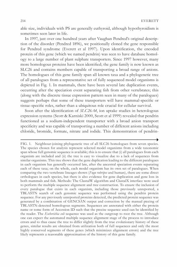
214 EVERETT
able size, individuals with PS are generally euthyroid, although hypothyroidism is sometimes seen later in life.
In 1997, just over one hundred years after Vaughan Pendred’s original descrip-tion of the disorder (Pendred 1896), we positionally cloned the gene responsible for Pendred syndrome (Everett et al 1997). Upon identifi cation, the encoded protein of this gene (which we named pendrin) was seen to have database homol-ogy to a large number of plant sulphate transporters. Since 1997 however, many more homologous proteins have been identifi ed; the gene family is now known as SLC26 and contains members capable of transporting a broad range of anions. The homologues of this gene family span all known taxa and a phylogenetic tree of all paralogues from a representative set of fully sequenced model organisms is depicted in Fig. 1. In mammals, there have been several late duplication events, occurring after the speciation event separating fi sh from other vertebrates; this (along with the discrete tissue expression pattern seen in many of the paralogues) suggests perhaps that some of these transporters will have mammal-specifi c or tissue-specifi c roles, rather than a ubiquitous role crucial for cellular survival.
Soon after the identifi cation of SLC26A4, ion uptake studies in heterologous expression systems (Scott & Karniski 2000, Scott et al 1999) revealed that pendrin functioned as a sodium-independent transporter with a broad anion transport specifi city and was capable of transporting a number of different anions including chloride, bromide, formate, nitrate and iodide. This demonstration of pendrin-
FIG. 1. Neighbour-joining phylogenetic tree of all SLC26 homologues from seven species. The species chosen for analysis represent selected model organisms from a wide taxonomic span whose full genomic sequence is available; this is to ensure that (i) all paralogues from each organism are included and (ii) the tree is easy to visualize due to a lack of sequences from similar organisms. This tree shows that the gene duplication leading to the different paralogues in each organism has generally occurred late, after the ancestral speciation events separating each of these taxa; on the whole, each model organism has its own set of paralogues. When comparing the two vertebrate lineages shown (Fugu rubripes and human), there are some direct orthologues in each species, but there is also evidence for gene duplication and gene loss in both mammals and fi sh. Methods: The ClustalW algorithm and ClustalX interface were used to perform the multiple sequence alignment and tree construction. To ensure the inclusion of every paralogue that exists in each organism, including those previously unreported, a TBLASTN search of each genomic sequence was performed using the pendrin protein sequence. For any previously unreported proteins detected, the predicted protein sequence was generated by a combination of GENCSAN output and correction by the manual piecing of TBLASTN-detected homologous segments. Sequences are annotated with either the protein name or some form of Accession ID such that the precise sequence used can be identifi ed by the reader. The Escherichia coli sequence was used as the outgroup to root the tree. Although one can expect the automated multiple sequence alignment stage of the process to introduce errors and to thus cause the tree to differ slightly from the true evolutionary history of these genes, similar results are obtained from utilization both of full sequences and only the most highly conserved segments of these genes (which minimizes alignment errors) and the tree likely represents a reasonable approximation of the history of this gene family.

THE ROLE OF PENDRIN IN THE INNER EAR 215
mediated iodide uptake immediately suggested that, in the thyroid, pendrin func-tioned as an apical porter of iodide from the thyrocyte to the follicular lumen (Fig. 2). This also explained the basis of the positive perchlorate discharge test observed in PS (Morgans & Trotter 1958), whereby radioactive iodide taken up by the thyroid is discharged from the thyroid again after administration of perchlorate. To briefl y explain the biochemical basis of this phenomenon, one should state that in normal individuals, any iodide taken up by the thyroid rapidly crosses the apical membrane to enter the follicular lumen. Here it is rapidly oxidized by the mem-brane-bound thyroid peroxidase and then bound to the tyrosine residues in thy-roglobulin, the precursor of thyroid hormones (Fig. 2). In individuals with PS, it

216 EVERETT
would appear that iodide transport across the apical membrane is retarded and iodide is thus trapped intracellularly. When perchlorate is administered, this com-petitively inhibits uptake of iodide by the sodium–iodide symporter (NIS) and the unbound intracellular iodide thus rapidly diffuses back across the basolateral mem-brane (down its high concentration gradient). It is therefore returned to the sys-temic circulation, causing a decrease in radioactivity over the thyroid and, as defi ned by this, a positive test result. The immunolocalization of pendrin to the apical membrane (Royaux et al 2000) further implicated pendrin’s role in apical iodide transport and therefore, although the precise mechanism of transport is unknown—some studies suggest that pendrin functions more like an ion channel at this location (Yoshida et al 2002), the function of pendrin in thyroid hormone biosynthesis has been at least partly defi ned.
SLC26A4 and pendrin in the inner ear
While gene identifi cation and determination of anion specifi city alone led to a hypothesis on the role of pendrin in the thyroid, its role in the inner ear remained more elusive. Although some speculated on the idea that hypothyroidism was responsible for the hearing loss, this seemed unlikely since the deafness associated with congenital hypothyroidism is (i) mild, (ii) not associated with the structural defects of the inner ear (Uziel et al 1981), and (iii) PS patients are generally euthy-roid. We therefore sought evidence that pendrin was directly involved in the audi-tory process in the inner ear.
First, several studies detected the presence of Slc26a4 and pendrin in mouse inner ears (Dou et al 2004, Everett et al 1999, Royaux et al 2003, Yoshino et al 2004). Collectively, all of these show that, in the cochlea, pendrin is located in the cells of the external sulcus and spiral prominence as well as the external sulcus root cells; in the vestibule, pendrin is located in the transitional cells surrounding all of the sensory epithelia, and in the endolymphatic duct and sac there is pendrin immunostaining at the apical membrane of the mitochondrial-rich cells. Tempo-rally, expression of Slc26a4 is seen from embryonic day 13 (E13) onwards (Everett et al 1999). Interestingly, although the precise role of these cells in reabsorption of endolymph has not been determined, it would appear that these pendrin-positive cells in the cochlea are key players not only in anion absorption but in the reabsorption of Na+ and K+ through non-selective cation channels (Marcus & Chiba 1999) and BK channels (Chiba & Marcus 2000).
Simultaneously, we also developed a mouse with a targeted disruption of the Slc26a4 gene (Everett et al 2001). Phenotypically, these mice are deaf, with a com-plete lack of auditory brainstem response waveforms at levels as high as 100 dB SPL (sound pressure level) in young adult mice. They also display signs of vestibu-lar dysfunction, such as circling, head-tilting, and head bobbing (for video clips

THE ROLE OF PENDRIN IN THE INNER EAR 217
of Slc26a4−/− mice, see http://research.nhgri.nih.gov/publications/pdsko/) and perform poorly on rotarod testing. Examination of the ears of these mice reveals profound dilatation of the endolymphatic compartment (Fig. 3A and 3B). This begins at around E15.5, which perhaps as one might expect, is shortly after the time that Slc26a4 expression begins. Ultrastructural examination by electron and confocal microscopy shows abnormalities and loss of the sensory hair cells in cochlea (Fig. 3C and 3D) and vestibule, thus explaining the deafness and imbalance. The utricu-lar and saccular maculae are often devoid of otoconia but, when they are present, they are often giant in nature (Fig. 3E and 3F). The abnormalities in these mice, however, are not limited to the sensory epithelia. Many of the other epithelial cells around the endolymphatic compartment take on an abnormal shape and lose their characteristic regular cobblestone appearance (Royaux et al 2003). These morpho-logical abnormalities are clearly seen in the marginal, intermediate, and basal cell layers of the stria vascularis as well as in the cells of the endolymphatic sac. The total thickness of the stria vascularis is also signifi cantly reduced, as is the thick-ness of the whole spiral ligament.
FIG. 2. Role of pendrin in thyroid hormone biosynthesis. In the thyroid, pendrin appears to mediate the transport of iodide across the apical membrane into the follicular lumen. Here it is rapidly oxidized by thyroperoxidase and bound to the tyrosine residues of thyroglobulin, the large globular precursor of thyroid hormones.

218 EVERETT
The severe abnormalities seen in these epithelial cells led us to wonder if they had ever been structurally normal, and therefore whether we were seeing degenera-tion or a deranged development of these areas. In the sensory cells at least, it appeared that hair cells of both the cochlea and the utricular and saccular maculae appeared completely normal until postnatal day 10 (Everett et al 2001), at which point degeneration began. It therefore seemed that two different pathological events were taking place, the fi rst of which caused a profound swelling of the endolymphatic compartment from the time of onset of Slc26a4 expression, and a second that caused some form of assault to a wide variety of epithelial cells border-ing the endolymphatic compartment, beginning at postnatal day 10 (which, inter-estingly, is the time that endolymphatic electrolyte composition becomes mature and the endocochlear potential is set up).
In an attempt to defi ne the nature of the assault to these epithelial cells, Royaux et al (2003) measured the composition of the endolymphatic fl uid in the scala media. They determined that the high concentration of K+ in this fl uid was largely maintained; however a complete loss of the endocochlear potential was seen, and the usual negative endocochlear potential (EP) induced by anoxia also failed to appear, presumably due to the loss of sensory hair cells. On further analysis they went on to determine that the loss of the endocochlear potential was due to the loss of KCNJ10 from the intermediate cells of the stria vascularis (Wangemann et al 2004), rather than from a direct effect of the loss of pendrin.
FIG. 3. Inner ear abnormalities seen in Slc26a4−/− mice (modifi ed from Everett et al 2001). (A, B) At postnatal day 1 (P1), a marked dilatation (hydrops) of the endolymphatic compart-ment is seen. The widening of the endolymphatic duct is particularly striking: compare the non-arrowed bars in each of the two panels (the common crus is marked with an arrowed bar because of the overlap of these two structures). (C, D) On scanning electron microscopy, degeneration of the cochlear sensory hair cells is seen in young adult mice. (E, F) Otoconia are often scarce or absent in Slc26a4−/− mice; when they are present, they are often giant in nature. Degeneration of the underlying otoconial membrane is also seen.

THE ROLE OF PENDRIN IN THE INNER EAR 219
Discussion
Results from numerous studies on the expression pattern/immunolocalization of Slc26a4/pendrin in the inner ear as well as those documenting the defects in Slc26a4−/− mice clearly show that pendrin directly participates in fl uid homeostasis in the inner ear. This refutes the hypothesis that all inner ear defects are secondary
FIG. 4. Pendrin in the endolymphatic sac. In the endolymphatic sac, pendrin immunolocal-izes to the mitochondria-rich cells, which protrude slightly from the epithelial surface. Various proteins involved in electrolyte transport are known to localize to these cells, although the mechanisms in which they likely synergistically act remain to be delineated. It is interesting to note the striking similarity between the localization of proteins in these cells and those in the renal cortical collecting duct and connecting tubule. It would appear that the mitochondria-rich cells are analogous to the renal non-A non-B intercalated cells. This renal/sac similarity is not limited to these cells. The ribosome-rich cells contain AQP2 in their apical membrane and vasopressin receptors in the basolateral membrane, paralleling the principal cells of the renal cortical collecting duct and connecting tubule. The presence of these two proteins in this loca-tion has important implications for fl uid homeostasis here since it suggests that these cells allow a vasopressin-mediated apical effl ux of water (out of the cell and into the endolymph) when systemic fl uid status requires the secretion of vasopressin; it would seem that this may have important implications for the treatment of disorders involving hydropic changes in this compartment.

220 EVERETT
to prenatal hypothyroidism. More importantly, the profound endolymphatic hydrops seen in Slc26a4−/− mice (beginning at the time of the onset of Slc26a4 expression) shows that Slc26a4 and the cells expressing it are responsible for the absorption of electrolytes from the endolymph (and thus reduction of the volume of the endolymphatic compartment). The clear visualization of this hydrops also likely explains the malformations of the inner ear in individuals with PS. While the cochlear Mondini malformation and the enlarged vestibular aqueduct were initially viewed as arrests in development, it now seems likely that both result from the dilatation of the endolymphatic compartment of the inner ear.
It is interesting to note that Slc26a4 is expressed in several discrete areas in the endolymphatic compartment of the inner ear and this may help explain the fi nd-ings of early research studies that sought to identify areas responsible for the reabsorption of endolymph. These studies showed that obliteration of the endol-ymphatic duct and sac induced an endolymphatic hydrops (Kimura & Schuknecht 1965) and this led to the suggestion that this structure was the area responsible for the endolymph reabsorption. Of course, this would require a longitudinal fl ow of endolymph from one area to the other, and from the apical turn of the cochlea to the basal turn. Later studies, in which markers were injected (Salt et al 1986) or iontophoresed (Salt & Thalmann 1988) into the scala media, however, suggested that the longitudinal fl ow rate in the cochlea is very low, and at a level incompatible with the idea of cochlear endolymph reabsorption occurring elsewhere. The expression of Slc26a4 in both the cochlea and in the endolymphatic duct and sac perhaps helps to explain these two paradoxical fi ndings; in a normally functioning inner ear, reabsorption of endolymph occurs throughout the endolymphatic com-partment; in the ear with an obliterated sac, however, the other regions are unable to compensate for the loss of this large area of absorptive epithelium, excess ves-tibular endolymph fl ows through into the cochlea, and a uniform hydrops through-out the endolymphatic compartment develops.
The mechanisms underlying fl uid homeostasis in the inner ear and the mainte-nance of the particularly usually electrolytic composition of the endolymph are currently still poorly understood. It has been shown that the stria vascularis and vestibular dark cells are responsible for the generation of the unusually high potas-sium concentration in the endolymph as well as, in the cochlea, its positive poten-tial of +80 mV, the EP; however, the mechanisms of cationic absorption as well as anionic homeostasis have yet to be elucidated. Since chloride is the major anion in the endolymph, one can speculate that pendrin is involved in a chloride uptake mechanism that serves to reduce the volume of the endolymph. It also seems pos-sible that pendrin plays a role in the regulation of pH in the endolymph. This notion is especially plausible given the otoconial irregularities seen in Slc26a4−/− mice. These abnormalities (which include the complete loss of otoconia or the formation of giant otoconia) have previously been observed after administration of acetazolamide (Kido et al 1991), a carbonic anhydrase inhibitor that has been
THE ROLE OF PENDRIN IN THE INNER EAR 221
shown to decrease the pH of fl uid in the endolymphatic duct and sac (Tsujikawa et al 1993). They occur, presumably, because of the effect of altered acid-base balance on the maintenance of anionic sites on otoconin-90 onto which the calcite matrix of calcium carbonate is seeded.
Further evidence for a role in acid–base balance is provided by pendrin’s role in the kidney, where it has been shown to be the long sought bicarbonate trans-porter of the B-intercalated cells of the cortical collecting duct (Royaux et al 2001, Wall et al 2003, Wall 2006, this volume). It is interesting to note the many similari-ties seen between the expression pattern of various solute carriers in these two different locations; clearly the development of these two areas is under the similar transcriptional control mechanisms during development, although it seems one should be cautious in making any assumptions that the cells in each location have a similar function. Nevertheless, in continuing the comparison, pendrin-positive/mitochondrial-rich cells in the endolymphatic duct and sac seem to have most similarity to the non-A non-B cells of the cortical collecting duct (CCD) and con-necting tubule (CNT), since both pendrin and vH+ATPase localize to the apical membrane (Dou et al 2004, Stankovic et al 1997), with one of the SLC4 anion transporters located at the basolateral membrane (Stankovic et al 1997) and car-bonic anhydrase (CA II) in the cytoplasm (Dou et al 2004). Unfortunately, the function of non-A non-B cells in the kidney is not clear, as they would not appear to be responsible for the secretion of either base or alkali. The ribosome-rich cells in the endolymphatic duct and sac would appear to also have similarity to cells of the CCD and CNT, in this case the principal cells. These have been shown to express AQP2 in apical membrane vesicles (Beitz et al 1999) although, due to the hypertonic nature of the endolymph with respect to perilymph or plasma (Konishi et al 1984), it would appear that mobilization of AQP2 to the apical membrane—mediated by activation of vasopressin receptors on the basolateral membrane of these cells (Beitz et al 1999, Kumagami et al 1998)—should cause an effl ux of water; this hypothesis is strengthened by the fact that administration of vaso-pressin causes an endolymphatic hydrops (Takeda et al 2000). It seems important to note at this stage that water entry into the endolymph at this site is mediated by vasopressin; one could hypothesize that this could have important implications for the treatment of endolymphatic hydrops, since it would suggest that endolym-phatic volume can be partially regulated by systemic fl uid status.
Although the development of Slc26a4−/− mice has undoubtedly revealed new insights into the role of pendrin in the inner ear, it would seem that one major problem with this model is the fact that secondary degenerative changes are seen throughout the epithelia surrounding the endolymphatic compartment. The loss of the endocochlear potential, for example, is not surprising given the secondary degeneration of the stria vascularis and does not result directly from the loss of pendrin. This stria vascularis is responsible for the secretion of K+ into the endol-ymphatic compartment, and the mechanism underlying this has been elucidated

222 EVERETT
(Fig. 5). KCNJ10, in the intermediate cells, is crucial to this process, and is respon-sible for the high intrastrial potential (and thus the endocochlear potential). Indeed, although the lack of Slc26a4 and pendrin during embryonic development leads to an initial hydrops, one can speculate that the loss of KCNJ10 in these mice may prevent further expansion of the endolymphatic compartment, since the secretory epithelium becomes as damaged as the absorptive epithelium. Certainly, mice with targeted disruptions of the transporters and channels active in this secretory process, i.e. KCNQ1 (Casimiro et al 2001, Lee et al 2000), KCNE1 (Vetter et al 1996), NKCC1 (Dixon et al 1999) and KCNJ10 (Marcus et al 2002) all show varying degrees of collapse of the endolymphatic compartment, the reverse of that seen in Slc26a4−/− mice.
Future studies
At this stage, it is clear that we are still in only the early stages of elucidating the mechanisms underlying fl uid and electrolyte homeostasis in the inner ear even if
FIG. 5. Mechanism of secretion of K+ in the stria vascularis. The Na+/K+ATPase located in the basolateral membrane of the strial marginal cells is responsible for the active pumping of K+ out of the intrastrial space. The presence of high K+ in intermediate cells, generated by the recycling of K+ through the basal cells and fi brocytes, causes effl ux of K+ into the intrastrial space down this concentration gradient, setting up a positive DC potential here. This in turn promotes the secretion of K+ into the endolymph through the strial marginal cells. In Slc26a4−/− mice, all three layers of this structure are visibly abnormal, and loss of the KCNJ10 protein has been demonstrated. The loss of the endocochlear potential can therefore be attrib-uted to this secondary effect.

THE ROLE OF PENDRIN IN THE INNER EAR 223
such knowledge will likely be rapidly acquired within the next decade. One must ask, however, whether full characterization of these mechanisms must be achieved before performing studies aiming to discover methods of correcting defects in these processes. As a result of the studies described above, it would appear that PS is fairly unique amongst the causes of childhood deafness as it may be amenable to treatment by pharmacotherapeutic means. Children with PS often have useable hearing in early childhood, and children with large vestibular aqueduct syndrome (LVAS), a disorder often associated with one mutation in SLC26A4—as well as one or more unknown genetic alteration(s) elsewhere (Pryor et al 2005)—the progression of hearing loss is even slower. It seems unlikely that hearing loss could be prevented altogether, but if its onset could be slowed, it could allow a child to develop language skills before a profound hearing loss occurred; this would forever provide them with better communication skills, or a better outcome after cochlear implantation. At present, it is not known whether the hearing loss in PS and LVAS is simply due to the trauma caused to the endolymphatic compartment due to increased pressure changes transmitted from the intracranial cavity through the widened vestibular aqueduct, or whether it is due to disturbances in the electro-chemical composition of the endolymph. If it were the former, it would seem that the hearing loss could only be prevented if therapeutic agents with good transpla-cental transfer were administered to the mother and this would clearly appear to have a limited therapeutic role. If the progression of hearing loss is due to distur-bances of endolymphatic composition or volume however, then it seems that a variety of drugs could be tested to see if the natural history of the disease process could be altered. Many drugs have been shown to alter the electrolytic composition of the endolymph, especially diuretics. Examples in this class include canreoate, an aldosterone antagonist that decreases the endolymphatic sac potential (Mori et al 1991) and OPC-31260, a vasopressin antagonist that decreases the degree of experimental hydrops in animal models (Takeda et al 2003). Furthermore, since secretion of endolymph appears to be mediated by b -adrenergic stimulation (Fauser et al 2004), even the use of drugs such as b -adrenergic antagonists may be of benefi t. Finally, if degeneration of the epithelia is due to free radical/oxidative damage, then free radical scavengers such as high-dose allopurinol may be of use. These studies appear to be the logical next step in trying to alter the disease process in PS and Slc26a4−/− mice and it would seem important to perform these studies in the near future.
References
Beitz E, Kumagami H, Krippeit-Drews P, Ruppersberg JP, Schultz JE 1999 Expression pattern of aquaporin water channels in the inner ear of the rat. The molecular basis for a water regu-lation system in the endolymphatic sac. Hear Res 132:76–84

224 EVERETT
Casimiro MC, Knollmann BC, Ebert SN et al 2001 Targeted disruption of the Kcnq1 gene produces a mouse model of Jervell and Lange-Nielsen Syndrome. Proc Natl Acad Sci USA 98:2526–2531
Chiba T, Marcus DC 2000 Nonselective cation and BK channels in apical membrane of outer sulcus epithelial cells. J Membr Biol 174:167–179
Dixon MJ, Gazzard J, Chaudhry SS, Sampson N, Schulte BA, Steel KP 1999 Mutation of the Na-K-Cl co-transporter gene Slc12a2 results in deafness in mice. Hum Mol Genet 8:1579–1584
Dou H, Xu J, Wang Z et al 2004 Co-expression of pendrin, vacuolar H+ -ATPase alpha4-subunit and carbonic anhydrase II in epithelial cells of the murine endolymphatic sac. J Histochem Cytochem 52:1377–1384
Everett LA, Belyantseva IA, Noben-Trauth K et al 2001 Targeted disruption of mouse Pds provides insight about the inner-ear defects encountered in Pendred syndrome. Hum Mol Genet 10:153–161
Everett LA, Glaser B, Beck JC et al 1997 Pendred syndrome is caused by mutations in a putative sulphate transporter gene (PDS). Nat Genet 17:411–422
Everett LA, Morsli H, Wu DK, Green ED 1999 Expression pattern of the mouse ortholog of the Pendred’s syndrome gene (Pds) suggests a key role for pendrin in the inner ear. Proc Natl Acad Sci USA 96:9727–9732
Fauser C, Schimanski S, Wangemann P 2004 Localization of beta1-adrenergic receptors in the cochlea and the vestibular labyrinth. J Membr Biol 201:25–32
Johnsen T, Larsen C, Friis J, Hougaard-Jensen F 1987 Pendred’s syndrome. Acoustic, vestibular and radiological fi ndings in 17 unrelated patients. J Laryngol Otol 101:1187–1192
Kido T, Sekitani T, Yamashita H, Endo S, Masumitsu Y, Shimogori H 1991 Effects of carbonic anhydrase inhibitor on the otolithic organs of developing chick embryos. Am J Otolaryngol 12:191–195
Kimura RS, Schuknecht H 1965 Membranous hydops in the inner ear of the guinea pig after the obliteration of the endolymphatic sac. Pract Otorhinolaryngol 27:343–345
Konishi T, Hamrick PE, Mori H 1984 Water permeability of the endolymph-perilymph barrier in the guinea pig cochlea. Hear Res 15:51–58
Kumagami H, Loewenheim H, Beitz E et al 1998 The effect of anti-diuretic hormone on the endolymphatic sac of the inner ear. Pfl ugers Arch 436:970–975
Lee MP, Ravenel JD, Hu RJ et al 2000 Targeted disruption of the Kvlqt1 gene causes deafness and gastric hyperplasia in mice. J Clin Invest 106:1447–1455
Marcus DC, Chiba T 1999 K+ and Na+ absorption by outer sulcus epithelial cells. Hear Res 134:48–56
Marcus DC, Wu T, Wangemann P, Kofuji P 2002 KCNJ10 (Kir4.1) potassium channel knockout abolishes endocochlear potential. Am J Physiol Cell Physiol 282:C403–407
Mondini C 1791 Anatomia surdi nati sectio: De Boroniensi scientarium et artium instituto atque acedemia commnetarii. Bononiae 7:419–431
Morgans ME, Trotter WR 1958 Association of congenital deafness with goitre; the nature of the thyroid defect. Lancet 1:607–609
Mori N, Yura K, Uozumi N, Sakai S 1991 Effect of aldosterone antagonist on the DC potential in the endolymphatic sac. Ann Otol Rhinol Laryngol 100:72–75
Pendred V 1896 Deaf mutism and goitre. Lancet ii:532Pryor SP, Madeo AC, Reynolds JC et al 2005 SLC26A4/PDS genotype-phenotype correlation
in hearing loss with enlargement of the vestibular aqueduct (EVA): evidence that Pendred syndrome and non-syndromic EVA are distinct clinical and genetic entities. J Med Genet 42:159–165

THE ROLE OF PENDRIN IN THE INNER EAR 225
Royaux IE, Suzuki K, Mori A et al 2000 Pendrin, the protein encoded by the Pendred syndrome gene (PDS), is an apical porter of iodide in the thyroid and is regulated by thyroglobulin in FRTL-5 cells. Endocrinology 141:839–845
Royaux IE, Wall SM, Karniski LP et al 2001 Pendrin, encoded by the Pendred syndrome gene, resides in the apical region of renal intercalated cells and mediates bicarbonate secretion. Proc Natl Acad Sci USA 98:4221–4226
Royaux IE, Belyantseva IA, Wu T et al 2003 Localization and functional studies of pendrin in the mouse inner ear provide insight about the etiology of deafness in pendred syndrome. J Assoc Res Otolaryngol 4:394–404
Salt AN, Thalmann R 1988 Interpretation of endolymph fl ow results: a comment on ‘Longi-tudinal fl ow of endolymph measured by distribution of tetraethylammonium and choline in scala media’. Hear Res 33:279–284
Salt AN, Thalmann R, Marcus DC, Bohne BA 1986 Direct measurement of longitudinal endolymph fl ow rate in the guinea pig cochlea. Hear Res 23:141–151
Scott DA, Karniski LP 2000 Human pendrin expressed in Xenopus laevis oocytes mediates chloride/formate exchange. Am J Physiol Cell Physiol 278:C207–211
Scott DA, Wang R, Kreman TM, Sheffi eld VC, Karniski LP 1999 The Pendred syndrome gene encodes a chloride-iodide transport protein. Nat Genet 21:440–443
Stankovic KM, Brown D, Alper SL, Adams JC 1997 Localization of pH regulating proteins H+ATPase and Cl-/HCO3- exchanger in the guinea pig inner ear. Hear Res 114:21–34
Takeda T, Takeda S, Kitano H, Okada T, Kakigi A 2000 Endolymphatic hydrops induced by chronic administration of vasopressin. Hear Res 140:1–6
Takeda T, Sawada S, Takeda S et al 2003 The effects of V2 antagonist (OPC-31260) on endol-ymphatic hydrops. Hear Res 182:9–18
Tsujikawa S, Yamashita T, Tomoda K et al 1993 Effects of acetazolamide on acid-base balance in the endolymphatic sac of the guinea pig. Acta Otolaryngol Suppl 500:50–53
Uziel A, Gabrion J, Ohresser M, Legrand C 1981 Effects of hypothyroidism on the structural development of the organ of Corti in the rat. Acta Otolaryngol 92:469–480
Vetter DE, Mann JR, Wangemann P et al 1996 Inner ear defects induced by null mutation of the isk gene. Neuron 17:1251–1264
Wall SM 2006 The renal physiology of pendrin (Slc26a4) and its role in hypertension. In: Epi-thelial anion transport in health and disease: the role of the SLC26 transporters family. Wiley, Chichester (Novartis Found Symp 273) p 231–243
Wall SM, Hassell KA, Royaux IE et al 2003 Localization of pendrin in mouse kidney. Am J Physiol Renal Physiol 284:F229–241
Wangemann P, Itza EM, Albrecht B et al 2004 Loss of KCNJ10 protein expression abolishes endocochlear potential and causes deafness in Pendred syndrome mouse model. BMC Med 2:30
Yoshida A, Taniguchi S, Hisatome I et al 2002 Pendrin is an iodide-specifi c apical porter responsible for iodide effl ux from thyroid cells. J Clin Endocrinol Metab 87:3356–3361
Yoshino T, Sato E, Nakashima T et al 2004 The immunohistochemical analysis of pendrin in the mouse inner ear. Hear Res 195:9–16
DISCUSSION
Markovich: Is there a thyroid phenotype in your mice?Everett: Not at the moment. Thyroid function tests are completely normal. If we
look at the size of the thyroids, these are normal too. It is interesting that other thyroid parameters differ quite considerably among strains. We are interested in

226 EVERETT
making a congenic onto C57BL/6J to see whether they have a thyroid phenotype.
Muallem: How can you get high K+ within the endolymph and still lose mem-brane potential? What channels are missing?
Everett: In the pendrin knockouts, KCNJ10 has been shown to be missing (Wangemann et al 2004). The stria vascularis is made up of three cell types: the marginal, intermediate and basal cells. There is an intrastrial space in between which does contain a positive endocochlear potential but it does not have the high K+. NKCC1, KCNE1 and KCNJ10 are involved. There are a whole range of trans-porters and channels which cause the endocochlear potential to become positive in the intrastrial compartment, but then the K+ is pumped out to create a high level in the endolymph itself through the marginal cells.
Muallem: Since you are getting K+ secretion you must be losing some kind of channel activity that is associated with pendrin to explain the loss of membrane potential.
Everett: The problem is, in the pendrin knockout most of the changes in endol-ymphatic composition are going to be secondary effects relating to the degenera-tion of the stria vascularis, so it is diffi cult to discern the primary effect of a lack of pendrin. In these mice there is clearly an endolymphatic hydrops. However, the fact that KCNJ10 is missing due to strial degeneration may actually be lessening the severity of the hydrops as a lack of KCNJ10 causes a partial collapse of the scala media. Without this secondary effect the hydrops could be worse. Perhaps a high K+ level is retained because of the endolymphatic hydrops and the ion com-position, owing to a lack of endolymph anion resorption by pendrin. If just KCNJ10 is missing there is collapse of the endocochlear compartment, loss of endocochlear potential and a reduced K+ concentration.
Welsh: What generates the voltage? Is it related to the K+ diffusion potential?Everett: It is actively pumped into the endolymph.Ashmore: There is meant to be an electrogenic pump between the marginal and
interstitial cells. This is considered to be the main cause.Welsh: Is the pump a K+ pump?Ashmore: It is an Na+/K+ -ATPase.Welsh: If that is still intact, based on the fact that you have high K+, why is the
voltage not there?Muallem: There must be some kind of a channel present that gets inactivated. It
would have to be a K+ channel of some sort.Everett: There are a lot of channels. There is KCNQ1 and NKCC1 on the mar-
ginal cell, and when these are knocked out the result is collapse of the endolym-phatic compartment and loss of the endocochlear potential.
Muallem: You are seeing a completely different phenotype. NKCC1 knockout is deaf, but this is because it results in complete loss of endolymphatic K+. Every
THE ROLE OF PENDRIN IN THE INNER EAR 227
time K+ is lost there is deafness. It seems that you are maintaining all the ion trans-porters and losing the potential.
Ashmore: There are two steps. You need to have K+ to load the scala media facing the hair cell stereocilia and on top of that you have to crank up the potential of the layer of cells that is facing the internal compartment of the stria vascularis. These two events seem to occur independently during development.
Alper: Is it a proton pump that does this?Ashmore: There are several different models, but I don’t think it is. It is a Na+/K+
exchanger.Alper: Where is this located?Knipper: At the basal cells of the stria vascularis. In the marginal cells I would
guess that KCNQ1 and KCNE1 may be affected. This may be the reason the current is lost. Once you have this kind of defect, the high turnover of these K+ ion channels, which passively drive K+ into the endolymph, appears to be affected within hours.
Muallem: She is getting the K+ into the endolymph. The K+ looks normal. So the channel has to be there.
Oliver: If I remember correctly, the endocochlear potential is essentially a diff-usion potential across the membrane of the intermediate cells. The intrastrial fl uid space is very low in K+ so there is a highly positive potential generated at the membrane of the intermediate cells by the potassium channel KCNJ10 (Kir4.1).
Knipper: I presume the problem is that as soon as you get rid of the inward rec-tifying Kir4.1 you lose the driving force. This is because the inward rectifying channel is the one responsible for keeping the K+ low in the intermedial space. If the potassium in the intermedial space is high because the Kir4.1 ion channel is missing, there is no driving force for passive transport of K+ from one side to the other.
Welsh: What role does pendrin play in this? Is it involved in absorption, for example?
Everett: There is no pendrin in the stria vascularis. It seems that most of the endolymphatic composition changes are resulting from the loss of stria vascularis and this presumably occurs as a secondary effect to the loss of pendrin. I assume that pendrin loss causes an early problem with endolymphatic homeos-tasis (and this precise defect will prove diffi cult to identify) and this then poisons the stria vascularis itself. It may be because of loss of the K+ recycling mechanism.
Knipper: I don’t understand. If you consider what the channel might do, you suggest that it transports iodine versus chloride in the thyroid. If I consider its expression pattern in the cochlear, it is striking how close it maps to deiodinase, the enzyme that catabolizes T4 to T3. Only T3 is active. If SLC26A4 is only there

228 EVERETT
to get rid of the iodine, this would make sense. If this is the case, can you exclude that the deafness has perhaps nothing or only indirectly to do with SLC26A4, but instead, may be the result of a reduced level of thyroid hormone during pregnancy, which in turn is the result of reduced levels of local T3. The reduced levels of local T3, however, may be the result of a loss of driving force for the deiodination required for T4 to T3 conversion due to PDS gene deletion. This would fi t nicely with the progressive hearing loss you see. During a critical time period the reduced level of thyroid hormone caused by the presumptive low function of deiodinases would lead to a failure of proper fi nal differentiation of hair cells or other compart-ments and deafness.
Everett: I agree that sensory hair cell defects are seen in hypothyroidism.Knipper: The phenotype you described would fi t with a loss of function of
deiodinases.Everett: So you are postulating that the swelling of the endolymphatic hydrops
is due to iodide.Quinton: What is the concentration of chloride?Everett: The only ion measurement study published so far is that of Dan Marcus’s
K+ measurements (Royaux et al 2003).Kere: Has pH been measured yet?Everett: This is going to be vital. I am quite sure that pendrin could be transport-
ing bicarbonate.Alper: Wangemann has found that pendrin-expressing cochlear epithelial cells
mediate Cl−/OH− and Cl−/formate exchange, and that cochlear endolymph in the deaf pendrin knockout mouse is 0.4 units more acidic than wild-type endolymph.
Ashmore: So pendrin is actually a chloride–bicarbonate exchanger?Alper: It can function in this way, yes.Quinton: If it were functioning as a chloride channel, wouldn’t this give you the
positive potential?Everett: I think it is involved in reabsorption of the endolymph. It is a secondary
effect.Quinton: How do you get that positive potential? Is this simply due to a proton
ATPase?Everett: In wild-type it is defi nitely created by the stria vascularis, and pendrin
is not located hereAlper: I’d like to ask the nephrologists in the audience, could this be obstruction
nephropathy? Is this a hydrostatic issue? Or is it going to be a pH and potential issue?
Aronson: I won’t answer that directly, but I was struck that proton ATPase and pendrin are in the same cells with carbonic anhydrase. The proton pump mutations

THE ROLE OF PENDRIN IN THE INNER EAR 229
can cause deafness also. If one is acting as a chloride–bicarbonate exchanger and the other is pumping protons, they have opposite effects on pH. This suggests that they have to do something together; knocking out either one disrupts this func-tion. In a sense, if you have a proton pump in parallel with a chloride–bicarbonate exchanger, the net effect is to have an inward chloride pump. This would in a sense be an electrogenic chloride uptake mechanism. This would also favour the tran-sepithelial potential difference that is generated. Is it possible that you need a proton pump and a chloride–bicarbonate exchanger working together, and losing either one causes deafness?
Muallem: Does mutation of the proton pump also lead to a collapse of the mem-brane potential?
Aronson: I don’t know.Welsh: Then you might expect too little fl uid. They are saying they have
hydrops.Soleimani: If there is a basolateral NKCC1 in these cells, then once you inhibit
pendrin on the apical membrane, the intracellular chloride should drop which should activate the basolateral NKCC1, if indeed there is one. It is possible that if this happens, NKCC1 is going to bring the chloride in. Can we get a similar sce-nario if there is an inactivating mutation in pendrin?
Ashmore: The issue is that pendrin is in a different location from all the other transporters we are talking about. This is the complexity.
Aronson: This model for the proton pump working with the chloride–bicarbonate exchanger would have an obligate role for carbonic anhydrase. As the proton pump generates hydroxyls you need to form bicarbonate to work the pendrin. There is a metabolon here that has the net effect of mediating electrogenic uptake of chloride.
Kere: In the chloride diarrhoea, one of the things that has not been men-tioned here is that not only is the output from the large intestine very high in chloride, but it is also acidic. The pH is entirely screwed up when A3 is not working.
Soleimani: CA2 mutants are not deaf.Alper: There is a lot of redundancy. I have an off-the-wall question. The sodium
iodide symporter NIS is also located in the mammary duct, and it is greatly up-regulated during lactation. How does iodide exit the apical side of the mammary duct for secretion?
Everett: Pendrin is expressed in mammary gland.Alper: Marlies Knipper was pursuing this issue earlier: one can certainly imagine
a neonatal iodide defi ciency in that context.Everett: It is diffi cult to explain the deafness features, but I agree that pendrin
could be acting as iodide transporter here.

230 EVERETT
References
Royaux IE, Belyantseva IA, Wu T et al 2003 Localization and functional studies of pendrin in the mouse inner ear provide insight about the etiology of deafness in pendred syndrome. J Assoc Res Otolaryngol 4:394–404
Wangemann P, Itza EM, Albrecht B et al 2004 Loss of KCNJ10 protein expression abolishes endocochlear potential and causes deafness in Pendred syndrome mouse model. BMC Med 2:30

The renal physiology of pendrin (SLC26A4) and its role in hypertensionSusan M. Wall
Renal Division, Emory University School of Medicine, 1639 Pierce Drive, NE Atlanta, GA 30322, USA
Abstract. SLC26A4 (pendrin, PDS) is a Na+ -independent, Cl−/HCO3−/OH− exchanger
that is expressed in the apical regions of type B and non-A, non-B intercalated cells within the cortical collecting duct (CCD), the connecting tubule and the distal convo-luted tubule where it mediates HCO3
− secretion and Cl− absorption. SLC26A4 is upregu-lated with aldosterone analogues and with Cl− restriction. While under basal conditions no renal abnormalities are observed in mice and humans with genetic disruption of SLC26A4 (Pendred syndrome), differences become apparent under conditions wherein the transporter is stimulated. Following treatment with aldosterone analogues, e.g. deoxycorticosterone pivalate (DOCP), weight gain and hypertension are observed in Slc26a4+/+ but not in Slc26a4−/− mice. During dietary NaCl restriction, a model in which serum aldosterone is appropriately increased, urinary volume and urinary excretion of Cl− are greater in Slc26a4−/− than in wild-type mice which results in apparent vascular volume contraction in Slc26a4−/− mice. Moreover, during NaCl restriction or following DOCP treatment, Slc26a4−/− mice have a higher serum HCO3
− than wild type mice from an impaired ability to excrete OH− equivalents. In conclusion, SLC26A4 regulates blood pressure and arterial pH, likely by participating in the renal regulation of net acid and Cl− excretion.
2006 Epithelial anion transport in health and disease: the role of the SLC26 transporters family. Wiley, Chichester (Novartis Foundation Symposium 273) p 231–243
Aldosterone increases blood pressure by acting on the collecting duct to stimulate the absorption of NaCl and acts on vascular tissue to increase resistance. Aldos-terone administration in vivo leads to increased absorption of Na+ by the kidney primarily through activation of the Na+ channel ENaC (Eaton et al 2001), the thiazide-sensitive NaCl cotransporter (TSC/NCC)(Kim et al 1998), and the Na+/K+ -ATPase (Ewart & Klip 1995). The result is aldosterone-induced Na+ retention and increased blood pressure. In rodents, administration of aldosterone analogues and a high NaCl diet has been used as a model of salt-sensitive hypertension (Kurtz & Morris 1983). In salt-sensitive hypertension, it has generally been assumed that intake of Na+, rather than Cl−, leads to increased blood pressure. However, in humans with salt-sensitive hypertension blood pressure varies more with intake of
231

232 WALL
Cl− than with intake of Na+ (Kurtz et al 1987). Nevertheless, identifi cation of aldosterone-sensitive Cl− transporters and the contribution of these Cl− trans-porters to salt-sensitive hypertension, has not been described fully (Ornellas et al 2002).
The collecting duct is the fi nal site of regulation for renal Cl− excretion. With administration of aldosterone in vivo transepithelial transport of both Cl− and HCO3
− increases dramatically along the collecting duct, particularly within the cortical collecting duct (CCD) (Knepper et al 1984, Star et al 1985, Garcia-Austt et al 1985, Hanley & Kokko 1978). In models of metabolic alkalosis, the CCD absorbs Cl− and secretes HCO3
− through transepithelial transport across inter-calated cells, rather than across principal cells (Schlatter et al 1990). This process is mediated by an apical Cl−/HCO3
− exchanger acting in series with a basolateral Cl− conductance (Schuster 1993) and an H+ -ATPase (Fig. 1) (Emmons 1993).
Intercalated cells represent the minority cell type within the CCD, connecting tubule (CNT) and distal convoluted tubule (DCT) and are classifi ed as A, B, or
AE1
Pds
Pds
ATPase
ATPase
ATPase
H+
H+
H+
HCO3-
Cl-
HCO3-
Cl-
Cl-
HCO3-
Intercalated Cell Subtypes
Interstitium Lumen
Type A
Type B
Non-A, non-B
FIG. 1. Intercalated cell subtypes: classifi cation of intercalated cell subtypes is based on the presence or absence of AE1 and the polarity of the H+ -ATPase. Pendrin expression is high in the apical regions of non-A, non-B intercalated cells and type B intercalated cells (Wall et al 2003).

RENAL PHYSIOLOGY OF PENDRIN 233
non-A, non-B based on the presence or absence of the anion exchanger, AE1, and the distribution of the H+ -ATPase within the cell (Alper et al 1989, Kim et al 1999, Schuster 1993, Teng-ummnuay et al 1996) (Fig. 1). Intercalated cells secrete or absorb net H+ equivalents depending on whether the H+ -ATPase is expressed on the apical or the basolateral plasma membrane (Brown et al 1988, Emmons 1993). Type A intercalated cells secrete H+ equivalents through the H+ -ATPase, which localizes to the apical plasma membrane and functions in series with the Cl−/HCO3
− exchanger, AE1 expressed on the basolateral plasma membrane (Teng-ummnuay et al 1996). In the type B intercalated cell, Cl−/HCO3
− exchange across the apical plasma membrane functions in series with the H+ -ATPase expressed on the basolateral plasma membrane (Alper et al 1989, Kim et al 1999, Teng-ummnuay et al 1996) to mediate secretion of OH− equivalents, particularly during metabolic alkalosis (Schuster 1993). As will be discussed below, SLC26A4 (PDS, pendrin) likely represents the gene responsible for apical anion exchange-mediated HCO3
− secretion (Royaux et al 2001, Soleimani et al 2001, Frische et al 2003, Verlander et al 2003) since both localize to the apical plasma membrane of the type B intercalated cell (Emmons & Kurtz 1994, Royaux et al 2001, Wall et al 2003, Kim et al 2002), both transport Cl− and HCO3
− (Scott et al 1999, Royaux et al 2001, Soleimani et al 2001, Wall et al 2004) and both are regulated in parallel (Frische et al 2003, Verlander et al 2003). As will be discussed below, by altering excretion of Cl− and OH− equivalents, SLC26A4 is an important regulator of both arterial pH and blood pressure.
Pendred syndrome
In 1896 Vaughan Pendred described an Irish family in which 2 of 10 children suf-fered from congenital deafness and goitre (Pendred 1896). Pendred syndrome accounts for up to 10% of all hereditary hearing loss and thus is recognized as one of the leading causes of congenital deafness (Everett et al 1997). Patients with Pendred syndrome have multinodular goitre, although they are generally euthyroid (Everett et al 1997, Pendred 1896).
While pendrin is highly expressed in kidney, the physiological signifi cance of pendrin in human kidney is not obvious since affected individuals and Slc26a4−/− mice (pendrin knockout mice) have no known acid-base or fl uid and electrolyte abnormalities and appear to have normal renal function at least under basal, unstimulated conditions (Royaux et al 2001). However, under conditions that stimulate pendrin in normal rodents, such as with administration of aldosterone analogues, differences between Slc26a4−/− and Slc26a4+/+ mice in acid–base and fl uid balance become apparent (Verlander et al 2003), due to reduced capacity of Slc26a4 null mice to secrete HCO3
− and absorb Cl− (Royaux et al 2001, Wall et al 2004).
234 WALL
Cloning and the tissue distribution of SLC26A4
The molecular structure of SLC26A4 was fi rst reported by Everett and colleagues (Everett et al 1997). In human, mouse and rat kidney, pendrin is expressed in the CCD, the CNT, the initial collecting tubule (iCT) and the DCT (Royaux et al 2001, Wall et al 2003, Kim et al 2002), where it localizes to the apical plasma membrane and apical cytoplasmic vesicles of both type B and non-A, non-B intercalated cells (Royaux et al 2001, Wall et al 2003, Kim et al 2002) (Fig. 1). Apical localization of pendrin to a subset of intercalated cells suggests that pendrin participates in the regulation of acid-base balance (Royaux et al 2001). However, intercalated cells also participate in the process of Cl− absorption and secretion (Wall et al 2001, Verlander et al 2003).
Aldosterone analogues up-regulate pendrin expression
Slc26a4 is up-regulated in a variety of models of metabolic alkalosis, such as with aldosterone analogues (Verlander et al 2003) and following ingestion of NaHCO3 (Frische et al 2003, Quentin et al 2004). Our laboratory has observed a modest increase in Slc26a4 mRNA and total pendrin protein expression following the administration of the aldosterone analogue, deoxycorticosterone pivalate (DOCP) (Verlander et al 2003). However, pendrin is regulated in this treatment model primarily through subcellular redistribution rather than changes in total protein expression (Verlander et al 2003).
The subcellular distribution of pendrin protein is highly regulated by aldoster-one analogues (Verlander et al 2003). In kidneys from control mice, pendrin immunoreactivity in type B intercalated cells was prevalent over apical cytoplasmic vesicles, with little immunolabels present along the apical plasma membrane. Fol-lowing DOCP treatment, however, the apical plasma membrane label density of Slc26a4 increased twofold in both type B intercalated cells and in non-A, non-B intercalated cells. Boundary length increased twofold in type B but did not change in non-A, non-B intercalated cells, whereas total protein expression per cell increased twofold in non-A, non-B, but does not change in type B intercalated cells. Thus, following the administration of aldosterone analogues, apical plasma membrane Slc26a4 expression rose twofold in non-A, non-B intercalated cells, and rose sixfold in type B intercalated cells (Verlander et al 2003).
Aldosterone analogues administered in vivo might up-regulate Slc26a4 indirectly such as through hypokalemia, volume expansion and/or metabolic alkalosis which are observed in this treatment model. However, since pendrin is up-regulated in many treatment models in which serum aldosterone levels are increased independ-ent of changes in serum K+, arterial pH and vascular volume status, aldosterone likely has a direct effect on Slc26a4 expression. However, pendrin is also regulated
RENAL PHYSIOLOGY OF PENDRIN 235
through aldosterone-independent mechanisms such as with changes in Cl− balance (Quentin et al 2004).
Slc26a4 mediates HCO3- secretion and Cl- absorption, which modulates
acid–base balance, fl uid balance and blood pressure
Because pendrin is a Na+ -independent, Cl−/HCO3− exchanger in heterologous
expression systems, we explored the role of Slc26a4 in the process of Cl− absorption and HCO3
− secretion in native kidney tissue. Thus, Slc26a4−/− and Slc26a4+/+ mice were treated with DOCP and given 50 mM NaHCO3 in their drinking water for 7 days to up-regulate pendrin (Royaux et al 2001, Frische et al 2003, Verlander et al 2003). CCD tubules from these mice were perfused in vitro in the presence of symmetric, physiological, HCO3
−/CO2-buffered solutions. Cl− absorption and HCO3
− secretion were observed in CCDs from wild-type mice, but were not observed in tubules from Slc26a4 null mice (Royaux et al 2001, Wall et al 2004). Thus Slc26a4 is a critical component of the Cl− absorption and HCO3
− secretion observed in the CCD following administration of aldosterone analogues and ingestion of NaHCO3. In mouse, Slc26a4-dependent Cl− absorption and HCO3
− secretion are probably greater in the CNT than in the CCD since pendrin expres-sion is much greater in the apical plasma membrane of the CNT than the CCD under both basal conditions and following the administration of DOCP (Wall et al 2003, Verlander et al 2003). Moreover, Na+ absorption is fourfold higher in CNT than CCD (Almeida & Burg 1982).
Although pendrin is highly expressed in kidney, under basal, unstimulated conditions, both Slc26a4 null mice and individuals with genetic disruption of the Slc26a4 gene (Pendred syndrome) have normal acid–base balance (Royaux et al 2001). However, following DOCP administration, arterial pH and serum HCO3
− are higher in Slc26a4−/− than Slc26a4+/+ mice, consistent with the reduced HCO3
− secretion observed in the CCD of Slc26a4−/− mice, which should limit correction of a metabolic alkalosis (Royaux et al 2001, Verlander et al 2003).
Since pendrin is a Cl− transporter, we explored the role of pendrin in salt and water balance. After consuming a NaCl-replete diet for 7 days, systolic blood pressure and weight change were similar in Slc26a4−/− and Slc26a4+/+ mice (Fig. 2) (Verlander et al 2003). Seven days after administration of DOCP and a NaCl-replete diet, wild type mice gained weight and their systolic blood pressure rose from 111 ± 1 (n = 7) to 132 ± 3 mmHg (n = 10, P < 0.05) (Verlander et al 2003). However, Slc26a4−/− mice treated with DOCP and pair-fed the same NaCl-replete diet had no change in blood pressure and did not gain weight (Fig. 2). Thus the cumulative excretion of NaCl over the treatment period was probably greater in Slc26a4−/− than Slc26a4+/+ mice.

236 WALL
A critical component of aldosterone-induced hypertension ( Jiang et al 2003) occurs through increased arterial sensitivity to vasoconstrictors (Tanoue et al 2002, Jiang et al 2003) and through structural changes in the vessel wall (Tanoue et al 2002). However, Slc26a4 expression in vascular tissue has not been detected
80
100
120
140
Pds (+/+) Pds (-/-)
-1.5
-1.0
-0.5
0.0
0.5
1.0
- DOCP - DOCP+ DOCP
*
Wei
ght C
hang
eO
ver
Tre
atm
ent P
erio
d (g
)S
ysto
lic B
lood
Pre
ssur
e(m
m H
g)
*
(n=6)
(n=6)(n=8)(n=7)
(n=9)
(n=9)
(n=11)
(n=12)
*P < 0.05
High NaCl High NaCl Low NaCl
(n=24)
(n=23)
(n=5)(n=5)
FIG. 2. Effect of DOCP and NaCl-restriction on weight and systolic blood pressure in Slc26a4−/− and Slc26a4+/+ mice. Mice were pair-fed a balanced NaCl-replete (0.78 mEq/day NaCl) or a NaCl-restricted (0.13 mEq/day) diet. After 7 days of diet alone or diet plus DOCP, systolic blood pressure and weight change over the treatment period were measured (Verlander et al 2003; Wall et al 2004).

RENAL PHYSIOLOGY OF PENDRIN 237
at the protein level. Thus, there is no evidence for a role of Slc26a4 in the regulation of vascular tone. Slc26a4 most likely modulates blood pressure, therefore, by medi-ating Cl− uptake by the kidney which expands vascular volume.
NaCl restriction up-regulates pendrin expression
We asked whether pendrin is up-regulated in treatment models that involve neither metabolic alkalosis nor increased intake of OH− equivalents. We evaluated pendrin expression in Slc26a4+/+ mice ingesting either a NaCl-restricted (0.13 mEq/day NaCl) or a NaCl-replete (0.78 mEq/day NaCl) diet, which fall within the range of NaCl intake found in normal rodent diets. We observed that arterial pH was the same in NaCl-replete and NaCl-restricted Slc26a4+/+ (wild-type) mice (Wall et al 2004), although Slc26a4 expression varied in the two groups (Wall et al 2004). In type B intercalated cells, dietary NaCl restriction caused a redistribu-tion of pendrin from cytoplasmic membrane vesicles to the apical plasma mem-brane, which led to a twofold increase in pendrin expression on the apical plasma membrane. To further study the physiological role of pendrin during NaCl restric-tion, we asked whether NaCl restriction unmasks a difference in acid–base or fl uid and electrolyte balance between Slc26a4+/+ and Slc26a4−/− mice. We observed that arterial pH was the same in NaCl-replete Slc26a4+/+ and Slc26a4−/− mice. However, during NaCl restriction, metabolic alkalosis and apparent vascular volume contraction were observed in Slc26a4−/− mice but not in Slc26a4+/+ mice (Wall et al 2004). Since Slc26a4 null and wild-type mice had similar blood pres-sure under either basal conditions or following moderate NaCl restriction, we asked whether differences in blood pressure become apparent when mice are further NaCl restricted. When pair-fed a NaCl-defi cient gelled diet (<0.1 mEq NaCl/day NaCl), systolic blood pressure in Slc26a4+/+ mice was 102.6 ± 3.4 mmHg (n = 4) versus 90.0 ± 3.9 mmHg (n = 4) in Slc26a4−/− mice (P < 0.05) (Wall et al 2004). One interpretation of these data is that pendrin-mediated Cl− absorption and HCO3
− secretion are required to maintain normal arterial pH and also to maintain vascular volume, and hence blood pressure, during NaCl restriction. During NaCl restriction, the reduced ability of Slc26a4−/− to absorb Cl− and secrete HCO3
− in the CCD causes metabolic alkalosis and apparent vascular volume contraction.
Conclusions
Slc26a4 is expressed in the apical regions of type B and non-A, non-B intercalated cells. It is regulated by aldosterone analogues and through changes in Cl− balance, independent of aldosterone. Slc26a4 is regulated primarily through changes in subcellular distribution. In kidney, Slc26a4 attenuates metabolic alkalosis and

238 WALL
maintains vascular volume, at least in part, by mediating secretion of OH− equiva-lents and uptake of Cl−.
Acknowledgements
This work was supported by NIH grant, DK 52935 (to S. M. Wall).
References
Almeida AJ, Burg MB 1982 Sodium transport in the rabbit connecting tubule. Am J Physiol 243:F330–334
Alper SL, Natale J, Gluck S, Lodish HF, Brown D 1989 Subtypes of intercalated cells in rat kidney collecting duct defi ned by antibodies against erythroid band 3 and renal vacuolar H+ -ATPase. Proc Natl Acad Sci USA 86:5429–5433
Brown D, Hirsch S, Gluck S 1988 Localization of a proton-pumping ATPase in rat kidney. J Clin Investig 82:2114–2126
Eaton DC, Malik B, Saxena NC, Al-Khalili OK, Yue G 2001 Mechanisms of aldosterone’s action on epithelial Na+ transport. J Membrane Biol 184:313–319
Emmons C 1993 New subtypes of rabbit CCD intercalated cells as functionally defi ned by anion exchange and H+-ATPase activity. J Am Soc Nephrol 4:838 (abstr)
Emmons C, Kurtz I 1994 Functional characterization of three intercalated cell subtypes in the rabbit outer cortical collecting duct. J Clin Investig 93:417–423
Everett LA, Glaser B, Beck JC et al 1997 Pendred syndrome is caused by mutations in a putative sulphate transporter gene (PDS). Nat Genet 17:411–422
Ewart HS, Klip A 1995 Hormonal regulation of the Na+ -K+ -ATPase: mechanisms underlying rapid and sustained changes in pump activity. Am J Physiol 269:C295–311
Frische S, Kwon T-H, Frokiaer J, Madsen KM, Nielsen S 2003 Regulated expression of pendrin in rat kidney in response to chronic NH4Cl or NaHCO3 loading. Am J Physiol 284:F584–593
Garcia-Austt J, Good DW, Burg MB, Knepper MA 1985 Deoxycorticosterone-stimulated bicarbonate secretion in rabbit cortical collecting duct: effects of luminal chloride removal and in vivo acid loading. Am J Physiol 249:F205–212
Hanley MJ, Kokko JP 1978 Study of chloride transport across the rabbit cortical collecting tubule. J Clin Investig 62:39–44
Jiang G, Cobbs S, Klein JD, O’Neill WC 2003 Aldosterone regulates the Na-K-2Cl cotrans-porter in vascular smooth muscle. Hypertension 41:1131–1135
Kim G-H, Masilamani S, Turner R, Mitchell C, Wade JB, Knepper MA 1998 The thiazide-sensitive Na-Cl cotransporter is an aldosterone-induced protein. Proc Natl Acad Sci USA 95:14552–14557
Kim J, Kim Y-H, Cha J-H, Tisher CC, Madsen KM 1999 Intercalated cell subtypes in connect-ing tubule and cortical collecting duct of rat and mouse. J Am Soc Nephrol 10:1–12
Kim Y-H, Kwon T-H, Frische S et al 2002 Immunocytochemical localization of pendrin in intercalated cell subtypes in rat and mouse kidney. Am J Physiol 283:F744–754
Knepper MA, Good DW, Burg MB 1984 Mechanism of ammonia secretion by cortical collect-ing ducts of rabbits. Am J Physiol 247:F729–738
Kurtz TW, Morris RC 1983 Dietary chloride as a determinant of ‘sodium-dependent’ hyperten-sion. Science 222:1139–1141
Kurtz TW, Hamoudi AAB, Morris RC 1987 ‘Salt-sensitive’ essential hypertension in men. N Engl J Med 317:1043–1048

RENAL PHYSIOLOGY OF PENDRIN 239
Ornellas DS, Nascimento DS, Christoph DH, Guggino WM, Morales MM 2002 Aldosterone and high-NaCl diet modulate ClC-2 chloride channel gene expression in rat kidney. Pfl ugers Arch 444:193–201
Pendred V 1896 Deaf-mutism and goitre. Lancet 2:532Quentin F, Chambrey R, Trinh-Trang-Tan MM et al 2004 The Cl-/HCO3- exchanger pendrin
in the rat kidney is regulated in response to chronic alterations in chloride balance. Am J Physiol 287:F1179–1188
Royaux IE, Wall SM, Karniski LP et al 2001 Pendrin, encoded by the pendred syndrome gene, resides in the apical region of renal intercalated cells and mediates bicarbonate secretion. Proc Natl Acad Sci USA 98:4221–4226
Schlatter E, Greger R, Schafer JA 1990 Principal cells of cortical collecting ducts of the rat are not a route of transepithelial Cl− transport. Pfl ugers Arch 417:317–323
Schuster VL 1993 Function and regulation of collecting duct intercalated cells. Annu Rev Physiol 55:267–288
Scott DA, Wang R, Kreman TM, Sheffi eld VC, Karniski LP 1999 The Pendred syndrome gene encodes a chloride-iodide transport protein. Nat Genet 21:440–443
Soleimani M, Greeley T, Petrovic S et al 2001 Pendrin: an apical Cl−/OH−/HCO3− exchanger
in the kidney cortex. Am J Physiol 280:F356–364Star RA, Burg MB, Knepper MA 1985 Bicarbonate secretion and chloride absorption by
rabbit cortical collecting ducts: role of chloride/bicarbonate exchange. J Clin Investig 76:1123–1130
Tanoue A, Koba M, Miyawaki S et al 2002 Role of the a1D-adrenergic receptor in the develop-ment of salt-induced hypertension. Hypertension 40:101–106
Teng-ummnuay P, Verlander JW, Yuan W, Tisher CC, Madsen KM 1996 Identifi cation of dis-tinct subpopulations of intercalated cells in the mouse collecting duct. J Am Soc Nephrol 7:260–274
Verlander JW, Hassell KA, Royaux IE et al 2003 Deoxycorticosterone upregulates Pds (Slc26a4) in mouse kidney: role of pendrin in mineralocorticoid-induced hypertension. Hypertension 42:356–362
Wall SM, Fischer MP, Mehta P, Hassell KA, Park SJ 2001 Contribution of the Na+ -K+ -2Cl− cotransporter (NKCC1) to Cl− secretion in rat outer medullary collecting duct. Am J Physiol 280:F913–921
Wall SM, Hassell KA, Royaux IE et al 2003 Localization of pendrin in mouse kidney. Am J Physiol 284:F229–241
Wall SM, Kim Y-H, Stanley L et al 2004 NaCl restriction upregulates renal Slc26a4 through subcellular redistribution: role in Cl− conservation. Hypertension 44:1–6
DISCUSSION
Markovich: Would Pendred syndrome patients suffer from hypertension? Assum-ing that the mouse model is representative of the human, you wouldn’t think so.
Wall: This hasn’t been studied.Welsh: They might be relatively protected. Have you looked at heterozygote
animals?Wall: Not for blood pressure.Welsh: There are some large genetic studies underway that are attempting to fi nd
genes that predispose to hypertension and also genes that protect. Has anyone studied the people who are heterozygous for pendrin?
240 WALL
Wall: Not for blood pressure.Kere: What do we know about the expression of other SLC26 family members
in the intercalated cells in humans?Soleimani: We haven’t looked at humans. We have seen A7 in mouse, mostly in
the outer medullary collecting duct but a little bit in the cortical collecting duct as well. A6 is absent. A11 is expressed in the collecting duct (personal observation) but we don’t know about its membrane localization. It doesn’t seem to co-localize with pendrin.
Wall: The distribution of pendrin is similar in human and rodent kidney (Royaux et al 2004).
Kere: We have a recent paper showing A6 and A7 both being present in the intercalated cells of the distal segment (Kujala et al 2005). In human kidney A6 is expressed in distal segments of proximal tubules, the thick and thin ascending loops of Henle, macula densa, distal convoluted tubules and a subpopulation of intercalated cells of collecting ducts. A7 is expressed in extraglomerular mesangial cells and a subpopulation of intercalating cells of the collecting duct.
Soleimani: Susan, what happens to H-ATPase in response to DOCA in pendrin knockout mice? These animals get alkalosis, presumably because of the imbalance of H-ATPase and pendrin.
Wall: Under basal conditions there is no renal phenotype in Slc26a4 null mice or in persons with Pendred syndrome (Royaux et al 2001, Wall et al 2004). There-fore, we asked whether the absence of a renal phenotype in these mice is because pendrin is not functionally important in kidney under basal conditions or because pendrin is important, but powerful compensatory mechanisms prevent changes in acid–base and/or fl uid and electrolyte balance. Therefore we measured H+ -ATPase expression in kidneys from wild-type and Slc26a4 null mice under basal conditions. We observed a marked decrease in H+ -ATPase expression in type B and non-A non-B intercalated cells from Slc26a4 null mice (Kim et al 2004). This adaptive response would be predicted to limit the cellular alkalinization expected in the absence of pendrin-mediated OH− secretion. We have not determined if renal H+ -ATPase expression differs in kidneys of wild-type and Slc26a4 null mice fol-lowing DOCP treatment.
Soleimani: You are using the model of DOCA treatment in mouse for 6 or 7 days. Fred Luft uses the same protocol and he monitors the cardiac output, an index of vascular volume. He doesn’t see any increase in cardiac output in response to DOCA, yet he sees that mice develop hypertension. The idea that the volume should go up and eventually the resistance is contradicted by his studies showing that DOCA-induced hypertension is not associated with increased volume. There is an additional aspect to these studies. The aldosterone receptor is heavily expressed in the arteries which you can readily detect by RT-PCR. The vascular smooth muscle (VSM) cells express the sodium–hydrogen exchanger NHE1. On the

RENAL PHYSIOLOGY OF PENDRIN 241
basis of what we know about aldosterone, is it possible that there is a chloride-bicarbonate exchanger in VSM cells, such as pendrin, that co-localizes and func-tions in parallel with NHE1 and is regulated by aldosterone? In such a scenario aldosterone would be increasing Na+ and Cl− infl ux into the vascular wall, there-fore initiating stretch or proliferation, leading to increased resistance and hyper-tension. This is our hypothesis at the moment and it is based, in part, on the extrarenal distribution of aldosterone (mineralocorticoid) receptor.
Wall: Aldosterone-induced hypertension has large vascular and neural compo-nents (Passmore et al 1985, Reid et al 1975, Takeda & Bunag 1980). However, pendrin protein expression has not been detected in either vascular or neural tissue.
Alper: Manoocher Soleimani, what mediates the chloride–formate exchange in the vascular smooth muscle that you once described?
Soleimani: After Peter Aronson showed the chloride–formate movement in kidney, we looked at heart and VSM. In both tissues we detected chloride–formate exchange. Presumably this component (chloride–formate exchange) in the cardiac cells is mediated via A6, but in arteries we fi nd abundant A7, which potentially could be the transporter mediating the chloride–formate exchange in VSM cells. We have looked for pendrin but we haven’t been successful in detecting it in the arteries.
Alper: Have you compared the Dahl salt-sensitive and salt-resistant rat strains? Have you sequenced pendrin or loaded them and looked at variable pendrin response to salt?
Wall: No, but this would be an interesting salt-sensitive hypertension model.Alper: This is one model for which one candidate gene after another has been
proposed and then dropped by the wayside. I have a question for both of you. Fisher rat thyroid cells are a favourite cell line for Na+ physiologists. Is pendrin there, and would Fisher rat thyroid cells without any manipulation serve as a good transepithelial iodide transport system?
Welsh: We and others use them because they behave almost like pavement: they don’t seem to perform much transepithelial electrolyte transport. This could be a nice model.
Everett: Pendrin is in FRTL5 cells.Soleimani: I thought pendrin expression was very low in the rat thyroid.Alper: These are cultured cells.Soleimani: Various studies have shown that in the rat thyroid the expression of
pendrin is almost non existent compared with human, whereas we know that in the kidney it is expressed in both human and rat. Rat thyroid expresses very little if any pendrin.
Everett: Len Kohn’s group have performed extensive thyroid studies in rat. They showed low expression. It is up-regulated by thyroglobulin (Royaux et al 2000).

242 WALL
Knipper: Is it known how the receptor manages to transfer the PDS to the mem-brane? You said that an agonist manages to get membrane expression of PDS?
Wall: The signalling cascade that regulates pendrin-mediated transport has not been studied.
Turner: Have you looked at the time scale of the insertion?Wall: No, but we plan to.Turner: It is probably fast.Wall: The time course of activation of pendrin by aldosterone is not known. In
rabbit cortical collecting tubules perfused in vitro, addition of aldosterone to the bath increases Na+ absorption within 3 hours (Wingo et al 1985).
Kere: We have A6 present apically in aquaporin 2-negative and H-ATPase positive cells, whereas A7 is basolateral in the ducts.
Wall: That is what you found: that A7 is basolateral.Soleimani: Yes. I’d like to return to the issue of hypertension and DOCA. I am
not sure we know why aldosterone causes hypertension, even though we talk about increased volume reabsorption in the kidney. Patients that have primary aldos-teronism have normal vascular volume. Some people have suggested that they might have had increased vascular volume initially. When Fred Luft monitors cardiac output in mouse for seven days, he shows that hypertension develops, but vascular volume doesn’t change. On the basis of the presence of the aldosterone receptors in the resistant arteries, we have done a clinical study on patients on dialysis. The majority of these patients have high plasma aldosterone levels and high blood pressure. We put them on aldosterone antagonist spironolactone, which should not have any effect on the vascular volume since these patients have no functioning kidneys. What we observe is that a good number of these patients show reduced blood pressure in response to the aldosterone antagonist. It has also been published that aldosterone antagonists reduce heart fi brosis in patients with congestive heart failure. I suspect there are extra-renal components to blood pres-sure lowering effects of aldosterone antagonists.
Wall: Aldosterone-induced hypertension has large vascular and neural compo-nents (Passmore et al 1985, Reid et al 1975, Takeda & Bunag 1980). So, clearly there are extra-renal components of aldosterone-induced hypertension.
Soleimani: In the model where they get hypertension, contrary to mouse the rat needs uninephrectomy to see the effect of DOCA on blood pressure.
Wall: The DOCP-treated mice we studied were not uninephrectomized.Aronson: I should like to comment further on this interesting observation that
pendrin has a role in chloride retention. Eladari’s group has done an additional model in which dietary chloride is varied but sodium is kept the same, doing this in a way where aldosterone is moving in the other direction, by varying KCl intake. He had an almost perfect correlation between chloride excretion and pendrin expression (Quentin et al 2004). Giving high KCl, which would suppress pendrin,

RENAL PHYSIOLOGY OF PENDRIN 243
would cause aldosterone to go in the other direction. There was some way that the chloride was being detected and turned into a change in pendrin expression.
References
Kim Y-H, Verlander JW, Matthews SW et al 2004 H+ -ATPase expression is markedly reduced in type B and non-A, non-B intercalated cells of Pds−/− mice. J Am Soc Nephrol 15:70A (Abstr)
Kujala M, Tienari J, Lohi H et al 2005 SLC26A6 and SLC26A7 anion exchangers have a distinct distribution in human kidney. Nephron Exp Nephrol 101:e50–58
Passmore JC, Whitescarver SA, Ott CE, Kotchen TA 1985 Importance of chloride for deoxy-corticosterone acetate-salt hypertension in the rat. Hypertension 7:I115–120
Quentin F, Chambrey R, Trinh-Trang-Tan MM et al 2004 The Cl−/HCO3− exchanger pendrin
in the rat kidney is regulated in response to chronic alterations in chloride balance. Am J Physiol Renal Physiol 287:F1179–1188
Reid JL, Zivin JA, Kopin IJ 1975 Central and peripheral adrenergic mechanisms in the develop-ment of deoxycorticosterone-saline hypertension in rats. Circ Res 37:569–579
Royaux IE, Suzuki K, Mori A et al 2000 Pendrin, the protein encoded by the Pendred syndrome gene (PDS), is an apical porter of iodide in the thyroid and is regulated by thyroglobulin in FRTL-5 cells. Endocrinology 141:839–845
Royaux IE, Wall SM, Karniski LP et al 2001 Pendrin, encoded by the pendred syndrome gene, resides in the apical region of renal intercalated cells and mediates bicarbonate secretion. Proc Natl Acad Sci USA 98:4221–4226
Takeda K, Bunag RD 1980 Augmented sympathetic nerve activity and pressor responsiveness in DOCA hypertensive rats. Hypertension 2:97–101
Wall SM, Kim Y-H, Stanley L et al 2004 NaCl restriction upregulates renal Slc26a4 through subcellular redistribution: role in Cl− conservation. Hypertension 44:1–6
Wingo CS, Kokko JP, Jacobson HR 1985 Effects of in vitro aldosterone on the rabbit cortical collecting tubule. Kidney Int 28:51–57

Interaction of prestin (SLC26A5) with monovalent intracellular anionsDominik Oliver, Thorsten Schächinger and Bernd Fakler
Physiologisches Institut, Universität Freiburg, Hermann-Herder-Str.7, 79104 Freiburg, Germany
Abstract. Outer hair cells (OHCs) of the mammalian cochlea are equipped with a specifi c form of cellular motility that is driven by changes of the membrane potential. This electromotility is a membrane-based process generated by the membrane protein prestin (SLC26A5). Current models suggest that prestin undergoes a force-generating confor-mational transition upon changes of the membrane potential. The voltage dependence of prestin needs to be mediated by a charged particle within the protein, a ‘voltage sensor’, that can move through the membrane electrical fi eld to trigger these conforma-tional rearrangements. Indeed, voltage sensor translocation can be measured as electrical charge transfer. Here, we review and extend data indicating that charge movement by prestin and consequently electromotility depend on the presence of small monovalent anions such as chloride and bicarbonate at the cytoplasmic side of the membrane. The voltage dependence of prestin varies with concentration and species of the anion present, consistent with a partial translocation of the anion through the membrane. Thus anions may act as extrinsic voltage sensors. These conclusions suggest that charge movement and subsequent conformational rearrangements may relate to anion transport by other SLC26 members. Insights into molecular properties of prestin may provide clues to common mechanisms of anion transport by SLC26 proteins.
2006 Epithelial anion transport in health and disease: the role of the SLC26 transporters family. Wiley, Chichester (Novartis Foundation Symposium 273) p 244–260
Outer hair cell (OHC) electromotility (Brownell et al 1985) comprises cycles of contraction and elongation of the cylindrical cell body in response to depolariza-tion and hyperpolarization, respectively (Ashmore 1987, Santos-Sacchi 1989). The length changes are extremely rapid and can follow sinusoidal electrical stimulation up to frequencies of at least 70 kHz in a cycle-by-cycle manner (Frank et al 1999). It is believed that this high-frequency motion can boost mechanical vibration within the cochlea, thereby providing a major component of the so-called ‘cochlear amplifi er’, an active mechanical process underlying the high sensitivity and fre-quency selectivity of the mammalian inner ear (Dallos 1992, Dallos & Evans 1995, Liberman et al 2002).
In contrast to conventional types of cellular motion that are based on motor proteins acting on cytoskeletal elements, OHC electromotility is an intrinsic mem-
244

INTERACTION OF PRESTIN WITH ANIONS 245
brane process. Motility persists even if the cytoskeleton has been digested with proteases (Kalinec et al 1992, Huang & Santos-Sacchi 1994) and can be evoked in membrane patches isolated from the OHC (Kalinec et al 1992, Gale & Ashmore 1997b). As a structural correlate of electromotility, the lateral membrane of OHCs contains a high density of particles (Forge 1991, Kalinec et al 1992), which are interpreted as individual elementary motors. Central to the understanding of how distributed elementary motors in the membrane may generate movement is the observation that electromotility has an ‘electrical signature’: it is associated with the transfer of electrical charge across the membrane that can be measured as a ‘gating current’ at the onset of a voltage jump, equivalent to gating currents of ion channels (Ashmore 1989, Santos-Sacchi 1991, Dallos & Fakler 2002).
Charge transfer and motility share the same voltage dependence, suggesting that the former results from the translocation of a charged particle (voltage sensor) within a membrane motor protein, which in turn directly triggers a force-producing conformational change. Voltage dependence is well described by a two-state Boltzmann function, again consistent with the voltage-dependent redistribution of an elementary motor between two conformational states. The charge movement is equivalent to a bell-shaped voltage dependent capacitance (e.g. Fig. 1A), usually termed non-linear capacitance (NLC), which can be measured conveniently using patch-clamp methods (for details see Dallos & Fakler 2002). Since motility and NLC share the same voltage dependence and kinetics, NLC is often used to characterize the properties of electromotility.
Using a subtractive OHC cDNA library Zheng, Dallos and colleagues (Zheng et al 2000) isolated a cDNA that was enriched in OHCs but absent from the non-motile inner hair cells. The protein product, which they termed prestin, is highly and exclusively expressed in the lateral membrane of the OHC (Belyantseva et al 2000, Zheng et al 2001). Prestin turned out to be the fi fth member of the SLC26 family of anion transporters (SLC26A5). Analysis of the amino acid sequence shows overall similarity to all other members: hydrophilic N- and C-termini and a largely hydrophobic core region that may contain 10 to 12 transmembrane spans. Both N- and C-termini are located intracellularly (Ludwig et al 2001, Zheng et al 2001). When expressed heterologously prestin generates all hallmarks of electro-motility: cells transfected with prestin are motile when stimulated electrically and exhibit the corresponding electrical signature, i.e. gating charge movement and NLC (Zheng et al 2000). Vice versa, OHCs from prestin knockout mice completely lack electromotility. Thus prestin is the motor protein of the OHC membrane.
Little is known about the molecular structure of prestin and the mechanisms that underlie force generation. Consistent with the primary sequences of all other SLC26 members, two sequence motifs are identifi ed by homology. The N-terminal half contains a highly conserved sequence stretch, the SLC26 transporter’s

246 OLIVER ET AL
signature (Prosite entry PS01130), and the cytoplasmic C-terminus contains a STAS (sulfate transporter and anti-sigma antagonist) domain (Prosite entry PS50801) (Zheng et al 2000, Mount & Romero 2004). Their role is as yet unknown. The work shown here represents fi rst steps towards the elucidation of the molecu-lar mechanisms that underlie the generation of electromotility, focusing on the identity of the voltage-sensing mechanism of prestin.
Methods
Electrophysiological and optical methods used for the analysis of charge move-ment, non-linear capacitance and cell motility have been detailed elsewhere (Oliver & Fakler 1999, Ludwig et al 2001, Oliver et al 2001, Dallos & Fakler 2002). Briefl y, NLC was measured using lock-in patch-clamp methods in native OHCs from rat, in patches excised from OHCs and from recombinant rat prestin expressed heter-ologously in CHO cells. To quantify the voltage dependence of NLC, the experi-mental bell-shaped capacitance-voltage curves were fi tted with the derivative of a fi rst-order Boltzmann function,
C CQ
e e
v v v v(V)= lin
max
-1 1
+
+
−α α α
-2 2
1
2
where Clin is residual linear membrane capacitance, V is membrane potential, Qmax is maximum voltage-sensor charge moved through the membrane electrical fi eld, V1/2 is voltage at half-maximal charge transfer and a is the slope factor of the voltage dependence. Clin is subtracted from the traces shown. V1/2 is the voltage at maximum NLC and in the two-state model half of the molecules reside in either state. The slope factor a signifi es the steepness of the voltage dependence; the larger a , the more shallow is the voltage dependence.
Results and discussion
Structure–function studies
In voltage-gated ion channels, the positively charged fourth transmembrane helix (S4 segment) serves as the voltage sensor (Stuhmer et al 1989, Papazian et al 1991). Movement of this sensor across the membrane is thought to generate gating charge and to trigger opening and closing of the channels. A similar mechanism might operate in prestin, thus we initially sought charged residues that could contribute to the voltage sensor. Because the closest homologue of prestin, SLC26A6, did not exhibit a comparable charge movement (Oliver et al 2001), the voltage sensor resi-

INTERACTION OF PRESTIN WITH ANIONS 247
dues of prestin should be unique, i.e. not conserved between SLC26A5 and A6. We thus neutralized all non-conserved positive and negative charges located in the putative transmembrane region of prestin either individually or in clusters by site-directed mutagenesis and tested the mutant prestin forms for NLC (Oliver et al 2001). All mutations neither abolished charge-movement nor reduced the apparent sensor charge, as indicated by an unchanged steepness of NLC. However, a subset of charge-neutralizing mutations shifted the voltage dependence to either hyper-polarized or depolarized voltages (Oliver et al 2001). This shift disagrees with a direct involvement in voltage sensing but may be explained by an electrostatic interaction of these residues with the voltage sensor or a local deformation of the electrical fi eld. The additional mutations shown in Fig. 1 support this interpreta-tion. Combination of the mutations D342Q and KKR233/235/236QQQ that individually shift the voltage dependence to depolarized and hyperpolarized volt-ages, respectively, yields the wild-type phenotype, indicating mutual compensation of both neutralizations. Vice versa, increasing the charge by adding an additional charged residue at a neighbouring position instead of neutralization at a position that affected voltage dependence, led to a shift of V1/2 opposite to neutralization. For example, V341D shifted V1/2 negatively to −166 mV, while the corresponding charge neutralization D342Q shifted V1/2 positively to 0 mV (Fig. 1B). These fi nd-ings would be consistent with a purely electrostatic interaction of the charged resi-dues with the voltage sensor.
Although these fi ndings suggest that the voltage sensor is not made up from charged residues within the core region of prestin, it may still comprise conserved amino acids not tested. Further, the apparent sensor charge (≈0.8 elementary charge) is much smaller than in ion channels and may well be supplied by several partial charges present in the membrane-embedded parts of the protein.
Anion dependence
Instead of being an intrinsic property of the protein, voltage dependence may as well be conferred by an external charged particle. In particular, electrogenic inter-action of an anion with prestin appeared feasible, given that prestin is a member of the SLC26 anion transporter family. We thus tested for the requirement of anions for charge movement/NLC. Both in recombinant prestin expressed in culture cells and in patches excised from native OHCs, NLC was abolished com-pletely and reversibly when monovalent intracellular anions (usually Cl− under control conditions) were replaced by divalents (sulfate) or large organic anions (Oliver et al 2001). In contrast, replacement of extracellular anions did not affect NLC.
Besides chloride at the intracellular side, other small inorganic anions, such as halides or bicarbonate, could support NLC (Oliver et al 2001). The amount of

248 OLIVER ET AL
FIG. 1. Shifts of voltage dependence induced by mutational neutralization of charged residues are additive. (A) Charge neutralization at position D342 results in a shift of NLC to positive potentials, whereas neutralization of a cluster of positively charged residues (‘C1’, i.e. K233Q/K235Q/R236Q) results in a shift towards negative potentials. Combination of both neutraliza-tions yields an intermediate (wild-type) phenotype. Note the differences in V1/2 (voltage at peak NLC) without changes of the steepness of the curve. NLC was measured in CHO cells trans-fected with the mutant version of rat prestin indicated. Data are normalized to peak NLC and smooth lines are fi ts of the derivative of a Boltzmann function to the measured data (see Methods). (B) V1/2 values obtained from experiments as shown in panel A (error bars indicate SD). Hatched area indicates V1/2 of wild-type prestin (mean ± SD). Combination of two oppo-sitely shifting mutants yields wild-type phenotype (‘C1 + D342Q’); addition of a charge at a neighbouring sequence position (e.g. V341D instead of D342Q) yields a shift opposite to the corresponding neutralization.

INTERACTION OF PRESTIN WITH ANIONS 249
charge moved and, equivalently, the amplitude of the NLC varied with the anion species present, with iodide yielding the largest and fl uoride the smallest NLC of the anions tested (Fig. 3A). While the steepness of NLC was constant for all anions, V1/2 strongly varied with the ion species. The shift of V1/2 along the voltage axis was well correlated with the amplitude of NLC supported by each ion, with the most negative voltage dependence occurring at the largest NLC (iodide, Fig. 3B). NLC decreased with reduction of the anion concentration, yielding dose–response curves that could be well fi tted will a Hill equation. The affi nity derived from such fi ts was 6 mM for Cl− and 44 mM for bicarbonate (Oliver et al 2001).
FIG. 2. Dependence of NLC on anion species present in the intracellular solution. NLC generated by native prestin was measured in inside-out patches excised from rat OHCs in the presence of various inorganic anions as the only anion present at the cytoplasmic face of the patch. (A) Peak NLC (mean ± SD) recorded with the different intracellular anions (150 mM) is shown normalized to the peak NLC obtained with 150 mM Cl− in each patch. (B) Shift in V1/2 obtained with the different anions is plotted relative to the V1/2 with Cl− in each patch. Data were not corrected for changes in liquid junction potentials (<20 mV).

250 OLIVER ET AL
In contrast to the constancy of slope observed with small anions, replacement with organic anions of increasing size, such as carboxylic acids, yielded decreasing steepness with increased molecule size (Oliver et al 2001). A different example obtained with alkyl-sulfonate ions of different alkyl chain lengths is presented in Fig. 4 (cf. Rybalchenko & Santos-Sacchi 2003). Similarly, the bulky organic anion
FIG. 4. Dependence of V1/2 on the Cl− concentration. (A) NLC measured in an excised patch as in Fig. 2 with the [Cl−] as indicated present at the intracellular side of the patch. Note that both peak NLC and V1/2 vary with [Cl−]. Sulfate was substituted for Cl− such that a constant ionic strength was maintained. (B) Summary of V1/2 data from 4 patches measured as in (A). Straight line indicates a fi t of a logarithmic function to the data. DV1/2 is the difference in V1/2 with respect to the value obtained with 150 mM Cl− in each patch.
FIG. 3. The steepness of voltage dependence of NLC decreases with increasing size of the intracellular organic anions. (A) NLC obtained from excised patches as in Fig. 2 with Cl−, methane-sulfonate or pentane-sulfonate (150 mM) at the intracellular side of the patch. Curves are shown normalized to peak NLC for each patch. (B) Slope factor of NLC obtained from Boltzmann fi ts (see Methods) in the presence of the various alkyl-sulfonates indicated.

INTERACTION OF PRESTIN WITH ANIONS 251
salicylate can support NLC, although with a very shallow voltage-dependence (Oliver et al 2001). Competition with chloride for the interaction with prestin can therefore explain the inhibition of normal electromotility by salicylate. In the simplest model of a voltage sensor, the slope of the voltage dependence is propor-tional to the total charge of the sensor and to the fraction of the membrane electric fi eld traversed by this charge (Woodhull 1973). The decrease of slope with increased anion size (at constant charge) is most easily explained by a reduced swing of the voltage sensor. This conclusion is entirely consistent with the anion itself being translocated through the electric fi eld, acting as an external voltage sensor. Thus bulky anions may be sterically hindered from reaching as far through the fi eld as small anions or may require larger voltage changes to be pushed through.
We conclude that intracellular anions may act as the voltage sensor, being trans-located through most of the membrane electrical fi eld by voltage changes (Oliver et al 2001). It is thus tempting to hypothesize that this anion translocation is equivalent to a partial reaction in the anion transport/exchange cycle of other SLC26 proteins. Notably, the anion selectivity derived from the amplitude of NLC with the different anions is similar to the transport selectivity determined for SLC26A4 (pendrin) (Scott & Karniski 2000). However, because extracellular anion concentration and species have no infl uence on NLC, it appears that the ion cannot unbind from prestin at the extracellular side.
One caveat to this simple model is that reducing the chloride concentration shifted the voltage-dependence to more positive potentials as demonstrated in Fig. 4 (Oliver & Fakler 2002, Rybalchenko & Santos-Sacchi 2003, Song et al 2005). In other words, at a lower anion concentration less negative intracellular potential is necessary to push an anion towards the extracellular side, contrary to what is expected for a simple anion translocation. Thus more complex reaction schemes may underlie the charge transfer and charged protein domains of prestin may contribute to it as well. An alternative model has been suggested in which anions bind to one or more distinct binding sites to regulate charge translocation allosteri-cally and thus regulate motility (Rybalchenko & Santos-Sacchi 2003, Song et al 2005). In this model, anion binding shifts the position of the NLC curve along the voltage axis. However, such a model predicts two populations of molecules either without or with Cl− bound which differ in the set point of voltage depend-ence. At any Cl− concentration NLC should then be the sum of two bell-shaped curves each with a fi xed V1/2 at the extreme voltages. This is not observed experi-mentally (Oliver & Fakler 2002, Rybalchenko & Santos-Sacchi 2003, Song et al 2005) (Fig. 4).
In any case, the essential role of anions for prestin’s function directly provokes the question of the molecular identity of the anion binding site. Future research may focus on the molecular mechanisms of prestin’s function. Does the NLC indeed result from physical translocation of anions through the electrical fi eld?

252 OLIVER ET AL
Alternatively, how much of the sensor charge is supplied by movable residues of the protein? And how does the sensor translocation subsequently trigger a gross conformational rearrangement that can even generate macroscopic cellular forces? Which are the domains of prestin that actually move upon changes in membrane voltage? An intriguing question is whether prestin may actually transport anions. Although extracellular anions do not appear to affect NLC (Oliver et al 2001) and preliminary transport studies did not reveal measurable ion fl uxes (Dallos & Fakler 2002), complete anion translocation may still occur at rates much lower than the extremely fast forward and reverse rates of charge transfer, under certain ionic or electrical conditions that are permissive for transport, or in the presence of an as yet unknown substrate.
Finally, answers to these questions may prove helpful for understanding the currently unresolved mechanism of anion transport by other SLC26 transporters.
References
Ashmore JF 1987 A fast motile response in guinea pig outer hair cells: the cellular basis of the cochlear amplifi er. J Physiol 388:323–347
Ashmore JF 1989 Transducer motor coupling in cochlear outer hair cells. In: Kemp D (ed) Mechanics of hearing. Plenum, New York, p 107–113
Belyantseva IA, Adler HJ, Curi R, Frolenkov GI, Kachar B 2000 Expression and localization of prestin and the sugar transporter GLUT-5 during development of electromotility in cochlear outer hair cells. J Neurosci 20:RC116
Brownell WE, Bader CR, Bertrand D, de Ribaupierre Y 1985 Evoked mechanical responses in isolated cochlear outer hair cells. Science 227:194–196
Dallos P 1992 The active cochlea. J Neurosci 12:4575–4585Dallos P, Evans BN 1995 High frequency motility of outer hair cells and the cochlear amplifi er.
Science 267:2006–2009Dallos P, Fakler B 2002 Prestin, a new type of motor protein. Nat Rev Mol Cell Biol
3:104–111Forge A 1991 Structural features of the lateral walls in mammalian cochlear outer hair cells.
Cell Tissue Res 265:473–483Frank G, Hemmert W, Gummer AW 1999 Limiting dynamics of high-frequency electrome-
chanical transduction of outer hair cells. Proc Natl Acad Sci USA 96:4420–4425Gale JE, Ashmore JF 1997b An intrinsic frequency limit to the cochlear amplifi er. Nature
389:63–66Huang G, Santos-Sacchi J 1994 Motility voltage sensor of the outer hair cell resides within the
lateral plasma membrane. Proc Natl Acad Sci USA 91:12268–12272Kalinec F, Holley MC, Iwasa KH, Lim DJ, Kachar B 1992 A membrane based force generation
mechanism in auditory sensory cells. Proc Natl Acad Sci USA 89:8671–8675Liberman MC, Gao J, He DZ, Wu X, Jia S, Zuo J 2002 Prestin is required for electromotility
of the outer hair cell and for the cochlear amplifi er. Nature 419:300–304Ludwig J, Oliver D, Frank G, Klocker N, Gummer AW, Fakler B 2001 Reciprocal electrome-
chanical properties of rat prestin: the motor molecule from rat outer hair cells. Proc Natl Acad Sci USA 98:4178–4183
Mount DB, Romero MF 2004 The SLC26 gene family of multifunctional anion exchangers. Pfl ugers Arch 447:710–721

INTERACTION OF PRESTIN WITH ANIONS 253
Oliver D, Fakler B 1999 Expression density and functional characteristics of the outer hair cell motor protein are regulated during postnatal development in rat. J Physiol (Lond) 519:791–800
Oliver D, Fakler B 2002 Functional properties of prestin—how the motormolecule works work. In: Gummer AW (ed) Biophysics of the cochlea. From molecules to models. World Scientifi c, Singapore, p 110–115
Oliver D, He DZ, Klocker N et al 2001 Intracellular anions as the voltage sensor of prestin, the outer hair cell motor protein. Science 292:2340–2343
Papazian DM, Timpe LC, Jan YN, Jan LY 1991 Alteration of voltage-dependence of Shaker potassium channel by mutations in the S4 sequence. Nature 349:305–310
Rybalchenko V, Santos-Sacchi J 2003 Cl− fl ux through a non-selective, stretch-sensitive conductance infl uences the outer hair cell motor of the guinea-pig. J Physiol 547:873–891
Santos-Sacchi J 1989 Asymmetry in voltage dependent movements of isolated outer hair cells from the organ of Corti. J Neurosci 9:2954–2962
Santos-Sacchi J 1991 Reversible inhibition of voltage-dependent outer hair cell motility and capacitance. J Neurosci 11:3096–3311
Scott DA, Karniski LP 2000 Human pendrin expressed in Xenopus laevis oocytes mediates chloride/formate exchange. Am J Physiol Cell Physiol 278:C207–211
Song L, Seeger A, Santos-Sacchi J 2005 On membrane motor activity and chloride fl ux in the outer hair cell: lessons learned from the environmental toxin tributyltin. Biophys J 88:2350–2362
Stuhmer W, Conti F, Suzuki H et al 1989 Structural parts involved in activation and inactivation of the sodium channel. Nature 339:597–603
Woodhull AM 1973 Ionic blockage of sodium channels in nerve. J Gen Physiol 61:687–708Zheng J, Shen W, He DZ, Long KB, Madison LD, Dallos P 2000 Prestin is the motor protein
of cochlear outer hair cells. Nature 405:149–155Zheng J, Long KB, Shen W, Madison LD, Dallos P 2001 Prestin topology: localization of
protein epitopes in relation to the plasma membrane. Neuroreport 12:1929–1935
DISCUSSION
Muallem: In the old days, when people were working on the pumps’ turnover cycle, it was possible to know whether there was just charge movement, and looking at actual transport you could determine if the ions are occluded during the pulse of exposure to the liganded ions. Is it possible to apply two fast pulses to see whether the time-course of the second pulse is the release from capacitance? The only one you showed going up and coming down was the 2 s time course. If the ion gets occluded, you should be able to apply one pulse, see the change in capacitance, and the second pulse should have a delay.
Oliver: If you assume occlusion of the ion, then the jump back to the initial voltage should produce a smaller charge. We don’t see anything like this; it is very symmetric. Jumping back, we always get exactly the same amount of charge returning.
Muallem: It must leave very quickly.Oliver: This makes sense for the operation of prestin, of course.

254 OLIVER ET AL
Ashmore: It might make more sense for the ‘slower’ prestins—the zebrafi sh ones. Is that possible?
Oliver: We also don’t see that.Muallem: What happens with fructose?Oliver: We have never tried it.Alper: How much of an individual’s or species’ frequency range is determined
by the arrangement of the BKCa channels (or some other determinant of tonotopic hearing) versus the range over which the amplifi cation occurs as determined by zebrafi sh prestin’s narrower and lower frequency spectrum and rat prestin’s broader and higher frequency?
Oliver: Tuning in the mammalian cochlea is all mechanical. In lower vertebrates such as amphibia there is an electrical tuning mechanism. A stimulus of the right frequency induces an electrical ringing in the cell. There is nothing like this in the mammalian cochlea.
Turner: It is an interesting idea that a member of a transport family can have a transport-related but different function. People who have worked with facilitated transport systems like this have for many years thought about what they called a ‘rocker model’: in one conformation the protein sits with its substrate binding site exposed on one side of the membrane, rather like an open clam. Then domains of the protein move or ‘rock’ relative to one another so that the substrate binding site is exposed on the other side of the membrane—so the clam simultaneously closes on one side and opens to the other. Recently, some of these transporters have been crystallized and they seem to look this way. Of course, we may see them that way because it is what we are expecting. But there have been crystals made of proteins that are thought to be locked in a certain conformation, and in the crystal structure the transmembrane helices are arranged in such a way that you see what looks like a substrate binding vestibule on one side of the membrane. But this kind of rocking motion doesn’t result in an increased volume of the protein; the protein just gets larger on one side of the membrane or the other. Your model implies that the protein gets bigger or at least stretches. Can you rec-oncile these ideas?
Oliver: Apparently, the prestin proteins produce a conformational change that makes a difference for the area occupied in the membrane. They would then have to work differently from a simple symmetrical rocker model. One important experiment in this direction was done in Jonathan Ashmore’s lab: they showed that it is possible to do the reverse thing, pulling on the membrane and thereby producing an electrical charge movement (Gale & Ashmore 1994). This is nice evidence that both components are present: the charge movement can induce this conformational change in the area of the membrane and, vice versa, changing the conformation can induce the charge movement. This reciprocal relation between the mechanical event and the electrical charge movement is very good evidence

INTERACTION OF PRESTIN WITH ANIONS 255
for the area model. If we think in terms of a rocking motion, this then needs to produce different areas in both conformations.
Turner: It looked like the diameter of the hair cell didn’t change; it was just the length.
Oliver: It does. The idea is that the volume of the cell is constrained. There are no volume changes. As the cell shortens, its diameter has to enlarge.
Welsh: As I understood your initial question, if there is a mechanism like that, then one might expect bending of the membrane. You don’t see this, do you? If the molecule were changing shape, then a bend should be generated in the membrane?
Turner: If you think of the prestin molecules as forming a belt around the cell, you could imagine that this sort of motion would constrict or relax the belt by expanding the volume of the outer or inner lipid leafl et.
Ashmore: I agree. The reason why we hearing types are hanging out with you transporter guys is because we thought that the rocker motion of a transporter is exactly what we need for a motor to drive outer hair cell motility. I think a rocker mechanism works as an area motor if the rocker is actually asymmetrical. It opens more in one direction than it does in the other. Then an asymmetrical rocker essentially becomes an area motor.
Welsh: Won’t this cause bending?Ashmore: It could cause bending, but it depends what the buffering around the
protein actually is. If the proteins are really crowded up close together then there may be no problems at all.
Welsh: It would be interesting to take some of amphipathic molecules that insert themselves into membranes from either one leafl et or the other and use them in your cells. Chin Kung has some interesting data with large mechanosensi-tive channels in bacteria and testing how bending infl uences their opening and closing.
Ashmore: Bill Brownell’s lab has been looking at specifi c molecules that distort the shape of the membrane. There is an interaction between the voltage depend-ence of prestin and the dosage of those compounds into the membrane.
Welsh: Do some of the molecules used for treating schizophrenia cause tinnitus or changes in hearing?
Ashmore: I am not sure. I think that tinnitus arises from a central rather than a peripheral mechanism.
Alper: Chlorpromazine is used as a red cell crenating agent, but it has many actions.
Welsh: Does chlorpromazine do anything to hearing?Oliver: In fact, chlorpromazine has some impact on prestin’s voltage
dependence, whether direct or indirect, but currently I don’t see how this will help understand how prestin works.

256 OLIVER ET AL
Romero: I have an offbeat question. It is well appreciated, even in the work you show with zebrafi sh prestin, that there are differences between dynamic hearing ranges between organisms. Have you thought about looking at species homologues of animals that have frequency domains that are radically skewed from what we have seen in rodents or zebrafi sh?
Oliver: I need to clarify: I am not saying that the charge movement I see with the zebrafi sh clone has anything to do with active electromechanical processes occurring with hearing in the zebrafi sh. As far as we know, there is no electromo-tility in any other auditory hair cell except for the mammalian outer hair cells (OHC). My idea was rather that in these other vertebrates or insects these SLC26s might have function as true transporters, and that what we are seeing here is a mirror effect of any aspect of the transport activity.
Muallem: Even in zebrafi sh you are fi nding these SLC26 transporters in mech-anosensing cells, so they are probably going to have some mechanosensing function.
Oliver: On the other hand, prestin’s function has to have evolved from some-where. Before prestin and active electromotility, there may have been an SLC26 member in this cell serving another function.
Romero: The fact that you see readily identifi able homologues in zebrafi sh and Drosophila implies that this protein was present pretty early on, for there to be something as readily identifi able as the prestin homologue. I know from looking in Drosophila myself, all of the other members of the larger SLC26 family are not readily identifi able as anything that looks like the mammalian gene products. I think you are right: there is conservation of a function that has been amplifi ed on through the course of evolution. It looks like a mechanical or frequency property that has been preserved.
Kere: You mentioned that the volume remains constant for those cells. This should mean that the stretching of prestin is unidimensional, rather than expan-sion in two dimensions. If it were to expand in two dimensions the circumference of the cell would also increase as well as length, resulting in an increase in volume.
Oliver: Are you talking about the volume of the cell or the volume of the molecule?
Kere: Both. If they are arranged like a belt, if you expand the belt then the diameter of the cell expands as well as the length of the belt.
Oliver: One basic idea is that the area change of prestin is isometric. The anis-otropy that you see with the whole cell is then probably due to specifi c cytoskeleton and structural constraints that the cell imposes onto the membrane.
Ashmore: It is a skin. Imagine a football full of water: you can squeeze it and turn it into a rugby ball without much effort but the volume remains the same.
INTERACTION OF PRESTIN WITH ANIONS 257
Turner: When the belt expands the cell has to shorten to keep the volume constant.
Ashmore: It does.Kere: That means it has to know what orientation it is in. If the belt (circumfer-
ence) becomes longer and also on the perpendicular length dimension the cell becomes bigger, then the volume expands.
Oliver: The simple answer is that the volume can’t expand that fast. We are talking about tens of kHz. You can’t have water fl uxes this fast. Then you can make many different kinds of shapes with the same area of cell surface. The change of the shape depends on the structural constraints of the cell.
Alper: What is your estimate of the single molecule gating charge, and the pro-portion of the electric fi eld that traverses? Is any of this pH dependent?
Oliver: We have no independent measures of the fraction of the fi eld that is tra-versed and the charge that is moved. We get the electrical fi eld times the charge that is moved, which gives the steepness of the Boltzmann slope. The experimental result is usually about 0.8–0.9 elementary charges moved, which may mean that one electrical charge is transferred through 80–90% percent of the whole electrical fi eld. With regard to pH, we can shift the voltage dependence along the voltage axis by changing either intracellular or extracellular pH.
Alper: Has anyone used group-specifi c reagents on whole OHC or transfected cells?
Oliver: Cysteine reactive reagents have been used in a recent study by Joe Santos-Sacchi (Santos-Sacchi & Wu 2004) and yielded moderate inhibitory effects on prestin-mediated non-linear capacitance.
Welsh: How does the balance sheet add up? You have an estimate of the number of molecules in the cell membrane. You say that charge movement is 1. How does that look compared with the number of molecules you estimate to be present in the cell surface?
Oliver: It fi ts within a factor of two. I am not aware of the actual numbers, but there is no real disagreement between these estimates.
Ashmore: A factor of two is pretty good is it not?!Welsh: These kinds of studies are so fraught with assumptions, that if you come
away with two, that’s the same as saying equal.Oliver: Perhaps this is far-fetched, but I would say that at least a part of the reac-
tions in this transport cycle should be voltage dependent or electrogenic.Welsh: I was surprised by your mutagenesis studies. Do they suggest that it is
just electrostatic; that it is just affecting the approach or avoidance of anions to the membrane? Are you sure it is not a vestibule effect, or a specifi c effect on the binding site?
Oliver: It might just deform the electrical fi eld that is seen by the anion. But we don’t know much about this.

258 OLIVER ET AL
Ashmore: When you do the mutagenesis studies, do you get the shift in direction that is compatible with it just being a surface charge effect on either the cytoplasm or externally?
Oliver: We don’t know on which side of the membrane these mutations are. There is no spatial pattern that we could extract from the presumed position of these charges. This is why it would be helpful to have a good topological model of the transporters.
Turner: Isn’t it necessary that those charges have to be able to move? If you mutated a static site on the molecule, one that didn’t move across the membrane, then you wouldn’t expect an effect.
Oliver: We would. If we removed a charged residue that undergoes voltage-dependent reorientation through the electric fi eld and therefore contributes to the observed charge movement then we should abolish the charge movement, but this is not what we see: we rather see that the voltage dependence of the charge that is actually moving is shifted to some degree.
Welsh: What if you go to higher divalent cation concentrations? Let’s say you raise magnesium or calcium concentration. How does this affect the voltage dependence?
Oliver: I guess that it would shift the voltage dependence. There are a huge number of different manipulations that can shift the voltage dependence of prestin along the voltage axis. If you change the tension in the membrane, for example, this will have an effect. We don’t understand yet why the V1/2 should shift with the different anions that we use.
Muallem: It occurs to me that these mutations that shift the voltage dependence by almost 50 mV are telling you that this is not likely to be a transport mode, but rather a charge movement. This incredible shift seems to me much more compat-ible with something that relates to a capacitance change.
Oliver: Do you mean that the size of the effect would be too large to have to do with transport?
Muallem: How do you get a transporter to cause such an incredible difference in voltage dependence of transport, by 50 mV on either side, and then you neutralize these charges and all of a sudden you get the same transport with the wild-type voltage dependence? It is not clear to me how you can do this with a transporter.
Turner: Isn’t the voltage driving the conformation of the transporter in one direction or the other?
Thomas: Maybe we are showing our bias here. We seem locked into talking of this as a partial reaction of a transporter. Perhaps it is something different and more dramatic; perhaps the protein is in one conformation, and when it binds an anion it is locked into that conformation. Without the anion it is relaxed, it simply can not stably populate the ordered conformation. Thus it could be more like an order–disorder reaction.

INTERACTION OF PRESTIN WITH ANIONS 259
Quinton: You could take it further than that. This sounds a lot like a piezoelectric effect in which there are no ions involved; you may be just shoving electrons around. Perhaps these effects have something to do with the ease by which the electrons can be displaced, and this is purely a ‘piezoelectric’ effect at the protein level.
Oliver: It might be. At least we know that we need these anions here.Kere: Can you measure the membrane tension in the OHCs?Oliver: I am not sure we could measure its absolute values, but we can change
it.Ashmore: In principle your experiment is possible. There are techniques using
atomic force microscopy. The problem is that we don’t know what holds these proteins together in the membrane. Can I come back to a point discussed earlier? The simplest form of this prestin model is that it is just a sort of vestibule which is then undergoing a deformation as the anions move in and out. Is there any evidence that any of the other SLC26 family members have a vestibule on the cytoplasmic surface? This is the implication of this model, unless the prestin topology is completely different from all the other members of the family.
Romero: You are asking whether there is a gating current. I showed results in my paper that would argue for that for A9 at least. To do the experiment correctly we would want to do something that really blocked the channel-like activity of the transporter, so you could get a much better look at the gating currents. But the current fall off after shutting down the clamp is the only way these can be described.
Ashmore: Do the transients you showed in your data have the characteristics of a gating charge?
Romero: They were a lot slower.Oliver: That was the whole oocyte, so you may not clamp fast enough to see the
kinetics.Alper: Is there a voltage dependence of the off-rate for the prestin inhibitor,
salicylate?Oliver: We didn’t look at the gating charge with salicylate but only at the
non-linear capacitance, which gives us the steady-state distribution of the confor-mations but not the kinetics.
Ashmore: We tried to look at this a long time ago. The salicylate binding curve looked as though it was voltage independent. In the light of new data we want to do those experiments again.
Welsh: What are the lipids like in the OHC membranes?Knipper: They have an unusually high amount of cholesterol. I don’t have the
numbers.Alper: What is the temperature dependence?
260 OLIVER ET AL
Ashmore: It is virtually nothing. It has a Q10 of about 1.2. It looks like a diffusion-limited step.
References
Gale JE, Ashmore JF 1994 Charge displacement induced by rapid stretch in the basolateral membrane of the guinea-pig outer hair cell. Proc Biol Sci 255:243–249
Santos-Sacchi J, Wu M 2004 Protein- and lipid-reactive agents alter outer hair cell lateral mem-brane motor charge movement. J Membr Biol 200:83–92
Final discussion
Chan: Many of the experiments we have discussed during the meeting have been done in oocytes. There have been noticeable differences between various labs. No one has mentioned that oocytes have a whole array of endogenous chan-nels. Have people compared the expression of endogenous channels after injection or transfection? It would be interesting to compare expression profi les of candidate genes that could be activated by the introduction of exogenous genes. Shmuel Muallem demonstrated that SLC26A3 and A6 could activate CFTR. They could also activate endogenous channels. The other possibility would be that depending on the time the oocyte is collected, different genes could be expressed at different stages of oocyte maturation. This could account for some of the differences seen.
Muallem: The fundamental properties of the transporters must be the same independent of expression system. My view is that these transporters do the same before and after transfection. I don’t think they switch mode of transport just because we are expressing the genes in this cell or another. I think expressing the transporters only in oocytes will always be problematic. Therefore it is important to verify what we see in oocytes in mammalian cells like HEK293 cells. If we can see current with similar characteristics in two expression systems, we tend to believe that this is not an oocyte artefact. One of the ways to solve the problem is to switch clones between labs and repeat the same experiments in different labs but using the same rigorous techniques. Unfortunately, in the case of A7 this didn’t work: Manoocher Soleimani sent us his clones and using his clones we saw the same thing we saw with our clones.
Soleimani: Your oocytes also have a very low membrane potential at baseline.Alper: We have heard from people who have baseline potentials of −60 mV. We
get around −40.Soleimani: We always get −50 to −55 mV at baseline.Muallem: This is hand waving! Transporters are transporters. We have to see the
same thing; it’s an important point. We need to express them in another cell type and see whether they show the same transport characteristics.
Alper: Dr Chan, you have raised an important point about subunits. Shmuel Muallem’s work has contributed the knowledge that the SLC26s can act as subu-nits of multitransporter complexes. This could be thought of as the old MinK, or it could be thought of as SUR1 or SUR2—it is missing something; like the G-protein regulated inward rectifi er K channels (GIRKs) where subunits missing
261

262 FINAL DISCUSSION
a required binding partner encounter that endogenous subunit in the Xenopus oocyte.
Romero: This missing partner is probably the best argument. The only thing that would argue against this is to propose clones doing similar things, and which have voltage changes in opposite directions. Shmuel’s data would imply that such clones interact with at least one other partner but in the same way. And his data would indicate that the partner interacts with them in a similar way to increase their function.
Knipper: I have a problem considering similarities or differences in the whole family. One aspect we haven’t discussed directly is whether in development all the members of the family are up-regulated or targeted in different cells prior to the time when the cell or organ starts to function. Or do they start working long before organs such as the kidney and pancreas are functioning? In human diseases this may be of functional relevance for describing the phenotype and the pathology. They probably only work in vivo when they are properly targeted. So all mechanisms involved in targeting may have functional implications and may lead to similar phenotypes. This kind of developmental aspect is something that I would be curious about.
Kere: We know that we don’t know a lot! What diseases should we be looking for with the different transporters? What would be our guesses for diseases for those SLC26’s that are still without a phenotype.
Case: Chronic pancreatitis.Soleimani: It seems that eventually one has to apply the knowledge that is gained
in vitro to the in vivo situation. Perhaps once we get into in vivo some of these con-troversies may be addressed. For example, if you look at A6, just by looking at chloride–oxalate exchange you will not guess that it can transport oxalate-stimulated NaCl unless this is studied in vivo. My point is that, similar to this, we can take A7, and once we delete it we can go back to the animal model and see what functions are missing. Depending on the tissue, we have seen that the same transporter may function predominantly in one mode in one tissue and in another mode in a different tissue. One should also take into consideration the effects of overexpressing these genes in tissues.
Knipper: If you look to the kind of difference between the human and rodent phenotype, the mutations may either affect the complete function of the organ, or perhaps a simple lowering in the number of correct targeted proteins in the mem-brane at the right time. Thus mutations within the gene, but also mutations in genes involved in targeting and scaffolding, may exhibit different redundancy in humans and rodent, and therefore be responsible for differences in the phenotype.
Kere: We would not be sitting here without genetics—without knowing fi rst that in human A2 mutations lead to diastrophic dysplasia, A3 leads to congenital chlo-

FINAL DISCUSSION 263
ride diarrhoea and A4 leads to Pendred syndrome. I appreciate that it is important to understand what these things are doing in cells, but we could use another kind of science as a shortcut, getting help from genetics to try to understand what these might be doing in different parts of the body.
Muallem: There are many cases of idiopathic pancreatitis that turned out to be linked to mutations in CFTR or trypsin, for example, and I would not be surprised if we were to fi nd a signifi cant linkage to A6 or even A3. On the basis of Min Goo’s data, A6 is the one where I would expect to see mutations affecting the function of the pancreas leading to chronic pancreatitis. One would think that there are going to be mutations in A9 linked to respiratory diseases. A9 is a potent Cl− channel that is expressed in the surface epithelium that is exactly where cystic fi brosis-like symptoms develop.
Soleimani: In the mouse if you knockout CFTR it does not develop any lung phenotype. This could pose a major problem for A9 knockout. In other words, one cannot be sure if A9 deletion is going to have any discernible phenotype in mouse lung if collaboration with CFTR is essential for the generation of lung phenotype, as it is in human.
Knipper: Juha Kere, could you not speculate about how to best proceed to get more successful genetically diagnosed cases of SLC26?
Kere: It is straightforward: we could, with reasonable cost, take all these SLC26 family members. For three genes we already know what the diseases are. We could tag all these genes with single nucleotide polymorphisms (SNPs), look for common variations in populations and then do gene association studies with patient and control cohorts. They would have to be large enough to have statisti-cal power. The genetic experiment is not too complicated, it is just a little expensive.
Alper: I would like to return to the difference between electrogenic ion exchange and whatever it is that you are calling A7. In the recent paper on A7 (Kim et al 2005) the activity has properties of electrogenic anion exchange, yet Shmuel went to some length not to call it this. What is it instead?
Muallem: It seems to me that it is a Cl− channel that has fi nite anion or HCO3−
permeabilities. The reason we defi ne it as an ion channel is that when we expressed it in oocytes we saw a strong current and absolutely no movement of HCO3
− of any sort. We saw movement of other anions. We were still not completely comfortable with that so we put it into HEK293 cells to look at currents. We got currents in excess of 800 pA. I am not aware of any transporter that functions as a coupled transporter that can generate close to 1 Na+ current in a small cell like HEK293 cells! Then we looked at the effect of pH and noticed regulation by intracellular pH, with the channel activated by acidifi cation. We can reproduce all of these fi ndings in both oocytes and HEK293 cells. All the HCO3
− movement looks pretty much miniscule when we go into the oocyte system, which has huge buffering

264 FINAL DISCUSSION
capacity. We came to the conclusion that if you generate so much current you cannot generate it by coupling to something else. If it looks like a channel, it is a channel.
Soleimani: This is important. We have examined A7. I also looked at Dr Muallem’s recent paper (Kim et al 2005). One important point is that in HEK cells when lumenal chloride is removed there is an intracellular alkalinization, and when chloride is returned the alkalinization disappears. Independent of whether this exchanger is electrogenic, this observation is consistent with the coupling between chloride and bicarbonate.
Ashmore: I have one word: pharmacology. This hasn’t entered our discussions much. Have people started to think about targeted drugs for this particular family of transporters?
Case: All of our lives have been made more diffi cult by the fact that we don’t have animals that produce anion blockers.
Welsh: It is time to bring our discussions to a close. I think this has been a wonderful meeting. This is an absolutely fascinating topic. What makes it particu-larly interesting is that this fi eld is so young. We are right at the beginning of understanding, and this makes things fun. Also, there has been tremendous and lively participation by the people in this room, and I thank you for your contributions.
Reference
Kim KH, Shcheynikov N, Wang Y, Muallem S 2005 SLC26A7 is a Cl- channel regulated by intracellular pH. J Biol Chem 280:6463–6470

Contributor index
265
Non-participating co-authors are indicated by asterisks. Entries in bold indicate papers; other
entries refer to discussion contributions.
A
Alper, S. L. 14, 15, 16, 17, 30, 31, 32, 33, 35, 37, 39, 52, 54, 55, 56, 68, 70, 71, 72, 73, 88, 104, 105, 107, 119, 120, 121, 122, 123, 124, 125, 139, 140, 141, 143, 145, 146, 159, 160, 175, 188, 189, 190, 208, 209, 210, 211, 212, 227, 228, 229, 241, 254, 255, 257, 259, 261, 263
Argent, B. E. 144, 164, 173, 174, 175, 189 Aronson, P. 14, 16, 36, 55, 70, 121, 124,
146, 148, 159, 160, 161, 162, 163, 174, 175, 209, 228, 229, 242
Ashmore, J. 15, 32, 33, 34, 35, 38, 39, 56, 59, 68, 69, 70, 71, 72, 73, 138, 143, 190, 226, 227, 228, 229, 254, 255, 256, 257, 258, 259, 260, 264
B
*Braig, C. 19*Broumand, V. 126
C
Case, M. 14, 33, 73, 86, 122, 139, 145, 146, 173, 174, 190, 262, 264
*Cetta, G. 193*Chambard, J.-M. 59 Chan, H. C. 14, 39, 89, 261*Chang, M.-H. 126*Chernova, M. N. 107*Choi, J. Y. 177*Cimerman, J. 19*Clark, J. S. 107
D
*Della Torre, S. 193*Dorwart, M. R. 177
E
Everett, L. A. 12, 15, 31, 32, 213, 225, 226, 227, 228, 229, 241
F
*Fakler, B. 244*Forlino, A. 193*Fraenzer, J. T. 19
G
Gray, M. A. 69, 106, 143, 144, 164, 188, 189
*Gualeni, B. 193
H
*Hegyi, P. 164 Höglund, P. 74, 86, 87, 88, 89, 90
I
Ishiguro, H. 174
K
Kere, J. 2, 12, 13, 14, 15, 16, 17, 31, 32, 39, 54, 87, 89, 228, 229, 240, 242, 256, 257, 259, 262, 263
Knipper, M. 12, 19, 30, 31, 32, 33, 34, 35, 36, 37, 38, 39, 40, 57, 70, 173, 209, 211, 227, 228, 242, 259, 262, 263
*Ko, S. B. H. 177
L
Lee, M. G. 31, 69, 87, 159, 186, 207

266 CONTRIBUTOR INDEX
M
Markovich, D. 12, 15, 17, 35, 42, 51, 53, 54, 122, 158, 160, 161, 174, 175, 207, 208, 209, 210, 211, 225, 239
*Mercado, A. 126 Mount, D. B. 12, 14, 15, 16, 32, 33, 36, 37,
54, 55, 56, 57, 69, 72, 87, 88, 89, 90, 120, 124, 125, 126, 139, 140, 141, 143, 146, 159, 160, 161, 162, 210, 211
Muallem, S. 13, 14, 30, 32, 33, 36, 51, 52, 53, 55, 56, 57, 68, 69, 72, 73, 86, 103, 104, 105, 119, 120, 123, 124, 139, 140, 143, 144, 146, 147, 163, 175, 177, 186, 188, 189, 190, 191, 192, 210, 226, 227, 229, 253, 254, 256, 258, 261, 263
O
Oliver, D. 33, 34, 35, 37, 39, 139, 227, 244, 253, 254, 255, 256, 257, 258, 259
P
*Pecora, F. 193*Piazza, R. 193*Plata, C. 126
Q
Quinton, P. M. 38, 68, 87, 89, 90, 121, 122, 142, 190, 207, 211, 228, 259
R
*Rácz, G. 164*Rakonczay, Z. 164 Romero, M. F. 53, 54, 56, 57, 69, 71, 73,
105, 122, 123, 125, 126, 138, 139, 140, 141, 142, 143, 144, 145, 146, 161, 190, 191, 192, 209, 256, 259, 262
Rossi, A. 193, 207, 208, 209, 210, 211, 212
S
*Schächinger, T. 244 Seidler, U. 14, 16, 53, 56, 57, 87, 88, 89,
103, 105, 120, 123, 141, 145, 146, 175, 191, 192
*Shcheynikov, N. 177 Soleimani, M. 13, 14, 16, 17, 34, 35, 52,
53, 55, 57, 70, 88, 91, 104, 105, 106, 123, 125, 140, 141, 142, 145, 146, 147, 159, 160, 162, 163, 188, 208, 229, 240, 241, 242, 261, 262, 263, 264
Stewart, A. K. 107, 160, 209*Superti-Furga, A. 193*Sussman, C. R. 126
T
*Tatangelo, L. 193 Thomas, P. J. 35, 69, 87, 177, 186, 187, 188,
207, 258*Tiszlavicz, L. 164*Tiveron, C. 193*Tóth, A. 164 Turner, R. J. 32, 69, 73, 160, 242, 254, 255,
257, 258
V
*Vandorpe, D. H. 107*Varga, G. 164*Varró, •• 164
W
Wall, S. M. 106, 231, 239, 240, 241, 242
*Weber, T. 19 Welsh, M. J. 1, 11, 12, 13, 15, 17, 30,
32, 34, 35, 38, 40, 51, 52, 53, 69, 71, 72, 90, 104, 105, 119, 122, 123, 124, 138, 139, 140, 141, 143, 145, 161, 162, 186, 187, 188, 192, 206, 208, 210, 226, 227, 229, 239, 241, 255, 257, 258, 259, 264
*Winter, H. 19
Z
*Zandi-Nejad, K. 126*Zeng, W. 177*Zimmermann, U. 19*Zolotarev, A. S. 107

Subject index
267
A
abts genes 117acetazolamide 122, 173, 220–1achondrodysplasia 55achondrogenesis IB 5, 193, 194acid-secreting a intercalated cells 96acid-secreting parietal cells 94–5AE1 95–7, 104–6, 121–2, 125AE2 94–5, 104–6, 128, 132, 145AE3 125AE4 99aldosterone analogues 231–2, 233, 234–7,
240–2alkali load method 169–71, 175–6alkalinization 142–4, 164, 174, 264alkyl-sulfonate anions 250allopurinol 223amiloride 70ammonium 99, 104, 111, 142, 145, 149, 191aquaporins 36, 64, 68, 72–3, 122, 242atelosteogenesis type II 5, 193, 194autoimmune thyroiditis 6AVP see vasopressin
B
b -adrenergic antagonists 223bicarbonate secretion 164–76
experimental procedures 167protein kinase C 164, 169–70, 174–5SLC26A3 164–6, 168, 175SLC26A6 164–6, 168, 175substance P 164, 167–71, 175see also Cl−/HCO3
− exchangebiglycan 209bisindolylmaleimide 164, 169, 171BMP see bone morphogenic proteinsbone 193, 196–9, 202–4, 209, 212
bone morphogenic proteins (BMP) 47butyrates 16, 75, 111
C
Ca2+ fl ux 103Ca2+ -activated Cl− channels (CACC) 52–3,
145, 165Caco-2 cells 169–70cAMP-regulated Cl− channels 178, 182
see also cyclic adenosine-3′,5′-monophosphate
cancer 17, 84canreoate 223carbonic anhydrases 122, 162, 209, 220–1,
228–9cardiac function 83–4cartilage 193, 194–8, 200, 202–5, 208–10CCD see cortical collecting ductCFEX 153, 156, 173CFTR see cystic fi brosis transmembrane
regulator proteinchlorpromazine 255cholestyramin 75chondrocytes 193, 196, 199–201, 204,
209–12chondroitin sulfate disaccharide
analysis 197, 202–3choroid plexus 13Cl− channels
bicarbonate secretion 165, 178electrogenic anion exchangers 128, 138,
145regulatory interactions 189sulfate transport 52–7vertebrate/insect homologues 32–3, 36
Cl− transport 68, 129–31Cl−/Cl− exchange 111–15, 121

268 SUBJECT INDEX
Cl−/HCO3− exchange 91–106
bicarbonate secretion 174–5, 177–8congenital chloride diarrhoea 74, 81–4,
117–18electrogenic anion exchangers 126–7,
131–3, 135, 139, 145–7gastrointestinal tract 91–5, 98–9, 104NaCl transport 148, 149–55proximal tubule 148, 149–55, 162regulatory interactions 180–1,
188–90renal function 91–3, 95–8, 104SLC26A1 98, 105SLC26A2 105SLC26A3 6, 16–17, 53, 91–2, 105,
111–18SLC26A4 91–2, 228–9, 232–3, 235–7SLC26A5 34, 35SLC26A6 7, 91–3, 105–11, 114–18, 120–1,
124–5SLC26A7 91, 94–8, 104–6, 125, 263SLC26A9 131–3, 135, 146–7stomach 94–5, 98
CLD see congenital chloride diarrhoeaCNT see connecting tubulecongenital chloride diarrhoea (CLD) 2,
11–12cardiac function 83–4Cl−/HCO3
− exchange 74, 81–4, 117–18clinical presentation 74–5genetic epidemiology 77incidence 76intestines 82, 84, 88male infertility 75, 83, 89–90mutations in SLC26A3 76–81, 84, 88–9pancreas, sweat glands and lungs 82–3,
86–7SLC26A3 5–6, 16–17, 48, 74–90sulfate transport 48
congenital deafness 9connecting tubule (CNT) 219, 221, 232–3,
235cortical collecting duct (CCD) 96, 219, 221,
232, 235cyclic adenosine-3′,5′-monophosphate
(cAMP) 103, 107–8, 111cysteine 206–7, 257cystic fi brosis 83, 90, 109, 145cystic fi brosis transmembrane regulator
protein (CFTR)bicarbonate secretion 164–6, 167
Cl−/HCO3− exchange 104, 108, 111,
117–18, 120–1congenital chloride diarrhoea 81, 87, 88,
90electrogenic anion exchangers 140,
142–3, 146NaCl transport 159R domain 183–4, 186–8regulatory interactions 177–92SLC26A3 261SLC26A7 261SLC26A9 263sulfate transport 52vertebrate/insect homologues 32
D
DCT see distal convoluted tubuledeafness 6, 9, 34–5, 39, 59
see also Pendred syndromedecorin 209Deiter’s cells 30–1deoxycorticosterone acetate (DOCA) 240,
242deoxycorticosterone pivalate (DOCP) 231,
234–6, 240dermatan sulfate disaccharide analysis 202dexamethasone 47diastrophic dysplasia (DTD) 2, 5, 46,
193–4, 205, 207, 2104,4′-diisothiocyanato-2,2′-disulfonate
(DIDS)bicarbonate secretion 164, 167–9Cl−/HCO3
− exchange 94, 99, 106electrogenic anion exchangers 128,
131NaCl transport 151–4, 160regulatory interactions 189SLC26A6 115sugar transport 70sulfate transport 42–5, 47
distal convoluted tubule (DCT) 232–3distortion product oto-acoustic emissions
(DPOAEs) 38diuretics 223DOCA see deoxycorticosterone acetateDOCP see deoxycorticosterone pivalateDPC 151–2DPOAEs see distortion product oto-acoustic
emissionsDRA see SLC26A3

SUBJECT INDEX 269
DTD see diastrophic dysplasiaDTDST see SLC26A2duodenum 7, 92–3, 99, 103dystroglycan 208
E
E3KARP 81electrogenic anion exchangers 126–47
alkalinization 142–4bicarbonate secretion 166cellular physiology 135–6Cl− channels 128, 138, 145Cl−/HCO3
− exchange 126–7, 131–3, 135, 139, 145–7
Na+ transport 133regulatory interactions 181respiratory acid–base compensation
128–9SLC4 transporters 128–9, 141SLC26A1 126SLC26A2 126–7SLC26A3 126–7, 129, 145SLC26A4 126–7SLC26A5 127, 138SLC26A6 126–7, 131–2, 135–6, 145SLC26A7 126, 131–2, 134–6, 139, 144SLC26A8 131SLC26A9 126, 129–36, 138–47SLC26A10 127voltage clamp studies 133–4
electroneutral anion exchange 104, 115–17
ENaC Na+ channel 231endolymphatic compartment 213, 217–23,
226epiphysis 198–200ethoxolamide 122
F
FGF see fi broblast growth factorsfi broblast growth factors (FGF) 204fi broblasts 193, 196, 199–201, 204, 209fl uid homeostasis 213, 219–23, 227formate exchange
electrogenic anion exchangers 126NaCl transport 148, 150–6, 160–1SLC26A4 228, 241
forskolin 93, 103, 179, 182fructose 60–7, 68–73, 254furosemide 34
G
G-protein regulated inward rectifi er K channels (GIRKs) 261–2
GAGs see glycosaminoglycansgall bladders 87gastrointestinal (GI) tract 13, 91–5, 98–9,
104, 208–9gene duplication 12GI tract see gastrointestinal tractGIRKs see G-protein regulated inward
rectifi er K channelsglibenclamide 182glucose 60–1, 64–6, 68GLUT transporters 60, 66, 69–70glycosaminoglycans (GAGs) 194, 197, 204,
208–9, 211–12goitres see Pendred syndromeGrave’s disease 6
H
H+/K+ pumps 128, 135H+ATPase 233, 240, 242haematopoeisis 209Hashimoto thyroiditis 6hearing disorders see Pendred syndromeheparan sulfate proteoglycans 204, 211–12hexose 67hippurates 155hydrops 220, 221, 223, 226hydroxide exchange
congenital chloride diarrhoea 77human/mouse polypeptides 107, 108,
111–12, 115NaCl transport 148, 149, 151, 153–5regulatory interactions 181SLC26A4 228
5-hydroxytryptamine 169hypertension 231–43hypokalemia 84, 98hyponatremia 75, 86hypothyroidism
SLC26A4 214, 216, 220, 228SLC26A5 22–5, 30–1, 35, 37
I
IBD see infl ammatory bowel diseaseindomethacin 103infertility 8, 17, 75, 83, 89–90infl ammatory bowel disease (IBD) 84

270 SUBJECT INDEX
inner ear 19–41, 59–73, 213–30, 244–60intercalated cells 96, 232–4, 237–8, 240intestine 82, 84, 88
see also gastrointestinal tract
K
K+ channel blockers 88K+ channels 31, 106K+ conductance 191–2K+ fl ux 216, 218, 221–2, 226–7kidneys
Cl−/base exchange 148–63Cl−/HCO3
− exchange 91–3, 95–8, 104congenital chloride diarrhoea 75disorders 13, 14hypertension 231–43SLC26A4 219, 221, 231–43
kyphosis 210
L
large vestibular aqueduct syndrome (LVAS) 223
lungs 82–3, 129LVAS see large vestibular aqueduct syndrome
M
male infertility 8, 17, 75, 83, 89–90mammary glands 229mannose 66MAP kinase inhibitors 97MCT see monocarboxylate transportersmedullary collecting duct 95–6, 106megalin 159metabolic alkalosis 237–8metaphysis 198, 200methane-sulfonate anions 250MgcRacGAP gene 8monocarboxylate transporters (MCT) 123mucine sulfation 207, 208multiple epiphyseal dysplasia type 4 5
N
N-acetylcysteine 207Na+ transport 133, 144, 216Na+ -dependent sulfate transporters 42, 53Na+ -independent sulfate transporters 42–3,
45Na+ –K+/2Cl− (NKCC) co-transport 34, 54,
140, 149, 175–6, 222, 226–9
Na+/H+ exchange (NHE)bicarbonate secretion 167congenital chloride diarrhoea 74–5, 81,
82, 89electrogenic anion exchangers 143hypertension 240–1NaCl transport 148, 149–52, 155, 159,
161–3Na+/K+ATPase 222, 226, 231NaCl restriction 235–7NaCl transport 148–63NBCe1 mutations 128, 133NBD see nucleotide binding domainnephrolithiasis 160NHE see Na+/H+ exchangenifl umate 143NIS see sodium–iodine symporternitrate exchange 189–91NKCC see Na+ –K+/2Cl− co-transportnon-linear capacitance (NLC) 245–52nucleotide binding domain (NBD) 187–8
O
OHCs see outer hair cellsomeprazole 75, 88ossifi cation 193, 198–9, 204osteoblasts 193, 196, 199–201, 204otoconia 218, 220outer hair cells (OHCs) 19–41
SLC26A5 244–9, 255–7, 259sugar transport 59–67, 69, 71
outer medullary collecting duct (OMCD) 95–6, 106
oxalate exchangeelectrogenic anion exchangers 126–7kidney and GI tract 92–3, 105NaCl transport 148, 150–2, 153–6,
159–60SLC26A6 107–8, 110–17, 120, 123–4,
262SLC26A7 262
oxalate transport 57, 179–80
P
pancreas 7bicarbonate secretion 164–76congenital chloride diarrhoea 82–3,
86electrogenic anion exchangers 139regulatory interactions 178

SUBJECT INDEX 271
SLC26A3 82–3, 86SLC26A5 263SLC26A6 115, 118
para-aminohippurate 155parietal cells 94–5PAT1 see SLC26A6PDS see SLC26A4PDZ domain 14–16, 81, 113
bicarbonate secretion 173–4cystic fi brosis transmembrane regulator
protein 177, 183–4NaCl transport 148, 156, 158–62SLC26A5 28, 30
Pendred syndrome 2, 12SLC26A4 213–16, 223, 231, 233, 239SLC26A5 31
pendrin see SLC26A4pentane-sulfonate anions 250pentose 70peptide YY 166phorbol 12,13-dibutyrate 164, 169–70phosphopantetheine attachment sites 46polythymidine tracks 90prestin see SLC26A5primary idiopathic myxedema 6prokaryotes 15prostaglandins 93, 103protein kinase C 164, 169–70, 174–5proteoglycan sulfation 193, 198, 202–4,
207–11proximal tubules 148–63
R
R domain 183–4, 186–8rat sulfate/anion transporter 1 see SLC26A1recessive multiple epiphyseal dysplasia
(rMED) 193, 194, 210red cells 73, 255renal see kidneysrespiratory acid–base compensation 128–9retinoic acid 47rMED see recessive multiple epiphyseal
dysplasiarocker model 254–5
S
salicylates 63–4, 251, 259salt-sensitive hypertension see hypertensionSAT1 see SLC26A1SCFA see short-chain fatty acid
secondary ossifi cation centres 193, 198–9, 204
secretin 167–8, 170–1, 174short-chain fatty acid (SCFA) butyrates 75skin 196–7, 200–4, 209skoliosis 210SLC2A1 66–7SLC4 polypeptides 108–9, 128–9, 141SLC13 gene family 42SLC26A1
associated diseases 2, 3–5, 13, 15Cl−/HCO3
− exchange 98, 105electrogenic anion exchangers 126localization of SLC26A2 208sulfate transport 42–6, 56–7
SLC26A2associated diseases 2, 4, 5, 15, 17chondroitin sulfate disaccharide
analysis 197, 202–3Cl−/HCO3
− exchange 105disease presentation 193–6electrogenic anion exchangers 126–7regulatory interactions 180sulfate transport 46–7, 51–2, 55, 131sulfate uptake assays 196–7, 199transgenic mouse models 193–212
SLC26A3associated diseases 2, 4, 5–6, 12bicarbonate secretion 164–6, 168, 175Cl−/Cl− exchange 111–13Cl−/HCO3
− exchange 74, 81–4, 91–2, 105, 111–18
congenital chloride diarrhoea 74–90cystic fi brosis transmembrane regulator
protein 261electrogenic anion exchangers 126–7,
129, 145expression profi le 76functional properties 76heart 83–4intestines 82, 84, 88male infertility 75, 83, 89–90mutations 76–81, 84, 88–9pancreas, sweat glands and lungs 82–3,
86–7regulation of pH 229regulatory interactions 179–84, 188–92sulfate transport 47–8, 51–6
SLC26A4acid–base balance 235–7associated diseases 2, 4, 12

272 SUBJECT INDEX
Cl−/HCO3− exchange 91–2, 232–3, 235–7
discovery and function 213–15electrogenic anion exchangers 126–7fl uid homeostasis 213, 219–23, 227hypertension 231–43immunolocalization 219inner ear 213–30kidneys 231–43localization 234metabolic alkalosis 237–8NaCl restriction 235–7NaCl transport 152–3Pendred syndrome 213–16, 223, 231, 233,
239regulation of pH 220regulatory interactions 179, 181–2sugar transport 59, 61–3, 66, 70–2sulfate transport 48–9thyroid hormone biosynthesis 215–17up-regulation 234–5, 237, 241vascular volume contraction 237–8
SLC26A5anion interactions 244–60associated diseases 2, 4, 6, 12–13conformational changes 37, 245, 254–5,
258–9electrogenic anion exchangers 127, 138expression in the inner ear 20–1inner ear 244–60membrane topology 31–3rocker model 254–5structure–function studies 246–7subcellular distribution 22–5, 28sugar transport 59, 60–4, 66–7, 69–73,
254sulfate transport 48–9transcriptional control 21–2, 25–7vertebrate/insect homologues 19–20, 25,
28, 40voltage sensor translocation 244, 246–7
SLC26A6associated diseases 2, 4, 7, 13–14, 16bicarbonate secretion 164–6, 168, 175Cl−/Cl− exchange 113–15, 121Cl−/HCO3
− exchange 91–3, 105, 107–11, 114–18, 120–1, 124–5
congenital chloride diarrhoea 83–4, 88electrogenic anion exchangers 126–7,
131–2, 135–6, 145electroneutral anion exchange 104,
115–17
human/mouse polypeptides 107–25hypertension 240–2lower organisms 117NaCl transport 148, 153–6, 160–3oxalate exchange 262oxalate transport 179–80regulatory interactions 179–84, 189–91structure–function studies 246–7sulfate transport 49, 52, 54transcriptional control 25
SLC26A7associated diseases 4, 7–8, 14Cl−/HCO3
− exchange 91, 94–8, 104–6, 125, 263
cystic fi brosis transmembrane regulator protein 261
electrogenic anion exchangers 126, 131–2, 134–6, 139, 144
hypertension 240–2NaCl transport 148, 156oxalate exchange 262regulation 96–7regulatory interactions 179subcellular localization 95–6sulfate transport 49, 52, 54traffi cking 97–8, 105–6
SLC26A8associated diseases 4, 8, 16–17electrogenic anion exchangers 131sulfate transport 49
SLC26A9associated diseases 4, 9Cl− transport 129–31Cl−/HCO3
− exchange 91, 98–9cloning and mRNA 129cystic fi brosis transmembrane regulator
protein 263electrogenic anion exchangers 126,
129–36, 138–47immunolocalization in stomach 134–5Na+ transport 133regulatory interactions 180sulfate transport 49, 54voltage clamp studies 133–4
SLC26A10associated diseases 4, 9, 12electrogenic anion exchangers 127sulfate transport 49
SLC26A11associated diseases 4, 9regulatory interactions 180

SUBJECT INDEX 273
sodium–iodine symporter (NIS) 216, 229somatostatin 166SP see substance Pspironolactone 242STAS domain see sulfate transporter anti-
sigma factor antagonist domainstomach 94–5, 98, 134–5, 142–3, 146stria vascularis 217–18, 221–2, 227–8substance P 164, 167–71, 175sugar transport 59–73
expression systems 61–3rate estimation 64salicylate blocking 63–4SLC26A4 59, 61–3, 66, 70–2SLC26A5 59, 60–4, 66–7, 69–73, 254study procedure 60–1swelling of cells 64–5, 68–9, 71–3voltage-dependence 71water co-transport 64–5, 68–70
sulfate exchangeelectrogenic anion exchangers 126–7NaCl transport 153SLC26A2 209SLC26A5 250SLC26A6 113–15
sulfate transferases 208sulfate transport 42–58
NaCl transport 151, 155SLC26A1 42–6, 56–7SLC26A2 46–7, 51–2, 55, 131SLC26A3 47–8, 51–6SLC26A4 48–9SLC26A5 48–9SLC26A6-A10 49, 52, 54, 109–10transporter signatures 46
sulfate transporter anti-sigma factor antagonist (STAS) domain
bicarbonate secretion 165congenital chloride diarrhoea 80, 84electrogenic anion exchangers 143regulatory interactions 177, 182–3, 186–8,
192SLC26A3 80, 84SLC26A5 246SLC26A6 108, 113
sulfate/anion transporter 1 see SLC26A1
sulfation of proteoglycans 193, 198, 202–4, 207–11
Sulp genes 117sweat glands 82–3, 87swelling of cells 64–5, 68–9, 71–3
T
T3 see triiodothyroninetetraiodothyronine (T4) 228tetrodotoxin (TTX) 103TGF see transforming growth factorsthiazide-sensitive NaCl cotransporter (TSC/
NCC) 231thyroglobulin 241thyroid
hormone biosynthesis 215–17hormone receptors 21–7SLC26A4 213–16, 225–6, 241SLC26A5 19, 21–8, 33sulfate transport 44see also Pendred syndrome
tinnitus 255transforming growth factors (TGF) 47tri-iodothyronine 44, 45triiodothyronine (T3) 228trypsin 263TSC/NCC see thiazide-sensitive NaCl
cotransporterTTX see tetrodotoxin
V
V317del mutation 84vascular smooth muscle (VSM) 240–1vascular volume contraction 237–8vasopressin (AVP) 96–7, 219, 221, 223versican 209vitamin D 44, 45voltage clamp studies 133–4voltage sensor translocation 244, 246–7voltage-dependent sugar transport 71VSM see vascular smooth muscle
W
water co-transport 64–5, 68–70