endoscopic third ventriculostomy in the treatment of
TRANSCRIPT
O¤print fromAdvances and Technical Standards in Neurosurgery, Vol. 31Edited by J.D. Pickard6 Springer-Verlag/Wien 2006 – Printed in Austria – Not for Sale
Endoscopic Third Ventriculostomy in the Treatment ofHydrocephalus in Pediatric Patients
C. Di Rocco1, G. Cinalli2, L. Massimi1, P. Spennato2, E. Cianciulli3,and G. Tamburrini1
1Pediatric Neurosurgical Unit, Catholic University Medical School, Rome, Italy
2Neuroendoscopy Unit, Department of Pediatric Neurosurgery, Santobono-
Pausilipon Children’s Hospital, Naples, Italy
3Department of Pediatric Neuroradiology, Santobono-Pausilipon Children’s
Hospital, Naples, Italy
With 28 Figures
Contents
Abstract . . . . . . . . . . . . . . . . . . . . . . . . . . . . . . . . . . . . . . . . . . . . . . . . . . . . . . . . . . . . . . . . . . . . . . 121
Historical Background . . . . . . . . . . . . . . . . . . . . . . . . . . . . . . . . . . . . . . . . . . . . . . . . . . . . . . . 121
Ventricular Anatomy. . . . . . . . . . . . . . . . . . . . . . . . . . . . . . . . . . . . . . . . . . . . . . . . . . . . . . . . . 124
Preoperative Evaluation of Ventricular Anatomy. . . . . . . . . . . . . . . . . . . . . . . . . 124
Neuroendoscopic Ventricular Anatomy . . . . . . . . . . . . . . . . . . . . . . . . . . . . . . . . . . . 126
Anatomy of the Frontal Horn of the Lateral Ventricles and of the
Foramen of Monro; Key-Points for Endoscopic Orientation . . . . . . . . . . 127
Anatomy and Endoscopic View of the Third Ventricle. . . . . . . . . . . . . . . . . 127Endoscopic Ventricular Anatomy Variations . . . . . . . . . . . . . . . . . . . . . . . . . . . . . 129
Modern Neuroendoscopic Instrumentation. . . . . . . . . . . . . . . . . . . . . . . . . . . . . . . . . . 133
Optic Devices . . . . . . . . . . . . . . . . . . . . . . . . . . . . . . . . . . . . . . . . . . . . . . . . . . . . . . . . . . . . . . 133
Flexible Fiberscopes. . . . . . . . . . . . . . . . . . . . . . . . . . . . . . . . . . . . . . . . . . . . . . . . . . . . . 133
Steerable Fiberscopes . . . . . . . . . . . . . . . . . . . . . . . . . . . . . . . . . . . . . . . . . . . . . . . . . . . 135
Rigid Fiberscopes . . . . . . . . . . . . . . . . . . . . . . . . . . . . . . . . . . . . . . . . . . . . . . . . . . . . . . . 140
Rigid Rod Lens Endoscopes . . . . . . . . . . . . . . . . . . . . . . . . . . . . . . . . . . . . . . . . . . . . 141
The Future: The Videoscope . . . . . . . . . . . . . . . . . . . . . . . . . . . . . . . . . . . . . . . . . . . . 145Camera and Monitor . . . . . . . . . . . . . . . . . . . . . . . . . . . . . . . . . . . . . . . . . . . . . . . . . . . . . . 147
Illumination. . . . . . . . . . . . . . . . . . . . . . . . . . . . . . . . . . . . . . . . . . . . . . . . . . . . . . . . . . . . . . . . 149
Accessories (Irrigation, Holders) . . . . . . . . . . . . . . . . . . . . . . . . . . . . . . . . . . . . . . . . . . 149
Neuronavigation and Stereotaxy . . . . . . . . . . . . . . . . . . . . . . . . . . . . . . . . . . . . . . . . . . 150
Operative Instruments . . . . . . . . . . . . . . . . . . . . . . . . . . . . . . . . . . . . . . . . . . . . . . . . . . . . . 152
Floor Perforation. . . . . . . . . . . . . . . . . . . . . . . . . . . . . . . . . . . . . . . . . . . . . . . . . . . . . . . . 152
Perforation With the Endoscope Itself . . . . . . . . . . . . . . . . . . . . . . . . . . . . . . . . . . 152
Monopolar or Bipolar Coagulation. . . . . . . . . . . . . . . . . . . . . . . . . . . . . . . . . . . . . 152

Decq Forceps. . . . . . . . . . . . . . . . . . . . . . . . . . . . . . . . . . . . . . . . . . . . . . . . . . . . . . . . . . . . 154
Laser. . . . . . . . . . . . . . . . . . . . . . . . . . . . . . . . . . . . . . . . . . . . . . . . . . . . . . . . . . . . . . . . . . . . . 155
Suction-Cutting (Grotenhuis) Device . . . . . . . . . . . . . . . . . . . . . . . . . . . . . . . . . . . 157‘‘Semisharp’’ Instruments . . . . . . . . . . . . . . . . . . . . . . . . . . . . . . . . . . . . . . . . . . . . . . . 157
Ultrasound Microprobes . . . . . . . . . . . . . . . . . . . . . . . . . . . . . . . . . . . . . . . . . . . . . . . . 157
Forceps and Scissors . . . . . . . . . . . . . . . . . . . . . . . . . . . . . . . . . . . . . . . . . . . . . . . . . . . . 157
Dilatation of the Stoma. . . . . . . . . . . . . . . . . . . . . . . . . . . . . . . . . . . . . . . . . . . . . . . . . . . . 158
Grasping or Biopsy Forceps . . . . . . . . . . . . . . . . . . . . . . . . . . . . . . . . . . . . . . . . . . . . 159
Fogarty Balloon . . . . . . . . . . . . . . . . . . . . . . . . . . . . . . . . . . . . . . . . . . . . . . . . . . . . . . . . . 159
Double Balloon Catheter. . . . . . . . . . . . . . . . . . . . . . . . . . . . . . . . . . . . . . . . . . . . . . . . 159
The ‘‘Urological’’ Device. . . . . . . . . . . . . . . . . . . . . . . . . . . . . . . . . . . . . . . . . . . . . . . . 159Decq Forceps. . . . . . . . . . . . . . . . . . . . . . . . . . . . . . . . . . . . . . . . . . . . . . . . . . . . . . . . . . . . 160
Opening of Liliequist’s Membrane . . . . . . . . . . . . . . . . . . . . . . . . . . . . . . . . . . . . . . . . 160
Indications. . . . . . . . . . . . . . . . . . . . . . . . . . . . . . . . . . . . . . . . . . . . . . . . . . . . . . . . . . . . . . . . . . . . 161
Pure Obstructive Hydrocephalus . . . . . . . . . . . . . . . . . . . . . . . . . . . . . . . . . . . . . . . . . . 161
Aqueductal Stenosis . . . . . . . . . . . . . . . . . . . . . . . . . . . . . . . . . . . . . . . . . . . . . . . . . . . . . 161
Primary Aqueductal Stenosis. . . . . . . . . . . . . . . . . . . . . . . . . . . . . . . . . . . . . . . . . . . . 162
Secondary Aqueductal Stenosis . . . . . . . . . . . . . . . . . . . . . . . . . . . . . . . . . . . . . . . . . 162
Pathogenesis of Hydrocephalus in Patients With Aqueductal Stenosis . 163Analysis of Results of ETV in Patients With Primary and Secondary
Aqueductal Stenosis . . . . . . . . . . . . . . . . . . . . . . . . . . . . . . . . . . . . . . . . . . . . . . . . . . . . . 164
Hydrocephalus in Posterior Cranial Fossa Tumors . . . . . . . . . . . . . . . . . . . . . . . 167
Pathogenesis . . . . . . . . . . . . . . . . . . . . . . . . . . . . . . . . . . . . . . . . . . . . . . . . . . . . . . . . . . . . . 167
Management Strategies: The Role of ETV . . . . . . . . . . . . . . . . . . . . . . . . . . . . . 167
Hydrocephalus With Possible Subarachnoid Spaces Impairment . . . . . . . . . 168
Posthemorrhagic Hydrocephalus of Premature Infants . . . . . . . . . . . . . . . . . 168
Pathogenesis . . . . . . . . . . . . . . . . . . . . . . . . . . . . . . . . . . . . . . . . . . . . . . . . . . . . . . . . . . . . . 169Results of ETV in Infants and Children With Posthemorrhagic Hydro-
cephalus . . . . . . . . . . . . . . . . . . . . . . . . . . . . . . . . . . . . . . . . . . . . . . . . . . . . . . . . . . . . . . . . . 170
Postinfectious Hydrocephalus . . . . . . . . . . . . . . . . . . . . . . . . . . . . . . . . . . . . . . . . . . . . . 171
Pathogenesis . . . . . . . . . . . . . . . . . . . . . . . . . . . . . . . . . . . . . . . . . . . . . . . . . . . . . . . . . . . . . 171
Results of ETV in Infants and Children With Postinfectious Hydroce-
phalus . . . . . . . . . . . . . . . . . . . . . . . . . . . . . . . . . . . . . . . . . . . . . . . . . . . . . . . . . . . . . . . . . . . 173
Hydrocephalus Associated with Dandy-Walker Syndrome . . . . . . . . . . . . . . . 173
Pathogenesis . . . . . . . . . . . . . . . . . . . . . . . . . . . . . . . . . . . . . . . . . . . . . . . . . . . . . . . . . . . . . 174Results of ETV in Children With Hydrocephalus and Dandy-Walker
Syndrome. . . . . . . . . . . . . . . . . . . . . . . . . . . . . . . . . . . . . . . . . . . . . . . . . . . . . . . . . . . . . . . . 175
Constrictive Hydrocephalus . . . . . . . . . . . . . . . . . . . . . . . . . . . . . . . . . . . . . . . . . . . . . . . 175
Hydrocephalus in Patients with Myelomeningocele . . . . . . . . . . . . . . . . . . . . 175
Pathogenesis . . . . . . . . . . . . . . . . . . . . . . . . . . . . . . . . . . . . . . . . . . . . . . . . . . . . . . . . . . . . . 176
Results of ETV in Myelomeningocele Patients . . . . . . . . . . . . . . . . . . . . . . . . . 177
Analysis of Outcome . . . . . . . . . . . . . . . . . . . . . . . . . . . . . . . . . . . . . . . . . . . . . . . . . . . . . . . . . 178
Early Results. . . . . . . . . . . . . . . . . . . . . . . . . . . . . . . . . . . . . . . . . . . . . . . . . . . . . . . . . . . . . . . 179Clinical Signs and Symptoms . . . . . . . . . . . . . . . . . . . . . . . . . . . . . . . . . . . . . . . . . . . 179
ICP Monitoring . . . . . . . . . . . . . . . . . . . . . . . . . . . . . . . . . . . . . . . . . . . . . . . . . . . . . . . . . 180
Neuroradiological Evaluation. . . . . . . . . . . . . . . . . . . . . . . . . . . . . . . . . . . . . . . . . . . 181
C. Di Rocco et al.120
ICP Monitoring and Management of Increased ICP During the
‘‘Adaptation Period’’ . . . . . . . . . . . . . . . . . . . . . . . . . . . . . . . . . . . . . . . . . . . . . . . . . . . . 186
Late Results. . . . . . . . . . . . . . . . . . . . . . . . . . . . . . . . . . . . . . . . . . . . . . . . . . . . . . . . . . . . . . . . 189Review of the Literature . . . . . . . . . . . . . . . . . . . . . . . . . . . . . . . . . . . . . . . . . . . . . . . . 189
Re-Obstruction of the Stoma . . . . . . . . . . . . . . . . . . . . . . . . . . . . . . . . . . . . . . . . . . . 196
Intellectual Outcome . . . . . . . . . . . . . . . . . . . . . . . . . . . . . . . . . . . . . . . . . . . . . . . . . . . . 197
Complications . . . . . . . . . . . . . . . . . . . . . . . . . . . . . . . . . . . . . . . . . . . . . . . . . . . . . . . . . . . . . . . . 198
Hemorrhages. . . . . . . . . . . . . . . . . . . . . . . . . . . . . . . . . . . . . . . . . . . . . . . . . . . . . . . . . . . . . . . 199
Neurological Disorders . . . . . . . . . . . . . . . . . . . . . . . . . . . . . . . . . . . . . . . . . . . . . . . . . . . . 201
Hypothalamic and Neurovegetative Disfunction. . . . . . . . . . . . . . . . . . . . . . . . . . 203
Other Complications. . . . . . . . . . . . . . . . . . . . . . . . . . . . . . . . . . . . . . . . . . . . . . . . . . . . . . . 204References . . . . . . . . . . . . . . . . . . . . . . . . . . . . . . . . . . . . . . . . . . . . . . . . . . . . . . . . . . . . . . . . . . . . 205
Abstract
Advances in surgical instrumentation and technique have lead to an exten-sive use of endoscopic third ventriculostomy in the management of pediat-ric hydrocephalus. The aim of this work was to point out the leading as-pects related to this technique. After a review of the history, which is nowalmost one century last, the analysis of the endoscopic ventricular anatomyis aimed to detail normal findings and possible anatomic variations whichmight influence the correct conclusion of the procedure. The overviewof modern endoscopic instrumentation helps to understand the technicalimprovements that have contributed to significantly reduce the operativeinvasiveness. Indications are analysed from a pathogenetic standpoint withthe intent to better understand the results reported in the literature. A fur-ther part of the paper is dedicated to the neuroradiological and clinicalmeans of outcome evaluation, which are still a matter of debate. Finallya review of transient and permanent surgical complications is performedlooking at their occurrence in di¤erent hydrocephalus etiologies.
Keywords: Hydrocephalus; endoscopic third ventriculostomy; pediatric age.
Historical Background
The increasing di¤usion of endoscopy in the surgical practice is one of themost impressive results of the continuous search of methodologies aimedat decreasing patient’s discomfort while maintaining a therapeutic e‰cacycomparable to that of traditional approaches requiring more invasive pro-cedures. Its progressive wider use is supported by a constantly developingtechnology. Actually, endoscopic third-ventriculostomy (ETV) has becomethe routine treatment for obstructive hydrocephalus in many neurosurgicalcenters and neuroendoscopic procedures are more and more employed forboth diagnostic and therapeutic purposes in various pathological condi-
Endoscopic Third Ventriculostomy 121
tions. In spite of its wide di¤usion in recent years, neuroendoscopy is, how-ever, an old technique.
The concept of internal visualization of the human cavities throughnatural orifices or small wounds was introduced by Bozzini in 1806(Bozzini 1806). He carried out the first endoscopic procedure with directedlight by using candlelight and a series of mirrors placed at an angle of 45�.Bozzini applied the methodology to the study of the urethra and the rec-tum. His experience was inherited by other authors during the centurywho attempted to improve the technique, especially the quality of the lightsource. The year 1879 was fundamental for endoscopy: Thomas Edisoninvented the electricity bulb and Nitze created a cystoscope to removebladder stones (Nitze 1879). This last instrument was widely used andmodified for the di¤erent human cavities and, thanks to the advancesin anesthesiology and in the medical care, actually opened the era of en-doscopy. The development of the computer chip TV camera during theEighties finally ratified the beginning of the modern endoscopic surgery(Stellato 1992).
Neuroendoscopy originated during the first years of the last centurywith the aim to find an e¤ective treatment for hydrocephalus. Its historystarted in 1910 when Lespinasse, an urologist, used a cystoscope to explorethe lateral ventricles of two hydrocephalic children in order to coagulatetheir choroid plexus (Lespinasse 1910). After a few years, Dandy (Dandy1918) reported the avulsion of the choroid plexus in five hydrocephalic chil-dren (four of them died during the operation) under direct cystoscopic vi-sualization. He made use of rigid Kelly’s cystoscope and alligator forceps,besides headlight and transillumination of the heads of his patients as lightsource; he called his instrumentation ‘‘ventriculoscope’’. In 1922 he pro-posed also a subfrontal approach to open the floor of the third ventricleby sacrificing an optic nerve (Dandy 1922). One year later, Mixter (Mixter1923) synthesized the ideas of Dandy in a single procedure and performeda third-ventriculo-cisternostomy using an urethroscope introduced throughthe anterior fontanel and a flexible probe in a 9-months-old child with non-communicating hydrocephalus. The presence of contrast dye (previouslyinjected in the lateral ventricle) in the lumbar subarachnoid space, demon-strated the success of the first ETV ever realized. In the same year, Fay andGrant (Fay and Grante 1923) were able to take the first endoscopic photo-graphs of the cerebral ventricles. In the following years the e¤orts ofthe scientists were addressed towards the improvement of the endoscopictechniques (Putnan 1943). In fact, although the procedures were correctlycarried out and based on a sound theory, the long-term results were notrewarding yet and the morbidity and mortality rate unacceptable. Poorlydesign of the instruments and optical apparatus were the main causes ofthe disappointing outcomes. For such a reason, Dandy, the ‘‘father of
C. Di Rocco et al.122
neuroendoscopy’’, was compelled to abandon ventriculoscopy after his firstattempts and devoted himself to the development of ventriculographictechniques and direct craniotomic approaches. Other authors, on the otherhand, looked for di¤erent surgical approaches (for example, stereotaxy) forthe treatment of hydrocephalus, so that endoscopic neurosurgery neverachieved a widespread popularity. The interest in neuroendoscopy furtherdeclined during the second half of the 20th century after the introduction oflow morbidity/mortality procedures for the implantation of CSF shuntingdevices.
Several factors have contributed to the renaissance of the neuroendo-scopy in the last two decades. Among them, the pioneer work of Bosma,who applied a 8-mm film registration in his interventions, and the introduc-tion by Harold Hopkins of a solid-rod lens system during 1960s. The sys-tem, which still represents the base of the current nonflexible endoscopes,was further enhanced by Guiot who introduced the solid quartz rod lenses,with their internal reflective properties. These innovations were the base forthe following development of modern instruments, as, for example, theductile Fukishima’s ventriculofiberscope introduced in 1973 (Fukushimaet al. 1973). The advent of microsurgery initially diverted neurosurgeons’interest from endoscopic neurosurgery, but afterwards made the neurosur-geons more confident with the neuro-microanatomy and more consciousabout the potential applications of the neuroendoscopy. The developmentof the computerized technology, either for diagnostic or therapeutic pur-poses, stimulated a renewed interest towards a technique which could rep-resent a valid alternative to the CSF shunt devices and their excessivelyhigh risk of infective complications and mechanical malfunctions. The firstmodern and important clinical experience with ETV in the management ofhydrocephalus was reported by Vries in 1978 (Vries 1978). In 1990, Joneset al. (Jones et al. 1990) reported about the possibility to manage di¤erenttypes of noncommunicating hydrocephalus. Their work became a mile-stone for the indications and the evaluation of the results after ETV. Anincreasing number of reports on large series of hydrocephalic patientstreated by ETV, which appeared in the literature in recent years, justifies theincreasingly wider use of the technique throughout the world. Many inno-vative clinical and experimental studies concerning the procedure itself, theinstrumentation and the possible integration with other techniques repre-sents the last steps in the di¤usion of ETV (Burtscher et al. 2002, Broggiet al. 2000, Decq et al. 2000, Foroutan et al. 1998, Horowitz et al. 2003,Vandertop et al. 1998).
Thanks to the improvement in the video imaging and in the endoscopicinstrumentation, the current use of the neuroendoscope is not limited onlyto the treatment of hydrocephalus. Though intraventricular surgery stillremains its main field of application (third-ventriculostomy, aqueductal
Endoscopic Third Ventriculostomy 123
plasty, septostomy of the septum pellucidum, choroid plexectomy, tumorbiopsy, arachnoid or colloid cysts marsupialization, . . .), endoscopy or theendoscopic-assisted microsurgery is more and more used in the manage-ment of intra-axial lesions and for skull base, spinal and peripheral nervessurgery. It is likely that neuroendoscopy will be further enriched by theadvances of three-dimensional imaging, image fusion, surgical armamenta-rium, and telepresence surgery in the very near future (Frazee and Shah1998).
Ventricular Anatomy
A correct preoperative study of individual ventricular anatomy and theendoscopic recognition of ventricular structures is mandatory to increasethe success rate of ETV and to decrease the percentage of operative com-plications. Anatomic anomalies and morphological variants of the cerebralventricular system have been indeed extensively described, most of them inrelation with the etiology of the hydrocephalus and the patient’s age; theirpotentially adverse e¤ects on the surgical procedure have been pointed out(Rohde and Gilsbach 2000, Rohde et al. 2001). Preoperatively, modernMagnetic Resonance (MR) apparatuses allow the 3D anatomic reconstruc-tion and measurement of linear distances in any chosen image plane, withthe possibility to evaluate anatomical characteristics in the single patient(Du¤ner et al. 2003, Rohde et al. 2001). Virtual neuroendoscopic planning,based on MR, 3D ultrasonography and neuronavigation systems have beenalso recently claimed to improve the safety of the endoscopic procedures(Du¤ner et al. 2003, Riegel et al. 2001). Furthermore a wealth of informa-tion comes from the improvement of intraoperative digital imaging (Decq2004, Grant 1998, Kamikawa et al. 2001c, Lang 1992, Longatti 2003, Okaet al. 1993a, Pavez Salinas 2004, Riegel et al. 2001, Rohde and Gilsbach2000, van Aalst et al. 2002). More precise endoscopic view reports are in-deed extremely helpful, as they increase our knowledge of the anatomicalvariations that can be found in hydrocephalus of di¤erent etiologies.
Preoperative Evaluation of Ventricular Anatomy
It is almost universally accepted that whenever an ETV procedure isplanned a MR study of the brain should be performed. Basic MR studiesallow to obtain an overall view of ventricular structures and their ana-tomical relationships, width of the cerebral mantle, size of the basal sub-arachnoid spaces, and basilar artery position. Three-D reconstructions anddistances measurements can be subsequently obtained and used in the pre-operative planning.
C. Di Rocco et al.124
In such a regard, the recently published study of Du¤ner et al. (Du¤neret al. 2003) is of particular importance. The authors compared preoperativeMR findings of thirty patients with a diagnosis of obstructive hydrocepha-lus and thirty healthy volunteers. After acquisition the images were ana-lyzed with a software that enabled the visualization of the three scanningplanes (sagittal, axial and coronal) through any free chosen point in theimage volume, the definition of oblique planes displaying two structuresof interest a time, and the measurement of angles and distances withineach of the selected planes. Significant anatomic di¤erences were found be-tween the two groups; in particular lateral ventricles height was 2.08 timeshigher in the hydrocephalic patients so as third ventricle width (4.39 timeslarger in the hydrocephalic group). The mean distance between anteriorand posterior commissures was 1.19 times longer in patients than in volun-teers and the distance between the ventricular system and the cortical sur-face was significantly higher in this latter group; moreover the mean size ofthe Monro foramina was about 20 times the size in hydrocephalic patientsif compared with normal individuals and it was larger than 5� 5 mm in 24of them. The position of an optimally located burr hole for third ventricu-lostomy was also calculated and was found to vary significantly betweendi¤erent patients. In an anterior-posterior direction it varied between 16.1mm in front of and 46.5 mm behind the coronal suture, with a mean valueof 8.2 mm behind the coronal suture, that is about 2 cm posterior to thepoint suggested by most authors (1 cm anterior to the coronal suture).The study provided two further interesting conclusions: the first is that arigid endoscope used for ETV should not exceed an external diameter of5 mm and the second is that they should be longer than 120 mm to allowa safe access to the floor of the third ventricle.
Another useful application of 3D MR reconstructions is relatedwith virtual neuroendoscopic procedures. Virtual MR neuroendoscopyhas been introduced in the clinical practice in late ’90s. Brutscher and co-workers (Brutscher et al. 1999) produced virtual endoscopic images of 5non-hydrocephalic brain specimens and compared the obtained imageswith intraventricular endoscopic views. The foramen of Monro, fornix, cho-roid plexus, clivus, mammillary bodies and basilar artery could be virtuallyvisualized and the images obtained were comparable to the actual views.Similar results were obtained by Auer and Auer (Auer and Auer 1998),who simulated several approaches to the ventricular system by virtualMR endoscopy in healthy volunteers as well as in patients with hydroce-phalus. Rohde et al. (Rohde et al. 2001) analyzed the sensitivity of virtualMRI endoscopy in detecting anatomic variations that could be found atsurgery. Seven anomalies of the normal ventricular anatomy were encoun-tered during ETV in 5 of 18 patients; five of the seven anomalies had beenalready identified by virtual MR with an overall sensitivity of 71%. All the
Endoscopic Third Ventriculostomy 125
missed information concerned anatomical variations of the third ventricu-lar floor. This anatomic structure invariably appeared as a defect when itwas translucent so as when it was thick and opaque or steeply inclined.According to the authors, the advantage of this virtual endoscopy of thethird ventricle region is that the surgeon can ‘‘look through’’ the third ven-tricular floor onto the first segment of the posterior cerebral arteries andonto the basilar artery tip and relate these data with the planned surgicalapproach. However, other authors have underlined the di‰culties to studythe anatomic relationships of the basilar bifurcation with the designatedsite of ventriculostomy due to the lack of segmentation between cerebralvessels and brain tissue (Jodicke et al. 2003). On these grounds alternativedevices for virtual neuroendoscopy have been proposed such as, for exam-ple, 3D ultrasonography (3D-US). This examination has the advantage ofan high resolution for ventricular pathologies and can be performed on aroutine basis without the need of sedation. Jodicke et al. (Jodicke et al.2003) recently evaluated the sensitivity of 3D US-based virtual neuroendo-scopy in the identification of parenchymal and vascular anatomical land-marks of the third ventricle. A software able to reconstruct sequential2D images in a 3D mathematical model was used and a power-dopplermode was employed to depict vessels in relation to ventricular walls. Inthe authors experience, the definition of ventricular and vascular struc-tures position was comparable to virtual MR neuroendoscopy. One mainadvantage of 3D US-based over MR virtual neuroendoscopy was the co-registration of parenchymal ventricular and vascular anatomy with onesingle image acquisition due to flow detection and coding using the sen-sitive Doppler properties of ultrasonography. On the other side, the nonvascular anatomical arrangement of the basal cisterns cannot be studiedon ultrasonography images, due to reflexion artifacts from the bony clivusand the narrow space of the basal cisterns with multiple anatomical bor-ders; for this reason the same authors maintained as essential a preopera-tive MR study.
Neuroendoscopic Ventricular Anatomy
The majority of ETV procedures are performed in patients with noticeabledilatation of the ventricular system. It should be therefore considered thatthe endoscopic anatomic views correspond to this kind of situation, theanatomic structures being often displaced and separated from each other.Regarding the cranial surface parameters most of the authors agree thatthe burr hole for ETV should be placed immediately (up to 1 cm) ante-rior to the coronal suture, 2–3 cm. from the midline, on the mid-pupillaryline (Decq 2004). The distance between the cortical surface and the frontalhorn of the lateral ventricle is extremely variable and should be individu-
C. Di Rocco et al.126
ally calculated. In the previously quoted paper by Du¤ner et al. (Du¤ner etal. 2003), this distance varied from 5.4 to 34.6 mm.
Anatomy of the Frontal Horn of the Lateral Ventricles and of theForamen of Monro; Key-Points for Endoscopic Orientation
The frontal horn of the lateral ventricle is delimited by a medial wall,formed by the septum pellucidum, an anterior wall and roof, formed bythe genu of the corpus callosum, a lateral wall, composed of the head ofthe caudate nucleus, and a narrow floor formed by the rostrum of the cor-pus callosum (Rhoton 2002). Most of the endoscopic orientation inside thefrontal horn is based on the visualization of the Monro foramen (MF).This structure is bound anteriorly and superiorly by the column of the for-nix and medially by the interventricular septum. The anterior septal vein isvisible along the septum and crosses the fornix column. The thalamusappears as a bulging on the posterolateral margin of the MF. The thala-mostriate vein passes backward at its boundary with the caudate nucleus,turns medially and empties into the internal cerebral vein. The choroidplexus constitutes the posterior wall of the MF; it is attached mediallyto the fornix by the tenia fornix and laterally to the thalamus by the teniathalami. It travels over the superior surface of the thalamus, either in astraight line or with a sinuous course. Reflecting posteriorly it contributesto the formation of the roof of the third ventricle. The posterior and medialmargin of the MF is also composed of the angle of anastomosis of the an-terior septal vein, choroidal veins (rarely visible within the choroid plexus)and thalamostriatal veins (Rhoton 2002). The Y-shaped angle betweenthese veins may be extremely variable, usually being 80–90�. The veinsusually have the same diameter, but in some case either the thalamos-triatal vein, or the anterior septal vein is much larger. Looking laterally tothe thalamostriate vein several a¿uent venous branches may be recog-nized, draining the anterior part of the caudate nucleus. The consequentstriped appearance has lead to the name of ‘‘striatum’’ for this anatomicstructure (Decq 2004).
Anatomy and Endoscopic View of the Third Ventricle
Once separated from cerebral hemispheres and viewed from inside thelateral ventricles, the third ventricle has nearly a prismatic shape in whichwe can distinguish a roof, a floor, an anterior, a posterior and two lateralwalls. The roof forms a gentle upward arch, extending from the foramen ofMonro anteriorly to the suprapineal recess posteriorly. It is composed byfour layers: one neural layer formed by the fornix, two thin membranouslayers of tela choroidea and a layer of blood vessels between the sheets of
Endoscopic Third Ventriculostomy 127
tela choroidea. During endoscopic procedures the roof of the third ventri-cle can almost only be seen from above when there is a partial or completeagenesia of the septum pellucidum; it appears as a thin vascularized trian-gular membrane peripherically bounded by the two columns of the fornix(Decq 2004, Grant 1998, Rhoton 2002). The floor of the third ventricleextends from the optic chiasm anteriorly to the orifice of the aqueduct ofSylvius posteriorly. When viewed from inferiorly, the structures formingthe floor include, from anterior to posterior the optic chiasm, the infundib-ulum of the hypothalamus, the tuber cinereum, the mammillary bodies, theposterior perforated substance and most posteriorly the part of the teg-mentum of the midbrain located above the medial aspect of the cerebralpeduncles. The optic chiasm is located at the junction of the floor and theanterior wall of the third ventricle, the inferior surface forming the anteriorpart of the floor, the superior surface constituting the lower part of the an-terior wall. During endoscopic procedures the optic chiasm is viewed as aprominence at the anterior margin of the floor. Immediately behind it andinferiorly a graysh hole, circumscribed by a pink anular ring represent theinfundibular recess. The thin whitish parenchimatous structure which canbe visible at the base of the infundibulum is the tuber cinereum (Vinaset al. 1996). One of the most important reference points inside the thirdventricle are the mammillary bodies, which form paired prominences onthe inner surface of the floor. Commonly, they form a narrow angle, butthey can be widely separated from each other and occasionally they arenot clearly recognizable (Decq 2004). Just anteriorly to the mammillarybodies and behind the tuber cinereum lies the premammillary recess, whichappears as an almost constantly translucent area; it can sometimes be verysmall or, on the contrary appear very large and even deep. Its anterior mar-gin is considered as the safest area to perform the orifice for third ventricu-lostomy (Vinas et al. 1996). The termination of the basilar artery and itsbranches, posterior cerebral arteries, or even the superior cerebellar arteriesmay be visible through under this recess, particularly in case of extremehydrocephalus. The part of the third ventricular floor between the mammil-lary bodies and the aqueduct of Sylvius has a smooth surface and is con-cave from side to side. This smooth surface lies above the anterior perfo-rated substance, a triangular area of gray matter which has a punctatedappearence due to multiple branches of the posterior cerebral arteries pass-ing through it and directed to the brainstem. The subarachnoid space un-der the floor of the third ventricle is the interpeduncular cistern. It has aconic shape and is bound posteriorly by the cerebral peduncles and anteri-orly by the Liliequist’s membrane. Laterally, the interpeduncular cistern issurrounded by the oculomotor, crural and ambient cisterns. A membranearising from the posterior edge of Liliequist’s membrane separates this cis-tern into two compartments: anterior and posterior. The anterior compart-
C. Di Rocco et al.128
ment contains the bifurcation of the basilar artery, the origin of both pos-terior cerebral arteries and both medial and lateral posterior choroidalarteries. The posterior compartment contains posterior thalamo-perforatingbranches, arising from the basilar and posterior cerebral arteries.
The anterior wall of the third ventricle extends from above the fora-mina of Monro to the optic chiasm below. During endoscopic proceduresonly its lower two-thirds can be seen; indeed, the upper third is hidden pos-terior to the rostrum of the corpus callosum. The part of the anterior wallthat can be endoscopically viewed is formed by the optic chiasm and thelamina terminalis. This last appears as a thin sheet of gray matter and piamater that attaches to the upper surface of the chiasm and stretches up-ward to fill the interval between the optic chiasm and the rostrum of thecorpus callosum (Rhoton 2002). Two arachnoid cisterns can be found un-derneath the anterior third ventricle: the chiasmatic cistern and the laminaterminalis cistern. The first one is bordered by the superior surface of theoptic nerves, chiasm, lamina terminalis cistern and Liliequist’s membrane.It commonly has extensions through the diafragma sellae and optic fora-men and contains the optic nerves, pituitary stalk, branches of the internalcarotid artery and ophtalmic artery. The lamina terminalis cistern liesabove the chiasmatic cistern. It is delimited by the superior surface of theoptic chiasm, the lamina terminalis, the rostrum of the corpus callosum,gyrus cinguli, interhemispheric fissure and gyrus recti; laterally it is bor-dered by the olfactory gyrus and the anterior cerebral membrane. Thelamina terminalis cistern contains both anterior cerebral artery and veins,the recurrent artery of Heubner, the anterior communicating arteries andveins, the fronto-orbital arteries and the most proximal A2 segments ofthe anterior cerebral arteries (Lang 1992, Oka et al. 1993a, Vinas et al.1994, Vinas et al. 1996). The lateral walls of the third ventricle are formedby the hypothalamus inferiorly and the thalamus superiorly. Endoscopi-cally they have an outline like the lateral silhouette of a bird’s head withan open beak. The head is formed by the oval medial surface of the thala-mus and the two beaks are respectively formed: the upper by the optic re-cess and the inferior by the infundibular recess. The columns of the fornixform distinct prominences in the lateral walls of the third ventricle, just be-low the foramina of Monro, but inferiorly they disappear under the sur-face of the floor (Kamikawa et al. 2001c, Oka et al. 1993a, Rhoton2002).
Endoscopic Ventricular Anatomy Variations
Variations in the anatomy of the lateral and third ventricles can be foundin more than one third of the cases (Decq 2004, Vinas et al. 1996). Some ofthem might hamper the correct conclusion of the endoscopic procedures,
Endoscopic Third Ventriculostomy 129
be responsible of a longer operating time and increase the complicationsrate. The most frequent anatomic variations regard the thickness of thefloor of the third ventricle and its position. In cases of acute hydrocephalus(shunt malfunction; hydrocephalus associated with posterior fossa tumors)the floor may be undistended and extremely thick, with the mammillarybodies hardly recognizable. On the contrary, in children with long-standinghydrocephalus (i.e. hydrocephalus associated with aqueductal stenosis) thefloor of the third ventricle can be extremely distended and bulge downwardinto the interpeduncular cistern because of the pressure gradient betweenthe third ventricle and the subarachnoid space. According to van Aalstet al. (van Aalst et al. 2002) this finding may complicate ETV procedures.In a recent paper these authors reported four cases of triventricular hydro-cephalus with deeply located third ventricular floor. The stretching of thefloor in front of the basilar artery increased the risks of damaging this vas-cular structure; moreover in all four cases an immediate upward ballooningof the third ventricular floor was observed soon after the performance ofthe stoma, completely filling the third ventricle, obscuring the vision ofthe fenestration site with consequent problems of orientation.
Two other common findings in cases of extreme and long-standinghydrocephalus are the ‘‘loss’’ of the posterior margin of the MF and thepartial or complete absence of the septum pellucidum, which may appearas a spiderweb structure; the only recognizable structure in the lateral ven-tricles may be the choroid plexus, which appears adherent to the outline ofthe thalamus laterally; through the dehiscence of the septum pellucidumthe controlateral anatomic ventricular structures may be seen (Decq 2004).
Rohde and Gilsbach (Rohde and Gilsbach 2000) recently compared theabove mentioned anatomical variations with a critical review of their per-sonal experience. The video recordings, operative reports and preoperativeMR images of 25 patients who underwent third ventriculostomy at theirinstitution were analyzed. All the patients were a¤ected by long standinghydrocephalus due to aqueductal stenosis in 18 cases, to obstruction ofthe fourth ventricle outlets in 4 patients and to presumed CSF malresorp-tion mechanisms in the last three patients. Overall 10 anatomic variantswere identified in 9 cases. Six anatomic variants were identified at the floorof the third ventricle. In four patients the floor of the third ventricle wasnot a thin transparent membrane, as could be expected in long-standinghydrocephalus, but a firm opaque structure. Blunt perforation was moretime-consuming and the stretching of the hypothalamic structures seemedto be higher than usual. The identification of the basilar artery was not pos-sible in two of these patients and repeated minor bleeding occurred in threeof them, leading to the abandoning of the surgical procedure in one case.The lack of su‰cient third ventricle dilatation lead to the abandoning ofthe surgical procedure in a further case. In two children the floor of the
C. Di Rocco et al.130
third ventricle atypically inclined steeply from the mammillary bodies tothe infundibular recess. This finding increased the operative time, becauseof the slipping o¤ of the tip of the perforation catheter and lead to a func-tionally insu‰cient stoma in one case. The other anatomic variations de-scribed by Rohde et al. regarded the Monro foramen; it was reduced insize in spite of chronic hydrocephalus in two cases with an associatedagenesia of the corpus callosum and septum and division of the bodyof the fornix in one case. Di¤erently from what occurred for the thirdventricle anatomic variations the ones related to the MF did not length-ened the operative time nor influenced the correct conclusion of the surgi-cal procedure.
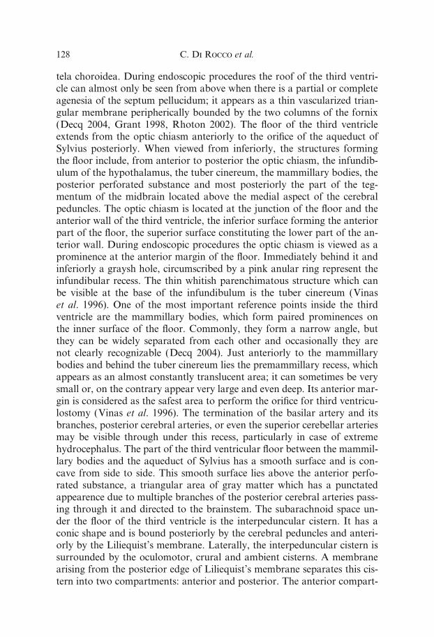
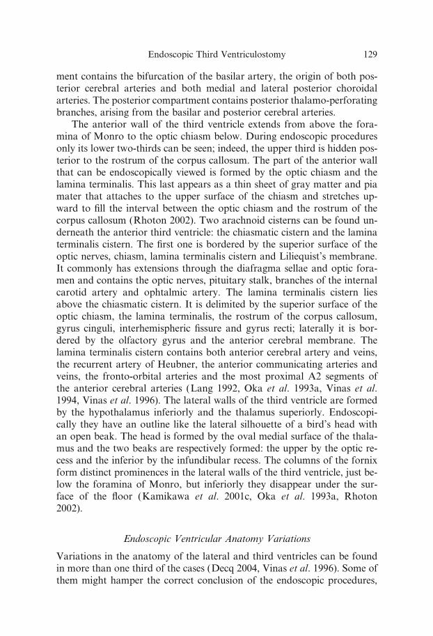
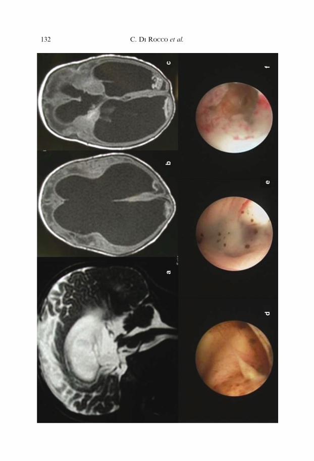
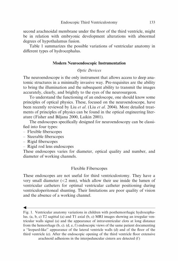
Di¤erent anatomic variations have been described in hydrocephalus ofdi¤erent etiologies. Kamikawa et al. (Kamikawa et al. 2001c) analyzed theventriculoscopic findings of four neonates with posthemorrhagic hydroce-phalus who underwent ETV. The choroid plexus was atrophic with hema-toma clots attached to it as well as at the orifice of the aqueduct of Sylviusin all cases. The septum pellucidum was widely fenestrated with a numberof small varices at the level of the septal veins in two cases. In all patientsfragments of old hematomas were scattered on the ventricular walls and onthe floor of the third ventricle, appearing brown because of the presence ofhemosiderin, an aspect defined by the authors as ‘‘leopard-like’’ (Fig. 1).At time distance from the hemorrhage thickening of the ventricular walls,due to fibrous scarring is common and septations can be found in the ven-tricular cavities as well as at the inlet of the aqueduct of Sylvius (Fig. 2).This could explain the occurrence of an acquired aqueductal stenosis insome of these patients (Scavarda et al. 2003). Septations as well as a thick-ened ventricular floor are common findings also in children with previousCSF infections. An extensive arachnoid sepimentation under the third ven-tricular floor might further complicate the correct conclusion of an ETVprocedure in this subset of patients (Riegel et al. 2001).
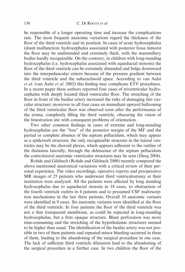
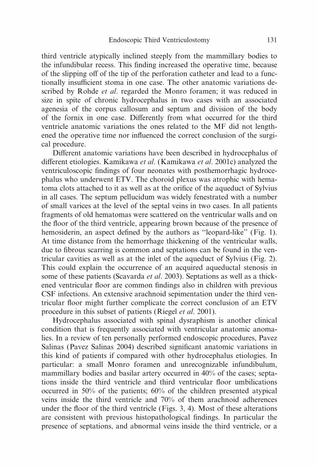
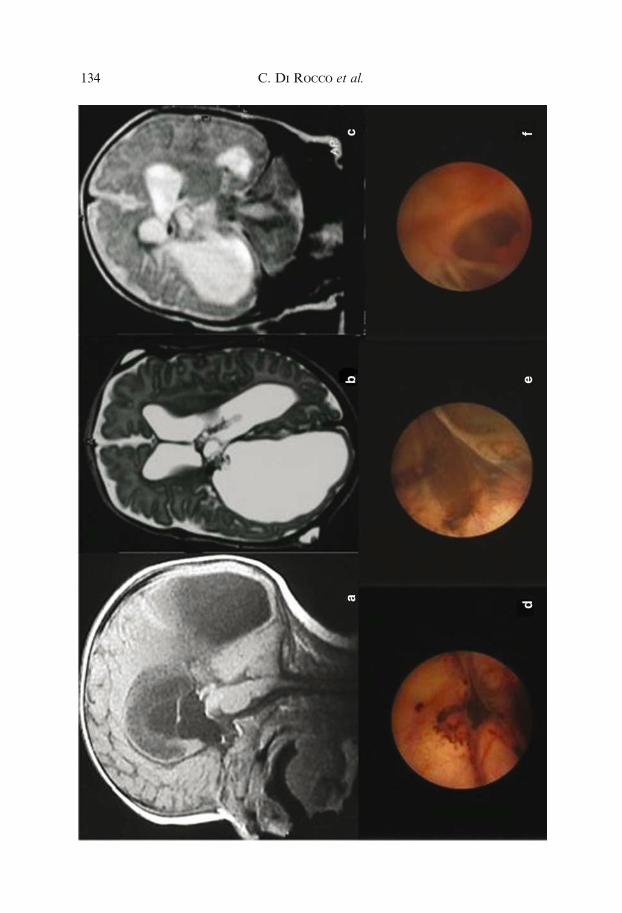
Hydrocephalus associated with spinal dysraphism is another clinicalcondition that is frequently associated with ventricular anatomic anoma-lies. In a review of ten personally performed endoscopic procedures, PavezSalinas (Pavez Salinas 2004) described significant anatomic variations inthis kind of patients if compared with other hydrocephalus etiologies. Inparticular: a small Monro foramen and unrecognizable infundibulum,mammillary bodies and basilar artery occurred in 40% of the cases; septa-tions inside the third ventricle and third ventricular floor umbilicationsoccurred in 50% of the patients; 60% of the children presented atypicalveins inside the third ventricle and 70% of them arachnoid adherencesunder the floor of the third ventricle (Figs. 3, 4). Most of these alterationsare consistent with previous histopathological findings. In particular thepresence of septations, and abnormal veins inside the third ventricle, or a
Endoscopic Third Ventriculostomy 131
C. Di Rocco et al.132
second arachnoidal membrane under the floor of the third ventricle, mightbe in relation with embryonic development alterations with abnormaldegrees of hypothalamus fusion.
Table 1 summarizes the possible variations of ventricular anatomy indi¤erent types of hydrocephalus.
Modern Neuroendoscopic Instrumentation
Optic Devices
The neuroendoscope is the only instrument that allows access to deep ana-tomic structures in a minimally invasive way. Pre-requisites are the abilityto bring the illumination and the subsequent ability to transmit the imagesaccurately, clearly, and brightly to the eyes of the neurosurgeon.
To understand the functioning of an endoscope, one should know someprinciples of optical physics. These, focused on the neuroendoscope, havebeen recently reviewed by Liu et al. (Liu et al. 2004). More detailed treat-ments of principles of physics can be found in the optical engineering liter-ature (Fisher and Biljana 2000, Laikin 2001).
The endoscopes specifically designed for neuroendoscopy can be classi-fied into four types:– Flexible fiberscopes– Steerable fiberscopes– Rigid fiberscopes– Rigid rod lens endoscopesThese endoscopes varies for diameter, optical quality and number, anddiameter of working channels.
Flexible Fiberscopes
These endoscopes are not useful for third ventriculostomy. They have avery small diameter (<2 mm), which allow their use inside the lumen ofventricular catheters for optimal ventricular catheter positioning duringventriculoperitoneal shunting. Their limitations are poor quality of visionand the absence of a working channel.
gFig. 1. Ventricular anatomy variations in children with posthemorrhagic hydrocepha-
lus. (a, b, c) T2 sagittal (a) and T1 axial (b, c) MRI images showing an irregular ven-
tricular walls signal (a) and the appearance of intraventricular clots at long distance
from the hemorrhage (b, c). (d, e, f ) endoscopic views of the same patient documenting
a ‘‘leopard-like’’ appearance of the lateral ventricle walls (d) and of the floor of the
third ventricle (e). After the endoscopic opening of the third ventricle floor extensive
arachnoid adhesions in the interpeduncular cistern are detected (f )
Endoscopic Third Ventriculostomy 133
C. Di Rocco et al.134
Steerable Fiberscopes
Flexible-Steerable endoscopes became a reality with the development offiber optic technology (Fukushima et al. 1973, Hecht 1999). They areconstructed of silica glass (which can be flexed without breaking) (Nobles
gFig. 2. Ventricular anatomy variations in children with posthemorrhagic hydrocepha-
lus. (a, b, c) T1 sagittal (a) and T2 axial (b) and coronal (c) images of an infant with
posthemorrhagic hydrocephalus. Intraventricular septa (a, b, c) and clots (b, c) lead to
a compartimentalization of the right lateral ventricle. The interventricular septum
seems to be preserved. (d, e, f ) endoscopic views of the same patient showing the aspect
of intraventricular clots (d) and of the right ventricle cella media septation (e); the
interventricular septum is opened and has a ‘‘spider-web’’ appearance (f )
Fig. 3. Ventricular anatomy variations in children with hydrocephalus and myelome-
ningocele. (a, b) T2 sagittal (a) and proton density coronal (b) MRI images of a 2 years
old child: the foramen of Monro is narrow and steeply oriented; the interventricular
septum is apparently preserved. (c, d) endoscopic views of the same patient confirming
the reduced size and the oblique orientation of the foramen of Monro; di¤erently from
what documented by the MRI images the inteventricular septum is widely opened
Endoscopic Third Ventriculostomy 135
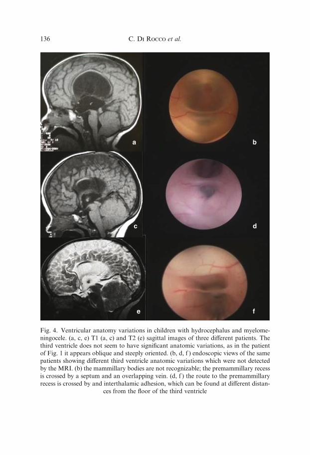
Fig. 4. Ventricular anatomy variations in children with hydrocephalus and myelome-
ningocele. (a, c, e) T1 (a, c) and T2 (e) sagittal images of three di¤erent patients. The
third ventricle does not seem to have significant anatomic variations, as in the patient
of Fig. 1 it appears oblique and steeply oriented. (b, d, f ) endoscopic views of the same
patients showing di¤erent third ventricle anatomic variations which were not detected
by the MRI. (b) the mammillary bodies are not recognizable; the premammillary recess
is crossed by a septum and an overlapping vein. (d, f ) the route to the premammillary
recess is crossed by and interthalamic adhesion, which can be found at di¤erent distan-
ces from the floor of the third ventricle
C. Di Rocco et al.136
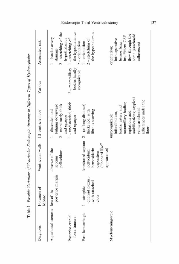
Table
1.Possible
VariationsofVentricularEndoscopic
Anatomyin
Di¤erentTypes
ofHydrocephalus
Diagnosis
Foramen
of
Monro
Ventricularwalls
IIIventricle
floor
Various
Associatedrisk
Aqueductalstenosis
loss
ofthe
posteriormargin
absence
ofthe
septum
pellucidum
1–distended
and
bulgingdownward
2–more
rarely
thick
andopaque
1–basilarartery
damage
2–stretchingofthe
hypothalamus
Posteriorcranial
fossatumors
1–undistended,thick
andopaque
2–mammillary
bodieshardly
recognizable
1–stretchingof
thehypothalamus
2–orientation
Post-hem
orrhagic
1–atrophic
choroid
plexus,
withattached
clots
fenestratedseptum
pellucidum;
hem
osiderin
deposition
(‘‘leopard
like’’
appearance)
2–(atlongdistance)
thickened,with
fibrousscarring
1–orientation
2–stretchingof
thehypothalamus
Myelomeningocele
unrecognizable
infundibulum,
basilararteryand
mammillary
bodies;
septationsand
umbilications;atypical
veins;arachnoid
adherencesunder
the
floor
orientation;
intraoperative
hem
orrhage;
insu‰cientCSF
flow
throughthe
soma(arachnoid
adherences)
Endoscopic Third Ventriculostomy 137
1998). The image formed by the objective lens is relayed to the eye lens bymultiple fibers contained in a very small package. Image fibers are formedin a coherent bundle that allows the image to be properly reconstructed onthe proximal end of the fiber. By contrast, light fibers are not constructed ina coherent fashion.
The main characteristic of the flexible and steerable fiberscope is thatthe last 4 cm can be oriented 100� upward and 160� downward. This isthe only system that makes looking and working around a corner possible.The diameter of the scope ranges from 2.3 to 4.6 mm (Table 2) allowing towork also in small ventricles and through a small foramen of Monro. Thesize of the device usually determines the number of individual fibers, conse-quently the number of pixels: the smaller the size, the fewer the number ofpixels. For each endoscope, designated fiberoptic cables and lighting equip-ment are used in combination with a standard camera and television mon-itor. The diameter of the working channel ranges from 1.0 mm to 1.5 mm,allowing the introduction of 3-French (1 mm) flexible instruments (microscissors, micro grasping forceps, micro biopsy forceps, monopolar elec-trodes, Fogarty balloon). Distinct irrigation channel and the outflow chan-nel are usually available. Access to the ventricle is possible using a dedicatedpeel-away sheath. Some steerable scopes with only one working channelpresent the limit of very slow irrigation when the instrument occupies thechannel. For this reason it is impossible to work under continuous irriga-
Table 2. List of main steerable neuroendoscopes with fiberoptic system suitable for
endoscopic third ventriculostomy
Manifacturer Model Outer
diameter
Channels* Working
channel
diameter
Bending
Aesculap1 flexible – steerable
endoscope
4.3 mm 1 W, 1 I/A 1.4 mm 280�
Codman &
Shurtle¤2
steerable
neuroendoscope
4.0 mm 1 W 1.0 mm 260�
Olympus flexible – steerable
endoscope
4.2 mm 1 Wþ 2 I/A 2.0 mm 180�
Storz3 neurofiberscope 2.9 mm
3.7 mm
1 Wþ 2 I/A
1 Wþ 2 I/A
1.2 mm
1.5 mm
290�
1 Aesculap – Tuttlingen, Germany; Wolf, Knittlingen, Germany.2 Codman & Shurtle¤ (Johnson & Johnson) New Brunswick, NJ.
3 Karl Storz GmbH & Co., Tuttlingen, Germany.
* W Working channel; I=A Irrigation/Aspiration channel.
C. Di Rocco et al.138
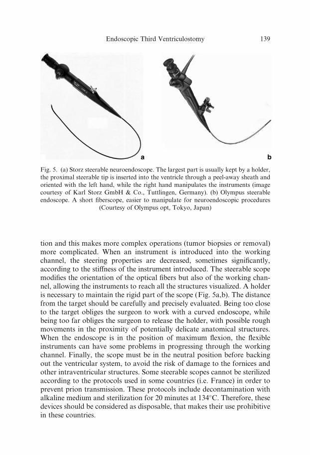
tion and this makes more complex operations (tumor biopsies or removal)more complicated. When an instrument is introduced into the workingchannel, the steering properties are decreased, sometimes significantly,according to the sti¤ness of the instrument introduced. The steerable scopemodifies the orientation of the optical fibers but also of the working chan-nel, allowing the instruments to reach all the structures visualized. A holderis necessary to maintain the rigid part of the scope (Fig. 5a,b). The distancefrom the target should be carefully and precisely evaluated. Being too closeto the target obliges the surgeon to work with a curved endoscope, whilebeing too far obliges the surgeon to release the holder, with possible roughmovements in the proximity of potentially delicate anatomical structures.When the endoscope is in the position of maximum flexion, the flexibleinstruments can have some problems in progressing through the workingchannel. Finally, the scope must be in the neutral position before backingout the ventricular system, to avoid the risk of damage to the fornices andother intraventricular structures. Some steerable scopes cannot be sterilizedaccording to the protocols used in some countries (i.e. France) in order toprevent prion transmission. These protocols include decontamination withalkaline medium and sterilization for 20 minutes at 134�C. Therefore, thesedevices should be considered as disposable, that makes their use prohibitivein these countries.
Fig. 5. (a) Storz steerable neuroendoscope. The largest part is usually kept by a holder,
the proximal steerable tip is inserted into the ventricle through a peel-away sheath and
oriented with the left hand, while the right hand manipulates the instruments (image
courtesy of Karl Storz GmbH & Co., Tuttlingen, Germany). (b) Olympus steerable
endoscope. A short fiberscope, easier to manipulate for neuroendoscopic procedures
(Courtesy of Olympus opt, Tokyo, Japan)
Endoscopic Third Ventriculostomy 139
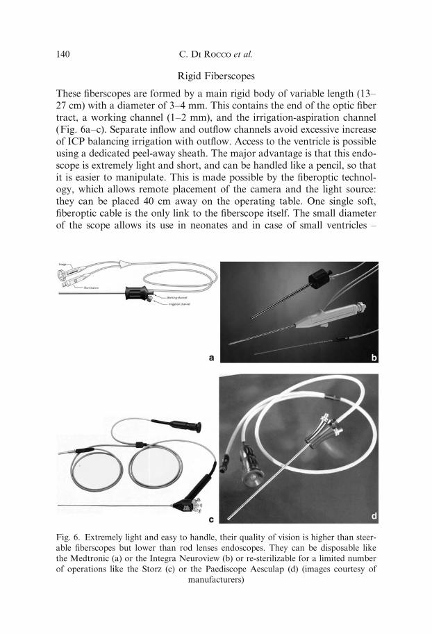
Rigid Fiberscopes
These fiberscopes are formed by a main rigid body of variable length (13–27 cm) with a diameter of 3–4 mm. This contains the end of the optic fibertract, a working channel (1–2 mm), and the irrigation-aspiration channel(Fig. 6a–c). Separate inflow and outflow channels avoid excessive increaseof ICP balancing irrigation with outflow. Access to the ventricle is possibleusing a dedicated peel-away sheath. The major advantage is that this endo-scope is extremely light and short, and can be handled like a pencil, so thatit is easier to manipulate. This is made possible by the fiberoptic technol-ogy, which allows remote placement of the camera and the light source:they can be placed 40 cm away on the operating table. One single soft,fiberoptic cable is the only link to the fiberscope itself. The small diameterof the scope allows its use in neonates and in case of small ventricles –
Fig. 6. Extremely light and easy to handle, their quality of vision is higher than steer-
able fiberscopes but lower than rod lenses endoscopes. They can be disposable like
the Medtronic (a) or the Integra Neuroview (b) or re-sterilizable for a limited number
of operations like the Storz (c) or the Paediscope Aesculap (d) (images courtesy of
manufacturers)
C. Di Rocco et al.140
small foramen of Monro (Table 3). The absence of a rigid rod lens systemallows a very wide working channel and a wide irrigating channel, makingit possible to use virtually all endoscopically designed surgical instruments,of any length. However, the fiberscopes with smaller diameter have smallerworking channel as well, allowing the use of 1 mm diameter instruments.
Vision is superior to that with the steerable fiberscopes because thenumber of optic fibers can be higher since there is no need for tip orienta-tion. More modern fiberscopes provide higher quality images, thanks to thepresence of as high as 30000 pixel fibers (Medtronic, Aesculap). Neverthe-less, the quality of vision cannot be compared to that o¤ered by rigid rodlens endoscopes.
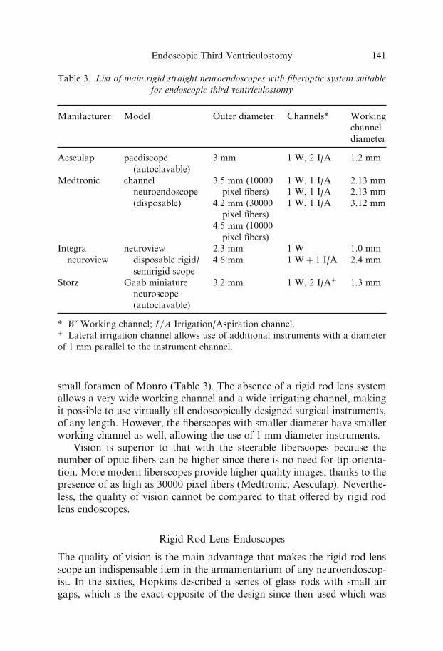
Rigid Rod Lens Endoscopes
The quality of vision is the main advantage that makes the rigid rod lensscope an indispensable item in the armamentarium of any neuroendoscop-ist. In the sixties, Hopkins described a series of glass rods with small airgaps, which is the exact opposite of the design since then used which was
Table 3. List of main rigid straight neuroendoscopes with fiberoptic system suitable
for endoscopic third ventriculostomy
Manifacturer Model Outer diameter Channels* Working
channel
diameter
Aesculap paediscope
(autoclavable)
3 mm 1 W, 2 I/A 1.2 mm
Medtronic channel
neuroendoscope(disposable)
3.5 mm (10000
pixel fibers)4.2 mm (30000
pixel fibers)
4.5 mm (10000
pixel fibers)
1 W, 1 I/A
1 W, 1 I/A1 W, 1 I/A
2.13 mm
2.13 mm3.12 mm
Integra
neuroview
neuroview
disposable rigid/
semirigid scope
2.3 mm
4.6 mm
1 W
1 Wþ 1 I/A
1.0 mm
2.4 mm
Storz Gaab miniatureneuroscope
(autoclavable)
3.2 mm 1 W, 2 I/Aþ 1.3 mm
* W Working channel; I=A Irrigation/Aspiration channel.þ Lateral irrigation channel allows use of additional instruments with a diameter
of 1 mm parallel to the instrument channel.
Endoscopic Third Ventriculostomy 141
composed by a series of small glass lenses interspersed with large air spaces(Fig. 7a,b). This technique forms the basis of most modern endoscopicsystems and bear his name (Nobles 1998, Siomin and Constantini 2004).Further reduction in light loss is achieved through the coating of the glasssurfaces with an ultrathin layer of magnesium fluoride. This layer markedlydecreases the reflection and improves the optic characteristics of endo-scopes and cameras (Nobles 1998, Shiau and King 1998, Siomin andConstantini 2004). With this technology the quality of vision is extremelysharp, and allows easier and more precise identification of the anatomicalstructures encountered, with an excellent visual definition of details. Therod lens system requires the presence of the camera and of the fiberopticcable for the cold light attached to the proximal extremity of the endo-scope (Figs. 8–11). The whole system requires good surgical training to bemanipulated freehand during navigation and throughout the whole surgi-cal procedure. A holder may be useful. The rigid lens system only allowstargets to be reached that are located on a straight line from the burr
Fig. 7. Traditional (a) and Rod lenses optic systems (b). In the rod lenses optical sys-
tem the air gap between lenses is significantly reduced, decreasing light dispersion and
increasing the stability of the system
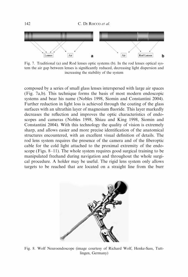
Fig. 8. Wolf Neuroendoscope (image courtesy of Richard Wolf, Henke-Sass, Tutt-
lingen, Germany)
C. Di Rocco et al.142
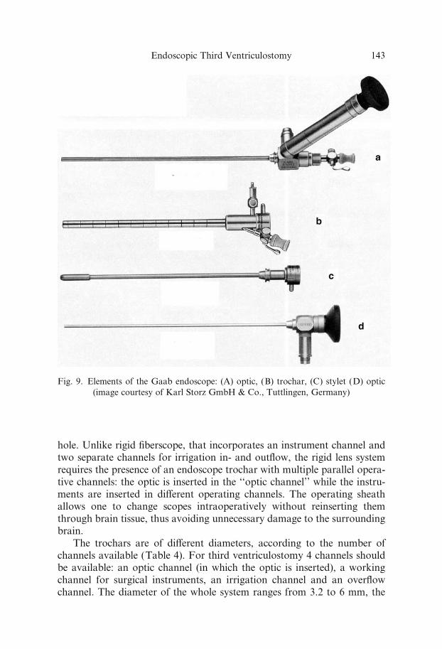
hole. Unlike rigid fiberscope, that incorporates an instrument channel andtwo separate channels for irrigation in- and outflow, the rigid lens systemrequires the presence of an endoscope trochar with multiple parallel opera-tive channels: the optic is inserted in the ‘‘optic channel’’ while the instru-ments are inserted in di¤erent operating channels. The operating sheathallows one to change scopes intraoperatively without reinserting themthrough brain tissue, thus avoiding unnecessary damage to the surroundingbrain.
The trochars are of di¤erent diameters, according to the number ofchannels available (Table 4). For third ventriculostomy 4 channels shouldbe available: an optic channel (in which the optic is inserted), a workingchannel for surgical instruments, an irrigation channel and an overflowchannel. The diameter of the whole system ranges from 3.2 to 6 mm, the
Fig. 9. Elements of the Gaab endoscope: (A) optic, (B) trochar, (C) stylet (D) optic
(image courtesy of Karl Storz GmbH & Co., Tuttlingen, Germany)
Endoscopic Third Ventriculostomy 143
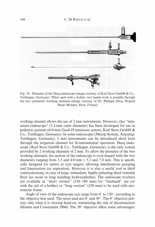
working channel allows the use of 2 mm instruments. However, also ‘‘min-iature endoscope’’ (3.2-mm outer diameter) has been developed for use inpediatric patients (4.0-mm Gaab II miniature system, Karl Storz GmbH &Co., Tuttlingen, Germany). In some endoscopes (Minop System, Aesculap,Tuttlingen, Germany), 1 mm instruments can be introduced short termthrough the irrigation channel for bi-instrumental operation. Decq endo-scope (Karl Storz GmbH & Co., Tuttlingen, Germany), is the only systemprovided by 2 working channels of 2 mm. To allow the presence of the twoworking channels, the section of the endoscope is oval-shaped with the twodiameters ranging from 3.5 and 4.0 mm� 5.2 and 7.0 mm. This is specifi-cally designed for tumor or cyst surgery allowing simultaneous graspingand fenestration (or aspiration). However it is also a useful tool in thirdventriculostomy in case of large, redundant, highly pulsating third ventriclefloor (as occur in long standing hydrocephalus). The endoscope trocharsare available in ‘‘short version’’ (150–160 mm) for ‘‘freehand’’ use (orwith the aid of a holder) or ‘‘long version’’ (250 mm) to be used with ster-eotactic frame.
Angle of view of the endoscope can range from 0� to 120�, according tothe objective lens used. The most used are 0� and 30�. The 0� objective por-tray only what it is viewing head-on, minimizing the risk of disorientation(Siomin and Constantini 2004). The 30� objective o¤ers some advantages:
Fig. 10. Elements of the Decq endoscope (image courtesy of Karl Storz GmbH & Co.,
Tuttlingen, Germany). When used with a holder, two hands work is possible through
the two symmetric working channels (image courtesy of Dr. Philippe Decq, Hopital
Henri Mondor, Paris, France)
C. Di Rocco et al.144
by simple rotation it provides an angle of view with a surface area twice aslarge as that obtained with 0� objective and it allows a better control of theinstruments because with the 30� objective the instruments introducedin the working channel (parallel to the endoscope) converge towards thecenter of the image (directed at 30�), while with the 0� objective the instru-ments remain in the periphery of the image. Major disadvantage of angledscopes is that the indirect image may cause the surgeon to become disori-ented. More than 30� angled objective are useful only to ‘‘look around thecorner’’ (Decq 2004).
The Future: The Videoscope
The future development of the technology of the endoscopes will allow thewide di¤usion of the group of Videoscopes. A videoscope is characterizedby a 1 CCD chip camera positioned at the proximal tip of the endoscope.
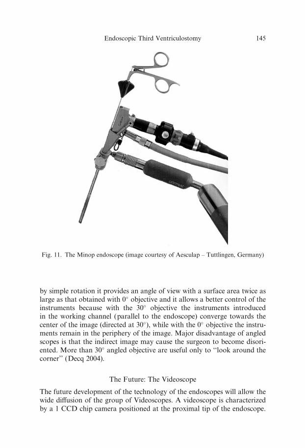
Fig. 11. The Minop endoscope (image courtesy of Aesculap – Tuttlingen, Germany)
Endoscopic Third Ventriculostomy 145
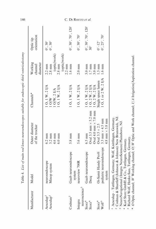
Table
4.Listofmain
rodlensesneuroendoscopes
suitable
forendoscopic
thirdventriculostomy
Manifacturer
Model
Outerdiameter
ofthetrochar
Channels*
Working
channel
diameter
Optictip
orientation
Aesculap1
neuroendoscope
6.2
mm
1O,1W,2I/A
2.2
mm
0� ,30�
Aesculap1
Minopsystem
3.2
mm
4.6
mm
6.0
mm
1O/W
1O/W
,2I/A
1O,1W,2I/A
2.8
mm
(optic/work)
2.8
mm
(optic/work)
2.2
mm
0� ,30�
Codman2
Gaabneuroendoscope
system
5.8
mm
1O,1W,2I/A
1.6
mm
0� ,30� ,70� ,120�
Integra
neuroscience3
neuroview
700R
5.6
mm
1O,1W,2I/A
2.0
mm
0� ,30� ,70�
Storz4
Gaabneuroendoscope
6.5
mm
1O,1W,2I/A
3.0
mm
0� ,30� ,70� ,120�
Storz4
Decq
Oval3.5
mm
�5.2
mm
Oval4.0
mm
�7.0
mm
1O,2W,2I/A
1O,2W,2I/A
1.7
mm
3.0
mm
30�
Storz4
Oi-SamiiHandyPro2
Oval3.5�2.5
Single
space
lumen
1.3
mm
0� –12�
Wolf5
pediatric
neuroendoscope
system
3.3
mm
�4.5
mm
4.8
mm
�5.8
mm
1O,1or2W,2I/A
1.6
mm
0� ,25� ,70�
1Aesculap–Tuttlingen,Germany;Wolf,Knittlingen,Germany.
2Codman&
Shurtle¤(Johnson&
Johnson)New
Brunsw
ick,NJ.
3NeuroNavigational(Integra
NeuroSciences)
Plainsboro,NJ.
4KarlStorz
GmbH
&Co.,Tuttlingen,Germany.
5Richard
Wolf,Henke-Sass,Tuttlingen,Germany.
*O
Opticchannel;W
Workingchannel;O=W
OpticandWork
channel;I=A
Irrigation/A
spirationchannel.
C. Di Rocco et al.146
This allows for the extreme simplification of the optical system, without theneed for complex and long rod lenses systems improving at the same timesignificatively the quality of vision if compared to the fiberscopes. Al-though the 1 CCD camera is somehow less performant than the 3 CCDcamera (see below) that can be used with rigid rod lenses systems and fiber-scopes, the proximity of the camera to the target of vision allows for excel-lent magnification and sharpness of the image. Moreover, the steerableproperties of the device are preserved because of the lack of rigid lens sys-tems. Some videoscope are already commercially available for ENT usein some countries, with an outer diameter of 6–8 mm; prototypes withsmaller outer diameter for neuroendoscopic use are under clinical valida-tion studies and should be available commercially in the next 2–3 years(Kamikawa et al. 2001a, Kamikawa et al. 2001b). The real, significant ad-vantage of this device is the excellent quality of vision (1 CCD cameralike), comparable to the 1 CCD rod lens systems, associated with the steer-able properties, allowing for perfect fusion of rigid and steerable systems(Fig. 12a–d).
Camera and Monitor
Two basic cameras are available: a single chip charged coupled device(CCD) and a three chip CCD. A good resolution for neuroendoscopy isavailable with 0.5 inches single chip cameras (resolution of 500 lines)(Schroeder et al. 2001). If the resolution is poor, the image needscomputer-enhancement. The three chip CCD produces images of betterquality (more than 800 horizontal lines) but is more expensive and heavier.So, most endoscopic system use single chip cameras. Some manifacturersproduce both the type of digital cameras (David 1 and David 3, Aesculap,Tuttlingen, Germany; Image1 and Image3, Karl Storz GmbH & Co., Tut-tlingen, Germany). In some models all the function can be controlled bythe surgeon in the operative field. Zoom endo-lens is useful to enlarge theimage section.
To achieve good quality images, a monitor with the highest possibleresolution should be selected. However, the resolution of the monitorshould not greatly exceed that of the camera. The size of the screen is lim-ited by the loss of quality when an image is enlarged. In fact, one shouldremember that the images of the camera are displayed over an area largerthan cross section of the optic cable. In monitor larger than 13 inches, thepicture is enlarged too much and decreases in quality. This is especiallytrue in case of fiberoptic endoscopes, where the spaces among the pixelsmay become evident. Larger monitors (19 or 20 inches) are useful for dis-playing multiple images (Cinalli 2004, Nobles 1998, Schroeder and Gaab1999, Siomin and Constantini 2004).
Endoscopic Third Ventriculostomy 147
C. Di Rocco et al.148
Illumination
Xenon light sources provide the best illumination for neuroendoscopic pro-cedures. The light is transmitted via fiber bundles from the light fountain tothe endoscope. Setting the light source to between 300 and 500 W providesa superior picture quality. Other types of light source, such as halogen, arenot able to generate a light bright enough for neuroendoscopy. Siomin andConstantini (Siomin and Constantini 2004) have calculated that, due to thesignificant light loss in the fiberoptic system, only 30% of the light gener-ated within the light source reaches the distal tip of the endoscope.
Accessories (Irrigation, Holders)
Imaging with an endoscope requires the clearest possible medium for opti-mum light with the lowest di¤raction. So, irrigation is important to assuregood visualization. It should be balanced by the egress of fluid. Careshould be given to avoid entrapment of fluid inside the ventricle: it maylead to disastrous sequels (Cinalli 2004, Teo 2004). Irrigation can be per-formed simply by hand with a catheter connected to the irrigation channelof the endoscope. It can be also provided with the use of a pump irrigatorfor which the flow is easily controlled using a foot switch (The Malis CMS-II Irrigation Module, Codman and Shurtle¤, Inc., Randolph, MA; Endos-copy Pump, Medtronic, Minneapolis, USA).
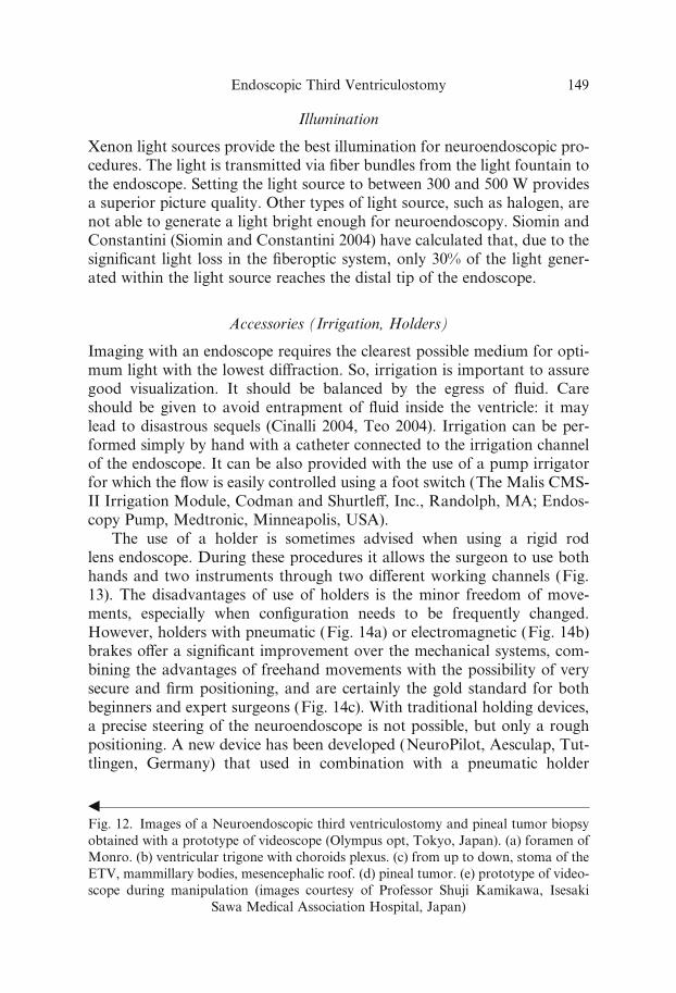
The use of a holder is sometimes advised when using a rigid rodlens endoscope. During these procedures it allows the surgeon to use bothhands and two instruments through two di¤erent working channels (Fig.13). The disadvantages of use of holders is the minor freedom of move-ments, especially when configuration needs to be frequently changed.However, holders with pneumatic (Fig. 14a) or electromagnetic (Fig. 14b)brakes o¤er a significant improvement over the mechanical systems, com-bining the advantages of freehand movements with the possibility of verysecure and firm positioning, and are certainly the gold standard for bothbeginners and expert surgeons (Fig. 14c). With traditional holding devices,a precise steering of the neuroendoscope is not possible, but only a roughpositioning. A new device has been developed (NeuroPilot, Aesculap, Tut-tlingen, Germany) that used in combination with a pneumatic holder
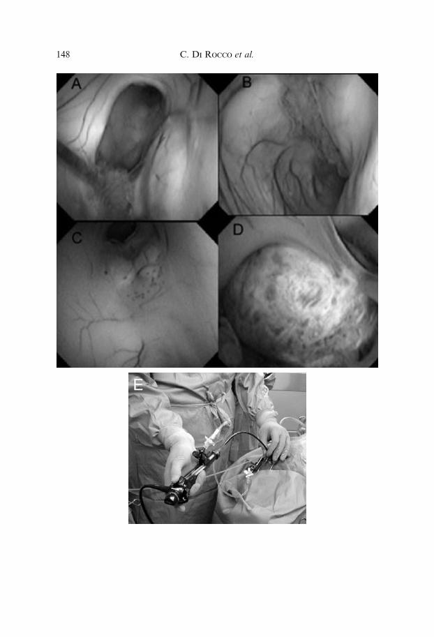
gFig. 12. Images of a Neuroendoscopic third ventriculostomy and pineal tumor biopsy
obtained with a prototype of videoscope (Olympus opt, Tokyo, Japan). (a) foramen of
Monro. (b) ventricular trigone with choroids plexus. (c) from up to down, stoma of the
ETV, mammillary bodies, mesencephalic roof. (d) pineal tumor. (e) prototype of video-
scope during manipulation (images courtesy of Professor Shuji Kamikawa, Isesaki
Sawa Medical Association Hospital, Japan)
Endoscopic Third Ventriculostomy 149
(Unitrac, Aesculap, Tuttlingen, Germany) allows, after positioning of theneuroendoscope, fine, sub-millimetric adjustment in the three dimensionalspace by three screws.
Neuronavigation and Stereotaxy
Stereotactic guidance was used before the advent of neuroendoscopy toperform third ventriculostomy (Ho¤man et al. 1980, Kelly 1991) and wasused in association with neuroendoscopy by several authors at the begin-ning of their experience (Grunert et al. 1994, Hellwig et al. 1998b, Hopf1999a). In fact, stereotactic guidance can be of some value only in choosingthe correct entry point and entering the lateral ventricle in a small-sizedventricular system. The limit of the technique is that the stereotactic framesare bulky and sometimes interfere with the endoscopic procedures andmost importantly, frame-based stereotactic systems do not provide an on-going intraoperative feedback to the surgeon about anatomical structuresencountered in the surgical field (Tirakotai et al. 2004).
A good alternative to traditional stereotactic frames can be the combi-nation with frameless neuronavigation (Alberti et al. 2001, Broggi et al.2000, Hopf et al. 1999a, Riegel et al. 2000, Riegel et al. 2002, Schroederet al. 2001, Tirakotai et al. 2004). Unlike based stereotaxis, frame-less nav-igation is still useful for intraoperative orientation, especially in casesof impaired visualization, distorted anatomy or narrowed ventricles. Inendoscopic third ventriculostomy, the use of neuronavigation may not be
Fig. 13. The use of a holder allows working with both hands if two working channels
are available (image courtesy of Dr. Philippe Decq, Hopital Henri Mondor, Paris,
France)
C. Di Rocco et al.150
necessary (Schroeder et al. 2001); however, in cases with thickened, non-translucent third ventricular floors, neuronavigation is useful for anatomi-cal orientation (Alberti et al. 2001, Tirakotai et al. 2004). Brain shift can bea major factor in influencing the accuracy of the target localization. Thisproblem occurs less often if some precautions are taken to prevent theabrupt change of CSF compartments or cystic lesion. The position of theburr hole should be at the highest point in order to minimize CSF loss.Moreover, brain distortion occurs rarely in midline structures and mostendoscopic procedures use midline structures as anatomical landmarks.
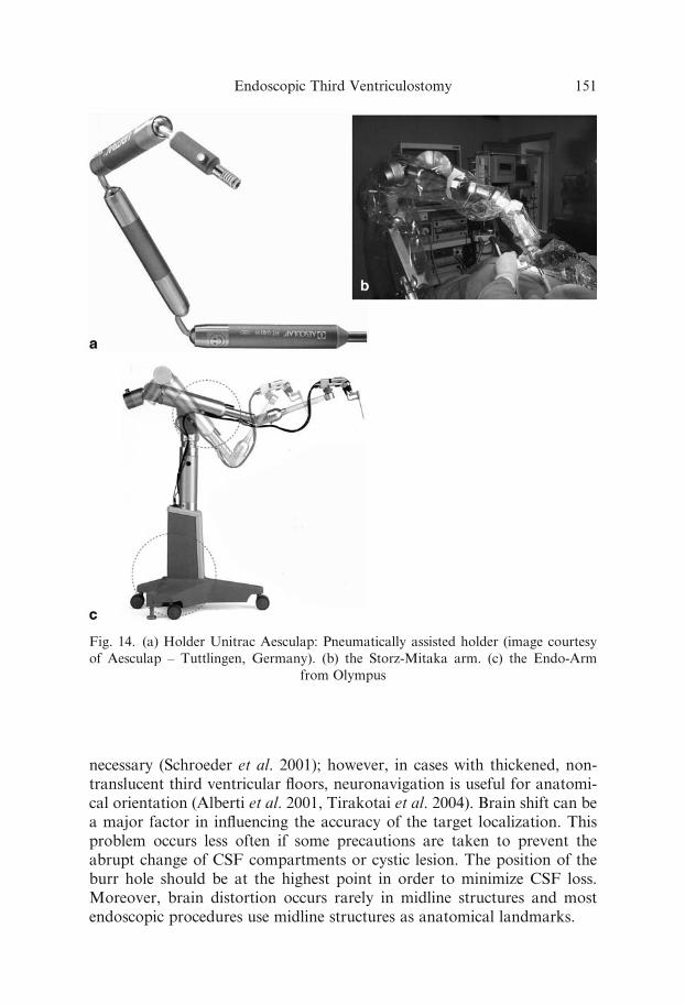
Fig. 14. (a) Holder Unitrac Aesculap: Pneumatically assisted holder (image courtesy
of Aesculap – Tuttlingen, Germany). (b) the Storz-Mitaka arm. (c) the Endo-Arm
from Olympus
Endoscopic Third Ventriculostomy 151
Neuronavigation requires a rigid three-pin head fixation, di‰cult toobtain in case of younger babies. Moreover the neuronavigation can becoupled only with rigid endoscopes.
Equipment for neuronavigation coupled with neuroendoscopy has beendiscussed in a recent paper by Tirakotai et al. (Tirakotai et al. 2004).
Operative Instruments
Operative instruments for neuroendoscopy include sharp micro scissor,blunt micro scissor, biopsy forceps, grasping forceps, monopolar and bipo-lar electrodes.
Floor Perforation
The perforation of the floor can be achieved mechanically (by either asharp instrument or, in combination with more force, a blunt instrument),electrically or with the aid of a laser.
Perforation With the Endoscope Itself
The endoscope can be gently pushed through the floor behind the clivus,stretching the fibers of the floor progressively until complete perforation isachieved and entry into the subarachnoid spaces is ensured by the sudden,direct visualization of the anatomical structures of the interpeduncularcistern (El-Dawlatly et al. 1999, El-Dawlatly et al. 2000, Teo and Jones1996). This technique has several inconveniences: the traction on the floorcan be significant and it is directly transmitted to the hypothalamic struc-tures situated above. Until perforation is achieved this is a blind procedure,with no visual control of the depth reached by the endoscope or of thespace remaining behind the membrane to be perforated.
Monopolar or Bipolar Coagulation
It is the most widely used technique (Cinalli 2004, Hellwig et al. 1999,Sainte-Rose and Chumas 1996). The advantages are evident. The point atwhich to perforate can be precisely chosen: if the floor is translucent, the tipof the coagulating wire can be positioned where the interpeduncular cisternis wider, as far as possible from the basilar bifurcation. Without applyingcautery current, the tip of the wire can be used as a probe to ‘‘palpate’’ thefloor of the third ventricle or to pierce it (Siomin and Constantini 2004).The coagulation is especially useful when the floor is very large and float-ing in the lumen of the ventricle: it allows the catheter tip to adhere to thechosen point, avoiding the natural tendency of the tip to slide. Coagulation
C. Di Rocco et al.152
should be used at the lowest e¤ective energy to bring about coagulationof the floor. In most cases it is not necessary to maintain the coagulationuntil the perforation is achieved. A very short coagulation (<1 s) is usuallysu‰cient to weaken the floor enough to allow perforation easily and atrau-matically with the inactive probe, avoiding the risk of entering the interpe-duncular cistern with an electric device on.
Both monopolar and bipolar coagulation are useful in this regard. Co-agulation is also useful to achieve hemostasis. Most bleedings are venouswith a slow flow, and can be managed only with irrigation. Sometimes aFogarty balloon can be used to tamponade a bleeding vessel or the marginsof a cutting (i.e. the stoma in the floor of the third ventricle). However, insome instances, neurosurgeon must appeal to coagulation to achieve hemo-stasis. Monopolar cautery can be used in both cutting and coagulationmodes to achieve fenestration, dissection or cauterisation. The use of elec-trical current can be associated with some problems (Vandertop et al.1998). The pathway of the currents flowing out of a tip cannot be con-trolled because the fluid in the ventricles is conductive and the current flowsalong the way of least resistance. Moreover energy losses caused by resis-tance in the leads makes them less e‰cient, so that very high currents couldbe necessary. Thus, tissue adherence and thermal damage of surroundingneural tissue are the major limitations of these instruments. However, thethermal damage to the hypothalamic region following coagulation hasnever been accurately studied. It may perhaps explain the fever sometimesobserved after third ventriculostomy (Decq et al. 2000, Decq 2004, Sainte-Rose and Chumas 1996). Because of these problems, Heilman and Cohen(Heilman and Cohen 1991) invented a ‘‘saline torch’’: a device that sendsa jet of saline past a monopolar wire. The saline acts as a conductor andcoagulation can be achieved without direct contact with the probe (Shiauand King 1998).
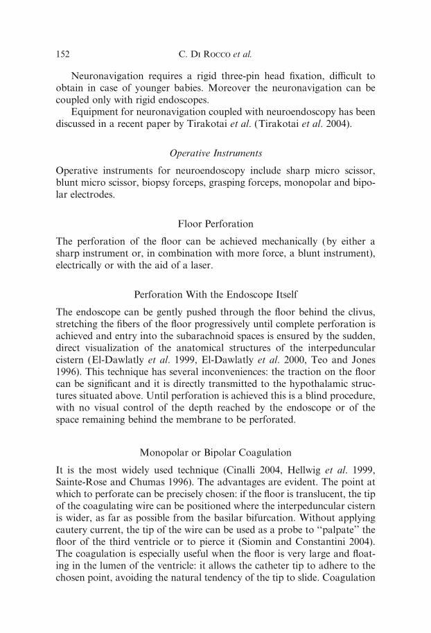
Bipolar cautery may represent a more controlled method of coagula-tion: it has demonstrated minimal current spread; it permits sharply demar-cated coagulation fields and precise cuts; damage to lateral or underlyingstructures is kept to a minimum. Therefore, bipolar coagulator should bepreferred (Shiau and King 1998). The simplest way to achieve bipolar co-agulation is through a fork electrode (Aesculap 2.1-mm fork electrode).The use of grasping bipolar forceps (2.5 mm – Codman & Shurtle¤, John-son & Johnson, Raynham, MA) allows the surgeon to pick up tissue fordissection and fenestration, and to coagulate vessels of more than 2 mmin diameter. Riegel et al. (Riegel et al. 2002) developed a new microbipolarforceps (ERBE Elektromedizin GmbH, Tubingen, Germany) that can beused for grasping, dissection, dilation, shrinkage of tissue, and precise co-agulation even of larger vessels. The branches of the forceps are moved viaelastic deformation of the metal without the use of a mechanical joint of
Endoscopic Third Ventriculostomy 153
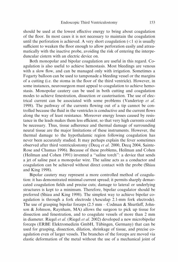
any kind (Fig. 15). The instrument has a outer diameter of 1.5 mm alongits entire length and is compatible with most working channels of neuro-endoscopes. It can be opened up to a width of 6 mm. So, it can be usedeither to perforate or to enlarge the stoma. Bipolar electrodes of di¤erentshape are also available and are extremely e¤ective for coagulation andperforation (Fig. 16).
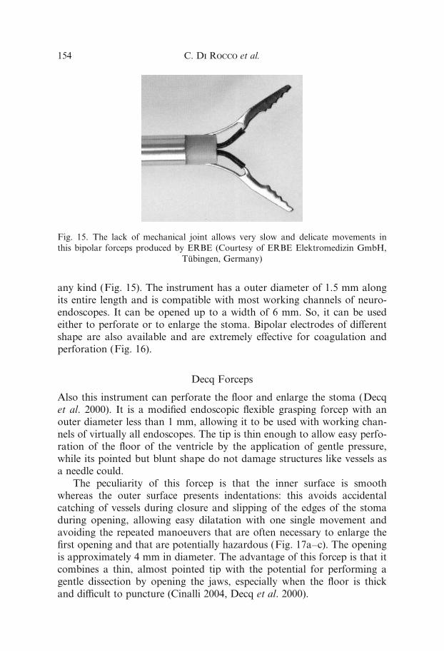
Decq Forceps
Also this instrument can perforate the floor and enlarge the stoma (Decqet al. 2000). It is a modified endoscopic flexible grasping forcep with anouter diameter less than 1 mm, allowing it to be used with working chan-nels of virtually all endoscopes. The tip is thin enough to allow easy perfo-ration of the floor of the ventricle by the application of gentle pressure,while its pointed but blunt shape do not damage structures like vessels asa needle could.
The peculiarity of this forcep is that the inner surface is smoothwhereas the outer surface presents indentations: this avoids accidentalcatching of vessels during closure and slipping of the edges of the stomaduring opening, allowing easy dilatation with one single movement andavoiding the repeated manoeuvers that are often necessary to enlarge thefirst opening and that are potentially hazardous (Fig. 17a–c). The openingis approximately 4 mm in diameter. The advantage of this forcep is that itcombines a thin, almost pointed tip with the potential for performing agentle dissection by opening the jaws, especially when the floor is thickand di‰cult to puncture (Cinalli 2004, Decq et al. 2000).
Fig. 15. The lack of mechanical joint allows very slow and delicate movements in
this bipolar forceps produced by ERBE (Courtesy of ERBE Elektromedizin GmbH,
Tubingen, Germany)
C. Di Rocco et al.154
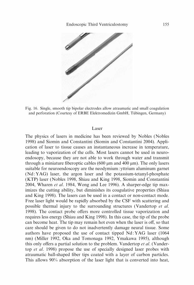
Laser
The physics of lasers in medicine has been reviewed by Nobles (Nobles1998) and Siomin and Constantini (Siomin and Constantini 2004). Appli-cation of laser to tissue causes an instantaneous increase in temperature,leading to vaporization of the cells. Most lasers cannot be used in neuro-endosopy, because they are not able to work through water and transmitthrough a miniature fiberoptic cables (600 mm and 400 mm). The only laserssuitable for neuroendoscopy are the neodymium:yttrium aluminum garnet(Nd :YAG) laser, the argon laser and the potassium-tetanyl-phosphate(KTP) laser (Nobles 1998, Shiau and King 1998, Siomin and Constantini2004, Wharen et al. 1984, Wong and Lee 1996). A sharper-edge tip max-imizes the cutting ability, but diminishes its coagulative properties (Shiauand King 1998). The lasers can be used in a contact or non-contact mode.Free laser light would be rapidly absorbed by the CSF with scattering andpossible thermal injury to the surrounding structures (Vandertop et al.1998). The contact probe o¤ers more controlled tissue vaporization andrequires less energy (Shiau and King 1998). In this case, the tip of the probecan become heat. The tip may remain hot even when the laser is o¤, so thatcare should be given to do not inadvertently damage neural tissue. Someauthors have proposed the use of contact tipped Nd:YAG laser (1064nm) (Miller 1992, Oka and Tomonaga 1992, Ymakawa 1995), althoughthis only o¤ers a partial solution to the problem. Vandertop et al. (Vander-top et al. 1998) propose the use of specially designed laser probes withatraumatic ball-shaped fiber tips coated with a layer of carbon particles.This allows 90% absorption of the laser light that is converted into heat,
Fig. 16. Single, smooth tip bipolar electrodes allow atraumatic and small coagulation
and perforation (Courtesy of ERBE Elektromedizin GmbH, Tubingen, Germany)
Endoscopic Third Ventriculostomy 155
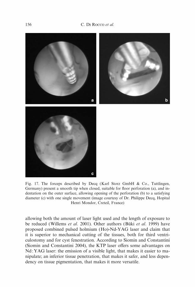
allowing both the amount of laser light used and the length of exposure tobe reduced (Willems et al. 2001). Other authors (Buki et al. 1999) haveproposed combined pulsed holmium (Ho)-Nd-YAG laser and claim thatit is superior to mechanical cutting of the tissues, both for third ventri-culostomy and for cyst fenestration. According to Siomin and Constantini(Siomin and Constantini 2004), the KTP laser o¤ers some advantages onNd:YAG laser: the emission of a visible light, that makes it easier to ma-nipulate; an inferior tissue penetration, that makes it safer, and less depen-dency on tissue pigmentation, that makes it more versatile.
Fig. 17. The forceps described by Decq (Karl Storz GmbH & Co., Tuttlingen,
Germany) present a smooth tip when closed, suitable for floor perforation (a), and in-
dentation on the outer surface, allowing opening of the perforation (b) to a satisfying
diameter (c) with one single movement (image courtesy of Dr. Philippe Decq, Hopital
Henri Mondor, Creteil, France)
C. Di Rocco et al.156
Nd:YAG laser and KTP laser are more useful in case of tumor re-moval and cyst fenestration than in case of third ventriculostomy. In fact,great care should be given when using a laser to perform a third ventricu-lostomy, since a case of injury of the vessels of the interpeduncolar cisternhas been reported (McLaughlin et al. 1997).
Suction-Cutting (Grotenhuis) Device
The suction-cutting device (Synergetics), developed recently by Grotenhuis,is composed of a thin suction cannula that can be introduced through anoperative channel at least 2 mm in diameter. The outer surface and theedges of the inlet of the cannula are smooth, whereas small blades areinserted into the lumen of the cannula. When the tip of the cannula comesinto contact with the floor of the third ventricle, the suction hole on thehandle is closed and the membrane is sucked into the lumen of the cannula.Rotation of the cannula allows section of only the tissue aspirated into thelumen, limiting the risk of accidental injury to vascular structures.
‘‘Semisharp’’ Instruments
The cautious blunt perforation is usually safe, but in case of more resistantfloor of the third ventricle a forceful pushing of the instruments is necessaryand might be dangerous. Surgical tools specifically designed for safe perfo-ration of a resistant and/or thick floor have a semisharp, slightly angulatedtip that, directed anteriorly and pushed inferiorly along the clivus, wouldallow safe perforation minimizing the risk of injury of the basilar artery(Kehler et al. 1998).
Ultrasound Microprobes
Ultrasound microprobes have been specifically designed for use throughthe working channel of the endoscope (6 French) or paraendoscopically(8 French). These probes o¤er the major advantage of direct visualiza-tion of the anatomical structures of the interpeduncolar and prepontine cis-terns and can be used for blunt perforation of the floor. This allows saferperforation under the double control of the floor of the third ventricle(endoscopic) and of the anatomical and vascular structures hidden behindthe floor membrane (ultrasonographic) (Paladino et al. 2000, Resch andReisch 1997, Resch and Perneczky 1998, Resch 2003) (Fig. 18a–c).
Forceps and Scissors
Instruments of various design are commercially available, suitable for rigidor flexible endoscopes. These include: grasping forceps, biopsy forceps
Endoscopic Third Ventriculostomy 157
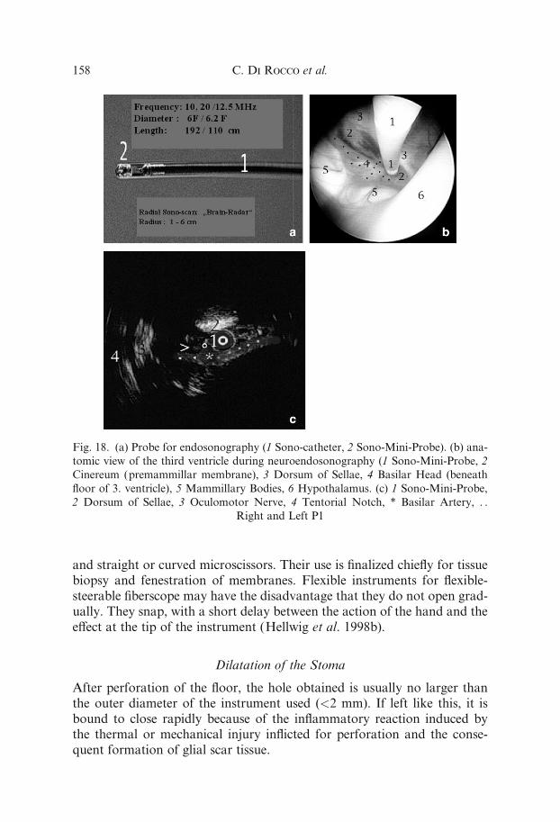
and straight or curved microscissors. Their use is finalized chiefly for tissuebiopsy and fenestration of membranes. Flexible instruments for flexible-steerable fiberscope may have the disadvantage that they do not open grad-ually. They snap, with a short delay between the action of the hand and thee¤ect at the tip of the instrument (Hellwig et al. 1998b).
Dilatation of the Stoma
After perforation of the floor, the hole obtained is usually no larger thanthe outer diameter of the instrument used (<2 mm). If left like this, it isbound to close rapidly because of the inflammatory reaction induced bythe thermal or mechanical injury inflicted for perforation and the conse-quent formation of glial scar tissue.
Fig. 18. (a) Probe for endosonography (1 Sono-catheter, 2 Sono-Mini-Probe). (b) ana-
tomic view of the third ventricle during neuroendosonography (1 Sono-Mini-Probe, 2
Cinereum (premammillar membrane), 3 Dorsum of Sellae, 4 Basilar Head (beneath
floor of 3. ventricle), 5 Mammillary Bodies, 6 Hypothalamus. (c) 1 Sono-Mini-Probe,
2 Dorsum of Sellae, 3 Oculomotor Nerve, 4 Tentorial Notch, * Basilar Artery, . .
Right and Left P1
C. Di Rocco et al.158
Grasping or Biopsy Forceps
Dilatation can be achieved by introducing a grasping or biopsy forceps intothe hole closed, then carefully opening it. This technique is usually rela-tively safe, but has several drawbacks. The dorsal surface of the forceps issmooth, so that the edges of the hole slip on this surface during opening:this necessitates several manoeuvres of opening and closing the forceps toobtain a satisfactory result, especially in the case of a thick, nontranslucentfloor. These repeated manoeuvres can result in accidental grasping of a per-forating vessel in the interpeduncular cistern.
Fogarty Balloon
Dilatation with a Fogarty balloon (usually 3 or 4 French) is much safer,since both the tip of the catheter and the surface of the balloon are smooth.For the same reason, the edges of the stoma slip very easily on this surface,requiring repeated inflations before the stoma is dilated to the largest diam-eter allowed by the balloon (4–5 mm). Fogarty catheter is the cheapest andmost common used device; the balloon must be inflated with saline. Thisallows the balloon to inflate very gradually, without the sudden inflationthat is quite traumatic and is usually observed when only air is used forinflation or too much air remains trapped within the balloon when salineis used.
Double Balloon Catheter
A balloon specifically designed for third ventriculostomy (Lighttouch bal-loon, Integra Neuroscience, Biot, France) o¤ers the ideal solution since theinflatable part is a dumbbell-shaped silicone membrane. The narrowestpart is marked with a black dot when the balloon is deflated. The catheteris introduced deflated into the stoma and advanced until the black dot is atthe level of the floor. Unlike Fogarthy balloon, the double balloon cathetermust be inflated with air. The proximal part inflates first, then the distalone (Fig. 19). The floor membrane remains trapped between the two bal-loons and the stoma is gently dilated with further air inflation to the largestextent allowed with one single maneuver. This technique remains by farthe simplest, safest, and fastest and should be recommended. However,the outer diameter of the available double balloon catheter is larger than1 mm and cannot be used in small instrument channels.
The ‘‘Urological’’ Device
A variant of the balloon technique has been proposed using instrumentsdesigned for stone extraction in urological endoscopic surgery (Wong and
Endoscopic Third Ventriculostomy 159
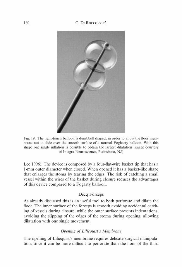
Lee 1996). The device is composed by a four-flat-wire basket tip that has a1-mm outer diameter when closed. When opened it has a basket-like shapethat enlarges the stoma by tearing the edges. The risk of catching a smallvessel within the wires of the basket during closure reduces the advantagesof this device compared to a Fogarty balloon.
Decq Forceps
As already discussed this is an useful tool to both perforate and dilate thefloor. The inner surface of the forceps is smooth avoiding accidental catch-ing of vessels during closure, while the outer surface presents indentations,avoiding the slipping of the edges of the stoma during opening, allowingdilatation with one single movement.
Opening of Liliequist’s Membrane
The opening of Liliequist’s membrane requires delicate surgical manipula-tion, since it can be more di‰cult to perforate than the floor of the third
Fig. 19. The light-touch balloon is dumbbell shaped, in order to allow the floor mem-
brane not to slide over the smooth surface of a normal Fogharty balloon. With this
shape one single inflation is possible to obtain the largest dilatation (image courtesy
of Integra Neuroscience, Plainsboro, NJ)
C. Di Rocco et al.160
ventricle itself. Bipolar coagulation should be preferred; monopolar coagu-lation should be avoided because of the proximity of the basilar artery. Inany case, only smooth instruments should be used at this level and thegrasping forceps should also be avoided.
Disclosures: The authors have no financial interest in the material pre-sented and discussed in this chapter.
Indications
A correct clinical indication is the most important factor influencing thesuccess rate of ETV. Advances in MR imaging have improved the under-standing of the specific physiopathologic mechanism at the base of thedi¤erent forms of hydrocephalus consequently representing an essential ad-junct for the correct patient selection. A more accurate preoperative diag-nosis of mixed types of communicating hydrocephalus, where obstructivefactors may still play a role, has lead to extend the indications for ETV topatients once excluded, namely subjects with posthemorrhagic hydrocepha-lus, postinfectious hydrocephalus and hydrocephalus in myelomeningocele.However, several points are still under discussion. The first is the role ofage. In past years, many authors reported low success rates in infants inde-pendently from hydrocephalus etiology (Buxton et al. 1998a, Buxton et al.1998b, Hopf et al. 1999b, Kim et al. 2000). Actually an increasing numberof papers have challenged this assumption recently. In fact, when infantswith pure obstructive hydrocephalus are considered, success rates up to85% were reported (Beems and Grotenhuis, 2002, Javadpour et al. 2001).A second important point that has merged in recent years is that theprimary hydrocephalus features may change in time in some patients; forexample, ‘‘reopening’’ of subarachnoid spaces may occur in cases of post-hemorrhagic or postinfectious hydrocephalus in children who initially pre-sented with purely communicating forms of hydrocephalus; an acquiredaqueductal stenosis is propounded as the long term main etiological factoraccounting for the hydrocephalus persistence (Cinalli et al. 1998, Smythet al. 2003).
Pure Obstructive Hydrocephalus
Aqueductal Stenosis
Aqueductal stenosis (AS) is responsible for 6–66% of cases of hydrocepha-lus in children and 5–49% in adults (Hirsch et al. 1986). Many classifica-tions of this clinical condition were proposed; the first and most importantdistinction is between primary stenosis, due to intrinsic pathology of theaqueduct itself and secondary stenosis, which follows a compression ofthe aqueduct from mass lesions (i.e. tectal tumors, pineal region tumors,
Endoscopic Third Ventriculostomy 161
Galen vein region vascular malformations) or in patients with communi-cating hydrocephalus from excessive dilatation of the occipital horns.
Primary Aqueductal Stenosis
Although pure primarily aqueductal stenosis is considered as a congenitalcondition, hydrocephalus secondary to AS is frequently diagnosed duringadolescence or in adulthood. Di¤erent ‘‘evolution theories’’ have beenproposed to explain this phenomenon. Head trauma, a small subarachnoidhemorrhage or a viral infection with benign meningitis have all beenclaimed as anatomic causes of aqueductal stenosis decompensation (Cinalliet al. 2004a, Jellinger, 1986, Lapras et al. 1986). A functional mechanismmight also contribute: progressive enlargement of the lateral and thirdventricles would lead to a distortion of the brainstem and kinking of theaqueduct (Nugent et al. 1979, Raimondi et al. 1976).
Secondary Aqueductal Stenosis
CSF flow obstruction in pure obstructive hydrocephalus may be the conse-quence of aqueductal compression by lesions arising within or around theSylvian aqueduct. Tectal gliomas and hamartomas are actually consideredone of the most frequent causes of this kind of condition. Unlike themajority of other di¤use brainstem tumors tectal tumors usually have anindolent clinical course, often remaining stable in size for several years(Chapman, 1990, Pollack et al. 1994).
Hydrocephalus in children with tectal plate tumors commonly becomessymptomatic when the tumor is small in size; indeed a circular restrictionof the aqueduct occurs due to supependymal tumor growth. Brainstemclinical signs are often lacking; in most of the patients the managementof hydrocephalus warrants a favorable long-term outcome (Cinalli et al.2004a).
Di¤erently from what occurs with tectal plate tumors, pineal regiontumors are usually larger in size when hydrocephalus becomes clinicallymanifest. In these cases, Sylvian aqueduct obstruction is generally the re-sult of a distortion of the whole mesencephalon from outside by part ofthe tumor (Rieger et al. 2000). Vascular malformations are a rare causeof secondary Sylvian aqueduct obstruction; abnormal draining veins ofmidbrain arteriovenous malformations may obstruct the CSF flow crossingthe aqueduct so as large fusiform aneurysms of the basilar artery. Hydro-cephalus in children with vein of Galen aneurysms has a multifactorial ori-gin; beside venous hypertension a tectal plate compression and tonsillarherniation may contribute to the development of hydrocephalus (Russell,1940). On these grounds, ETV may be considered in children with persis-
C. Di Rocco et al.162
tent hydrocephalus after aneurysm/AVM endovascular treatment accord-ing to Cinalli et al. (Cinalli et al. 2004a).
Pathogenesis of Hydrocephalus in Patients With Aqueductal Stenosis
It is actually accepted that CSF flow through the aqueduct is not unidirec-tional but has a systolic and a dyastolic cycle. According to recent MRIstudies, a downward displacement of CSF would occur during systole.The decrease of blood volume and the recoil of CSF displaced in the lum-bar sac during the dyastolic phase of the cardiac cycle would lead to a par-tial upward reversal of CSF flow. A net downward movement of CSF willresult, corrisponding to CSF production (0.0067 ml� s) (Enzmann andPelec, 1991, Quencer et al. 1990). Changes in aqueductal size and shapealter this volume flow rate and pattern. In aqueductal stenosis a decreaseof CSF flow and an increase up to ten times of its normal velocity occurs,with a consequent increase of wall shear stresses (Jacobson et al. 1999).Increasing pressure within the aqueduct may be responsible for secondarygliosis and for a progressive narrowing; in the ‘‘forking form’’, CSF flowturbulence and stasis further contribute to CSF accumulation. The conse-quent increase of CSF pulsations during systole lead to periventricularedema, partial displacement of blood from parenchymal vessels and furtheraccumulation of CSF in the lateral and third ventricles (Cinalli et al.,2004a).
The role of CSF pulsations in the pathogenesis of ventricular dilatationin this kind of hydrocephalus is confirmed by a previous experimental workdone at our institution. In this experience, the e¤ects of increasing the am-plitude of the intraventricular CSF pulse pressure in lambs was studied.Intraventricular pulse pressure was artificially modified by inserting a pul-sating balloon inside the lateral ventricles; modifications of ICP waves,mean CSF pressure and ventricular anatomic structures were recorded.No significant changes in mean CSF pressure were observed. On the con-trary, ICP waves were significantly modified, with maximal changes whenthe mechanical pulses were in phase with the physiological (arterial) ones.Moreover, an increase of ventricular volume, with the pathological evi-dence of stretching of the ependymal layers and periventricular whitematter spongiosis was already obtained when the amplitude of the pulseintraventricular pressure was increased three times the basal value. In theanimals, in which the intraventricular pulse pressure was mechanicallyincreased as much as six times the control value, a necrotic lesion of theperiventricular structures, associated with the ventricular dilatation, oc-curred (Pettorossi et al. 1978).
An alternative pathophysiological mechanism has been proposed ininfants in order to explain the relatively less satisfactory results of ETV
Endoscopic Third Ventriculostomy 163
in this specific subset of patients. Oi et al. (Oi et al. 2004) prospectivelyanalyzed 12 infantile cases of aqueductal stenosis with CT ventriculo-cisternography. Nine of them revealed to have a predominant transependy-mal intraparenchymal CSF outflow, with a minor involvement of the aque-duct pathway. On these grounds, the authors proposed that CSF dynamicsundergo an evolutionary mechanism: a transependymal CSF absorption(also defined as minor pathway) would be favored in neonates and infantsdue to the incomplete development of Pacchionian bodies; once ended, thematuration process of subarachnoid spaces the CSF flow through the aque-duct (major pathway) and to the cranial vault would prevail.
Analysis of Results of ETV in Patients With Primaryand Secondary Aqueductal Stenosis
Aqueductal stenosis is considered the ideal indication for third ventriculos-tomy. When patients with an exclusively obstructive triventricular hydroce-phalus are selected, the success rate is quite homogeneous and stable, beingbetween 63% and 92% in most series (Boschert et al. 2003). The influenceof age on the final result has been long discussed. Jones et al. (Jones et al.1994) reported a 40% success rate of ventriculostomy in children with triv-entricular hydrocephalus operated on within the age of two years. For thesame pathology with a clinical onset within the age of two years, but oper-ated on later in childhood and adolescence the success rate increased to71%. Kim et al. (Kim et al. 2000) reported age as a significant predictorof outcome. Of the six patients younger than one year of age, only two(33.3%) had good outcomes, whereas 19 out of 23 patients older than oneyear of age (82.6%) were successfully treated with ETV. Similar resultswere reported by Grunert et al. (Grunert et al. 2003): in their personal ex-perience a statistically worse success rate was found in infants (22.3%) ifcompared with children aged over one year (71.4%) and adults (81.6%). In-complete development of the subarachnoid spaces and consequent impair-ment of CSF absorption are the main factors which are claimed to explainwhy ETV fails more frequently in infants (Buxton et al. 1998a, Buxtonet al. 1998b, Hopf et al. 1999b, Kim et al. 2000). However, consideringexclusively children with pure obstructive hydrocephalus, other factorsmight have influenced the reported ETV failure rate in infants. First ofall, in most of these series a second endoscopic view was never or notalways performed, in order to estabilish ventriculostomy patency and thisfactor might further have negatively influenced the overall outcome. In amulticenter study Siomin et al. (Siomin et al. 2001) reported repeat ETVas e¤ective as primary procedures with an overall 65% success rate. Javad-pour et al. (Javadpour et al. 2001) analyzed the ETV success rate in a seriesof 21 infants with obstructive hydrocephalus. The success rate in 7 infants
C. Di Rocco et al.164
with congenital aqueductal stenosis was 71%; a redo third ventriculostomywas performed with success in one of the two initial failures with a finalsuccess rate of 86%. In a recent personal communication, Wagner et al.(Wagner and Koch, 2004) confirmed this finding, reporting the occlusionof the ventriculostomy site as the most frequent factor causing ETV failurein infants. The lower ICP with slowing of CSF flow through the stomamightexplain ventriculostomy obstruction in this subset of patients (Boschertet al. 2003).
Another important point to consider is the time for defining ETV fail-ure. According to Beems and Grotenhuis (Beems and Grotenhuis, 2002),infants have a longer adaptation time if compared with their older counter-part. Fourty-five percent of the 35 patients, aged less than two years, whounderwent successful ETV at their institution required a relatively longadaptation time (mean adaptation time: 1 week). This is important to rec-ognize, because it means that these young patients should not be treatedtoo soon using a shunting device if the ETV does not lead to immediaterelief of the symptoms.
Finally, concerning older series, a further factor which may have leadto a lower success of ETV in infants is the adopted surgical technique. Intheir earliest experience Cinalli et al. (Cinalli et al. 1999) reported a higherfailure rate in infants (<6 months of age) operated on under ventriculo-graphic guidance. In a subsequent series of 119 cases with triventricularhydrocephalus who underwent endoscopic third ventriculostomy, no signif-icant di¤erence was found between the two groups with an overall successrate of 79% (<6 months) and 71% (>6 months) respectively. Direct visual-ization of the ventricle floor and the anatomical landmarks of the interpe-duncular cistern under magnified conditions allow a greater safety and e‰-cacy of the procedure. Moreover, the larger size and more precise locationof the stoma in the floor of the third ventricle might play a role in youngpatients, in whom the risk of secondary obstruction may be high.
A further discussed point is the influence of AS etiology on the successrate of ETV procedures. Most authors report ETV as e¤ective in primaryas in secondary forms of aqueductal stenosis (Boschert et al. 2003, Fuku-hara et al. 2000, Hopf et al., 1999b, Rieger et al. 2000). Pople et al. (Popleet al. 2001) reported ETV to be e¤ective in the control of the hydrocepha-lus in 17 out of 18 cases with pineal region tumors. Macarthur et al. (Mac-arthur et al. 2001) referred a long-term (median follow-up: 12 months) con-trol of the hydrocephalus in 39 out of 47 pediatric patients (82.9%) withsecondary neoplastic AS. Similar results were reported by Rieger et al.(Rieger et al. 2000) in a series of 7 patients with pineal region tumors. Asuccessful control of the hydrocephalus was obtained in all cases; howeversix of the seven children also underwent tumor removal few days after theneuroendoscopic procedure, this factor possibly contributing to the long
Endoscopic Third Ventriculostomy 165
term results. In contrast with what previously said, Goh and Abbott(Goh and Abbott, 2000) recently reported a 49% failure rate of ETV in 63patients with tumoral AS (mean follow-up: 11.4 months). This result com-pared unfavorably with their 65% success rate obtained in children withprimary aqueductal stenosis. An analysis of the di¤erent series does notseem to show di¤erences in the prevalence of tumor location. Neverthlessnone of the reported authors details histological types nor the presence orabsence of subarachnoid tumor seeding, a factor which might specificallyinfluence the success of ventriculocisternostomy. This is particularly truefor some pineal region tumors (i.e. germinal cell tumors) and it is con-firmed by the fact that when exclusively benign lesions are selected (i.e.tectal plate gliomas) the success rates of ETV are almost homogeneous(ranging between 65% and 100%) and comparable with those obtained inpatients with primary AS (Grunert et al. 2003, Macarthur et al. 2001).Moreover, as part of the therapeutic protocol, endoscopic tumor biopsywas combined with ETV in selected cases of some of these series; eitherminor intraoperative bleeding and/or increase in CSF proteins which canfollow the bioptic step of endoscopic surgical procedures may conditionthe functional patency of the ventriculostomy site.
Another point that has been investigated in recent years is the role ofETV as secondary procedure in children with AS and previously implantedmalfunctioning or infected shunt. Most series report results comparable tothose obtained in primary procedures with overall success rates rangingfrom 62.9 to 82.3% (Boschert et al. 2003, Cinalli et al. 2004). The relativevariability of these results can again be ascribed to di¤erent selectioncriteria and di¤erent methods in the evaluation of results. Boschert et al.(Boschert et al. 2003) found that two of the three failures in their seventeenpatients series occurred in children with shunt malfunction and a previoushistory of shunt infection, suggesting a concurrent CSF absorption impair-ment in the pathogenesis of the hydrocephalus for these cases. However,other authors have pointed out that previous shunt infections or infectedshunt malfunctions do not influence outcome. Jones et al. (Jones et al.1990) reported on 4 patients having ETV for AS and infected shunt mal-function having a 75% success rate. Buxton et al. (Buxton et al. 2003) re-ferred that 33% of their 88 patients had previously su¤ered one or moreshunt infection, but they did not observe significant di¤erences in their out-come if compared with those who did not have a history of an infectedshunt.
Regarding evaluation of results Cinalli et al. (Cinalli et al. 1998) re-ported 30 patients, having a 76.7% ETV success rate, but many patientsin their series were left with a potentially patent shunt in situ; only sevenpatients were left in the initial stages to rely on the NTV alone (6 with theirshunts removed and one with it clipped). More stringent criteria were used
C. Di Rocco et al.166
by Buxton et al. (Buxton et al., 2003) who retrospectively analyzed a per-sonal series of 27 children with AS who underwent secondary third ventri-culostomy at their institution. The shunt was removed or tied o¤ in theneck in all cases; the final success rate was 62.9%.
Hydrocephalus in Posterior Cranial Fossa Tumors
The incidence of preoperative hydrocephalus in children with posteriorfossa tumors is around 60–80%; however only 15–25% of these patientsrequire a permanent ‘‘shunting’’ procedure. Younger age, the severity ofhydrocephalus at diagnosis, midline tumor localization, degree of tumorremoval and the use of substitute dural grafts are all factors considered toincrease this risk (Sainte-Rose et al. 2001).
Pathogenesis
The development of hydrocephalus in children with posterior fossa tumorsis directly related at diagnosis with the nature and localization of the tu-mor. Tumors arising or secondarily filling the IV ventricle primarily ob-struct the ventricular cavity and its foraminal outlets. On the other hand,cerebellar hemisphere tumors induce an anatomical distortion of the fourthventricle with a secondary occlusion of CSF pathways. In patients withmalignant tumors, subarachnoid seeding may contribute, impairing CSFabsorption. Though, logically, tumor removal should re-estabilish the com-munication between the fourth ventricle cavity and the subarachnoidspaces one-fourth to one third of these patients remain hydrocephalic(Sainte-Rose 2004). In the immediate postoperative period the surgicalsubarachnoid hemorrhage and the presence of cerebellar swelling may con-tribute to increase the resistance in CSF circulation. Subsequently the de-velopment of adhesions at the level of the fourth ventricle outlets and theadjacent cisterns may permanently alter CSF dynamics.
Management Strategies: The Role of ETV
Di¤erent protocols have been proposed for the management of hydroce-phalus in children with posterior fossa tumors. Actually, most pediatricneurosurgeons use a combination of corticosteroids, early tumor surgeryand external ventricular drainage where needed (Fritsch et al. 2004, Sainte-Rose 2004). Children with persistent postoperative hydrocephalus can bealternatively treated with ETV or shunt implantation. An important draw-back of this protocol is the risk of CSF infections which is related with ex-ternal ventricular drainage positioning (10% in the series of Rappaport andShalit; 4.9% in the series of Schmid) (Rappaport and Shalit, 1989, Schmid
Endoscopic Third Ventriculostomy 167
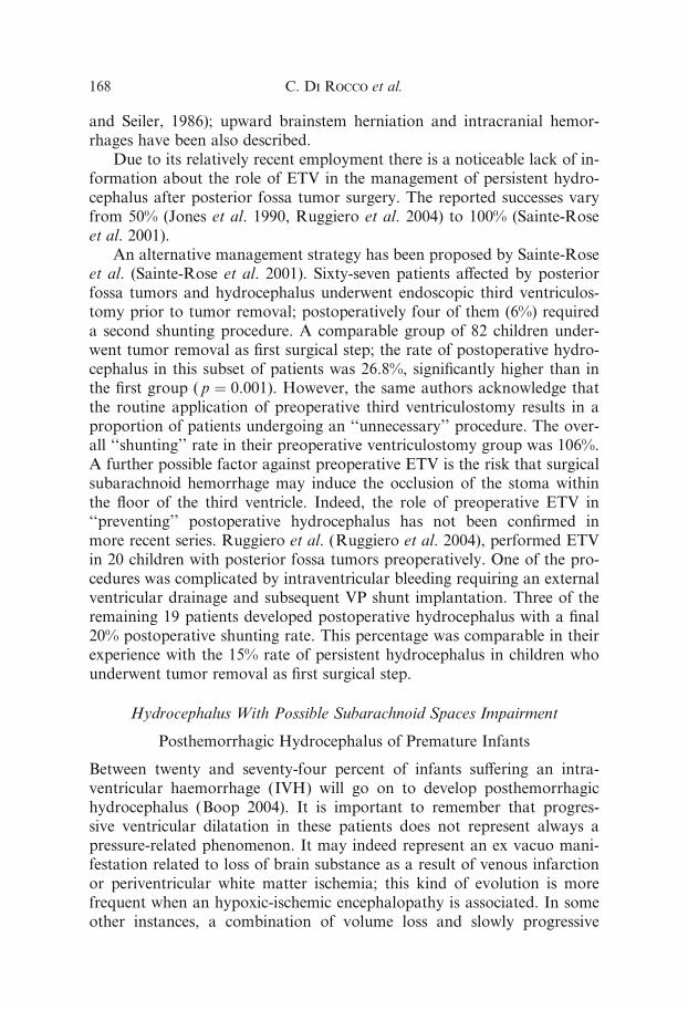
and Seiler, 1986); upward brainstem herniation and intracranial hemor-rhages have been also described.
Due to its relatively recent employment there is a noticeable lack of in-formation about the role of ETV in the management of persistent hydro-cephalus after posterior fossa tumor surgery. The reported successes varyfrom 50% (Jones et al. 1990, Ruggiero et al. 2004) to 100% (Sainte-Roseet al. 2001).
An alternative management strategy has been proposed by Sainte-Roseet al. (Sainte-Rose et al. 2001). Sixty-seven patients a¤ected by posteriorfossa tumors and hydrocephalus underwent endoscopic third ventriculos-tomy prior to tumor removal; postoperatively four of them (6%) requireda second shunting procedure. A comparable group of 82 children under-went tumor removal as first surgical step; the rate of postoperative hydro-cephalus in this subset of patients was 26.8%, significantly higher than inthe first group ( p ¼ 0.001). However, the same authors acknowledge thatthe routine application of preoperative third ventriculostomy results in aproportion of patients undergoing an ‘‘unnecessary’’ procedure. The over-all ‘‘shunting’’ rate in their preoperative ventriculostomy group was 106%.A further possible factor against preoperative ETV is the risk that surgicalsubarachnoid hemorrhage may induce the occlusion of the stoma withinthe floor of the third ventricle. Indeed, the role of preoperative ETV in‘‘preventing’’ postoperative hydrocephalus has not been confirmed inmore recent series. Ruggiero et al. (Ruggiero et al. 2004), performed ETVin 20 children with posterior fossa tumors preoperatively. One of the pro-cedures was complicated by intraventricular bleeding requiring an externalventricular drainage and subsequent VP shunt implantation. Three of theremaining 19 patients developed postoperative hydrocephalus with a final20% postoperative shunting rate. This percentage was comparable in theirexperience with the 15% rate of persistent hydrocephalus in children whounderwent tumor removal as first surgical step.
Hydrocephalus With Possible Subarachnoid Spaces Impairment
Posthemorrhagic Hydrocephalus of Premature Infants
Between twenty and seventy-four percent of infants su¤ering an intra-ventricular haemorrhage (IVH) will go on to develop posthemorrhagichydrocephalus (Boop 2004). It is important to remember that progres-sive ventricular dilatation in these patients does not represent always apressure-related phenomenon. It may indeed represent an ex vacuo mani-festation related to loss of brain substance as a result of venous infarctionor periventricular white matter ischemia; this kind of evolution is morefrequent when an hypoxic-ischemic encephalopathy is associated. In someother instances, a combination of volume loss and slowly progressive
C. Di Rocco et al.168
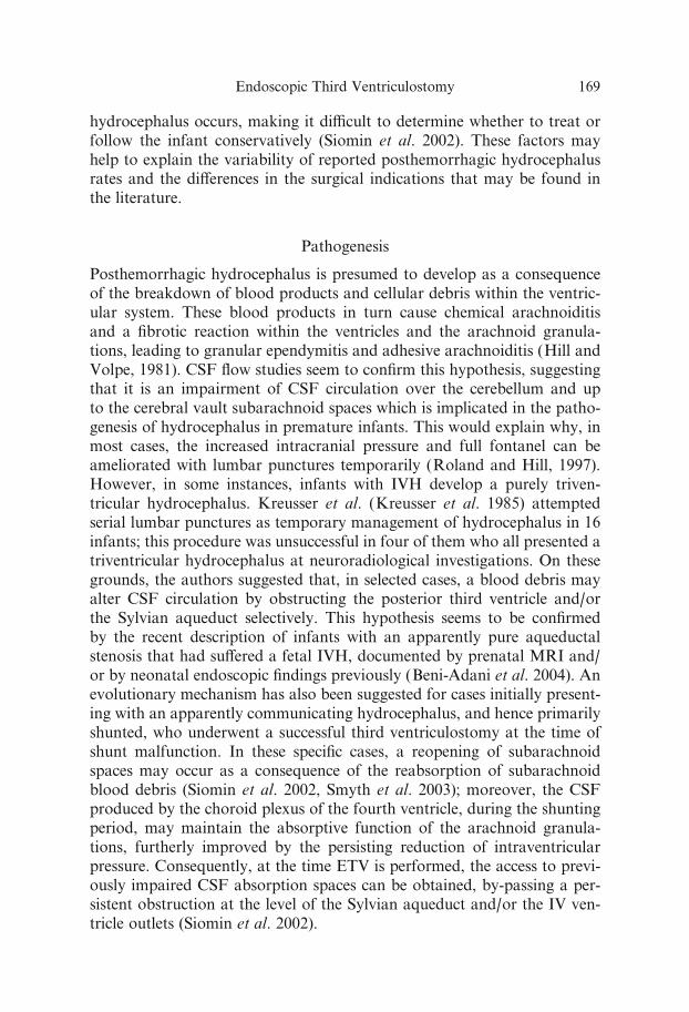
hydrocephalus occurs, making it di‰cult to determine whether to treat orfollow the infant conservatively (Siomin et al. 2002). These factors mayhelp to explain the variability of reported posthemorrhagic hydrocephalusrates and the di¤erences in the surgical indications that may be found inthe literature.
Pathogenesis
Posthemorrhagic hydrocephalus is presumed to develop as a consequenceof the breakdown of blood products and cellular debris within the ventric-ular system. These blood products in turn cause chemical arachnoiditisand a fibrotic reaction within the ventricles and the arachnoid granula-tions, leading to granular ependymitis and adhesive arachnoiditis (Hill andVolpe, 1981). CSF flow studies seem to confirm this hypothesis, suggestingthat it is an impairment of CSF circulation over the cerebellum and upto the cerebral vault subarachnoid spaces which is implicated in the patho-genesis of hydrocephalus in premature infants. This would explain why, inmost cases, the increased intracranial pressure and full fontanel can beameliorated with lumbar punctures temporarily (Roland and Hill, 1997).However, in some instances, infants with IVH develop a purely triven-tricular hydrocephalus. Kreusser et al. (Kreusser et al. 1985) attemptedserial lumbar punctures as temporary management of hydrocephalus in 16infants; this procedure was unsuccessful in four of them who all presented atriventricular hydrocephalus at neuroradiological investigations. On thesegrounds, the authors suggested that, in selected cases, a blood debris mayalter CSF circulation by obstructing the posterior third ventricle and/orthe Sylvian aqueduct selectively. This hypothesis seems to be confirmedby the recent description of infants with an apparently pure aqueductalstenosis that had su¤ered a fetal IVH, documented by prenatal MRI and/or by neonatal endoscopic findings previously (Beni-Adani et al. 2004). Anevolutionary mechanism has also been suggested for cases initially present-ing with an apparently communicating hydrocephalus, and hence primarilyshunted, who underwent a successful third ventriculostomy at the time ofshunt malfunction. In these specific cases, a reopening of subarachnoidspaces may occur as a consequence of the reabsorption of subarachnoidblood debris (Siomin et al. 2002, Smyth et al. 2003); moreover, the CSFproduced by the choroid plexus of the fourth ventricle, during the shuntingperiod, may maintain the absorptive function of the arachnoid granula-tions, furtherly improved by the persisting reduction of intraventricularpressure. Consequently, at the time ETV is performed, the access to previ-ously impaired CSF absorption spaces can be obtained, by-passing a per-sistent obstruction at the level of the Sylvian aqueduct and/or the IV ven-tricle outlets (Siomin et al. 2002).
Endoscopic Third Ventriculostomy 169
Results of ETV in Infants and ChildrenWith Posthemorrhagic Hydrocephalus
In the past, a history of hemorrhage was considered as a contraindicationfor ETV, because most patients were considered to su¤er a communicatingform of hydrocephalus. With the extension of the indications, an increasingnumber of patients with hydrocephalus and a history of ventricular hemor-rhage has been included in ETV management protocols. Actually, the mostrelevant paper that can be found in the literature is the one of Siomin et al.(Siomin et al. 2002) reporting the results of a multicentric retrospectivestudy on the role of ETV in children with posthemorrhagic and postinfec-tious hydrocephalus. Overall 36 children su¤ered a history of hemorrhagewith an ETV success rate of 55.6%. A striking di¤erence was documentedbetween primary and secondary ETV procedures. At a mean follow-upof 1.87G 1.6 years, the 13 posthemorrhagic patients who had primarilyreceived a VP shunt had a 100% ETV success rate. On the other hand, allthe premature infants in whom ETV was performed as first line of treat-ment subsequently required a shunt. This result is confirmed by otherauthors. Buxton et al. (Buxton et al. 1998b) reported on 16 infants (meancorrected age: 8.9 months) with posthemorrhagic hydrocephalus who allunderwent ETV as primary treatment for their hydrocephalus with a suc-cess rate of 30%. More recently Smyth et al. (Smyth et al. 2003) described a71.4% success rate in 7 children (mean age: 9.2), all the successes beingdocumented after secondary ETV procedures.
Javadpour et al. (Javadpour et al. 2001) suggested that a further mech-anism which may negatively influence the results of ETV in infants withposthemorrhagic hydrocephalus is their low intracranial pressure; ICPcompensating mechanisms due to expanding opened sutures may impairto build up a su‰cient pressure gradient across the stoma and the arach-noid granulations, with secondary insu‰cient CSF reabsorption and ven-triculostomy closure. In their experience only three out of ten infants weresuccessfully treated with ETV, two of them requiring two endoscopic pro-cedures, because of first closure of the ventriculostomy.
Two further factors that influenced the outcome of infants who had un-dergone ETV in the multicentric study of Siomin et al. (Siomin et al. 2002)were a history of associated CSF infection (the overall ETV success rate inthis specific subset of patients was 10%) and the time interval between theonset of hydrocephalus and ETV (mean temporal gap of 6.34G 8.31 yearssuccessful results versus 3.15G 5.16 years for treatment failures). The latterfactor probably influenced the results obtained by Beems and Grotenhuistoo (Beems and Grotenhuis 2002). In contrast with previous reports, theseauthors did not find significant di¤erence between the results of primary(44.4%) and secondary (50%) ETV procedures; however the time interval
C. Di Rocco et al.170
from the hydrocephalus clinical appearance and the surgical operation wasless than two years in the children who required a second procedure subse-quently.
Postinfectious Hydrocephalus
In the past, postinfectious hydrocephalus was commonly considered as acommunicating form of hydrocephalus, the predominant pathogenetic fac-tor being the inflammatory obstruction of the basal cisterns and cerebralvault subarachnoid spaces. The progressing knowledge of pathophysiologyin di¤erent CNS infectious diseases has lead to a reevaluation of this clini-cal entity that should actually be regarded as a complex disease.
Pathogenesis
The consequences of a CNS infectious disease on CSF dynamics dependon the age of the child at the time of the primary infection (prenatal,neonatal, postnatal) as well as on the infectious agent (bacterial, viral,parasitic).
Prenatal Infections. Toxoplasmosis is one of the most frequent prenatalinfections involving the CNS. In this condition, the parasites invade anddestroy the ependymal lining of the ventricles. In acute cases extensivenecrosis of the cerebral hemispheres can be observed with associated signif-icant cerebral tissue loss, which contributes to the ventricular enlargement.Most authors consider post-toxoplasmosis hydrocephalus a consequenceof severe leptomeningeal inflammation blocking the subarachnoid spaces(Kaiser 1985, Ciurea et al. 2004). However, a significant percentage ofthese children present with purely triventricular hydrocephalus. Accordingto the experimental model of Stahl (Stahl et al. 1997), the inflammatory de-bris may selectively obstruct, in these cases, the Sylvian aqueduct. More-over, ventricular enlargement would lead to a compression of the midbrainfurther limiting CSF flow to the IV ventricle.
Another prenatal infection which is commonly related to CNS diseasesis Cytomegalovirus infection. However, the occurrence rate of hydrocepha-lus in these children is relatively low (10–15%) (Ciurea et al. 2004). In mostcases an extensive involvement of the cerebral structures takes place lead-ing to a di¤use encephalomalacia and consequent ex vacuo ventricular di-latation. Patients with an active hydrocephalus usually present with a dif-fuse involvement of the leptomeninges, revealed by an extensive contrastenhancement on CT scans. Obstruction of the Sylvian aqueduct with con-
Endoscopic Third Ventriculostomy 171
sequent triventricular hydrocephalus was only occasionally described (Perl-man and Argyle, 1992).
Neonatal Infections. The majority of CSF infections in the neonatal periodare due to bacterial agents. A recent study on very low birth weight neo-nates with meningitis identified coagulase-negative staphylococci as themost common cause (43% of episodes), followed by other gram-positivebacteria (19%) and gram-negative bacteria (17%) (Doctor et al. 2001).The incidence of postinfective hydrocephalus in this specific subset ofpatients ranges between 50% and 75% (Ciurea et al. 2004). It usuallyoccurs within 2–3 weeks following the initial diagnosis, but it may developmonths or years after the acute phase of the disease. If hydrocephalusdevelops soon after the resolution of the infection it is more frequently ofthe communicating type with tetraventricular dilatation; obstructive formshave more commonly been described in patients developing late hydroce-phalus. As in the posthemorrhagic cases, the hypothesis is that the se-quence of fibrosis, thickening of the leptomeninges and consequent obliter-ation of the subarachnoid spaces may be reversible, with, however, thepossible persistence of an impaired CSF flow at the level of the Sylvian aq-ueduct or the basal cisterns (Siomin et al. 2002). In some cases the inflam-matory reaction may lead to a loculation of the CSF spaces and the forma-tion of multiple intraventricular septations. This process is more frequentafter gram-negative and mycotic infections which are associated with amore severe subependymal inflammatory reaction.
Postneonatal Infections. About 60–75% of postneonatal bacterial meningi-tis in children are caused by Haemophilus influenzae type B. However,hydrocephalus in these patients is uncommon occurring in less than 10%of the cases (Daoud et al. 1998). One of the hypothesis for this low rate isthat Haemophilus B ventriculitis usually causes choroid plexuses atrophy,with consequently reduced CSF production (Ciurea et al. 2004). In contrastwith Gram negative CSF infections, viral ventriculitis are frequently fol-lowed by the development of hydrocephalus. Viruses inclusions cause gran-ular ependymitis and ependimal cell loss or fusion; this process frequentlyinvolves the Sylvian aqueduct leading to secondary aqueductal stenosis;however, some authors have pointed out that the damaged ependyma itselfmay contribute to this process because of the secondarily induced reductionin CSF transportation and replacement (Ciurea et al. 2004). In non en-demic countries hydrocephalus, secondary to tuberculous meningitis is arare occurrence.
It is more frequently of the communicating type and secondary to ex-tensive meningeal involvement of the basal cisterns; in rare cases the mass
C. Di Rocco et al.172
e¤ect of an intracranial tuberculoma may lead to direct or secondary aque-ductal stenosis (Shoeman et al. 2000).
Results of ETV in Infants and ChildrenWith Postinfectious Hydrocephalus
A relatively low number of cases of infants and children with post-infectious hydrocephalus treated with ETV was reported in literature,mostly as a subgroup of combined series. In the multicentric study ofSiomin et al. (Siomin et al. 2002), children with a history of meningitis,ventriculitis or shunt infection represented only 2.1% (27 cases) of all thirdventriculostomies performed in seven international neurosurgical centers;at a mean follow-up of 1.87G 1.6 years the rate of success was 55.6% (15cases). The severity of the infection, the number of episodes (less or morethan 2), location of the inflammatory process (meninges, ventricular sys-tem, CSF shunt device) or the type of the agent (bacterial, yeast, unknownculture) did not appear to a¤ect outcome with statistical significance. Onthe contrary, a history of associated hemorrhage had a negative predictiveimpact (ETV success rate: 10%); actually, at a review of endoscopic videorecordings in this last subgroup of patients demonstrated a high incidence(53.8%) of interpeduncular fossa adhesions, a factor which might haveaccounted for the failure of the procedure at least in some patients.
Age was a significant factor with regards to the outcome, youngerpatients showing a greater rate of treatment failures. This observation wasconfirmed in other series and has a particular significance in infants; whenpatients under 2 years of age are selectively considered the reported ETVsuccess rate ranges between 0% and 44.4% (Fukuhara et al., 2000, Siominet al. 2002, Smyth et al. 2003). The role of the age factor is further supportedby the relatively high success rate reported, on the contrary, for secondaryETV procedures. For example, Smyth et al. (Smyth et al. 2003) obtained a60% success rate in five cases of postinfective hydrocephalus, three of themprimarily shunted and then treated by ETV at the time of shunt malfunc-tion. Similarly to what propounded in cases of posthemorrhagic hydroce-phalus, also in post-infectious hydrocephalus the resolution of the acute in-flammatory reaction and the temporary control of CSF dynamics assuredby the presence of an extrathecal CSF shunt may allow an improvement ofCSF absorption mechanisms with time. ETV would then enhance the CSFaccess to the subarachnoid spaces by-passing a possible persistent obstruc-tion at the level of the aqueduct or fourth ventricle outlets.
Hydrocephalus Associated with Dandy-Walker Syndrome
According to the original definition of Dandy and Blackfan, the termDandy-Walker syndrome (DWS) indicate the association of: 1) cystic dila-
Endoscopic Third Ventriculostomy 173
tation of the fourth ventricle; 2) partial or complete absence of the cerebel-lar vermis and 3) hydrocephalus. The remarkable variability of the patho-logical features of the disease has posed some problems in distinguishingthis entity from other types of posterior fossa cysts, such as persistentBlake’s pouch, retrocerebellar cyst, megacisterna magna and arachnoidcysts. Further di‰culties were determined by the consistent controversy inthe meaning of the terminology adopted by the various authors. Actuallythe di¤erential diagnosis is done on radiological criteria; according toBarkovich (Barkovich et al. 1989), the main distinguishing factor betweenthese di¤erent pathologies is the extension of the communication betweenthe cystic cavity and the fourth ventricle: 1) a wide communication is docu-mented in Dandy-Walker and Dandy Walker variant syndromes which arealso characterized by a more or less extended dysgenesis of the cerebellarvermis; 2) a limited communication via the vallecula can be found inpatients with mega cisterna magna; 3) a complete absence of communi-cation occurs in posterior fossa arachnoid cysts. Hydrocephalus is typicalonly of children with DWS; however it is not always, in fact, associatedwith the disease. Hirsch (Hirsch et al. 1984) reported a 90% incidenceof an associated hydrocephalus in their clinical series, but they arguedthat it could be an overestimation, since children are referred to neurosur-geons only when they develop active hydrocephalus and require surgicaltreatment.
Pathogenesis
The relevance of IV ventricle outlets atresia as a pathogenetic factor ofhydrocephalus in children with DWS is debated; indeed the foramina ofLushcka and Magendie have occasionally been found to be patent in thesepatients. It has also to be considered that more than 80% of Dandy-Walkerinfants are not hydrocephalic at birth (Cinalli et al. 2004b). Shaw et al.(Shaw et al. 1995) described atresia of one or both foramina of Lushckain 20% of autoptic normal brains. Barr (Barr, 1948) observed nonpatencyof the foramina of Magendie in 1% of autopsies. On these grounds otherfactors have been claimed to contribute to the development of hydrocepha-lus in these patients. Aqueductal stenosis, due to a primary developmentdefect or secondary to herniation of the vermis may be important in somespecific cases (Cinalli et al. 2004b). Furthermore, the obstruction of CSFflow can also be distal to the outlets of the fourth ventricle. Glasauer(Glasauer, 1975) noted at isotope cisternography that occasionally the sub-arachnoid space anterior to the medulla and basal cisterns was not patent.Other authors have confirmed that the subarachnoid spaces can be abnor-mally developed in children with DWS (Hirsch et al. 1984). Finally, the se-vere malformation of the posterior fossa with elevation of the tentorium,the torcular Herophili and the transverse sinuses can lead to a lenghtening
C. Di Rocco et al.174
of the venous sinuses and to their direct compression from the posteriorfossa cyst (Cinalli et al. 2004b). Subsequently, the possible role of venoushypertension should not be underestimated.
Results of ETV in Children With Hydrocephalusand Dandy-Walker Syndrome
Due to the relative rarity of the disease the papers that can be found inthe literature on this subject are essentially limited to case reports or de-scription of very limited series of patients. Hirsch et al. (Hirsch et al.1984) and Ho¤man et al. (Ho¤man et al. 1980) reported a 50% successrate (2 out of four patients joining the two series). All the endoscopic pro-cedures were performed under an exclusively radioscopic control, a factorwhich might have influenced the correct conclusion of the procedure. Morerecently, a higher success rate was reported by Cinalli (Cinalli, 1999) whodescribed three long term outcomes out of four personal cases.
In children with secondary aqueductal stenosis a combined ETV andaqueductal stenting placement has been suggested. Mohanty (Mohanty,2003) successfully performed this kind of procedure in two out of threepatients (66.6%). Intraoperative endoscopic confirmation of aqueductpatency might be useful in order to select the appropriate surgical proce-dure (Jodicke et al. 2003).
One of the conditions that seems to negatively influence the results ofETV in patients with Dandy-Walker syndrome is the presence of associ-ated CNS malformations, such as agenesis of the corpus callosum whichmight preoperatively allow the escape of CSF into the convexity subarach-noid spaces. Contrast dynamic CT scans, MRI with flow studies and flow-sensitive phase-contrast cine MRI images might help to make the correctpatient’s selection (Cinalli et al. 2004b). Anatomic modifications of theinterpeduncular cistern, the orientation of the third ventricular floor, thehigher position of the tip of the basilar artery and the displacement ofthe brainstem against the clivus are the conditions which usually makethe endoscopic procedure di‰cult to perform. Endoneurosonography andtransendoscopic Doppler ultrasound were proposed as technical adjunctsin order to identify intraoperatively parenchimal and vascular landmarks(Jodicke et al. 2003). Controlled CSF leak through the endoscope may al-low a slow decompression of the cyst, which in turn may reduce the masse¤ect on the brainstem and recreate the space for a safe ETV.
Constrictive Hydrocephalus
Hydrocephalus Patients with Myelomeningocele
The exact incidence of hydrocephalus in myelomeningocele patients is notknown. In most cases it is not present at birth but it develops in the first few
Endoscopic Third Ventriculostomy 175

weeks or months of life. Postnatal neuroimaging studies, obtained beforeclosure of the spinal defect, have documented the presence of hydrocepha-lus in 15.25% of the cases; however, the proportion of patients who subse-quently require shunting reaches up to 80–90% in most surgical series. Nocorrelation has been shown between the level of the lesion and the presenceof hydrocephalus (Teo and Jones, 1996).
Pathogenesis
A variety of factors are implicated in the pathogenesis of hydrocephalus inchildren with myelomeningocele which characteristically may combine thefeatures of the communicating and obstructive forms of hydrocephalus.The Chiari type II malformation, aqueduct stenosis, anomalous venousdrainage, and the closure of an open myelomeningocele all contribute tothe development of hydrocephalus. In particular, the hypoplasic posteriorcranial fossa and the hindbrain anomalies, typical of the Chiari II malfor-mation, usually result in an overcrowing of the nervous and vascular struc-tures and reduction of the CSF spaces, with secondary caudal dislocationof the IV ventricle and cerebellar tonsils with the upper cervical canal(Sgouros 2004). The consequent increased resistance to venous outflowand venous hypertension, may account for the development of a ‘‘commu-nicating’’ form of hydrocephalus. On the other side, the mechanical distor-tion and deformation of the brain stem can secondarily lead to a functionalaqueductal stenosis. Furthermore the tonsillar herniation may also con-tribute to the obstructive component of this type of hydrocephalus, impair-ing CSF flow at the level of the fourth ventricle outlets. The relevant role ofCSF obstruction in the pathogenesis of hydrocephalus in myelomeningo-cele patients seems to be confirmed by recent series reporting on intrauter-ine repair of opened spina bifida and the possible correlation between theseverity of Chiari type II malformation and the occurrence of hydrocepha-lus. Tulipan et al. (Tulipan et al. 1998) and Sutton et al. (Sutton et al. 1999)described a significant reduction of the hindbrain herniation occurrence inchildren who underwent intrauterine closure of myelomeningocele if com-pared with historical controls. The incidence of associated hydrocephaluswas reduced from 91% to 59% in the series of Tulipan. Only one of thenine survivors in the series of Sutton required ventriculoperitoneal shunt-ing. Hence these authors postulated that the lower incidence of hydroce-phalus in their series had to be related to the absence of the obstructinge¤ect of the hindbrain herniation at the level of the foramen magnum.However other authors have claimed this assumption to be oversimplisticas the intrauterine procedure had induced significant changes in volume ofthe posterior fossa, with consequent improved flow through the aqueduct,improved compliance of CSF flow around the brain stem and the tentorial
C. Di Rocco et al.176
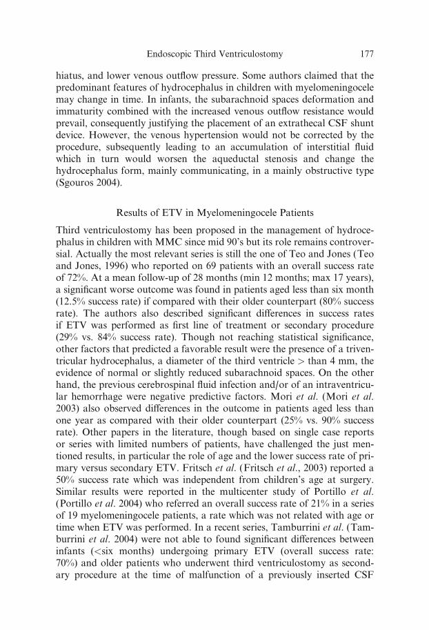
hiatus, and lower venous outflow pressure. Some authors claimed that thepredominant features of hydrocephalus in children with myelomeningocelemay change in time. In infants, the subarachnoid spaces deformation andimmaturity combined with the increased venous outflow resistance wouldprevail, consequently justifying the placement of an extrathecal CSF shuntdevice. However, the venous hypertension would not be corrected by theprocedure, subsequently leading to an accumulation of interstitial fluidwhich in turn would worsen the aqueductal stenosis and change thehydrocephalus form, mainly communicating, in a mainly obstructive type(Sgouros 2004).
Results of ETV in Myelomeningocele Patients
Third ventriculostomy has been proposed in the management of hydroce-phalus in children with MMC since mid 90’s but its role remains controver-sial. Actually the most relevant series is still the one of Teo and Jones (Teoand Jones, 1996) who reported on 69 patients with an overall success rateof 72%. At a mean follow-up of 28 months (min 12 months; max 17 years),a significant worse outcome was found in patients aged less than six month(12.5% success rate) if compared with their older counterpart (80% successrate). The authors also described significant di¤erences in success ratesif ETV was performed as first line of treatment or secondary procedure(29% vs. 84% success rate). Though not reaching statistical significance,other factors that predicted a favorable result were the presence of a triven-tricular hydrocephalus, a diameter of the third ventricle > than 4 mm, theevidence of normal or slightly reduced subarachnoid spaces. On the otherhand, the previous cerebrospinal fluid infection and/or of an intraventricu-lar hemorrhage were negative predictive factors. Mori et al. (Mori et al.2003) also observed di¤erences in the outcome in patients aged less thanone year as compared with their older counterpart (25% vs. 90% successrate). Other papers in the literature, though based on single case reportsor series with limited numbers of patients, have challenged the just men-tioned results, in particular the role of age and the lower success rate of pri-mary versus secondary ETV. Fritsch et al. (Fritsch et al., 2003) reported a50% success rate which was independent from children’s age at surgery.Similar results were reported in the multicenter study of Portillo et al.(Portillo et al. 2004) who referred an overall success rate of 21% in a seriesof 19 myelomeningocele patients, a rate which was not related with age ortime when ETV was performed. In a recent series, Tamburrini et al. (Tam-burrini et al. 2004) were not able to found significant di¤erences betweeninfants (<six months) undergoing primary ETV (overall success rate:70%) and older patients who underwent third ventriculostomy as second-ary procedure at the time of malfunction of a previously inserted CSF
Endoscopic Third Ventriculostomy 177
shunt device (overall success rate: 60%). Such discordant results suggestthat particular caution should be paid in these type of hydrocephalus,when planning ETV.
On the other hand, the slow and insidious progression which character-izes the evolution of this specific form of hydrocephalus in a large portionof subjects, might lead to a wrong claiming of a successful result after thesurgical operation in patients who actually continue to have hydrocepha-lus. Subsequently, strict neuroradiological, ophtalmological and neuro-psychological follow-up control is mandatory.
Analysis of Outcome
ETV, by forming a communication between the third ventricle and thesubarachnoid spaces, restores an almost ‘‘physiological’’ CSF circulationin case of obstructive hydrocephalus. The recent views on the pathophysi-ology of hydrocephalus (Greitz 2004) enhance the importance of the intra-cranial compliance, that is widely dependent on the free movement of theCSF between the cranial and the spinal subarachnoid spaces. Despite ofthe accuracy of patients selection, several reports show that in a significantnumber of cases the symptoms and signs of increasing intracranial pressurerelated to hydrocephalus are however not controlled by the procedure(Cinalli et al. 1999, Hirsh et al. 1986). The phenomenon is more frequentlyobserved in the immediate post-operative period (early failures), being onlysporadically observed in a later phase, usually within 5–6 years from theoperation (late failures). Recurrence of hydrocephalus beyond this periodhas been reported exceptionally (Beems and Grotenhuis 2004, Tuli et al.1999). Pre-requisite of a successful ventriculostomy is the patency of thedistal subarachnoid pathways: the di‰culty to detect pre-operatively thepresence of coexisting obstruction in the basilar cisterns or in the subarach-noid space of the surface may account for the 25–40% failure rate stillreported in literature (Cinalli et al. 1999, Fritsch and Mehdorn 2002,Javadpour et al. 2001). A reliable non-invasive pre-operative test is notavailable yet; invasive tests have been proposed, but they are di‰cult topropose on a routine basis, especially in children (Bech et al. 1999, Czos-nyka et al. 1996, Magnaes 1982, Magnaes 1989). Therefore, generally fail-ures continue to be detected post-operatively on the basis of clinical, instru-mental and radiological data.
The evaluation of the results immediately following the procedure maybe sometimes di‰cult. After the opening of the third ventriculostomy, theincreased amount of circulating CSF may require a so-called ‘‘adaptationperiod’’ (Bellotti et al. 2001, Cinalli 2004, Hopf et al. 1999b) to circulateor to be re-absorbed. This could explain the persistence of symptoms andincreasing ventriculomegaly at CT scan examination, that can be errone-
C. Di Rocco et al.178
ously interpreted as failure of the procedure and indication to perform aVP shunt.
Unfortunately, a widely accepted method to accurately detect early fail-ures is not available and management of post-operative intracranial hyper-tension is controversial. Accurate pressure measurements associated, whennecessary, to CSF withdrawal may allow the patients to reach an equilib-rium in CSF dynamics in safe condition.
The persistence of dilatation of the ventricular system can be oftenobserved following ETV also in the late post-operative phase in otherwiseasymptomatic subjects. Such a ventricular dilation raises the question ofits possible influence on intellectual outcome, especially when ETV is per-formed in very young children. Even though the findings seem not to becorrelated with adverse e¤ects, the absence of long-term follow-up observa-tion in large series suggests the opportunity of prospective, multicentric,randomized studies for the direct comparison with he already availablelong-term outcomes of extrathecal CSF shunting procedures.
Early Results
Clinical Signs and Symptoms
Symptoms and signs of intracranial hypertension resolve immediately fol-lowing a successful procedure in the great majority of the operated onpatients. In particular, Parinaud’s sign, if present preoperatively, is ex-pected to disappear from the earliest post-operative moment; persistenceof this sign should be taken as a potential predictor of failure (Cinalli2004, Schijns and Beuls 2002). Papilledema disappears within 2–3 weeks.Feng et al. (Feng et al. 2004) found that an early satisfactory clinical re-sponse (within two weeks after the surgical operation) provided a high cor-relation with the overall ETV success.
The possible persistence of clinical symptoms using high ICP can be amajor pitfall in the evaluation of the patient and can lead to the wrong,premature conclusion of a failure of the technique, with consequent deci-sion to implant or re-implant the CSF shunt device. Symptoms and signsof persistently high ICP are relatively rare in patients operated on primarilyfor primitive aqueductal stenosis or obstructive triventricular hydrocepha-lus of any nature. In these patients, the most frequent sign of increased ICPare swelling at the level of the coronal incision due to subcutaneous accu-mulation of CSF or, in some cases, CSF leaks. In our experience of ICPmonitoring post-operatively, patients operated on primarily can toleratevery high levels of ICP without complaining of any symptom. The situa-tion is di¤erent when ETV is performed on patients operated on for shuntmalfunction or for slit ventricle syndrome. These patients may have signifi-
Endoscopic Third Ventriculostomy 179
cantly lowered intracranial compliance due to cephalocranial dispropor-tion induced by the shunt, and may exhibit a progressive clinical deteriora-tion as their ICP may tend to increase in the first 7–10 days after surgery.Consequently, ICP monitoring and safety external ventricular catheter(EVC) to be placed into the same corticotomy track produced by the endo-scope should be considered as mandatory in these patients. In case ofsymptoms of high ICP seriated lumbar punctures should be performedaccording to the algorithm reported in Fig. 20. EVC should be kept closedand open only if lumbar punctures cannot manage the situation.
ICP Monitoring
The persistence of high ICP in the post-operative period followingventriculo-cisternostomy, is a well known phenomenon which was re-ported in the literature since the earliest descriptions of Matson’s ventriculo-subarachnoid shunt (Matson 1969, Cinalli 2004). The explanations pro-posed so far are not conclusive, varying from the hypothesis of theprogressive dilatation of the subarachnoid spaces not used to receive the
Fig. 20. Algorythm of evaluation of the early post-operative days following ETV and
management of high ICP with or without symptoms and signs of increased intracranial
pressure
C. Di Rocco et al.180
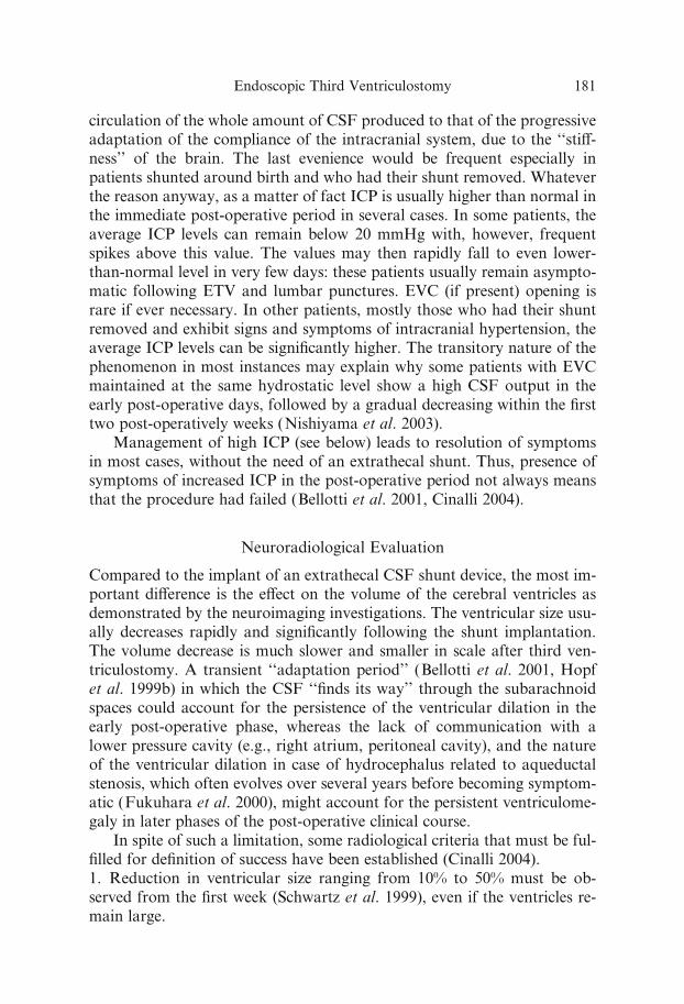
circulation of the whole amount of CSF produced to that of the progressiveadaptation of the compliance of the intracranial system, due to the ‘‘sti¤-ness’’ of the brain. The last evenience would be frequent especially inpatients shunted around birth and who had their shunt removed. Whateverthe reason anyway, as a matter of fact ICP is usually higher than normal inthe immediate post-operative period in several cases. In some patients, theaverage ICP levels can remain below 20 mmHg with, however, frequentspikes above this value. The values may then rapidly fall to even lower-than-normal level in very few days: these patients usually remain asympto-matic following ETV and lumbar punctures. EVC (if present) opening israre if ever necessary. In other patients, mostly those who had their shuntremoved and exhibit signs and symptoms of intracranial hypertension, theaverage ICP levels can be significantly higher. The transitory nature of thephenomenon in most instances may explain why some patients with EVCmaintained at the same hydrostatic level show a high CSF output in theearly post-operative days, followed by a gradual decreasing within the firsttwo post-operatively weeks (Nishiyama et al. 2003).
Management of high ICP (see below) leads to resolution of symptomsin most cases, without the need of an extrathecal shunt. Thus, presence ofsymptoms of increased ICP in the post-operative period not always meansthat the procedure had failed (Bellotti et al. 2001, Cinalli 2004).
Neuroradiological Evaluation
Compared to the implant of an extrathecal CSF shunt device, the most im-portant di¤erence is the e¤ect on the volume of the cerebral ventricles asdemonstrated by the neuroimaging investigations. The ventricular size usu-ally decreases rapidly and significantly following the shunt implantation.The volume decrease is much slower and smaller in scale after third ven-triculostomy. A transient ‘‘adaptation period’’ (Bellotti et al. 2001, Hopfet al. 1999b) in which the CSF ‘‘finds its way’’ through the subarachnoidspaces could account for the persistence of the ventricular dilation in theearly post-operative phase, whereas the lack of communication with alower pressure cavity (e.g., right atrium, peritoneal cavity), and the natureof the ventricular dilation in case of hydrocephalus related to aqueductalstenosis, which often evolves over several years before becoming symptom-atic (Fukuhara et al. 2000), might account for the persistent ventriculome-galy in later phases of the post-operative clinical course.
In spite of such a limitation, some radiological criteria that must be ful-filled for definition of success have been established (Cinalli 2004).1. Reduction in ventricular size ranging from 10% to 50% must be ob-served from the first week (Schwartz et al. 1999), even if the ventricles re-main large.
Endoscopic Third Ventriculostomy 181
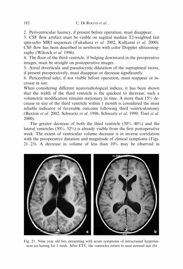
2. Periventricular lucency, if present before operation, must disappear.3. CSF flow artifact must be visible on sagittal median T2-weighted fastspin-echo MRI sequences (Fukuhara et al. 2002, Kulkarni et al. 2000).CSF flow has been described in newborns with color Doppler ultrasonog-raphy (Wilcock et al. 1996).4. The floor of the third ventricle, if bulging downward in the preoperativeimages, must be straight on postoperative images.5. Atrial diverticula and pseudocystic dilatation of the suprapineal recess,if present preoperatively, must disappear or decrease significantly.6. Pericerebral sulci, if not visible before operation, must reappear or in-crease in size.When considering di¤erent neuroradiological indices, it has been shownthat the width of the third ventricle is the quickest to decrease; such avolumetric modification remains stationary in time. A more than 15% de-crease in size of the third ventricle within 1 month is considered the mostreliable indicator of favorable outcome following third ventriculostomy(Buxton et al. 2002, Schwartz et al. 1996, Schwartz et al. 1999, Tisel et al.2000).
The greater decrease of both the third ventricle (30%–40%) and thelateral ventricles (30%–32%) is already visible from the first postoperativeweek. The extent of ventricular volume decrease is in inverse correlationwith the preoperative duration and magnitude of clinical symptoms (Figs.21–23). A decrease in volume of less than 10% may be observed in
Fig. 21. Nine year old boy presenting with acute symptoms of intracranial hyperten-
sion (a) lasting for 1 week. After ETV, the ventricles return to near-normal size (b)
C. Di Rocco et al.182
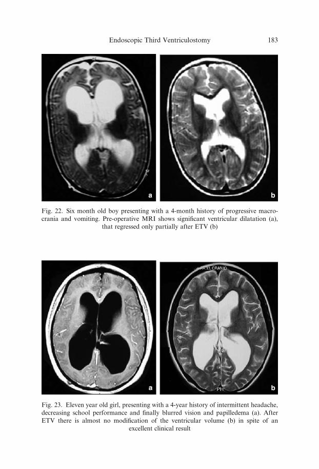
Fig. 22. Six month old boy presenting with a 4-month history of progressive macro-
crania and vomiting. Pre-operative MRI shows significant ventricular dilatation (a),
that regressed only partially after ETV (b)
Fig. 23. Eleven year old girl, presenting with a 4-year history of intermittent headache,
decreasing school performance and finally blurred vision and papilledema (a). After
ETV there is almost no modification of the ventricular volume (b) in spite of an
excellent clinical result
Endoscopic Third Ventriculostomy 183
patients with long-standing chronic symptoms (Schwartz et al. 1999). Thedownward deviation and flattening of the brainstem reverts within 1 year,whereas the width and height of the lateral ventricles continue to decreasesteadily for at least 2 years (Oka et al. 1993b).
Nevertheless radiographic evaluation in the immediate post-operativeperiod is challenging. CT scan during the adaptation period may not revealany modification or even serial scans may show re-enlargement of theventricles following initial reduction in size, leading to the misdiagnosis offailure. Feng et al. (Feng et al. 2004) reported satisfactory results in 5 of 10patients in which a persistent increase of the ventricular size was observedafter surgery and they concluded that the size of the ventricles is not a goodpredictor in the evaluation of the outcome within 3 months following sur-gery. The same paradoxical re-enlargement of ventricular volume after aninitial decrease has been described by other authors (Saint George et al.2004).
Cine-phase contrast MRI, showing directly the patency of the stoma,could be useful as a prognostic factor for a successful ETV. However, thedata of the literature are controversial: Kulkarni et al. (Kulkarni et al.2000) demonstrated a statistically significant relation between evidence ofpostoperative aqueductal CSF on MRI and clinical success. Feng et al.(Feng et al. 2004) in a recent paper found a good correlation betweenthe finding of a patent stoma and outcome. They documented that 12of 16 patients (75%) in whom a positive flow void was observed on post-operative (within two weeks) cine MRI had a successful ETV, even if 4patients, in whom a positive flow void was shown, experienced treatmentfailure. Other reports (Cinalli et al. 1999, Goumnerova and Frim 1997)had shown that cine MRI has not high sensibility and specificity in predict-ing outcome. The exam is more useful as an element of comparison to aprevious similar exam in the same patient than evaluated independently.For this reason, we perform a cine PC MRI in the first post-operativeweek before discharge in order to have a baseline exam when the stomais certainly patent to compare with similar studies carried out in thelate post-operative period (Fig. 20). Early post-operative MRI is also use-ful in order to detect modifications of the profile of the third ventricle andof the lateral ventricle: in cases where cine PC is not conclusive and flowvoid artifact is not well visible on sagittal or coronal T2 turbo spin echo.The morphological analysis of the profile of the third ventricle is prob-ably the most reliable evidence of occlusion of the stoma (Fig. 24a–g).The anatomical details can be visible well before symptoms and signsof increased intracranial pressure occur, and should prompt the surgeonto re-operate the patient even if asymptomatic in order to avoid situa-tions of extreme emergency with possible fatal complications (Hader et al.2002).
C. Di Rocco et al.184
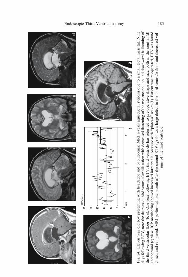
Fig.24.Eleven
yearold
boypresentingwithheadacheandpapilledem
a:MRIrevealsaqueductalstenosisdueto
asm
alltectalmass
(a).Nine
daysfollowingETV,note
thedecreasedthirdventriculardilatationwithdecreasedflatteningofthemesencephalonanddownward
ballooningof
thethirdventricularfloor(b,c).OneyearfollowingETV,thirdventricle
hasreturned
topre-operativeshapeandsize,both
inthesagittal(d)
andcoronal(e)view.IC
Pmonitoringshowed
increasedintracranialpressure
with‘‘plateau’’waves
(f).Patientwasre-operated,ETVwasfound
closedandre-opened.MRIperform
edonemonth
after
thesecondETV
(g)showsalargedefectin
thethirdventricle
flooranddecreasedvol-
umeofthethirdventricle
Endoscopic Third Ventriculostomy 185
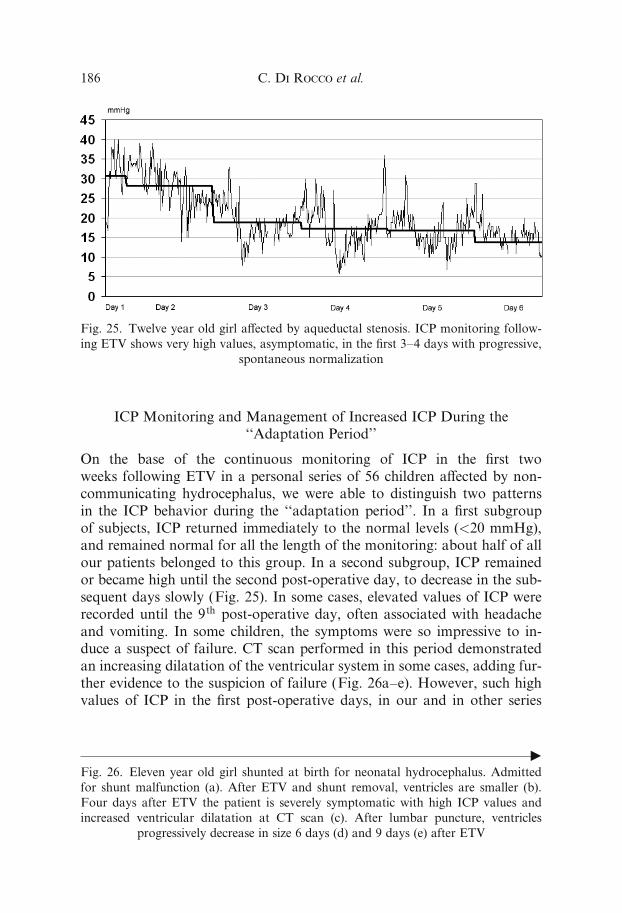
ICP Monitoring and Management of Increased ICP During the‘‘Adaptation Period’’
On the base of the continuous monitoring of ICP in the first twoweeks following ETV in a personal series of 56 children a¤ected by non-communicating hydrocephalus, we were able to distinguish two patternsin the ICP behavior during the ‘‘adaptation period’’. In a first subgroupof subjects, ICP returned immediately to the normal levels (<20 mmHg),and remained normal for all the length of the monitoring: about half of allour patients belonged to this group. In a second subgroup, ICP remainedor became high until the second post-operative day, to decrease in the sub-sequent days slowly (Fig. 25). In some cases, elevated values of ICP wererecorded until the 9th post-operative day, often associated with headacheand vomiting. In some children, the symptoms were so impressive to in-duce a suspect of failure. CT scan performed in this period demonstratedan increasing dilatation of the ventricular system in some cases, adding fur-ther evidence to the suspicion of failure (Fig. 26a–e). However, such highvalues of ICP in the first post-operative days, in our and in other series
Fig. 25. Twelve year old girl a¤ected by aqueductal stenosis. ICP monitoring follow-
ing ETV shows very high values, asymptomatic, in the first 3–4 days with progressive,
spontaneous normalization
fFig. 26. Eleven year old girl shunted at birth for neonatal hydrocephalus. Admitted
for shunt malfunction (a). After ETV and shunt removal, ventricles are smaller (b).
Four days after ETV the patient is severely symptomatic with high ICP values and
increased ventricular dilatation at CT scan (c). After lumbar puncture, ventricles
progressively decrease in size 6 days (d) and 9 days (e) after ETV
C. Di Rocco et al.186
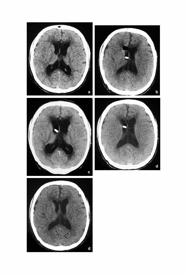

(Bellotti et al. 2001, Hopf et al. 1999b), were not necessarily associatedwith late failure. Actually, their incidence was not statistically significantwhen compared to that observed in cases of true failure. In the only 4 casesin which the procedure failed in the first 15 days (early failure), the ICP,after an initial decrease that lasted for 2–4 days, rapidly rose again betweenthe 5th and 8th day to reach high values unaccettable for the patient, con-sequently suggesting the need for a further surgical procedure (second ETVor CSF shunt implant).
ICP monitoring is very useful to detect intracranial hypertension.However, how to adequately manage high ICP is still under debate. Someauthors (Bellotti et al. 2001, Boschert et al. 2003, Hopf et al. 1999b, Nishi-yama et al. 2003, Oi et al. 2000), have proposed leaving an external ven-tricular drainage in place during the procedure, to allow intermittent CSFdrainage during the periods of pathological ICP elevation. According tothese authors, this would allow transient drop in intraventricular pressure,allowing re-expansion of the intracranial subarachnoid spaces and facili-tating CSF circulation toward the convexity. The same result can be ob-tained by performing CSF evacuation by lumbar taps under ICP monitor-ing (Cinalli 2004). Post-operative withdrawl CSF to restore a normal ICPwas already suggested by Matson in 1969 (Matson 1969) and by otherslater (Ho¤man et al. 1980, Jaksche and Loew 1986, Oka et al. 1995). Insome cases lumbar tapping had been repeated 24–48 hours later; a thirdtap had been rarely necessary (Fig. 27). The positive e¤ects on the ICPlast longer than can be explained by the simple subtraction of the small
Fig. 27. Three year old boy shunted at birth for hydrocephalus and aqueductal steno-
sis. At the time of shunt malfunction, ETV was performed and shunt was removed.
Post-operative ICP monitoring shows high ICP values. Lumbar punctures were
performed (arrows) because of persisting symptoms and signs of increased ICP
C. Di Rocco et al.188
amount of CSF, usually limited to 5–10 ml. One possible explanation is theincreased compliance of the spinal subarachnoid spaces (SAS) and theincreased pressure gradient between third ventricle and the SAS of the pos-terior cranial fossa induced by the CSF subtraction. This would facilitateCSF flow through the third ventriculostomy and, in turn, the reduction ofthe ventricular volume, so allowing the re-expansion of the intracranialsubarachnoid spaces. In patients with intracranial mass lesions, in whichETV was performed before tumor removal, we refrained from using lum-bar tapping, for the risk of tonsillar herniation and preferred to implantan external drainage in the ventricle to be utilized to remove CSF whennecessary.
In conclusion ICP must return to normal values within 10 days afterETV. Should ICP remain elevated with persistent symptoms of intracranialhypertension, we suggest to perform at least two lumbar taps before con-cluding that the procedure failed (Fig. 27).
The algorythm of post-operative patient’s evaluation we utilized in thefirst weeks after ETV is shown in Figure 4. A central role is attributed toICP monitoring in subjects above one year of age. In newborns, ICP mon-itoring is very e¤ectively replaced by fontanel examination.
Late Results
Review of the Literature
As shown on Table 5, di¤erent authors describe various definitions of fail-ure by taking into account several factors, such as the need of an externalCSF shunt, the clinical symptomatology, or the changes of the ventricularvolume. According to this variability, outcome evaluation can di¤er asmuch as 30% in the same group of patients depending on the criteria usedfor definition of success. Moreover, in the same group of patients, di¤erentcriteria of success have been reasonably used according to the di¤erentprocedures. Most authors include neuroradiological criteria to confirmthe clinical success (Beems and Grotenhuis 2004, Brockmeyer et al. 1998,Cinalli et al. 1999, Gorayeb et al. 2004, Teo 1998). According to theseauthors, the ventricular system must decrease in size or at least remainstable and radiographic signs of active hydrocephalus must disappear. Incase of progressive dilatation of the ventricles (despite the absence of symp-tomatology) a further surgical procedure (ETV re-do or VP shunt inser-tion, according to the criteria discussed below) is warranted. Only Fenget al. (Feng et al. 2004) considered successful 5 cases in which clinical im-provement occurred despite of the progressive dilatation of the ventricularsystem, and did not implant a shunt. Actually long-term e¤ect of persistentventriculomegaly (present in about 30% of patients) (Goumnerova and
Endoscopic Third Ventriculostomy 189
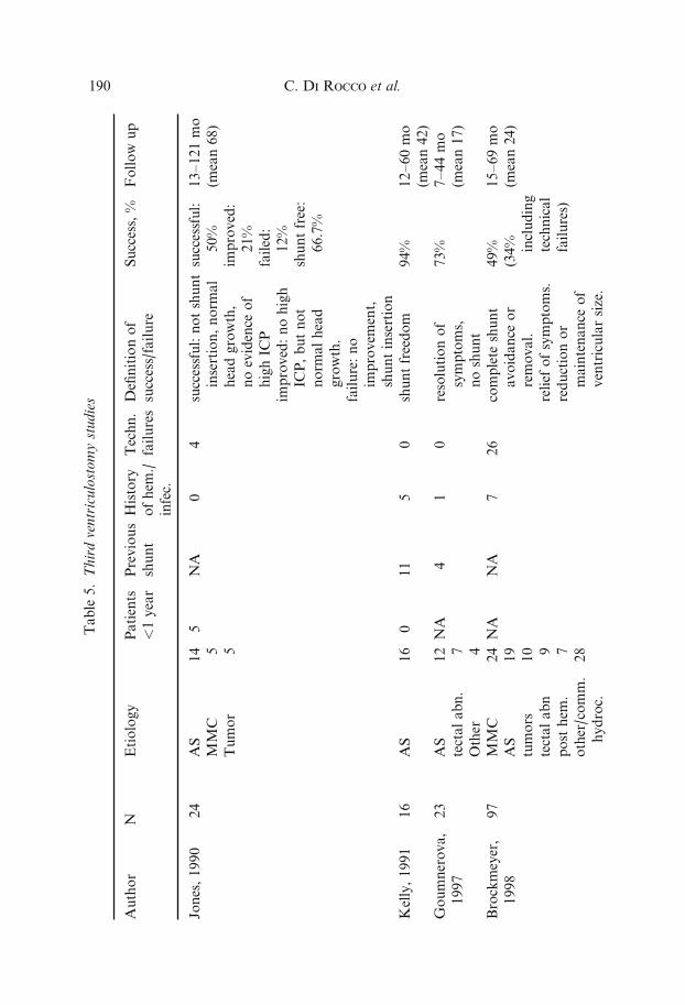
Table
5.Thirdventriculostomystudies
Author
NEtiology
Patients
<1year
Previous
shunt
History
ofhem
./
infec.
Techn.
failures
Definitionof
success/failure
Success,%
Follow
up
Jones,1990
24
AS
MMC
Tumor
14 5 5
5NA
04
successful:notshunt
insertion,norm
al
headgrowth,
noevidence
of
highIC
P
improved:nohigh
ICP,butnot
norm
alhead
growth.
failure:no
improvem
ent,
shuntinsertion
successful:
50%
improved:
21%
failed:
12%
shuntfree:
66.7%
13–121mo
(mean68)
Kelly,1991
16
AS
16
011
50
shuntfreedom
94%
12–60mo
(mean42)
Goumnerova,
1997
23
AS
tectalabn.
Other
12 7 4
NA
41
0resolutionof
symptoms,
noshunt
73%
7–44mo
(mean17)
Brockmeyer,
1998
97
MMC
AS
tumors
tectalabn
post
hem
.
other/comm.
hydroc.
24
19
10 9 7
28
NA
NA
726
complete
shunt
avoidance
or
removal.
relief
ofsymptoms.
reductionor
maintenance
of
ventricularsize.
49%
(34% including
technical
failures)
15–69mo
(mean24)
C. Di Rocco et al.190
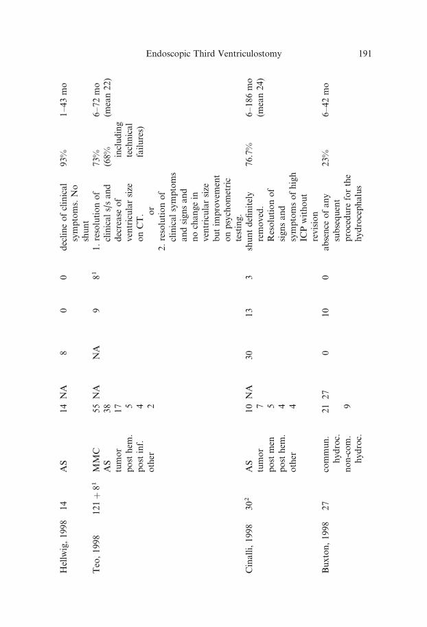
Hellwig,1998
14
AS
14
NA
80
0declineofclinical
symptoms.No
shunt
93%
1–43mo
Teo,1998
121þ81
MMC
AS
tumor
post
hem
.
post
inf.
other
5538
17 5 4 2
NA
NA
981
1.resolutionof
clinicals/sand
decrease
of
ventricularsize
onCT. or
2.resolutionof
clinicalsymptoms
andsignsand
nochangein
ventricularsize
butim
provem
ent
onpsychometric
testing.
73%
(68% including
technical
failures)
6–72mo
(mean22)
Cinalli,1998
302
AS
tumor
post
men
post
hem
.
other
10 7 5 4 4
NA
30
13
3shuntdefinitely
removed.
Resolutionof
signsand
symptomsofhigh
ICPwithout
revision
76.7%
6–186mo
(mean24)
Buxton,1998
27
commun.
hydroc.
non-com.
hydroc.
21 9
27
010
0absence
ofany
subsequent
procedure
forthe
hydrocephalus
23%
6–42mo
Endoscopic Third Ventriculostomy 191
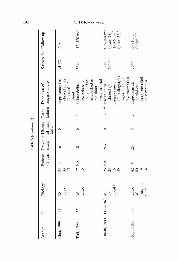
Table
5(C
ontinued
)
Author
NEtiology
Patients
<1year
Previous
shunt
History
ofhem
./
infec.
Techn.
failures
Definitionof
success/failure
Success,%
Follow
up
Choi,1999
71
AS
tumor
other
33
33 5
06
00
improvem
entin
clinicalstatus.
Noneedofa
shunt
91.5%
NA
Tuli,1999
32
AS
tumor
17
15
NA
00
0failure
defined
accordingto
theguidelines
established
in
theshunt
designed
trial
56%
12–120mo
Cinalli,1999
119þ943AS
toxo
pinealt.
other
126
23
15
49
NA
NA
07þ123resolutionof
clinicals/s.
disappearance
of
allradiographic
signsofactive
hydrocephalus
72%
65%3
0.2–108mo
(mean25)
1–209mo3
(mean76)3
Hopf,1999
95
tumor
AS
hem
/inf.
other
42
40 9 4
425
92
improvem
ent:
partialor
complete
relief
ofsymptoms
76%4
3–71mo
(mean26)
C. Di Rocco et al.192
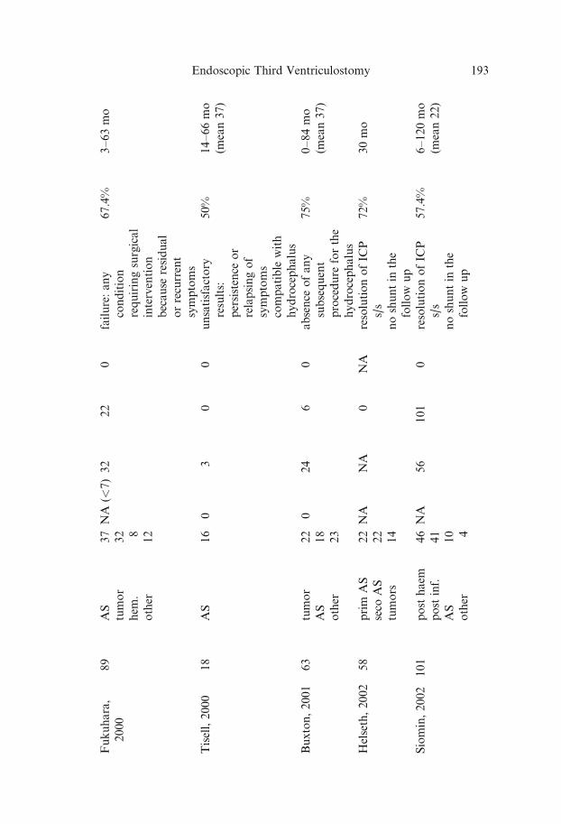
Fukuhara,
2000
89
AS
tumor
hem
.
other
37
32 8
12
NA
(<7)32
22
0failure:any
condition
requiringsurgical
intervention
because
residual
orrecurrent
symptoms
67.4%
3–63mo
Tisell,2000
18
AS
16
03
00
unsatisfactory
results:
persistence
or
relapsingof
symptoms
compatible
with
hydrocephalus
50%
14–66mo
(mean37)
Buxton,2001
63
tumor
AS
other
22
18
23
024
60
absence
ofany
subsequent
procedure
forthe
hydrocephalus
75%
0–84mo
(mean37)
Helseth,2002
58
prim
AS
seco
AS
tumors
22
22
14
NA
NA
0NA
resolutionofIC
P
s/s
noshuntin
the
follow
up
72%
30mo
Siomin,2002
101
post
haem
post
inf.
AS
other
46
41
10 4
NA
56
101
0resolutionofIC
P
s/s
noshuntin
the
follow
up
57.4%
6–120mo
(mean22)
Endoscopic Third Ventriculostomy 193
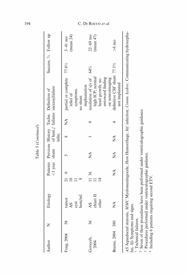
Table
5(C
ontinued
)
Author
NEtiology
Patients
<1year
Previous
shunt
History
ofhem
./
infec.
Techn.
failuresDefinitionof
success/failure
Success,%
Follow
up
Feng,2004
58
tumor
AS
cyst
hem
/inf.
21
18
11 8
05
8NA
partialorcomplete
relief
of
symptoms.
noshunt
implantation
77.6%
3–41mo
(mean24)
Gorayeb,
2004
36
AS
chiariII
other
11
11
14
36
NA
10
resolutionofs/sof
highIC
P;norm
al
headgrowth;no
untoward
finding
onneuroim
aging
64%
22–69mo
(mean47)
Beems,2004
380
NA
NA
NA
NA
4definitiveCSFshunt
notim
planted
77.1%
>6mo
ASAqueductalstenosis,MMCMyelomeningocele,Hem
Hem
orrhage,Inf.infection,Comm.hydroc.Communicatinghydrocepha-
lus,S/sSymptomsandsigns.
1Technicalfailures.
2Seven
oftheseprocedureshavebeenperform
edunder
ventriculographic
guidance.
3Proceduresperform
edunder
ventriculographic
guidance.
4Including6patients
requiringsecondETV.
C. Di Rocco et al.194
Frim 1997) on neuropsychological developmental is not fully known. Teo(Teo 1998) highlights the importance of maintaining a close vigilance onthe patients whose ventricles remain large, by the means of neuropsycho-logical testing. He recommended to shunt those individuals in which a neu-ropsychological deterioration is documented during the follow up, despitethe absence of symptoms and signs of acute intracranial hypertension (seebelow).
Iantosca et al. (Iantosca et al. 2004), reviewing the influence of hydro-cephalus etiology on outcome, found three groups with di¤erent rates ofsuccess: a group with high success rates (>75%), that included patientsa¤ected by acquired aqueductal stenosis, tumor, cyst or infectious lesionsobstructing third ventricular outflow, tectal, pineal, thalamic or intraven-tricular tumor, shunt malfunction; a group of intermediate success rates(about 50%) that included patients a¤ected by myelomeningocele (previ-ously shunted), tumor obstructing fourth ventricle outflow, congenital aque-ductal stenosis, Dandy-Walker malformation, slit ventricles syndrome,recurrent or intractable shunt infection/malfunction; and a group with alow success rate (<50%) that included patients a¤ected by myelomeningo-cele (previously unshunted) and posthaemorrhagic/postmeningitc hydroce-phalus. These authors, on the basis of their experience (Drake 1993, Ian-tosca et al. 2004) and that of others (Jones 1990), discourage the use ofETV in patients who have undergone prior radiation therapy, becausepoor success rates, altered anatomy (in particular thickness of the thirdventricle floor) and increased risk of bleeding.
Thereafter, the best results are achieved in series in which only hydro-cephalus secondary to aqueductal stenosis or benign mass lesions is consid-ered (Choi et al. 1999, Cinalli et al. 1999, Hellwig et al. 1998a, Helseth et al.2002, Kelly 1991). However, when only patients a¤ected by obstructive tri-ventricular hydrocephalus are selected the success rate is actually quitehomogeneous and stable, being above 60–70% at any age (Fritsch andMehdorn 2002, Gorayeb et al. 2004, Javadpour et al. 2001), even if a his-tory of haemorrhage or infection is present (Siomin et al. 2002). All the se-ries studied with actuarial method (Buxton et al. 2001, Cinalli et al. 1999,Elbabaa et al. 2001, Feng et al. 2004, Fukuhara et al. 2000, Helseth et al.2002, Tuli et al. 1999) show a long term success rate above 70% withfollow-up ranging from 4 years to 9 years (Fig. 28). Only Tuli et al. (Tuliet al. 1999) reported a 44% failure rate in a group of pediatric patients af-fected by obstructive tri-ventricular hydrocephalus. This may be secondaryto the di¤erent definition of failure used in the di¤erent papers (Table 5).While most authors define failure as the need of extracranial shunt (table 1),Tuli et al. defined failure in a manner similar to the guidelines established inthe shunt design trial (Drake et al. 1998), accounting for an unusually highrate of failure in selected patients. However, all long-term studies seem to
Endoscopic Third Ventriculostomy 195

show that most of the failures occur during the first year after surgery, andonly sporadically after; basically no late failure has been described after the5th year from ETV. Thus, close follow-up is mandatory in the first 5–6years, and parents must be informed that immediate neurosurgical assess-ment is required if symptoms and signs of intracranial hypertension ap-pear. Such failures can manifest with a very acute clinical onset, and ifnot recognized early may have dramatic consequences, leading to suddendeath (Hader et al. 2002). As argued by Iantosca et al. (Iantosca et al.2004), ‘‘education and regular follow up are important to counteract thefalse sense of security that may arise in the absence of a shunt’’.
Re-Obstruction of the Stoma
The lack of multicentric, prospective, randomized studies account for thecurrent lack of reliable figures about the long term outcome of the childrentreated with ETV. However, Feng et al. (Feng et al. 2004) calculated a pro-portion of 75–80% functioning ETV at 1 year, Cinalli et al. (Cinalli et al.1999) a functioning rate of 72% at 15 years, excluding technical failures.
Fig. 28. Comparison of the seven actuarial studies available in the literature showing
long term success rate studied with actuarial method following ETV in obstructive
hydrocephalus. All the studies, except Tuli, remain between 70 and 85% at 5 years,
no failures being observed beyond that date
C. Di Rocco et al.196
Most failures of ETV occur in the first year following surgery; however,late failures have been reported, usually secondary to obstruction of thestoma by the growth of gliotic ependymal scarring tissue (Cinalli et al.1999, Elbabaa et al. 2001, Hader et al. 2002, Hayashi et al. 2000, Kochet al. 2002, Mohanty et al. 2002, Siomin et al. 2001, Tuli et al. 1999).
If the patient is readmitted with recurrent signs of intracranial hyper-tension and increased ventricular dilatation on CT scan or MRI, the mostlikely diagnosis is obstruction or severe narrowing of the third ventriculos-tomy. This must be immediately confirmed by sagittal median T2-weightedfast spin-echo MRI sequences, which will show disappearance of the flowartifact and recurrence of the indirect signs of occlusion of the stoma(Elbabaa et al. 2001, Koch et al. 2002, Siomin et al. 2002). The treatmentfor obstruction can be reopening or enlargement of the stoma; this proce-dure carries the same success rate as the primary treatment (>65%) andshould be preferred in a first instance to shunt implantation. If MRI atthis stage shows good flow through the stoma, then a ventriculo-peritonealshunt should be inserted.
The ‘‘delayed open-stoma’’ failure can rarely be observed also in chil-dren, especially in patients who have multiple potential etiological factorsfor hydrocephalus (e.g., hemorrhage, meningitis). This is the most insidiousform of failure because of the di‰culties in diagnosing it. Patients canpresent mild developmental delay that is sometimes di‰cult to diagnosewithout appropriate psychomotor testing. Radiology reveals large ven-tricles (stable at follow-up) and open stoma at MRI with clearly visiblepericerebral CSF and no transependymal resorption. Clinically, papille-dema is usually absent and only mild hyperreflexia can be observed, butprogressive macrocrania in the first 2–3 years of life is almost the rule.ICP monitoring in these patients reveals abnormal baseline ICP valuesand B waves. After ventriculo-peritoneal shunting, patients usually showsignificant improvements in psychomotor testing.
Intellectual Outcome
Only a few studies focusing on neuropsychological outcome are available.Burtscher et al. (Burtscher et al. 2003) in a prospective study evaluatedsix patients a¤ected by late onset aqueductal stenosis using standardizedpsychometric testing pre- and postoperatively. They found improvementof pre-operative deficits (usually combined deficits of memory and frontal-executive function) in all patients (full recovery in two, good recovery inthree and moderate recovery in one). Ventricular size diminished in allthese cases but never reached normal size. When comparable series ofpatients with aqueductal stenosis treated by insertion of VP shunt or ETVat the same Institution were analysed, no significant di¤erences in the post-
Endoscopic Third Ventriculostomy 197
operative IQ or in the neurological, endocrinologic, social and behavioralstatus could be detected (Hirsch et al. 1986, Sainte-Rose 1992, Tuli et al.1999). A controlled randomized study comparing neuroendoscopic versusnon neuroendoscopic treatment of hydrocephalus in children seems toshow that the outcome of the patients treated initially with a neuroendo-scopic procedure is significantly better than that of the patients initiallytreated with extrathecal CSF shunt implantation, but this observationrequires further investigation in multicentric studies (Kamikawa et al.2001b).
Complications
Although indications, surgical technique and technological advances re-garding ETV have been largely reported, the complications of the tech-nique have been the subject of a surprisingly scarce interest (Teo et al.1996, Schroeder et al. 2002). Furthermore, only a few papers discuss thequality of the complications in term of cost-e¤ectiveness among ETV andtraditional treatments (Garton et al. 2002, Richard et al. 2000). Such a phe-nomenon depends on the low rate of complications itself but also reflectsthe bias of a recently ‘‘re-introduced’’ technique with the obvious limita-tions in the evaluation of the long-term outcomes Anyway, the data so farcumulated may allow a su‰ciently reliable analysis and the comparisonwith those related to the traditional treatment of hydrocephalus, based onthe placement of an extrathecal CSF shunt device.
The overall morbidity rate related to the shunting procedures is esti-mated to be about 40–50% in the patients operated on before the secondyear of age and near 30% in those ones treated afterwards (Drake et al.2000). Mechanical malfunctions (60% of the cases), infections (1–40%,mean 8.5%), over/underdrainage (10%) and post-inflammation intraven-tricular septa (6%) are the most frequent causes of complication of theCSF shunt devices (Di Rocco et al. 1994, Drake et al. 2000). The reportedoverall complication rate associated with ETV is lower, ranging from 6 to20% (Buxton and Jonathan 2000, Hopf et al. 1999b, Teo and Jones 1996).The experience of the neuroendoscopic team is one of the most influencingfactor (Schroeder et al. 2002). Some authors report even a nil rate of com-plications (Boschert et al. 2003).
The complications of ETV may be divided into ‘‘significant’’ and ‘‘non-significant’’. Significant complications such as hemorrhages, post-operativeneurological deficits, hypothalamic dysfunction, severe bradycardia, epi-lepsy, infections, CSF leak are more frequently observed after ETV thanafter shunt placement. On the other hand, infections are more commonlyassociated to the placement of CSF shunt devices.
C. Di Rocco et al.198
In most cases, the significant complications of ETV are only transient.The rate of significant complications ranges around 7% of the cases withonly one sixth of them leading to permanent neurological damages (Broggiet al. 2000, Cinalli et al. 1998, Fukuhara et al. 2000, Sainte-Rose and Chu-mas 1996, Schroeder et al. 2002, Teo et al. 1991, Teo et al. 1996). Many ofthese complications, often the most severe ones, occur during the openingof the floor of the third ventricle, a phenomenon which accounts for a largenumber of techniques and instruments proposed so far to carry out theventriculocisternostomy (Decq et al. 2004, Guiot 1973, Kehler et al. 1998,Kunz et al. 1994, Lewis and Crone 1994, Oka et al. 1993b, Paladino et al.2000, Rieger et al. 1996, Vandertop et al. 1998, Vries 1978, Wellons et al.1999). On the other hand, non-significant complications are linked up totransient intra-operative accidents (small bleeding, least contusions of theventricular wall or the brain, short episodes of bradycardia) that do not af-fect the success of the operation and do not cause any significant damages.Their rate ranges from 5 to 13% in the largest series (Schroeder et al. 2002,Teo et al. 1996) even if their exact occurrence is di‰cult to establish be-cause they are not always reported or perhaps misunderstood. The loss ofsubstance due to the introduction of the endoscopic instrumentation and itsmovements inside the brain parenchyma might be one of them (Ascho¤2002).
Data about the mortality rate for ETV show an incidence meanly lowerthan 1%, often equal to 0% in many series (Abtin et al. 1998, Hopf et al.1999b, Javadpour et al. 2001, Schroeder et al. 2002, Siomin et al. 2001).This incidence is comparable to the very low one observed for extrathecalshunts (0.1%) (Di Rocco et al. 1994) and it confirms ETV is a safe surgicalprocedure. In fact, death usually occurs after catastrophic bleedings fromlarge vessels or, less frequently, because of severe secondary complications(infections) in patients with poor clinical conditions (Schroeder et al. 2002,Singh et al. 2003). Nevertheless, deaths after late closure of the fenestratedfloor of the third ventricle have been recently reported (Abtin et al. 1998,Hader et al. 2002, Javadpour et al. 2003, Jones et al. 1996). This is the mostinsidious complication of ETV, mainly because sudden, unexpected andpotentially fatal. Nonetheless, a potential and simple solution, as placinga ventricular catheter and a subcutaneous reservoir, has been successfullyused (Mobbs et al. 2003).
Hemorrhages
The control of the intraoperative bleeding during ETV may be challeng-ing since the minimally invasive procedures often do not give the surgeonenough space to arrest it. Moreover, the chances to arrest a blood loss arereduced by a poor three-dimensional vision. Currently, however, the risk of
Endoscopic Third Ventriculostomy 199
severe hemorrhage during ETV is rather low (Abtin et al. 1998, Buxtonand Jonathan 2000, Schroeder et al. 1999).
The damage to the vessels is brought through a mechanical (endoscope,balloon, forceps) or a thermal injury (electrocoagulation, lasers) (Abtinet al. 1998, Buxton and Jonathan 2000, McLaughlin et al. 1997, Schroederet al. 1999). The risk of vascular damage is higher when monopolar/bipolaror laser probe are used as their thermal e¤ect is not much predictable andit may add itself to the mechanical one (Vandertop et al. 1998). Correctinstruments in expert hands and continuous irrigation are the only toolsavailable to decrease this risk.
Intraventricular bleeding from the small subependymal vessels, due tothe traumatic rhexis of these delicate vascular structures caused by theimpact with the endoscopic instrumentation, is the most frequent hemor-rhagic complication happening during ETV (Choi et al. 1999, Cinalli et al.1999, Fukuhara et al. 2000, Gangemi et al. 1999, Sainte-Rose and Chumas1996). The reported incidence ranges from 1 to 3% (Beems and Grotenhuis2004, Fukuhara et al. 2000, Gangemi et al. 1999) but higher figures can behypothesized (Koch and Wagner 2004, Schroeder et al. 2004). Usually, it isa non-significant complication that can be easily managed and stoppedby (extensive) irrigation with lactate Ringer’s solution or, less frequently,through a prudent coagulation of the bleeding vessel. On the contrary,mild hemorrhages (hypothalamic, thalamostriate, septal veins) sometimesmake the irrigation ine¤ective, so a¤ecting the operative visibility and thepossibility to perform a safe electrocoagulation. In these cases, the proce-dure has to be abandoned and an extracranial CSF drainage is placed(Buxton et al. 1998b, Cinalli et al. 1999). This ‘‘technical’’ failure doesnot prevent the possibility to try again the ETV after the clearing of theCSF, with a risk of closure of the stoma following the aseptic ventriculitisjust a little increased (Cinalli et al. 1999, Fukuhara et al. 2000, Hopf et al.1999b). In some instances, the venous bleeding results in a subependymalclot that may clear up spontaneously without the need to abort the surgicalprocedure, as reported by Schonauer et al. (Schonauer et al. 2000). Other-wise, the management of the hematoma requires a microsurgical approach(Hopf et al. 1999b).
Cerebral hemorrhagic contusion, due to the damage toward the pialvessels at the introduction of the endoscope (Schroeder et al. 2002, Vander-top et al. 1998), and extradural hematoma (Choi et al. 1999, Cinalli et al.1998) are uncommon.
The damage of large vascular structures is equally rare, occurring inabout 1% of ETVs (Abtin et al. 1998, Cinalli et al. 1998, McLaughlinet al. 1997, Schroeder et al. 1999, Schroeder et al. 2002). The basilar arteryand its branches (P1 segment of the posterior cerebral artery, perforatingvessels) are usually involved but also the injury to the anterior cerebral ar-
C. Di Rocco et al.200
tery or to the pericallosal branch has been described (Buxton and Jonathan2000, Cohen 1993). The surgeon’s prudence and her/his anatomical knowl-edge, besides her/his skill to succeed in ‘‘seeing’’ the arteries or their pulsa-tions through the ventricular floor, can explain this low rate. It is recom-mended to perforate the floor of the third ventricle halfway between theinfundibular recess and the mammillary bodies, in the midline, in order tominimize the risk of vascular as well as neurological and hypothalamicdamage (Schroeder et al. 1999). The use of ultrasonic or neuronavigationguidance to reduce the incidence of vascular complications has been rec-ommended (Broggi et al. 2000, Strowitzki et al. 2002). An unfavorableanatomy (small or too thick or excessively elastic floor of the third ventri-cle) and a poor intraoperative visibility are important risk factors. Basilarartery perforation appears as a intraventricular bloody flooding that sud-denly obscures the operating field. The management consists of quick re-moval of the endoscope and placement of an external ventricular drainthrough which a copious irrigation must be performed till the bleeding isarrested. After that, the drainage can be use to infuse thrombin in orderto achieve the lysis of the intraventricular clot (Abtin et al. 1998). Alsoblood replacement may be necessary and an adequate neuroanesthesiologicmonitoring is mandatory. The preventive placement of a peelaway sheathin the third ventricle has been proved to be very helpful to avoid theblood accumulation, so preventing an herniation syndrome and saving thepatient’s life (Abtin et al. 1998). The radiological findings of post-operativevascular complications include the demonstration of more or less exten-sive subarachnoid hemorrhage, intraventricular clots, persistent hydroce-phalus and possible pseudoaneurysm formation (McLaughlin et al. 1997,Schroeder et al. 1999). The hydrocephalus is usually treated by ventriculo-peritoneal shunt but the traumatic aneurysm could need a craniotomy tobe excluded (Abtin et al. 1998, McLaughlin et al. 1997). In spite of thereported successful management, the injury to the basilar artery remains adramatic event, potentially fatal (Schroeder et al. 1999). Sometimes, fatalmajor intraventricular bleedings occur without the possibility to establishtheir source (Husain et al. 2003).
Neurological Disorders
Also the damage to the nervous structures resulting in both di¤use or focalnervous functional impairment are caused by mechanical and/or thermalinjury, either directly or indirectly. The incidence of such a type of compli-cation is, however, lower than that of vascular complications (Beems andGrotenhuis 2004, Schroeder et al. 2004). Indirect injuries derive fromvascular ischemic impairment, sometimes as the consequence of one ofthe significant hemorrhagic complications seen before. Whatever the mech-
Endoscopic Third Ventriculostomy 201
anism, the result is often limited to a transient morbidity (non-permanentsignificant complication). In most cases the damage involve the fornix, thethalami or the mammillary bodies (Schroeder et al. 2002). Actually, longterm neurological complications are very rare as demonstrated by onlyanedoctical reports (two cases of hemiparesis and one case of midbraindamage (Jones et al. 1994), one case of confusion after herniation syn-drome and one case of oculomotor palsy (Schroeder et al. 2002), one caseof ankle clonus and speech delay (McLaughlin et al. 1997). Nevertheless,it is possible that some of the complications remain unreported (Schroederet al. 2004).
Among the complications leading to di¤use impairment of cerebralfunction, the decrease in level of consciousness and confusion are the mostcommon, mainly depending on focal diencephalic/brain stem lesions orextensive SAH (Brockmeyer et al. 1998, Buxton and Jonathan 2000, Fuku-hara et al. 2000, Schroeder et al. 2002), and seldom resulting from post-herniation syndrome (Beems and Grotenhuis 2004). Transient memorydisfunctions seem to share a similar pathogenesis (Baskin et al. 1998,Choi et al. 1999, Ferrer et al. 1997, Handler et al. 1994). The fornix andthe mammillary bodies, thanks to their neural connections to the hippo-campus, exert an important role in the acquisition and consolidation ofnewly learned information. The contusions of these two endoscopic land-mark during the surgical procedure may result in impairment of immediatememory, though rarely (Benabarre et al. 2001). Psychiatric complicationsof ETV (disinhibition, aggressive behavior) are exceptional and usually re-lated to primary or secondary lesions of the frontal lobe (Benabarre et al.2001, Buxton and Jonathan 2000). Also epilepsy is rare after ETV, in mostcases resulting from an impaired general clinical condition (Siomin et al.2001), from a previous extensive brain damage (Handler et al. 1994) orfrom associated metabolic disfunction (Vaicys and Fried 2000). Someauthors even assert that ETV minimizes the risk of late seizures, comparedto traditional shunts, thanks to the lack of a permanent irritating foreignbody (ventricular drain) (Kramer et al. 2001).
Focal neurological deficits result from localized lesions, more oftenischemic than mechanical, a¤ecting the long neural pathways (internal cap-sule, brain stem), with consequent side motor impairment (hemiparesis)(Brockmeyer et al. 1998, Buxton and Jonathan 2000, Cinalli et al. 1999,Sainte-Rose and Chumas 1996, Teo et al. 1991), the III–VI cranial nervesand/or their nuclei, with consequent oculomotion palsy (Buxton and Jon-athan 2000, Gangemi et al. 1999, Siomin et al. 2001), or the optic chiasm(hemianopia) (Beems and Grotenhuis 2004). The case of Horner’s syn-drome due to a probable hypothalamic su¤ering, reported by Fukuharaet al. (Fukuhara et al. 2000), and the case of peduncular hallucinosis(visual hallucinations resulting from midbrain, pontine or diencephalic
C. Di Rocco et al.202
lesions), described by Kumar et al. (Kumar et al. 1999), are still unique ascomplications of ETV.
Although uncommonly, focal and generalized neurological deficits, asconsequence of both direct and indirect mechanism, can occur simultane-ously. The case of a young patient, who underwent ETV because of a ven-triculoperitoneal shunt twice complicated by subdural hematoma, is em-blematic (Buxton and Jonathan 2000): the opening of the ventricular floorcaused a bleeding from a perforating branch of the left anterior cerebralartery, with ESA and consequent vasospasm with bilateral cerebral infarc-tion, and a lesion of the right III cranial nerve. The patient initially devel-oped a right oculomotor palsy and drowsiness, both transient; afterwards,a left hemiparesis and behavior disfunction, both transient; finally, a latehyperphagia.
Brain herniation syndrome is unusual but potentially dangerous neuro-logical complication of ETV, though only transient morbidity has beenattributed to it so far. It can result from intraventricular overirrigation(Brockmeyer et al. 1998, Schroeder et al. 2002) or from sudden hydroce-phalus resolution in case of posterior fossa tumor (upward herniation)(Sainte-Rose et al. 2001).
Hypothalamic and Neurovegetative Disfunction
As expected, the anatomy of the third ventricle makes the hypothalamusvulnerable during ETV. Besides all the other mechanisms seen before, theinjuries to the hypothalamus can arise from the distortion of its nucleicaused by the strain of the ventricular floor or by overirrigation. Thismechanism might be liable of one of the most frequent and less studiedcomplications of ETV, the bradycardia. In fact, it has been found that theincidence of the phenomenon at the opening of the stoma may exceed arate of 40%, because of the distortion of the hypothalamic autonomicnuclei (El-Dawlatly et al. 2000). However, this complication is clinicallynon-significant in most cases, rarely requiring a temporary interruption ofthe procedure. Seldom it leads to cardiac asystole and transient cardiacarrest (Fukuhara et al. 2000, Teo et al. 1996).
Ventricular tachycardia, followed by ventricular fibrillation and near-fatal cardiac arrest, reported in 14-years-old girl, was explained on thebase of a similar mechanism (rapid and/or excessive distension of the thirdventricle due to the irrigation) (Handler et al. 1994).
A few cases of transient respiratory arrest following ETV were lasodescribed and attributed to the patient’s general clinical condition afterextubation rather than to the procedure itself (Enya et al. 1997, Siominet al. 2001).
Endoscopic Third Ventriculostomy 203
Diabetes insipidus is a common complication among the hypothalamicimbalances due to ETV but, since it is basically transient, its actual inci-dence is not well known (Schroeder et al. 2002, Teo and Jones 1996, Teoet al. 1996). The damage concerns the supraoptic and paraventricular nu-clei or, more probably, their connections to the hypophyseal median emi-nence (Coulbois et al. 2001). It usually a¤ects the patient for a few daysafter the operation and regresses spontaneously or after a short treatmentby vasopressin (Coulbois et al. 2001, Teo and Jones 1996). To date, onlythree cases of persistent diabetes insipidus have been described (Di Roioet al. 1999, Schroeder et al. 2002, Beems and Grotenhuis 2004).
Other possible hypothalamic endocrinologic or metabolic disfunctionsare uncommon. They include secondary amenorrhea (Teo et al. 1996), in-appropriate secretion of antidiuretic hormone/hyponatriemia (Javadpouret al. 2001, Vaicys and Fried 2000), hyperphagia (Buxton and Jonathan2000, Teo et al. 1996), and loss of thirst (Schroeder et al. 2002, Teo et al.1996).
Transient hyperthermia is reported as possible result of hypothalamicmanipulation, though it is often di‰cult to exclude a concomitant asepticirritation of the ependyma (Sainte-Rose 1992, Sainte-Rose and Chumas1996). In all the cases, however, it is mandatory to rule out a secondaryinfective complication when the fever persists after the first 24–48 post-operative hours. On the other hand, hypothermia, plausibly due to exces-sively cold irrigating fluid, is a not rare finding during the early recoveryafter ETV (Singh et al. 2003).
Other Complications
Infections account for the majority of non-specific complications of ETV.They are facilitated by the presence of a previous infected shunt or an ex-ternal drainage more than depending on the endoscopic procedure itself(Fukuhara et al. 2000, Hopf et al. 1999b, Schroeder et al. 2002). Accordingto the literature, the approximate overall rate ranges from 1 to 5% (Beemsand Grotenhuis 2004, Schroeder et al. 2004, Teo et al. 1996) and includeswound infections (Jones et al. 1994, Siomin et al. 2001, Teo et al. 1991),ventriculitis (Abtin et al. 1998, Brockmeyer et al. 1998, Buxton et al.1998b, Jones et al. 1994, Teo et al. 1991), and meningitis (Gangemi et al.1999, Hopf et al. 1999b, Schroeder et al. 2002). This low incidence rateaccounts for the current favor of ETV, especially when the data related toextrathecal CSF shunts (range 1–40%, mean 8.5%) (Whitehead and Kestle2001) are taken into consideration. In spite of their generally benign courseand the e¤ectiveness of the antibiotic drugs (Schroeder et al. 2004), CSFinfections following ETV are a very feared complication of the procedurebecause of the risk of arachnoiditis and consequent obliteration of the CSF
C. Di Rocco et al.204
pathways. Actually, postoperative meningitis resulted as an independentrisk factor of failure of ETV from the multivariate analysis of Fukuharaet al. (Fukuhara et al. 2000).
CSF leak is a characteristic complication of ETV (Buxton et al. 1998b,Gangemi et al. 1999, Schroeder et al. 2002, Siomin et al. 2001, Tamburriniet al. 2004, Teo et al. 1996), especially in infants (thin skin, immature sub-arachnoid spaces) (Teo 2004). Though in many cases it can be success-fully managed by intermittent tapping from lumbar or ventricular drainage(Cinalli 2004, Husain et al. 2003, Siomin et al. 2001), revealing the possibil-ity of a gradual adaptation of the CSF hydrodynamics to the ETV (Nishi-yama et al. 2003), this type of complication represents a risk factor forinfections and, in particular, it may be the early sign of a failure of the pro-cedure and the need of a shunt placement.
Subdural fluid collections (hematomas or hygromas) are reported asthe second frequent complication involving the vascular structures afterthe intraventricular blood hemorrhages (Oka et al. 1993b, Fukuhara et al.2000, Jones et al. 1994, Sainte-Rose and Chumas 1996, Teo et al. 1991).Their incidence, however, is relatively low, being less than 2% in largeseries (Genitori et al. 2004, Schroeder et al. 2002, Schroeder et al. 2004).These extracerebral collections are likely related to the quick drop of theintracranial pressure following the opening of the stoma (Sgaramella et al.2004) and/or, more probably, to the possible acute CSF leak at the begin-ning or at the end of the procedure (Teo 2004). The phenomenon is espe-cially common in children with a very thin brain cortex and massivehydrocephalus (Schonauer et al. 2000). They are quite often asymptomaticand do not require any further surgical procedure (Fukuhara et al. 2000,Schroeder et al. 2002). Only in a few cases, they need to be evacuated, pro-ducing a transient morbidity (Jones et al. 1990, Sgaramella et al. 2004).The case of a 3-months-old child, who developed a cardiorespiratory arrestbecause of a massive subdural collection after ETV and who was success-fully treated by resuscitation and urgent aspiration of the fluid collection, isexceptional (Mohanty et al. 1997).
Finally, tension pneumocephalus is a rarely reported (Hamada et al.2004) but dangerous complication of ETV, likely depending on the CSFleak during the procedure.
References
1. Abtin K, Thompson BG, Walker ML (1998) Basilar artery perforation as a
complication of endoscopic third ventriculostomy. Pediatr Neurosurg 28:
35–41
2. Alberti O, Riegel T, Hellwig D, Bertalan¤y H (2001) Frameless navigation
and endoscopy. J Neurosurg 95: 541–543
Endoscopic Third Ventriculostomy 205
3. Ascho¤ A (2002) III Ventriculostomy. EANS Winter Meeting, Rome,
March 19th
4. Auer LA, Auer DP (1998) Virtual endoscopy for planning and simulation ofminimally invasive neurosurgery. Neurosurgery 43: 529–548
5. Barkovich AJ, Kjos BO, Norman D, Edwards MS (1989) Revised classifica-
tion of posterior fossa cysts and cyst-like malformations based on the results
of multiplanar MR imaging. AJNR 10: 977–988
6. Barr (1948) Observations on the foramen of Magendie in a series of human
brains. Brain 71: 281
7. Baskin JJ, Manwaring KH, Rekate HL (1998) Ventricular shunt removal:
the ultimate treatment of the slit ventricle syndrome. J Neurosurg 88: 478–484
8. Bech RA, Bogeskov L, Borgesen SE, Juhler M (1999) Indications for shunt
insertion or 3rd ventriculostomy in hydrocephalic children, guided by lumbar
and intraventricular infusion tests. Child’s Brain 15: 213–218
9. Beems T, Grotenhuis JA (2002) Is the success rate of endoscopic third ven-
triculostomy age dependent? An analysis of the results of third ventriculos-
tomy in young children. Child’s Nerv Sist 18: 605–608
10. Beems T, Grotenhuis JA (2004) Long term complications and definition offailures of neuroendoscopic procedures. Child’s Nerv Syst 20: 868–877
11. Bellotti A, Rapana A, Iaccarino C, Schonauer (2001) Intracranial pressure
monitoring after endoscopic third ventriculostomy: an e¤ective method to
manage the ‘adaptation period’. Clin Neurol Neurosurg 103: 223–227
12. Benabarre A, Ibanez J, Boget T, Obiols J, Martınez-Aran A, Vieta E (2001)
Neuropsychological and psychiatric complications in endoscopic third ventri-
culostomy: a clinical case report. J Neurol Neurosurg Psychiatry 71: 268–271
13. Beni-Adani L, Bental Y, Ben-Sira L (2004) Does aqueductal stenosis reallyexist as an independent diagnosis or does it represent an old fetal hemor-
rhage misdiagnosed on real time fetal US? Child’s Nerv Syst 20: 663
14. Boop FA (2004) Post-hemorrhagic hydrocephalus of prematurity. In: Cinalli
G, Meixner W, Sainte-Rose C (eds) Pediatric hydrocephalus, Springer-
Verlag, Milan, pp 121–132
15. Boschert J, Hellwig D, Krauss JK (2003) Endoscopic third ventriculostomy
for shunt dysfunction in occlusive hydrocephalus: long-term follow up and
review. J Neurosurg 98: 1032–103916. Bozzini P (1806) Lichtleiter; eine Erfindung zur Ashauung innerer Theile und
Krankheiten nebst der Abbildung. In: Hufeland CW (ed) J der practischen
Arzneykunde und Wundarrzneykunst, Berlin, 24: 107–124
17. Brockmeyer D, Abtin K, Carey L, Walker M (1998) Endoscopic third ven-
triculostomy: an outcome analysis. Pediatr Neurosurg 28: 236–240
18. Broggi G, Dones I, Ferroli P, Franzini A, Servello D, Duca S (2000) Image
guided neuroendoscopy for third ventriculostomy. Acta Neurochir (Wien)
142: 893–89919. Buki A, Doczi T, Veto F, Horvath Z, Gallyas F (1999) Initial clinical expe-
rience with a combined pulsed holmiumneodymium-YAG laser in minimally
invasive neurosurgery. Minim Invasive Neurosurg 42: 35–40
C. Di Rocco et al.206
20. Burtscher J, Bale R, Dessl A, Eisner W, Twerdy K, Sweeney RA, Felber S
(2002) Virtual endoscopy for planning neuro-endoscopic intraventricular
surgery. Minim Invasive Neurosurg 45: 24–3121. Burtscher J, Bartha L, Twerdy K, Eisner W, Benke T (2003) E¤ect of endo-
scopic third ventriculostomy on neuropsychological outcome in late onset
idiopathic aqueductal stenosis: a prospective study. J Neurol Neurosurg Psy-
chiatry 74: 222–225
22. Burtscher J, Dessl A, Maurer H, Seiwald M, Felber S (1999) Virtual neuro-
endoscopy, a comparative magnetic resonance and anatomical study. Minim
Invas Neurosurg 42: 113–117
23. Buxton N, Jonathan P (2000) Cerebral infarction after neuroendoscopicthird ventriculostomy: case report. Neurosurgery 46: 999–1002
24. Buxton N, Macarthur D, Mallucci C, Punt J, Vloeberghs M (1998a) Neuro-
endoscopy in the premature population. Child’s Nerv Syst 14: 649–652
25. Buxton N, Macarthur D, Mallucci C, Punt J, Vloeberghts M (1998b) Neuro-
endoscopic third ventriculostomy in patients less than 1 year old. Pediatr
Neurosurg 29: 73–76
26. Buxton N, Macarthur D, Robertson I, Punt J (2003) Neuroendoscopic third
ventriculostomy for failed shunts. Surg Neurol 60: 193–20027. Buxton N, Turner B, Ramli N, Vloeberghs M (2002) Changes in third ven-
tricular size with neuroendoscopic third ventriculostomy: a blinded study. J
Neurol Neurosurg Psychiatry 72: 385–387
28. Chapman PH (1990) Indolent gliomas of the midbrain tectum. Concepts
Pediatr Neurosurg 10: 97–107
29. Choi JU, Kim DS, Kim SH (1999) Endoscopic surgery for obstructive
hydrocephalus. Yonsei Med J 40: 600–607
30. Cinalli G (1999) Alternatives to shunting. Child’s Nerv Syst 15: 718–73131. Cinalli G (2004) Endoscopic third ventriculostomy. In: Cinalli G, Maixner
WJ, Sainte-Rose C (eds) Pediatric Hydrocephalus, Springer, Milan, pp
361–388
32. Cinalli G, Sainte-Rose C, Chumas P, Zerah M, Brunelle F, Lot G, Pierre-
Khan A, Renier D (1999) Failure of third ventriculostomy in the treatment
of aqueductal stenosis in children. J Neurosurg 90: 448–454
33. Cinalli G, Salazar C, Mallucci C, Yada JZ, Zerah M, Sainte-Rose C (1998)
The role of endoscopic third ventriculostomy in the management of shuntmalfunction. Neurosurgery 43: 1323–1329
34. Cinalli G, Spennato P, Cianciulli E, D’Armiento M (2004a) Hydrocephalus
and aqueductal stenosis. In: Cinalli G, Meixner W, Sainte-Rose C (eds)
Pediatric Hydrocephalus, Springer-Verlag, Milan, pp 279–294
35. Cinalli G, Spennato P, Del Basso De Caro ML, Buonocore MC (2004b)
Hydrocephalus and the Dandy-Walker malformation. In: Cinalli G, Meixner
W, Sainte-Rose C (eds) Pediatric Hydrocephalus, Springer-Verlag, Milan, pp
279–29436. Ciurea AV, Coman TC, Mircea D (2004) Postinfectious hydrocephalus in
children. In: Cinalli G, Meixner W, Sainte-Rose C (eds) Pediatric Hydroce-
phalus, Springer-Verlag, Milan, pp 201–218
Endoscopic Third Ventriculostomy 207
37. Cohen AR (1993) Endoscopic ventricular surgery. Pediatr Neurosurg 19:
127–134
38. Coulbois S, Boch A-L, Philippon J (2001) Diabete insipide apres ventriculo-cisternostomie par voie endoscopique. A propos d’un cas et revue de la litter-
ature. Neurochirurgie 47: 435–438
39. Czosnyka M, Whitehouse M, Smielewski P, Simac S, Pickard JD (1996)
Testing of cerebrospinal compensatory reserve in shunted and non-shunted
patients: a guide to interpretation based on an observational study. J Neurol
Neurosurg Psychiatry 60: 549–558
40. Dandy WE (1918) Extirpation of the choroid plexuses of the lateral
and fourth ventricle in communicating hydrocephalus. Ann Surg 68: 569–579
41. Dandy WE (1922) An operative procedure for hydrocephalus. Bull Johns
Hopkins Hosp 33: 189–190
42. Daoud AS, Omari H, al-Sheyyab M, Abuekteish F (1998) Indications and
benefits of computer tomography in childhood bacterial meningitis. J Trop
Pediatr 44: 167–169
43. Decq P (2004) Endoscopic anatomy of the ventricles. In: Cinalli G, Maixner
WJ, Saint-Rose C (eds) Pediatric Hydrocephalus, Springer, Milan, pp 351–359
44. Decq P, Le Guerinel C, Palfi S, Djindjian M, Keravel Y, Nguyen J (2000) A
new device for endoscopic third ventriculostomy. J Neurosurg 93: 509–512
45. Di Rocco C, Marchese E, Velardi F (1994) A survey of the first complication
of newly implanted CSF shunt devices for the treatment of nontumoral
hydrocephalus. Cooperative survey of the 1991–1992 Education Committee
of the ISPN. Child’s Nerv Syst 10: 312–327
46. Di Roio C, Mottolese C, Cayrel V, Berlier P, Artru F (1999) Ventriculosto-mie du troisieme ventricule et diabete insipide. Ann Fr Anesth Reanim 18:
776–778
47. Doctor BA, Newman N, Minich NM, Taylor HG, Fanaro¤ AA, Hack M
(2001) Clinical outcomes of neonatal meningitis in very-low birth-weight
infant. Clin Pediatr 40: 473–480
48. Drake JM (1993) Ventriculostomy for treatment of hydrocephalus. Neuro-
surg Clin North Am 4: 657–666
49. Drake JM, Kestle JRW, Milner R, Cinalli G, Boop F, Piatt J Jr, Hainess S,Schi¤ SJ, Cochrane DD, Steinbok P, McNeil N (1998) Randomized trial
of cerebrospinal fluid shunt valve design in pediatric hydrocephalus. Neuro-
surgery 43: 294–303
50. Drake JM, Kestle JRW, Tuli S (2000) CSF shunts 50 years on – past, present
and future. Child’s Nerv Syst 16: 800–804
51. Du¤ner F, Shi¤bauer H, Glenser D, Skalej M, Freudenstein D (2003) Anat-
omy of the cerebral ventricular system for endoscopic neurosurgery: a mag-
netic resonance study. Acta Neurochir 145: 359–36852. Elbabaa SK, Steinmetz M, Ross J, Moon D, Luciano M (2001) Endoscopic
third ventriculostomy for obstructive hydrocephalus in the pediatric popula-
tion: evaluation of outcome. Eur J Pediatr Surg 11 (suppl. 1): 552–554
C. Di Rocco et al.208
53. El-Dawlatly AA, Murshid WR, El-Khwsky F (1999) Endoscopic third ven-
triculostomy: a study of intracranial pressure vs heamodynamic changes.
Minim Invas Neurosurg 42: 198–20054. El-Dawlatly AA, Murshid WR, Elshimy A, Magboul MA, Samarkandi A,
Takrouri MS (2000) The incidence of bradycardia during endoscopic third
ventriculostomy. Anesth Analg 91: 1142–1144
55. Enya S, Masuda Y, Terui K (1997) Respiratory arrest after a ventriculo-
scopic surgery in infants: two case reports. Masui 46: 416–420 [Japanese]
56. Enzmann DR, Pelec NJ (1991) Normal flow pattern in intracranial and spi-
nal cerebrospinal fluid defined with phase-contrast cine-MR imaging. Radiol
178: 467–47457. Fay T, Grant FC (1923) Ventriculoscopy and intraventricular photography
in internal hydrocephalus. JAMA 80: 461–463
58. Feng H, Huang G, Liao X, Fu K, Tab H, Pu H, Cheng Y, Liu W, Zhao D
(2004) Endoscopic third ventriculostomy in the management of obstructive
hydrocephalus: an outcome analysis. J Neurosurg 100: 626–633
59. Ferrer E, Santamarta D, Garcia-Fructuoso G, Caral L, Rumia J (1997)
Neuroendoscopic management of pineal region tumors. Acta Neurochir
139: 12–2160. Fischer RE, Biljana T (2000) Optical system design, McGraw Hill, New
York
61. Foroutan M, Mafee MF, Dujovny M (1998) Third ventriculostomy, phase-
contrast cine MRI and endoscopic techniques. Neurol Res 20: 443–448
62. Frazee JG, Shah AS (1998) Interactive surgery and telepresence. In: King W,
Frazee J, De Salles A (eds) Endoscopy of the Central and Peripheral Ner-
vous System, Thieme, New York, 1998, pp 243–251
63. Fritsch MJ, Mehdorn M (2002) Endoscopic intraventricular surgery fortreatment of hydrocephalus and loculated CSF space in children less than
one year of age. Pediatr Neurosurg 36: 183–188
64. Fritsch MJ, Mehdorn HM (2003) Indication and controversies for endo-
scopic third ventriculostomy in children. Child’s Nerv Syst 19: 706–707
65. Fritsch MJ, Dorner L, Kienke S, Claviez A, Mehdorn HM (2004) Indication
for endoscopic third ventriculostomy in children with posterior fossa tumors.
Child’s Nerv Syst 20: 668
66. Fukuhara T, Luciano MG, Kowalski RJ (2002) Clinical features of thirdventriculostomy failures classified by fenestration patency. Surg Neurol 58:
102–110
67. Fukuhara T, Vorster SJ, Luciano MG (2000) Risk factors for failure of en-
doscopic third ventriculostomy for obstructive hydrocephalus. Neurosurgery
46: 1100–1111
68. Fukushima T, Ishijima B, Hirakaw K, Nakamura N, Samo K (1973) Ventri-
culofiberscope: a new technique for endoscopic diagnosis and operation. J
Neurosurg 38: 251–25669. Gangemi M, Donati P, Maiuri F, Longatti P, Godano U, Mascari C (1999)
Endoscopic third ventriculostomy for hydrocephalus. Minim Invasive Neu-
rosurg 42: 128–132
Endoscopic Third Ventriculostomy 209
70. Garton HJL, Kestle JRW, Cochrane DD, Steinbok P (2002) A cost-
e¤ectiveness analysis of endoscopic third ventriculostomy. Neurosurgery 51:
69–7871. Genitori L, Peretta P, Mussa F, Giordano F (2004) Endoscopic third ventri-
culostomy in children: are age and etiology of hydrocephalus predictive
factors influencing the outcome in primary and secondary treated patients?
A series of 328 patients and 353 procedures. II CURAC Congress, Munich,
October 8th
72. Glasauer FE (1975) Isotope cisternography and ventriculography in congen-
ital anomalies of the central nervous system. J Neurosurg 43: 18–26
73. Goh KYC, Abbott R (2000) Is endoscopic third ventriculostomy of benefitin tumour related aqueductal stenosis? Child’s Nerv Syst 16: 127–128
74. Gorayeb RP, Cavalheiro S, Zymberg SY (2004) Endoscopic third ventricu-
lostomy in children younger than 1 year of age. J Neurosurg (Pediatrics 5)
100: 427–429
75. Goumnerova LC, Frim D (1997) Treatment of hydrocephalus with third
ventriculostomy: outcome and CSF flow patterns. Pediatr Neurosurg 27:
149–152
76. Grant JA (1998) An endoscopic view of the anterior commissure. PediatrNeurosurg 28: 42
77. Greitz D (2004) Radiological assessment of hydrocephalus: new theories and
implications for therapy. Neurosurg Rev 27: 145–165
78. Grunert P, Perneczki A, Resch K (1994) Endoscopic procedures through the
foramen of Monro under stereotactic conditions. Mim Invasive Neurosurg
37: 2–8
79. Grunert P, Charalampaki P, Hopf N, Filippi R (2003) The role of third
ventriculostomy in the management of obstructive hydrocephalus. MinimInvasive Neurosurg 46: 16–21
80. Guiot G (1973) Ventriculo-cisternostomy for stenosis of aqueduct of Silvius.
Puncture of the floor of the third ventricle with a leucotome under television
control. Acta Neurochir 28: 275–289
81. Hader WJ, Drake J, Cochrane D, Sparrow O, Johnson ES, Kestle J (2002)
Death after late failure of third ventriculostomy in children. Report of three
cases. J Neurosurg 97: 211–215
82. Hamada H, Hayashi N, Kurimoto M, Umemura K, Hirashima Y, NogamiK, Endo S (2004) Tension pneumocephalus after neuroendoscopic procedure
– case report. Neurol Med Chir (Tokyo) 44: 205–208
83. Handler MH, Abbott R, Lee M (1994) A near-fatal complication of endo-
scopic third ventriculostomy: case report. Neurosurgery 35: 525–528
84. Hayashi N, Hamada H, Hirashima Y, Kurimoto M, Takaku A, Endo S
(2000) Clinical features in patients requiring reoperation after failed endo-
scopic procedures for hydrocephalus. Minim Invasive Neurosurg 43: 181–186
85. Hecht J (1999) City of light: the story of fiber optics, Oxford UniversityPress, New York
86. Heilman CB, Cohen AR (1991) Endoscopic ventricular fenestration using
‘‘saline torch’’. J Neurosurg 74: 224–229
C. Di Rocco et al.210
87. Hellwig D, Haag R, Bartel V, Riegel T, Eggers F, Becker R, Bertalan¤y H
(1999) Application of new electrosurgical devices and probes in endoscopic
neurosurgery. Neurol Res 21: 67–7288. Hellwig D, Heinemann A, Riegel T (1998a) Endoscopic third ventriculos-
tomy in treatment of obstructive hydrocephalus caused by primary aqueduc-
tal stenosis. In: Hellwig D, Bauer B (eds) Minimally Invasive Techniques for
Neurosurgery, Springer, Berlin, pp 65–72
89. Hellwig D, Riegel T, Bertalan¤y H (1998b) Neuroendoscopic techniques
in treatment of intracranial lesions. Minim Invasive Ther Allied Technol 7:
123–135
90. Helseth E, Due-Tonnessen B, Egge A, Eide PK, Meling T, Lundar T, FroslieKF (2002) Behandling av Hydrocephalus med endoskopisk tredjeventrikkel-
stomi. Tidsskr Nor Loegeforen 122: 994–998
91. Hill A, Volpe J (1981) Seizures, hypoxic-ischemic brain injury, and intraven-
tricular hemorrhage in the newborn. Ann Neurol 10: 109–121
92. Hirsch JF, Pierre-Kahn A, Renier D, Sainte-Rose C, Hoppe-Hirsch E (1984)
The Dandy-Walker malformation. A review of forty cases. J Neurosurg 61:
512–522
93. Hirsch JF, Hirsch E, Sainte-Rose C, Renier D, Pierre-Kahn A (1986) Steno-sis of the aqueduct of Sylvius. Etiology and treatment. J Neurosurg Sci 30:
29–36
94. Ho¤man HJ, Harwood-Nash D, Gilday DL, Craven MA (1980) Percutane-
ous third ventriculostomy in the management of non-communicating hydro-
cephalus. Neurosurgery 7: 313–321
95. Hopf NJ, Grunert P, Darabi K, Busert C, Bettag (1999a) Frameless neuro-
navigation applied to endoscopic neurosurgery. Minim Invasive Neurosurg
42: 187–19396. Hopf NJ, Grunert P, Fries G, Resch KDM, Perneczky A (1999b) Endo-
scopic third ventriculostomy: outcome analysis of 100 consecutive proce-
dures. Neurosurgery 44: 795–806
97. Horowitz M, Ramzipoor K, Mair A, Miller S, Rappard G, Spiro R, Purdy P
(2003) Experimental third ventriculostomy performed using endovascular
surgical techniques and their adaptation to percutaneous intradural neurona-
vigation: proof of concept cadaver study. Neurosurgery 53: 387–392
98. Husain M, Jha D, Vatsal DK, Thaman D, Gupta A, Husain N, Gupta RK(2003) Neuroendoscopy surgery – experience and outcome analysis of 102
consecutive procedure in a busy neurosurgical center of India. Acta Neuro-
chir 145: 369–376
99. Iantosca MR, Hader WJ, Drake JM (2004) Results of endoscopic third ven-
triculostomy. Neurosurg Clin N Am 15: 67–75
100. Jacobson EE, Fletcher DF, Morgan MK, Johnston IH (1999) Computer
modelling of the CSF flow dynamics of aqueductal stenosis. Med Biol Eng
Comput 37: 59–63101. Jaksche H, Loew F: Burr hole third ventriculostomy (1986) An unpopular
but e¤ective procedure for treatment of certain forms of occlusive hydroce-
phalus. Acta Neurochir 79: 48–51
Endoscopic Third Ventriculostomy 211
102. Javadpour M, Mallucci C, Brodbelt A, Golash A, May P (2001) The impact
of endoscopic third ventriculostomy on the management of newly diagnosed
hydrocephalus in infants. Pediatr Neurosurg 35: 131–135103. Javadpour M, May P, Mallucci C (2003) Sudden death secondary to delayed
closure of endoscopc third ventriculostomy. Br J Neurosurg 17: 266–269
104. Jellinger G (1986) Anatomomorphology of nontumoral aqueductal stenosis.
J Neurosurg Sci 30: 1–16
105. Jodicke A, Berthold LD, Scharbrodt W, Schroth I, Reiss I, Neubauer BA,
Boker DK (2003) Endoscopic surgical anatomy of the paediatric third ventri-
cle studied using virtual neuroendoscopy based on 3-D ultrasonography.
Child’s Nerv Syst 19: 325–331106. Jones FRC, Stening WA, Brydon M (1990) Endoscopic third ventriculos-
tomy. Neurosurgery 26: 86–92
107. Jones RFC, Kwok BCT, Stening WA (1996) Endoscopic III ventriculos-
tomy. How long does it last? Child’s Nerv Syst 12: 364–365
108. Jones RFC, Kwok BCT, Stening WA, Vonau M (1994) The current status of
endoscopic third ventriculostomy in the management of non-communicating
hydrocephalus. Minim Invasive Neurosurg 37: 28–36
109. Kaiser G (1985) Hydrocephalus following toxoplasmosis. Z Kinderkir 40(Suppl. 1): 10–11
110. Kamikawa S, Inui A, Kobayashi N, Kuwamura K, Kasuga M, Yamadori T,
Tamaki N (2001b) Endoscopic treatment of hydrocephalus in children: a
controlled study using newly developed Yamadori-type ventriculoscopes.
Minim Invasive Neurosurg 44: 25–30
111. Kamikawa S, Inui A, Kobayashi N, Tamaki N, Yamadori T (2001c) Intra-
ventricular hemorrhage in neonates: endoscopic findings and treatment by
the use of our newly developed Yamadori-type 8 ventriculoscope. Min InvasNeurosurg 44: 74–78
112. Kamikawa S, Inui A, Tamaki N, Kobayashi N, Yamadori T (2001a) Appli-
cation of flexible neuroendoscope to intracerebroventricular arachnoid cyst
in children – Use of videoscope. Minim Invas Neurosurg 44: 186–189
113. Kehler U, Gliemroth J, Knopp U, Arnold H (1998) How to perforate safely
a resistant floor of the third ventricle? Technical note. Minin Invasive Neuro-
surg 41: 198–199
114. Kelly PJ (1991) Stereotactic third ventriculostomy in patients with nontu-moral adolescent/adult onset aqueductal stenosis and symptomatic hydroce-
phalus. J Neurosurg 75: 865–873
115. Kim SK, Wang KC, Cho BK (2000) Surgical outcome of pediatric hydroce-
phalus treated by endoscopic III ventriculostomy: prognostic factors and in-
terpretation of postoperative neuroimaging. Child’s Nerv Syst 16: 161–169
116. Koch D, Grunert P, Filippi R, Hopf N (2002) Re-ventriculostomy for treat-
ment of obstructive hydrocephalus in cases of stoma dysfunction. Minim
Invasive Neurosurg 45: 158–163117. Koch D, Wagner W (2004) Endoscopic third ventriculostomy in infants of
less than 1 year of age: which factors influence the outcome? Child’s Nerv
Syst 20: 405–411
C. Di Rocco et al.212
118. Kramer U, Kanner AA, Siomin V, Harel S, Constantini S (2001) No evi-
dence of epilepsy following endoscopic third ventriculostomy: a short-term
follow up. Pediatr Neurosurg 34: 121–123119. Kreusser K, Tarby T, Kovnar E, Taylor DA, Hill A, Volpe JJ (1985) Serial
lumbar punctures for at least temporary amelioration of neonatal posthe-
morrhagic hydrocephalus. Pediatrics 75: 719–724
120. Kulkarni AV, Drake JM, Armstrong DC, Dirks PB (2000) Imaging correlates
of successful endoscopic third ventriculostomy. J Neurosurg 92: 915–919
121. Kumar R, Behari S, Wahi J, Banerji D, Sharma K (1999) Peduncular hallu-
cinosis: an unusual sequel to surgical intervention in the suprasellar region.
Br J Neurosurg 13: 500–503122. Kunz U, Goldman A, Bader C, Waldbaur H, Oldenkott P (1994) Endo-
scopic fenestration of the 3rd ventricular floor in aqueductal stenosis. Minim
Invasive Neurosurg 37: 42–47
123. Laikin M (2001) Method of lens design, Dekker, New York
124. Lang J (1992) Topographic anatomy of preformed intracranial spaces. Acta
Neurochir (Wien) (suppl.) 54: 1–10
125. Lapras C, Bret P, Patet JD, Huppert J (1986) Hydrocephalus and aqueductal
stenosis. Direct surgical treatment by interventriculostomy (aqueduct cannu-lation). J Neurosurg Sci 30: 47–53
126. Lespinasse VL (1910) In: Davis L (ed) Neurological Surgery, Lea &
Febinger, Philadelphia, 1936, p 405
127. Lewis AI, Crone KR (1994) Advances in neuroendoscopy. Contemp Neuro-
surg 16: 1–6
128. Liu CY, Wang MY, Apuzzo MLJ (2004) The physics of image formation in
the neuroendoscope. Childs Nerv Syst 20: 777–782
129. Longatti P, Martinuzzi A, Fiorindi A, Malobabic S, Carteri A (2003) Endo-scopic anatomic features of the triangular recess. Neurosurgery 52: 1491–1494
130. Macarthur DC, Buxton N, Vloeberghs M, Punt J (2001). The e¤ectiveness of
neuroendoscopic interventions in children with brain tumors. Child’s Nerv
Syst 17: 589–594
131. Magnaes B (1982) Cerebrospinal fluid hydrodynamics in adult patients
with benign non-communicating hydrocephalus: one-hour test shunting and
balanced cerebrospinal fluid infusion test to select patients for intracranial
bypass operation. Neurosurgery 11: 769–775132. Magnaes B (1989) Hydromechanical testing in non-communicating hydroce-
phalus to select patients for microsurgical third ventriculostomy. Br J Neuro-
surg 3: 443–450
133. Matson DD (1969) Neurosurgery of Infancy and Childhood, 2nd edition,
Charles C Thomas, Springfield, Illinois
134. McLaughlin MR, Wahlig JB, Kaufmann AM, Albright AL (1997) Trau-
matic basilar aneurysm after endoscopic third ventriculostomy: case report.
Neurosurgery 41: 1400–1404135. Miller MN (1992) Organisation of the neuroendoscopy suite. In: Manwaring
KH, Crone KR (eds) Neuroendoscopy, vol 1, Mary Ann Liebert, New York,
pp 9–15
Endoscopic Third Ventriculostomy 213
136. Mixter WJ (1923) Ventriculoscopy and puncture of the floor of the third
floor. Preliminary report of a case. Boston Med Surg J 1: 277–278
137. Mobbs RJ, Vonau M, Davies MA (2003) Death after late failure of endo-scopic third ventriculostomy: a potential solution. Neurosurgery 53: 384–
386
138. Mohanty A, Anandt B, Reddy MS, Sastry KVR (1997) Contralateral mas-
sive acute subdural collection after endoscopic third ventriculostomy – a case
report. Minim Invasive Neurosurg 37: 28–36
139. Mohanty A, Vasudev MK, Sampath S, Radhesh S, Sastry, Kolluri VR
(2002) Failed endoscopic third ventriculostomy in children: management
options. Pediatr Neurosurg 37: 304–309140. Mohanty A (2003) Endoscopic third ventriculostomy with cystoventricular
stent placement in the management of Dandy-Walker malformation: techni-
cal case report of three patients. Neurosurgery 53: 1223–1229
141. Mori H, Nishiyama K, Tanaka R (2003) Endoscopic third ventriculostomy
in shunt malfunction and slit-ventricle syndrome. Child’s Nerv Syst 19: 690
142. Murshid WR (2000) Endoscopic third ventriculostomy: towards more indi-
cations for the treatment of non-communicating hydrocephalus. Minim
Invas Neurosurg 43: 75–82143. Nishiyama K, Mori H, Tanaka R (2003) Changes in cerebrospinal fluid
hydrodynamics following endoscopic third ventriculostomy for shunt-
dependent noncommunicating hydrocephalus. J Neurosurg 98: 1027–1031
144. Nitze M (1879) Eine neue Beobachtungs und Untersuchungs Methode fur
Harnrohre, Harnblse und Rectum. Wien Med Wochenschr 24: 649–652
145. Nobles A (1998) The physics of neuroendoscopic systems and the instrumen-
tation. In: Jimenez DF (ed) Intracranial Endoscopic Neurosurgery, Park
Ridge, IL: American Association of Neurological Surgeons, pp 1–12146. Nugent GR, Al-Mefty O, Chou S (1979) Communicating hydrocephalus as a
cause of aqueductal stenosis. J Neurosurg 51: 812–818
147. Oi S, Shibata M, Tominaga J, Honda Y, Shinoda M, Takei F, Tsugane R,
Matsuzawa K, Sato O (2000) E‰cacy of neuroendoscopic procedures in
minimally invasive preferential management of pineal region tumors: a pro-
spective study. J Neurosurg 93: 245–253
148. Oi S, Hamada H, Nonaka Y, Kusaka Y, Samii A, Samii M (2004) Proposal
of a ‘‘evolution theory in CSF dynamics’’: a significant factor a¤ecting thefailure of neuroendoscopic ventriculostomy in treatment of hydrocephalus.
Child’s Nerv Syst 20: 662
149. Oka K, Go Y, Kin Y, Tomonaga M (1993a) An observation of the third
ventricle under flexible fiberoptic ventriculoscope: normal structure. Surg
Neurol 40: 273–277
150. Oka K, Go Y, Kin Y, Utsunomiya H, Tomonaga M (1995) The radio-
graphic restoration of the ventricular system after third ventriculostomy.
Minim Invasive Neurosurg 38: 158–162151. Oka K, Tomonaga M (1992) Instruments for flexible endoneurosurgery.
In: Manwaring KH, Crone KR (eds) Neuroendoscopy, vol 1, Mary Ann
Liebert, New York, pp 17–28
C. Di Rocco et al.214
152. Oka K, Yamamoto M, Ikeda K, Tomonaga M (1993b) Flexible endo-
neurosurgical therapy for aqueductal stenosis. Neurosurgery 33: 236–
243153. Paladino J, Rotim K, Stimac D, Pirker N, Stimac A (2000) Endoscopic third
ventriculostomy with ultrasonic contact microprobe. Minim Invasive Neuro-
surg 43: 132–134
154. Pavez Salinas A (2004) Description of endoscopic ventricular anatomy in
meningomyelocele. Child’s Nerv Syst (in press)
155. Perlman JM, Argyl C (1992) Lethal cytomegalovirus infection in preterm
infants: clinical, radiological, and neuropathological findings. Ann Neurol
31: 64–68156. Pettorossi VE, Di Rocco C, Mancinelli R, Caldarelli M, Velardi F (1978)
Communicating hydrocephalus induced by mechanically increased ampli-
tude of the intraventricular cerebrospinal fluid pulse pressure: rationale and
method. Exp Neurol 59: 30–39
157. Pollack JK, Pang D, Albright AL (1994) The long term outcome in children
with late-onset aqueductal stenosis resulting from benign intrinsic tectal
tumors. J Neurosurg 80: 681–688
158. Pople IK, Athanasiou TC, Sandeman DR, Coakham HB (2001) The role ofendoscopic biopsy and third ventriculostomy in the management of pineal
region tumours. Br J Neurosurg 15: 305–311
159. Portillo S, Zuccaro G, Fernandez-Molina A, Houssay A, Sosa F, Konsol O,
Jaimovich R, Olivella E, Ledesma J, Guevara M, Ajler G, Picco P (2004)
Endoscopic third ventriculostomy in the treatment of pediatric hydrocepha-
lus. A multicentric study. Child’s Nerv Syst 20: 666–667
160. Putnan TJ (1943) The surgical treatment of infantile hydrocephalus. Surg
Gynecol Obst 76: 171–182161. Quencher RM, Donovan Post MJ, Hinks RS (1990) Cine MR in the evalua-
tion of normal and abnormal CSF flow: intracranial and intraspinal studies.
Neuroradiology 32: 371–391
162. Raimondi AJ, Clark SJ, McLone DG (1976) Pathogenesis of aqueductal oc-
clusion in congenital murine hydrocephalus. J Neurosurg 45: 66–77
163. Rappaport ZH, Shalit MN (1989) Perioperative external ventricular drain-
age in obstructive hydrocephalus secondary to infratentorial brain tumours.
Acta Neurochir 96: 118–121164. Resch KDM (2003) Endo-neuro-sonography: first clinical series (52 cases).
Childs Nerv Syst 19: 137–144
165. Resch KDM, Perneczky A (1998) Endo-neuro-sonography: basics and cur-
rent use. In: Hellwig D, Bauer BL (eds) Minimally Invasive Techniques for
Neurosurgery, Springer, Berlin Heidelberg New York, pp 21–31
166. Resch KDM, Reisch R (1997) Endo-neuro-sonography: anatomical aspects
of the ventricles. Minim Invasive Neurosurg 1: 2–7
167. Rhoton AL Jr (2002) The lateral and third ventricles. Neurosurgery 51(suppl. 1): 207–270
168. Richard HK, Seeley HM, Okane C, Pickard JD (2000) ICP 11, Cambridge,
July 22–26th
Endoscopic Third Ventriculostomy 215
169. Riegel T, Alberti O, Hellwig D, Bertalan¤y H (2001) Operative management
of third ventriculostomy in cases of thickened, non-translucent third ventric-
ular floor: technical note. Minim Invas Neurosurg 44: 65–69170. Riegel T, Alberti O, Retsch R, Shiratori V, Hellwig D, Bertalan¤y H (2000)
Relationships of virtual reality neuroendoscopic simulations to actual imag-
ing. Minim Invasive Neurosurg 43: 176–180
171. Riegel T, Freudenstein D, Alberti O, Du¤ner F, Hellwig D, Bartel V, Berta-
lan¤y H (2002) Novel multipurpose bipolar instrument for endoscopic neu-
rosurgery. Neurosurgery 51: 270–274
172. Rieger A, Rainov NG, Sanchin L, Schoop G, Burkert W (1996) Ultrasound-
guided endoscopic fenestration of the third ventricular floor for non-communicating hydrocephalus. Minim Invasive Neurosurg 39: 17–20
173. Rieger A, Rainov NG, Brucke M, Marx T, Holz C (2000) Endoscopic third
ventriculostomy is the treatment of choice for obstructive hydrocephalus due
to pediatric pineal tumors. Minim Invas Neurosurg 43: 83–86
174. Rohde V, Gilsbach JM (2000) Anomalies and variants of the endoscopic
anatomy for third ventriculostomy. Min Invas Neurosurg 43: 111–117
175. Rohde V, Krombach GA, Stru¤ert T, Gilsbach JM (2001) Virtual MRI
endoscopy: detection of anomalies of the ventricular anatomy and its possi-ble role as a presurgical planning tool for endoscopic third ventriculostomy.
Acta Neurochir 143: 1085–1091
176. Roland E, Hill A (1997) Intraventricular hemorrhage and posthemorrhagic
hydrocephalus: current and potential future interventions. Clin Perinatol 1:
589–605
177. Ruggiero C, Cinalli G, Spennato P, Aliberti F, Cianciulli E, Trischitta V,
Maggi G (2004) Endoscopic third ventriculostomy in the treatment of hydro-
cephalus in posterior fossa tumors in children. Child’s Nerv Syst 20: 828–833178. Russel DS, Nevin S (1940) Aneurysm of the great vein of Galen causing in-
ternal hydrocephalus: report of two cases. J Pathol Bacteriol 51: 447–448
179. Saint George E, Natarajan K, Sgouros S (2004) Changes in ventricular vol-
ume in hydrocephalic children following successful endoscopic third ventri-
culostomy. Childs Nerv Syst 20: 834–838
180. Sainte-Rose C (1992) Third ventriculostomy. In: Manwaring KH, Crone K
(eds) Neuroendoscopy, Mary Ann Libert, New York, pp 47–62
181. Sainte-Rose C, Chumas P (1996) Endoscopic third ventriculostomy. TechNeurosurg 1: 176–184
182. Sainte-Rose C, Cinalli G, Roux FE, Maixner V, Chumas PD, Mansour M,
Carpentier A, Bourgeois M, Zerah M, Pierre-Kahn A, Renier D (2001) Man-
agement of hydrocephalus in pediatric patients with posterior fossa tumors:
the role of endoscopic third ventriculostomy. J Neurosurg 95: 791–797
183. Sainte-Rose C (2004) Hydrocephalus in pediatric patients with posterior
fossa tumours. In: Cinalli G, Meixner W, Sainte-Rose C (eds) Pediatric
Hydrocephalus, Springer-Verlag, Milan, pp 155–162184. Scavarda D, Bednarek N, Litre F, Koch C, Lena G, Morville P, Rousseaux
P (2003) Acquired aqueductal stenosis in preterm infants: an indication for
neuroendoscopic third ventriculostomy. Child’s Nerv Syst 19: 756–759
C. Di Rocco et al.216
185. Schijns OE, Beuls EA (2002) Parinaud’s syndrome as a sign of acute obstruc-
tive hydrocephalus: recovery after acute ventriculostomy. Ned Tijdschr Gen-
eeskd 46: 1136–1140186. Schmid UD, Seiler RW (1986) Management of obstructive hydrocephalus
secondary to posterior fossa tumours by steroids and subcutaneous ventricu-
lar catheter reservoir. J Neurosurg 65: 649–653
187. Schoeman JF, Honey EM, Loock DB (1996) Raised ICP in a child with
cryptococal meningitis: CT evidence of a distal CSF block. Child’s Nerv
Syst 12: 568–571
188. Schonauer C, Bellotti A, Tessitore E, Parlato C, Moraci A (2000) Traumatic
subependymal hematoma during endoscopic third ventriculostomy in a pa-tient with a third ventricle tumor: case report. Minim Invasive Neurosurg
43: 135–137
189. Schroeder HWS, Gaab MR (1999) Intracranial neuroendoscopy. Neurosurg
Focus 6 (4): Article 1
190. Schroeder HWS, Niendorf W-R, Gaab MR (2002) Complications of endo-
scopic third ventriculostomy. J Neurosurg 96: 1032–1040
191. Schroeder HWS, Oertel J, Gaab MR (2004) Incidence of complications in
neuroendoscopic surgery. Child’s Nerv Syst 20: 878–883192. Schroeder HWS, Wagner W, Tschiltschke W, Gaab MR (2001) Frameless
neuronavigation in intracranial endoscopic neurosurgery. J Neurosurg 94:
72–79
193. Schroeder HWS, Warzok RW, Assaf JA, Gaab MR (1999) Fatal subarach-
noid hemorrhage after endoscopic third ventriculostomy. Case report. J
Neurosurg 90: 153–155
194. Schwartz TH, Ho B, Prestigiacomo CJ, Bruce JN, Feldstein NA, Goodman
RR (1999) Ventricular volume following third ventriculostomy. J Neurosurg91: 20–25
195. Schwartz TH, Yoon SS, Cutruzzola FW, Goodman RR (1996) Third ventri-
culostomy: post-operative ventricular size and outcome. Minim Invasive
Neurosurg 39: 122–129
196. Sgaramella E, Castelli G, Sotgiu S (2004) Chronic subdural collection after
endoscopic third ventriculostomy. Acta Neurochir 146: 529–530
197. Sgouros S (2004) Hydrocephalus with myelomeningocele. In: Cinalli G,
Meixner W, Sainte-Rose C (eds) Pediatric Hydrocephalus, Springer-Verlag,Milan, pp 133–144
198. Shaw CM, Alvord EC (1995) Hydrocephalus. In: Duckett S (ed) Pediatric
Neuropathology, Williams and Wilkins, Baltimore pp 149–211
199. Shiau JSC, King WA (1998) Neuroendoscopes and instruments. In: Jimenez
DF (ed) Intracranial Endoscopic Neurosurgery, Park Ridge, IL: American
Association of Neurological Surgeons, pp 13–27
200. Singh D, Gupta V, Goyal A, Singh H, Sinha S, Singh AK, Kumar S (2003)
Endoscopic third ventriculostomy in obstructed hydrocephalus. NeurologyIndia 51: 39–42
201. Siomin V, Weiner H, Wiso¤ J, Cinalli G, Pierre-Khan A, Sainte-Rose C,
Abbott R, Elran H, Beni-Adani L, Ouaknine G, Constantini S (2001) Re-
Endoscopic Third Ventriculostomy 217
peat endoscopic third ventriculostomy: is it worth trying? Child’s Nerv Syst
17: 551–555
202. Siomin V, Cinalli G, Grotenhuis A, Golash A, Oi S, Kothbauer K, WeinerH, Roth J, Beni-Adani L, Pierre-Kahn A, Takahashi M, Mallucci C, Abbott
R, Wiso¤ J, Constantini S (2002) Endoscopic third ventriculostomy for
patients with cerebrospinal fluid infections and/or hemorrhage. J Neurosurg
97: 519–524
203. Siomin V, Constantini S (2004) Basic principles and equipment in neuro-
endoscopy. Neurosurg Clin N Am 15: 19–31
204. Smyth MD, Tubbs RS, Wellons JC 3rd, Oakes WJ, Blount JP, Grabb PA
(2003) Endoscopic third ventriculostomy for hydrocephalus secondary tocentral nervous system infection or intraventricular hemorrhage in children.
Pediatr Neurosurg 39: 258–263
205. Stahl W, Kaneda Y (1997) Pathogenesis of murine toxoplasmic hydrocepha-
lus. Parasitology 114: 219–229
206. Stellato TA (1992) History of laparoscopic surgery. Surg Clin North Am 72:
997–1002
207. Strowitzki M, Kiefer M, Steudel W-I (2002) A new method of ultrasonic
guidance of neuroendoscopic procedures. J Neurosurg 96: 628–632208. Sutton LM, Adzick NS, Bilaniuk LT, Johnson MP, Crombleholme TM,
Flake AW (1999) Improvement in hindbrain herniation demonstrated by
serial fetal magnetic resonance imaging following fetal surgery for myelome-
ningocele. JAMA 282: 1826–1831
209. Tamburrini G, Caldarelli M, Massimi L, Ramirez-Reyes G, Di Rocco C
(2004) Primary and secondary third ventriculostomy in children with hydro-
cephalus and myelomeningocele. Child’s Nerv Syst 20: 666
210. Teo C, Jones RFC, Stening WA (1991) Neuroendoscopic third ventriculos-tomy. In: Matsumoto S (ed) Hydrocephalus: pathogenesis and treatment.
Springer, Berlin Heidelberg New York Tokyo, pp 65–78
211. TeoC, JonesR (1996)Management of hydrocephalus by endoscopic third ven-
triculostomy in patients with myelomeningocele. Pediatr Neurosurg 25: 57–63
212. Teo C, Rahman S, Boop FA, Cherny B (1996) Complications of endoscopic
neurosurgery. Child’s Nerv Syst 12: 248–253
213. Teo C (1998) Third ventriculostomy in the treatment of hydrocephalus: Ex-
perience with more than 120 cases. In: Hellwig D, Bauer B (eds) MinimallyInvasive Techniques for Neurosurgery. Springer, Berlin Heidelberg New
York Tokyo, pp 73–76
214. Teo C (2004) Complications of endoscopic third ventriculostomy. In: Cinalli
G, Maixner J, Saint-Rose C (eds) Pediatric Hydrocephalus, Springer, Milan,
pp 411–420
215. Tirakotai W, Bozinov O, Sure U, Riegel T, Bertalan¤y H, Hellwig D (2004)
The evolution of stereotactic guidance in neuroendoscopy. Child’s Nerv Syst
20: 790–795216. Tisell M, Almstrom O, Stephensen H, Tullberg M, Wikkelso C (2000) How
e¤ective is endoscopic third ventriculostomy in treating adult hydrocephalus
caused by primary aqueductal stenosis? Neurosurgery 46: 104–110
C. Di Rocco et al.218
217. Tuli S, Alshail E, Drake JM (1999) Third ventriculostomy versus cerebrospi-
nal fluid shunt as a first procedure in pediatric hydrocephalus. Pediatr Neu-
rosurg 30: 11–15218. Tulipan M, Hernanz-Schulman M, Bruner JP (1998) Reduced hindbrain
herniation after intrauterine myelomeningocele repair: a report of four cases.
Pediatr Neurosurg 29: 274–278
219. Vaicys C, Fried A (2000) Transient hyponatriemia complicated by seizures
after endoscopic third ventriculostomy. Minim Invasive Neurosurg 43: 190–
191
220. van Aalst J, Beuls EAM, van Nie FA, Vles JSH, Cornips EMJ (2002) Acute
distortion of the anatomy of the third ventricle during third ventriculostomy.J Neurosurg 96: 597–599
221. Vandertop WP, Verdaasdonk RM, Van Swol CFP (1998) Laser assisted neu-
roendoscopy using a neodymium-yttrium aluminium garnet or diode contact
laser with pretreated fiber tips. J Neurosurg 88: 82–92
222. Vinas FC, Dujovny N, Dujovny M (1996) Microanatomical basis for the
third ventriculostomy. Minim Invas Neurosurg 39: 116–121
223. Vinas FC, Fandino R, Dujovny M, Chavez V (1994) Microsurgical anatomy
of the supratentorial arachnoidal trabecular membranes and cisterns. NeurolRes 16: 417–424
224. Vries J (1978) An endoscopic technique for third ventriculostomy. Surg Neu-
rol 9: 165–168
225. Wagner W, Koch D (2004) Why does third ventriculostomy fail more often
in infants? Child’s Nerv Syst 20: 666
226. Wellons JC III, Bagley CA, George TM (1999) A simple and safe techniques
for endoscopic third ventriculocisternostomy. Pediatr Neurosurg 30: 219–
223227. Wharen REJ, Anderson RE, Scheithauer B, Sundt TM (1984) The Nd:YAG
laser in neurosurgery: Part 1. Laboratory investigations: dose-related biolog-
ical response of neural tissue. J Neurosurg 60: 531–539
228. Whitehead WE, Kestle JRW (2001) The treatment of cerebrospinal fluid
shunt infections. Pediatr Neurosurg 35: 205–210
229. Wilcock DJ, Jaspan T, Punt J (1996) CSF flow through third ventriculos-
tomy demonstrated with colour Doppler ultrasonography. Clin Radiol 51:
127–129230. Willems PW, Vandertop WP, Verdaasdonk RM, van Swol CF, Jansen GH
(2001) Contact laser-assisted neuroendoscopy can be performed safely by
using pretreated ‘black’ fibre tips: experimental data. Lasers Surg Med 28:
324–329
231. Wong TT, Lee LS (1996) A method of enlarging the opening of the third
ventricular floor for flexible endoscopic third ventriculostomy. Child’s Nerv
Syst 12: 396–398
232. Yamakawa K (1995) Instrumentation for neuroendoscopy. In: Cohen AR,Haines SJ (eds) Minimally invasive techniques in neurosurgery. Williams &
Wilkins, Baltimore, pp 6–13
Endoscopic Third Ventriculostomy 219