embryology of the heart
TRANSCRIPT
Development of the Cardiovascular System

• Endothelial strands appear from the the angioblastic cords in the cardiogenic mesoderm by 3rd week (myoblasts & Blood islands)
• Arrange in the form of cords to form 2 heart tubes.• The 2 tubes unite with the lateral ford of embryo
to form a single tubular heart• Heart begins to beat by 22nd or 23rd and blood flow
begins by 28th day.• Till then the need is met by simple diffusion.

Cardiac progenitor cells lie in the epiblast, immediately adjacent to cranial end of primitive streak.
From there, they migrate through the streak into splanchnic layer of lateral plate mesoderm
form horseshoe shaped cluster of cells – primary heart field (PHF)


Cells in PHF are induced by the underlying pharyngeal endoderm to form cardiac myoblasts.
With time, the islands unite and form a horseshoe-shaped endothelial-lined tube surrounded by myoblasts.
This region is known as the cardiogenic field.
Patterning of PHF occurs between 16-18 days, same time as that for the entire embryo.
PHF is specified on both sides from lateral to medial to become atria, LV and most of RV.

Cephalocaudal Folding
• Initially , cardiogenic area is anterior to the oropharyngeal membrane and the neural plate.
• Rapid cephalad growth of CNS extends it over the central cardiogenic area
• Because of cephalic folding of the embryo, the oropharyngeal membrane is pulled forward
• The heart & pericardial cavity move first to the cervical region & finally to the thorax.

Figures showing effects of the rapid growth of the brain on positioning of the heart. A. 18 days. B. 20 days. C. 21 days. D. 22 days

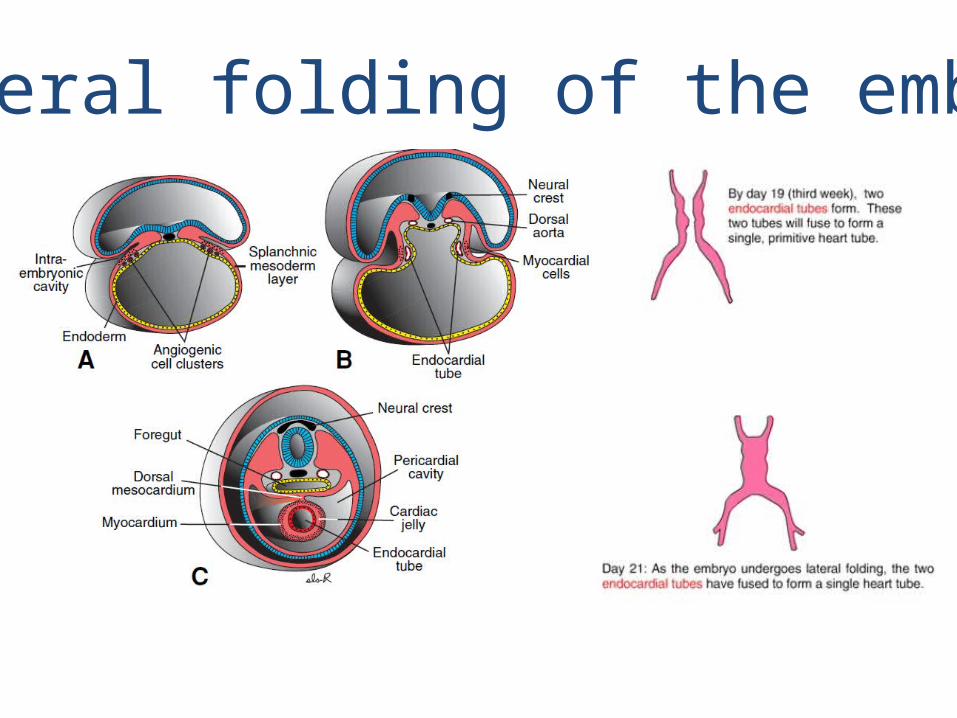
Lateral folding of the embryo
• Due to lateral folding, the pair of endothelial tubes merge except at their caudalmost ends.
• Simultaneously, the crescent part of the horseshoe-shaped area expands to form the future outflow tract and ventricular regions.
• Thus, the heart becomes a continuous expanded tube consisting of an inner endothelial lining and an outer myocardial layer.
• It receives venous drainage at its caudal pole and begins to pump blood out of the first aortic arch into the dorsal aorta at its cranial pole.
Lateral folding of the embryo

10
Development of layers of myocardium
• During these events, the myocardium thickens and secretes a thick layer of extracellular matrix, rich in hyaluronic acid, that separates it from the endothelium called cardiac jelly.
• In addition, mesothelial cells on the surface of the septum transversum form the proepicardium near the sinus venous and migrate over the heart to form most of the epicardium.
• The remainder of the epicardium is derived from mesothelial cells originating in the outflow tract region.

• Thus, the heart tube consists of three layers:
1. Endocardium – forming the internal lining of the tube;
2. Myocardium – forming the muscular wall;
3. Epicardium or visceral pericardium – covering the outside of the tube.
• This epicardium is responsible for formation of the coronary arteries, including their endothelial lining and smooth muscle.

Development of Pericardium
• The developing heart tube bulges more and more into the pericardial cavity.
• Initially, the tube remains attached to the dorsal side of pericardial cavity by the dorsal mesocardium.
• No ventral mesocardium is ever formed. • With further development, the dorsal mesocardium
disappears, creating the transverse pericardial sinus, which connects both sides of pericardial cavity.
• The heart is now suspended in the cavity by blood vessels at its cranial and caudal poles

Heart tube
• The heart is at first seen in the form of right and left endothelial heart tubes that soon fuse with each other.
• It has series of dilatations• Bulbus cordis• Ventricle• Atrium• Sinus venosus


bulbus cordis
truncus arteriosus
aortic roots
ventricle
atrium
sinus venosus


Formation of the cardiac loop
• Heart tube normally loops or folds to the right occurs during the fourth week and is completed by end of 28 days.
• Cephalic part (ventricle) is displaced ventrally , caudally and the right & the caudal part (atrium) portion of the tube is displaced dorsally cranially and to the left


• The bulbus cordis develops 3 divisions :– Proximal part – forms the trabeculated part of RV. – Midportion (conus cordis) – forms the outflow tracts of
both ventricles. – Distal part (truncus arteriosus) – forms the roots and
proximal portion of the aorta and pulmonary artery.
• The junction between the primitive ventricle and the bulbus cordis (bulboventricular sulcus) remains narrow. It is called the primary interventricular foramen .

• The smooth walled heart tube begins to form primitive trabeculae proximal and distal to the primary interventricular foramen.
• The bulbus temporarily remains smooth walled.
• The primitive ventricle, which is now trabeculated, is called the primitive LV.
• Likewise, the trabeculated proximal third of the bulbus cordis may be called the primitive RV.

• The atrial portion, initially a paired structure outside the pericardial cavity, forms a common atrium and is incorporated into the pericardial cavity.
• The AV junction remains narrow and forms the AV canal, which connects the common atrium and the early embryonic ventricle
• The conotruncal portion, initially on the right side, shifts gradually to a more medial position due to formation of 2 transverse dilations of the atrium, bulging on each side of the bulbus cordis .

Formation of the Cardiac Septae
The major septae are formed between the 27 and 37th days of development
It is a simultanuous process if the following areas
1. Septum formation in the common atrium
2. Septum formation in the atrioventricular canal
3. Septum formation in the truncus arteriosus and conus cordis
4. Septum formation in the ventricles

Septum formation in the Common Atria
At the end of the 4th week, a sickle-shaped crest grows from the roof of thecommon atrium into the lumen. This crest is the first portion of the septum
primum
A. 30 days .B. Same stage as A, viewed from the right.

Septum formation of the Common Atria
C. 33 days. D. Same stage as C, viewed from the right
When the lumen of the right atrium expands as a result of incorporation ofthe sinus horn, a new crescent-shaped fold appears. This new fold, the septum secundum never forms a complete partion in the atrial cavity

Septum formation of the common atria
E. 37 days; F. Newborn.G. The atrial septum from the right; same stage as F.
When the upper part of the septum primum gradually disappears, theremaining part becomes the valve of the oval foramen.

Further differentiation of the Atria• Primitive RA enlarges by incorporation of the right sinus horn.
• From Primitive LA, a single embryonic pulmonary vein develops as an outgrowth of the posterior left atrial wall, just to the left of the septum primum.
• This vein gains connection with veins of the developing lung buds.
• During further development, the 4 Pulmonary Veins are incorporated, forming the large smooth walled part of the adult LA.

Further differentiation of the Atria
Both the wall of the right sinus horn (blue) and the pulmonary veins (red) are incorporated into the heart to form the smooth-walled parts of the atria.

Septum formation of the Atrioventricular Canal
At the end of the 4th week, two mesenchymal cushions, the atrioventricular endocardial cushions, appear at the superior and inferior borders of the atrioventricular canal, two additional lateral cushions appear at the left and right borders.
At the end of the 5th week there is complete fusion of the superior and inferior cushions with complete division of the canal into left and right orifices.

Atrioventricular Valves
After the endocardial cushions fuse, each AV orifice is surrounded by local proliferations of mesenchymal tissue.
Bloodstream hollows and thins out tissue on the ventricular surface of these proliferations, forming valves that remain attached to the ventricular wall by muscular cords.
Finally, muscular tissue in the cords degenerates and is replaced by dense connective tissue.

Septum formation of the Ventricles
End of the 4th week the two primitive ventricles start to expand.
The medial walls of the expanding ventricles become apposed and gradually merge, forming the muscular interventricular septum

Septum formation of the Truncus Arteriosus and Conus Cordis
5th week, pairs of opposing ridges appear in the truncus and conus cordis

Closure of Interventricular Septum
A. 6 weeks.
B. Beginning of the 7th week.
C. End of the 7th week.
Combined Proliferations of the right and left conus cushions, along with the inferior
endocardial cushion
Close the interventricular foramen and form the membranous portion of the
interventricular septum.
Semilunar Valves
When partitioning of the truncus is almost complete, primordia of the semilunar valves become visible as pair of small tubercles found on the main truncus swellings.
One of each pair is assigned to the pulmonary and aortic channels, respectively
A third tubercle appears in both channels opposite the fused truncus swellings.
Gradually the tubercles hollow out at their upper surface, forming the semilunar valves.
Recent evidence shows that neural crest cells contribute to formation of these valves.
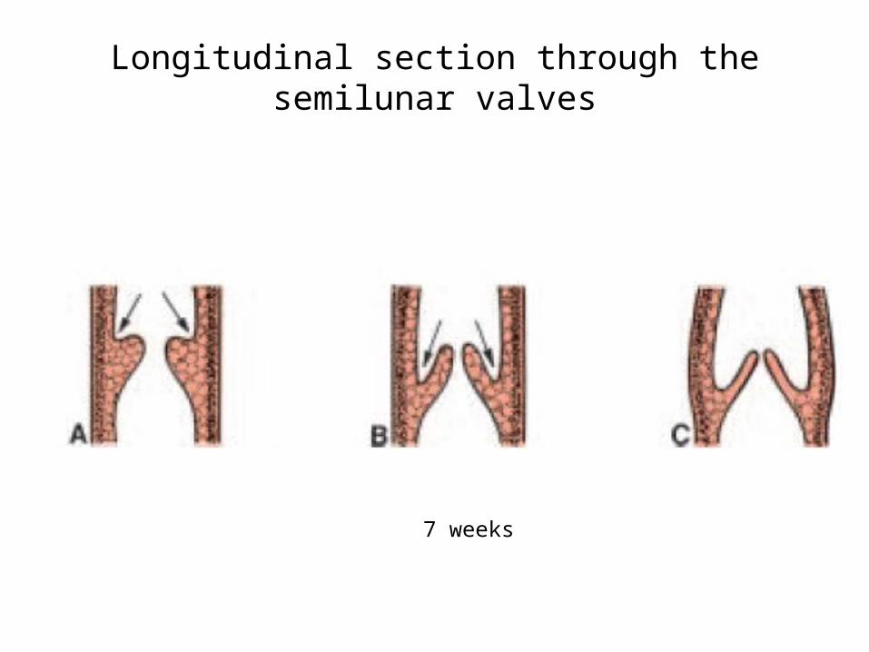
Longitudinal section through the semilunar valves
6 weeks 7 weeks 9 weeks

Development of the Sinus Venosus In mid 4th week, the sinus venosus receives venous blood
from the right and left sinus horns.
Each horn receives blood from 3 important veins: 1. vitelline or omphalomesenteric vein,2. umbilical vein, 3. common cardinal vein.
Entrance of the sinus shifts to the right primarily caused by left-to-right shunts of blood occuring in the venous system.
The right horn, now the only communication between the original SV and the atrium, is incorporated into the RA.


38
Development of the Sinus Venosus
When the left common cardinal vein is obliterated at 10 weeks, all that remains of the left sinus horn is the oblique vein of the LA and coronary
sinus

39
Development of Venous Valves
The sinuatrial orifice, is flanked on each side by a valvular fold, the right and left venous valves.
Dorsocranially, the valves fuse, forming a ridge known as the septum spurium.
The left venous valve and the septum spurium fuse with the developing atrial septum.
The superior portion of the right venous valve disappears entirely.
The inferior portion develops into two parts: (1) valve of the inferior vena cava (2) valve of the coronary sinus.
The crista terminalis forms the dividing line between the original trabeculated part of the RA and the smooth-walled part,from the right sinus horn
