echocardiography of congenital heart · pdf fileobjectives overview of embryology understand...
TRANSCRIPT

Pediatric Echocardiography
Sarah Clauss MDChildren’s National Medical Center
Washington DC

What is your career?
A. Adult Echocardiographic Sonographer
B. Pediatric Echocardiography Sonographer
C. Adult and Pediatric
D. Radiology
E. Other

Objectives
Overview of Embryology
Understand Pediatric Echocardiography
Congenital Heart Disease
• Common lesions
• Complex lesions

Congenital Heart Defects7-10/1,000 Live Births
DIAGNOSIS (Balt-Wash) PERCENT
Ventricular septal defect 26%
Tetralogy of Fallot 9%
Atrioventricular septal defect 9%
Atrial septal defect 8%
Pulmonary valve stenosis 7%
Coarctation of the Aorta 7%
Hypoplastic left heart syndrome 6%
D-Transposition 5%

CHD in Adults
30,000 babies born with CHD per year
20,000 surgeries for CHD per year
85% survive into adulthood
Over 1.2 million adults with CHD
Increasing at 5% per year
8,500 per year reach adulthood
Less than 10% disabled

Evolution of Cardiac SurgeryDiagnosis 1950’s 1960’s 1970’s 1980’s 1990’s 2000’s
ASD Rare
Repair
Repair
older child
Repair age
4
Repair age 2 Repair age
2-3
Device
closure
VSD Rare
Repair
Repair
>10 kg or
palliate
Repair < 1
year or
palliate
Repair 6
months or prn
Repair
premature
infants
PDA Repair Repair Repair Repair Repair
TOF Palliate Late
Repair in
adults
Repair
after
palliation
Repair 2-8
months or
prn
TGA No
survivors
Rare
Survivors
Atrial
Repair
Transitional
Decade
Arterial
Repair
Single
Ventricle
Comfort
care
Palliate Rare
Fontan
Fenestrated
Fontan
Lateral
Tunnel
Extra-
cardiac
Fontan
HLHS Comfort
care
Comfort
care
Surgery in
Boston
Comfort vs.
high risk
surgery
Surgery &
Fetal
Diagnosis

Embryology 101
19 Days: Two endocardial tubes have formed – these tubes will fuse to form a common, single primiative heart tube22 Days: Heart tube begins to beat23 Days: Folding commences30 Days: Primitive circulation 9 weeks (56 Days): All major structures identified
(In humans, several months of gestation remain for emergence of HLHS, PS, etc)

The Cardiac Crescent and the Tube Heart
From Heart Development, 1999
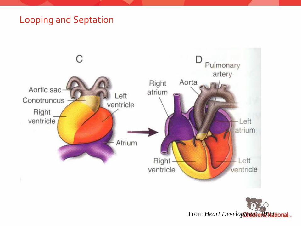
Looping and Septation
From Heart Development, 1999

23
4
5
6
From
Dr. R.
Anderson

How do Congenital Heart Defects form?
Complex interaction between environmental and genetic etiology
• Multifactorial
• 5-8% chance of recurrence
Environmental exposures may influence micro-uterine environment and either turn on or off needed protein development

Echo timeline
1793 Italian priest studied bats
1845 Austrian scientist Christian Doppler
WWII Sonar detected submarines
1954 Hertz & Edler
• (A&B mode echocardiogram)
M-mode ultrasound early 1970’s
2D echo late 1970’s
Doppler Echo 1980’s
• Pulsed wave Doppler
• Continuous wave Doppler
• Color Doppler

Pediatric Echo is Different
Anatomy and physiology over function
Segmental approach for complex patients
Improved resolution
• Heart is closer to chest wall
• Higher frequency transducers
• TEE rarely necessary for diagnosis
Inversion of apical and subcostal images

Echo in CHD
Doppler echo
• Pulsed wave Doppler
• Quantitation of intracardiac hemodynamics
(Modified Bernoulli Equation Δ P= 4 x v22 )
– Valvar regurgitation
– Intracardiac shunts
– LVOT/RVOT obstruction
• Ventricular function
– Systolic
– Diastolic (mitral inflow, pulmonary venous
inflow)

Echo in CHD
Continuous wave Doppler
• Non-invasive measurements of mean and
peak transvalvar gradients
• Valvar stenosis
• Prediction of Ventricular Pressure (modified
Bernoulli equation)
• VSD- LV: RV pressure gradient
• TR/PR RV, PA pressure

DopplerSpectral Display
Pulsed Wave (PW) Continuous Wave (CW)
Aortic Valve Velocity
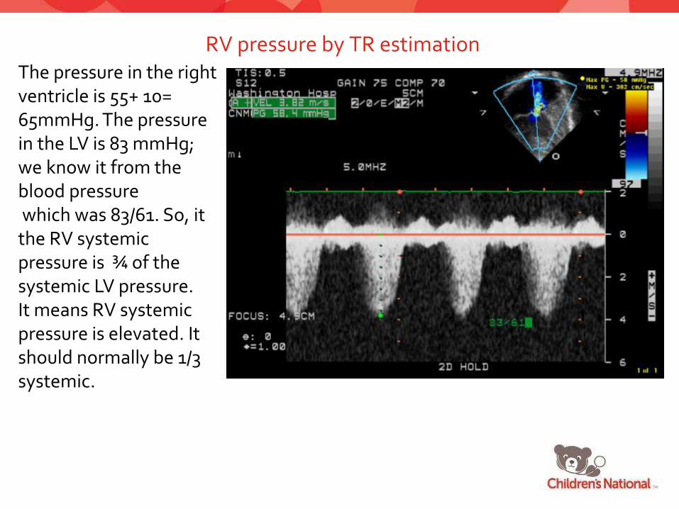
RV pressure by TR estimationThe pressure in the right ventricle is 55+ 10= 65mmHg. The pressure in the LV is 83 mmHg; we know it from the blood pressurewhich was 83/61. So, it the RV systemic pressure is ¾ of the systemic LV pressure. It means RV systemic pressure is elevated. It should normally be 1/3 systemic.

Echo in CHD
Color Doppler
• Direction of cardiac flow
• TAPVR vs. LSVC
• Velocity and Turbulence of cardiac flow
• Conduit obstruction
• Identification of intracardiac shunts
– VSD, PDA, ASD
• Assessment of Post-op CHD
– Shunt patency, residual intracardiac shunt

Questions….
1. How much time should you spend trying to obtain Doppler of TR when there is a HUGE ventricular septal defect?
2. What if your patient has a single ventricle, if you measure the TR what does that estimate?
3. Why is it important to Doppler a VSD?
4. If you see funny blood flow, should you invert your color scale?
5. The doctor wants to know if there is pulmonary hypertension in a NICU baby, but there is no TR, is there another way to answer that question?


Classification and Terminology of Cardiovascular Anomalies

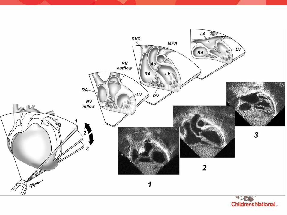
Morphologic/Segmental approach
Define morphologic—not spatial—anatomy• Which atrium is the Right? Left?
• Which ventricle is the Right? Left?
• Which great artery is which?
Define segmental anatomy• Segments: Atrium, Ventricles, Great Arteries
• What is the position of each segment relative to each other?• Is the RA on the right? Is it connected to the RV? Is it connected to
the PA?
• Is the LA on the left? Is it connected to the LV? Is it connected to the Aorta?
Predict the physiology• What is the physiology predicted by the segmental connections?
• Normal? Transposition? Obstructed flow?
• What is the physiology predicted by flow in the ductus? Across the foramen?

Cardiac base-apex axis and orientation in the chest
Levocardia Mesocardia Dextrocardia

Cardiac situs (sidedness)

Example: Cardiac sidedness
Situs solitus normal cardiac sidedenss Situs ambiguus, right isomerism

Differentiation between the atria
The morphologic RA has a smooth or sinusal portion, which is found between the interatrial septum and the crista terminalis. It receives the drainage of the superior and inferior venae cavae and the coronary sinus. The trabecular portion is characterized by the presence of pectinate muscles, which are directed from crista terminalis to the base of the right atrial appendage. The RA appendage is wide and its edge is blunt.
RA appendage is broad based and triangularly shaped (like Snoopy’s nose), with pectinate muscles that extend into the body of the right atrium.
The anatomic LA is totally smooth and lacks pectinate muscles. It receives the drainage of the pulmonary veins, and LA appendage has a narrow base and fingerlike appearance (like Snoopy’s ears) with pectinate muscles confined within the appendage.


RA and TV valve characteristics
Right atrium:•Limbus of fossa ovalis (limb of oval fossa)•Large pyramidal appendage (Snoopy’s nose)•Crista terminalis (terminal crest)•Pectinate muscles•Receives venae cavae and coronary sinus*
Tricuspid valve:•Low septal annular attachment•Septal cordal attachments•Triangular orifice (midleaflet level)•Three leaflets and commissures•Three papillary muscles•Empties into right ventricle

LA and MV valve characteristics
Left atrium:•Ostium secundum•Small fingerlike appendage (Snoopy’s ear)•No crista terminalis•No pectinate muscles•Receives pulmonary veins*
Mitral valve:•High septal annular attachment•No septal cordal attachments•Elliptical orifice (midleaflet level)•Two leaflets and commissures•Two large papillary muscles•Empties into left ventricle

Differentiation between the atria
The only structures that are constant and allow differentiation between the right and left atria are the appendages!
The drainage of the systemic and pulmonary veins does not permit the conclusive identification of the atria, as drainage sites are sometimes anomalous. The atrial septum cannot always be used either, because it can have defects or be absent.

Ventricles-characteristics
Right ventricle:Tricuspid-pulmonary discontinuityMuscular outflow tractSeptal and parietal bandsLarge apical trabeculationsCoarse septal surfaceCrescentic in cross sections* (* variable)Thin free wall (3–5 mm)*Receives tricuspid valve Pulmonary valve empties into main pulmonary artery

Ventricles-characteristics
Left ventricle:•Mitral-aortic continuity•Muscular-valvular outflow tract•No septal or parietal band•Small apical trabeculations•Smooth upper septal surface•Circular in cross section* (* variable)•Thick free wall (12–15 mm)*•Receives mitral valve•Aortic valve•Empties into ascending aorta

Ventricular features (summary)
•Features of the morphologic RV:•Coarse trabeculae with prominent septal band, parietal band, and moderator band. •Septophillic attachments of the tricuspid valve (attachments to septum and free wall)•Well-developed infundibulum (= conus= cone of muscle beneath the semilunar valve) which results in fibrous dyscontinuity between the tricuspid and semilunar valves•Features of the morphologic LV:•Smooth septal surface, fine trabeculae•Septophobic attachments of the mitral valve (attachments only to free wall)•No infundibulum which results in fibrous continuity of the mitral and semilunar valves

Atrioventricular connections

Examples of atrioventricularconnections:
A. ConcordanceB. DiscordanceC. Double-inlet LVD.Ticuspid atresia:
absent right A-V connection

Overriding and straddling

Arterial SegmentA- normal, Pa- anterior, leftAo-posterior, right

Ventriculoarterial connection- 5 possible

To summarize…….The Cardiac Segments
Viscera and atria• Abdominal situs• Systemic and pulmonary venous return• Atrial anatomy
Atrioventricular canal• AV valves and atrioventricular septum
Ventricles• Ventricular anatomy (D- or L-looping)• Ventricular size and proportion• Ventricular septum
Conus• Ventricular outflow tracts
Great arteries• Semilunar valves• Great arteries

Common Lesions

ASD
RV
LV
LA
RV Dilation
RV
LV
Diastolic Septal Flattening

Atrial Septal Defects
Secundum ASD
Primum ASD
Sinus Venosus defect
• Not truly a deficiency
of the atrial septum,
but the same
physiology as an ASD
Common atrium

Atrial Septal Development
http://www.med.unc.edu/embryo_images/unit-welcome/welcome_htms/contents.htm
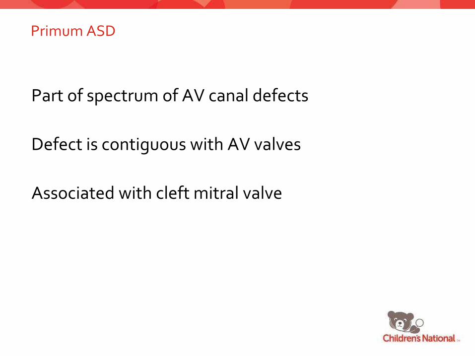
Primum ASD
Part of spectrum of AV canal defects
Defect is contiguous with AV valves
Associated with cleft mitral valve

Sinus Venosus Defects
Deficiency in the wall between the right pulmonary veins and the RA
PAPV-DRAINAGE• SVC type = RUPV
• Inferior type = RLPV

Sinus Venosus ASD

ASD: Clinical Correlation
Usually diagnosed in childhood
Asymptomatic
F>M
Systolic ejection murmur and widely split fixed S2
EKG may show RBBB or RVH


Devices for ASD Closure
Cardio-SEAL Amplatzer

Amplatzer Occlusion of Atrial Septal Defect
Clockwise from above:
Transcatheter delivery of
Amplatzer device, which is
positioned across the
atrial septal defect
Left: Amplatzer device in
place

Newborn infant noted to be breathing heavy in New born nursery
Chest xray demonstrates increased lung markings.
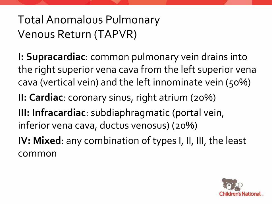
Total Anomalous Pulmonary Venous Return (TAPVR)
I: Supracardiac: common pulmonary vein drains into the right superior vena cava from the left superior vena cava (vertical vein) and the left innominate vein (50%)
II: Cardiac: coronary sinus, right atrium (20%)
III: Infracardiac: subdiaphragmatic (portal vein, inferior vena cava, ductus venosus) (20%)
IV: Mixed: any combination of types I, II, III, the least common

TAPVR

Partial Anomalous Pulmonary Venous Return (PAPVR)Right veins (more common):RASVC ( RUPV to the RA or base of the SVC-sinus venosus ASD)IVCLeft veins:Innominate veinCoronary sinus Rarely: SVC, IVC, right atrium, or left subclavian vein

“Very loud murmur” heard prior to hospital discharge
Baby is well, feeding, growing, pink, passed new pulse ox screening

The Ventricular Septum
AV canal septum (1)
Muscular septum including the trabecular portion (2) and the septal band (3)
Conal septum (4)
Conoventricular
Membranous
Inlet
Malalignment

The Ventricular Septum
Left ventricular view
AV canal septum (1)
Muscular septum including the trabecular portion (2) and the septal band (3)
Conal septum (4)

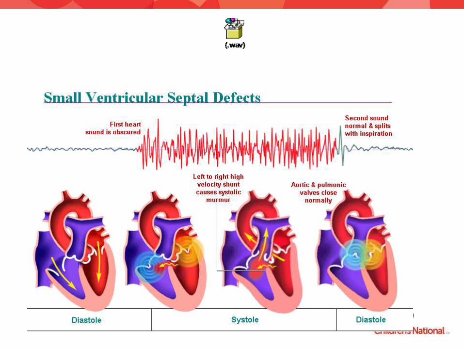
VSD: Clinical Correlation
Size and pulmonary vascular resistance determines clinical presentation
• Fetal transition
Symptoms are determined by the size of the shunt
• Size of defect
• Presence of other anomalies
• Extracardiac abnormalities

VSD: Clinical Correlation
Spontaneous resolution
Or not…
Pulmonary disease
• Eisenmenger’s syndrome
Aortic regurgitation


Restrictive Membranous VSD
4.7 m/sec
87 mm Hg
Normal RVP

Unrestrictive Membranous VSD

Atrioventricular Canal Defect- Complete

Common AV Canal (CAVC)
Endocardial Cushion Defect (ECD)
Atrioventricular Septal Defect (AVSD)
Failure of the AV canal to develop properly and form tricuspid, mitral valves and portions of atrial and ventricular septae

DefinitionsSpectrum of defects
• Incomplete CAVC = lack the VSD component or ASD component
• Partial CAVC = synonym for incomplete CAVC OR = primum ASD with cleft mitral valve
• Transitional CAVC = small VSD component
• Balanced/Unbalanced

Atrioventricular Canal Defect – Partial

AV Septal Defect
Complete
Anterior
Leaflet
Posterior
Leaflet
VSD
ASD

Bonus points…
You are doing the echo on a baby and diagnosis her with an Unbalanced AVC.
You are having a hard time imaging the aortic arch.
Are you concerned, or do you think to yourself, the arch is always hard to image, these babies have no necks, they can’t stand when I put my transducer there….I am sure its fine, I just can’t see it right now…..

You are called to NICU to echo 28 week premature baby, weight is 600 gm, every time you try to image the baby’s HR falls and alarms go off…

Patent Ductus Arteriosus

Patent Ductus Arteriosus

PDA: Clinical Correlation
Closed in 90% of infants by 48 hours of life• Prematuring, altitude
Anatomy• Derived from the left 6th embryonic arch
Closure• Muscular constriction→endothelium→thrombosis→fibrous strand
Physiology↔ shunting• Symptoms proportional to shunting
MurmurEKG
• Ventricular hypertrophy


Doppler of the PDA (L-R shunt)
CW Doppler tracing (right) seen above the baseline indicating flow toward the probe from the descending aorta through the PDA to the PA. The peak velocity is reached in late systole 4 m/s. L-R shunt
Color flow Doppler (left) showing a L-R shunt from the descending aorta through the PDA to the PA (red: towards the probe)

Doppler of the PDA (bidirectional shunt)
CW Doppler from an infant with pulmonary artery hypertension and PDA. The negative deflection in systole below the baseline arises from the R-L shunt through the PDA from the PA to the Dao (away from the TDX).The positive deflection (late systole-into late diastole) arises from L-R shunt through the PDA from the Dao to the PA
Bidirectional blood flow through the PDAcan be a normal finding in newborn infants due to high pulmonary resistance

Doppler of the PDA (R-L shunt)
The Doppler spectral tracing shows evidence of severe pulmonary hypertension and no evidence of a L-R shunt through the PDA. The shunt is R-L from the ductus arteriosus to the Dao (blue: away from the TDX)

Patent Ductus Arteriosus –
Ligation and Division

Occlusion of Intracardiac and Vascular ShuntsCoil embolization of PDA
Left, top: Catheter crosses the PDA
from the aortic side and delivers a coil.
Left, bottom: Withdrawal of catheter,
leaving coil in PDA

Amplatzer Ductal Occluders
Amplatzer ductal occluder
Illustration courtesy AGA Medical Group
Aorta angiogram with device
occlusion of PDA, lateral view

Right Heart Obstructive Lesions

Pulmonary Valve Stenosis
Valve anatomy
• Doming, fused commissures
• Thickened, immobile
• Subvalvar obstruction
• Supravalvar obstruction
Post stenotic dilation
RVH
RV

PS: Clinical Correlation
Asymptomatic
Murmur at birth
EKG
• RAD, RVH proportional to obstruction
Management
• Balloon dilation
Excellent outcome

Pulmonary Artery Branch Stenosis
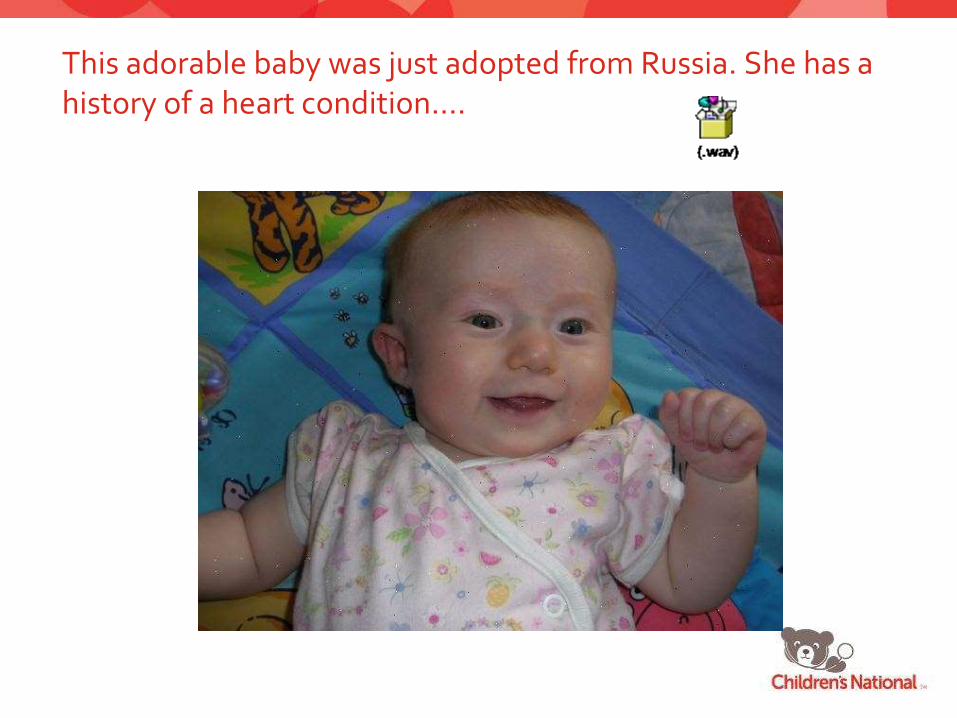
This adorable baby was just adopted from Russia. She has a history of a heart condition….

Tetralogy of Fallot : “Maladie Bleu” 1888
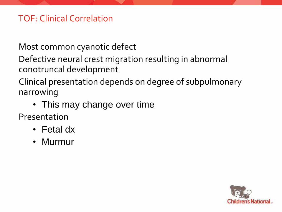
TOF: Clinical Correlation
Most common cyanotic defect
Defective neural crest migration resulting in abnormal conotruncal development
Clinical presentation depends on degree of subpulmonary narrowing
• This may change over time
Presentation
• Fetal dx
• Murmur

Variations in TOF
• “Mexican Tet”
• Hypoplastic or absent conal septum
• Tetralogy with absent pulmonary valve
• Rudimentary pulmonary valve leaflets result in fetal pulmonary regurgitation, PA dilation
• Airway and lung development is compromised in severe cases
• Tetralogy with CAVC
• Tetrology with pulmonary atresia

TOF: Clinical Correlation
• Cyanosis due to right to left shunting at ventricular level
• Degree of cyanosis is proportional to amount of right ventricular outflow tract obstruction
• Dynamic factors may worsen cyanosis• Tet Spell→ no murmur→ deeply cyanotic
• EKG • RVH, RAD, RAE
• CXR• Boot shaped heart


Tetralogy of Fallot

Transcatheter Pulmonary Valve- 2010
• Catheter delivered prosthetic pulmonary valve
• Made from bovine jugular vein
• Sewn within a platinum-iridium ballon expandable stent
• For use in patients with a surgically placed conduit from the RV to the PA
• Used to treat significant conduit valve insufficiency and/or stenosis that would otherwise require surgical conduit replacement


Double Outlet Right Ventricle (DORV)
• Describes a relationship where the PA and Aorta both arise from the anatomic RV
• “DORV” is normal during heart development
• Incidence 1 – 1.5% of patients with CHD
• 1 per 10,000 live births
• Possible association with trisomy 13 and trisomy 18
• Van Praagh – both great arteries arise from the morphologically RV
• NO mitral - aortic fibrous continuity
• Two functional ventricles in which a VSD provides the only outlet for one ventricle
• Anderson - 50% override rule – “if >50% of the aorta is over the RV, its DORV”

Left Heart Obstruction

Aortic Stenosis
Valve, sub-valvar or supravalvar
Clinical manifestations
• Mild-moderate assymptomatic
• Severe
• Depends on age of patient
• Management
• Cath vs. surgery


You are called to the emergency room to perform an echo on a baby that is listless and pale.
He has not been eating well over the last 24 hours
The ER doctor wants to know if they need to call cardiology….

You decide to start with parasternal imaging, you notice the LV function is very very bad….where should you image next?

Coarctation of the Aorta
Narrowed
Isthmus
AAo


Coarctation of the Aorta
Aberrant ductal tissue within
the wall of the aorta
All coarcts are “juxtaductal”
Must look for other left heart
Disease (aortic & mitral valve)

The next day you are staffing a Children’s clinic and the nurse tells you the blood pressure in the legs of the next patient are the same as the arms.
The doctor is busy and asks that you perform an echo while she finishes the previous patient…


Descending AO Doppler

Doppler “drag”

Interrupted Aortic Arch
-Type A = After the subclavian artery,
probably an extreme form of
coarctation with obliteration of the lumen
-Type B = Between the LCC and LSCA,
most common, defect of arch
remodeling/neural crest
-Type C = Between the Carotid arteries,
most rare

Complex Lesions

The nurse from the nursery calls you frantic, there is a baby that is blue.
He was born earlier today, he seemed ok, his birth weight was 8 pounds, uncomplicated pregnancy and delivery…

D-Transposition of the Great Arteries

D-TGA
First described by Baillie 1797
Natural history: >90% mortality in infancy
Incidence: ~5% of congenital heart disease
Rare association with syndromes or other anomalies
Male:Female = 2:1
Possible association with infant of diabetic mother

D-TGA
Ventriculo-arterial discordance
Circulation in parallel
RA=>RV=>Ao
LA=>LV=>PA
Must have mixing at atrial or ventricular level to survive

D-Transposition Balloon Septostomy

Arterial Switch Procedure

Long Term Postoperative ConcernsArterial Switch Operation
Neo-pulmonary stenosis
Coronary abnormalities
• Obstruction and stenosis
• Decreased flow reserve
Neo-aortic insufficiency
• Almost always trivial/mild
LV function

Mustard Repair

Atrial Baffle RepairLong Term Sequelae
On going late mortality risk
• 20% mortality at 20 years
Arrhythmia
SVC obstruction -- 14-17%
IVC obstruction -- 1%
Baffle Leak -- Significant 1-2%
Systemic AV valve regurgitation -- 30%
Systemic Ventricular Failure -- 15-20%

Transposition of the Great Arteries – L Type
Congenitally Corrected Transposition”
Atrio-ventricular and ventriculo-arterial discordance (“double discordance”)
RA LV PA
LA RV Ao
May be an isolated, asymptomatic finding or may be associated with other heart malformations
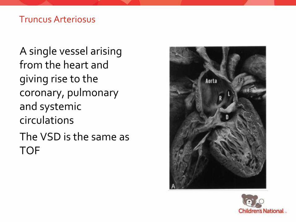
Truncus Arteriosus
A single vessel arising from the heart and giving rise to the coronary, pulmonary and systemic circulations
The VSD is the same as TOF

Truncus Arteriosus


AP Window
Communication between aorta and PA

Hypoplastic Left Heart Syndrome
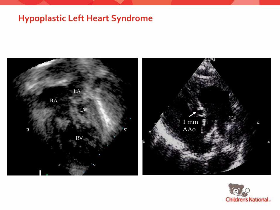
Hypoplastic Left Heart Syndrome
RV
LV
RA
LA
MPA
1 mmAAo

BT Shunt: History
1930: Vivien Thomas hired as
Alfred Blalock’s lab assistant
1924: Failing to obtain a
surgical residency at Hopkins,
Alfred Blalock goes to
Vanderbilt and begins research
on traumatic shock1938: Rabbit models with
subclavian to PA anastomosis
fail to produce pulmonary HTN
1941: Blalock and Thomas
move to Hopkins
1941: Coarctation relief with
subclavian to descending aorta
shunt
1943: Helen Taussig, a
Hopkins pediatrics residency
graduate, approaches Blalock
about help for “blue babies”
1944: “Anna,” a dog with a
surgically created mixing lesion,
successfully undergoes end-to-side
subclavian-to-PA anastomosis,
lives 15 years
November 29, 1944: Eileen Saxon,
a 15-month-old 4.5 kg undergoes
successful systemic-to-pulmonary
shunt by Blalock with Thomas
directly over his shoulder


Norwood I: Anatomy
1. Atrial septectomy
2. Ligation of main pulmonary artery and construction of neo-aorta
3. Sano Modification/Modified BT Shunt

BT Shunt

Norwood I: Sano
Sano modification
• RV-to-PA conduit
• Eliminates
competitive flow to
PAs in diastole
• Enhances coronary
perfusion
RV

Sano Shunt

Bidirectional Glenn: Anatomy
• End-to-side anastomosis
of SVC to undivided right
pulmonary artery
• Includes takedown of BT shunt
• Allows flow to both lungs
from SVC via passive flow

Glenn Shunt

Glenn Doppler

Fontan: Variations
Lateral tunnel runs within RA, using free wall plus conduit as baffle for IVC blood
• Fenestrations: R-to-Lshunting through the fenestration hypoxemia
• Improve cardiac output, minimize systemic venous hypertension, decrease post-op thoracostomy drainage
• Can later be closed by cath
Extracardiac is IVC to MPA• Generally has lower rate
of complications

Fenestrated Fontan

Hypoplastic Left Heart SyndromePalliative ReconstructionStage I -- Norwood Procedure
• Birth
Stage II -- Bi-directional Cavopulmonary Shunt
• 4-6 months
Stage III-- Fontan Procedure
• 18-24 months for lateral tunnel
procedure
• > 15 kg for extracardiac procedure


QUESTION 1A tachypneic 2 month old is not growing well and has a murmur. An echocardiogram is obtained:
E
c
SYSTOLE DIASTOLE
QUESTION 1 (CONT)All of the following statements are likely to be true except:
A. The patient is at increased risk to have Down Syndrome
B. The patient may not need surgery
C. The patient has an endocardial cushion defect
D. The patient has a normal oxygen saturation
E. The patient may have a small mitral valve cleft after surgical repair
E
c

QUESTION 2A cyanotic newborn has the following echocardiogram:
E
c

QUESTION 2 (CONT)All of the following statements are likely to be true except:
A. The pulmonary artery gives rise to the coronary arteries.
B. The right ventricle pumps blood to the body
C. Oxygenated blood is pumped to the lungs
D. The left ventricle pumps blood to the body
E. The right ventricular pressure is greater than or equal to the left ventricular pressure
E
c

QUESTION 3A 40 year old with atrial fibrillation has the following echo:
E
c
SYSTOLE DIASTOLE

QUESTION 3 (CONT)Subsequent imaging is most likely to reveal the following
A. Tetralogy of Fallot
B. Large membranous ventricular septal defect
C. Large patent ductus arteriosus
D. Large secundum atrial septal defect
E. No structural cardiac defect
E
c

QUESTION 4A 3 month old with a loud murmur and intermittent perioral cyanosis has the following echo:
E
c
LV
RV
Ao
LA
RVOT

QUESTION 4 (CONT)All of the following statements are likely to be true except:
A. The aorta is overriding the left and right ventricle
B. There is a large ventricular septal defect
C. There is pulmonary stenosis
D. The right ventricular pressure is increased
E. The pulmonary artery pressure is increased
E
c

QUESTION 5An asymptomatic 9 month old with a loud murmur and a BP of 79/48 and has the following parasternal long axis 2D and CW Doppler findings:
E
c
LA
LV
RV
LV
LA
RV
CW

QUESTION 5 (CONT)The most likely diagnosis is:
A. Membranous VSD, normal RV pressure
B. Membranous VSD, elevated RV pressure
C. Muscular VSD, normal RV pressure
D. Muscular VSD, elevated RV pressure
E. Tricuspid regurgitation, elevated RV pressure
E
c
Acknowledgements
Unattributed illustrations are from Nadas’ Pediatric Cardiology
Amy L. Juraszek
Margaret Lasota
Children’s National Medical Center
• 202-476-4880 Physician Line
• 202-476-5579 Echo Lab