elucidating the small regulatory rna repertoire of the sea
TRANSCRIPT

University of Plymouth
PEARL https://pearl.plymouth.ac.uk
Faculty of Science and Engineering School of Biological and Marine Sciences
Elucidating the small regulatory RNA
repertoire of the sea anemone
Anemonia viridis based on whole
genome and small RNA sequencing
Hall-Spencer, JM
http://hdl.handle.net/10026.1/10695
10.1093/gbe/evy003
Genome Biology and Evolution
All content in PEARL is protected by copyright law. Author manuscripts are made available in accordance with
publisher policies. Please cite only the published version using the details provided on the item record or
document. In the absence of an open licence (e.g. Creative Commons), permissions for further reuse of content
should be sought from the publisher or author.

Elucidating the small regulatory RNA repertoire of the sea anemone
Anemonia viridis based on whole genome and small RNA sequencing
Ilona Urbarova* 1
, Hardip Patel 2
, Sylvain Forêt 3
, Bård Ove Karlsen 4
, Tor Erik
Jørgensen 5, Jason M. Hall-Spencer
6,7 and Steinar D. Johansen*
1,5
1 Department of Medical Biology, Faculty of Health Sciences, UiT – The Arctic University
of Norway, N9037 Tromsø, Norway
2 Genomics and Predictive Medicine, Genome Biology Department, John Curtin School of
Medical Research, ANU College of Medicine, Biology and Environment, Australian
National University, Canberra, ACT 2601, Australia
3 Evolution, Ecology and Genetics, Research School of Biology, Australian National
University, Canberra, ACT 2601, Australia
4 Research Laboratory and Department of Laboratory Medicine, Nordland Hospital,
N8092, Bodø, Norway
5 Genomics Group, Faculty of Biosciences and Aquaculture, Nord University, N8049
Bodø, Norway
6 Marine Biology and Ecology Research Centre, University of Plymouth, Plymouth PL4
8AA, UK
7 Shimoda Marine Research Centre, University of Tsukuba, Shimoda City, Shizuoka 415-
0025, Japan
*Authors for Correspondence: Ilona Urbarova, Department of Medical Biology, UiT - The
Arctic University of Norway, MH building, N9019, Tromsø, Norway, [email protected]
Steinar Daae Johansen: [email protected]
© The Author(s) 2018. . Published by Oxford University Press on behalf of the Society for Molecular Biology and Evolution.
This is an Open Access article distributed under the terms of the Creative Commons Attribution Non-Commercial License
(http://creativecommons.org/licenses/by-nc/4.0/), which permits non-commercial re-use, distribution, and reproduction in any medium,
provided the original work is properly cited. For commercial re-use, please contact [email protected] Downloaded from https://academic.oup.com/gbe/advance-article-abstract/doi/10.1093/gbe/evy003/4827693by University of Plymouth useron 30 January 2018

2
Abstract
Cnidarians harbour a variety of small regulatory RNAs that include microRNAs (miRNAs)
and PIWI-interacting RNAs (piRNAs), but detailed information is limited. Here we report the
identification and expression of novel miRNAs and putative piRNAs, as well as their genomic
loci, in the symbiotic sea anemone Anemonia viridis. We generated a draft assembly of the A.
viridis genome with putative size of 313 Mb that appeared to be composed of about 36%
repeats, including known transposable elements. We detected approximately equal fractions of
DNA transposons and retrotransposons. Deep sequencing of small RNA libraries constructed
from A. viridis adults sampled at a natural CO2 gradient off Vulcano Island, Italy, identified 70
distinct miRNAs. Eight were homologous to previously reported miRNAs in cnidarians, while
62 appeared novel. Nine miRNAs were recognized as differentially expressed along the
natural seawater pH gradient. We found a highly abundant and diverse population of piRNAs,
with a substantial fraction showing ping-pong signatures. We identified nearly 22% putative
piRNAs potentially targeting transposable elements within the A. viridis genome. The A.
viridis genome appeared similar in size to that of other hexacorals with a very high divergence
of transposable elements resembling that of the sea anemone genus Exaiptasia. The genome
encodes and expresses a high number of small regulatory RNAs, which include novel
miRNAs and piRNAs. Differentially expressed small RNAs along the seawater pH gradient
indicated regulatory gene responses to environmental stressors.
Keywords: Coastal ecology, CO2 seep, ocean acidification; miRNA; piRNA; transposable
elements
Downloaded from https://academic.oup.com/gbe/advance-article-abstract/doi/10.1093/gbe/evy003/4827693by University of Plymouth useron 30 January 2018
3
Introduction
Two major classes of small regulatory RNAs in eumetazoans are microRNAs (miRNAs) and
PIWI-interacting RNAs (piRNAs). These classes are distinct in terms of their sizes,
biogenesis, biological function and origin (Ghildiyal and Zamore 2009). Compared to
Bilateria, knowledge about cnidarian small RNAs remains scarce. To date, small RNAs have
only been reported in four cnidarians; the non-symbiotic sea anemone Nematostella vectensis,
the stony corals Stylophora pistillata and Acropora digitifera, and the hydroid Hydra
magnipapillata (Chapman et al. 2010; Gajigan and Conaco 2017; Grimson et al. 2008;
Krishna et al. 2013; Liew et al. 2014; Moran et al. 2014; Wheeler et al. 2009).
miRNAs represent a well-studied class of small RNAs that usually range in size from
20 to approximately 24 nt (miRBase, http://mirbase.org). In animals, miRNAs are initially
transcribed as RNA polymerase II transcripts (pri-miRNAs), which are further processed by
the RNases Drosha and Dicer into stem-loop precursor miRNAs (pre-miRNAs) and mature
miRNAs, respectively. One strand of the mature miRNA duplex is usually incorporated into
the RNA-induced silencing complex (RISC) (Gregory et al. 2005; Schwarz et al. 2003), and it
guides the whole complex to complementary mRNA for post-transcriptional gene silencing. In
plants, the miRNA biogenesis pathway involves a Dicer-like 1 (DCL-1) protein that is
responsible for both cropping and slicing miRNA precursors in the nucleus (Voinnet 2009).
The miRNA silencing mechanism is fundamentally different in animals and plants. While
animal miRNAs usually perform translational repression through partial base-pairing to target
mRNAs, plant miRNAs mostly bind with full or nearly full complementarity leading to
targeted mRNA cleavage (Bartel 2009). Cnidarian miRNAs appear to contain plant-like
features in their biogenesis and the post-transcriptional gene silencing follows a plant-like
regulatory pathway (Moran et al. 2013; Moran et al. 2014). Among cnidarians, Nematostella
was reported to express 87 distinct miRNAs, compared to 26, 31 and 126 miRNAs in
Downloaded from https://academic.oup.com/gbe/advance-article-abstract/doi/10.1093/gbe/evy003/4827693by University of Plymouth useron 30 January 2018

4
Acropora, Stylophora and Hydra, respectively (Chapman et al. 2010; Gajigan and Conaco
2017; Grimson et al. 2008; Krishna et al. 2013; Liew et al. 2014; Moran et al. 2014; Wheeler,
et al. 2009). Interestingly, only one miRNA (miR-100) was found conserved between the
bilaterian and the cnidarian species. miRNAs have several important regulatory roles in plants
and animals (Bartel 2004; Ghildiyal and Zamore 2009; Vashisht and Nodine 2014).
Expression profiling indicated the cnidarian miRNAs to be involved in developmental
regulation, regeneration and thermal stress resilience (Krishna et al. 2013; Moran et al. 2014;
Gajigan and Conaco 2017). However, their roles in other biological processes, including other
environmental stress responses, have not been investigated in detail.
piRNAs are usually between 23 and 30 nt in size (Krishna et al. 2013; Liew et al.
2014; Moran et al. 2014). The single-stranded piRNA precursors are either derived from
transposable elements or from specific piRNA genomic clusters, and they do not require Dicer
nuclease activity for their processing (Das et al. 2008; Houwing et al. 2007; Vagin et al. 2006).
piRNAs represent a highly diverse class of small regulatory RNAs, reaching several thousand
distinct members within a single organism (Aravin et al. 2006; Kawamura et al. 2008). The
uniqueness of piRNAs arises from phased production of primary piRNAs (Han et al. 2015;
Mohn et al. 2015). A secondary piRNA pathway serves for piRNA amplification by a ‘ping-
pong loop’ mechanism (Brennecke et al. 2007; Gunawardane et al. 2007). The two distinct
piRNA populations (primary and secondary) show opposite orientation and complementarity
in their first 10 nt positions (Brennecke et al. 2007; Gunawardane et al. 2007). piRNAs lack
universal sequence conservation, except that the primary and secondary piRNAs show a
preference for an uracil residue at the 5’end (1U) and an adenine residue at the 10th
position
(10A), respectively. In all cnidarians investigated so far, piRNAs appear to be highly abundant
compared to miRNAs and short-interfering RNAs (siRNAs) (Gajigan and Conaco 2017;
Grimson et al. 2008; Juliano et al. 2014; Krishna et al. 2013; Moran et al. 2014; Praher et al.
Downloaded from https://academic.oup.com/gbe/advance-article-abstract/doi/10.1093/gbe/evy003/4827693by University of Plymouth useron 30 January 2018

5
2017). The biological role of piRNAs is not well understood, but their most important
function seems to be guiding PIWI proteins to suppress transposon activity in animal germ
cells (Brennecke et al. 2007; Gunawardane et al. 2007). piRNA profiling in cnidarians
(Nematostella and Hydra) suggested a similar role in transposon silencing, but proposed
broader silencing functionalities as well (Grimson et al. 2008; Juliano et al. 2014; Praher et al.
2017).
The sea anemone Anemonia viridis exposed to natural ocean acidification conditions
appears to be physiologically acclimatized to low pH and optimizes its energy utilization
under elevated pCO2 through an increased autotrophic input (Suggett et al. 2012, Horwitz et
al. 2015). Our recent transcriptome sequencing from the same sampling site indicates
increased expression of stress-related transcripts, repression of global synthesis and boost in
certain retrotransposon elements at low pH in A. viridis (Urbarova I., unpublished results). In
plants, it is known that small RNAs can regulate species tolerance to stress via post-
transcriptional silencing (Sunkar et al. 2007). We therefore wanted to elucidate if
acclimatization responses of A. viridis that we observe at the transcriptome level could be
caused by small RNA-mediated post-transcriptional regulation. Here we report whole genome
and small RNA library sequencing of the symbiotic sea anemone A. viridis, sampled at a
natural seawater pH gradient off Vulcano Island, Sicily - Italy. We mainly aimed to identify
novel small RNA species in A. viridis, but also elucidate their possible involvement in the
acclimatization responses to low pH conditions. We detected 70 distinct miRNA species, and
assessed differentially expressed small RNAs. Most of the putative piRNAs contained features
typical of primary piRNAs and a large fraction showed ping-pong signatures. Our study
indicates possible regulatory gene responses of small RNAs to low pH.
Materials and Methods
Downloaded from https://academic.oup.com/gbe/advance-article-abstract/doi/10.1093/gbe/evy003/4827693by University of Plymouth useron 30 January 2018

6
Sampling
The temperate symbiotic sea anemone A. viridis (the Snakelocks Anemone) was collected at
Levante Bay, North Vulcano Island, Sicily - Italy. Acidification conditions are created here by
the release of CO2 into the seawater from a natural vent site at -1 m depth (Boatta et al. 2013;
Johnson et al. 2013). Sampling was performed on May 13 and 14, 2013, at the depth of 1-2
meters at >350 m from the vent site along a gradient of decreasing pH (~ pH 7.6 and pH 7.9),
and at a control location at ~800 m from the vent site with pH corresponding to ambient
seawater levels (~ pH 8.2). For simplicity, we are referring to average pH values throughout
this work as reported in Johnson et al. (2013). A total of nine individuals of A. viridis were
sampled in two days (three from each location). Small pieces of tissue (≤ 0.5 cm) from body
wall, tentacles and oral disc of each individual were collected and stored separately at 4°C in
RNAlater (ThermoFisher Scientific, Waltham, MA USA) during transport from the sampling
site to laboratory. Then, RNAlater solution was removed and all samples were frozen at -80°C
before further processing steps.
Reference genome assembly
DNA from one individual of A. viridis (pH 8.2) was extracted using Wizard(R) Genomic DNA
Purification Kit (Promega, Madison, Wisconsin, USA). Two whole genome paired-end
libraries (2 x 150 bp) were constructed and sequenced on Illumina HiSeq2500 at Eurofins
MWG Operon (Germany). The paired-end reads were processed using Trimmomatic (Bolger
et al. 2014). Adapters were removed and reads were trimmed and quality filtered using sliding
window with Phred score > 20. A bias at the first nine nucleotides was removed by trimming
these bases, and reads with length < 40 bp were discarded. SGA preqc tool was run pooling
the forward and reverse reads from the two libraries together (Simpson 2014). Platanus, a de
novo genome assembler for highly heterozygous diploidic organisms, was then used to
Downloaded from https://academic.oup.com/gbe/advance-article-abstract/doi/10.1093/gbe/evy003/4827693by University of Plymouth useron 30 January 2018

7
assemble reads with k-mer length of 51 (Kajitani et al. 2014). To be able to assess our A.
viridis genome assembly for repeat-enriched regions using RepeatMasker (Smit et al. 2013-
2015), we first filtered out short reads from our reference assembly using N75 statistics,
resulting in 210,233 sequences with sizes larger than 173 bp. The filtered assembly was then
assessed for repeat-enriched regions using RepeatMasker (Smit et al. 2013-2015), with a
custom library created by RepeatModeler, which integrates RECON, RepeatScout, and
Tandem Repeats Finder (TRF) de-novo repeat finding tools to build a repeat library for an
assembly (Smit and Hubley 2008-2015). In addition to RepeatMasker annotation, the repeat-
enriched regions were extracted from the assembly and transposable element annotation was
performed as described previously (Baumgarten et al. 2015; Chapman et al. 2010). The
annotation pipeline included then also a TBLASTX run using RepBase database (Bao et al.
2015), version 22.09 (e-value < 10-20
), and a BLASTX search (e-value < 10-10
) against a
custom-made non-redundant database of proteins encoded by transposable elements (TEs;
NCBI keywords: retrotransposon, transposase, reverse transcriptase, gypsy, copia). These two
databases were separately queried against our reference genome assembly and the best
annotation was chosen based on alignment coverage and score. A combined tabular output
from the searches was further run through two Perl scripts, “blast92gff3.pl” with additional
options -lowscore 0.0001 -alignmax 9999 -exonType exon
(http://arthropods.eugenes.org/EvidentialGene/evigene/scripts/blast92gff3.pl) and the
“overbestgene2.pl” (http://iubio.bio.indiana.edu/gmod/tandy/perls/overbestgene2.perl) to
create a gff file from blast results and to remove overlapping blast hits, respectively. The
results were imported into IBM SPSS Statistics software (version 23), where counting of
transposable elements was performed. Sequence regions corresponding to transposable
elements in our reference genome assembly were then extracted from our scaffolds using
BLAST fastacmd tool (Altschul et al. 1990; Altschul et al. 1997) and used as a reference for
Downloaded from https://academic.oup.com/gbe/advance-article-abstract/doi/10.1093/gbe/evy003/4827693by University of Plymouth useron 30 January 2018

8
piRNA analyses.
RNA extraction
Each tissue sample (without excess RNAlater solution) was immediately transferred from -
80°C to 1 ml cold TRIzol reagent (ThermoFisher Scientific, Waltham, MA, USA). The tissue
was then crushed using Precellys tissue homogenizer at 6000 rpm for 30 seconds (Stretton
Scientific, Stretton, UK) to minimize degradation of RNA. RNA was twice extracted by
chloroform, and subsequently precipitated in isopropanol at 4 ºC overnight, washed with 70%
ethanol, and rehydrated in Nuclease-Free Water (ThermoFisher Scientific, Waltham, MA,
USA). The RNA quality was examined using the Agilent 2100 Bioanalyzer (Agilent
technologies, Santa Clara, CA, USA) and quantity of the samples was measured using Qubit
2.0 fluorometer (ThermoFisher Scientific, Waltham, MA, USA). Only high quality samples
with RNA integrity number (RIN) equal to 7 or higher were used in library constructions.
Small RNA sequencing
Nine individuals of A. viridis representing three different pH conditions (8.2, 7.9 and 7.6)
were included in small RNA sequencing. Total RNA from three different tissue samples of an
individual was pooled at equal amounts. The small RNA fraction was enriched using PureLink
miRNA Isolation Kit (ThermoFisher Scientific, Waltham, MA, USA). Libraries were prepared
only from high quality RNA samples (RIN ≥ 7) following the SOLiD Total RNA-Seq Kit
protocol (ThermoFisher Scientific, Waltham, MA, USA). Different A. viridis small RNA
libraries were barcoded and sequencing was performed on three lanes of a SOLiD™ 6-Lane
FlowChip using SOLiD 5500xl sequencer at the Nord University (Bodø, Norway).
Downloaded from https://academic.oup.com/gbe/advance-article-abstract/doi/10.1093/gbe/evy003/4827693by University of Plymouth useron 30 January 2018

9
Discovery of novel miRNA
After removing low quality (quality score < 18) and less complex sequences from our raw
small RNA sequencing data set, adapter sequences were trimmed away using
trimSOLiDAdaptor.pl Perl script keeping only sequences equal to or longer than 18 nt. The
filtered reads were mapped in colour space using Bowtie (Langmead et al. 2009) to our A.
viridis genome reference with parameters --integer-quals -l 18 -M 20 --best --strata -e 150 --
nomaqround --maxbts 800 --tryhard -a --col-cqual --col-keepends --mapq 20 --threads 14 --
chunkmbs 200. These options select the best alignments based on the seed mismatches only
and mismatches outside the seed region are ignored. Therefore, we needed to perform
additional filtering using processBowtieAlignments.pl Perl script to select alignments with the
minimum mismatches along the whole reads. Both Perl scripts are available at
https://github.com/patelhardip/bitx.git. Mapped reads from each condition were then pre-
processed by bwa_sam_converter.pl Perl script (Friedländer et al. 2012), outputting two files
essential for running miRDeep2 software tool for the novel miRNA predictions (Friedländer
et al. 2012). The two files were used as input into miRDeep2.pl Perl script (Friedländer et al.
2012) that was run for identification of novel miRNAs in A. viridis. In addition, known
miRNAs of three related species, N. vectensis, S. pistillata and H. magnipapillata (Krishna et
al. 2013; Liew et al. 2014; Moran et al. 2014) that were available at the time of the analysis,
have been used in the prediction pipeline. These miRNA sequences were downloaded from
miRBase, release 21 (Kozomara and Griffiths-Jones 2014). Output from miRDeep2 software
was then inspected manually, keeping only predicted miRNAs with miRDeep2 score larger
than 10 and with significant randfold value (p-value < 0.05). Small RNA sequencing data
from each individual were assessed separately for the presence of novel miRNAs. Only
miRNAs identified in at least two individuals were considered further. Possible tRNA
contamination was examined by running tRNAscan on the reference genome (Lowe and Eddy
Downloaded from https://academic.oup.com/gbe/advance-article-abstract/doi/10.1093/gbe/evy003/4827693by University of Plymouth useron 30 January 2018

10
1997). Further, presence of rRNA sequences in predicted hairpin structures was tested by
querying against a custom database combining known rRNA sequences from N. vectensis and
A. viridis. No contamination was found in either case. In addition, to ensure that our miRNA
candidates come from the host, assembled scaffolds of A. viridis were screened for their
possible contamination by symbiont DNA using genomes of Symbiodinium minutum,
Symbiodinium microadriaticum and Symbiodinium kawaguti (Aranda et al. 2016; Shoguchi et
al. 2013; Lin et al. 2015). 1642 scaffolds (mainly short ones with length ~ 100bp) that were
highly similar (e-value < 10-20
) to Symbiodinium genomes in the A. viridis genome assembly
were filtered out prior to the small RNA alignment. Genomic setting of aligned putative
miRNAs was inspected for overlapping regions corresponding to open reading frames
(ORFs). Our scaffolds were searched for ORFs using OrfPredictor
(http://bioinformatics.ysu.edu/tools/OrfPredictor.html) (Min et al. 2005).
miRNA analyses
Expression of selected miRNAs was confirmed by quantitative PCR (qPCR). Six Locked
Nucleid Acid (LNA) probes targeting the predicted miRNAs were designed using online
miRNA qPCR designer tool from Exiqon (Vedbaek, Denmark). cDNA was synthesized from
three individuals (10 ng of total RNA each) per condition using miRCURY LNA™ Universal
RT microRNA PCR, Polyadenylation and cDNA synthesis kit II (Exiqon, Vedbaek, Denmark)
following the instruction manual. Small RNA for the qPCR analysis was isolated from the
same samples that were used for preparation of small RNA libraries. qPCR was performed in
duplicates using miRCURY LNA microRNA PCR, ExiLENT SYBR Green master mix
(Exiqon, Vedbaek, Denmark) in 10 µl. miRNAs were assessed for differential expression
among the sampling sites with differing pH by edgeR (FDR < 0.05) (Robinson et al. 2010).
Mature miRNAs were aligned to their precursor sequences. miRNAs with less than 20 counts
Downloaded from https://academic.oup.com/gbe/advance-article-abstract/doi/10.1093/gbe/evy003/4827693by University of Plymouth useron 30 January 2018

11
in less than three conditions were not considered. We searched for putative animal-like
miRNA targets by Probability of Interaction by Target Accessibility (PITA) software, based on
target complementarity and site accessibility (Kertesz et al. 2007). Coding regions were
predicted from the A. viridis transcriptome (Urbarova I, unpublished results) by
TransDecoder, version 2.0.1 (Haas et al. 2013) and used as input into PITA software. The
results were filtered based on the change in free energy (ddG) of miRNAs binding to its
targets (ddG < - 10 kcal/mol), seed length of 8 nt with no mismatches and no wobble pairs.
Only targets fulfilling these criteria were considered further. We then checked the predicted
targets from PITA for more extensive complementarity to the miRNAs using FASTA v36
(Pearson and Lipman 1988) as previously described (Moran et al. 2014), and scored the
alignments accordingly. Blast hits were obtained using NCBI nr database (e-value < 10-5
) and
GO terms were assigned to the potential mRNA targets using B2G4Pipe Blast2GO pipeline
(Götz et al. 2008).
Search for putative piRNAs
Raw reads from SOLiD sequencing were quality filtered and adapter sequences were
trimmed, as described previously for miRNA discovery. However, only sequences equal to or
longer than 23 nt were kept for the piRNA analyses. Sequences were further filtered for reads
mapping to miRNA precursors identified in the present study and reads mapping to rRNAs
using rRNA databases according to Praher et al. (2017). Sequences in colour space were
aligned using Bowtie with the same parameters as for the miRNA alignment, but allowing for
maximum three mismatches with the ‘seed length’ of 23 nt (-l 23 -n 3). We refrained from
mapping our reads to unique locations in the A. viridis reference genome due to presence of
many sequence stretches in assembled scaffolds that most probably correspond to the same
genomic locations. The aligned sequences were filtered for the best matches using the same
Downloaded from https://academic.oup.com/gbe/advance-article-abstract/doi/10.1093/gbe/evy003/4827693by University of Plymouth useron 30 January 2018

12
custom Perl script as for filtering the miRNA alignments and filtered for reads with 1U or 10A
sequence signatures. TE-targeting potential of putative piRNAs was then assessed by
including only putative piRNAs mapping antisense to the transposable elements. Overlap
probabilities of the putative piRNA sequences with opposite orientation were analysed using
signature.py script (Antoniewski 2014). The script computes the probability of an antisense
read overlapping a sense read with defined length and assigns each overlap length a z-score.
The overlapping signatures of the putative piRNAs were inspected in more detail by running
PingPongPro v1.0 software (http://sourceforge.net/projects/pingpongpro/). This software also
served for inspection of transposon silencing by putative piRNAs. Silencing of transposable
elements by the putative piRNAs was only considered if FDR (qValue) < 0.01 and if it was
supported by at least 10 putative piRNA reads with ping-pong signatures normalized to the
transposon length. To inspect data for presence of piRNA clusters, the putative piRNA reads
were mapped onto the masked genome reference produced by RepeatMasker (Smit et al.
2013-2015) as described previously in colour space using Bowtie, reporting at maximum five
valid alignments. Finally, our sorted alignment files were submitted to piClust software (Jung
et al. 2014). Here, the Eps parameter was set to 1000 and MinReads to 50.
Results
Genome reference assembly and search for repeat-enriched regions
Total DNA from a single A. viridis polyp (normal seawater conditions, pH 8.2) was extracted
and subjected to whole genome sequencing on the Illumina HiSeq2500 platform (fig. 1).
Sequencing generated 43 billion nucleotides (nt) of genomic data, which corresponded to 144
million paired-end reads (table 1). The basic genome characteristics were determined using
the SGA preqc software tool (Simpson 2014), showing an estimated genome size of 313 Mb
Downloaded from https://academic.oup.com/gbe/advance-article-abstract/doi/10.1093/gbe/evy003/4827693by University of Plymouth useron 30 January 2018

13
(approximately 140 times coverage). Adapters were trimmed and the reads were quality
filtered before de novo genome assembly, which created about 1.1 million short scaffolds with
N50 = 2,087. This genome assembly is highly fragmented, but it was sufficient for the
mapping of small RNAs and the identification of transposable elements (fig. 1).
The A. viridis genome assembly was inspected for repeat and low complexity regions
using the RepeatMasker software tool (Smit et al. 2013-2015), and about 36% of the genome
reference was found to contain repetitive regions. About 27.5% of the repetitive sequences
could be assigned to previously known repetitive elements, but most of these sequences (25%
of the genome) could not be classified into any assigned category (supplementary table S1,
Supplementary Material online). Repeat annotation identified only about 8.3% of genome to
be comprised of transposable elements (TEs). These are similar observations as made
previously for symbiotic sea anemone Exaiptasia sp. (formerly known as Aiptasia sp.;
Grajales and Rodríguez 2014) (Baumgarten et al. 2015). From the identified TE fraction,
about half (44.4%) were retrotransposons, and amongst them, non-long terminal repeat (non-
LTR) retrotransposons were predominating (supplementary table S1, Supplementary Material
online).
miRNA discovery
Small RNA libraries from nine individual polyps of A. viridis, sampled at three different
seawater pH conditions (at normal seawater pH 8.2, as well as at pH 7.9 and pH 7.6) were
prepared and subjected to sequencing on the SOLiD 5500xl platform (fig. 1). The sequencing
generated approximately 116 million reads (≥ 18 nt) after adapter trimming and quality
filtering (table 2; fig. 2). Despite the fragmented nature of our genome draft assembly, a high
proportion of the small RNA reads mapped to the genomic reference (between 88 to 92%,
table 2). This indicates that even a very preliminary genome assembly is sufficient for the
Downloaded from https://academic.oup.com/gbe/advance-article-abstract/doi/10.1093/gbe/evy003/4827693by University of Plymouth useron 30 January 2018

14
discovery of small RNAs.
A. viridis miRNAs were identified by the miRDeep2 software tool (Friedländer et al.
2012), and a representative analysis result is shown in fig. 3A. We predicted in total 70 high-
confidence miRNA candidates (20 to 25 nt) for A. viridis, including 61, 60, and 65 distinct
miRNA species at pH 8.2, 7.9, and 7.6, respectively (table 3). Most miRNAs were detected in
all pH conditions studied (n = 51), 14 miRNAs in two different pH conditions, and five
miRNAs were detected in one pH condition only (table 3). Eight candidate miRNAs in A.
viridis were apparently homologous to those reported in other cnidarian species (avi-miR-
temp-100, 2022, 2023, 2025, 2028, 2030, 2036 and 2037) (fig. 3B). The predicted miRNA
with the highest miRDeep2 score (avi-miR-temp-100), and which was highly expressed in all
pH conditions, was identical in sequence to miR-100 in N. vectensis and S. pistillata (fig. 3B)
(Grimson et al. 2008; Liew et al. 2014; Moran et al. 2014). Similarly, avi-miR-temp-2022,
2023, and 2025 were identical to the corresponding miRNAs in N. vectensis (Moran et al.
2014). Other A. viridis miRNAs (avi-miR-temp-2028, 2030, 2036, 2037) have one nucleotide
substitution compared to that of the N. vectensis homolog (fig. 3B). Multiple precursor
sequences for some of the predicted miRNAs were found by miRDeep2 (table 3). All mature,
star, and precursor sequences are presented in supplementary table S2, with read counts for
each pH condition in supplementary table S3, Supplementary Material online.
Genome context of identified miRNAs
In sea anemones, little is known about the genomic miRNA clusters that generate pri-
miRNAs. Therefore, we searched the A. viridis reference genome sequence for the presence
of putative miRNA clusters. We identified four clusters, three contained two miRNA
sequences (avi-miR-temp-11 and 66; avi-miR-temp-28 and 27; avi-miR-temp-64 and 67) and
one cluster contained three miRNAs (avi-miR-temp-2, 13 and 39) (supplementary fig. S1,
Downloaded from https://academic.oup.com/gbe/advance-article-abstract/doi/10.1093/gbe/evy003/4827693by University of Plymouth useron 30 January 2018

15
Supplementary Material online). No open reading frames (ORFs) spanning cluster regions
could be predicted, implying that the miRNAs were transcribed as independent transcription
units. Further clustering of miRNA loci could not be assessed due to the fragmented nature of
the genome assembly.
We then asked if any of the expressed miRNAs were co-localized with predicted
transposable elements in the A. viridis genome reference. One miRNA (avi-miR-temp-58)
was found encoded within a DNA transposon (fig. 4A). avi-miR-temp-58 was detected only
at pH 7.9 in the small RNA sequencing experiment (but in all three individuals inspected).
However, we detected avi-miR-temp-58 in all conditions studied by a quantitative PCR
(qPCR) approach (see below), though in higher abundance at pH 7.9 (supplementary fig. S2,
Supplementary Material online). avi-miR-temp-58 and its precursor was predicted to create a
1 nt 3’overhang (fig. 4B). The latter feature suggests a group II pre-miRNA that requires a 3’-
end mono-uridylation for further Dicer processing (Heo et al. 2012).
Differential miRNA expression upon seawater pH gradient
All high-confidence miRNAs were included in differential expression analyses. Despite that
19 candidate miRNAs could not be detected in all conditions studied, only nine miRNAs were
recognized as differentially expressed between conditions by edgeR (FDR < 0.05) (fig. 5;
supplementary tables S4 and S5, Supplementary Material online) (Robinson et al. 2010).
Here, avi-miR-temp-37, 52, 56, 58 and 59 appeared up-regulated at pH 7.9, while avi-miR-
temp-13, 29, 48 and 60 appeared down-regulated at pH 7.9 (fig. 5). Six miRNAs were then
selected for verification analysis by qPCR (avi-miR-temp-37, 58, 60, 100, 2023, 2028), where
three miRNAs homologous to Nematostella with apparently unaffected expression levels in
the different pH conditions served as controls (supplementary fig. S2, Supplementary Material
online). The control miRNAs (avi-miR-temp-100, 2023, and 2028) were detected by qPCR in
Downloaded from https://academic.oup.com/gbe/advance-article-abstract/doi/10.1093/gbe/evy003/4827693by University of Plymouth useron 30 January 2018

16
all pH conditions at similar expression levels, and thus are in good agreement with results
generated from small RNA sequencing. The miRNA avi-miR-temp-37 was detected only at
pH 7.9 and only in one individual at pH 7.6, and avi-miR-temp-60 was detected only at pH
8.2. However, in contrast with the observation from small RNA sequencing, we detected the
presence of avi-miR-temp-58 in all conditions studied, though in higher abundance at pH 7.9
(supplementary fig. S2, Supplementary Material online).
We then searched for putative mRNA targets of 13 selected miRNAs that were
differentially expressed along the pH gradient (avi-miR-temp-13, 29, 37, 48, 52, 56, 58, 59
and 60) (fig. 5), detected only in one pH condition (avir-miR-temp-48, 58, 59, 60 and 65), or
detected only at low pH, i.e. at pH 7.6 and/ or pH 7.9 (avi-miR-temp-37, 48, 50, 57, 58, 59,
60, 64 and 65) (table 3). Differentially expressed mRNAs previously identified in A. viridis in
the same individuals from the same sampling experiment (Urbarova I, unpublished results)
were assessed as potential targets. After stringent filtering criteria, including full seed
matching with extended pairing, we identified 9 out of the 13 selected miRNAs that could
potentially target 13 of the differentially expressed mRNAs along the low pH gradient
(supplementary table S6, Supplementary Material online). Although we could not consistently
detect miRNA upregulation and its mRNA target downregulation, we made one interesting
observation. We detected avi-miR-temp-50, present only at pH 7.6 and pH 7.9, to target an
RNase HI domain of a DIRS1 retrotransposon. The corresponding transcript was found
downregulated both at pH 7.6 and pH 7.9 compared to pH 8.2, which could mean that this
domain is inactivated and reverse transcription is therefore inhibited.
Search for putative piRNAs and their characteristics
Most small RNA sequences present in our data set showed a distinct peak at 27 to 29 nt in the
small RNA size distribution plot (fig. 2), and most likely represent PIWI-interacting RNAs
Downloaded from https://academic.oup.com/gbe/advance-article-abstract/doi/10.1093/gbe/evy003/4827693by University of Plymouth useron 30 January 2018

17
(piRNAs). Based on an earlier report on piRNA signatures in cnidarians (Moran et al. 2014),
we explored the trimmed and quality filtered small RNA reads with minimum length of 23 nt
and with the typical base preference signatures (hereafter called putative piRNAs) in our data
set. The putative piRNAs were aligned to two different reference data sets; the A. viridis
reference genome, and the transposable elements identified in the genome. In total, about 83%
to 90% of reads ≥ 23 nt aligned to the reference genome were putative piRNAs (table 2), with
about 14% to 42% mapping to transposable elements (supplementary table S7, Supplementary
Material online), including unclassified fraction of repeats. Most of the putative piRNA reads
mapped to the reference genome and transposable elements showed strong preference for 1U
(fig. 6), a feature consistent with the primary piRNA population. Most of the putative piRNAs
are found in genomic clusters and the majority of piRNA cluster loci appear unistranded (61-
68%), where piRNAs are transcribed from one strand of the piRNA locus (supplementary fig.
S3, Supplementary Material online).
About one third of the genome scaffolds contained expressed piRNA loci
(supplementary table S8, Supplementary Material online). We observed more expressed
piRNA loci at pH 7.9 than at pH 8.2 or pH 7.6. Putative piRNAs were found to map to about
24-30% of the identified transposable elements in all conditions and in all individuals
(supplementary table S9, Supplementary Material online). These included both DNA
transposons and retrotransposons (supplementary fig. S4, Supplementary Material online).
Interestingly, retrotransposons appeared more frequently targeted by piRNAs than DNA
transposons (supplementary table S9, Supplementary Material online).
Ping-pong piRNA amplification signature in A. viridis
We further investigated if ping-pong signatures, e.g. 10 nt overlap of putative piRNAs with
opposite direction, were common in our data set. The probability of overlap by 1 to 30 nt of
Downloaded from https://academic.oup.com/gbe/advance-article-abstract/doi/10.1093/gbe/evy003/4827693by University of Plymouth useron 30 January 2018

18
putative piRNAs with opposite orientation was assessed. The data exhibited strong ping-pong
signatures, since most reads showed preference for 10 nt 5’overlaps of putative piRNAs with
opposite orientation in all conditions, in all individuals, and for all reference data sets (z-score
> 5). Probability of other overlaps was much lower (z-score < 1) (fig. 7A). We could detect
~14-15% putative piRNAs mapping to identified transposable elements, and up to 42%
(supplementary table S7, Supplementary Material online) when including the unclassified
fraction of identified repeats (supplementary table S1, Supplementary Material online). Only
around 10% of putative piRNAs mapped to transposable elements showed ping-pong
signatures. A slightly higher proportion of putative piRNA reads with ping-pong signatures
was found to map to the genome reference outside the repeat-enriched regions (~17%).
Majority of these most probably represent protein-coding genes. When assessing the base-
preferences of piRNAs with ping-pong signatures, we found that a substantial amount of the
putative piRNAs had 1U preference (primary piRNAs) (fig. 7B). Sequences mapping to
transposable elements showed preference for 1U in both sense and antisense orientation (figs.
7B and 7C). Shorter antisense reads (27 nt) mapped to transposable elements showed minor
preference for 10A (secondary piRNAs) (fig. 7B). Only around 8-9% of the putative piRNAs
appeared to be targeting transposable elements in all conditions studied (supplementary table
S7, Supplementary Material online). This fraction further increased nearly up to 22% when
including the unclassified fraction of the identified repeats in the genome (supplementary
table S7, Supplementary Material online). These might potentially represent very divergent
transposable elements, as reported also for Exaiptasia sp. (Baumgarten et al. 2015). Higher
fraction of putative piRNAs targeting transposable elements is also expected to be found
during development or when extracting specifically germline cells from A. viridis, as observed
in N. vectensis (Praher et al. 2017).
Ping-pong activity is mainly linked to transposon silencing (Brennecke et al. 2007;
Downloaded from https://academic.oup.com/gbe/advance-article-abstract/doi/10.1093/gbe/evy003/4827693by University of Plymouth useron 30 January 2018

19
Gunawardane et al. 2007). Therefore, we investigated if ping-pong activity changes could be
observed at different pH conditions. Transposable elements were only considered silenced if a
significant ping-pong activity feature could be detected within the transposon region, with
FDR (qValue) < 0.01. We found that a possible ping-pong dependent suppression varied
among individuals in each condition, and examples from the BEL and Gypsy LTR
retrotransposons are shown in supplementary figs. S5 and S6, Supplementary Material online.
However, only a small fraction of the identified transposable elements appeared silenced by
the ping-pong pathway in all conditions studied (< 1%; supplementary table S10,
Supplementary Material online).
Discussion
Here we report a preliminary draft genome reference sequencing of the symbiotic sea
anemone A. viridis, with an estimated genome size of approximately 313 Mb. The partially
assembled reference genome was used to assess transposable element and small RNA loci. We
also performed small RNA sequencing along a natural seawater pH gradient and identified
differentially expressed RNA candidates. In A. viridis, we found 70 distinct miRNAs and
thousands of putative piRNAs, suggesting that small RNAs are widespread regulators in the
control of gene expression and transposable element silencing in this species.
The estimated genome size of A. viridis appears intermediate compared to the sea
anemones Exaiptasia sp. (260 Mb) and Nematostella vectensis (329/ 450 Mb), slightly less
than the stony coral Acropora digitifera (420 Mb), and substantially smaller than the
freshwater hydroid Hydra magnipapillata (1.3 Gb) (Baumgarten et al. 2015; Chapman et al.
2010; Putnam et al. 2007; Shinzato et al. 2011). Thus, a general trend is that hexacorals
harbour relatively small genomes. We found that about 36% of the A. viridis genome contains
repeated sequences, which is a higher fraction than Exaiptasia and Nematostella (both 26%)
Downloaded from https://academic.oup.com/gbe/advance-article-abstract/doi/10.1093/gbe/evy003/4827693by University of Plymouth useron 30 January 2018

20
and Acropora (13%), but less than Hydra (57%) (Baumgarten et al. 2015; Chapman et al.
2010; Putnam et al. 2007; Shinzato et al. 2011). There is a significant heterogeneity in the
distribution of classes and subclasses of transposable elements among the investigated
cnidarians. Whereas Hydra contains approximately equal fractions of DNA transposons and
retrotransposons, Acropora harbours four times as many retrotransposons than DNA
transposons. There are also significant differences between the sea anemones Nematostella
and Exaiptasia. The non-symbiotic Nematostella was reported to carry about four times more
DNA transposons than retrotransposons (Putnam et al. 2007), which contrasts that of the
symbiotic Exaiptasia with slightly more retrotransposons than DNA transposons (Baumgarten
et al. 2015). Our data from the symbiotic A. viridis does not appear to resemble any previously
sequenced cnidarian in terms of the transposable element distribution, even though it contains
approximately equal fractions of DNA transposons and retrotransposons. It is interesting to
note that the Gypsy element is the most frequent LTR retrotransposon in all the cnidarian
species, including A. viridis.
We identified 70 distinct miRNAs in A. viridis, and 61 of these were detected in
normal seawater conditions at pH 8.2. Only eight miRNAs were similar to previously known
miRNAs in Nematostella (Grimson et al. 2008; Moran et al. 2014), six in Acropora (Gajigan
and Conaco 2017), five in Stylophora (Liew et al. 2014) and two in Hydra (Krishna et al.
2013). These results support that taxonomically restricted miRNAs are common to cnidarians,
including A. viridis – an observation seen mainly in plants, and which could be explained by
high sequence turnover rates of miRNAs, as suggested by Moran et al. (2017). In agreement
with other reports in cnidarians (Gajigan and Conaco 2017; Grimson et al. 2008; Liew et al.
2014; Moran et al. 2014; Wheeler et al. 2009), we detected only one miRNA in A. viridis (avi-
miR-temp-100) to be conserved with miRNAs in bilaterians. This miRNA belongs to the miR-
100 family, and it was found identical in sequence to nve-miR-100 and spi-miR-100 in
Downloaded from https://academic.oup.com/gbe/advance-article-abstract/doi/10.1093/gbe/evy003/4827693by University of Plymouth useron 30 January 2018

21
Nematostella and Stylophora, respectively (Grimson et al. 2008; Liew et al. 2014; Moran et al.
2014; Wheeler et al. 2009). In bilaterians, including nematodes and humans, miR-100 makes a
cluster in the genome together with let-7 and miR-125, and regulates transcripts involved in
multiple cellular and developmental processes, as well as cancer progression (Christodoulou
et al. 2010; Li et al. 2015; Sokol 2012). The absence of miR-51/miR-100 family was first
reported in nematodes to result in lethality during development (Shaw et al. 2010). In A.
viridis, as well as in other cnidarians, miR-100 appears to be transcribed from an individual
gene locus, but its biological role in gene repression is not well established. In the coral
Stylophora, Liew and co-workers speculated that miR-100 could be involved in the
calcification process (Liew et al. 2014). However, since sea anemones lack any sort of
calcified skeleton, other processes have to be regulated by miR-100 in sea anemones.
We identified and described four miRNA clusters within the A. viridis genome.
However, a more detailed analysis in regard to clustering of individual miRNAs was not
possible due to insufficient contiguity of our draft assembly. Therefore, we cannot exclude
that some miRNAs predicted in our study form additional miRNA clusters, where miRNA
pairs are located further apart. Interestingly, one of the identified miRNA locus localized
inside a DNA transposon (fig. 4), and this miRNA (avi-miR-temp-58) appeared expressed
mostly at seawater pH 7.9. The formations of small RNAs from transposable element loci are
not unusual among animals, and dozens of publications inspecting various species have
reported miRNAs originating from transposable elements (reviewed in Roberts et al. 2014).
However, to our knowledge avi-miR-temp-58 is the first example of a TE-encoded miRNA
reported in any cnidarian. We found nine miRNAs to be differentially expressed in A. viridis
along the seawater pH gradient, indicating that miRNA-based gene repression might be
involved in compensating environmental stressors. Here, we identified few potential mRNA
targets, including stress-related and mobile element proteins.
Downloaded from https://academic.oup.com/gbe/advance-article-abstract/doi/10.1093/gbe/evy003/4827693by University of Plymouth useron 30 January 2018

22
PIWI-interacting RNAs (piRNAs) have previously been reported in Nematostella
(Grimson et al. 2008; Praher et al. 2017). This sea anemone contains two piRNA classes,
where class I possesses an unknown function during germline development and class II is
involved in gene silencing, including transposons, by the ping-pong mechanism. In A. viridis,
we found a high number of expressed piRNA candidates, apparently representing both piRNA
classes, even though we did not specifically extract and analyse germline cells in our study.
However, we were not able to characterize piRNA gene loci at high resolution in A. viridis
due to presence of many short scaffolds in the genome assembly.
A relatively high proportion of our piRNA reads showed a strong enrichment for
uridine at 5’ends (1U) and a higher probability to carry an adenine at the nucleotide 10 (10A).
In addition, the majority of sense and antisense putative piRNA reads showed an overlap by
exactly 10 nucleotides. This bi-directional production of piRNA reads with 10 nt offset
indicated a ping-pong dependent piRNA biogenesis. However, only a small fraction of the
putative piRNA reads that mapped to transposable elements showed ping-pong signatures.
While we could detect nearly 22% putative piRNAs potentially targeting transposons, only
around 10% showed ping-pong signatures in all pH conditions studied. This might be caused
by very high divergence of transposable elements in A. viridis, an observation made
previously in Exaiptasia sp. (Baumgarten et al. 2015). Another possible explanation is that
piRNAs may fulfil various functions mainly during development or in female adults. Here,
TE-targeting piRNAs could be connected to the process of oogenesis and serve the
maintenance of the germline genome, as recently reported by Praher et al. (2017). However, it
indicates that piRNAs in cnidarians may also have additional function to that of transposable
element silencing, a notion supported by observations in Hydra (Juliano et al. 2014; Krishna
et al. 2013). More detailed characteristics of the putative piRNA population remain to be
elucidated once better genome assembly and gene predictions are available for A. viridis. One
Downloaded from https://academic.oup.com/gbe/advance-article-abstract/doi/10.1093/gbe/evy003/4827693by University of Plymouth useron 30 January 2018

23
important aim of our study was to identify and assess differentially expressed small RNAs
along a natural seawater pH gradient. We found high amounts of putative piRNA reads in all
the different pH conditions. While it was difficult to detect any significantly differentially
expressed piRNAs or piRNA clusters at this point, we noted an increase in putative piRNA
expression at pH 7.9 compared to pH 7.6 and pH 8.2. One possible biological implication
could be less restricted transposon activities at seawater pH 7.6 compared to pH 7.9.
Conclusion
The A. viridis genome appears similar in size and in transposable element divergence to that
of Exaiptasia sp., a related sea anemone with a symbiotic lifestyle resembling that of
Anemonia spp. The A. viridis genome encodes and expresses a high number of small
regulatory RNAs, and when compared to the sea anemone Nematostella, a large fraction
(89%) of miRNAs appear taxonomically restricted. A. viridis expresses a high amount of
candidate piRNA sequences with putative functions in transposable element silencing and in
other still unknown cellular functions. Some small RNAs appeared differentially expressed
along a seawater pH gradient, suggesting a regulatory role in the response to environmental
stressors.
Author Contributions
I.U. and S.D.J. designed the study; I.U. collected, analysed and interpreted the data; S.F., H.P.,
B.O.K. and I.U. designed workflows for the data analyses; H.P. wrote two Perl scripts for
processing of miRNA sequencing data; I.U. and J.M.H-S. organised and performed the
fieldwork; T.E.J. sequenced the small RNA libraries; all authors helped I.U. and S.D.J.
prepare the manuscript for publication. All authors (except S.F.) reviewed, commented and
Downloaded from https://academic.oup.com/gbe/advance-article-abstract/doi/10.1093/gbe/evy003/4827693by University of Plymouth useron 30 January 2018

24
approved the final manuscript for publication. S.F. recently passed away; S.F. reviewed,
commented and approved an earlier version of the final manuscript.
Acknowledgements
We thank to Sebastian Uhrig, Johannes Gutenberg University of Mainz, for advices using
PingPongPro software tool, Inuk Jung, Seoul National University, for assistance in running
piClust software tool, and Professor Don Gilbert, Indiana University, for advices on filtering
of the transposable element search output. We also thank members of the RAMP research
group at UiT and the Genomics group at Nord University for practical support and
discussions. We also thank two anonymous reviewers for their valuable comments and
suggestions that helped us to improve the manuscript. This work was supported by grants
from the Research Council of Norway (CoralSeq; to S.D.J.); and Tromsø Research
Foundation (to S.D.J.).
Author notes
Data deposition
Paired-end whole genome sequencing reads and small RNA raw sequencing data sets of nine
individuals of Anemonia viridis used in this study have been deposited in NCBI’s Sequence
Read Archive (SRA) under BioProject accession number PRJNA396679. Draft genome
assembly was deposited in European Nucleotide Archive (ENA) under accession number
PRJEB23133. The data will be made available upon publication.
Literature Cited
Downloaded from https://academic.oup.com/gbe/advance-article-abstract/doi/10.1093/gbe/evy003/4827693by University of Plymouth useron 30 January 2018

25
Altschul SF, Gish W, Miller W, Myers EW, Lipman DJ. 1990. Basic local alignment search tool. J Mol
Biol. 215:403-410.
Altschul SF, et al. 1997. Gapped BLAST and PSI-BLAST: a new generation of protein database
search programs. Nucleic Acids Res. 25:3389-3402.
Antoniewski C. 2014. Computing siRNA and piRNA overlap signatures. Methods Mol Biol. 1173:135-
146.
Aranda M, et al. 2016. Genomes of coral dinoflagellate symbionts highlight evolutionary adaptations
conducive to a symbiotic lifestyle. Sci Rep. 6:39734.
Aravin A, et al. 2006. A novel class of small RNAs bind to MILI protein in mouse testes. Nature
442:203-207.
Bao W, Kojima KK, Kohany O. 2015. Repbase Update, a database of repetitive elements in eukaryotic
genomes. Mob DNA 6:11.
Bartel DP. 2004. MicroRNAs: genomics, biogenesis, mechanism, and function. Cell 116:281-297.
Bartel DP. 2009. MicroRNAs: target recognition and regulatory functions. Cell 136:215-233.
Baumgarten S, et al. 2015. The genome of Aiptasia, a sea anemone model for coral symbiosis. Proc
Natl Acad Sci U S A. 112:11893–11898.
Boatta F, et al. 2013. Geochemical survey of Levante Bay, Vulcano Island (Italy), a natural laboratory
for the study of ocean acidification. Mar Pollut Bull. 73:485-494.
Bolger AM, Lohse M, Usadel B. 2014. Trimmomatic: a flexible trimmer for Illumina sequence data.
Bioinformatics 30:2114-2120.
Brennecke J, et al. 2007. Discrete small RNA-generating loci as master regulators of transposon
activity in Drosophila. Cell 128:1089-1103.
Chapman JA, et al. 2010. The dynamic genome of Hydra. Nature 464:592-596.
Downloaded from https://academic.oup.com/gbe/advance-article-abstract/doi/10.1093/gbe/evy003/4827693by University of Plymouth useron 30 January 2018

26
Christodoulou F, et al. 2010. Ancient animal microRNAs and the evolution of tissue identity. Nature
463:1084-1088.
Das PP, et al. 2008. Piwi and piRNAs act upstream of an endogenous siRNA pathway to suppress Tc3
transposon mobility in the Caenorhabditis elegans germline. Mol Cell 31:79-90.
Friedländer MR, Mackowiak SD, Li N, Chen W, Rajewsky N. 2012. miRDeep2 accurately identifies
known and hundreds of novel microRNA genes in seven animal clades. Nucleic Acids Res. 40:37-52.
Gajigan AP, Conaco C 2017. A microRNA regulates the response of corals to thermal stress. Mol Ecol.
26:3472-3483.
Ghildiyal M, Zamore PD. 2009. Small silencing RNAs: an expanding universe. Nat Rev Genet. 10:94-
108.
Götz S, et al. 2008. High-throughput functional annotation and data mining with the Blast2GO suite.
Nucleic Acids Res. 36:3420-3435.
Grajales A., Rodríguez E. 2014. Morphological revision of the genus Aiptasia and the family
Aiptasiidae (Cnidaria, Actiniaria, Metridioidea). Zootaxa. 3826:55-100.
Gregory RI, Chendrimada TP, Cooch N, Shiekhattar R. 2005. Human RISC couples microRNA
biogenesis and posttranscriptional gene silencing. Cell 123:631-640.
Grimson A, et al. 2008. Early origins and evolution of microRNAs and Piwi-interacting RNAs in
animals. Nature 455:1193-1197.
Gunawardane LS, et al. 2007. A slicer-mediated mechanism for repeat-associated siRNA 5' end
formation in Drosophila. Science 315:1587-1590.
Haas BJ, et al. 2013. De novo transcript sequence reconstruction from RNA-seq using the Trinity
platform for reference generation and analysis. Nat Protoc. 8:1494-1512.
Han BW, Wang W, Li C, Weng Z, Zamore PD. 2015. Noncoding RNA. piRNA-guided transposon
cleavage initiates Zucchini-dependent, phased piRNA production. Science 348:817-821.
Downloaded from https://academic.oup.com/gbe/advance-article-abstract/doi/10.1093/gbe/evy003/4827693by University of Plymouth useron 30 January 2018

27
Heo I, et al. 2012. Mono-uridylation of pre-microRNA as a key step in the biogenesis of group II let-7
microRNAs. Cell 151:521-532.
Horwitz R, Borell EM, Yam R, Shemesh A, Fine M. 2015. Natural high pCO2 increases autotrophy in
Anemonia viridis (Anthozoa) as revealed from stable isotope (C, N) analysis. Sci Rep. 5:8779.
Houwing S, et al. 2007. A role for Piwi and piRNAs in germ cell maintenance and transposon
silencing in Zebrafish. Cell 129:69-82.
Johnson VR, et al. 2013. Responses of marine benthic microalgae to elevated CO2. Mar Biol.
160:1813-1824.
Juliano CE, et al. 2014. PIWI proteins and PIWI-interacting RNAs function in Hydra somatic stem
cells. Proc Natl Acad Sci U S A 111:337-342.
Jung I, Park JC, Kim S. 2014. piClust: a density based piRNA clustering algorithm. Comput Biol
Chem. 50:60-67.
Kajitani R, et al. 2014. Efficient de novo assembly of highly heterozygous genomes from whole-
genome shotgun short reads. Genome Res. 24:1384-1395.
Kawamura Y, et al. 2008. Drosophila endogenous small RNAs bind to Argonaute 2 in somatic cells.
Nature 453:793-797.
Kertesz M, Iovino N, Unnerstall U, Gaul U, Segal E. 2007. The role of site accessibility in microRNA
target recognition. Nat Genet. 39:1278-1284.
Kozomara A, Griffiths-Jones S. 2014. miRBase: annotating high confidence microRNAs using deep
sequencing data. Nucleic Acids Res. 42:D68-73.
Krishna S, et al. 2013. Deep sequencing reveals unique small RNA repertoire that is regulated during
head regeneration in Hydra magnipapillata. Nucleic Acids Res. 41:599-616.
Langmead B, Trapnell C, Pop M, Salzberg SL. 2009. Ultrafast and memory-efficient alignment of
short DNA sequences to the human genome. Genome Biol. 10:R25.
Downloaded from https://academic.oup.com/gbe/advance-article-abstract/doi/10.1093/gbe/evy003/4827693by University of Plymouth useron 30 January 2018

28
Li C, et al. 2015. Multiple Roles of MicroRNA-100 in Human Cancer and its Therapeutic Potential.
Cell Physiol Biochem. 37:2143-2159.
Liew YJ, et al. 2014. Identification of microRNAs in the coral Stylophora pistillata. PLoS One
9:e91101.
Lin S, et al. 2015. The Symbiodinium kawagutii genome illuminates dinoflagellate gene expression
and coral symbiosis. Science 350:691-694.
Lowe TM, Eddy SR. 1997. tRNAscan-SE: a program for improved detection of transfer RNA genes in
genomic sequence. Nucleic Acids Res. 25:955-964.
Min XJ, Butler G, Storms R, Tsang A. 2005. OrfPredictor: predicting protein-coding regions in EST-
derived sequences. Nucleic Acids Res. 33:W677-680.
Mohn F, Handler D, Brennecke J. 2015. Noncoding RNA. piRNA-guided slicing specifies transcripts
for Zucchini-dependent, phased piRNA biogenesis. Science 348:812-817.
Moran Y, Praher D, Fredman D, Technau U. 2013. The evolution of miRNA pathway protein
components in Cnidaria. Mol Biol Evol. 30:2541-2552.
Moran Y, et al. 2014. Cnidarian microRNAs frequently regulate targets by cleavage. Genome Res.
24:651-663.
Moran Y, Agron M, Praher D, Technau U. 2017. The evolutionary origin of plant and animal
microRNAs. Nat Ecol Evol. 1:27.
Pearson WR, Lipman DJ. 1988. Improved tools for biological sequence comparison. Proc Natl Acad
Sci U S A. 85:2444-2448.
Praher D, et al. 2017. Characterization of the piRNA pathway during development of the sea anemone
Nematostella vectensis. RNA Biol. 7:1-15
Putnam NH, et al. 2007. Sea anemone genome reveals ancestral eumetazoan gene repertoire and
genomic organization. Science 317:86-94.
Downloaded from https://academic.oup.com/gbe/advance-article-abstract/doi/10.1093/gbe/evy003/4827693by University of Plymouth useron 30 January 2018

29
Roberts JT, Cardin SE, Borchert GM. 2014. Burgeoning evidence indicates that microRNAs were
initially formed from transposable element sequences. Mob Genet Elements 4:e29255.
Robinson MD, McCarthy DJ, Smyth GK. 2010. edgeR: a Bioconductor package for differential
expression analysis of digital gene expression data. Bioinformatics 26:139-140.
Schwarz DS, et al. 2003. Asymmetry in the assembly of the RNAi enzyme complex. Cell 115:199-
208.
Shaw WR, Armisen J, Lehrbach NJ, Miska EA. 2010. The conserved miR-51 microRNA family is
redundantly required for embryonic development and pharynx attachment in Caenorhabditis elegans.
Genetics 185:897-905
Shinzato C, et al. 2011. Using the Acropora digitifera genome to understand coral responses to
environmental change. Nature 476:320-323.
Shoguchi E, et al. 2013. Draft assembly of the Symbiodinium minutum nuclear genome reveals
dinoflagellate gene structure. Curr Biol. 23:1399-1408.
Simpson JT. 2014. Exploring genome characteristics and sequence quality without a reference.
Bioinformatics 30:1228-1235.
Smit AFA, Hubley R. 2008-2015. RepeatModeler Open-1.0. http://www.repeatmasker.org.
Smit AFA, Hubley R, Green P. 2013-2015. RepeatMasker Open-4.0 http://www.repeatmasker.org.
Sokol NS. 2012. Small temporal RNAs in animal development. Curr Opin Genet Dev. 22:368-373.
Suggett DJ, et al. 2012. Sea anemones may thrive in a high CO2 world. Glob Chang Biol. 18:3015-
3025.
Sunkar R, Chinnusamy V, Zhu J, Zhu JK. 2007. Small RNAs as big players in plant abiotic stress
responses and nutrient deprivation. Trends Plant Sci. 12:301–309.
Vagin VV, et al. 2006. A distinct small RNA pathway silences selfish genetic elements in the germline.
Downloaded from https://academic.oup.com/gbe/advance-article-abstract/doi/10.1093/gbe/evy003/4827693by University of Plymouth useron 30 January 2018

30
Science 313:320-324.
Vashisht D, Nodine MD. 2014. MicroRNA functions in plant embryos. Biochem Soc Trans. 42:352-
357.
Voinnet O. 2009. Origin, biogenesis, and activity of plant microRNAs. Cell 136:669-687.
Wheeler BM, et al. 2009. The deep evolution of metazoan microRNAs. Evol Dev. 11:50-68.
Figure legends
FIG. 1. Data analysis overview. DNA and RNA were isolated from A. viridis adult polyps
sampled from a natural seawater pH gradient (at normal seawater pH 8.2, and at low seawater
pH 7.9 and 7.6) off Vulcano Island, Sicily – Italy. Only one polyp (pH 8.2) was used for DNA
extraction and was subjected to paired-end sequencing on the Illumina HiSeq2500 platform.
Sequencing reads were assembled into a draft genome reference. Subsequently, repeat-
enriched regions, including transposable elements were identified and annotated in this
assembly. Nine polyps were used for small RNA library preparation and sequencing on the
SOLiD 5500xl platform. Sequencing reads were further used for novel miRNA discovery and
description of putative piRNA reads.
FIG. 2. Sequence length representation of small RNAs in A. viridis. Distribution of small
RNA reads after adapter trimming and quality filtering in one individual sampled from pH
8.2. Two distinct peaks could be observed; first around 22 nt representing both miRNA and
siRNA reads, and second around 28 nt representing putative piRNA reads.
Downloaded from https://academic.oup.com/gbe/advance-article-abstract/doi/10.1093/gbe/evy003/4827693by University of Plymouth useron 30 January 2018

31
FIG. 3. Identified miRNAs in A.viridis with similarity to known miRNAs. (A) A typical
prediction result from miRDeep2 software tool showing the miRNA precursor, mature and
star sequence and their abundances in the sample. Shown is avi-miR-temp-100 precursor with
top sequence alignments in the sample for each strand. (B) Alignments of novel miRNAs
from A. viridis to known miRNAs from other species. Sequences of our predicted miRNAs
from A. viridis (denoted as avi-miR-temp) were aligned to known miRNA sequences from H.
sapiens (hsa-miR), N. vectensis (nve-miR), H. magnipapillata (hma-miR), S. pistillata (spi-
miR) and A. digitifera (adi-miR). (temp = temporary; miRNAs that are not yet registered in
the miRBase)
FIG. 4. Precursor of avi-miR-temp-58 and its DNA transposon localization. (A) The
miRNA precursor of avi-miR-temp-58 was found localized in a DNA transposon. A schematic
representation of the scaffold region is depicted above the DNA sequence. Only the part of the
scaffold with similarity to the DNA transposon is shown. The DNA transposon has homology
to a transposable element from Crassostrea gigas. The miRNA precursor is marked in colour
and the whole sequence is underlined. Mature miRNA is indicated in red and star sequence in
violet. (B) Hairpin structure of avi-miR-temp-58 precursor with 1 nt 3’overhang.
FIG. 5. Differentially expressed miRNAs under low pH conditions. Nine miRNAs were
found differentially expressed among the sampling sites (edgeR, FDR < 0.05). This included
two miRNAs detected in all pH conditions studied (avi-miR-temp-13 and 29) and seven
miRNAs that could be detected in only one or two different pH conditions. Five miRNAs
were differentially expressed between pH 7.6 and pH 7.9, three downregulated
(avi-miR-temp-52, 58 and 59) and two upregulated (avi-miR-temp-48 and 60) at pH 7.6
compared to pH 7.9. Only one miRNA was detected differentially expressed between pH
Downloaded from https://academic.oup.com/gbe/advance-article-abstract/doi/10.1093/gbe/evy003/4827693by University of Plymouth useron 30 January 2018
32
7.6 and pH 8.2 (avi-miR-temp-37), and it was upregulated at pH 7.6 compared to pH 8.2.
Eight miRNAs were found differentially expressed between pH 7.9 and pH 8.2, three
downregulated (avi-miR-temp-13, 29 and 48) and five upregulated (avi-miR-temp-37, 52,
56, 58 and 59) at pH 7.9 compared to pH 8.2. Differentially expressed miRNAs were then
hierarchically clustered into heatmap based on counts per million (cpm) and scaled by row.
FIG. 6. Base preferences of putative piRNA reads mapped to various data sets. Shown are
base preferences of putative piRNA reads mapping to sense (A, C) and antisense (B, D) strand
of genome (A, B) and transposable elements (TEs; C, D). Base preferences did not
significantly differ at various pH conditions. Depicted is always one sequence set of specific
length from one condition. The Y-axis represents the entropy score for the base bias.
FIG. 7. Ping-pong pathway signature. (A) Overlap probabilities of sense and antisense reads
mapping to the genome and transposable elements (TEs). Overlap probabilities in all the
different pH conditions are shown. (B) Depicted are base preferences of putative piRNA reads
with ping-pong signatures mapping to the genome and TEs. The Y-axis represents the entropy
score for the base bias. (C) Shown are putative piRNA reads aligned to a TE. Three regions
with identified ping-pong signatures are highlighted. Green reads correspond to sense strand,
and red reads to antisense strand.
Downloaded from https://academic.oup.com/gbe/advance-article-abstract/doi/10.1093/gbe/evy003/4827693by University of Plymouth useron 30 January 2018

Table 1. The amount of reads gained from genome sequencing of A. viridis.
1 Sequencing of barcoded genome libraries was performed in one lane of Illumina HiSeq2500 sequencing machine in 2x150 bp mode. The amount of sequences presented here is the number of raw paired-end sequences obtained after the run.
Sequencing index No. of paired-end raw reads 1 No. of trimmed and quality filtered reads
% paired-end reads kept after filtering
CTTGTA 82,428,617 71,676,503 87.0 GCCAAT 61,246,666 52,649,432 86.0
Downloaded from https://academic.oup.com/gbe/advance-article-abstract/doi/10.1093/gbe/evy003/4827693by University of Plymouth useron 30 January 2018

Table 2. The amount of reads gained from small RNA sequencing of A. viridis.
Individuals Raw reads 1 Filtered small RNA reads (≥ 18 nt) 2
% Reads aligned to the genome
(≥ 18 nt)
Filtered small RNA reads (≥ 23 nt) 3
Reads aligned to genome (≥ 23 nt)
Putative piRNA reads aligned to
genome 4
% Putative piRNAs aligned
to genome
pH 7.6 - 1 16,111,517 12,117,933 88.5 9,831,771 7,681,304 6,643,549 86.5
pH 7.6 - 2 12,492,090 11,175,766 89.5 9,465,543 7,721,730 6,795,032 88.0
pH 7.6 - 3 13,794,761 10,609,870 89.1 8,921,147 7,238,217 6,310,624 87.2
pH 7.9 - 1 18,815,344 15,837,619 91.5 13,235,131 11,175,365 10,001,436 89.5
pH 7.9 - 2 20,094,253 17,369,794 90.0 16,247,704 13,837,596 11,960,594 86.4
pH 7.9 - 3 16,794,089 15,567,040 89.3 14,958,985 12,687,556 11,353,938 89.5
pH 8.2 - 1 13,066,319 12,109,753 88.0 11,317,833 9,184,500 8,163,612 88.9
pH 8.2 - 2 17,379,705 10,309,153 90.3 8,124,195 6,661,625 5,543,870 83.2
pH 8.2 - 3 13,492,380 11,225,436 90.3 8,769,865 7,189,261 6,271,324 87.2 1 Small RNA libraries from each individual were barcoded, pooled and sequencing was performed on three lanes of a SOLiD™ 6-Lane FlowChip using the SOLiD 5500xl sequencer. The amount of sequences presented here is the sum of raw reads from the three lanes. 2,3 These reads had adapter removed and were quality filtered. They differ only according to the size filtering. 4 Reads (≥ 23 nt) aligned to the reference genome and filtered for piRNA sequence signatures (1U and 10A).
Downloaded from https://academic.oup.com/gbe/advance-article-abstract/doi/10.1093/gbe/evy003/4827693by University of Plymouth useron 30 January 2018

Table 3. The list of 70 predicted miRNAs in A. viridis from various pH conditions.
Temporary miRNA name Mature sequence Leng
th
Stem-loop
length
Present in
condition 1
Similarity to known miRNAs
avi-miR-temp-100 acccguagauccgaacuugugg 22 56 all nve-miR-100-5p avi-miR-temp-2022 uuugcuaguugcuuuugucccgc 23 52; 53 all nve-miR-2022-3p avi-miR-temp-2023 aaagaaguacaagugguaggg 21 53 all nve-miR-2023-3p avi-miR-temp-2025 uuuuuuagcccgcggaaguugu 22 53 all nve-miR-2025-3p avi-miR-temp-2028 aaauguuccugcuuguuccug 21 48 all nve-miR-2028-5p avi-miR-temp-2030 uagcauaacauaguaagagauu 22 52 all nve-miR-2030-5p avi-miR-temp-2036 uauauuguacgacucucaucguag 24 54 all nve-miR-2036-3p avi-miR-temp-2037 ugugauuggagacuuuuaucgu 22 54 all nve-miR-2037-3p avi-miR-temp-1 gaucaagucaaauacaucucu 21 48 all avi-miR-temp-2 uaucaaggcagucuuaccauau 22 54 all avi-miR-temp-3 uacaaauguuacgcagcagaac 22 55 all avi-miR-temp-4 ugacauugcugcccgaaucucc 22 88 all avi-miR-temp-5 uuuaauguuacugcucguucc 21 53 all avi-miR-temp-6 aauuucaaauauccacugauuga 23 55 all avi-miR-temp-7 uugagcaucuguugcaugucua 22 53 all avi-miR-temp-8 aucaucgccacuagcaucguca 22 55 all avi-miR-temp-9 aagggcaagacaauagaauuuca 23 59 all avi-miR-temp-10 cuugauaguacuuuugccuugc 22 52 pH 7.9, pH 8.2 avi-miR-temp-11 uaguagguucuuauaagcuauu 22 55 all avi-miR-temp-12 uauaagucuaggcugguuaaga 22 56 all avi-miR-temp-13 auacugaacuugaaagaagugau 23 55 all avi-miR-temp-14 aaacgcuguucuugguaguca 21 55 all avi-miR-temp-15 uaacaaagcaguuuggcuguau 22 55 all avi-miR-temp-16 ucuggcugauuugaagaaaga 21 51 all avi-miR-temp-17 acaucaaacaaagcaguuug 20 53 all avi-miR-temp-18 auuacccguaaauaaauucaau 22 54 all avi-miR-temp-19 aaccccaacgcgggccucugg 21 51 all avi-miR-temp-20 uuaguuugcacucauuugcugg 22 55 all avi-miR-temp-21 auuacccagaauggggccuuu 21 55 all avi-miR-temp-22 uauucuccaaaaauucacaagg 22 52 all avi-miR-temp-23 uaaacuaguugauaggauugu 21 51 all avi-miR-temp-24 acagauugcggcaaccgugcag 22 72; 88 all avi-miR-temp-25 ucaaauguugcgcagcagaac 21 55 all avi-miR-temp-26 ugcugcaguuuagacugaccuc 22 53 all avi-miR-temp-27 uccucaaguuuugauuguaauac 23 51; 52 all
Temporary miRNA name Mature sequence Leng
th
Stem-loop length
Present in condition 1
avi-miR-temp-28 uucuuaaguuuugauuguaauac 23 52 all avi-miR-temp-29 aucuacugauacuaaguauccg 22 54 all avi-miR-temp-30 uuucuguaguacuuuauccuggc 23 54 all avi-miR-temp-31 uauucaaucagucuggcuguua 22 52 all avi-miR-temp-32 ucuuuugauaaauaccaccaaca 23 56 all avi-miR-temp-33 uacucugaaguguacuuagugu 22 53; 54 all avi-miR-temp-34 gauaugauauaauauguaugug 22 57 all avi-miR-temp-35 uauacauauuuaguaucgauaucag 25 57; 58 all avi-miR-temp-36 uaaauacacaauaucuauagcagu 24 55 all avi-miR-temp-37 uaugguagugauguuuagaaa 21 49 pH 7.6, pH 7.9 avi-miR-temp-38 ccggacaaugagaauagcuga 21 56 all avi-miR-temp-39 ugaucaauaaaagaaacaucguu 23 54 all avi-miR-temp-42 uaucacauuuaaaacacucaug 22 53 all avi-miR-temp-43 ucauacgauauuuuucacuagu 22 55; 56 all avi-miR-temp-44 aaccucaugucagagaucaaa 21 53 pH 7.6, pH 8.2 avi-miR-temp-45 acagagccuccuuuaaccuccu 22 60 pH 7.6, pH 8.2 avi-miR-temp-47 ugguagaacaaguaacuugcugc 23 55 pH 7.6, pH 8.2 avi-miR-temp-48 gaaaaagacauuuagagacuug 22 56 pH 7.6 avi-miR-temp-49 aaugucaccaaguuucgacca 21 50 pH 7.6, pH 8.2 avi-miR-temp-50 aggcccuggggaaacaaugga 21 54 pH 7.6, pH 7.9 avi-miR-temp-52 uggaugcucaauuugccaauugc 23 75 pH 7.9, pH 8.2 avi-miR-temp-53 aacuuaaaacaaaaaucucccu 22 53 pH 7.6, pH 8.2 avi-miR-temp-54-1 aucuauucacugugggcguccagu 24 54 all avi-miR-temp-54-2 aucuauucauugugggcguccagu 24 55 all avi-miR-temp-55 uacuacuuugacaaugugaugg 22 53; 77 pH 7.6, pH 8.2 avi-miR-temp-56 aggucagucuaaacugcagca 21 54; 55 pH 7.6, pH 8.2 avi-miR-temp-57 gcuuugaaaauguaaagaaca 21 50 pH 7.6, pH 7.9 avi-miR-temp-58 ugcaguauucaguaugcacua 21 65 pH 7.9 avi-miR-temp-59 ucggcgccggucacgcgauaga 22 52 pH 7.9 avi-miR-temp-60 caagcuauaaauuccaacuga 21 50 pH 7.6 avi-miR-temp-61 ucgaguaaaauauuacagaaaug 23 54 pH 7.6, pH 8.2 avi-miR-temp-64 ucaucucuuguggcuugacauu 22 51 pH 7.6, pH 7.9 avi-miR-temp-65 uggugcaguuuagacugacccuu 23 54 pH 7.9 avi-miR-temp-66 cuagauuaugagagcuuaugu 21 53 all avi-miR-temp-67 ugugugaaaacaugacaagaucu 23 50 all
1 Presence of two numbers in this column indicates that two different miRNA precursors (pre-miRNAs) of the same mature miRNAs have been detected in our genome assembly. All nucleotide sequences of mature and star miRNAs and pre-miRNA precursors are listed in supplementary table S2, Supplementary Material online.
Downloaded from https://academic.oup.com/gbe/advance-article-abstract/doi/10.1093/gbe/evy003/4827693by University of Plymouth useron 30 January 2018

FIG. 1
RNA isolation
body wall tentacles mouth piece
Pooling equal RNA amounts
Small RNA enrichment
Preparation of barcoded libraries
SOLiD 5500xl sequencing
Trimming and quality filtering
Size filtering (≥ 18 nt)
Size filtering (≥ 23 nt)
DNA isolation
Illumina HiSeq 2500 sequencing
Trimming and quality filtering
Genome assemblywith Platanus
Mapping to genome in colour space
Novel miRNA predictionsby miRDeep2
Mapping to genome and transposable elements
in colour space
9 individuals(3 individuals/condition)
1 individual(control condition)
Assessment of ping-pong signatures
miRNA pipeline
putative piRNA pipeline
qPCR verification
Identification of repetitive DNA by RepeatMasker
Annotation of repeat-enriched fraction, including
transposable elements
Differential expression with edgeR
Downloaded from https://academic.oup.com/gbe/advance-article-abstract/doi/10.1093/gbe/evy003/4827693by University of Plymouth useron 30 January 2018

FIG. 2
sequence length in nt
num
ber o
f rea
ds
20 25 30 35
0
2e+06
4e+06
6e+06
18 20 22 24 26 28 30 32 3419 21 23 27 29 31 33
1e+06
3e+06
5e+06
Downloaded from https://academic.oup.com/gbe/advance-article-abstract/doi/10.1093/gbe/evy003/4827693by University of Plymouth useron 30 January 2018

FIG. 3
AProvisional ID : scaffold25818_len18526_cov78_2565Score total : 428917.1Score for star read(s) : 3.9Score for read counts : 428907.3Score for mfe : 1.3Score for randfold : 1.6Score for cons. seed : 3Total read count : 841295Mature read count : 799715Loop read count : 0Star read count : 41580
5' a cc c g u a g a u c
cg a a c u u g
ug g
u ug
uc
ua
ccu
ccc
caaguuuu
gaucuacgga
acu
3'
freq.
length
1
0.75
0.5
0.25
01
Mature
23 35
Star
57
guaggcaguguuguuugucgacccguagauccgaacuugugguugucuaccuccccaaguuuugaucuacggaacuaaaaucaacgcuaauaugaccaggaacauccaagga -3'5'- obs
guaggcaguguuguuugucgacccguagauccgaacuugugguugucuaccuccccaaguuuugaucuacggaacuaaaaucaacgcuaauaugaccaggaacauccaagga exp
......(((((((.((......(((((((((.(((((((.((..(....)..)).))))))).)))))))))......)).))))))).......((.(((...)))..)). reads mm sample
....................acccguagauccgaacuugugg...................................................................... 737600 0 seq
....................acccguagauccgaacuugu........................................................................ 26766 0 seq
....................acccguagauccgaacuugug....................................................................... 21383 0 seq
....................acccguagauccgaacuug......................................................................... 4646 0 seq
....................acccguagauccgaacuugugU...................................................................... 1627 1 seq
......................................................ccaaguuuugaucuacggaacu.................................... 30488 0 seq
......................................................ccaaguuuugaucuacggaac..................................... 8550 0 seq
......................................................ccaaguuuugaucuacggaa...................................... 1460 0 seq
hsa-miR-100 aacccguagauccgaacuugug-nve-miR-100 -acccguagauccgaacuuguggspi-miR-100 -acccguagauccgaacuuguggadi-miR-100 -ucccguagauccgaacuuguggavi-miR-temp-100 -acccguagauccgaacuugugg ********************
nve-miR-2022 uuugcuaguugcuuuugucccgc-avi-miR-temp-2022 uuugcuaguugcuuuugucccgc-spi-miR-2022 uuugcuaguugcuuuugucccguuadi-miR-2022 uuugcuaguugcuuuugucccguhma-miR-2022 uuugcuaguugcuuuuguccccu- *********************
nve-miR-2023 aaagaaguacaagugguagggspi-miR-2023 aaagaaguacaagugguagggadi-miR-2023 aaagaaguacaagugguagggavi-miR-temp-2023 aaagaaguacaagugguaggg *********************
nve-miR-2025 uuuuuuagcccgcggaaguuguadi-miR-2025 auuuuuagcccgcggaaguugcavi-miR-temp-2025 uuuuuuagcccgcggaaguugu ********************
nve-miR-2028 uaauguuccugcuuguuccuaavi-miR-temp-2028 aaauguuccugcuuguuccug *******************
hma-miR-2030 uagcauaacauuguaagaaacaavi-miR-temp-2030 uagcauaacauaguaagagauunve-miR-2030 uagcauaacauuguaagagauuspi-miR-2030 uagcauaacauuguaagagaucadi-miR-2030 uagcauaacauuguaagagaucu *********** ****** *
spi-miR-2036 uauauuguacgacucucaucguguadi-miR-2036 uauauuguacgacucucaucgugnve-miR-2036 uauauuguacgacucucaucgua-avi-miR-temp-2036 uauauuguacgacucucaucguag **********************
nve-miR-2037 ugugauuggagacuuuuaccguavi-miR-temp-2037 ugugauuggagacuuuuaucgu ****************** ***
B
Downloaded from https://academic.oup.com/gbe/advance-article-abstract/doi/10.1093/gbe/evy003/4827693by University of Plymouth useron 30 January 2018
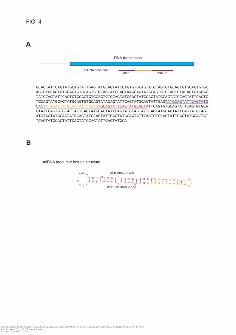
GCACCATTCAGTATGCAGTATTGAGTATGCAGTATTCAGTGTGCAGTATGCAGTGTGCAGTGTGCAGTGTGCAGTGTGCAGTGTGCAGTGTGCAGTGTGCAGTATGCAGTAAGCAGTATGCAGTGTGCAGTGTACAGTGTGCAGTATGCAGTATTCAGTGTGCAGTGTGCAGTGTGCAGTATGCAGTATGCAGTATGCAGTATGCAGTATTCAGTGTGCAGTATGCAGTATGCAGTGTGCAGTATACAGTATTCAGTATGCACTATTGAGTATGCAGTATTCAGTATGCACTATTGAGTATGCAGTATTCAGTGTGCAGTATTCAGTATGCACTATTCAGTATGCAGTATTCAGTGTGCAGTATTCAGTGTGCACTATTCAGTATGCACTATTGAGTATGCAGTATTCAGTATGCAGTATTCAGTATGCAGTATGTAGTATGCAGTATGCAGTATGCACTATTGAGTATGCAGTATTCAGTGTGCACTATTCAGTATGCACTATTCAGTATGCACTATTGAGTATGCAGTATTGAGTATGCA
A
B
DNA transposon
star mature
5'u au g c agu a u u c
a g u a u g c a cua u u g a g u a u gc
aguauucagugugcaguauuc
aguaugcacua
3'
miRNA precursor hairpin structure
star sequence
mature sequence
miRNA precursor
FIG. 4
Downloaded from https://academic.oup.com/gbe/advance-article-abstract/doi/10.1093/gbe/evy003/4827693by University of Plymouth useron 30 January 2018

avi-miR-temp_60
avi-miR-temp_48
avi-miR-temp_59
avi-miR-temp_58
avi-miR-temp_37
avi-miR-temp_52
avi-miR-temp_13
avi-miR-temp_29
avi-miR-temp_56
1 3 2 2 1 3 1 3 2
pH 8.2pH 7.9
individualconditionpH 8.2pH 7.6 pH 7.6
−1.5 −1 −0.5 0 0.5 1 1.5Row Z−Score
Colour Key
FIG. 5
Downloaded from https://academic.oup.com/gbe/advance-article-abstract/doi/10.1093/gbe/evy003/4827693by University of Plymouth useron 30 January 2018

FIG. 6
A
WebLogo 3.3
0.0
1.0
2.0
bits
GACU
G
AUC
G
UA
5UGCA
C
C
GUA
A
U
C
C
UA
10G
CUA
G
CAU
ACU
CGUA
GACU
15AUC
G
UA
GCAU
U
C
C
GAU
20GCUA
G
UA
C
AU
25CUA
G
UAC
G
CA
U
AU
C
30G
AUC
Sense putative piRNA reads aligned to genome
BAntisense putative piRNA reads aligned to genome
WebLogo 3.3
0.0
1.0
2.0
bits
CGAU
C
G
5C
A
U
10CGUA
C
AU
15A
UUU
20 25U C
Sense putative piRNA reads aligned to TEs
WebLogo 3.3
0.0
1.0
2.0
bits
A
CG
UU
CAG
AC
UGA
5UU
A
UU
10A
UU UU
15U
20 25C
U
C
WebLogo 3.3
0.0
1.0
2.0
bits
CGAU
ACG
5A
U
10G
CUA
UU
15U
20 25U
C
DAntisense putative piRNA reads aligned to TEs
Downloaded from https://academic.oup.com/gbe/advance-article-abstract/doi/10.1093/gbe/evy003/4827693by University of Plymouth useron 30 January 2018

FIG. 7
A
WebLogo 3.30.0
1.0
2.0
bits
GCAU
U
CAG
C
UA
UAG
5AU
C
AU
C
A
U
C
AU
10C
AU
G
C
AU
U
15AU
UA
A
20A
UA
U
25U
A
CU
Long putative piRNA reads with 10 nt overlap mapping to genome
WebLogo 3.30.0
1.0
2.0
bits
CGAU
CAG
A
5U
UA
C
AU
C
U
10GCUA
G
AU
15A
20 25
Short putative piRNA reads with 10 nt overlap mapping to genome
Long putative piRNA reads with 10 nt overlap mapping to TEs
WebLogo 3.30.0
1.0
2.0
bits
GCAU
CAG
A
G
CUAG
5GCAU
GCAU
ACU
UAC
10G
CAU
C
U
GCAU
G
C
AU
15GACU
G
CU
G
ACU
A
U
C
20C
G
AU
C
UA
C
G
A
UC
G
AU
AUC
25G
UC
C
AU
C
UG
CAU
WebLogo 3.30.0
1.0
2.0
bits
GCAU
UCAG
G
CUA
UAG
5G
AU
G
CAU
10GCUA
A
UU
CCU
U
15CAU
G
C
AU
C
A
UC
UU
20GCAU
A
C
UC
A
UA
C
25CU
Short putative piRNA reads with 10 nt overlap mapping to TEs
B
GAACUCGUUACUGAUGCCUCAUCGAACUCGUUACUGAUGCCUCAUCGAACUCGUUACUGAUGCCUCAUC
CUUUGAGAACUUCAACAACUUGAGCAAUCUUUGAGAACUUCAACAACUUGAGCAAU
UCUUUGAGAACUUCAACAACUUGAGCAAUUUUGAGAACUUCAACAACUUGAGCAAU
10 nt10 nt10 nt
UGAGUCUACAUUUUACCGCAUGCAGCGUGAGUCUACAUUUUACCGCAUGCAGCG
CGGACAGGAAGUAAAGACUCAGAUGUCGGACAGGAAGUAAAGACUCAGAUGUCGGACAGGAAGUAAAGACUCAGAUGU
CUGACAUGGAAAAACAGGCACUACAGACUGACAUGGAAAAACAGGCACUACAGA
ACAACCCGACCGUAGAAGACUGUACCUACAACCCGACCGUAGAAGACUGUACCU
UACAACCCGACCGUAGAAGACUGUACCU
forward strand
reverse strand
10 nt
10 nt
10 nt
Overlap probabilities of putative piRNA reads mapping to genome
Overlap size [nt]
Ove
rlapp
ing
prob
abilit
y0.
000.
020.
040.
060.
080.
10
pH 7.6pH 7.9pH 8.2
1 2 3 4 5 6 7 8 9 10 12 14 16 18 20 22 24 26 28 3011 13 15 17 19 21 23 25 27 29
Overlap probabilities of putative piRNA reads mapping to TEs
Over
lappin
g pr
obab
ility
0.00
0.02
0.04
0.06
0.08
0.10
0.12
pH 7.6pH 7.9pH 8.2
1 2 3 4 5 6 7 8 9 10 12 14 16 18 20 22 24 26 28 3011 13 15 17 19 21 23 25 27 29
Overlap size [nt]
C
Downloaded from https://academic.oup.com/gbe/advance-article-abstract/doi/10.1093/gbe/evy003/4827693by University of Plymouth useron 30 January 2018