elimination of plasma membrane phosphatidylinositol...
TRANSCRIPT
2084 Research Article
IntroductionPhosphoinositides are a family of phospholipids containingmyo-inositol as their headgroup. Despite a relatively lowabundance in biological membranes, phosphoinositidesregulate a myriad of cellular processes owing to their high rateof metabolic turnover. One such lipid, phosphatidylinositol4,5-bisphosphate [PtdIns(4,5)P2], is required for many aspectsof cellular physiology, including multiple stages of membranetraffic (Czech, 2003; Hammond et al., 2004; Janmey andLindberg, 2004). In particular, PtdIns(4,5)P2 is implicated inthe fusion of secretory vesicles with the plasma membrane inresponse to a specific stimulus (Martin, 2001), a process knownas regulated exocytosis.
Studies in neuroendocrine cells revealed that a process ofATP-dependent priming is required before secretory vesiclesare competent to undergo regulated exocytosis (Hay andMartin, 1992; Holz et al., 1989). Early work demonstrated thatATP is required, at least in part, to generate phosphoinositidesand that depletion of phosphoinositides prevented priming(Eberhard et al., 1990). Subsequent work has revealed
requirements for enzymes that generate PtdIns(4,5)P2 duringATP-dependent priming (Hay et al., 1995; Hay and Martin,1993; Wiedemann et al., 1996), indicating a specificrequirement for this lipid. Recent genetic approaches, wherebycellular PtdIns(4,5)P2 levels had been manipulated inendocrine cells showed corresponding changes in the numberof primed vesicles (Gong et al., 2005; Milosevic et al., 2005;Olsen et al., 2003). Furthermore, use of the monogamousPtdIns(4,5)P2-binding pleckstrin homology (PH) domain fromphospholipase C �1 (PLC�1) fused to green fluorescent protein(GFP) has identified plasma membrane pools of PtdIns(4,5)P2as being important in the regulation of exocytosis inneuroendocrine cells (Aikawa and Martin, 2003; Holz et al.,2000), hippocampal neurons (Micheva et al., 2001) andpancreatic beta cells (Lawrence and Birnbaum, 2003).
How does plasma membrane PtdIns(4,5)P2 mediate vesiclepriming? Several proteins required for regulated exocytosisbind PtdIns(4,5)P2 in vitro, including Mints (Okamoto andSudhof, 1997), Rabphilin 3A (Chung et al., 1998),synaptotagmins (Bai and Chapman, 2004) and CAPS
The inositol lipid phosphatidylinositol (4,5)-bisphosphate[PtdIns(4,5)P2] is involved in a myriad of cellular processes,including the regulation of exocytosis and endocytosis. Inthis paper, we address the role of PtdIns(4,5)P2 incompound exocytosis from rat peritoneal mast cells. Thisprocess involves granule-plasma membrane fusion aswell as homotypic granule membrane fusion and occurswithout any immediate compensatory endocytosis. Using anovel quantitative immunofluorescence technique, wereport that plasma membrane PtdIns(4,5)P2 becomestransiently depleted upon activation of exocytosis, and isnot detected on the membranes of fusing granules.Depletion is caused by phospholipase C activity, and ismandatory for exocytosis. Although phospholipase C isrequired for Ca2+ release from internal stores, the majorityof the requirement for PtdIns(4,5)P2 hydrolysis occursdownstream of Ca2+ signalling – as shown in permeabilised
cells, where the inositol (1,4,5)-trisphosphate–Ca2+
pathway is bypassed. Neither generation of thePtdIns(4,5)P2 metabolite, diacylglycerol (DAG) or simpleremoval and/or sequestration of PtdIns(4,5)P2 aresufficient for exocytosis to occur. However, treatment ofpermeabilised cells with DAG induces a small potentiationof exocytosis, indicating that it may be required. Wepropose that a cycle of PtdIns(4,5)P2 synthesis andbreakdown is crucial for exocytosis to occur in mast cells,and may have a more general role in all professionalsecretory cells.
Supplementary material available online athttp://jcs.biologists.org/cgi/content/full/119/10/2084/DC1
Key words: Diacylglycerol, Exocytosis, Neomycin,Phosphoinositide, Phospholipase C
Summary
Elimination of plasma membrane phosphatidylinositol(4,5)-bisphosphate is required for exocytosis frommast cellsGerald R. V. Hammond1,*,‡, Stephen K. Dove2, Alastair Nicol3, Jef A. Pinxteren4, Daniel Zicha3 andGiampietro Schiavo1,‡
1Molecular Neuropathobiology, Cancer Research UK London Research Institute, Lincoln’s Inn Fields Laboratories, London WC2A 3PX, UK2School of Bioscience, University of Birmingham, Birmingham, B15 2TT, UK3Light Microscopy Laboratories, Cancer Research UK London Research Institute, Lincoln’s Inn Fields Laboratories, London WC2A 3PX, UK4Flanders Interuniversity Institute for Biotechnology, Department of Medical Protein Research, Albert Baertsoenkaai 3, 9000 Ghent, Belgium*Present address: Department of Pharmacology, University of Cambridge, Tennis Court Road, Cambridge, CB2 1PD, UK‡Authors for correspondence (e-mail: [email protected]; [email protected])
Accepted 1 February 2006Journal of Cell Science 119, 2084-2094 Published by The Company of Biologists 2006doi:10.1242/jcs.02912
Jour
nal o
f Cel
l Sci
ence
2085Loss of PtdIns(4,5)P2 during exocytosis
(Grishanin et al., 2004). Indeed, it has even been suggestedthat the interaction between PtdIns(4,5)P2 and synaptotagminI plays a direct role in the membrane fusion process (Bai etal., 2004). However, exactly how PtdIns(4,5)P2 regulatesexocytosis is still far from clear. Furthermore, exocytosisfrom neurons and endocrine cells is accompanied bycompensatory endocytosis (Gundelfinger et al., 2003), aprocess itself reliant on PtdIns(4,5)P2 (Wenk and De Camilli,2004). Therefore, it can be difficult to resolve precisely atwhich stage in the exo-endocytic cycle PtdIns(4,5)P2 acts; forexample, neurons from mice deficient in phosphatidylinositol4-phosphate 5-kinase I� (PIPK I�) bear defects at severalstages of the synaptic vesicle cycle, with a predominantimpairment in vesicle retrieval after exocytosis (Di Paolo etal., 2004).
Upon activation, mast cells release pro-inflammatorymediators in an acute, rapid and massive exocytosis fromaround a thousand pre-formed granules, a process termeddegranulation. This involves compound exocytosis, wherebyonly an outer cohort of granules undergo heterotypic fusionat the plasma membrane; the remaining granules fuse withneighbouring-fused granules (pseudo-heterotypic fusion), in awave that propagates from the surface into the cell (Alvarez deToledo and Fernandez, 1990). As a result, a series of largeintracellular cavities communicating with the extracellularmilieu and containing the granule cores are formed. Notably,mast cell exocytosis proceeds without significant compensatoryendocytosis during or immediately following degranulation(Fernandez et al., 1984). Like neuroendocrine cells, mast cellsrequire an ATP-dependent priming reaction prior to exocytosis(Howell et al., 1989), which again involves the generation ofphosphoinositides (Pinxteren et al., 2001). However, mast cellsfrom PIPK I�-deficient mice display reduced PtdIns(4,5)P2levels yet, intriguingly, augmented degranulation (Sasaki et al.,2005). Therefore, it seems likely that PtdIns(4,5)P2 plays a key– yet poorly defined – role in mast cell exocytosis.
In this paper, we have employed a novel quantitativeimmunofluorescence technique to follow PtdIns(4,5)P2dynamics during compound exocytosis from rat peritonealmast cells (RPMCs). We report that PtdIns(4,5)P2 is present atthe plasma membrane and absent from the granule membranes;during exocytosis, PtdIns(4,5)P2 is eliminated from the plasmamembrane and remains absent from membranes of fusinggranules. This depletion is mediated by phosphatidylinositol-specific phospholipase C (PLC), the activity of which isrequired for exocytosis beyond its established role in Ca2+
signalling.
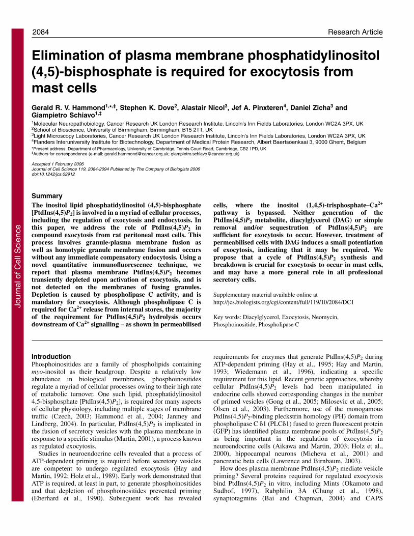
ResultsPtdIns(4,5)P2 is present at the plasma membrane ofresting mast cellsTo investigate a potential role for PtdIns(4,5)P2 in exocytosisfrom mast cells, we began by establishing the subcellularlocalisation of this lipid. RPMCs are not amenable totransfection with GFP-tagged probes. Therefore, we used thespecific monoclonal antibody 2C11 against PtdIns(4,5)P2(Osborne et al., 2001; Thomas et al., 1999), opting for astaining protocol performed at 4°C to preserve membranelocalisation of the lipid (Watt et al., 2002). Discontinuousthread-like structures labelled by the 2C11 antibody wereobserved at the cell surface (Fig. 1A), which are reminiscent
of the plasma membrane folds seen in mast cells when viewedby scanning electron microscopy (Burwen and Satir, 1977).Since a similar pattern was observed with the generic plasmamembrane dye, CM-DiI (Fig. 1A), the staining is consistentwith a homogenous distribution of PtdIns(4,5)P2 within the
Fig. 1. PtdIns(4,5)P2 is enriched at the plasma membrane of restingRPMCs. (A) The monoclonal anti-PtdIns(4,5)P2 antibody 2C11stains the plasma membrane. RPMCs were fixed and stained at 4°Cwith (a,b) 2C11 (red) and Draq5TM (blue) or (c) CM-DiI as describedin Materials and Methods; (a) shows an equatorial confocal section,(b,c) are projections of confocal sections taken at 1 �m intervalsthroughout the cell. (B) 2C11 binds PtdIns(4,5)P2 andPtdIns(3,4,5)P3. 2C11 was pre-incubated with 1000-fold molarexcess of the indicated inositol phosphate before staining as in (A);equatorial confocal sections are shown. (C) 2C11 specifically detectsPtdIns(4,5)P2 on the plasma membrane of RPMCs. Cells werepreincubated with 50 �M of the indicated PH domain before staining(b,c), or with 1 mM neomycin during staining with 2C11 (a);equatorial confocal sections are shown. (D) PtdIns(4,5)P2 colocaliseswith the cortical actin cytoskeleton. Cells were co-stained with 2C11(red) and Alexa Fluor 488-phalloidin (green) as described inMaterials and Methods. A projection of confocal sections taken at 1�m intervals throughout the cell is shown. (A-C) Micrographs are atthe same magnification, except (Ac), which is at the samemagnification as (D). Bars, 10 �m.
Jour
nal o
f Cel
l Sci
ence
2086
plasma membrane, and we could not detect any evidence forlocal enrichment of PtdIns(4,5)P2 at this level of resolution.
We performed two independent sets of controls todemonstrate that 2C11 was detecting PtdIns(4,5)P2 in RPMCs.First, the antibody was pre-incubated with a molar excess ofvarious inositol phosphates before applying the antibodyto cells. Under these conditions, both Ins(1,4,5)P3 andIns(1,3,4,5)P4 competed with the cellular antigen (Fig. 1B),indicating that this could be PtdIns(4,5)P2 or PtdIns(3,4,5)P3.However, it is unlikely that PtdIns(3,4,5)P3 is giving rise to thestaining observed, because this molecule is not detectable inresting RPMCs whereas PtdIns(4,5)P2 is abundant (G.R.V.H.and S.K.D., unpublished observations). To formally excludethis possibility, we performed a second set ofcontrol experiments: cells were pre-incubatedwith agents that would specifically bindendogenous phosphoinositide, thereforepreventing their labelling with the antibody.Binding of 2C11 was effectively prevented byincubation of the cells with neomycin, anaminoglycoside antibiotic that binds with highaffinity to several phosphoinositides, includingPtdIns(4,5)P2 and PtdIns(3,4,5)P3 (Fig. 1B)(Schacht, 1978) (S.K.D., unpublishedobservations). In parallel, cells were pre-incubated with GST-tagged probes of exquisitespecificity: the PH domain from PLC�1, whichbinds PtdIns(4,5)P2 (Lemmon et al., 1995), andthe 2G splice-variant of the PH domain fromGRP1, which is specific for PtdIns(3,4,5)P3(Klarlund et al., 2000). Only the PH domainfrom PLC�1 effectively competed with 2C11(Fig. 1C), demonstrating that the antibody isindeed detecting endogenous PtdIns(4,5)P2.
Resting RPMCs contain a cortical actincytoskeleton (Norman et al., 1996), which hasbeen linked to plasma membrane PtdIns(4,5)P2in a number of cell types (Janmey andLindberg, 2004). Indeed, when mast cells werecounter-stained for F-actin with fluorescentphalloidin, we observed an excellentcolocalisation with PtdIns(4,5)P2 (Fig. 1D).Thus, resting RPMCs appear to contain a poolof PtdIns(4,5)P2 at the plasma membrane,juxtaposed to the cortical F-actin network.Importantly, we did not observe any staining ofinternal granule membranes (Fig. 1A-C).
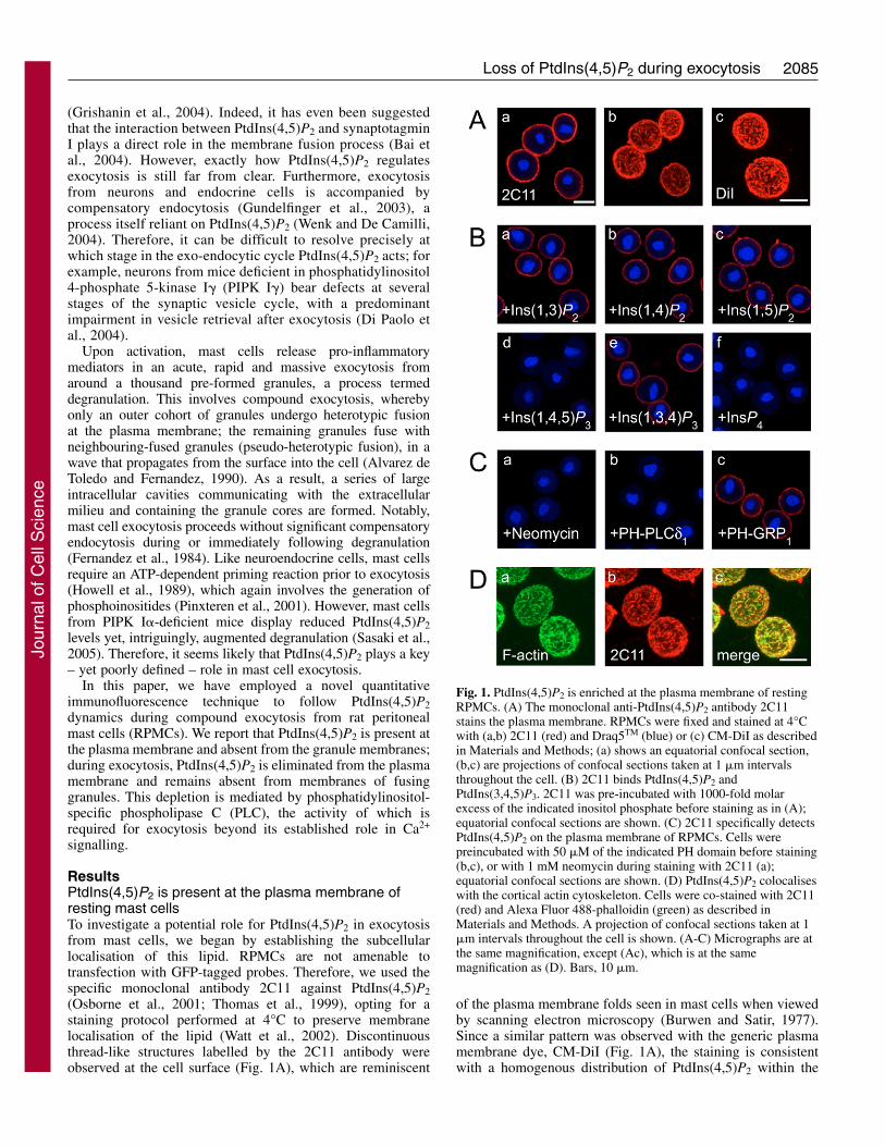
PtdIns(4,5)P2 dynamics during exocytosisMast cell exocytosis involves both granule-plasma membrane and granule-granulemembrane fusion. Therefore, if PtdIns(4,5)P2were required for membrane fusion, we wouldexpect either relocation of plasma membranePtdIns(4,5)P2 to granule membranes orsynthesis of PtdIns(4,5)P2 at the these sitesduring exocytosis. To test these hypotheses, westimulated cells for various times with thepolycationic agonist, compound 48/80 (48/80).Cells were then fixed and stained with both2C11 and fluorescent concanavalin A, a
Journal of Cell Science 119 (10)
membrane impermeable reagent that binds to granule coreswith high affinity. Only granule cores that have been exposedto the extracellular milieu are labelled by concanavalin A,making this reagent an effective tool to monitor exocytosis(Norman et al., 1996).
Surprisingly, cells stimulated for short periods whendegranulation was well underway showed almost completeelimination of plasma membrane PtdIns(4,5)P2, whichremained absent from the membranes of fusing granules (Fig.2A). Only after a further 2.5 minutes, well after the completionof exocytosis (Penner, 1988), was PtdIns(4,5)P2 seen to returnat the plasma- as well as granule-membranes at levelscomparable with resting cells (Fig. 2A,B). Analysis of single-
Fig. 2. Plasma membrane PtdIns(4,5)P2 is transiently depleted during degranulation.(A) Cells were fixed with ice-cold 3% glutaraldehyde and stained either at rest orafter stimulation for the indicated times with compound 48/80. First and third row ofpanels show merged images of 2C11 (red) and Alexa Fluor 647-concanavalin A(blue). Second and fourth row of panels show fluorescence intensity profiles for2C11. Bar, 10 �m. Ctrl indicates that 2C11 has been omitted during staining. *,contaminating neutrophil. (B,C) 2C11 fluorescence was quantified as described inMaterials and Methods. (B,C) Mean fluorescence (± s.e.m.) normalised to the restinglevel (B), distribution of fluorescence at the indicated time points (C); n>100 cells pertime point. (D) RPMCs were loaded with unlabelled or [2-3H]inositol, and stimulatedfor the indicated time period with compound 48/80. Cells were either fixed andstained with 2C11 and quantified as in B (2C11), or extracted and analysed forPtdIns(4,5)P2 content by HPLC ([2-3H]-PtdIns(4,5)P2).
Jour
nal o
f Cel
l Sci
ence
2087Loss of PtdIns(4,5)P2 during exocytosis
cell fluorescence intensity revealed that after 15 seconds ofstimulation with 48/80, the majority of cells had undergonecomplete elimination of PtdIns(4,5)P2, because the intensitywas comparable with that of cells stained with only secondaryantibody (Fig. 2C). Thus the ~80% drop in fluorescenceintensity observed after 15 seconds (Fig. 2B) representselimination of PtdIns(4,5)P2 in ~80% of the cells (Fig. 2C); theremaining ~20% overlap with the fluorescence observed inresting cells (Fig. 2C), because these cells had yet to beginexocytosis (e.g. Fig. 2A, 15 seconds, lower cell).
Is the 2C11 antibody an accurate tool with which to studyPtdIns(4,5)P2 dynamics? In principle, PtdIns(4,5)P2 binding toan endogenous effector might produce a decrease in staining.Although this seems unlikely given the harsh fixationprocedure used in these experiments, we directly measuredPtdIns(4,5)P2 by metabolic labelling of mast cells with [2-3H]-inositol. RPMCs were left at rest or stimulated with 48/80 forup to 60 seconds; cells were then either fixed and stained, orlysed in acid and lipids were extracted and analysed by HPLC.Once again, we saw evidence of transient depletion of plasmamembrane PtdIns(4,5)P2 (Fig. 2D and supplementary materialFig. S2). The relative decrease in fluorescence intensity wasless marked in cells labelled with inositol for 19 hours(compare Fig. 2B and 2D). However, this was owing todecreased fluorescence intensity in the resting population oflabelled RPMCs (compare Fig. 2C with supplementarymaterial Fig. S2B), rather than an incomplete elimination ofPtdIns(4,5)P2 in stimulated cells. Furthermore, the labelledRPMCs displayed a more rapid recovery of PtdIns(4,5)P2 afterlabelling in vitro for 19 hours. However, despite thesedifferences, the levels of [2-3H]-PtdIns(4,5)P2 were inexcellent agreement with the fluorescence data (Fig. 2D).
Biochemical estimates of PtdIns(4,5)P2 levels tended to beslightly higher than fluorescence estimates after stimulation(Fig. 2D). We reasoned that this might be due to the existenceof a second, minor pool of PtdIns(4,5)P2 not detected aftersaponin permeabilisation (see Materials and Methods).Many cells contain a detergent-resistant nuclear pool ofphosphoinositides (Hammond et al., 2004). Indeed, afterextraction of membranes with Triton X-100, we observed apunctate staining of RPMC nuclei (supplementary materialFig. S1A), consistent with previous reports using 2C11 in othercells (Osborne et al., 2001). This staining was observed toincrease dramatically after stimulation of cells with 48/80(supplementary material Fig. S1A,B). Although directcomparison of membrane and nuclear PtdIns(4,5)P2 pools wasnot possible owing to the different staining protocols, thenuclear pool contained approximately 1% of the fluorescencecompared with the membrane pool, accounting for thediscrepancy with biochemical measurements (Fig. 2D).Therefore, we conclude that after the onset of exocytosis,plasma membrane PtdIns(4,5)P2 becomes depleted from mastcells, while a minor nuclear pool increases.
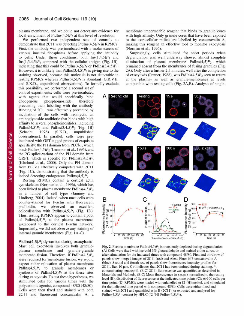
PLC mediates the depletion of plasma membranePtdIns(4,5)P2A clue to the cause of PtdIns(4,5)P2 depletion duringexocytosis was given by experiments akin to those illustratedin Fig. 2D. When the total phosphoinositide levels are plottedas a function of time after stimulation, a steady decline toapproximately 85% of control after 60 seconds is observed
(data not shown). This depletion was predominately fromPtdIns, and indicates the action of a phospholipase.
To test whether PLC caused the depletion of plasmamembrane PtdIns(4,5)P2, we used two inhibitors of PLC: thethiol-reactive U73-122 (Horowitz et al., 2005) and Et-18-OMe(Powis et al., 1992). U73-122 inhibited exocytosis with anapparent IC50 of ~2 �M, whereas the inactive analogue U73-343 had an IC50 approximately tenfold higher (Fig. 3A). Et-18-OMe inhibited exocytosis with an IC50 of ~27 �M (Fig.3B), whereas the vehicle control had no discernible effect. Bothinhibitors caused release of �-hexosaminidase (�-hex) fromunstimulated cells at higher concentrations, indicating cell lysis(data not shown). Pre-incubation of cells with 5 �M U73-122or 40 �M Et-18-OMe prevented exocytosis and blocked the
Fig. 3. Phospholipase C causes the depletion of plasma membranePtdIns(4,5)P2, and the release of Ca2+ from intracellular stores.(A,B) PLC inhibitors prevent mast cell degranulation. RPMCs werepre-incubated with U73-122 and/or U73-343 for 5 minutes (A), orEt-18-OMe (or EtOH, as vehicle control) for 20 minutes (B), at theindicated concentration before stimulation with 48/80 at 25°C. After10 minutes, the medium was assayed for released �-hexosaminidase(�-hex) activity. Data are means of triplicates ± s.e.m. (C) PLCinhibitors block depletion of PtdIns(4,5)P2. Mast cells were untreated(ctrl) or incubated with 5 �M BAPTA/AM or as described in A andB. Subsequently, degranulation was evoked with 48/80 for 30seconds. Cells were fixed and stained with 2C11 (red) and AlexaFluor 647-concanavalin A (blue). Bar, 10 �m. (D) PLC inhibitorsblock Ca2+ signalling. RPMCs were loaded for 20 minutes withFluo3/AM, pre-incubated with the indicated compound as in A-C orfor 20 minutes with 5 �M BAPTA/AM, and activated with 48/80.Normalised fluorescence intensity traces are shown from a singlerepresentative cell for each condition.
Jour
nal o
f Cel
l Sci
ence

2088
depletion of PtdIns(4,5)P2, whereas no effect of 5 �M U73-343 was observed (Fig. 3C). Thus, PLC activity seems to causethe reduction in plasma membrane PtdIns(4,5)P2, and isrequired for exocytosis.
Bone marrow derived mast cells require PLC�2 for Ca2+
release from intracellular stores as well as for exocytosis (Wenet al., 2002), suggesting a similar function in RPMCs. To testthis possibility, mast cells were loaded with the Ca2+ indicatorFluo3/AM and stimulated with 48/80. In agreement withprevious studies (Penner, 1988), we observed a rapid increasein the intracellular Ca2+ concentration in control cells afteractivation, which declines slowly (Fig. 3D). When cells weretreated with 5 �M U73-122 this change in cytosolic Ca2+ levelswas completely abolished (Fig. 3D), whereas U73-343 waswithout effect. Thus, it appears that PLC is required for theCa2+ release from intracellular stores, consistent with theprevious observation that injection of neomycin, which alsoinhibits PLC, blocks Ca2+ release from intracellular stores inRPMCs (Penner, 1988).
Is Ca2+ release from internal stores strictly required forexocytosis to occur? This seems to be the case, because loadingcells with the cell-permeant Ca2+ chelator BAPTA/AMinhibits exocytosis. However, depletion of plasma membranePtdIns(4,5)P2 was also inhibited (Fig. 3C). This was mostprobably due to the reduction of basal Ca2+ levels and thusinhibition of PLC, which is a Ca2+-dependent enzyme (Rhee etal., 1989).
PLC activity in permeabilised mast cellsTo investigate whether hydrolysis of bulk PtdIns(4,5)P2 wasrequired for mast cell exocytosis, we utilised cellpermeabilisation with streptolysin-O (SL-O). PermeabilisedRPMCs undergo exocytosis in the presence of Ca2+ bufferedin the �M range, although provision of GTP or its non-hydrolysable analogue GTP�S is absolutely required (Howellet al., 1987). Since Ca2+ is buffered with EGTA, Ca2+ releasefrom stores will not lead to an elevation in Ca2+ concentration,thus bypassing Ins(1,4,5)P3-mediated Ca2+ release.
When RPMCs are permeabilised for 3 minutes beforefixation in the presence of 100 nM free Ca2+ (pCa 7) and 100�M MgATP, PtdIns(4,5)P2 displays a distribution similar tothat observed in intact, resting cells and no exocytosis isdetected (Fig. 4A). However, if cells are permeabilised in thepresence of MgATP, 10 �M GTP�S and 10 �M free Ca2+ (pCa5) a stochastic activation of the cells is observed (Hide et al.,1993): the majority of cells appeared to have degranulated after3 minutes, although a minority have not yet begun exocytosis.The proportion of mast cells undergoing exocytosis underthese conditions increases with time, although for individualcells exocytosis may take a matter of seconds (G.R.V.H.,unpublished observations). Strikingly, in cells that are yetto begin exocytosis, PtdIns(4,5)P2 remains on the plasmamembrane and may even be increased (Fig. 4A). On thecontrary, cells that undergo exocytosis show almost completeelimination of PtdIns(4,5)P2. No recovery of PtdIns(4,5)P2 isobserved in any of the cells within 3 minutes (Fig. 4A). In tenexperiments, the proportion of degranulated cells varied widelyfrom 44% to 100%, with a mean ± s.d. of 79%±20%.
What causes the depletion of PtdIns(4,5)P2 frompermeabilised cells? Under these conditions, PLC,phosphoinositide 3-kinases and inositol phosphatases, all of
Journal of Cell Science 119 (10)
which can deplete plasma membrane PtdIns(4,5)P2, may beactivated. Treatment of cells with U73-122 duringpermeabilisation blocked exocytosis and the depletion ofPtdIns(4,5)P2, whereas U73-343 was without effect (Fig. 4B,Fig. 5A). At 40 �M, Et-18-OMe did not have such dramaticeffects in permeabilised cells, but still decreased the proportionof degranulating cells by approximately 70% relative tountreated controls (Fig. 4D). Neomycin at 1-3 mM (Fig. 4C)also blocked exocytosis, although results were extremelyvariable at 300 �M (Fig. 4C). However, in some cells 1-3 mMneomycin appeared to cause a reduction in the extent ofPtdIns(4,5)P2 staining despite a lack of degranulation (Fig.5A). This is possibly caused by the interaction of neomycin
Fig. 4. PLC causes depletion of PtdIns(4,5)P2 from permeabilisedmast cells. (A) RPMCs were permeabilised with SL-O in thepresence of 100 �M MgATP and 3 mM Ca:EGTA at pCa 7, or 100�M MgATP, 10 �M GTP�S and 3 mM Ca:EGTA at pCa 5 for 3minutes at 30°C. Cells were then fixed and stained with 2C11 (red)and Alexa Fluor 647-concanavalin A (blue). Bar, 10 �m. (B-D) PLCinhibitors prevent PtdIns(4,5)P2 depletion and block degranulation ofpermeabilised cells. Mast cells were permeabilised as described in(A) at pCa 5, 100 �M MgATP and 10 �M GTP�S, in the presence ofU73-122 or U73-343 (B), neomycin (C), 40 �M Et-18-OMe, 100�M LY294002 or LY303511, and 5 mM �-GP (D) as indicated.(E) RPMCs were permeabilised in the presence of 100 �M MgATPand 300 �M Ca:EGTA at pCa 8 at 30°C. After 2 minutes, theindicated peptide was added to 100 �M. After a further 5 minutes,cells were activated by the addition of the indicated buffers at thesame concentrations as (A). The numbers of degranulated cells werecounted, and the numbers normalised to the control value for eachexperiment. Values represent the means of three or more independentexperiments ± s.e.m., with the exceptions of U73-343 andLY303511, which are means ± range of duplicate experiments. 0.4%EtOH is the vehicle control for 40 �M Et-18-OMe.
Jour
nal o
f Cel
l Sci
ence

2089Loss of PtdIns(4,5)P2 during exocytosis
with PtdIns(4,5)P2, which can effectively inhibit2C11 labelling at these concentrations (Fig. 1B).
We also used the myristoylated alanine-rich C-kinase substrate basic effector domain (MARCKS-ED) because this peptide was shown to laterallysequester PtdIns(4,5)P2 in the membrane, inhibitingPLC but sparing other interactions such as with thePH domain from PLC�1 (Gambhir et al., 2004).Equilibration of 100 �M peptide into permeabilisedRPMCs abolished exocytosis (Fig. 4E) as well asbreakdown of PtdIns(4,5)P2 (Fig. 5B). By contrast,a mutant peptide in which four Phe residues aremutated to Ala (FA-MARCKS-ED) was shown todisplay a weaker interaction with PtdIns(4,5)P2 atthe membrane and, consequently, a far less potentinhibition of PLC (Gambhir et al., 2004); thispeptide affected neither exocytosis (Fig. 4E) orplasma membrane PtdIns(4,5)P2 depletion (Fig.5B). Together, these results are therefore consistentwith a role for PLC-mediated PtdIns(4,5)P2breakdown in exocytosis.
By contrast, use of �-glycerophosphate (�-GP, 5mM) as a generic phosphatase inhibitor did noteffect exocytosis or depletion of PtdIns(4,5)P2 (Fig.4D, Fig. 5A). At 100 �M, the phosphoinositide3-kinase inhibitor LY294002 produced a 20%reduction in the number of degranulating cells (Fig.4D). However, its inactive analogue LY303511(Vlahos et al., 1994) produced an identical effect(Fig. 4D), allowing us to conclude that theinhibition was not due to impairment ofphosphoinositide 3-kinases.
RPMCs not undergoing exocytosis were repletewith plasma membrane PtdIns(4,5)P2, whereascells that showed signs of degranulation showedalmost complete elimination of PtdIns(4,5)P2 (Fig.4A, Fig. 5). We occasionally observed cells that hadnot degranulated yet still lost their PtdIns(4,5)P2staining, but these were always a minority (< 5%)for most of the conditions shown in Figs 4 and 5.The presence of U73-122 increased this proportionto approximately 7% at 5 �M and as high as12% at 10 �M. The lack of PtdIns(4,5)P2immunoreactivity might be due to the action ofphosphatases in the absence of PLC-mediatedPtdIns(4,5)P2 hydrolysis. However, we neverobserved cells with signs of compound exocytosisthat retained PtdIns(4,5)P2 under any experimentalcondition tested. Therefore, the depletion ofplasma membrane PtdIns(4,5)P2 observed frompermeabilised cells is indispensable for exocytosisto occur and appears to be mediated by PLC.
We also tested the effect of U73-122 and Et-18-OMe on therelease of �-hex from permeabilised RPMCs in suspension andfound both inhibited, with apparent IC50s of 2 and 20 �M,respectively (supplementary material Fig. S3). Once again,U73-343 had only a minor effect, and Et-18-OMe became lyticat higher concentrations. Our results with U73-122 and U73-343 are consistent with those reported independently byanother group (Gloyna et al., 2005) in SL-O permeabilisedmast cells.
Downstream of PLC activationHow does hydrolysis of PtdIns(4,5)P2 by PLC controlexocytosis? Although in a permeabilised cell system withbuffered Ca2+ we can rule out a role for Ins(1,4,5)P3 inmediating Ca2+ release, there might be other functions for thisinositol phosphate. However, Ins(1,4,5)P3 and Ins(1,4)P2 atconcentrations of up to 100 �M failed to modify exocytosisfrom permeabilised cells in the presence or absence of PLCinhibitors (data not shown). Furthermore, a degradation
Fig. 5. (A) Mast cells were permeabilised with SL-O in the presence of 100 �MMgATP, 10 �M GTP�S and 3 mM Ca:EGTA at pCa 5 for 3 minutes at 30°C asin Fig. 4A, in the presence of 10 �M U73-122 or U73-343, 3 mM neomycin, 40�M Et-18-OMe, 100 �M LY294002, 100 �M LY303511 or 5 mM �-GP.(B) Cells were permeabilised in the presence of 300 �M Ca:EGTA at pCa 8;after two minutes, the indicated peptide was added to 100 �M. After a further 5minutes at 30°C, Ca:EGTA at pCa 5 and GTP�S were added to the same finalconcentrations as (A), and the cells incubated for 5 minutes. Cells were thenfixed and stained with 2C11 (red) and Alexa Fluor 647-concanavalin A (blue).Top panels show merged images of 2C11 and concanavalin A; bottom panelsshow the fluorescence intensity profile for 2C11. Bar, 10 �m.
Jour
nal o
f Cel
l Sci
ence
2090
product of Ins(1,4,5)P3 is probably not involved because mastcell exocytosis is insensitive to mM concentrations of LiCl,(Cockcroft et al., 1987), which block the dephosphorylation ofIns(1,4)P2. Finally, Ins(1,4,5)P3 can be phosphorylated by anInsP3 3-kinase and enter into the metabolic pathway of inositolpolyphosphates. These molecules have many functionsincluding regulation at multiple steps of membrane traffic(Irvine and Schell, 2001). However, because mast cellexocytosis proceeds when the cells are depleted of ATP(Howell et al., 1987), InsP3 3-kinase activity is probably notrequired for exocytosis. This leaves us with two non-mutuallyexclusive possibilities. First, the other product of PLC, 1,2-diacyl-sn-glycerol (DAG) may be required. Second,elimination of plasma membrane PtdIns(4,5)P2 may be neededfor exocytosis, as has been shown for phagosome formation(Scott et al., 2005), inactivation of certain ion channels (Suhand Hille, 2005) and for Salmonella invasion (Terebiznik et al.,2002).
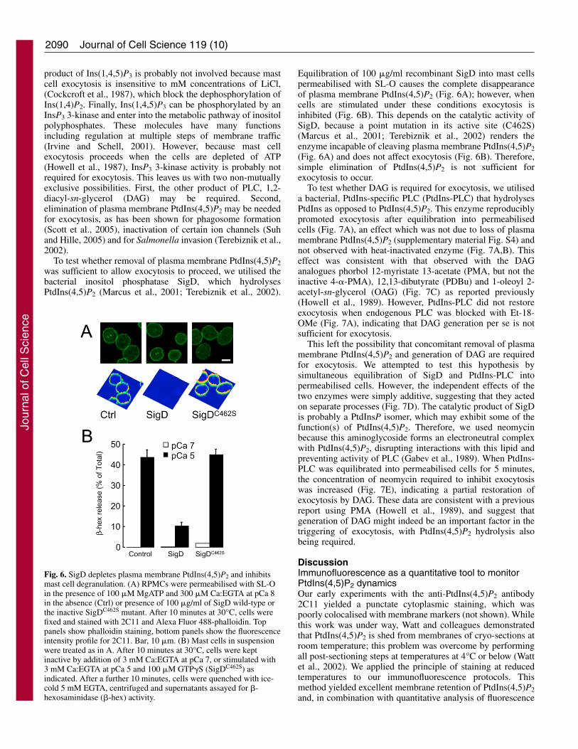
To test whether removal of plasma membrane PtdIns(4,5)P2was sufficient to allow exocytosis to proceed, we utilised thebacterial inositol phosphatase SigD, which hydrolysesPtdIns(4,5)P2 (Marcus et al., 2001; Terebiznik et al., 2002).
Journal of Cell Science 119 (10)
Equilibration of 100 �g/ml recombinant SigD into mast cellspermeabilised with SL-O causes the complete disappearanceof plasma membrane PtdIns(4,5)P2 (Fig. 6A); however, whencells are stimulated under these conditions exocytosis isinhibited (Fig. 6B). This depends on the catalytic activity ofSigD, because a point mutation in its active site (C462S)(Marcus et al., 2001; Terebiznik et al., 2002) renders theenzyme incapable of cleaving plasma membrane PtdIns(4,5)P2(Fig. 6A) and does not affect exocytosis (Fig. 6B). Therefore,simple elimination of PtdIns(4,5)P2 is not sufficient forexocytosis to occur.
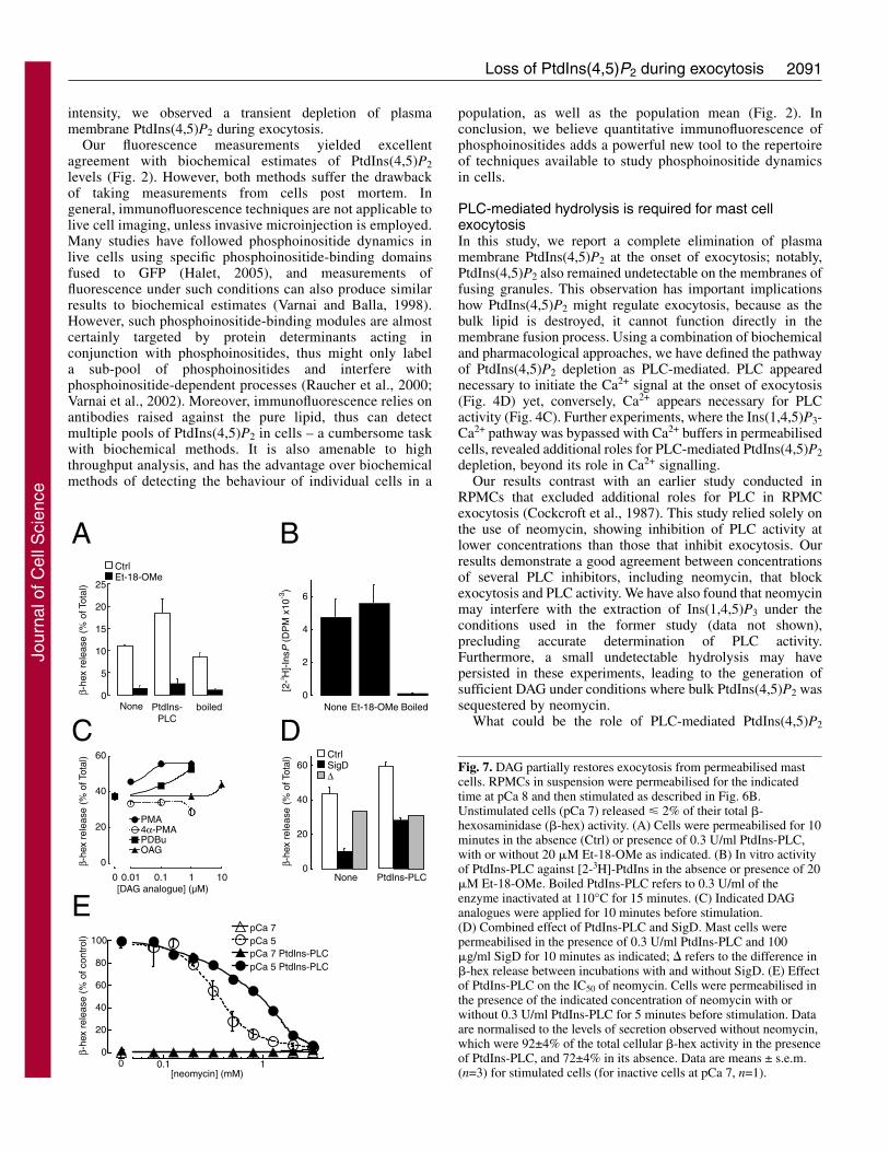
To test whether DAG is required for exocytosis, we utiliseda bacterial, PtdIns-specific PLC (PtdIns-PLC) that hydrolysesPtdIns as opposed to PtdIns(4,5)P2. This enzyme reproduciblypromoted exocytosis after equilibration into permeabilisedcells (Fig. 7A), an effect which was not due to loss of plasmamembrane PtdIns(4,5)P2 (supplementary material Fig. S4) andnot observed with heat-inactivated enzyme (Fig. 7A,B). Thiseffect was consistent with that observed with the DAGanalogues phorbol 12-myristate 13-acetate (PMA, but not theinactive 4-�-PMA), 12,13-dibutyrate (PDBu) and 1-oleoyl 2-acetyl-sn-glycerol (OAG) (Fig. 7C) as reported previously(Howell et al., 1989). However, PtdIns-PLC did not restoreexocytosis when endogenous PLC was blocked with Et-18-OMe (Fig. 7A), indicating that DAG generation per se is notsufficient for exocytosis.
This left the possibility that concomitant removal of plasmamembrane PtdIns(4,5)P2 and generation of DAG are requiredfor exocytosis. We attempted to test this hypothesis bysimultaneous equilibration of SigD and PtdIns-PLC intopermeabilised cells. However, the independent effects of thetwo enzymes were simply additive, suggesting that they actedon separate processes (Fig. 7D). The catalytic product of SigDis probably a PtdInsP isomer, which may exhibit some of thefunction(s) of PtdIns(4,5)P2. Therefore, we used neomycinbecause this aminoglycoside forms an electroneutral complexwith PtdIns(4,5)P2, disrupting interactions with this lipid andpreventing activity of PLC (Gabev et al., 1989). When PtdIns-PLC was equilibrated into permeabilised cells for 5 minutes,the concentration of neomycin required to inhibit exocytosiswas increased (Fig. 7E), indicating a partial restoration ofexocytosis by DAG. These data are consistent with a previousreport using PMA (Howell et al., 1989), and suggest thatgeneration of DAG might indeed be an important factor in thetriggering of exocytosis, with PtdIns(4,5)P2 hydrolysis alsobeing required.
DiscussionImmunofluorescence as a quantitative tool to monitorPtdIns(4,5)P2 dynamicsOur early experiments with the anti-PtdIns(4,5)P2 antibody2C11 yielded a punctate cytoplasmic staining, which waspoorly colocalised with membrane markers (not shown). Whilethis work was under way, Watt and colleagues demonstratedthat PtdIns(4,5)P2 is shed from membranes of cryo-sections atroom temperature; this problem was overcome by performingall post-sectioning steps at temperatures at 4°C or below (Wattet al., 2002). We applied the principle of staining at reducedtemperatures to our immunofluorescence protocols. Thismethod yielded excellent membrane retention of PtdIns(4,5)P2and, in combination with quantitative analysis of fluorescence
Fig. 6. SigD depletes plasma membrane PtdIns(4,5)P2 and inhibitsmast cell degranulation. (A) RPMCs were permeabilised with SL-Oin the presence of 100 �M MgATP and 300 �M Ca:EGTA at pCa 8in the absence (Ctrl) or presence of 100 �g/ml of SigD wild-type orthe inactive SigDC462S mutant. After 10 minutes at 30°C, cells werefixed and stained with 2C11 and Alexa Fluor 488-phalloidin. Toppanels show phalloidin staining, bottom panels show the fluorescenceintensity profile for 2C11. Bar, 10 �m. (B) Mast cells in suspensionwere treated as in A. After 10 minutes at 30°C, cells were keptinactive by addition of 3 mM Ca:EGTA at pCa 7, or stimulated with3 mM Ca:EGTA at pCa 5 and 100 �M GTP�S (SigDC462S) asindicated. After a further 10 minutes, cells were quenched with ice-cold 5 mM EGTA, centrifuged and supernatants assayed for �-hexosaminidase (�-hex) activity.
Jour
nal o
f Cel
l Sci
ence
2091Loss of PtdIns(4,5)P2 during exocytosis
intensity, we observed a transient depletion of plasmamembrane PtdIns(4,5)P2 during exocytosis.
Our fluorescence measurements yielded excellentagreement with biochemical estimates of PtdIns(4,5)P2levels (Fig. 2). However, both methods suffer the drawbackof taking measurements from cells post mortem. Ingeneral, immunofluorescence techniques are not applicable tolive cell imaging, unless invasive microinjection is employed.Many studies have followed phosphoinositide dynamics inlive cells using specific phosphoinositide-binding domainsfused to GFP (Halet, 2005), and measurements offluorescence under such conditions can also produce similarresults to biochemical estimates (Varnai and Balla, 1998).However, such phosphoinositide-binding modules are almostcertainly targeted by protein determinants acting inconjunction with phosphoinositides, thus might only labela sub-pool of phosphoinositides and interfere withphosphoinositide-dependent processes (Raucher et al., 2000;Varnai et al., 2002). Moreover, immunofluorescence relies onantibodies raised against the pure lipid, thus can detectmultiple pools of PtdIns(4,5)P2 in cells – a cumbersome taskwith biochemical methods. It is also amenable to highthroughput analysis, and has the advantage over biochemicalmethods of detecting the behaviour of individual cells in a
population, as well as the population mean (Fig. 2). Inconclusion, we believe quantitative immunofluorescence ofphosphoinositides adds a powerful new tool to the repertoireof techniques available to study phosphoinositide dynamicsin cells.
PLC-mediated hydrolysis is required for mast cellexocytosisIn this study, we report a complete elimination of plasmamembrane PtdIns(4,5)P2 at the onset of exocytosis; notably,PtdIns(4,5)P2 also remained undetectable on the membranes offusing granules. This observation has important implicationshow PtdIns(4,5)P2 might regulate exocytosis, because as thebulk lipid is destroyed, it cannot function directly in themembrane fusion process. Using a combination of biochemicaland pharmacological approaches, we have defined the pathwayof PtdIns(4,5)P2 depletion as PLC-mediated. PLC appearednecessary to initiate the Ca2+ signal at the onset of exocytosis(Fig. 4D) yet, conversely, Ca2+ appears necessary for PLCactivity (Fig. 4C). Further experiments, where the Ins(1,4,5)P3-Ca2+ pathway was bypassed with Ca2+ buffers in permeabilisedcells, revealed additional roles for PLC-mediated PtdIns(4,5)P2depletion, beyond its role in Ca2+ signalling.
Our results contrast with an earlier study conducted inRPMCs that excluded additional roles for PLC in RPMCexocytosis (Cockcroft et al., 1987). This study relied solely onthe use of neomycin, showing inhibition of PLC activity atlower concentrations than those that inhibit exocytosis. Ourresults demonstrate a good agreement between concentrationsof several PLC inhibitors, including neomycin, that blockexocytosis and PLC activity. We have also found that neomycinmay interfere with the extraction of Ins(1,4,5)P3 under theconditions used in the former study (data not shown),precluding accurate determination of PLC activity.Furthermore, a small undetectable hydrolysis may havepersisted in these experiments, leading to the generation ofsufficient DAG under conditions where bulk PtdIns(4,5)P2 wassequestered by neomycin.
What could be the role of PLC-mediated PtdIns(4,5)P2
β-he
x re
leas
e (%
of T
otal
)
0
5
10
15
20
25
CtrlEt-18-OMe
None PtdIns-PLC
boiled
A
0
2
4
6
None Et-18-OMe Boiled
B
[2-3 H
]-In
sP (
DP
M x
10-3
)
0
20
40
60
0 0.01 0.1 1 10
PMA4α-PMAPDBuOAG
[DAG analogue] (µM)
β-he
x re
leas
e (%
of T
otal
)
C
0
20
40
60
None PtdIns-PLC
CtrlSigDΔ
β-he
x re
leas
e (%
of T
otal
)
D
0
20
40
60
80
100
0 0.1 1
pCa 7pCa 5
pCa 5 PtdIns-PLC
[neomycin] (mM)
β-he
x re
leas
e (%
of c
ontr
ol)
EpCa 7 PtdIns-PLC
Fig. 7. DAG partially restores exocytosis from permeabilised mastcells. RPMCs in suspension were permeabilised for the indicatedtime at pCa 8 and then stimulated as described in Fig. 6B.Unstimulated cells (pCa 7) released � 2% of their total �-hexosaminidase (�-hex) activity. (A) Cells were permeabilised for 10minutes in the absence (Ctrl) or presence of 0.3 U/ml PtdIns-PLC,with or without 20 �M Et-18-OMe as indicated. (B) In vitro activityof PtdIns-PLC against [2-3H]-PtdIns in the absence or presence of 20�M Et-18-OMe. Boiled PtdIns-PLC refers to 0.3 U/ml of theenzyme inactivated at 110°C for 15 minutes. (C) Indicated DAGanalogues were applied for 10 minutes before stimulation.(D) Combined effect of PtdIns-PLC and SigD. Mast cells werepermeabilised in the presence of 0.3 U/ml PtdIns-PLC and 100�g/ml SigD for 10 minutes as indicated; � refers to the difference in�-hex release between incubations with and without SigD. (E) Effectof PtdIns-PLC on the IC50 of neomycin. Cells were permeabilised inthe presence of the indicated concentration of neomycin with orwithout 0.3 U/ml PtdIns-PLC for 5 minutes before stimulation. Dataare normalised to the levels of secretion observed without neomycin,which were 92±4% of the total cellular �-hex activity in the presenceof PtdIns-PLC, and 72±4% in its absence. Data are means ± s.e.m.(n=3) for stimulated cells (for inactive cells at pCa 7, n=1).
Jour
nal o
f Cel
l Sci
ence
2092
depletion during exocytosis? PLC depletes PtdIns(4,5)P2 fromincipient phagosomes, causing disassembly of the surroundingF-actin (Scott et al., 2005). Furthermore, SigD eliminatesplasma membrane PtdIns(4,5)P2, causing a reduction inmembrane cytoskeletal rigidity to facilitate invasion bySalmonella typhimurium (Terebiznik et al., 2002). Mast cellscontain an F-actin cortex that breaks down at the onset ofdegranulation (Nishida et al., 2005; Norman et al., 1996).Disruption of this cortex is seen to enhance degranulation(Borovikov et al., 1995; Martin-Verdeaux et al., 2003), and theaugmented degranulation by PIPK I�-deficient mast cells ispartially mimicked by depletion of the F-actin cortex in wild-type cells (Sasaki et al., 2005). Notably, although SigDeliminated plasma membrane PtdIns(4,5)P2, it failed to reducethe actin cortex in RPMCs (Fig. 5A). Therefore, it seemspossible that PtdIns(4,5)P2 breakdown is associated with thedisassembly of the cortical actin cytoskeleton. However,stabilisation of this cortex with phalloidin is not seen to affectexocytosis (Norman et al., 1996), so simple removal of an F-actin ‘barrier’ seems unlikely. Alternatively, removal of plasmamembrane PtdIns(4,5)P2 might lead to the dissociation of anF-actin-associated factor. Notably, the SNARE protein SNAP-23, which relocates from plasma to granule membranes forcompound exocytosis, is associated with F-actin inside themembrane folds of resting RPMCs (Guo et al., 1998).
The enhancement of exocytosis from RPMCs by DAG andits analogues, along with its sparing effect on the inhibition byneomycin (Fig. 7) lead us to suspect that PLC-mediatedgeneration of this lipid is also be important for exocytosis, evenif it is not sufficient. However, it is unlikely to act throughDAG-kinase or protein kinase C (PKC), because mast cellexocytosis does not require ATP (Howell et al., 1987), and isinsensitive to PKC inhibitors or PKC pseudosubstrate peptides(Gloyna et al., 2005; Howell et al., 1989; Pinxteren et al., 2001;Shefler et al., 1998). Instead, it might act through another C1-domain-containing protein (Brose and Rosenmund, 2002) suchas Munc13, which is the sole target of DAG forneuroexocytosis (Rhee et al., 2002).
A general role for PLC in exocytosis?We have demonstrated a crucial role for bulk hydrolysis ofplasma membrane PtdIns(4,5)P2 in mast cell exocytosis. Mightsuch a mechanism operate during exocytosis in other celltypes? Several lines of evidence lead us to suspect so. First, alate activation of PLC is entirely consistent with a role ofPtdIns(4,5)P2 in priming of exocytosis. Second, there is anobligate role for DAG generation during exocytosis in neuronsto facilitate activation of Munc13 (Brose and Rosenmund,2002). Third, activation of exocytosis stimulates PLC in anumber of cell types, including adrenal chromaffin cells(Whitaker, 1985) and RPMCs (Cockcroft and Gomperts,1979). Finally, U73-122 and isoform-specific PLC antibodieswere found to inhibit chromaffin-cell exocytosis (O’Connell etal., 2003). However, PtdIns(4,5)P2 depletion might not beapparent in neural and endocrine cells, because sustainedsynthesis of PtdIns(4,5)P2 would be required to permit multiplerounds of vesicle priming and compensatory endocytosis thataccompany exocytosis.
ConclusionWe have used a novel quantitative immunofluorescence
Journal of Cell Science 119 (10)
technique to follow PtdIns(4,5)P2 dynamics during exocytosisfrom mast cells. We have established that elimination ofplasma membrane PtdIns(4,5)P2 by PLC is required forexocytosis, independent of the Ins(1,4,5)-P3–Ca2+ pathway.Further work will be required to understand the molecular basisbehind this pathway, but it most likely involves bothconsumption of PtdIns(4,5)P2 and concomitant production ofDAG. We propose that such a cycle underpins the regulationof priming and exocytosis in the majority of professionalsecretory cells.
Materials and MethodsMaterials and recombinant proteinsEt-18-OMe, U73-122 and U73-343 were from Calbiochem, neomycin from AlexisBiochemicals and B. cereus PtdIns-specific phospholipase C from Sigma.Radionuclides were from Amersham Biosciences. All other reagents of analyticalquality or higher were obtained from standard commercial sources.
GST-tagged PH-PLC�1 was a kind gift of M. Katan (Institute of Cancer Research,London, UK); GST-tagged PH-GRP1 was generated as described (Klarlund et al.,2000) and cloned into a modified pGEX-4T3 vector. His6-SigD wild-type andC462S (Marcus et al., 2001) were kind gifts of B. Finlay (Michael SmithLaboratories, University of British Columbia, Vancouver, Canada). Plasmids weretransformed into BL21(DE3) and grown to an OD600 0.8-1.4; protein expressionwas induced with 400 �M isopropyl �-D-1-thiogalactopyranoside for 3 hours at37°C (PH-PLC�1 or PH-GRP1), or 100 �M for 16 hours at 18°C (SigD). Bacteriawere harvested, washed twice in PBS containing 0.05% Tween-20, and lysed in aFrench press upon resuspension in breaking buffer [PBS with EDTA-free proteaseinhibitor tablets (Roche), 2 mM EDTA, 4 �g/ml pepstatin, 0.1% 2-mercaptoethanoland either 0.05% Tween-20 (for PH-PLC�1 and PH-GRP1) or 1% Triton X-100 and10 mM imidazole (SigD)]. Lysates were then cleared of insoluble material bycentrifugation at 27,000 g for 15 minutes followed by 160,000 g for 20 minutes.Supernatants were then purified on glutathione-agarose or Ni-agarose and elutedwith 2.5 mM reduced glutathione or 250 mM imidazole, respectively. Finally,proteins were dialysed into intracellular buffer (IB: 20 mM PIPES-NaOH, 137 mMNaCl, 2.7 mM KCl, 1 mM MgCl2, 1 mg/ml BSA, pH 6.8) without BSA, flash-frozenin liquid nitrogen and stored at –80°C.
Stimulation of cells and secretion assayRPMCs were purified from male retired-breeder Sprague Dawley rats (CharlesRiver) as described previously (Gomperts and Tatham, 1992). Briefly, 50 ml ofperitoneal washings (in 0.9% NaCl, 1 mg/ml BSA) were pelleted, resuspended to8.5 ml in extracellular buffer (EB: 20 mM HEPES-NaOH, 137 mM NaCl, 2.7 mMKCl, 2 mM MgCl2, 1 mM CaCl2, 5.6 mM glucose, 1 mg/ml BSA, pH 7.2) andfiltered through a nylon mesh. Mast cells were then purified from the suspensionby centrifugation through a 1.5 ml Percoll cushion at 1.113 g/ml. Purified RPMCswere washed once and seeded on glass eight-well multi-test slides (MPBiomedicals) or plastic 96-well plates (Corning) for 30 minutes at roomtemperature.
When left intact, cells were pre-incubated as described in figure legends beforestimulating with 10 �g/ml compound 48/80 (Sigma) in EB for the stated timeperiods. For permeabilisation experiments, adherent cells were rinsed twice with 5mM EGTA in IB to remove extracellular Ca2+. Subsequently, they were chilled onice and incubated for 8 minutes in IB containing 1.6 IU/ml streptolysin-O (SL-O:iTest plus Ltd). Excess SL-O was then removed by rinsing once with ice-cold IB,and ice-cold stimulation buffers were added. These consisted of IB containing 0.3-3 mM Ca:EGTA and 100 �M MgATP; GTP�S (Lithium salt solution, RocheMolecular Biochemicals) was included at 10 or 100 �M, as were compounds orrecombinant protein as described in the figure legends. Permeabilisation was theninitiated by warming the slides to 30°C.
For secretion assays on cells in suspension, experiments were performed exactlyas described previously (Pinxteren et al., 2001), using a fluorometric assay forsecreted �-hexosaminidase (Gomperts and Tatham, 1992).
Immunofluorescence stainingAt the end of the stimulation period, mast cells on multi-test slides weretransferred to ice and quenched with ice-cold fixative (either 3% glutaraldehydeor 0.2% glutaraldehyde with 4% paraformaldehyde in PBS); they were then leftto fix for 3 hours at 4°C. All subsequent steps were performed either on ice or at4°C, with care taken not to allow slides to warm above 4°C. After fixation, thecells were rinsed thrice with 50 mM NH4Cl in PBS, stained for 5 minutes with200 �g/ml Alexa Fluor 647-concanavalin A (Molecular Probes) and rinsed twicein PBS. Cells were then permeabilised and blocked for 4 hours with 0.5% saponin,5% normal goat serum (NGS, Gibco RBL) and 50 mM NH4Cl in sodiumglutamate buffer (NaGB: 20 mM PIPES-NaOH, 137 mM sodium glutamate, 2mM MgCl2, 1 mg/ml BSA, pH 6.8). For competition by PH domains, 50 �M of
Jour
nal o
f Cel
l Sci
ence
2093Loss of PtdIns(4,5)P2 during exocytosis
the relevant recombinant proteins were included. This solution was then removedand replaced with antibody solution (0.1% saponin and 5% NGS in NaGB),containing 16 �g/ml anti-PtdIns(4,5)P2 antibody 2C11 (Osborne et al., 2001;Thomas et al., 1999). For competition experiments, 16 �g/ml 2C11 was pre-incubated with a 10,000-fold molar excess (approximately 200 �M, assuming 10PtdIns(4,5)P2-binding sites per molecule of 2C11 IgM) of inositol polyphosphates(Cell Signals Inc.). Antibody-inositol polyphosphate complexes were allowed toform for 30 minutes at room temperature, before chilling the mixture on ice. Cellswere incubated with 2C11 overnight, and then washed twice for 10 minutes withNaGB, before adding 10 �g/ml Alexa Fluor 555-anti mouse IgM with or without5 U/ml Alexa Fluor 488-phalloidin or 5 �M BODIPY-ceramide:BSA (MolecularProbes) in antibody solution for 4 hours. Cells were then washed four times for10 minutes with NaGB; DAPI (Roche) or Draq5TM (Alexis Biochemicals) at1:2000 were included in the second wash when used. Cells were next post-fixedfor 10 minutes on ice, then 5 minutes at room temperature. Finally, cells wererinsed four times with 50 mM NH4Cl in PBS and mounted in Mowiol4-88. Fornuclear PtdIns(4,5)P2 staining, the above protocol was modified by fixing cellswith 4% paraformaldehyde, replacing NaGB with PBS, and using blocking- andantibody-solutions which contain 5% NGS, 0.2% Triton X-100. All stages wereperformed in the cold except for the blocking step, which was performed at roomtemperature.
When staining the plasma membrane, cells were incubated with 10 �M CM-DiI18
(Molecular Probes) for 10 minutes, rinsed twice in EB and then fixed for 3 hourswith 0.2% glutaraldehyde, 4% paraformaldehyde in PBS at 4°C. Cells were rinsedand mounted as above.
Confocal microscopy and PtdIns(4,5)P2 image analysis Images were acquired on a Zeiss 510 LSM confocal microscope equipped with 405,488, 543 and 633 nm laser excitation lines, using a 63 1.4 NA PlanApochromatoil-immersion lens. Image intensity profiles were created with the Zeiss LSM 3.2software.
For quantitative image analysis, 3D-image stacks comprising four (nuclei) or six(whole cell) 4 �m sections were acquired using a 40 1.3 NA PlanApochromatoil-immersion lens and 4 averaging. Image stacks were saved in the original Zeissformat, with 2C11 (Alexa Fluor 555, Alexa Fluor 543 nm excitation) in the redchannel. Laser-power- and detector-gain and -offset were set such that 2C11 signalwas never saturated, and background fluorescence from secondary antibody justdetectable. BODIPY-ceramide (488 nm excitation) was in the green channel andnuclei (DAPI, 405 nm excitation or Draq5TM, 633 nm excitation) in the blue; thedetector gain was set such that both signals just saturated the detector. Image stackswere then analysed using a custom written journal in MetaMorph 6.3 image analysissoftware (Molecular Devices). The journal performed the following operations:Image stacks were separated into red, green and blue channels and the fluorescenceintensities for each image channel in the stacks summed. Nuclei were detected inthe blue channel using the ‘Count Nuclei’ application module and a binary mask ofthe nuclei generated. The detected nuclei were used as markers for watershedsegmentation of the green channel (cell location) image. This generated boundariesbetween cells and between touching cells. Visual inspection of such images revealedaccurate separation of touching cells in the vast majority of cases. To analyse 2C11fluorescence in individual cells, a cell body mask obtained from the segmented greenimage was used to extract the total red (2C11 labelling) intensities of the individualcells from the red channel image. To quantify for nuclear PtdIns(4,5)P2, a ‘CountNuclei’ derived nuclear mask was used to extract the total intensities of theindividual nuclei from the red channel image. Data were analysed in Excelspreadsheet software (Microsoft).
[2-3H]inositol labelling and HPLCRPMCs prepared from four Sprague Dawley rats (500 g) were washed in Medium199 (Gibco RBL) supplemented with 1 mg/ml BSA, 60 �g/ml penicillin and 100�g/ml streptomycin and then seeded in four 35 mm dishes (2 ml/dish) in thepresence of 25 �Ci/ml [2-3H]inositol (Amersham Biosciences). After 19 hours at37°C, 10% CO2, cells were rinsed five times with EB, and stimulated with 48/80.Reactions were stopped by removing 48/80 and lysing cells at the indicated timeswith 0.5 ml 1 M HCl, supplemented with 5 mM tetrabutyl ammonium hydrogensulphate. Lipids were then extracted as described (Jackson et al., 1992); extractswere dried under a stream of nitrogen, deacylated and analysed by HPLC (Dove etal., 1997).
Ca2+ imagingCells were seeded in the presence of 2 �M Fluo3/AM (Molecular Probes) for 30minutes, before incubating in the presence of the stated compounds. Cells were thenstimulated with 48/80 whilst images were acquired at approximately threeframes/second with a 100 1.25 NA PlanApochromat oil-immersion lens (Nikon)mounted on a Nikon Diaphot 200 inverted microscope, using a standard Nikon FITCB-2A filter. Exposure time was 111 milliseconds. Fluorescence changes weremeasured within defined regions of interest encompassing whole cells using Trackersoftware (Kinetic Imaging), and fluorescence intensity at a given frame (Ft)normalised to the initial fluorescence intensity (F0).
In vitro assay of PtdIns-PLCPtdIns-PLC (0.3 U/ml) was assayed in 20 �l of IB (without BSA) with or withoutEt-18-OMe. Heat-inactivated PtdIns-PLC was treated at 110°C for 15 minutes. Tostart the reaction, 20 �l of 1 �M PtdIns spiked with 10 nCi [2-3H]-PtdIns in 0.16%octylglucoside was added, and the reaction allowed to proceed for 10 minutes at30°C. The reaction was stopped with 50 �l 1 M HCl; 200 �l of CHCl3:MeOH (1:1)was then added, the mixture vortexed and the aqueous phase separated from theorganic by brief centrifugation. Then, 50 �l of aqueous phase was assayed forreleased [2-3H]-InsP by scintillation counting.
We thank Victoria Heath, Bob Michell, Peter Parker, Peter Tathamand Steffi Bohnert for critical reading of the manuscript and valuablediscussion. This work was supported by Cancer Research UK(G.R.V.H., A.N., D.Z. and G.S.), the Wellcome trust (S.K.D.) and theFlanders Interuniversity Institute for Biotechnology (J.A.P.). S.K.D.is a Royal Society University Research Fellow.
ReferencesAikawa, Y. and Martin, T. F. (2003). ARF6 regulates a plasma membrane pool of
phosphatidylinositol(4,5)bisphosphate required for regulated exocytosis. J. Cell Biol.162, 647-659.
Alvarez de Toledo, G. and Fernandez, J. M. (1990). Compound versus multigranularexocytosis in peritoneal mast cells. J. Gen. Physiol. 95, 397-409.
Bai, J. and Chapman, E. R. (2004). The C2 domains of synaptotagmin – partners inexocytosis. Trends Biochem. Sci. 29, 143-151.
Bai, J., Tucker, W. C. and Chapman, E. R. (2004). PIP2 increases the speed of responseof synaptotagmin and steers its membrane-penetration activity toward the plasmamembrane. Nat. Struct. Mol. Biol. 11, 36-44.
Borovikov, Y. S., Norman, J. C., Price, L. S., Weeds, A. and Koffer, A. (1995).Secretion from permeabilised mast cells is enhanced by addition of gelsolin:contrasting effects of endogenous gelsolin. J. Cell Sci. 108, 657-666.
Brose, N. and Rosenmund, C. (2002). Move over protein kinase C, you’ve got company:alternative cellular effectors of diacylglycerol and phorbol esters. J. Cell Sci. 115, 4399-4411.
Burwen, S. J. and Satir, B. H. (1977). Plasma membrane folds on the mast cell surfaceand their relationship to secretory activity. J. Cell Biol. 74, 690-697.
Chung, S. H., Song, W. J., Kim, K., Bednarski, J. J., Chen, J., Prestwich, G. D. andHolz, R. W. (1998). The C2 domains of Rabphilin3A specifically bindphosphatidylinositol 4,5-bisphosphate containing vesicles in a Ca2+-dependent manner.In vitro characteristics and possible significance. J. Biol. Chem. 273, 10240-10248.
Cockcroft, S. and Gomperts, B. D. (1979). Evidence for a role of phosphatidylinositolturnover in stimulus-secretion coupling. Studies with rat peritoneal mast cells.Biochem. J. 178, 681-687.
Cockcroft, S., Howell, T. W. and Gomperts, B. D. (1987). Two G-proteins act in seriesto control stimulus-secretion coupling in mast cells: use of neomycin to distinguishbetween G-proteins controlling polyphosphoinositide phosphodiesterase andexocytosis. J. Cell Biol. 105, 2745-2750.
Czech, M. P. (2003). Dynamics of phosphoinositides in membrane retrieval and insertion.Annu. Rev. Physiol. 65, 791-815.
Di Paolo, G., Moskowitz, H. S., Gipson, K., Wenk, M. R., Voronov, S., Obayashi, M.,Flavell, R., Fitzsimonds, R. M., Ryan, T. A. and De Camilli, P. (2004). ImpairedPtdIns(4,5)P2 synthesis in nerve terminals produces defects in synaptic vesicletrafficking. Nature 431, 415-422.
Dove, S. K., Cooke, F. T., Douglas, M. R., Sayers, L. G., Parker, P. J. and Michell, R.H. (1997). Osmotic stress activates phosphatidylinositol-3,5-bisphosphate synthesis.Nature 390, 187-192.
Eberhard, D. A., Cooper, C. L., Low, M. G. and Holz, R. W. (1990). Evidence that theinositol phospholipids are necessary for exocytosis. Loss of inositol phospholipids andinhibition of secretion in permeabilized cells caused by a bacterial phospholipase Cand removal of ATP. Biochem. J. 268, 15-25.
Fernandez, J. M., Neher, E. and Gomperts, B. D. (1984). Capacitance measurementsreveal stepwise fusion events in degranulating mast cells. Nature 312, 453-455.
Gabev, E., Kasianowicz, J., Abbott, T. and McLaughlin, S. (1989). Binding ofneomycin to phosphatidylinositol 4,5-bisphosphate (PIP2). Biochim. Biophys. Acta979, 105-112.
Gambhir, A., Hangyas-Mihalyne, G., Zaitseva, I., Cafiso, D. S., Wang, J., Murray,D., Pentyala, S. N., Smith, S. O. and McLaughlin, S. (2004). Electrostaticsequestration of PIP2 on phospholipid membranes by basic/aromatic regions ofproteins. Biophys. J. 86, 2188-2207.
Gloyna, W., Schmitz, F. and Seebeck, J. (2005). Inhibition of phospholipase C-independent exocytotic responses in rat peritoneal mast cells by U73122. Regul. Pept.125, 179-184.
Gomperts, B. D. and Tatham, P. E. (1992). Regulated exocytotic secretion frompermeabilized cells. Methods Enzymol. 219, 178-189.
Gong, L. W., Di Paolo, G., Diaz, E., Cestra, G., Diaz, M. E., Lindau, M., De Camilli,P. and Toomre, D. (2005). Phosphatidylinositol phosphate kinase type I gammaregulates dynamics of large dense-core vesicle fusion. Proc. Natl. Acad. Sci. USA 102,5204-5209.
Grishanin, R. N., Kowalchyk, J. A., Klenchin, V. A., Ann, K., Earles, C. A., Chapman,E. R., Gerona, R. R. and Martin, T. F. (2004). CAPS acts at a prefusion step in dense-core vesicle exocytosis as a PIP2 binding protein. Neuron 43, 551-562.
Jour
nal o
f Cel
l Sci
ence
Gundelfinger, E. D., Kessels, M. M. and Qualmann, B. (2003). Temporal and spatialcoordination of exocytosis and endocytosis. Nat. Rev. Mol. Cell Biol. 4, 127-139.
Guo, Z., Turner, C. and Castle, D. (1998). Relocation of the t-SNARE SNAP-23 fromlamellipodia-like cell surface projections regulates compound exocytosis in mast cells.Cell 94, 537-548.
Halet, G. (2005). Imaging phosphoinositide dynamics using GFP-tagged proteindomains. Biol. Cell 97, 501-518.
Hammond, G., Thomas, C. L. and Schiavo, G. (2004). Nuclear phosphoinositides andtheir functions. Curr. Top. Microbiol. Immunol. 282, 177-206.
Hay, J. C. and Martin, T. F. (1992). Resolution of regulated secretion into sequentialMgATP-dependent and calcium-dependent stages mediated by distinct cytosolicproteins. J. Cell Biol. 119, 139-151.
Hay, J. C. and Martin, T. F. (1993). Phosphatidylinositol transfer protein required forATP-dependent priming of Ca(2+)-activated secretion. Nature 366, 572-575.
Hay, J. C., Fisette, P. L., Jenkins, G. H., Fukami, K., Takenawa, T., Anderson, R. A.and Martin, T. F. (1995). ATP-dependent inositide phosphorylation required forCa(2+)-activated secretion. Nature 374, 173-177.
Hide, I., Bennett, J. P., Pizzey, A., Boonen, G., Bar-Sagi, D., Gomperts, B. D. andTatham, P. E. (1993). Degranulation of individual mast cells in response to Ca2+ andguanine nucleotides: an all-or-none event. J. Cell Biol. 123, 585-593.
Holz, R. W., Bittner, M. A., Peppers, S. C., Senter, R. A. and Eberhard, D. A. (1989).MgATP-independent and MgATP-dependent exocytosis. Evidence that MgATP primesadrenal chromaffin cells to undergo exocytosis. J. Biol. Chem. 264, 5412-5419.
Holz, R. W., Hlubek, M. D., Sorensen, S. D., Fisher, S. K., Balla, T., Ozaki, S.,Prestwich, G. D., Stuenkel, E. L. and Bittner, M. A. (2000). A pleckstrin homologydomain specific for phosphatidylinositol 4, 5-bisphosphate (PtdIns-4,5-P2) and fusedto green fluorescent protein identifies plasma membrane PtdIns-4,5-P2 as beingimportant in exocytosis. J. Biol. Chem. 275, 17878-17885.
Horowitz, L. F., Hirdes, W., Suh, B. C., Hilgemann, D. W., Mackie, K. and Hille, B.(2005). Phospholipase C in living cells: activation, inhibition, Ca2+ requirement, andregulation of M current. J. Gen. Physiol. 126, 243-262.
Howell, T. W., Cockcroft, S. and Gomperts, B. D. (1987). Essential synergy betweenCa2+ and guanine nucleotides in exocytotic secretion from permeabilized rat mast cells.J. Cell Biol. 105, 191-197.
Howell, T. W., Kramer, I. M. and Gomperts, B. D. (1989). Protein phosphorylation andthe dependence on Ca2+ and GTP-gamma-S for exocytosis from permeabilised mastcells. Cell. Signal. 1, 157-163.
Irvine, R. F. and Schell, M. J. (2001). Back in the water: the return of the inositolphosphates. Nat. Rev. Mol. Cell Biol. 2, 327-338.
Jackson, T. R., Stephens, L. R. and Hawkins, P. T. (1992). Receptor specificity ofgrowth factor-stimulated synthesis of 3-phosphorylated inositol lipids in Swiss 3T3cells. J. Biol. Chem. 267, 16627-16636.
Janmey, P. A. and Lindberg, U. (2004). Cytoskeletal regulation: rich in lipids. Nat. Rev.Mol. Cell Biol. 5, 658-666.
Klarlund, J. K., Tsiaras, W., Holik, J. J., Chawla, A. and Czech, M. P. (2000). Distinctpolyphosphoinositide binding selectivities for pleckstrin homology domains of GRP1-like proteins based on diglycine versus triglycine motifs. J. Biol. Chem. 275, 32816-32821.
Lawrence, J. T. and Birnbaum, M. J. (2003). ADP-ribosylation factor 6 regulates insulinsecretion through plasma membrane phosphatidylinositol 4,5-bisphosphate. Proc. Natl.Acad. Sci. USA 100, 13320-13325.
Lemmon, M. A., Ferguson, K. M., O’Brien, R., Sigler, P. B. and Schlessinger, J.(1995). Specific and high-affinity binding of inositol phosphates to an isolatedpleckstrin homology domain. Proc. Natl. Acad. Sci. USA 92, 10472-10476.
Marcus, S. L., Wenk, M. R., Steele-Mortimer, O. and Finlay, B. B. (2001). Asynaptojanin-homologous region of Salmonella typhimurium SigD is essential forinositol phosphatase activity and Akt activation. FEBS Lett. 494, 201-207.
Martin, T. F. (2001). PI(4,5)P(2) regulation of surface membrane traffic. Curr. Opin. CellBiol. 13, 493-499.
Martin-Verdeaux, S., Pombo, I., Iannascoli, B., Roa, M., Varin-Blank, N., Rivera, J.and Blank, U. (2003). Evidence of a role for Munc18-2 and microtubules in mast cellgranule exocytosis. J. Cell Sci. 116, 325-334.
Micheva, K. D., Holz, R. W. and Smith, S. J. (2001). Regulation of presynapticphosphatidylinositol 4,5-biphosphate by neuronal activity. J. Cell Biol. 154, 355-368.
Milosevic, I., Sorensen, J. B., Lang, T., Krauss, M., Nagy, G., Haucke, V., Jahn, R.and Neher, E. (2005). Plasmalemmal phosphatidylinositol-4,5-bisphosphate levelregulates the releasable vesicle pool size in chromaffin cells. J. Neurosci. 25, 2557-2565.
Nishida, K., Yamasaki, S., Ito, Y., Kabu, K., Hattori, K., Tezuka, T., Nishizumi, H.,Kitamura, D., Goitsuka, R., Geha, R. S. et al. (2005). Fc{epsilon}RI-mediated mastcell degranulation requires calcium-independent microtubule-dependent translocationof granules to the plasma membrane. J. Cell Biol. 170, 115-126.
Norman, J. C., Price, L. S., Ridley, A. J. and Koffer, A. (1996). The small GTP-bindingproteins, Rac and Rho, regulate cytoskeletal organization and exocytosis in mast cellsby parallel pathways. Mol. Biol. Cell 7, 1429-1442.
O’Connell, G. C., Douglas, S. A. and Bunn, S. J. (2003). The involvement of specific
phospholipase C isozymes in catecholamine release from digitonin permeabilizedbovine adrenal medullary chromaffin cells. Neurosci. Lett. 342, 1-4.
Okamoto, M. and Sudhof, T. C. (1997). Mints, Munc18-interacting proteins in synapticvesicle exocytosis. J. Biol. Chem. 272, 31459-31464.
Olsen, H. L., Hoy, M., Zhang, W., Bertorello, A. M., Bokvist, K., Capito, K., Efanov,A. M., Meister, B., Thams, P., Yang, S. N. et al. (2003). Phosphatidylinositol 4-kinaseserves as a metabolic sensor and regulates priming of secretory granules in pancreaticbeta cells. Proc. Natl. Acad. Sci. USA 100, 5187-5192.
Osborne, S. L., Thomas, C. L., Gschmeissner, S. and Schiavo, G. (2001). NuclearPtdIns(4,5)P(2) assembles in a mitotically regulated particle involved in pre-mRNAsplicing. J. Cell Sci. 114, 2501-2511.
Penner, R. (1988). Multiple signaling pathways control stimulus-secretion coupling in ratperitoneal mast cells. Proc. Natl. Acad. Sci. USA 85, 9856-9860.
Pinxteren, J. A., Gomperts, B. D., Rogers, D., Phillips, S. E., Tatham, P. E. andThomas, G. M. (2001). Phosphatidylinositol transfer proteins and protein kinase Cmake separate but non-interacting contributions to the phosphorylation state necessaryfor secretory competence in rat mast cells. Biochem. J. 356, 287-296.
Powis, G., Seewald, M. J., Gratas, C., Melder, D., Riebow, J. and Modest, E. J. (1992).Selective inhibition of phosphatidylinositol phospholipase C by cytotoxic ether lipidanalogues. Cancer Res. 52, 2835-2840.
Raucher, D., Stauffer, T., Chen, W., Shen, K., Guo, S., York, J. D., Sheetz, M. P.and Meyer, T. (2000). Phosphatidylinositol 4,5-bisphosphate functions as a secondmessenger that regulates cytoskeleton-plasma membrane adhesion. Cell 100, 221-228.
Rhee, J. S., Betz, A., Pyott, S., Reim, K., Varoqueaux, F., Augustin, I., Hesse, D.,Sudhof, T. C., Takahashi, M., Rosenmund, C. et al. (2002). Beta phorbol ester- anddiacylglycerol-induced augmentation of transmitter release is mediated by Munc13sand not by PKCs. Cell 108, 121-133.
Rhee, S. G., Suh, P. G., Ryu, S. H. and Lee, S. Y. (1989). Studies of inositolphospholipid-specific phospholipase C. Science 244, 546-550.
Sasaki, J., Sasaki, T., Yamazaki, M., Matsuoka, K., Taya, C., Shitara, H., Takasuga,S., Nishio, M., Mizuno, K., Wada, T. et al. (2005). Regulation of anaphylacticresponses by phosphatidylinositol phosphate kinase type I {alpha}. J. Exp. Med. 201,859-870.
Schacht, J. (1978). Purification of polyphosphoinositides by chromatography onimmobilized neomycin. J. Lipid Res. 19, 1063-1067.
Scott, C. C., Dobson, W., Botelho, R. J., Coady-Osberg, N., Chavrier, P., Knecht, D.A., Heath, C., Stahl, P. and Grinstein, S. (2005). Phosphatidylinositol-4,5-bisphosphate hydrolysis directs actin remodeling during phagocytosis. J. Cell Biol. 169,139-149.
Shefler, I., Taube, Z., Medalia, O. and Sagi-Eisenberg, R. (1998). Basic secretagoguesactivate protein tyrosine phosphorylation and release of arachidonic acid in mast cellsvia a novel protein kinase C and phosphatidylinositol 3-kinase-dependent mechanism.Eur. J. Immunol. 28, 3468-3478.
Suh, B. C. and Hille, B. (2005). Regulation of ion channels by phosphatidylinositol 4,5-bisphosphate. Curr. Opin. Neurobiol. 15, 370-378.
Terebiznik, M. R., Vieira, O. V., Marcus, S. L., Slade, A., Yip, C. M., Trimble, W. S.,Meyer, T., Finlay, B. B. and Grinstein, S. (2002). Elimination of host cellPtdIns(4,5)P(2) by bacterial SigD promotes membrane fission during invasion bySalmonella. Nat. Cell Biol. 4, 766-773.
Thomas, C. L., Steel, J., Prestwich, G. D. and Schiavo, G. (1999). Generation ofphosphatidylinositol-specific antibodies and their characterization. Biochem. Soc.Trans. 27, 648-652.
Varnai, P. and Balla, T. (1998). Visualization of phosphoinositides that bind pleckstrinhomology domains: calcium- and agonist-induced dynamic changes and relationshipto myo-[3H]inositol-labeled phosphoinositide pools. J. Cell Biol. 143, 501-510.
Varnai, P., Lin, X., Lee, S. B., Tuymetova, G., Bondeva, T., Spat, A., Rhee, S. G.,Hajnoczky, G. and Balla, T. (2002). Inositol lipid binding and membrane localizationof isolated pleckstrin homology (PH) domains. Studies on the PH domains ofphospholipase C delta 1 and p130. J. Biol. Chem. 277, 27412-27422.
Vlahos, C. J., Matter, W. F., Hui, K. Y. and Brown, R. F. (1994). A specific inhibitorof phosphatidylinositol 3-kinase, 2-(4-morpholinyl)-8-phenyl-4H-1-benzopyran-4-one(LY294002). J. Biol. Chem. 269, 5241-5248.
Watt, S. A., Kular, G., Fleming, I. N., Downes, C. P. and Lucocq, J. M. (2002).Subcellular localization of phosphatidylinositol 4,5-bisphosphate using the pleckstrinhomology domain of phospholipase C delta1. Biochem. J. 363, 657-666.
Wen, R., Jou, S. T., Chen, Y., Hoffmeyer, A. and Wang, D. (2002). Phospholipase Cgamma 2 is essential for specific functions of Fc epsilon R and Fc gamma R. J.Immunol. 169, 6743-6752.
Wenk, M. R. and De Camilli, P. (2004). Protein-lipid interactions and phosphoinositidemetabolism in membrane traffic: insights from vesicle recycling in nerve terminals.Proc. Natl. Acad. Sci. USA 101, 8262-8269.
Whitaker, M. (1985). Polyphosphoinositide hydrolysis is associated with exocytosis inadrenal medullary cells. FEBS Lett. 189, 137-140.
Wiedemann, C., Schafer, T. and Burger, M. M. (1996). Chromaffin granule-associatedphosphatidylinositol 4-kinase activity is required for stimulated secretion. EMBO J. 15,2094-2101.
Journal of Cell Science 119 (10)2094
Jour
nal o
f Cel
l Sci
ence