electric and magnetic fields and health: review of the ... · electric and magnetic fields and...
TRANSCRIPT
Health Sciences Practice
Electric and Magnetic Fields and Health: Review of the Scientific Research from March 1, 2012 to December 31, 2016
1508330.000 - 8072
Electric and Magnetic Fields and Health: Review of the Scientific Research from March 1, 2012 to December 31, 2016 Prepared for BC Hydro 333 Dunsmuir St. Vancouver, B.C. V6B 5R3 Prepared by Exponent 149 Commonweals Drive Menlo Park, California 94025 February 21, 2017 Exponent, Inc.
February 21, 2017
1508330.000 - 8072 i
Contents
Page
List of Figures iii
List of Tables iv
Acronyms and Abbreviations v
Limitations vii
Executive Summary viii
Introduction 1
1 Background: Electric and Magnetic Fields 2
2 Methods for evaluating scientific research 5
2.1 Heath risk assessment approach 5
2.2 Hazard identification/weight-of-evidence review 5
2.3 Evaluation of epidemiologic studies 7 2.3.1 Association vs. causation 11 2.3.2 Meta- and pooled analyses 13 2.3.3 Assessment of EMF exposure in epidemiologic studies 14
2.4 Evaluation of experimental research 16 2.4.1 General research methods 16 2.4.2 Experimental methods for cancer research 18 2.4.3 Experimental methods for developmental toxicity 19 2.4.4 Evaluating the cumulative body of experimental evidence 20
3 Conclusions of weight-of-evidence reviews of EMF and health 22
3.1 Weight of evidence reviews by national and international scientific agencies 22
3.2 Standards and guidelines for limiting exposure to EMF 27 3.2.1 Status of EMF guidelines 27 3.2.2 Comparison of ICES and ICNIRP guidelines 29 3.2.3 Implications for human health 30
3.3 Precautionary approaches 31 3.3.1 General definition 31
February 21, 2017
1508330.000 - 8072 ii
3.3.2 WHO recommendations regarding precautionary measures for EMF 32 3.3.3 Canadian perspective on precautionary approaches 33
4 Human Health Research 35
4.1 Cancer 36 4.1.1 Childhood leukemia 36 4.1.2 Childhood brain cancer 47 4.1.3 Breast cancer 49 4.1.4 Other adult cancers 54 4.1.5 In vivo studies of carcinogenesis 59
4.2 Reproductive and developmental effects 73
4.3 Neurodegenerative disease 78
5 Electromagnetic hypersensitivity 84
6 Possible Effects of ELF Electric and Magnetic Fields on Implanted Cardiac Devices 87
7 Fauna and Flora Research 92
7.1 Fauna 92
7.2 Flora 92
Glossary 93
References 97
February 21, 2017
1508330.000 - 8072 iii
List of Figures
Page
Figure 1. Basic design of cohort and case-control studies 9
Figure 2. Interpretation of an odds ratio in a case-control study 9
Figure 3. Basic IARC method for classifying exposures based on evidence for potential carcinogenicity 11
February 21, 2017
1508330.000 - 8072 iv
List of Tables
Page
Table 1. Hill’s guidelines for evaluating causation in epidemiologic data* 12
Table 2. Criteria for evaluating experimental studies as applied to EMF exposures* 21
Table 3. Reference levels for whole body exposure to 60-Hz fields: general public 28
Table 4. Studies of childhood leukemia (2012-2016) 46
Table 5. Studies of childhood brain cancer (2012-2016) 49
Table 6. Studies of breast cancer (2012-2016) 53
Table 7. Studies of adult brain cancer (2012-2016) 56
Table 8. Studies of adult leukemia/lymphoma (2012-2016) 58
Table 9. Studies of in vivo carcinogenesis (2012-2016) 72
Table 10. Studies of reproductive and developmental effects (2012-2016) 77
Table 11. Studies of neurodegenerative diseases (2012-2016) 83
Table 12. Studies of electromagnetic hypersensitivity (2012-2016) 86
Table 13. Studies of EMI (2012-2016) 90

February 21, 2017
1508330.000 - 8072 v
Acronyms and Abbreviations
μT Microtesla
AC Alternating current
ACGIH American Conference of Governmental Industrial Hygienists
ALL Acute lymphocytic leukemia
ALS Amyotrophic lateral sclerosis
CI Confidence interval
CNS Central nervous system
DMBA 7, 12-dimethylbenz[a]anthracene
EFHRAN European Health Risk Assessment Network on Electromagnetic Fields Exposure
ELF Extremely low frequency
EMF Electric and magnetic fields
EMI Electromagnetic interference
FPTRPC Federal-Provincial-Territorial Radiation Protection Commission
G Gauss
Hz Hertz
IARC International Agency for Research on Cancer
ICD Implantable cardioverter-defibrillator
ICES International Committee for Electromagnetic Safety
ICNIRP International Commission on Non-Ionizing Radiation Protection
IEI Idiopathic environmental intolerance
kV/m Kilovolts per meter
mG Milligauss
MNPCE Micronucleated polychromatic erythrocytes
OR Odds ratio
RR Relative risk
SCENIHR Scientific Committee of Emerging and Newly Identified Health Risks
TWA Time-weighted average
UDS Unscheduled DNA synthesis
V/m Volts per meter
February 21, 2017
1508330.000 - 8072 vi
WHO World Health Organization
February 21, 2017
1508330.000 - 8072 vii
Limitations
At the request of BC Hydro, Exponent prepared this summary report on the status of research
related to power frequency electric and magnetic field exposure and health. The findings
presented herein are made to a reasonable degree of scientific certainty. This report is limited to
the papers reviewed and may not include all information in the public domain. Exponent
reserves the right to supplement this report and to expand or modify opinions based on review
of additional material as it becomes available, through any additional work, or review of
additional work performed by others.
The scope of services performed during this investigation may not adequately address the needs
of other users of this report, and any re-use of this report or its findings, conclusions, or
recommendations presented herein are at the sole risk of the user. The opinions and comments
formulated during this assessment are based on observations and information available at the
time of the investigation. No guarantee or warranty as to future life or performance of any
reviewed condition is expressed or implied.
February 21, 2017
1508330.000 - 8072 viii
Executive Summary
This report was prepared at the request of BC Hydro to provide a summary and overview on the
status of scientific research related to extremely low frequency (ELF) electric and magnetic
fields (EMF) exposure and health. This report also fulfills a recurring directive from British
Columbia Utilities Commission to monitor and report on ELF EMF research on a regular basis.
Electric and magnetic fields are produced by both natural and man-made sources that surround
us in our daily lives. Power-frequency EMF, part of the ELF range of the electromagnetic
spectrum that includes frequencies up to 300 Hertz (ICNIRP, 1998), are invisible fields
surrounding all objects that generate, use, or transmit electricity. People are almost constantly
exposed to ELF EMF in their homes, workplaces, schools, hospitals, and other environments,
because the use of electricity and the supporting electricity network are essential parts of
technologically-advanced societies. Sources of ELF EMF in our everyday environment include,
for example, appliances, wiring in homes, and electric motors, as well as distribution and
transmission lines.
This report provides an overview of scientific methods used for studying potential health effects
of environmental exposures, specifically reviews the scientific disciplines most relevant for
human health (epidemiologic and laboratory animal studies), and reviews methods used for
health risk assessments. This report then provides a summary and evaluation of epidemiologic
studies on selected health outcomes, including childhood cancers, adult cancers, reproductive
and developmental effects, neurodegenerative diseases, electromagnetic hypersensitivity, and in
vivo experimental studies, focusing on carcinogenesis, published from March 1, 2012 to
December 31, 2016, and identified through a systematic review of the literature.
Since the late 1970s, potential health effects related to ELF EMF have been the focus of
extensive scientific research. Because of the amount and complexity of the scientific studies in
this area, comprehensive evaluations of the available scientific evidence have been performed
for health and scientific agencies by panels comprised of independent scientists with expertise in
relevant scientific disciplines. The general public and policy makers should look to the
February 21, 2017
1508330.000 - 8072 ix
conclusions of reviews such as these for guidance. In the past two decades a number of national
and international health and scientific agencies have assembled panels that conducted
comprehensive evaluations of the scientific literature to assess if the evidence points to a causal
link between exposure to ELF EMF and adverse human health effects.
One of the most comprehensive health risk assessments of the EMF ELF literature that critically
reviewed the cumulative epidemiologic and laboratory research was conducted by the World
Health Organization that published its report in 2007. Similar evaluations in prior years were
also conducted, among others, by the National Institute for Environmental Health Sciences in
the United States, the International Agency for Research on Cancer, the Federal-Provincial-
Territorial Radiation Protection Committee in Canada, and the National Radiological Protection
Board in the United Kingdom, while more recent evaluations have been conducted by the
Swedish Radiation Safety Authority and the European Union’s Scientific Committee on
Emerging and Newly Identified Health Risks. Overall, none of these agencies has concluded
that long-term exposure to ELF EMF is known to cause any adverse health effect, including
cancer and other illnesses. Recent research results, including the scientific literature that has
been reviewed in this report, do not provide new evidence to alter this conclusion.
February 21, 2017
1508330.000 - 8072 1
Introduction
Exponent was requested by BC Hydro to prepare a summary of the current research related to
extremely low frequency (ELF) electric and magnetic fields (EMF) and health. This report
provides an update to Exponent’s 2007, 2010, and 2012 reports.1 The previous Exponent
reports evaluated research results published up to March 1, 2012, and assessed their potential
impact on the conclusions reached by the World Health Organization (WHO) in its
comprehensive risk assessment that reviewed research through 2005 (WHO, 2007). This report
evaluates research published between March 1, 2012 and December 31, 2016, to determine if
new research developments justify changes to the conclusions of previous weight-of-evidence
reviews. This report also provides an update to the British Columbia Utilities Commission on
the status of EMF health research since 2012.
This report follows the general structure of the previous Exponent reports to BC Hydro and
discusses the scientific topics covered in the previous reports. Sections 1 and 2 of this report
provide the reader with a framework for understanding the discussion in later sections. Section
1 provides background information on EMF, and Section 2 outlines the standard scientific
methods used to evaluate research. Section 3 summarizes the conclusions of recent weight-of-
evidence reviews of ELF EMF prepared by scientific organizations. Section 4 provides an
evaluation of epidemiologic studies on selected health outcomes (childhood cancers, adult
cancers, reproductive and developmental effects, neurodegenerative diseases) and in vivo
experimental studies, focusing on carcinogenesis, published from March 1, 2012 to December
31, 2016, identified through a systematic review of the literature. Sections 5, 6, and 7 address
additional topics with relevance to an EMF risk assessment. A glossary of scientific terms is
included at the end of the report to provide additional clarification.
1 EMF and Health – Comprehensive Review and Update of the Scientific Research, January 15, 2010 through
March 1, 2012 (Exponent, 2012); EMF and Health – Review and Update of the Scientific Research, September 2007 through January 2010 (Exponent, 2010); EMF and Health – Review and Update of the Scientific Research (Exponent, 2007).
February 21, 2017
1508330.000 - 8072 2
1 Background: Electric and Magnetic Fields
Electric and magnetic fields are produced by both natural and man-made sources that surround
us in our daily lives. Man-made EMF is found wherever electricity is generated, delivered, or
used, including near power lines, wiring in homes, workplace equipment, electrical appliances,
power tools, and electric motors. In North America, EMF from these sources changes direction
and intensity 60 times, or cycles, per second—a frequency of 60 Hertz (Hz)—and are often
referred to as power-frequency EMF.2 Power-frequency EMF is part of the ELF range that
includes frequencies up to 300 Hz (ICNIRP, 1998). Natural sources of EMF include, for
example, the earth’s static magnetic field and the electric fields created by the normal
functioning of our nervous and cardiovascular system.
Electric fields occur as the result of the voltage applied to electrical conductors and equipment.
Electric-field levels are expressed in measurement units of volts per meter (V/m) or kilovolts
per meter (kV/m); 1 kV/m is equal to 1,000 V/m. Electric fields are easily blocked by most
objects such as buildings, walls, trees, and fences. As a result, the major indoor sources of
electric fields are the many appliances and equipment we use within our homes and workplaces.
Electric-field levels increase in strength as voltage increases and are present even if an electrical
device is turned off but plugged in; field strength diminishes quickly, however, as one increases
distance from the source.
Magnetic fields are produced by the movement of electricity. Magnetic-field levels are
expressed as magnetic flux density in units called gauss (G), or in milligauss (mG), where 1 G
equals 1,000 mG.3 The magnetic-field level associated with a particular object (e.g., an
appliance or power line) depends largely on various operating characteristics of the source and
on the amount of current (i.e., electricity) flowing through the object. Unlike electric fields,
magnetic fields are only present when an appliance or electrical device is turned on or a power
line is energized. Similar to electric fields, magnetic fields diminish in strength quickly as
2 Electrical facilities in many countries outside North America operate at a frequency of 50 Hz. 3 Scientists also refer to magnetic flux density at these levels in units of microtesla. Magnetic flux density in
milligauss units can be converted to microtesla by dividing by 10 (i.e., 1 milligauss = 0.1 microtesla).
February 21, 2017
1508330.000 - 8072 3
distance increases from the source, but unlike electric fields they are not easily blocked by
conductive objects.
ELF EMF is ubiquitous in modern society because of the abundance of electrical sources in our
environments. Every person’s average EMF exposure is defined by the environments where
they spend time, the sources they encounter in those locations, and the duration of any exposure;
any substantial changes to these variables may result in a change in average exposure. If
someone worked as a welder or lived in a home with faulty wiring, for example, his or her
average EMF exposure may be elevated during these periods. This ubiquitous and changing
nature of EMF exposure makes it difficult to describe and quantify.
Electric fields in the home range up to approximately 0.01 kV/m in the center of rooms (away
from appliances) and up to 0.25 kV/m near appliances (WHO, 1984). In most homes, the
magnetic-field level measured in the center of rooms (away from appliances) is approximately
1 mG, resulting principally from indoor sources (Zaffanella, 1993). Based on a sample taken in
the United States, the estimated daily average exposure to magnetic fields is approximately 1-2
mG for about 76% of the population (Zaffanella, 1997). In Canada, the average magnetic-field
exposure in a sample of 382 children from five provinces, including British Columbia, was
measured as 1.2 mG using wearable personal magnetic-field meters (Deadman et al., 1999).
While increased magnetic-fields levels may be measured immediately under distribution and
transmission lines, the distance of most buildings from a power line’s right-of-way reduces the
effect of these sources on magnetic-field levels measured inside a home or office, since the
intensity of magnetic fields diminishes quickly with distance from the source. In fact, typical
sources of the highest magnetic fields encountered indoors are electrical appliances. For
example, a publication by the U.S. National Institute of Environmental Health Sciences
(NIEHS, 2002) reported that the median magnetic field at 6 inches from a sample of appliances
was 6 mG (baby monitor), 7 mG (color televisions), 9 mG (electric oven), 14 mG (computers),
90 mG (copier), 200 mG (microwave ovens), 300 mG (hair dryer), and 600 mG (can opener).4
4 Mobile phones and their antennas, wireless communication networks, and radios of all types (AM, FM, police,
and fire) operate using radio frequency fields, which represent a frequency (i.e., millions and billions of Hz) within the electromagnetic spectrum much higher than ELF EMF.
February 21, 2017
1508330.000 - 8072 4
Because the frequency of electromagnetic energy is a key factor in determining its interaction
with living things, and the interaction mechanisms relevant for ELF EMF are very different
from those relevant for higher frequency fields (e.g., radio frequency or solar energy), only
studies of ELF EMF are directly relevant to assessing the potential biological and health effects
of power-frequency fields. The focus of this report is on power-frequency EMF (i.e., the ELF
EMF fields produced by the generation, transmission, and use of electricity); thus, only ELF
EMF studies will be reviewed in this report.5
5 The major focus of the review is magnetic-field exposure. Research has focused on magnetic fields because,
among other reasons, conductive objects effectively shield electric fields, and power lines have little effect on the potential long-term average electric-field exposure of nearby residents.
February 21, 2017
1508330.000 - 8072 5
2 Methods for evaluating scientific research
2.1 Heath risk assessment approach
The standard scientific method for determining whether an exposure in the environment (such as
chemical, physical, or biological agents) can affect human health is a health risk assessment.6
Health risk assessments include four general steps: hazard identification, dose-response
assessment, exposure assessment, and specific risk characterization. The process starts with a
systematic identification and evaluation of the entire relevant body of research to determine if
any health risks are associated with an exposure (hazard identification/weight-of-evidence
review).7 A follow-up question to hazard identification is, “if the exposure does cause any
health risks, at what level do they occur?” (dose-response assessment). A risk assessment then
characterizes the exposure circumstances of the situation under consideration (exposure
assessment). Finally, using the findings from the hazard identification and dose-response
assessment as a basis, a summary evaluation is provided (risk characterization).
2.2 Hazard identification/weight-of-evidence review
Science is more than a collection of facts; rather, it is a method of obtaining information and of
reasoning to ensure that the information is accurate and correctly describes physical and
biological phenomena. Many misconceptions in human reasoning occur when people casually
observe and interpret their observations and experience (e.g., if a person develops a headache
after eating a particular food, he or she may mistakenly ascribe the headache to the food). The
proximity or co-occurrence of events or conditions, however, does not necessarily indicate a
causal relationship. Scientists use systematic methods to evaluate observations and assess the
potential impact of a specific agent on human health.
6 Some of the scientific panels that have reviewed EMF research have described the risk assessment process in the
introductory sections of their reviews or in separate publications (ICNIRP, 2002; IARC, 2006; SCENIHR, 2007; SSI, 2007; WHO, 2007; HCN, 2009; SSM, 2010; SCENIHR, 2012).
7 The terms weight-of-evidence review and hazard identification are used interchangeably in this report to denote a systematic review process involving the review of experimental and epidemiologic research to arrive at conclusions about possible health risks.
February 21, 2017
1508330.000 - 8072 6
The scientific process involves looking at all the evidence on a particular issue in a systematic
and thorough manner (i.e., a weight-of-evidence review or hazard identification). This process
is designed to ensure that more weight is given to studies of better quality and that studies with a
given result are not selected out from the available evidence to advocate or suppress a
preconceived idea of an adverse effect. Three broad steps define a weight-of-evidence review: a
systematic search of the published literature to identify relevant studies, an evaluation of each
identified study to determine its strengths and weaknesses, and an overall evaluation of the data,
giving more weight to higher-quality studies.
Data from several types of studies must be evaluated together in a weight-of-evidence review,
including epidemiologic observations in people, experimental studies in animals (in vivo), and
experimental studies in isolated cells and tissues (in vitro). Epidemiologic and experimental
studies complement one another because the inherent limitations of epidemiologic studies are
addressed in experimental studies and vice versa. Similar to puzzle pieces, the results of
epidemiologic and experimental studies are placed together to provide a picture of the possible
relationship between exposure to a particular agent and disease.
Epidemiology is the scientific discipline that studies the patterns of disease occurrence in human
populations and the factors that influence those patterns. Epidemiologic studies are critical for
determining the causes of diseases and play a primary role in a human health risk assessment.
Epidemiologic studies are observational in nature, in that they examine and analyze people in
their normal lives with the investigators having little control over the many factors that affect
disease. Such studies are designed to quantify and evaluate the association between exposures
(e.g., a high fat diet) and health outcomes (e.g., coronary artery disease). An association is a
statistical measure of how things vary together. Scientists may report, for example, that people
with coronary artery disease eat a diet that is lower in fiber content compared to people without
the disease (i.e., a negative association), or that persons with coronary artery disease eat a diet
that is higher in fat compared to persons without the disease (i.e., a positive association).
Epidemiologic studies can identify factors that may contribute to the development of disease but
typically they are not used as the sole basis for drawing inferences about cause-and-effect
relationships. Additional results from experimental research needs to be considered as well.

February 21, 2017
1508330.000 - 8072 7
In contrast to epidemiologic studies, experimental studies are conducted under controlled
laboratory conditions designed to test specific hypotheses. In vivo studies can strictly control
and measure the exposure levels in the exposed groups as well as control and measure other
factors such as food intake, housing conditions, and temperature that may have an effect on the
outcome in all groups of exposed and unexposed animals. Generally, experimental studies are
required to establish cause-and-effect relationships, but the results of experimental studies by
themselves may not always be directly extrapolated to predict effects in human populations.
Therefore, it is both necessary and desirable that biological responses to agents that could
present a potential health threat be explored by epidemiologic methods in human populations, as
well as by experimental studies in the research laboratory.
A weight-of-evidence review is essential for arriving at a valid conclusion about causation
because no individual study is capable of assessing causation independently. Rather, evaluating
causation is an inferential process that is based on a comprehensive assessment of all the
relevant scientific research. The final conclusion of a weight-of-evidence review is a
conservative evaluation of the strength in support of a causal relationship. If a clear causal
relationship is indicated by the data, the conclusion is that the exposure is a known cause of the
disease. In most cases, however, because of limitations in study methods, the relationship is not
clear and the exposure is characterized as probably related, possibly related, unclassifiable, or
probably not related (IARC, 2006). Few exposures are categorized as either known
or unlikely causes of cancer (IARC, 2006).
2.3 Evaluation of epidemiologic studies
This section briefly describes the two most commonly used and most informative designs used
in epidemiologic studies (cohort and case-control designs) and the major issues that are relevant
to evaluating their results.
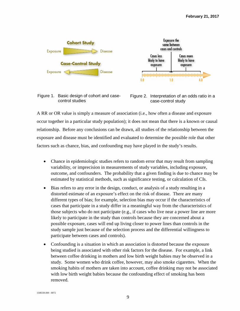
A case-control study (Figure 1) compares the characteristics of people who have been diagnosed
with a disease (i.e., cases) to a group of people who do not have the disease (i.e., controls). The
prevalence and extent of past exposure to a particular agent is estimated in both groups to assess
whether the cases have a higher exposure level than the controls, or vice versa.
February 21, 2017
1508330.000 - 8072 8
In a case-control study, an odds ratio (OR) is used to estimate the association quantitatively. An
OR is the ratio of the odds of being exposed among the cases to the odds of being exposed
among the controls. If an OR is equal to 1.0, the general interpretation is that there is no
association between the exposure and disease in the study. If the OR is greater than 1.0, there is
a positive association between the exposure and disease in the study and the inference is that the
exposure may increase the risk of the disease (Figure 2). A negative association is indicated
when the OR is less than 1.0. Epidemiologists typically quantify the precision of the estimated
measures of association by calculating confidence intervals (CI), which is the margin of error,
usually set at 95% by convention, around the point estimates. The 95% CI represents a range of
values that are expected to include the underlying effect estimate in the population 95% of the
time if samples for studies were repeatedly drawn from the underlying population. When the
95% CI for the effect estimate excludes the null value of 1.0, the result is also commonly
referred to as statistically significant.
In a cohort study, the researchers start with the identification of a pre-defined study population
(i.e., individuals who are free of the disease), determine their exposure status, then follow them
over time to see if persons with a certain exposure develop disease at a higher or lower rate
compared to unexposed persons (Figure 1). Cohort studies are evaluated statistically in a
similar manner as case-control studies, although the risk estimate is referred to as a relative risk
(RR). The RR is equal to the risk of disease in the exposed group divided by the risk of disease
in the unexposed group, with values greater than 1.0 suggesting that the exposed group has a
higher risk of disease.
February 21, 2017
1508330.000 - 8072 9
Figure 1. Basic design of cohort and case-
control studies
Figure 2. Interpretation of an odds ratio in a case-control study
A RR or OR value is simply a measure of association (i.e., how often a disease and exposure
occur together in a particular study population); it does not mean that there is a known or causal
relationship. Before any conclusions can be drawn, all studies of the relationship between the
exposure and disease must be identified and evaluated to determine the possible role that other
factors such as chance, bias, and confounding may have played in the study’s results.
• Chance in epidemiologic studies refers to random error that may result from sampling variability, or imprecision in measurements of study variables, including exposure, outcome, and confounders. The probability that a given finding is due to chance may be estimated by statistical methods, such as significance testing, or calculation of CIs.
• Bias refers to any error in the design, conduct, or analysis of a study resulting in a distorted estimate of an exposure’s effect on the risk of disease. There are many different types of bias; for example, selection bias may occur if the characteristics of cases that participate in a study differ in a meaningful way from the characteristics of those subjects who do not participate (e.g., if cases who live near a power line are more likely to participate in the study than controls because they are concerned about a possible exposure, cases will end up living closer to power lines than controls in the study sample just because of the selection process and the differential willingness to participate between cases and controls).
• Confounding is a situation in which an association is distorted because the exposure being studied is associated with other risk factors for the disease. For example, a link between coffee drinking in mothers and low birth weight babies may be observed in a study. Some women who drink coffee, however, may also smoke cigarettes. When the smoking habits of mothers are taken into account, coffee drinking may not be associated with low birth weight babies because the confounding effect of smoking has been removed.
February 21, 2017
1508330.000 - 8072 10
As part of the weight-of-evidence review process, each study’s design and methods are critically
evaluated to determine if and how chance, bias, and confounding may have affected the results,
and, as a result, the weight that should be placed on the study’s findings.
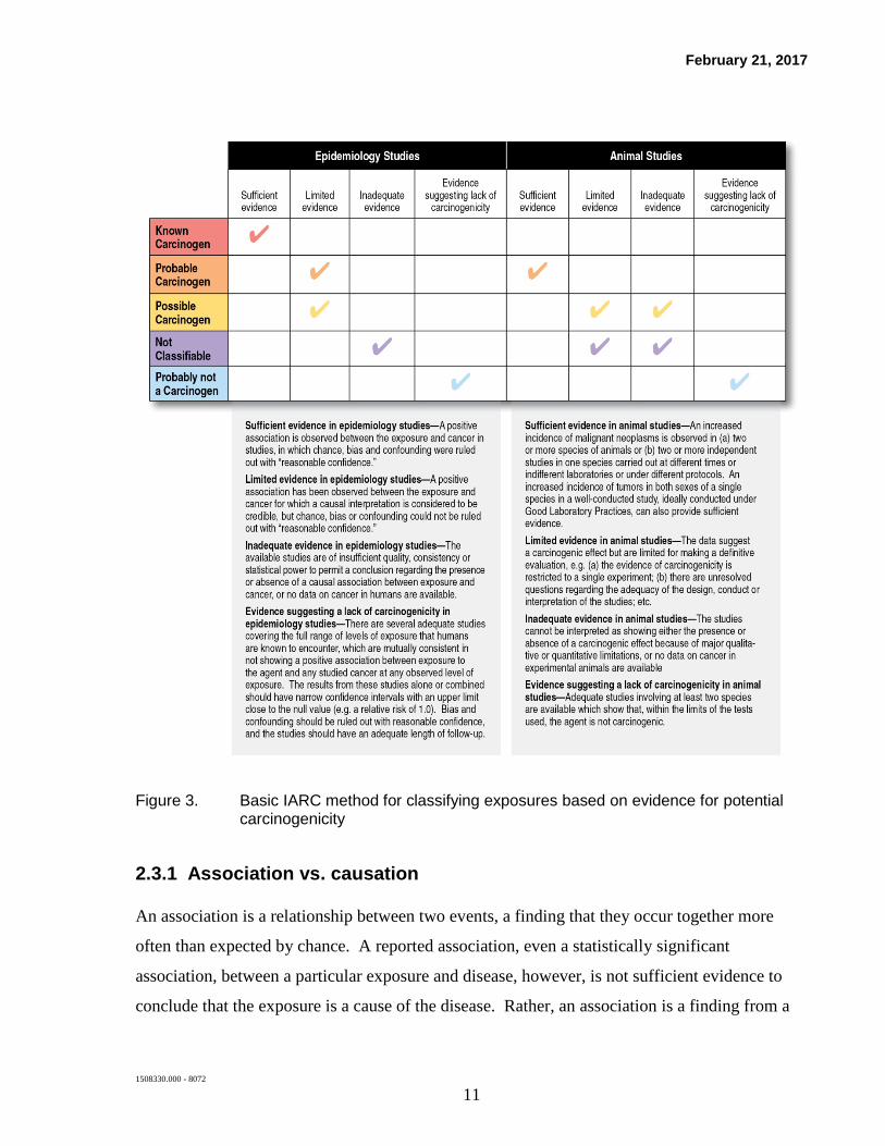
A formal procedure for classifying scientific data has been developed by the International
Agency for Research on Cancer (IARC). The IARC classifies epidemiologic and in vivo studies
as providing sufficient, limited, or inadequate evidence (Figure 3) in support of carcinogenicity,
or evidence suggesting a lack of carcinogenicity. In epidemiologic studies, the role of chance,
bias, and confounding on the observed association must be ruled out with reasonable confidence
to designate the evidence as sufficient. If the role these factors may play in the calculated
statistical association cannot be ruled out with reasonable confidence, then the data is classified
as providing limited evidence. Inadequate evidence describes a data set that lacks quality,
consistency, or power for conclusions to be drawn regarding causality. The categories on the
left in Figure 3 (e.g., known, probable, etc.) are based on the combined evaluations of
epidemiologic and in vivo studies. Other biological data relevant to the evaluation of
carcinogenicity and its mechanisms are considered, depending on the relevance to the agent
under study.
February 21, 2017
1508330.000 - 8072 11
Figure 3. Basic IARC method for classifying exposures based on evidence for potential carcinogenicity
2.3.1 Association vs. causation
An association is a relationship between two events, a finding that they occur together more
often than expected by chance. A reported association, even a statistically significant
association, between a particular exposure and disease, however, is not sufficient evidence to
conclude that the exposure is a cause of the disease. Rather, an association is a finding from a
February 21, 2017
1508330.000 - 8072 12
particular study; evaluating causation is an inferential process that combines the totality of
evidence (including epidemiologic studies that have measured associations) in a weight-of-
evidence review.
In order to support a cause-and-effect relationship, the overall data, or evidence, must present a
logically coherent and consistent picture. Various guidelines have been used to assist in the
evaluation of the plausibility of a cause-and-effect relationship between a particular exposure
and disease. These guidelines, commonly referred to as Hill’s criteria after the British
physician who outlined them (Hill, 1965), typically form the foundation of causal inference
(Rothman and Greenland, 1998). Since the publication of Hill’s criteria in 1965, numerous
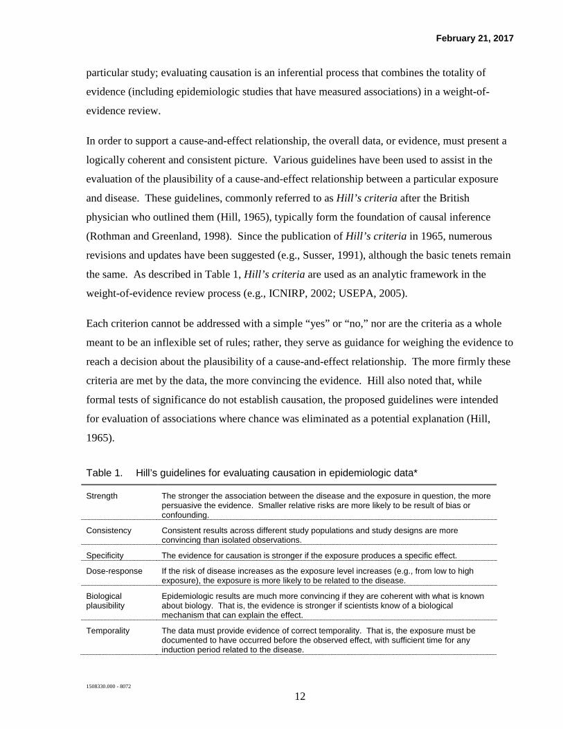
revisions and updates have been suggested (e.g., Susser, 1991), although the basic tenets remain
the same. As described in Table 1, Hill’s criteria are used as an analytic framework in the
weight-of-evidence review process (e.g., ICNIRP, 2002; USEPA, 2005).
Each criterion cannot be addressed with a simple “yes” or “no,” nor are the criteria as a whole
meant to be an inflexible set of rules; rather, they serve as guidance for weighing the evidence to
reach a decision about the plausibility of a cause-and-effect relationship. The more firmly these
criteria are met by the data, the more convincing the evidence. Hill also noted that, while
formal tests of significance do not establish causation, the proposed guidelines were intended
for evaluation of associations where chance was eliminated as a potential explanation (Hill,
1965).
Table 1. Hill’s guidelines for evaluating causation in epidemiologic data*
Strength The stronger the association between the disease and the exposure in question, the more persuasive the evidence. Smaller relative risks are more likely to be result of bias or confounding.
Consistency Consistent results across different study populations and study designs are more convincing than isolated observations.
Specificity The evidence for causation is stronger if the exposure produces a specific effect.
Dose-response If the risk of disease increases as the exposure level increases (e.g., from low to high exposure), the exposure is more likely to be related to the disease.
Biological plausibility
Epidemiologic results are much more convincing if they are coherent with what is known about biology. That is, the evidence is stronger if scientists know of a biological mechanism that can explain the effect.
Temporality The data must provide evidence of correct temporality. That is, the exposure must be documented to have occurred before the observed effect, with sufficient time for any induction period related to the disease.
February 21, 2017
1508330.000 - 8072 13
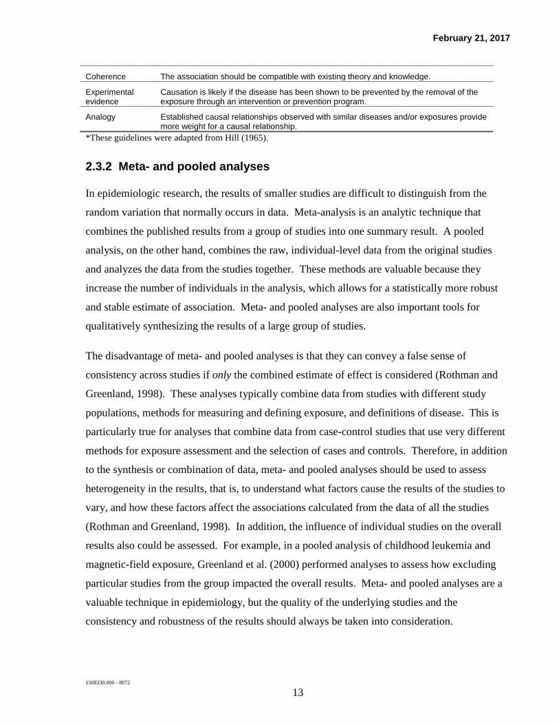
Coherence The association should be compatible with existing theory and knowledge.
Experimental evidence
Causation is likely if the disease has been shown to be prevented by the removal of the exposure through an intervention or prevention program.
Analogy Established causal relationships observed with similar diseases and/or exposures provide more weight for a causal relationship.
*These guidelines were adapted from Hill (1965).
2.3.2 Meta- and pooled analyses
In epidemiologic research, the results of smaller studies are difficult to distinguish from the
random variation that normally occurs in data. Meta-analysis is an analytic technique that
combines the published results from a group of studies into one summary result. A pooled
analysis, on the other hand, combines the raw, individual-level data from the original studies
and analyzes the data from the studies together. These methods are valuable because they
increase the number of individuals in the analysis, which allows for a statistically more robust
and stable estimate of association. Meta- and pooled analyses are also important tools for
qualitatively synthesizing the results of a large group of studies.
The disadvantage of meta- and pooled analyses is that they can convey a false sense of
consistency across studies if only the combined estimate of effect is considered (Rothman and
Greenland, 1998). These analyses typically combine data from studies with different study
populations, methods for measuring and defining exposure, and definitions of disease. This is
particularly true for analyses that combine data from case-control studies that use very different
methods for exposure assessment and the selection of cases and controls. Therefore, in addition
to the synthesis or combination of data, meta- and pooled analyses should be used to assess
heterogeneity in the results, that is, to understand what factors cause the results of the studies to
vary, and how these factors affect the associations calculated from the data of all the studies
(Rothman and Greenland, 1998). In addition, the influence of individual studies on the overall
results also could be assessed. For example, in a pooled analysis of childhood leukemia and
magnetic-field exposure, Greenland et al. (2000) performed analyses to assess how excluding
particular studies from the group impacted the overall results. Meta- and pooled analyses are a
valuable technique in epidemiology, but the quality of the underlying studies and the
consistency and robustness of the results should always be taken into consideration.
February 21, 2017
1508330.000 - 8072 14
2.3.3 Assessment of EMF exposure in epidemiologic studies
One of the most crucial aspects in the review of any epidemiologic study is an evaluation of
how exposure was measured or assessed. A good exposure metric should measure the element
that is hypothesized to cause the disease at the etiologically relevant time in the disease process.
Estimating exposure to EMF is difficult because 1) EMF is ubiquitous; 2) exposure is often
estimated retrospectively; and 3) there is currently no accepted biological mechanism for
carcinogenicity or any other disease process, so the appropriate exposure metric and timing is
unknown. In the absence of substantive knowledge about a specific mechanism by which
magnetic fields could affect normal cells, the focus on long-term exposure is based upon the
standard assumption that exposure that affects the development of cancer requires repeated
exposure at elevated levels, as does tobacco smoke, alcohol, sunlight, chemicals, and other
agents in the environment that are known to cause cancer. Investigators have used different
types of magnetic-field assessment methods, including measurements and calculations, to
estimate a person’s long-term time-weighted average (TWA) exposure. One method of
estimating a person’s TWA exposure is to sum all magnetic-field exposure encountered during
the day (e.g., while at work or school, at home, at a grocery store, shopping, etc.), weight each
estimate by the time spent in that environment, and divide that value by the total time of interest.
Historical exposure to residential magnetic fields has been estimated in epidemiologic studies
using a variety of surrogates, including:
• Classification of potential magnetic-field exposure from nearby power lines based on the number and thickness of power-line conductors and their distance to nearby residences (wire code categories);
• Simple distance from overhead or underground power lines;
• Instantaneous, spot (short-term) measurements in particular locations of a home;
• Long-term stationary measurements of magnetic fields (typically over 24- or 48-hour periods) in a room where a person spends most of his or her time, or measurements taken by a device that is carried by the person (personal monitoring); and
• Calculated magnetic-field levels based on information on loading, height, configuration, etc., of nearby transmission lines.
February 21, 2017
1508330.000 - 8072 15
In general, long-term exposure using personal magnetic-field measurements are frequently
considered as the most appropriate measures, because they estimate exposure from all magnetic-
field sources and directly estimate a person’s total exposure. Personal monitoring results,
however, are strongly influenced by behavior and the person’s environment, thus, any change in
behavior and the environment between the time of measurement and the etiologically relevant
time period may still result in exposure misclassification. Also, even long-term measurements
typically capture exposure during a 24- or 48-hour period, and may not fully represent average
exposure over months or years. Other methods typically capture exposure from one type of
source. Personal magnetic-field measurements are obtained by wearing a personal exposure
meter, which can take single readings each minute to estimate average magnetic-field exposure
over the measurement period. Since this type of measurement may be cost prohibitive in some
locations, the investigators of a study of Canadian children evaluated what proxy exposure
measures might best predict the child’s 48-hour average magnetic-field exposure (Armstrong et
al., 2001). Stationary 24-hour measurements in a child’s bedroom were a good predictor of 48-
hour personal exposure, and spot measurements around the perimeter of the child’s home were a
moderately good predictor. Wire code categories, on the other hand, were not found to be an
accurate predictor of a child’s exposure (Armstrong et al., 2001).
It is important to note that estimates of magnetic-field exposure in epidemiologic studies
represent estimates of long-term exposure potentially from all sources over months or years, and
should not be compared to the magnetic-field values measured on a single occasion, and at a
single, fixed location. It is evident that brief encounters with higher magnetic fields (for
example, walking under a distribution or transmission line, at home in front of a refrigerator or
television, or at a grocery store near the freezer) would not significantly alter the long-term
exposure of a person to magnetic fields, as reflected in their TWA exposure, because they
typically spend a very small fraction of their time at these locations.
Much of the research on EMF is related to occupational exposures, given the higher range of
exposure levels encountered in the occupational environment. The main limitation of these
studies, however, has been the methods used to assess exposure, with early studies relying
simply on a person’s occupational title (often taken from a death certificate) and later studies
linking a person’s full or partial occupational history to representative average exposures for
February 21, 2017
1508330.000 - 8072 16
each occupation (i.e., a job exposure matrix). The latter method, while it represents
advancement over earlier methods, still has some important limitations, as highlighted in a
review by Kheifets et al. (2009) summarizing an expert panel’s findings.8 While a person’s
occupation may provide some indication of the overall magnitude of their occupational
magnetic-field exposure, it does not take into account the possible variation in exposure due to
different job tasks within occupational titles, the frequency and intensity of contact to relevant
exposure sources, or variation by calendar time. A study of the 48-hour exposure of 543
workers in Italy found that job exposure matrices were a poor indicator of actual occupational,
magnetic-field exposure levels (Gobba et al. 2011). A study by Mee and colleagues (2009) also
confirmed that job exposure matrices could be improved by linking occupational classifications
with industry or information on participation in certain tasks of interest (e.g., use of welding
equipment or work near power lines) based on their measurements of personal occupational
magnetic-field exposures in the United Kingdom.
2.4 Evaluation of experimental research
2.4.1 General research methods
Experimental studies of humans, animals, and cells and tissues complement epidemiologic
studies. Both epidemiologic and experimental approaches are needed because, although people
are the species of interest, they have large variations in their genetic makeup, exposures, dietary
intake, and health-related behaviors that may affect health outcomes. In laboratory animals,
these variables can be well controlled to provide more precise information regarding the effects
of an exposure. In epidemiologic studies, it is difficult to control for these variables because
scientists are merely observing individuals going about their ordinary lives. Taken together,
epidemiology, in vivo, and in vitro studies provide a more complete picture of a possible disease
etiology than any one of these study types alone.
A wide variety of approaches is available for assessing the possible adverse effects associated
with exposures in experimental studies. The two general types of experimental studies are in
8 Kheifets et al. (2009) reports on the conclusions of an independent panel organized by the Energy Networks
Association in the United Kingdom in 2006 to review the current status of the science on occupational EMF exposure and identify the highest priority research needs.

February 21, 2017
1508330.000 - 8072 17
vivo and in vitro studies. In vivo studies include studies that examine the potential effects of
exposures on human volunteers (usually short-term studies examining short-term effects) and
studies of whole animals that could also examine long-term effects. In vitro studies are
designed to evaluate the way that the exposure may interact with cells and tissues outside of the
body, which may provide information on mechanism of action.
In vivo studies
Studies in which laboratory animals receive high exposures in a controlled environment provide
an important basis for evaluating the safety of environmental, occupational, and drug exposures.
These approaches are widely used by health agencies to assess risks to humans from medicines,
chemicals, and physical agents (Health Canada, 2000; WHO, 2010; IARC, 2002 preamble;
USEPA, 2002; USEPA, 2005). From a public health perspective, long-term (chronic) studies in
which animals undergo exposure over most of their lifetime, or during their entire pregnancy,
are of high importance in assessing potential risks of cancer and other adverse effects. In these
long-term studies, researchers examine a large number of parameters and anatomical sites to
assess changes and adverse effects in body organs, cells, and tissues.
These data are used in the hazard identification step of the risk assessment process to determine
whether an environmental exposure is likely to produce cancer or damage organs and tissues.
Health Canada mandates that lifetime in vivo studies or in vivo studies of exposures during
critical sensitive periods are conducted to assess potential toxicity to humans (Health Canada,
1994). Furthermore, the U.S. Environmental Protection Agency’s position is that, “…the
absence of tumors in well-conducted, long-term animal studies in at least two species provides
reasonable assurance that an agent may not be a carcinogenic concern for humans” (USEPA,
2005, pp. 2-22).
In vitro studies
In vitro studies are used to investigate the mechanisms for effects that are observed in living
organisms. The relative value of in vitro tests to human health risk assessment is less than that
of in vivo and epidemiologic studies because responses of cells and tissues outside the body may
not reflect the response of those same cells if maintained in an intact living system, so their
February 21, 2017
1508330.000 - 8072 18
relevance cannot be assumed (IARC, 1992). It may be difficult to extrapolate from simple
cellular systems to complex, higher organisms to predict risks to health because the mechanism
underlying effects observed in vitro may not correspond to the mechanism underlying complex
processes like carcinogenesis. In addition, the results of in vitro studies cannot be interpreted in
terms of potential human health risks unless they are performed in a well-studied and validated
test system. For these reasons, the IARC and other agencies treat data from in vitro studies as
supplementary to data obtained from epidemiologic and in vivo studies.
Convincing evidence for a mechanism that explains an effect observed in experimental or
epidemiologic studies can add weight to the assessment of cause and effect, and in some cases
may clarify reasons for different results among species, or between animals and humans. In
vitro studies, however, are not used directly by any health agency to assess risks to human
health. Therefore, this report focuses on epidemiologic studies and also discusses in vivo
experimental research with relevance to carcinogenesis and relies on the conclusions of
scientific panels with regard to in vitro data.
2.4.2 Experimental methods for cancer research
Cancer research in the laboratory includes studies of various stages of cancer development.
Research has established that cells may take several steps to change from ordinary cells to the
uncontrolled growth typical of cancer. Cancer usually begins with a mutation, that is, an
irreversible change in the genetic material of the cell, a process also called cancer initiation or
cancer induction. Additional steps (also called cancer promotion), must also occur for a
cancerous cell to develop into a tumor. A carcinogenic agent may affect either or both the
initiation and promotion phases of cancer development. Exposures that affect both initiation
and promotion are sometimes called complete carcinogens.
In vitro assays isolate specific cells or microorganisms in glassware in the laboratory to assess
the likelihood that exposure to the agent can cause mutations, a step considered necessary in the
initiation of cancer. Initiation tests have also been developed in animals, in which scientists
expose them for less than lifetime periods to determine whether an exposure caused changes
typical for early stage cancers in specific tissues such as liver, breast, or skin.
February 21, 2017
1508330.000 - 8072 19
Other tests are designed to ascertain whether a specific exposure can stimulate tumor growth
(i.e., promotion) in an animal in which the cellular changes typical of initiation have already
occurred. Studies of promotion typically include two steps: first, exposing the experimental
animals to a chemical known to initiate cancer, and second, exposing the animals to the agent to
be tested as a promoter. The occurrence of cancer in animals exposed to an initiator and the
potential promoter is compared to the occurrence of cancer that develops in animals exposed
only to the initiator.
The failure of early EMF research to produce mutations in the DNA of cells in vitro was a factor
in directing scientists to focus on studies of promotion.
2.4.3 Experimental methods for developmental toxicity
Studies in animals also are used to assess whether an exposure can pose a risk to the unborn
children of pregnant women. Experimental studies in pregnant animals provide a means for
isolating the exposure in question from the myriad of other factors that can affect prenatal
development. The results of these well-controlled in vivo studies are used by regulatory
agencies to assess prenatal risk and help set human exposure limits (NTP, 2015; USEPA, 1991,
1998).
To test the potential for an exposure to affect fetal development, pregnant mammals such as
mice, rats, or rabbits are exposed from the time the embryo is implanted in the uterus to the day
before delivery. Variations in study design include preconception exposure of the female in
addition to exposure during gestation, and even further exposure after the animal is born.
Protocols generally specify that doses be set below the levels known to cause maternal toxicity,
that unexposed controls are maintained at the same time period, and that the animals’ health is
monitored throughout the study. Endpoints measured include maternal body weight and weight
change, the numbers and percent of live offspring, fetal body weight, the sex ratio, and external,
soft tissue, or skeletal variations and malformations. The uterus can also be examined to assess
the number of implantations and fetuses that have been lost, as an indication of miscarriage
(USEPA, 1998).
February 21, 2017
1508330.000 - 8072 20
2.4.4 Evaluating the cumulative body of experimental evidence
Key factors in evaluating individual experimental studies for a weight-of-evidence review
include the details of the protocol; the plan for selecting animals and conducting and analyzing
the study; the adequacy of the dose levels selected; the way in which the study was actually
conducted, including adherence to good laboratory practices in animal housing and monitoring;
and the evaluation of the effects on toxicity, tumors, or malformations, considering both
biological and statistical issues (USEPA, 2005).
As an example of a protocol, consider the long-term in vivo study, a major tool for determining
whether a chemical can produce cancer in humans. Standard protocols usually specify at least
50 animals of each sex per dose level, in each of three different dose groups. One of these is a
high-level dose group termed the maximum tolerated dose, which is close to, but below, the
level that increases mortality or produces significant morbidity. Additional dose levels are used
below this maximum. An unexposed group, or control, is maintained under the same conditions
during the same time period for comparison. This study design permits a separate evaluation of
the incidence rate for each tumor type in the exposed group compared to the unexposed control
group. Statistical methods are used to assess the role of chance in any differences in the rates
between exposed and unexposed, or among the dose groups. If effects are observed in a study,
other studies are conducted because similarity of results in different studies, laboratories, and
species strengthens the evidence.
Specific methods are used to reduce subjectivity and avoid systematic error, or bias, in scientific
experiments (NRC, 1997). These are summarized in Table 2, including the random assignment
of subjects to control or comparison groups, the unbiased collection of information (e.g.,
researchers are not aware of, or are “blind” to the exposure), and the need for replication of
results. As with Hill’s criteria, each guideline for evaluating causation in experimental studies
is not met with a simple “yes” or “no,” rather, they serve as guidance for weighing the evidence
to reach a decision about cause-and-effect. The more firmly these criteria are met by the
studies, the more convincing the evidence.

February 21, 2017
1508330.000 - 8072 21
Table 2. Criteria for evaluating experimental studies as applied to EMF exposures*
Avoiding unwanted effects
Experimental techniques should be chosen to avoid effects of intervening factors such as microshocks, noise, corona discharges, vibrations and chemicals.
Exposure classification
Extreme care should be taken to determine the effective EMF field, voltage, or current in the organism.
Sensitivity The sensitivity of the experiments should be adequate to ensure a reasonable probability that an effect would be detected if it existed.
Objectivity The experimental and observational techniques, methods and conditions should be objective. “Blind” scoring (where the investigator making the observations is unaware of the experimental variable being tested) should be used whenever there is a possibility of investigator bias. “Double-blind” protocols (where neither the investigator making the observations nor the experimental subject are aware of the experimental variable being tested) should be used in studies of people when the experimental subjects’ perceptions may be unwittingly influenced.
Statistical significance
If an effect is claimed, the result should be demonstrated at a level where chance is an unlikely explanation.
Consistency The results of a given experiment should be internally consistent among different ways of analyzing the data, and consistent across studies with respect to the effects of interest.
Quantifiable results The results should be quantifiable and replicable. In the absence of independent confirmation, a result should not be viewed as definitive.
Appropriateness of methodologies
The biological and engineering methodologies should be sound and appropriate for the experiment.
*These criteria were adapted from NRC (1997).
February 21, 2017
1508330.000 - 8072 22
3 Conclusions of weight-of-evidence reviews of EMF and health
Scientists, scientific organizations, and regulatory agencies worldwide use the weight-of-
evidence approach to assess potential health risks associated with exposures. These expert
groups typically include many scientists with diverse skills and background that reflect the
different research approaches required to answer questions about health. Using a weight-of-
evidence approach as an analytic framework, each group provides its scientific consensus based
on a review of the evidence.
3.1 Weight of evidence reviews by national and international scientific agencies
The following scientific organizations have assembled multidisciplinary panels of scientists to
conduct weight-of-evidence reviews and arrive at conclusions about the possible risks
associated with ELF EMF (in ascending, chronological order of their most recent publication):9
• The National Institute for Environmental Health Sciences assembled a 30-person Working Group to review the cumulative body of epidemiologic and experimental data and provide conclusions and recommendations to the US government (NIEHS, 1998, 1999).
• The IARC completed a full carcinogenic evaluation of EMF in 2002.
• The Federal-Provincial-Territorial Radiation Protection Committee (FPTRPC), an intergovernmental, Canadian committee assembled to harmonize the standards and practices for radiation protection within federal, provincial, and territorial jurisdictions, conducted a review in 1998 and an update in 2005 (FPTRPC, 1998; FPTRPC, 2005). The FPTRPC most
9 We are aware of other published summaries of the EMF research. With an increase in transmission
infrastructure development and the advent of the Internet, the release of reviews and summaries now occurs regularly. This update is restricted to summaries that used a weight-of-evidence approach, and for which a multidisciplinary scientific panel reviewed the epidemiologic and experimental evidence (either in its entirety or since the organization’s previous report), and offered conclusions about causality. Other reviews and summaries that did not follow this approach are not addressed because they do not assist in making science-based risk assessments and conclusions. Specifically, the BioInitiative (BI) Group’s report that was posted on the internet is not included in our report, because, among other shortcomings, the BI report is not a comprehensive review of the literature and is not based on the scientific weight-of-evidence method.
February 21, 2017
1508330.000 - 8072 23
recently released a statement from their Working Group in November 2008 summarizing their opinion on exposure to EMF (FPTRPC, 2008).10
• The National Radiological Protection Board11 of the United Kingdom issued full evaluations of the research in 1992, 2001, and 2004, with supplemental updates and topic-specific reports published in the interim and subsequent to their last full evaluation in 2004 (NRPB, 1992, 1994a, 1994b, 2001a, 2001b, 2004; HPA, 2006). In a letter addressing a related topic in 2009, the Director of the HPA reiterated their position with regard to ELF EMF and appropriate precautionary measures (HMG, 2009).
• The WHO released a review in June 2007 as part of its International EMF Program to assess the scientific evidence of possible health effects of EMF in the frequency range from 0 to 300 Gigahertz.
• The Health Council of the Netherlands, using other major scientific reviews as a starting point, evaluated recent studies in several periodic reports (HCN, 2001; HCN, 2004; HCN, 2005; HCN, 2007; HCN, 2009a). The HCN also released an advisory letter that addressed the topic of power lines and Alzheimer’s disease (HCN, 2009b).
• The European Commission funded the European Health Risk Assessment Network on Electromagnetic Fields Exposure (EFHRAN), a network of experts convened to perform health risk assessments and provide scientifically-based recommendations to the Commission. EFHRAN consulted other major reviews and evaluated epidemiologic and experimental research published after August 2008 to provide an updated health assessment (EFHRAN, 2010, 2012).
• The International Commission on Non-Ionizing Radiation Protection (ICNIRP), the formally recognized organization for providing guidance on standards for non-ionizing radiation exposure for the WHO, published a review of the cumulative body of epidemiologic and experimental data on ELF-EMF in 2003. The ICNIRP released exposure guidelines in 2010 that updated their 1998 exposure guidelines. For both guidelines, they relied heavily on previous reviews of the literature related to long-term exposure,
10 Health Canada refers to the FTPRPC as the authority on issues related to EMF. The FPTRPC established an
ELF Working Group to carry out periodic reviews, recommend appropriate actions, and provide position statements that reflect the common opinion of intergovernmental authorities.
11 The National Radiological Protection Board merged with the Health Protection Agency in April 2005 to form its Radiation Protection Division, and in April 2013, the Health Protection Agency became part of Public Health England.
February 21, 2017
1508330.000 - 8072 24
but provided some relevant conclusions as part of their update process (ICNIRP, 1998, 2010).
• The European Union’s Scientific Committee on Emerging and Newly Identified Health Risks (SCENIHR) issued its most recent report to the Health Directorate of the European Commission in 2015 that updated previous conclusions (SSC, 1998; CSTEE, 2001; SCENIHR, 2007, 2009, 2015).
• The Swedish Radiation Protection Authority, using other major scientific reviews as a starting point, evaluated current studies in several annual reports published in 2007 and 2008 (SSI, 2007, 2008). The Swedish Radiation Safety Authority, which superseded the Swedish Radiation Protection Authority in 2008, and has “national collective responsibility within the areas of radiation protection and nuclear safety” including EMF research, continue to publish annual reports (SSM, 2010, 2013, 2014, 2015, 2016).
The most comprehensive assessment of EMF was conducted by the WHO and published in June
2007; their report updated a previous evaluation of ELF EMF by the IARC in 2002. Exponent’s
2007 report focused on the conclusions of WHO (2007) and provided an update by reviewing
literature published from December 2005 (the approximate cut-off date for WHO) through
September 2007. Exponent’s 2010 report reviewed research through January 2010, and the
2012 report reviewed the research through March 1, 2012. This report will again focus on
describing and updating the conclusions of the WHO (2007) report, while noting the other
scientific organizations that have published their reviews in the interim.
Overall, the published conclusions of scientific review panels have been consistent. None of the
panels concluded that either electric fields or magnetic fields are a known or likely cause of any
adverse health effect at the long-term, low exposure levels found in the environment. The only
known effects of exposure to EMF are acute or short-term effects (such as nerve and muscle
stimulation). Existing guidelines from ICNIRP are set to limit short-term exposure at levels
much higher than those encountered in public locations, including publicly-accessible areas near
electrical facilities.
Most of the uncertainty and controversy surrounding magnetic-field exposure is related to the
research on childhood leukemia. Some epidemiologic studies reported that children with
leukemia were more likely to live closer to power lines, or have higher estimates of magnetic-
February 21, 2017
1508330.000 - 8072 25
field exposure, compared to children without leukemia; other epidemiologic studies did not
report this statistical association. When a number of relevant studies were combined in a single
analysis, no association was evident at lower exposure levels, but a weak association was
reported between childhood leukemia and estimates of average magnetic-field exposure greater
than 3-4 mG (Ahlbom et al., 2000; Greenland et al., 2000). These pooled analyses provide
some evidence for an association between magnetic fields and childhood leukemia; however,
because of the inherent uncertainty associated with observational epidemiologic studies, the
results of these pooled analyses were considered to provide only limited epidemiologic support
for a causal relationship; chance, bias and confounding could not be ruled out with reasonable
confidence. Further, in vivo studies have not found that magnetic fields induce or promote
cancer in animals exposed for their entire lifespan under highly-controlled conditions, nor have
in vitro studies found a cellular mechanism by which magnetic fields could induce
carcinogenesis.
Considering all the evidence together, the WHO, as well as other scientific panels, classified
magnetic fields as a possible cause of childhood leukemia (NRPB, 2001a; IARC, 2002;
ICNIRP, 2003; HCN, 2004; WHO, 2007). The term possible denotes an exposure for which
epidemiologic evidence points to a statistical association, but other explanations cannot be ruled
out as the cause of that statistical association (e.g., bias and confounding) and experimental
evidence does not support a cause-and-effect relationship (Figure 3).
While much additional research has been published since the WHO evaluation, the main
conclusions of scientific organizations remained consistent—the scientific evidence does not
establish that exposure to low level ELF EMF is the cause of any cancer (including childhood
leukemia) or non-cancer adverse health effects (WHO, 2007; HPA, 2009; EFHRAN, 2012;
ICNIRP, 2010; SCENIHR, 2015; SSM, 2016).
The WHO and more recent reviews, however, continue to recommend further research to
reconcile results from epidemiologic studies on childhood leukemia and the lack of evidence
from experimental studies through innovative research. Researchers believe that the
development of childhood leukemia, like any other cancer, is influenced by a multitude of
February 21, 2017
1508330.000 - 8072 26
different factors, such as genetics, environmental exposures, and infectious agents (see e.g.,
Buffler et al., 2005; McNally and Parker, 2006).
Although some questions remain, the epidemiologic evidence does not support a cause-and-
effect relationship between magnetic fields and adult leukemia/lymphoma or brain cancer, with
the data being described as inadequate or weak (WHO, 2007; EFHRAN, 2012; SCENIHR,
2015; SSM, 2016). Scientific organizations have concluded that there is strong evidence in
support of no relationship between magnetic fields and breast cancer or cardiovascular disease
(WHO, 2007; SSI, 2008; ICNIRP, 2010; EFHRAN, 2012; SSM, 2016). Although two
epidemiologic studies reported a statistical association between peak magnetic-field exposure
and miscarriage, a serious bias in how these studies were conducted was identified and various
scientific panels concluded that these biases preclude making any conclusions about
associations between magnetic-field exposure and miscarriage (HCN, 2004; NRPB, 2004;
WHO, 2007; ICNIRP, 2010; SCENIHR, 2016). While an association between some
neurodegenerative diseases (i.e., Alzheimer’s disease and amyotrophic lateral sclerosis [ALS])
and estimates of higher average occupational magnetic-field exposure has been reported in
earlier studies, more recent studies showed mixed results; scientific panels have described this
research as weak and inadequate and recommended additional research in this area (WHO,
2007; HCN, 2009b; ICNIRP, 2010; EFHRAN, 2012; SCENIHR, 2015; SSM, 2016).
In summary, reviews published by scientific organizations using weight-of-evidence methods
have concluded that the cumulative body of research to date does not support the hypothesis
that electric or magnetic fields cause any long-term adverse health effects at the levels we
encounter in our everyday environments.
The Working Group of the FPTRPC concluded the following with respect to ELF EMF and
health in a statement released in 2008:
In summary, it is the opinion of the Federal-Provincial-Territorial Radiation Protection Committee that there is insufficient scientific evidence showing exposure to EMFs from power lines can cause adverse health effects such as cancer.
The FPTRPC conclusion is consistent with statements by Health Canada on its website:
February 21, 2017
1508330.000 - 8072 27
There is no conclusive evidence of any harm caused by exposures at levels found in Canadian homes and schools, including those located just outside the boundaries of power line corridors.12
3.2 Standards and guidelines for limiting exposure to EMF
3.2.1 Status of EMF guidelines
Two international scientific organizations, ICNIRP and the International Committee for
Electromagnetic Safety (ICES), have published guidelines for limiting public exposure to EMF
(ICES, 2002; ICNIRP, 2010). The health outcomes examined in most EMF epidemiologic and
in vivo studies primarily have addressed magnetic fields, mainly because structures and
vegetation provide some shielding that limits residential exposure to electric fields from power
lines; however, these EMF guidelines recommend limits for both electric and magnetic fields.
These guideline limits are set to prevent known and established effects after consideration of the
scientific evidence regarding potential effects of both long-term and short-term exposures.
Because the only established effects are the short-term direct, acute health effects (i.e.,
perception, annoyance, and the stimulation of nerves and muscles) that can occur at high levels
of exposure, the guidelines are set to protect against these acute effects. With respect to long-
term effects, the ICNIRP review concluded the following:
It is the view of ICNIRP that the currently existing scientific evidence that prolonged exposure to low frequency magnetic fields is causally related with an increased risk of childhood leukemia is too weak to form the basis for exposure guidelines. In particular, if the relationship is not causal, then no benefit to health will accrue from reducing exposure (ICNIRP, 2010; p. 824).
Although ICNIRP and ICES have the same objectives13 and used similar methods, the
recommended limits for exposure of the general public to EMF at the frequencies used to
transmit electricity differ, as seen in Table 3.
12 https://www.canada.ca/en/health-canada/services/home-garden-safety/electric-magnetic-fields-power-lines-
electrical-appliances.html; website update on July 6, 2016; accessed on January 24, 2017.
February 21, 2017
1508330.000 - 8072 28
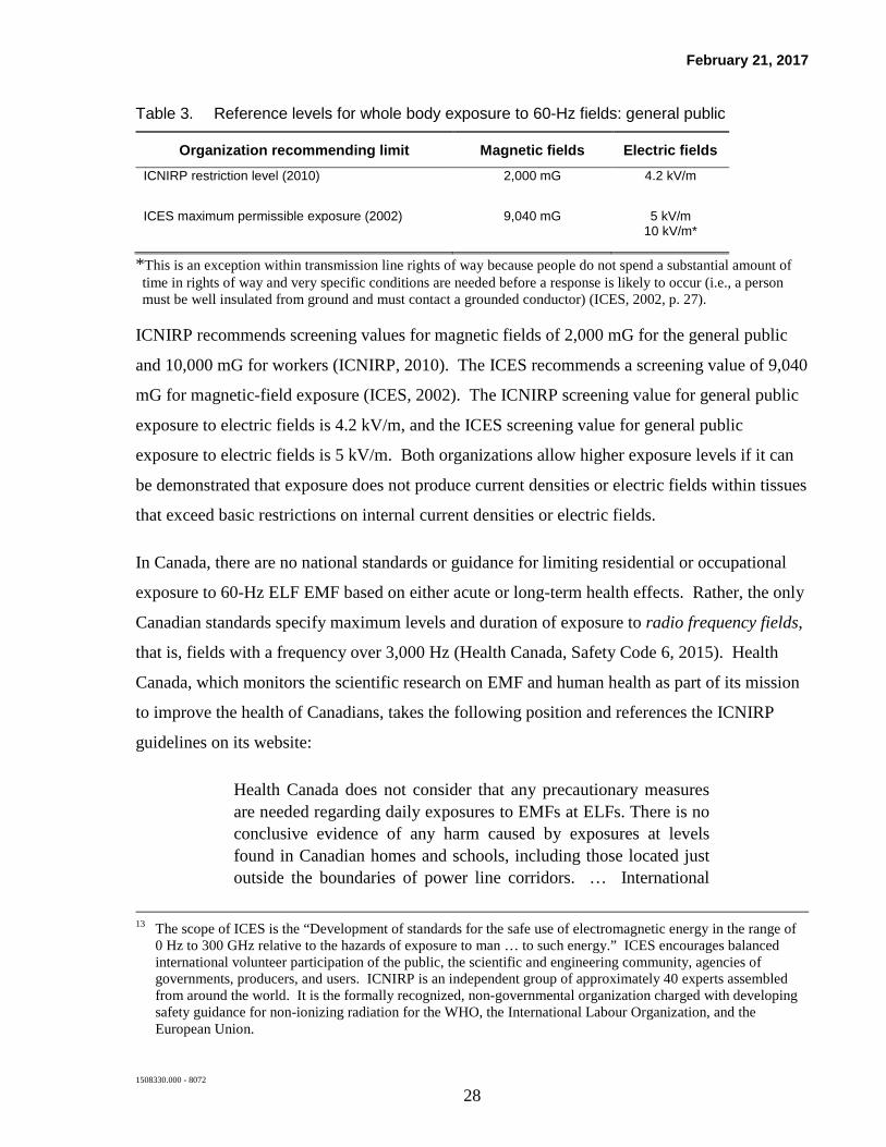
Table 3. Reference levels for whole body exposure to 60-Hz fields: general public
Organization recommending limit Magnetic fields Electric fields ICNIRP restriction level (2010) 2,000 mG 4.2 kV/m
ICES maximum permissible exposure (2002) 9,040 mG 5 kV/m 10 kV/m*
*This is an exception within transmission line rights of way because people do not spend a substantial amount of time in rights of way and very specific conditions are needed before a response is likely to occur (i.e., a person must be well insulated from ground and must contact a grounded conductor) (ICES, 2002, p. 27).
ICNIRP recommends screening values for magnetic fields of 2,000 mG for the general public
and 10,000 mG for workers (ICNIRP, 2010). The ICES recommends a screening value of 9,040
mG for magnetic-field exposure (ICES, 2002). The ICNIRP screening value for general public
exposure to electric fields is 4.2 kV/m, and the ICES screening value for general public
exposure to electric fields is 5 kV/m. Both organizations allow higher exposure levels if it can
be demonstrated that exposure does not produce current densities or electric fields within tissues
that exceed basic restrictions on internal current densities or electric fields.
In Canada, there are no national standards or guidance for limiting residential or occupational
exposure to 60-Hz ELF EMF based on either acute or long-term health effects. Rather, the only
Canadian standards specify maximum levels and duration of exposure to radio frequency fields,
that is, fields with a frequency over 3,000 Hz (Health Canada, Safety Code 6, 2015). Health
Canada, which monitors the scientific research on EMF and human health as part of its mission
to improve the health of Canadians, takes the following position and references the ICNIRP
guidelines on its website:
Health Canada does not consider that any precautionary measures are needed regarding daily exposures to EMFs at ELFs. There is no conclusive evidence of any harm caused by exposures at levels found in Canadian homes and schools, including those located just outside the boundaries of power line corridors. … International
13 The scope of ICES is the “Development of standards for the safe use of electromagnetic energy in the range of
0 Hz to 300 GHz relative to the hazards of exposure to man … to such energy.” ICES encourages balanced international volunteer participation of the public, the scientific and engineering community, agencies of governments, producers, and users. ICNIRP is an independent group of approximately 40 experts assembled from around the world. It is the formally recognized, non-governmental organization charged with developing safety guidance for non-ionizing radiation for the WHO, the International Labour Organization, and the European Union.
February 21, 2017
1508330.000 - 8072 29
exposure guidelines for exposure to EMFs at ELFs have been established by the International Commission on Non-Ionizing Radiation Protection (ICNIRP). These guidelines are not based on a consideration of risks related to cancer. Rather, the point of the guidelines is to make sure that exposures to EMFs do not cause electric currents or fields in the body that are stronger than the ones produced naturally by the brain, nerves and heart. EMF exposures in Canadian homes, schools and offices are far below these guidelines.14
The sections below discuss the similarities and differences between the ICNIRP and ICES
standards, and the public health implications of the differences.
3.2.2 Comparison of ICES and ICNIRP guidelines
In both the ICES and ICNIRP standard setting process, a group of scientists conducted extensive
reviews of the scientific research regarding health effects. The scientists reviewed the
epidemiologic and experimental evidence and concluded that the evidence was insufficient to
warrant the development of standards on the basis of hypothesized long-term health effects,
such as cancers. Each organization reached a consensus that the most sensitive endpoints—the
substantiated adverse effects that would occur at the lowest level of exposure—are short-term
reactions to electrostimulation of nerves and muscle. These are direct, acute reactions to high
levels of exposure, not severe or life-threatening events.
Each organization developed its recommended exposure limit in two steps. The first step was to
identify the lowest level of electrical forces inside the body that is likely to produce the
stimulation of nerves and muscle. This internal level, or dose, is further lowered by safety
factors to develop what is referred to as the basic restriction. As the term indicates, the basic
restriction is the limit for internal dose recommended for exposed populations. This internal
dose limit is the foundation of both the ICNIRP and ICES standards because both electric fields
and magnetic fields can induce electrical forces in the body.
The ICNIRP and ICES basic restrictions are set well below the value at which an adverse effect
was observed in experiments. This is because they incorporate dose reduction factors, also 14 https://www.canada.ca/en/health-canada/services/home-garden-safety/electric-magnetic-fields-power-lines-
electrical-appliances.html; website updated on July 6, 2016; accessed on January 24, 2017.
February 21, 2017
1508330.000 - 8072 30
known as safety factors, to account for potential sources of uncertainty. For example, both
groups consider the potentially higher sensitivity in vulnerable groups as a reason for using a
safety factor.
The second step in the standard setting process involves developing the reference level. A
reference level is developed because a basic restriction cannot be directly measured. The
reference level is the measurable level of electric fields and magnetic fields at the location of
interest; these levels are outside of the body, and are used as a screening value to maintain the
internal level identified as the basic restriction. These reference levels represent conservative
limits, meaning that if the reference level (i.e., the screening level) is exceeded, it does not
necessarily follow that the basic restriction is exceeded. As ICNIRP explains, “In many
practical exposure situations external power frequency electric fields at the reference levels will
induce current densities in central nervous tissues that are well below the basic restrictions.
Recent dosimetry calculations indicate that the reference levels for power-frequency magnetic
fields are conservative guidelines relative to meeting the basic restrictions on current density for
both public and occupational exposures” (ICNIRP, 1998).
3.2.3 Implications for human health
The underlying question for people who make decisions about public health and safety is
whether the ICNIRP reference value (4.2 kV/m) implies greater safety simply because it is
lower and includes a larger safety factor. In developing public health standards, safety factors
are used when uncertainty is recognized, and the general rule is that smaller safety factors are
needed as the relevant information on risk to humans is improved. Although ICNIRP uses a
larger safety factor, it applies that safety factor to a higher estimated threshold level. ICES uses
a smaller safety factor, but has used highly specific data on human responses, leading to a lower,
presumably more precise, estimated threshold level. It is essential to understand that for effects
where thresholds are identified, the goal of the standard setting process is to set the exposure
limit where no effects will occur in the population. Therefore, further lowering of the exposure
limit is not expected to have any additional health benefit. For additional perspective on the
question of the safety of exceeding ICNRIP exposure limits up to the level of the ICES limits,
February 21, 2017
1508330.000 - 8072 31
consider that ICNIRP states that EMF guidelines are conservative, and that the ICNIRP
recommended limit for occupational exposure is 8.3 kV/m (ICNIRP, 1998, 2010).
3.3 Precautionary approaches
3.3.1 General definition
A precautionary policy for risk management of possible, but unproven, adverse effects emerged
in Europe in the 1970s regarding environmental issues. The precautionary principle refers to the
idea that, when evidence does not support the suggestion that an exposure is a cause of a
particular disease but where a risk is perceived or uncertainty exists, precautionary measures
may be taken that are proportional to the perceived level of risk, with science as the basis for
estimating that risk. A key element of precautionary approaches is the recognition that a real
risk from the exposure may not exist, and its necessary corollary is that the reduction of
exposure may not decrease any adverse effects in the population.
The European Commission prepared a report in 2000 to clarify the precautionary principle
because this idea had been subject to controversy and variability in interpretation.15 Their report
explained that the implementation of the precautionary principle should be science based,
starting with a complete scientific evaluation, and the range of actions taken should depend on
the extent of the risk and the degree of uncertainty surrounding the occurrence of adverse
effects. They provided guidelines for the application of the precautionary principle or other risk
management measures specifying five general principles: proportionality, non-discrimination,
consistency, examination of costs and benefits of actions, and examination of scientific
developments.16
15 Commission of the European Communities, Communication on the Precautionary Principle, Brussels 03
February 2000 (http://europa.eu.int/comm./off/com/health_consumer/precaution.htm). 16 Proportionality: "Measures...must not be disproportionate to the desired level of protection and must not aim at
zero risk." Nondiscrimination: "comparable situations should not be treated differently and... different situations should
not be treated in the same way, unless there are objective grounds for doing so." Consistency: "measures...should be comparable in nature and scope with measures already taken in equivalent
areas in which all the scientific data are available."
February 21, 2017
1508330.000 - 8072 32
A variant of the precautionary principle called prudent avoidance has been favored as a policy
option for EMF by some national and local governments. The WHO describes this as “using
simple, easily achievable, low to modest (prudent) cost measures to reduce individual or public
EMF exposure, even in the absence of certainty that the measure would reduce risk” (WHO,
2002).
3.3.2 WHO recommendations regarding precautionary measures for EMF
The scientific evaluation completed by the WHO also discusses general policy strategies for
risk management, and provides a summary table of different policy strategies employed
worldwide specifically for EMF exposure in the general public (WHO, 2007, Chapter 13). The
WHO recommended the following precautionary measures (WHO, 2007, adapted from pp. 372-
373):
• Countries are encouraged to adopt international science-based guidelines.
• Provided that the health, social, and economic benefits of electric power are not compromised, implementing very low-cost precautionary procedures to reduce exposures is reasonable and warranted.
• Policy-makers and community planners should implement very low-cost measures when constructing new facilities and designing new equipment including appliances.
• Changes to engineering practice to reduce ELF exposure from equipment or devices should be considered, provided that they yield other additional benefits, such as greater safety or involve little or no cost.
• When changes to existing ELF sources are contemplated, ELF field reductions should be considered alongside safety, reliability, and economic aspects.
Examination of the benefits and costs of action or lack of action: "This examination should include an
economic cost/benefit analysis when this is appropriate and feasible. However, other analysis methods...may also be relevant."
Examination of scientific developments: "The measures must be of a provisional nature pending the availability of more reliable scientific data"... "Scientific research shall be continued with a view to obtaining more complete data."
February 21, 2017
1508330.000 - 8072 33
• Local authorities should enforce wiring regulations to reduce unintentional ground currents when building new or rewiring existing facilities, while maintaining safety. Proactive measures to identify violations or existing problems in wiring would be expensive and unlikely to be justified.
• National authorities should implement an effective and open communication strategy to enable informed decision-making by all stakeholders; this should include information on how individuals can reduce their own exposure.
• Local authorities should improve planning of ELF EMF-emitting facilities, including better consultation between industry, local government, and citizens when siting major ELF EMF-emitting sources.
• Government and industry should promote research programs to reduce the uncertainty of the scientific evidence on the health effects of ELF field exposure.
In summary, the general recommendation of the WHO is as follows:
Countries are encouraged to adopt international science-based guidelines. In the case of EMF, the international harmonization of standard setting is a goal that countries should aim for (WHO, 2006). If precautionary measures are considered to complement the standards, they should be applied in such a way that they do not undermine the science-based guidelines (WHO, 2007, p. 367).
3.3.3 Canadian perspective on precautionary approaches
The Government of Canada has published “A Framework for the Application of Precaution in
Science-based Decision Making About Risk” (2003). One of the basic general principles is that
sound scientific information must be the basis for both deciding whether or not to implement
precautionary measures and determining what precautionary measures, if any, are implemented.
The document clarifies that “Scientific advisors should give weight to peer-reviewed science
and aim at sound and reasonable evidence on which to base their judgments” (p. 8).
The FPTRPC stated the following with respect to precautionary measures in 2008: “In the
context of power-frequency EMFs, health risks to the public from such exposures have not been
February 21, 2017
1508330.000 - 8072 34
established; therefore, it is the opinion of the FPTRPC that any precautionary measures applied
to power lines should favour low cost or no cost options.”
Health Canada recommended no precautionary measures to the public in a statement updated in
2016:
Health Canada does not consider that any precautionary measures are needed regarding daily exposures to EMFs at ELFs. There is no conclusive evidence of any harm caused by exposures at levels found in Canadian homes and schools, including those located just outside the boundaries of power line corridors.17
A framework for applying the precautionary principle to public health issues in Canada has been
proposed by four Canadian public health physicians that closely matches the conceptual
approach recommended by the European Commission and the approach of the FPTRPC and
Health Canada in addressing EMF health concerns (Weir et al., 2010).
17 https://www.canada.ca/en/health-canada/services/home-garden-safety/electric-magnetic-fields-power-lines-
electrical-appliances.html; website update on July 6, 2016; accessed on January 24, 2017.
February 21, 2017
1508330.000 - 8072 35
4 Human Health Research
This section provides a summary and assessment of the literature published up to December 31,
2016, to determine whether recent findings are consistent with the conclusions of the scientific
panels reviewed in Section 3, particularly the conclusions of the WHO’s evaluation. In three
previous reports, Exponent reviewed the literature through March 1, 2012 (Exponent, 2007,
2010, 2012).
This assessment reviews literature indexed in Pub-Med between March 1, 2012, and December
31, 2016. In carrying out this update, we considered the totality of the science (not just the new
information) to determine if changes in the national and international health risk assessments
were warranted. This assessment uses a weight-of-evidence approach with standard
epidemiologic principles and Hill’s criteria as an analytic foundation. All relevant research
discussed below is taken into consideration and more weight is assigned to studies that are well-
designed and well-conducted, because studies with better methods provide stronger evidence.
Therefore, this assessment reflects the current knowledge of research related to EMF and the
health concerns reviewed.
As noted by the ICNIRP and IARC, there has been no consistent or strong evidence to explain
how EMF exposure could affect biological processes in cells and tissues. In addition, such data
are supplementary to epidemiologic and in vivo studies, and are used rarely by health agencies
to directly identify hazards to human health. For that reason, this review systematically
addresses epidemiologic studies of various health concerns and in vivo studies relevant for
carcinogenesis, but relies largely on reviews and the conclusions of scientific panels with regard
to studies of mechanism.
A structured literature review was conducted to identify new epidemiologic and in vivo peer-
reviewed research published on 50- or 60-Hz alternating current (AC) ELF EMF between March 1,
2012, and December 31, 2016. A large number of search strings referencing the exposure and
health outcomes of interest, as well as authors that regularly publish in this area, were included as
February 21, 2017
1508330.000 - 8072 36
search terms in the PubMed database.18 This report focuses on the health outcomes that have
received the most attention—cancer, reproductive or developmental effects, and neurodegenerative
diseases. To be included in this review, epidemiologic studies on these health outcomes must have
assessed EMF exposure beyond a self-reported job title.19 Many other health effects have been
studied (suicide, depression, cardiovascular effects, effects on the immune system, etc.), but for
brevity and because research on these topics have evolved slowly, they are not summarized here.
We note, however, that for these outcomes no substantive evidence has been identified by any
previous comprehensive reviews. Electrical hypersensitivity is discussed separately in Section 5.
The WHO report continues to remain a good resource for the status of research on these other
areas of health research (WHO, 2007).
4.1 Cancer
4.1.1 Childhood leukemia
Since the late 1970s, numerous epidemiologic studies have evaluated the relationship between
childhood leukemia and some proxy of magnetic-field exposure. When independently
evaluated, the studies showed mixed and varying results. Some of the largest and most
advanced studies did not show a clear relationship between magnetic-field exposure and
leukemia (Linet et al., 1997; UKCCS, 2000) and the five-province study of Canadian children
by McBride and colleagues (1999). When two, independent pooled analyses combined the data
from several of these studies, however, results showed an approximate two-fold statistically
significant association between average magnetic-field exposure above 3-4 mG and childhood
leukemia (Ahlbom et al., 2000; Greenland et al., 2000). This result means that the children with
leukemia in these studies were about two times more likely to have had average magnetic-field
18 PubMed is a service of the U.S. National Library of Medicine that includes over 26 million citations from
MEDLINE and other life science journals for biomedical articles back to the 1950s. PubMed includes links to full text articles and other related resources (http://www.ncbi.nlm.nih.gov/PubMed/).
19 Studies that only report associations between the health outcome under investigation and job titles that are presumed to have high levels of magnetic-field exposure were identified and scanned, but are not evaluated further in this report for several reasons. First, job titles are a crude method of estimating exposure because they do not capture the variety of a person’s occupational history or the variety of exposures a person may encounter within one occupation. Furthermore, hypothesis-generating case-control analyses that calculate associations for many occupations are subject to the bias associated with multiple comparisons. These studies provide relatively little information in a weight-of-evidence review, particularly when studies are available with more thorough exposure evaluations (as is the case for the large number of studies related to magnetic-field exposures).
February 21, 2017
1508330.000 - 8072 37
exposure above 3-4 mG, compared to the children in the control group. Average exposure at
this level is rare; several surveys show that approximately 0.5-7 percent of children have time-
averaged exposures in excess of 3 mG, and 0.4-3.3 percent have time-averaged exposures in
excess of 4 mG (WHO, 2007).20 Because of the rarity of exposure to magnetic fields in the 3-4
mG range, analyses have suggested that a small proportion of childhood leukemia cases would
be attributed to magnetic fields, if a true relationship existed (Greenland and Kheifets, 2006;
Kheifets et al., 2006). A more recent pooled analysis that combined data from studies published
between 2000 and 2010 (Kheifets et al., 2010a) reported results comparable to those reported by
the earlier pooled analyses (Ahlbom et al., 2000; Greenland et al., 2000), but the reported
association in the highest exposure category was weaker than reported previously and no longer
statistically significant based on the more recent studies. The studies included in the more
recent pooled analysis had limitations similar to those included in the earlier analyses.
The epidemiologic studies of childhood leukemia and EMF were limited in many ways, such
that chance, bias, and confounding could not be ruled out as explanations for the association,
which has been the overall conclusion of the reviews issued by the IARC (2002), WHO (2007),
SCENIHR (2015), and other agencies. Thus, it was unclear whether exposure to magnetic fields
in the range of 3-4 mG had any relationship with the development of childhood leukemia or
whether the association was simply a consequence of chance or some error in some aspects of
study design, conduct, or analysis. In addition, experimental studies did not suggest that
magnetic fields are carcinogenic—these studies did not indicate any consistent increase in
cancer in animals when they were exposed to high levels of magnetic fields over the course of
their lifetime (see “In vivo studies of carcinogenesis” below), and there was no known
mechanism by which magnetic fields cause cancer.
Relevant studies published since 2012
Because of the limited epidemiologic evidence observed in earlier childhood leukemia studies,
childhood leukemia remained one of the most studied health outcome of EMF epidemiologic
research. A number of large case-control studies on EMF and childhood leukemia studies have
20 The failure to understand the difference between calculated or measured spot values of the magnetic field and
estimates of long-term average magnetic-field exposure above 4 mG has been discussed by Bailey and Wagner (2008).
February 21, 2017
1508330.000 - 8072 38
been published in recent years from several European countries and from the United States
(Sermage-Faure et al., 2013; Bunch et al., 2014, 2015, 2016; Pedersen et al., 2014a, 2014b,
2015; Magnani et al., 2014; Crespi et al., 2016). French investigators used geocoded
information to determine residential addresses and power line locations in a study of residential
proximity to power lines and childhood leukemia between 2002 and 2007. The investigators
reported no statistically significant association, overall, between distance to power lines and
leukemia based on 2,779 cases and 30,000 controls (Sermage-Faure et al., 2013). In a sub-
analysis, however, they noted a statistically non-significant association for residential addresses
within 50 meters of 225-kV to 400-kV lines, but this was based on a small number of cases
(n=9). In subsequent correspondence, some scientists criticized the study for its limitations in
exposure assessment, namely the substantial inaccuracies in geocoding data for a large fraction
of the study subjects and the inability of residential distance to power lines to accurately predict
EMF exposure (Bonnet-Belfais et al., 2013; Clavel et al., 2013).
Danish epidemiologists published several population-based case-control studies of power lines
and childhood leukemia (Pedersen et al., 2014a, 2014b, 2015). In one of the studies that
included 1,698 cases of childhood leukemia and 3,396 healthy control children (Pedersen et al.,
2014a), the authors reported no associations for childhood leukemia with residential distance to
power lines. In methodological study conducted using data on the same study population, the
authors observed no influence on the results when adjustments were made for socioeconomic
status, mother’s age, birth order, domestic radon exposure, or traffic-related air pollution,
suggesting no confounding by these factors (Pedersen et al., 2014b). While the authors reported
a statistical interaction between distance to power lines and radon exposure, they attributed
these findings to chance, as these results were based on a small number of cases. In a more
recent case-control study by the same research team (Pedersen et al., 2015), the investigators
identified all subjects from the Danish Cancer Registry who were diagnosed with a first primary
leukemia (n=1536), central nervous system (CNS) tumor (n=1324), or malignant lymphoma
(n=417) in Denmark before the age of 15 years between 1968 and 2003. The study population
of this paper mostly overlapped with the study subjects in their paper described above. For each
case, two to five controls (n=9129), matched on sex and year of birth, were selected randomly
from the Danish childhood population. Average magnetic-field exposure levels were calculated
from overhead 50- to 400-kV power lines based on residential addresses of all study subjects
February 21, 2017
1508330.000 - 8072 39
from 9 months before birth until the diagnosis of cases or corresponding time of controls.
Statistically non-significant associations were observed for all cancers combined and for the
three types of cancers examined separately in the highest exposure category (≥4 mG compared
to the lowest exposure category (<1 mG). The study’s strengths include its large sample size,
the inclusion of residential history and exposure assessment from the start of pregnancy
throughout their children’s entire lifetime up to diagnosis, control for some potential
confounders (including radon exposure, traffic-related air pollution, socioeconomic status) and
the reliance on reliable population-based cancer and population registries in Denmark. The
main limitations include the use of calculated magnetic-field levels for exposure assessment
relying on input data (e.g., historical line loading and distance to residence) with unknown
accuracy.
Bunch et al. (2014) provided an update to an earlier study (Draper et al., 2005) in the United
Kingdom. The updated study included a 13-year longer study period, included children from
Scotland in addition to England and Wales, and included 132-kV lines in addition to 275-kV and
400-kV transmission lines. The updated study (Bunch et al., 2014) represents the largest case-
control study to date on EMF exposure and childhood cancers, including over 53,000 childhood
cancer cases, diagnosed between 1962 and 2008, and over 66,000 healthy control children. The
authors reported no associations, overall, between residential proximity to power lines with any
of the voltage categories and any of the childhood cancers. The statistical association that was
reported for childhood leukemia in the earlier study (Draper et al., 2005) was no longer apparent
in the updated and more complete data set. An analysis by calendar time indicated that the
association was observed only in the earlier decades (1960s and 1970s) but not in the later
decades (1980s and later) within the study period (Bunch et al., 2014). The results reported by
Bunch et al. (2014) weaken the argument that the associations observed in the earlier study were
due to magnetic-field effects. Infectious etiology, potentially acting through population mixing,
has been proposed to explain the associations observed in the earlier years, however, no
empirical data are available in support of this hypothesis (Jeffers, 2014).
The same investigators, using data from the same study population, also examined the
relationship between residential distance to high-voltage underground cables (mostly AC
275 kV and 400 kV) and childhood cancer (Bunch et al., 2015). Over 52,000 cases of childhood
February 21, 2017
1508330.000 - 8072 40
cancer occurring between 1962 and 2008 in England and Wales, along with their matched
controls, were included in these analyses. No statistically significant associations or exposure-
response trends were reported between childhood leukemia and distance to power lines or
calculated magnetic-field levels from the underground cables. The authors concluded that their
results further detract from the hypothesis that exposure to magnetic fields explains the
associations observed in earlier studies.
Additional analyses from the same research team (Bunch et al., 2016) indicated that the
associations observed during the earlier years of the study period were more pronounced among
older children (aged 10 to 14 years), and were not related to the age of power lines, but were
associated with year of birth and year of cancer diagnosis. This finding implies that whatever
factors might have resulted in the apparent association increase in the earlier years of the study
are less likely to be linked to the newly built or existing power lines, and more likely to be
related to a yet to be identified characteristic of the population (or chance variation) in those
years. Analyses by regions of the country did not suggest any clear pattern. The authors
concluded that their findings, overall, do not provide support for the etiologic role of magnetic
fields in the reported associations.
A large case-control epidemiologic study of childhood cancer, including leukemia (n=5,788) and
CNS tumors (n= 3,308) diagnosed between 1986 and 2008 and residential proximity to high-
voltage overhead power lines (60 kV to 500 kV) was conducted in California (Crespi et al.,
2016; Kheifets et al., 2015). Records from the California Cancer Registry were used to identify
cases, while controls, matched on age and sex, were selected from the California Birth Registry.
Birth record was also obtained for cases. Distance between the address at birth and the nearest
power line was estimated using geographic information systems and aerial imaging from
Google Earth for all subjects; while site visits were made to homes of a subset of subjects.
Overall, no consistent associations were reported for leukemia or CNS tumor with residential
distance to power lines with voltage of 200 kV and above. Among children with addresses
closer than 50 meters to 200+ kV power lines, a statistically non-significant association was
reported for childhood leukemia, but not for CNS tumors. No associations were reported for
either leukemia or CNS tumors when lower voltage lines were also included in the analyses. In
a separate publication, details of magnetic-field calculations for the same study populations also
February 21, 2017
1508330.000 - 8072 41
were presented (Vergara et al., 2015); thus, it may be anticipated that associations between cases
and controls based calculated field levels will be investigated in the future.
The large sample sizes, resulting in higher statistical precision, and the population-based
designs, minimizing the potential for selection bias, are significant strengths of these recently
published studies. Exposure assessment in these studies, however, relied primarily on
residential distance to power lines, which is known to be a poor predictor of actual magnetic-
field exposure in the homes. The use of geographic information systems in these studies to
determine distance represents an additional limitation (Chang et al., 2014). Based on post-hoc
analyses of data from the British study, Swanson et al. (2014a) concluded that geocoding
information that is not based on exact address but only on post code information is “probably
not acceptable for assessing magnetic-field effects” (Swanson et al., 2014a, p. N81).
An Italian case-control epidemiologic study of residential exposure to 50-Hz magnetic fields
and childhood leukemia (Magnani et al., 2014; Salvan et al., 2015) included 412 leukemia cases
(age 10 years or less) diagnosed between 1998 and 2001 and 587 health controls. The
investigators conducted 24 to 48-hr measurements of magnetic fields in the children’s bedroom.
They employed conditional logistic regression to calculate the RR for childhood leukemia, with
adjustment for potentially confounding variables, considering a number of exposure metrics
(measures of central tendency or peak-exposure measures; continuous or categorical exposures
based on measurements during nighttime, weekend, or entire measurement periods) in their
analyses. The potential role of residential mobility of the subjects in the observed associations
also was assessed. The authors reported no consistent exposure-response patterns in their study.
The study’s main limitations included the potential for differential participation based on the
subjects’ case-control and socioeconomic status, which in combination may result in a reference
group that is not representative of the underlying population at risk. This, in turn, may bias the
calculated effect estimates. The low prevalence of highly-exposed subjects (particularly
exposure above 3 mG) substantially limits the statistical power of the study.
A couple of studies of EMF and childhood leukemia with small sample sizes and limited
methodologies were also published from the Czech Republic and Iran (Jirik et al., 2012; Tabrizi
and Bigdoli, 2015; Tabrizi and Hossein, 2015). The Czech study was a hospital-based case-
February 21, 2017
1508330.000 - 8072 42
control study that included 79 cases and 79 matched controls (Jirik et al., 2012). Exposure was
measured in the participants’ homes, in the “vicinity” of the residences, and the participants’
schools. The authors reported no association between the measured magnetic field and
childhood leukemia risk. The study was small and provided insufficient information on the
methods of case ascertainment, control selection, subject recruitment, and exposure assessment
to fully assess its quality. The study from Iran was a cross-sectional study of 22 cases of
childhood acute lymphoblastic leukemia and 100 controls (Tabrizi and Bigdoli, 2015). The
authors reported a statistically significant association with “prenatal and postnatal childhood
exposure to high voltage power lines” (Tabrizi and Bigdoli, 2015, p. 2347). Because of its
cross-sectional design, very small size, and the complete lack of information on exposure
assessment, the study would carry very little weight, if any, in an overall evaluation. An
apparent duplication of the study with near identical results and limitations was also published
(Tabrizi and Hossein, 2015). A subsequent letter to the editor highlighted the major flaws in the
study, pointed out the apparent duplication and suggested the retraction of the second
publication (Dechent and Driessen, 2016).
An international study by Schüz et al. (2012) examined the survival of childhood leukemia cases
following their diagnosis in relation to their estimated magnetic-field exposure. The study that
included exposure and clinical data on more than 3,000 childhood leukemia cases from Canada,
Denmark, Germany, Japan, the United Kingdom, and the United States was motivated by earlier
observations suggesting poorer survival among childhood leukemia cases with exposure to
higher than average magnetic fields (Foliart et al., 2006; Svendsen et al., 2007). The Schüz et
al. (2012) pooled analysis reported no association between magnetic-field exposure and overall
survival or relapse of disease among children with leukemia following their diagnosis.
British researchers conducted a study to examine potential associations between occupational
exposures of fathers and the risk of childhood leukemia among their children (Keegan et al.,
2012). A total of 15,785 childhood leukemia cases, diagnosed between 1962 and 2006, and a
similar number of matched controls were included in the analyses. The study investigated 33
occupational exposures, including EMF. The authors reported that the fathers’ occupational
exposure to EMF was not statistically significantly associated with leukemia among their
children when all types of leukemia, lymphoid leukemia (the most common type), or myeloid
February 21, 2017
1508330.000 - 8072 43
leukemia were considered. The authors reported a statistically significant increase for an ill-
defined subset of leukemia cases classified as “other types” that included only 7% of the
leukemia cases.
Chinese researchers have published a number of meta-analyses in recent years. Zhao et al.
(2014a) combined nine case-control studies of EMF and childhood leukemia published between
1997 and 2013 in their meta-analysis. They reported a statistically significant association
between average exposure above 4 mG and all types of childhood leukemia (OR 1.57; 95% CI
1.03‒2.4). The meta-analysis provided little new insight compared to the previously published
pooled analyses that included some of the same studies. Su et al. (2016) included 11 case-
control studies and 1 cohort study of parental EMF exposure and childhood leukemia in the
offspring in their meta-analysis. They reported no overall association between either maternal
or paternal occupational EMF exposure and childhood leukemia. The authors noted, however,
that they observed an association when they combined small and low-quality studies, but not
when they combined larger and high-quality studies. Zhang et al. (2016) combined
epidemiologic studies of all types of cancer in their meta-analyses, including studies of adult
and childhood cancers. Since various adult and childhood cancers have very different etiologies
and biological mechanisms, it is scientifically not defensible to expect that any specific
exposure will have an identical effect on the risk of all types of cancers, which renders the
study’s main results mostly meaningless, or difficult to interpret at best.
The potential role of corona ions from AC power lines in childhood cancer development was
investigated by British epidemiologists (Swanson et al., 2014b) in the previously discussed
childhood cancer study (Bunch et al., 2014). This work followed up on a hypothesis suggesting
that charged aerosol particles generated by corona activity might increase exposure to ambient
airborne substances leading to increased risk of certain cancers, including childhood cancers.
The authors relied upon an improved model to predict exposure to corona ions using
meteorological data on wind conditions, power line characteristics, and proximity to residential
address. Swanson et al. (2014b) concluded that their results provided no empirical support for
the corona ion hypothesis. While subsequent correspondence included some criticism on the
model’s validity (Jeffers, 2015), no scientifically sound explanation was offered to counter or
refute the authors’ conclusions (Swanson et al., 2015).
February 21, 2017
1508330.000 - 8072 44
Researchers have continued to examine the potential role of causal and alternative non-causal
explanations for the reported statistical associations between EMF and childhood leukemia in
some of the epidemiologic studies. Swanson and Kheifets (2012) proposed that if the biological
mechanism explaining the epidemiologic association involves free radicals then, due to the
small timescale of the reactions, the effects of ELF EMF and the earth’s geomagnetic fields
would be similar. An analysis that evaluated whether the earth’s geomagnetic field modified the
effects reported in ELF EMF childhood leukemia epidemiologic studies from various parts of
the world did not provide support for the hypothesis. Swanson (2013) examined differences in
residential mobility among residents who lived at varying distances from power lines in order to
assess if these differences in mobility may explain the statistical association of leukemia with
residential proximity to power lines. The study reported some variations in residential mobility,
“but only small ones, and not such as to support the hypothesis” (Swanson, 2013, p. N9). The
potential role of selection bias in the association between childhood leukemia and residential
magnetic-field exposure was evaluated in a study from California (Slusky et al., 2014).
Exposure to EMF was assessed by wire code categories among participant and nonparticipant
subjects in the Northern California Childhood Leukemia Study. While the authors reported
systematic differences between participant and nonparticipant subjects in both wire code
categories and socioeconomic status, these differences did not appear to influence the
association between childhood leukemia and exposure estimates. The limitations of the study
include the use of wire code categories to assess exposure, which is known to be a poor
predictor for actual magnetic-field exposure, and that the study showed no association between
magnetic fields and childhood leukemia among the participant subjects.
Most recent reviews concluded that the statistical association observed in epidemiologic studies
of EMF and childhood leukemia remains unexplained and that there are no data from either
laboratory animal studies or mechanistic studies to provide support or explain a potential
carcinogenic effect (Ziegelberger et al., 2011; Teepen and van Dijck, 2012; Grellier et al., 2014;
Schüz et al., 2016). Leitgeb (2014, 2015), on the other hand, concluded that this combined
analysis of 36 childhood leukemia epidemiologic studies did not support an overall association
between ELF magnetic-field exposure and childhood leukemia when results from all
epidemiologic studies are considered together. He reached his conclusions after plotting ORs as
a function of the number of exposed cases and the publication year of the studies. No reliable
February 21, 2017
1508330.000 - 8072 45
conclusion could be drawn based on this analysis, however, because the employed method is not
a conventional meta- or pooled analysis and it does not consider any of the design features,
characteristics, and limitations of the individual studies (e.g., exposure assessment methods,
potential sources of bias).
Grellier et al. (2014) estimated that, if the association was causal, ~1.5% to 2% of childhood
leukemia cases in Europe might be attributable to ELF EMF. They conclude that “this
contribution is relatively small and is characterised [sic] by considerable uncertainty” (Grellier
et al., 2014, p. 61). A recent evaluation by a European Union funded research consortium
concluded that recent research results have not provided new evidence that would change the
overall conclusion reached by IARC in 2001, and the current evidence is consistent with the
possibly carcinogenic classification (Schüz et al., 2016).
Assessment of residential exposure to EMF among children also continues to be of interest.
While not linked to any specific health outcomes, EMF exposure assessment studies of children
have recently been reported from Australia, Italy, Spain, and Switzerland (Karipidis, 2015;
Struchen et al., 2016; Liorni et al., 2016; Gallastegi et al., 2016).
In summary, the association between childhood leukemia and magnetic fields remains
unexplained. Some of the most recent studies with large sample sizes and methodological
advancements showed no statistically significant associations between estimates of residential
exposure to EMF and childhood leukemia (e.g., Bunch et al., 2014; Pedersen et al., 2014a, 2015;
Crespi et al., 2016). While these studies provided no further support for an association and
somewhat weaken the overall evidence, the overall conclusion on the epidemiologic data
remains that it provides limited evidence for an association. This conclusion also expressed by
the most recent reviews by scientific organizations (e.g., SCENIHR, 2015; SSM, 2016).
It should also be noted that magnetic fields are just one area in the large body of research on the
possible causes of childhood leukemia. There are many other hypotheses under investigation
that point to possible genetic, environmental, and infectious explanations for childhood
leukemia, which have similar or stronger support in epidemiology studies (Ries et al., 1999;
McNally and Parker, 2006; Belson et al., 2007; Rossig and Juergens, 2008; Ma et al., 2009;
Eden 2010; Rudant et al., 2015). Nevertheless, it has been estimated that, even if the observed
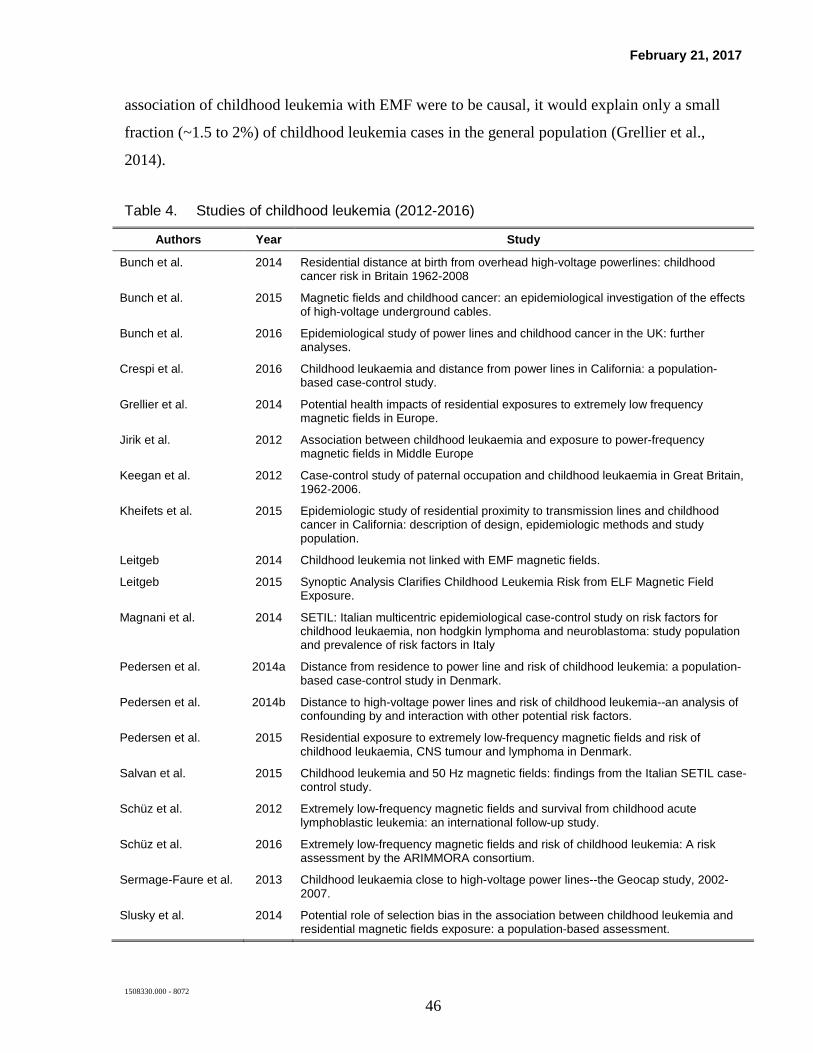
February 21, 2017
1508330.000 - 8072 46
association of childhood leukemia with EMF were to be causal, it would explain only a small
fraction (~1.5 to 2%) of childhood leukemia cases in the general population (Grellier et al.,
2014).
Table 4. Studies of childhood leukemia (2012-2016)
Authors Year Study
Bunch et al. 2014 Residential distance at birth from overhead high-voltage powerlines: childhood cancer risk in Britain 1962-2008
Bunch et al. 2015 Magnetic fields and childhood cancer: an epidemiological investigation of the effects of high-voltage underground cables.
Bunch et al. 2016 Epidemiological study of power lines and childhood cancer in the UK: further analyses.
Crespi et al. 2016 Childhood leukaemia and distance from power lines in California: a population-based case-control study.
Grellier et al. 2014 Potential health impacts of residential exposures to extremely low frequency magnetic fields in Europe.
Jirik et al. 2012 Association between childhood leukaemia and exposure to power-frequency magnetic fields in Middle Europe
Keegan et al. 2012 Case-control study of paternal occupation and childhood leukaemia in Great Britain, 1962-2006.
Kheifets et al. 2015 Epidemiologic study of residential proximity to transmission lines and childhood cancer in California: description of design, epidemiologic methods and study population.
Leitgeb 2014 Childhood leukemia not linked with EMF magnetic fields.
Leitgeb 2015 Synoptic Analysis Clarifies Childhood Leukemia Risk from ELF Magnetic Field Exposure.
Magnani et al. 2014 SETIL: Italian multicentric epidemiological case-control study on risk factors for childhood leukaemia, non hodgkin lymphoma and neuroblastoma: study population and prevalence of risk factors in Italy
Pedersen et al. 2014a Distance from residence to power line and risk of childhood leukemia: a population-based case-control study in Denmark.
Pedersen et al. 2014b Distance to high-voltage power lines and risk of childhood leukemia--an analysis of confounding by and interaction with other potential risk factors.
Pedersen et al. 2015 Residential exposure to extremely low-frequency magnetic fields and risk of childhood leukaemia, CNS tumour and lymphoma in Denmark.
Salvan et al. 2015 Childhood leukemia and 50 Hz magnetic fields: findings from the Italian SETIL case-control study.
Schüz et al. 2012 Extremely low-frequency magnetic fields and survival from childhood acute lymphoblastic leukemia: an international follow-up study.
Schüz et al. 2016 Extremely low-frequency magnetic fields and risk of childhood leukemia: A risk assessment by the ARIMMORA consortium.
Sermage-Faure et al. 2013 Childhood leukaemia close to high-voltage power lines--the Geocap study, 2002-2007.
Slusky et al. 2014 Potential role of selection bias in the association between childhood leukemia and residential magnetic fields exposure: a population-based assessment.
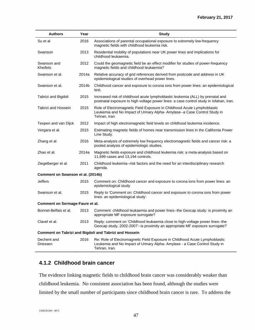
February 21, 2017
1508330.000 - 8072 47
Authors Year Study
Su et al 2016 Associations of parental occupational exposure to extremely low-frequency magnetic fields with childhood leukemia risk.
Swanson 2013 Residential mobility of populations near UK power lines and implications for childhood leukaemia.
Swanson and Kheifets
2012 Could the geomagnetic field be an effect modifier for studies of power-frequency magnetic fields and childhood leukaemia?
Swanson et al. 2014a Relative accuracy of grid references derived from postcode and address in UK epidemiological studies of overhead power lines.
Swanson et al. 2014b Childhood cancer and exposure to corona ions from power lines: an epidemiological test.
Tabrizi and Bigdoli 2015 Increased risk of childhood acute lymphoblastic leukemia (ALL) by prenatal and postnatal exposure to high voltage power lines: a case control study in Isfahan, Iran.
Tabrizi and Hossein 2015 Role of Electromagnetic Field Exposure in Childhood Acute Lymphoblastic Leukemia and No Impact of Urinary Alpha- Amylase--a Case Control Study in Tehran, Iran.
Teepen and van Dijck 2012 Impact of high electromagnetic field levels on childhood leukemia incidence.
Vergara et al. 2015 Estimating magnetic fields of homes near transmission lines in the California Power Line Study.
Zhang et al. 2016 Meta-analysis of extremely low frequency electromagnetic fields and cancer risk: a pooled analysis of epidemiologic studies.
Zhao et al. 2014a Magnetic fields exposure and childhood leukemia risk: a meta-analysis based on 11,699 cases and 13,194 controls.
Ziegelberger et al. 2011 Childhood leukemia--risk factors and the need for an interdisciplinary research agenda.
Comment on Swanson et al. (2014b)
Jeffers 2015 Comment on: Childhood cancer and exposure to corona ions from power lines: an epidemiological study
Swanson et al. 2015 Reply to 'Comment on: Childhood cancer and exposure to corona ions from power lines: an epidemiological study'.
Comment on Sermage-Faure et al.
Bonnet-Belfais et al. 2013 Comment: childhood leukaemia and power lines--the Geocap study: is proximity an appropriate MF exposure surrogate?
Clavel et al. 2013 Reply: comment on 'Childhood leukaemia close to high-voltage power lines--the Geocap study, 2002-2007'--is proximity an appropriate MF exposure surrogate?
Comment on Tabrizi and Bigdoli and Tabrizi and Hossein
Dechent and Driessen
2016 Re: Role of Electromagnetic Field Exposure in Childhood Acute Lymphoblastic Leukemia and No Impact of Urinary Alpha- Amylase - a Case Control Study in Tehran, Iran.
4.1.2 Childhood brain cancer
The evidence linking magnetic fields to childhood brain cancer was considerably weaker than
childhood leukemia. No consistent association has been found, although the studies were
limited by the small number of participants since childhood brain cancer is rare. To address the
February 21, 2017
1508330.000 - 8072 48
issue of small numbers, the WHO research recommendations included the conduct of a pooled
analysis of epidemiologic studies of childhood brain cancer and EMF. Following up on these
recommendations, both a meta-analysis and a pooled analysis were published (Mezei et al.,
2008; Kheifets et al., 2010b). A meta-analysis is similar to a pooled analysis, but only the
published results from the individual studies are combined as opposed to the raw data. Overall,
no association was reported in the meta-analysis, but an analysis of five studies with information
on calculated or measured magnetic fields greater than 3-4 mG found a combined OR that was
elevated but not statistically significant (OR=1.68, 95% CI=0.83-3.43) (Mezei et al., 2008). The
authors stated that an increased risk of childhood brain tumors could not be excluded at this high
exposure level, but that the similarity of this result to the findings of the pooled analyses of
childhood leukemia data suggests that control selection bias is operating in both analyses. In
direct response to the WHO’s recommendation, Kheifets et al. (2010b) pooled data from 10
studies on childhood brain cancer and residential magnetic-field exposure. Similar to the pooled
analysis of childhood leukemia (Kheifets et al., 2010a), there were few cases in the upper
exposure categories; however, contrary to the childhood leukemia results, no consistent
associations were reported for childhood brain cancer in the pooled analysis (Kheifets et al.,
2010b). While some elevated ORs were observed, they were not statistically significant and no
dose-response patterns were observed. The authors concluded that their results provide little
evidence for an association between magnetic fields and childhood brain cancer.
Relevant studies published since 2012
Some of the large epidemiologic studies of childhood cancer discussed in the childhood
leukemia section above also examined the potential relationship between residential proximity
to overhead and underground transmission lines and childhood brain cancer (Bunch et al.,
2014; Bunch et al., 2015; Bunch et al., 2016; Pedersen et al., 2015; Crespi et al., 2016). The
case-control epidemiologic study by Bunch et al. (2014), described earlier, also included cases
of brain cancer (n=11,968) and other solid tumors (n=21,985) diagnosed among children in the
United Kingdom between 1962 and 2008. No statistically significant associations were
reported between residential proximity to overhead lines and childhood brain cancer in any of
the analyses. In additional analyses, no clear patterns were identified when brain cancer
occurrence among younger and older children were examined separately (Bunch et al., 2016),
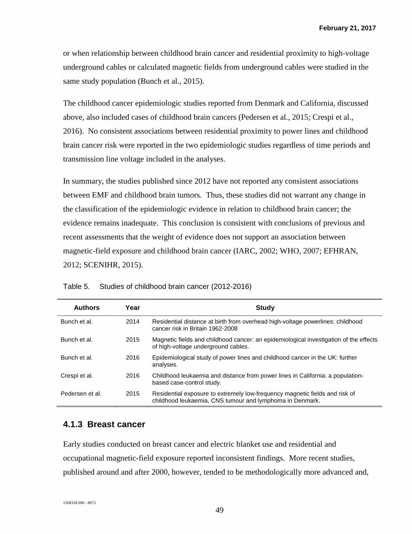
February 21, 2017
1508330.000 - 8072 49
or when relationship between childhood brain cancer and residential proximity to high-voltage
underground cables or calculated magnetic fields from underground cables were studied in the
same study population (Bunch et al., 2015).
The childhood cancer epidemiologic studies reported from Denmark and California, discussed
above, also included cases of childhood brain cancers (Pedersen et al., 2015; Crespi et al.,
2016). No consistent associations between residential proximity to power lines and childhood
brain cancer risk were reported in the two epidemiologic studies regardless of time periods and
transmission line voltage included in the analyses.
In summary, the studies published since 2012 have not reported any consistent associations
between EMF and childhood brain tumors. Thus, these studies did not warrant any change in
the classification of the epidemiologic evidence in relation to childhood brain cancer; the
evidence remains inadequate. This conclusion is consistent with conclusions of previous and
recent assessments that the weight of evidence does not support an association between
magnetic-field exposure and childhood brain cancer (IARC, 2002; WHO, 2007; EFHRAN,
2012; SCENIHR, 2015).
Table 5. Studies of childhood brain cancer (2012-2016)
Authors Year Study
Bunch et al. 2014 Residential distance at birth from overhead high-voltage powerlines: childhood cancer risk in Britain 1962-2008
Bunch et al. 2015 Magnetic fields and childhood cancer: an epidemiological investigation of the effects of high-voltage underground cables.
Bunch et al. 2016 Epidemiological study of power lines and childhood cancer in the UK: further analyses.
Crespi et al. 2016 Childhood leukaemia and distance from power lines in California: a population-based case-control study.
Pedersen et al. 2015 Residential exposure to extremely low-frequency magnetic fields and risk of childhood leukaemia, CNS tumour and lymphoma in Denmark.
4.1.3 Breast cancer
Early studies conducted on breast cancer and electric blanket use and residential and
occupational magnetic-field exposure reported inconsistent findings. More recent studies,
published around and after 2000, however, tended to be methodologically more advanced and,
February 21, 2017
1508330.000 - 8072 50
overall, reported no consistent associations. The WHO in its 2007 report concluded that the
body of research they reviewed was higher in quality compared with the early studies, and that
there was strong support for consensus statements that magnetic-field exposure does not
influence the risk of breast cancer.21 Studies published following the WHO review and included
in the previous Exponent reports supported this conclusion. The WHO recommended no further
research with respect to breast cancer and magnetic-field exposure, although the epidemiologic
evidence was still classified as inadequate.
Relevant studies published since 2012
In spite of the WHO conclusion that no further research is needed on EMF and breast cancer, a
number of case-control and cohort studies have been published that examined the relationship
between residential or occupational exposure to EMF and breast cancer. A large case-control
study in the United Kingdom investigated the occurrence of several types of adult breast cancer,
including leukemia, brain tumors, and malignant melanoma, in relation to magnetic-field
exposure and residential distance to high voltage power lines (Elliott et al., 2013). The British
researchers included 29,202 incident cases of female breast cancer, diagnosed between 1974 and
2008 in England and Wales, along with a total number of 79,000 controls between the age of 15
and 74 years in their study. Geographical information system databases were used to identify
the location of power lines and residential addresses. Magnetic-field exposure was calculated
for control and case addresses for the year of diagnosis and 5 years prior to diagnosis. Female
breast cancer risk showed no association with distance to power lines or with estimated
magnetic fields. In subsequent correspondence, several scientists expressed criticism regarding
the study’s exposure assessment, exposure categorization, and the potential for confounding in
the study (de Vocht, 2013; Philips et al., 2013; Schüz, 2013).
The potential relationship between occupational exposure to EMF and breast cancer was
examined in studies from the United Kingdom, the Netherlands, China, and Canada. In the
United Kingdom, Sorahan (2012) analyzed cancer incidence among more than 80,000 electricity
21 The WHO concluded, “Subsequent to the IARC monograph a number of reports have been published concerning
the risk of female breast cancer in adults associated with ELF magnetic field exposure. These studies are larger than the previous ones and less susceptible to bias, and overall are negative. With these studies, the evidence for an association between ELF exposure and the risk of breast cancer is weakened considerably and does not support an association of this kind” (WHO 2007, p. 307).
February 21, 2017
1508330.000 - 8072 51
generation and transmission workers between 1973 and 2008. Standardized rates for various
types of cancer were calculated among the workers compared to rates observed in the general
population. The author reported no statistically significant increases for breast cancer among
either men or women, and reported no trend for breast cancer incidence with year of hire, years
of being employed, or years since leaving employment. The study’s prospective nature and its
large sample size are among its strengths. The study’s limitations include the lack of analysis of
cancer rates by calculated magnetic-field exposures, and the use of an external reference group.
In the Netherlands, Koeman et al. (2014) investigated cancer incidence in relation to
occupational exposure to ELF magnetic fields in a cohort of about 120,000 men and women.
The researchers identified 2,077 breast cancer cases among women and no breast cancer among
men in the cohort. A job-exposure matrix was used to assign exposure to ELF magnetic fields
based on job titles. Based on a case-cohort analysis, the authors reported no association
between breast cancer and any of the exposure metrics, including estimated ELF magnetic-field
exposure, the length of employment, or cumulative exposure in the exposed jobs.
Breast cancer incidence was studied by Li et al. (2013) among more than 267,000 female textile
workers in Shanghai. A total of 1,687 incidence cases of breast cancer were identified in the
cohort between 1989 and 2000. The cases were compared to 4,702 non-cases using a case-
cohort approach. Exposure assessment was based on complete work history and a job-exposure
matrix specifically developed for the cohort. The authors reported no association between
cumulative exposure and risk of breast cancer regardless of age, histological type, and whether a
lag period was used or not. As a well-known epidemiologist and EMF researcher commented in
an accompanying editorial, the study was well-designed and it added further evidence to the
already large pool of data not supporting an association between ELF EMF and breast cancer
(Feychting, 2013). The editorial opined that additional studies on breast cancer “have little new
knowledge to add,” given the considerable improvement in study quality over time in breast
cancer epidemiologic studies, and that the evidence has been “consistently negative” (Feychting,
2013, p. 1046).
A population-based case-control study of occupational exposure to magnetic fields and male
breast cancer in Canada was reported by Grundy et al. (2016). The researchers identified 115
February 21, 2017
1508330.000 - 8072 52
cases in eight Canadian provinces through the provincial cancer registries between 1994 and
1998. A total of 570 age- and gender-matched controls were selected from provincial health
insurance plans or using random digit dialing. Self-administered questionnaires were used to
gather information on demographic characteristics and occupational history. Magnetic-field
exposure was classified into three categories (<3, 3 to <6, and ≥6 mG) based on expert review of
the jobs held by the study subjects. The authors reported statistically non-significant risk
increases with highest average exposure ≥6 mG compared to exposure <3 mG, and with having
an exposed job (≥3 mG) for at least 30 years compared to never having an exposed job.
Chinese investigators have published several meta-analyses in recent years for both female and
male breast cancer (Chen et al., 2013; Sun et al., 2013; Zhao et al., 2014b). One of the meta-
analyses for female breast cancer included 23 case-control studies published between 1991 and
2007. Based on all 23 studies, the authors reported a small, but statistically significant
association between breast cancer and ELF magnetic-field exposure (OR 1.07; 95% CI 1.02-
1.13). The authors also reported marginally significant small increases in estimated OR for
estrogen receptor positive and premenopausal cancer (OR 1.11) (Chen et al., 2013). The
conclusion of the authors that ELF magnetic fields might be related to breast cancer is contrary
to the conclusion of the WHO and other risk assessment panels, and may be explained by the
reliance of Chen et al, (2013) on earlier and methodologically less advanced studies in the
analysis. Zhao et al. (2014b) combined results from 16 case-control epidemiologic studies of
ELF EMF and breast cancer published between 2000 and 2007 in their meta-analysis, and
reported a weak but statistically significant association, which appeared to be stronger among
non-menopausal women. The conclusion of Zhao et al. (2014b) that ELF magnetic fields might
be related to breast cancer is contrary to the conclusion of the WHO and other risk assessment
panels. This, again, similar to the previously discussed meta-analysis, may be explained by the
inclusion of earlier and methodologically less advanced studies in the Zhao et al. (2014b)
analysis. Sun et al (2013) included 7 case-control and 11 cohort studies in their meta-analysis of
male breast cancer. With one exception, all included studies were occupational epidemiologic
studies of ELF magnetic-field exposure. The authors reported a statistically significant
association between male breast cancer and exposure to ELF EMF (OR 1.32; 95% CI 1.14-1.52)
in their overall analysis. However, methodological limitations, the small number of cases in the
individual studies, and the potential for publication bias may explain their overall findings.
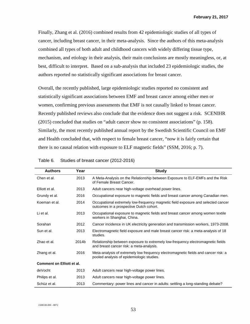
February 21, 2017
1508330.000 - 8072 53
Finally, Zhang et al. (2016) combined results from 42 epidemiologic studies of all types of
cancer, including breast cancer, in their meta-analysis. Since the authors of this meta-analysis
combined all types of both adult and childhood cancers with widely differing tissue type,
mechanism, and etiology in their analysis, their main conclusions are mostly meaningless, or, at
best, difficult to interpret. Based on a sub-analysis that included 23 epidemiologic studies, the
authors reported no statistically significant associations for breast cancer.
Overall, the recently published, large epidemiologic studies reported no consistent and
statistically significant associations between EMF and breast cancer among either men or
women, confirming previous assessments that EMF is not causally linked to breast cancer.
Recently published reviews also conclude that the evidence does not suggest a risk. SCENIHR
(2015) concluded that studies on “adult cancer show no consistent associations” (p. 158).
Similarly, the most recently published annual report by the Swedish Scientific Council on EMF
and Health concluded that, with respect to female breast cancer, “now it is fairly certain that
there is no causal relation with exposure to ELF magnetic fields” (SSM, 2016; p. 7).
Table 6. Studies of breast cancer (2012-2016)
Authors Year Study Chen et al. 2013 A Meta-Analysis on the Relationship between Exposure to ELF-EMFs and the Risk
of Female Breast Cancer.
Elliott et al. 2013 Adult cancers near high-voltage overhead power lines.
Grundy et al. 2016 Occupational exposure to magnetic fields and breast cancer among Canadian men.
Koeman et al. 2014 Occupational extremely low-frequency magnetic field exposure and selected cancer outcomes in a prospective Dutch cohort.
Li et al. 2013 Occupational exposure to magnetic fields and breast cancer among women textile workers in Shanghai, China.
Sorahan 2012 Cancer incidence in UK electricity generation and transmission workers, 1973-2008.
Sun et al. 2013 Electromagnetic field exposure and male breast cancer risk: a meta-analysis of 18 studies.
Zhao et al. 2014b Relationship between exposure to extremely low-frequency electromagnetic fields and breast cancer risk: a meta-analysis.
Zhang et al. 2016 Meta-analysis of extremely low frequency electromagnetic fields and cancer risk: a pooled analysis of epidemiologic studies.
Comment on Elliott et al.
deVocht 2013 Adult cancers near high-voltage power lines.
Philips et al. 2013 Adult cancers near high-voltage power lines.
Schüz et al. 2013 Commentary: power lines and cancer in adults: settling a long-standing debate?
February 21, 2017
1508330.000 - 8072 54
Authors Year Study Comment on Li et al.
Feychting 2013 Invited commentary: extremely low-frequency magnetic fields and breast cancer--now it is enough!
4.1.4 Other adult cancers
In general, scientific panels have concluded that the scientific evidence is inadequate to
establish a causal link between other adult cancers and exposure to magnetic fields, but due to
the inherent nature of scientific research, the possibility cannot be entirely ruled out (IARC,
2002; WHO 2007). Most epidemiologic studies of EMF and adult cancers (in addition to breast
cancer) examined leukemia, lymphoma, and brain cancer, and studies of these outcomes will be
discussed in detail below. Adult cancers other than leukemia, lymphoma, and cancers of the
brain and breast were examined sporadically, and no consistently replicated findings were
identified by any of the expert panels for adult cancer outcomes. Since studies with better
exposure assessment methods do not report stronger or more consistent findings, scientific
panels concluded that the evidence for an association is weak and the observed inconsistency is
probably due to chance or bias. The IARC classified the epidemiologic data with regard to adult
leukemia, lymphoma, and brain cancer as “inadequate” in 2002, and the WHO confirmed this
classification in 2007, with the remaining uncertainty attributed mainly to limitations in
exposure assessment methods.
Much of the research on EMF and adult cancers is related to occupational exposure, given the
higher range of exposure levels encountered in the occupational environment. The main
limitations of these studies, however, are the methods used to assess exposure, with early studies
relying simply on a person’s occupational title (often taken from a death certificate) and later
studies linking a person’s full or partial occupational history to representative average exposure
for each occupation (i.e., a job exposure matrix). The latter method, while representing a
methodological advancement, still has some important limitations as highlighted by Kheifets et
al. (2009). While a person’s occupation may provide some indication of the overall magnitude
of their occupational magnetic-field exposure, it does not take into account the possible
variation in exposure due to different job tasks within occupational titles, the frequency and
intensity of contact to relevant exposure sources, or variation by calendar time. Furthermore,
February 21, 2017
1508330.000 - 8072 55
since scientists do not know any mechanism by which magnetic fields could lead to cancer, an
appropriate exposure metric is also unknown.
4.1.4.1. Adult brain cancer
Epidemiologic studies on EMF and adult brain cancer, published following the WHO
assessment, provided no additional consistent evidence for an association (e.g., Johansen et al.,
2007; Coble et al., 2009; Baldi et al., 2011; Marcilio et al., 2011). In a meta-analysis of
occupational EMF exposure and leukemia and brain cancer (Khefeits et al., 2008), a small but
statistically significant increase of leukemia and brain cancer was reported in relation to the
highest estimate of magnetic-field exposure in the individual studies. Several findings,
however, led the authors to conclude that magnetic-field exposure is not responsible for the
observed associations. For example, Khefeits et al. (2008) reported a weaker association in the
more recent studies than the observed association in their previous meta-analysis (Kheifets et
al., 1995), whereas a stronger association would be expected if there were a true relationship
since the quality of the studies has improved over time. The authors concluded that “the lack of
a clear pattern of EMF exposure and outcome risk does not support a hypothesis that these
exposures are responsible for the observed excess risk” (Kheifets et al., 1995, p. 677).
Relevant studies published since 2012
Some of the adult cancer epidemiologic studies discussed in the breast cancer section also
included brain cancers in the analyses. The study of residential proximity and magnetic-field
exposure from power lines in the United Kingdom (Elliott et al., 2013), also included brain
cancer cases (n=6,781). The authors reported no statistically significant risk increase for brain
cancer with either distance or estimated magnetic-field levels in the study.
The British cohort study of electricity generation and transmission workers (Sorahan, 2012;
2014a) also studied brain cancer cases. Both internal comparisons (within the cohort of
workers) and external comparisons (to the general population of the United Kingdom) were
made in the study, and the author also considered cumulative, recent, and distant occupational
exposures to occupational ELF EMF. No increased risk for brain cancer among either men or
women was observed, and no trend was reported for brain cancer risk with year of hire, years of
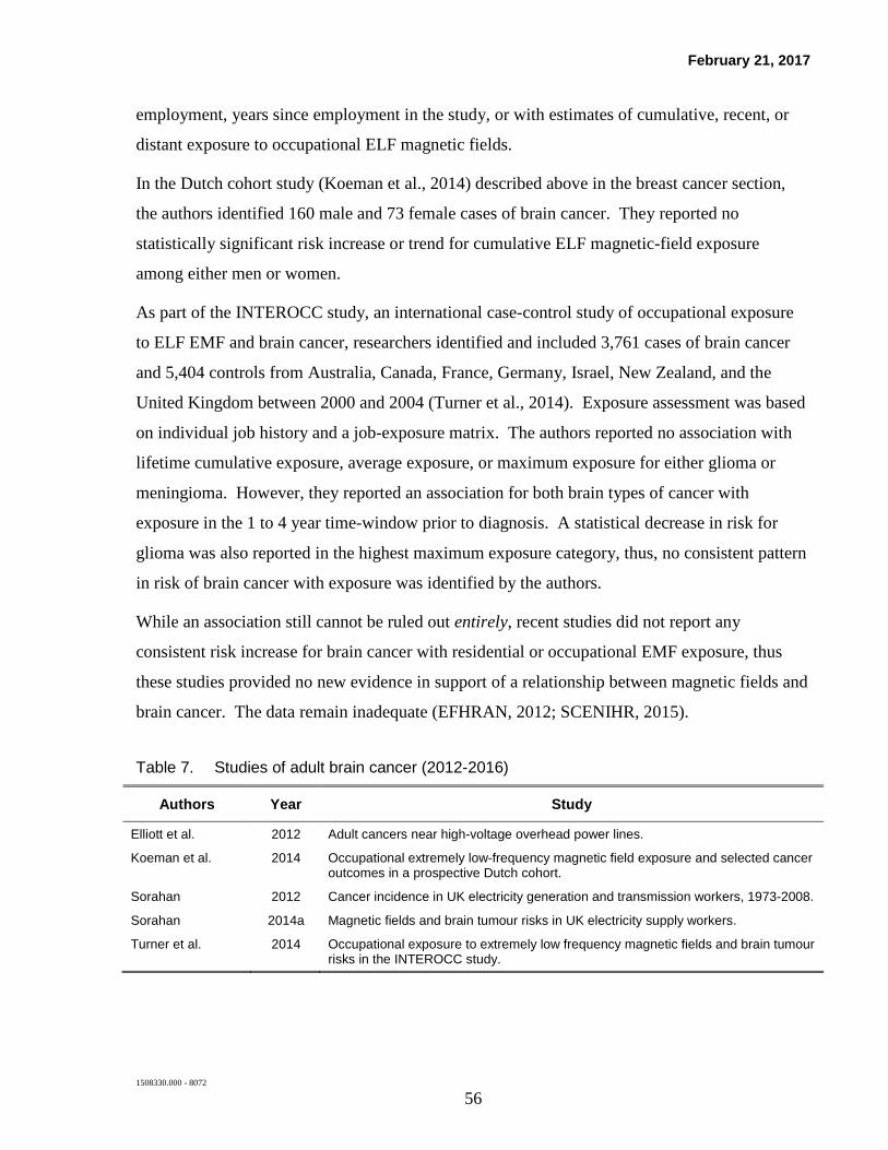
February 21, 2017
1508330.000 - 8072 56
employment, years since employment in the study, or with estimates of cumulative, recent, or
distant exposure to occupational ELF magnetic fields.
In the Dutch cohort study (Koeman et al., 2014) described above in the breast cancer section,
the authors identified 160 male and 73 female cases of brain cancer. They reported no
statistically significant risk increase or trend for cumulative ELF magnetic-field exposure
among either men or women.
As part of the INTEROCC study, an international case-control study of occupational exposure
to ELF EMF and brain cancer, researchers identified and included 3,761 cases of brain cancer
and 5,404 controls from Australia, Canada, France, Germany, Israel, New Zealand, and the
United Kingdom between 2000 and 2004 (Turner et al., 2014). Exposure assessment was based
on individual job history and a job-exposure matrix. The authors reported no association with
lifetime cumulative exposure, average exposure, or maximum exposure for either glioma or
meningioma. However, they reported an association for both brain types of cancer with
exposure in the 1 to 4 year time-window prior to diagnosis. A statistical decrease in risk for
glioma was also reported in the highest maximum exposure category, thus, no consistent pattern
in risk of brain cancer with exposure was identified by the authors.
While an association still cannot be ruled out entirely, recent studies did not report any
consistent risk increase for brain cancer with residential or occupational EMF exposure, thus
these studies provided no new evidence in support of a relationship between magnetic fields and
brain cancer. The data remain inadequate (EFHRAN, 2012; SCENIHR, 2015).
Table 7. Studies of adult brain cancer (2012-2016)
Authors Year Study
Elliott et al. 2012 Adult cancers near high-voltage overhead power lines.
Koeman et al. 2014 Occupational extremely low-frequency magnetic field exposure and selected cancer outcomes in a prospective Dutch cohort.
Sorahan 2012 Cancer incidence in UK electricity generation and transmission workers, 1973-2008.
Sorahan 2014a Magnetic fields and brain tumour risks in UK electricity supply workers.
Turner et al. 2014 Occupational exposure to extremely low frequency magnetic fields and brain tumour risks in the INTEROCC study.
February 21, 2017
1508330.000 - 8072 57
4.2.4.2 Adult leukemia and lymphoma
Similar to adult brain cancer, the WHO classified the epidemiologic evidence with regard to
adult leukemia as “inadequate” (WHO 2007). Epidemiologic studies of adult leukemia and
lymphoma, published subsequent to the WHO report (2007), provided no consistent support for
an association (e.g., Johansen et al., 2007; Wong et al., 2010; Marcilio et al., 2011). As
described above, a small and statistically significant increase of leukemia in relation to the
highest estimate of magnetic-field exposure was reported in the meta-analysis by Kheifets et al.
(2008), but the authors concluded that the overall pattern of results (e.g., there was no
consistency in findings by leukemia subtype) did not support a causal relationship between EMF
and leukemia.
Relevant studies published since 2012
The British epidemiologic study of power lines and adult cancer (Elliott et al., 2013) also
included 7,823 cases of adult leukemia. The authors reported no elevated risk or trend for adult
leukemia in association with distance to or estimated magnetic-field exposure from high-voltage
power lines. In the cohort of electricity power plant and transmission workers in the United
Kingdom, Sorahan (2012) reported no increase in risk for leukemia, when compared to the
general population of the United Kingdom, either among men or women, and no increasing
trend was observed with length of employment. In a separate analysis, Sorahan also analyzed
leukemia risk in relation to estimated occupational exposure to ELF magnetic fields within the
cohort of employees; he reported that RR estimates were “unexceptional,” and were close to
unity for all exposure categories based on cumulative, recent, and distant exposures (Sorahan,
2014b). A statistical association for ALL in a sub-analysis was attributed by the author to
unusually low risk in the reference category (Sorahan, 2014b).
In the Dutch cohort study (Koeman et al., 2014), the authors identified 761 and 467
hematopoietic malignancies among men and women, respectively. They reported no increases
in risk or trend for these malignancies in association with cumulative exposure to ELF magnetic
fields among either men or women.
Rodriguez-Garcia and Ramos (2012) reported inverse correlations between acute myeloid
leukemia, ALL, and the distance to thermoelectric power plants and high-density power line
February 21, 2017
1508330.000 - 8072 58
networks, i.e., the fewer cases were recorded close to the power lines and power plants, in their
study of hematologic cancers in a region of Spain from 2000 to 2005. This study, however, has
severe limitations due to the use of aggregated data, rudimentary methods of exposure
assessment, and the lack of an adequate comparison group.
A large case-control study of occupational exposure to ELF EMF and electric shocks and acute
myeloid leukemia conducted in four Northern European countries (Finland, Iceland, Norway,
and Sweden) included 5,409 cases diagnosed between 1961 and 2005 and 27,045 controls
matched on age, sex, and country (Talibov et al., 2015). Lifetime occupational exposure to ELF
EMF and shocks were assessed using corresponding job-exposure matrices and were based on
jobs reported in the participating countries’ censuses. Work-related exposures to benzene and
ionizing radiation were controlled for in the analyses. The authors reported no associations
between leukemia and exposure to ELF EMF or electric shocks among either men or women.
Recent studies did not provide substantive new evidence in support of an association between
EMF exposure and leukemia and lymphoma in adults. While the possibility that there is a
relationship between adult lymphohematopoietic malignancies and magnetic-field exposure still
cannot be entirely ruled out as a result of scientific uncertainty due to study limitations, the
current scientific body of studies provides inadequate evidence for an association (EFHRAN,
2012, SCENIHR, 2015).
Table 8. Studies of adult leukemia/lymphoma (2012-2016)
Authors Year Study Elliott et al. 2013 Adult cancers near high-voltage overhead power lines.
Koeman et al. 2014 Occupational extremely low-frequency magnetic field exposure and selected cancer outcomes in a prospective Dutch cohort.
Rodriguez-Garcia and Ramos 2012 High incidence of acute leukemia in the proximity of some industrial facilities in El
Bierzo, northwestern Spain.
Sorahan 2012 Cancer incidence in UK electricity generation and transmission workers, 1973-2008.
Sorahan 2014b Magnetic fields and leukaemia risks in UK electricity supply workers.
Talibov et al. 2015 Occupational exposure to extremely low-frequency magnetic fields and electrical shocks and acute myeloid leukemia in four Nordic countries.
February 21, 2017
1508330.000 - 8072 59
4.1.5 In vivo studies of carcinogenesis
It is standard procedure to conduct studies on laboratory animals to determine whether exposure
to a specific agent leads to the development of cancer (USEPA, 2005). This approach is used
because all known human carcinogens also have been shown to cause cancer in laboratory
animals and such studies are better suited to determining causation than epidemiology studies
(IARC, 2002).
Magnetic field bioassays
The major focus of interest is on what are known as chronic bioassay studies in which animals,
including those with a particular genetic susceptibility to cancer, are exposed at high levels over
their entire lifespan or a large part of it and tissue evaluations are performed to assess the
incidence of tumors in many organs. These studies are considered one of the gold standards for
identifying carcinogenic agents and are often considered when establishing regulatory actions.
The 2007 WHO review described four large-scale, long-term studies of rodents exposed to
magnetic fields over the course of their lifetime that did not report increases in any type of
cancer (Mandeville et al., 1997; Yasui et al., 1997; McCormick et al., 1999; Boorman et al.,
1999a, 1999b). Some animals, however, developed a type of lymphoma similar to childhood
ALL (Fam and Mikhail, 1996), but other studies exposing transgenic mice predisposed to
develop leukemias to ELF magnetic fields did not report an increased incidence of this
lymphoma type (Harris et al., 1998; McCormick et al., 1999; Sommer and Lerchl, 2004).
Magnetic field exposure + known carcinogens
Other types of studies test whether the exposure of interest, in combination with a known
carcinogen, produces a promotional or co-carcinogenetic effect or whether the exposure in
combination with a known carcinogen and a known promoter produces a co-promotional effect.
These types of studies can be problematic in their interpretation because of the sometimes
limited nature of the interaction of the known carcinogens or tumor promoters with particular
tissues and because of the complexity of the study designs and conditions.
February 21, 2017
1508330.000 - 8072 60
Most studies reviewed by the WHO did not find evidence that magnetic-field exposure when
combined with chemical carcinogens affected the development of tumors in skin, liver, etc. For
over a decade, however, one laboratory in Germany has reported that the incidence of mammary
tumors caused by 7, 12-dimethylbenz[a]anthracene (DMBA) was generally, but inconsistently,
increased by magnetic-field exposure (Löscher et al., 1993, 1994, 1997; Baum et al., 1995;
Löscher and Mevissen, 1995; Mevissen et al., 1993a, 1993b, 1996a, 1996b, 1998). The reported
influence of magnetic fields on the carcinogenic effects of DMBA reported by the German
laboratory could not be replicated in studies from laboratories supported by the U.S. National
Toxicology Program (Anderson et al., 1999; Boorman et al.1999a, 1999b; NTP, 1999). The
WHO concluded that the inconsistent findings across laboratories may be due to differences in
experimental protocols or the use of different rat sub-strains, only some of which may be
susceptible to the promotional effects of magnetic fields on mammary tissue. Based on the
research available at the time, the WHO concluded that, “There is no evidence that ELF
exposure alone causes tumours. The evidence that ELF field exposure can enhance tumour
development in combination with carcinogens is inadequate” (WHO 2007, p. 322).
In light of the available evidence that exposure to magnetic fields alone does not increase the
occurrence of cancer, most studies published subsequently have investigated the potential
promotional or co-carcinogenic effects of magnetic-field exposure. These studies show that
long-term exposure to magnetic fields does not alter the incidence of brain tumors or
leukemia/lymphoma in rats and mice treated with the chemical initiators DMBA (Negishi et al.,
2008), ethylnitrosourea (Chung et al., 2008) or n-butylnitrosourea (Bernard et al., 2008) or the
cancer incidence rates or survival time in a strain of mice genetically predisposed to develop
leukemia (Chung et al., 2010). The German laboratory continues to report findings similar to
earlier work and have explored potential associations of magnetic-field exposure with the
expression of certain genes and proteins in mammary tissue of different rat strains (Fedrowitz
and Löscher, 2008; 2012).
Damage to DNA, tumor development, and oxidative stress
Another focus of animal research is on the potential for magnetic fields to damage the DNA
directly or in combination with known carcinogenic chemicals or x-rays (Lai and Singh, 2004).
February 21, 2017
1508330.000 - 8072 61
Studies have continued to look for evidence of DNA damage from magnetic-field exposure with
mixed results (Mariucci et al., 2010; Okudan et al., 2010). Yet, in other studies investigating the
therapeutic applications of magnetic fields, much higher level exposures to magnetic fields in
combination with cancer treatments have been reported to reduce tumor size or increase the
survival of animals injected with tumors (Berg et al., 2010; Wen et al., 2011) or to reduce the
development of pre-neoplastic lesions in the livers of rats initiated via chemicals and surgery
(Jiménez-Garcia et al., 2010).
Without good evidence that either cancer or DNA damage is caused by magnetic-field
exposures, a number of studies have investigated the role of magnetic-field exposures on
indicators of oxidative stress to tissues on the premise that oxidative stress is the mechanism that
connects magnetic-field exposure to cancer. However, these studies typically have involved
exposures far above guidelines for human exposure, have been of low quality, and have not
been designed to establish any direct relevance of any findings to cancer.
Reviewers for EFHRAN (2010) concluded that the in vivo research published up to July 2010
indicated a “lack of effect” of magnetic fields on cancer.
Relevant studies published since 2012
Magnetic field bioassays
The research themes that have been pursued in animal studies of cancer and biological processes
possibly related to cancer before 2012 have continued to be addressed in more recent in vivo
studies.
Two chronic cancer bioassays conducted at the Ramazinni Institute in Italy were reported in
2016 (Soffritti et al. 2016a, 2016b). In one of these studies, over 5,000 rats were said to be
exposed to 50-Hz magnetic fields at intensities of 0, 20, 200, 1,000, and 10,000 mG for 19 hours
per day starting before birth and continuing over their lifetime (Soffritti et al, 2016a).
Regarding male and female rats exposed to magnetic fields alone, partial results were reported
only for female rats exposed to 10,000 mG in an earlier report (Soffritti et al., 2010). There was
no effect on body weight or the incidence of mammary tumors (Soffritti et al., 2010). In a
second study (Soffritti et al., 2016b), male and female rats were exposed to 10,000 mG or
February 21, 2017
1508330.000 - 8072 62
control conditions and also exposed over their lifetime. The authors report no differences in the
water intake, body weight or survival of male or female rats exposed to magnetic fields
compared to controls. More important, there were no reported differences between these groups
with respect to benign or malignant tumors, including C-cell thyroid tumors or
hemolymphoreticular neoplasias. The results of these two studies would appear to be consistent
with previous chronic bioassay studies.
Exposures to 50-Hz magnetic fields were investigated in a modified bioassay design by Qi et al.
(2015). Ten pregnant mice were exposed for 12 hours per day to 500 mG magnetic fields and a
portion of the pups were further exposed for 15.5 months. No description of the exposure
system or housing was described and the control mice were not sham-exposed. The authors
reported that the body weights of both sexes in the exposed group were significantly lower than
the same sex control groups after 6 months of exposure. The incidence of tumors in the exposed
and control mice were not different; however, three of the exposed female mice were observed
with histological changes in the liver and bone marrow consistent with chronic myelogenous
leukemia. The study has severe limitations, including the lack of any description of the
exposure system, the absence of sham-controls, the failure to control for litter effects, and the
very small numbers of test subjects (n=66/group). A previous study with a similar design (i.e.,
prenatal and postnatal exposure) mice of the same strain to 5,000 or 50,000 mG magnetic fields
for 7 weeks and with a 78 week follow-up period) did not report any adverse effects or
differences in cancer in any of the examined tissues (Otaka et al., 2002).
Magnetic field exposure + known carcinogens
As part of the experiments performed by Soffritti et al. discussed above and in a third
experiment (Soffritti et al., 2016a), the investigators exposed animals to known carcinogens
(gamma rays or formaldehyde) in combination with magnetic field exposure. In 2010, Soffritti
et al. reported that 0.1 Gray of high-energy x-rays alone or in combination with exposure to 200
or 10,000 mG magnetic fields had no effect on mammary cancer in female rats (Soffritti et al.,
2010). Relying upon the exact same data as reported in this earlier paper, however, Soffritti et
al. (2016a) later found significant differences in an apparent post hoc analysis between the
control group and the 0.1 Gray + magnetic field exposure groups in the percent of mammary
February 21, 2017
1508330.000 - 8072 63
adenomas. The Soffritti et al. (2016a) paper presents many more analyses of the data for both
females and males based on small numbers of affected subjects (data not presented in the 2010
paper). Even so, the authors argue that exposure to magnetic fields enhances the effect of high
energy gamma rays on mammary tumor development.
The investigators from the Ramazzini Institute used the same exposure apparatus and general
methods to examine the effects of exposure to 50 mg/L formaldehyde, also a known carcinogen,
in drinking water for two years alone or in combination with exposure to a 10,000 mG, 50-Hz
magnetic field (Soffritti et al., 2016b). Controls were either unexposed (the same control group
as reported in Soffritti et al., 2016a) or treated with formaldehyde in drinking water only.
Malignant tumors, including C-cell carcinomas of the thyroid and lymphatic tumors, were
increased in the males exposed to magnetic fields and formaldehyde together compared to the
unexposed control, but the reported incidences were not substantially different than those seen
with formaldehyde treatment alone. No effects were seen in females, except for an increase in
thyroid adenomas and carcinomas in those rats administered formaldehyde alone. The results
for males were confounded by the substantially reduced water intake levels over the first year of
the study in males receiving formaldehyde in the drinking water with or without magnetic-field
exposure. Again, some of the tumor increases reported by Soffritti et al. (2010; 2016a; 2016b)
were based on limited numbers of affected animals; additionally, the time to tumor development
was not reported, and the study was carried out for the lifetime of the animals. Hence, while the
authors claim that the results of these studies suggest that magnetic fields increased the
carcinogenic effect of formaldehyde, the results do not clearly support this conclusion.
In addition to the limitations in the reporting and interpretation of the Soffritti et al. studies,
there are more serious problems with these studies including: 1) the absence of sham controls, for
which all conditions of housing, light, handling, etc., were kept the same as those of the exposed
groups (with the exception of exposure); 2) the allocation of individual rats to groups in a non-
random manner and without account of potential within-litter effects in the statistical analyses; and
3) histologic analyses that were not reported to have been performed without a priori knowledge of
the exposure condition to prevent bias in the scoring of the tissues. On this latter point, the EPA has
“decided not to rely on RI [Ramazzini Institute] data on lymphomas and leukemias in IRIS
[Integrated Risk Information System] assessments” (EPA, 2013), and has warned risk assessors
February 21, 2017
1508330.000 - 8072 64
about problems with the cancer bioassays conducted by the Ramazzini Institute. These problems
include the accuracy of the cancer diagnoses; the categorization of tumors; errors in identifying
cellular changes as leukemia/lymphoma in certain tissues that appear to be due to infections (lung)
and tissue inflammation; a unexplained significant rise in the incidence of leukemia/lymphomas
over time in control groups unrelated to the exposure under study; the lack of complete reporting
and documentation of analytical specifications; failure to control or analyze for potential litter
effects; and the use of common controls for multiple studies (Gift et al., 2013). Such factors
probably account for the more than two-fold difference between the incidence of mammary cancer
in unexposed controls in the Soffritti et al. (2010; 2016a) study and the incidence of mammary
cancer in unexposed controls reported in a study of identical design (Soffritti et al., 2014).
Several studies investigated the response of chromosomes and DNA to chemicals or ionizing
radiation when combined with magnetic-field exposure.22 Miyakoshi et al. (2012) compared the
frequency of chromosomal micronuclei in the brain astrocyte cells in multiple groups of six
newborn rats. Some groups were exposed to the DNA-damaging anti-cancer drug bleomycin at
two doses, to 50-Hz magnetic fields at 100,000 mG, or to appropriate control conditions.
Bleomycin increased the frequency of micronuclei in a dose-related fashion. Adding the
magnetic field did not affect the frequency of micronuclei in controls not treated with bleomycin
or at a bleomycin dose of 5 mG/kg. At 10 mG/kg, however, the magnetic field significantly
increased the frequency of micronuclei. Treatment with tempol, an antioxidant radical
scavenger, did not significantly reduce micronuclei in the control group, but did significantly
reduce the frequency of micronuclei in the group exposed to 100,000 mG. The limitations of
the study are several, including that while all exposures were administered to the pups in vivo
(apparently not by a standard randomization process), the astrocytes were cultured for 96 hours
in vitro before analysis; the standard protocol for micronuclei evaluation was developed for
bone marrow and blood erythrocytes, not astrocytes; there was minimal description of the
exposure system; the reliability of micronuclei identification was not assessed by an
independent observer; and the analysis was not done without a priori knowledge of the
exposure status of the cells. The study’s results, however, are consistent with the lead author’s
22 For context, it is important to recognize that most damage to chromosomes and DNA arise in the course of
ongoing cellular processes, not just due to environmental agents, and that some of the tools applied in the studies described are so sensitive that even DNA damage from common fluorescent lighting can be quantified (see e.g., Kennedy et al., 2012).
February 21, 2017
1508330.000 - 8072 65
previous study in which it was reported that magnetic fields with intensities greater than 50,000
mG were required to increase the mutational damage from x-rays in CHO-K1 cells (Miyakoshi
et al., 1999); this latter study was previously evaluated in the WHO (2007) report. It should be
noted, however, that the reported increase in mutations with x-rays + magnetic fields is not a
general phenomenon: the author’s colleagues also have reported that exposure to 50,000 mG 60-
Hz magnetic field did not increase the frequency of gene mutations caused by exposure to
ultraviolet light in 2RA and XP2OS(SV) cells (Mizuno et al., 2014).
Investigators at the Genome Damage and Stability Centre at the University of Sussex in the
United Kingdom applied sensitive, reproducible, and validated methods for the detection of
DNA damage in vivo to study the response of rapidly developing embryonic rat brains to x-rays
and magnetic fields. Woodbine et al (2015) exposed pregnant C57BL/6 mice in groups of four
in three experiments: 1) exposure to 0.1gray x-rays23 with analysis after 1, 3, 6, and 11 hours; 2)
exposure to 50-Hz, 3,000 mG magnetic fields for 9 hours with immediate analysis; and 3) 3
hours of magnetic-field exposure followed by x-rays and additional magnetic-field exposure for
up to 9 hours more with analysis of samples at 1, 3, 6, and 11 hours after x-ray treatment. The
investigators measured the average number of double-strand breaks per cell following exposure.
While x-rays significantly increased foci formation 1, 3, and 6 hours after exposure, concurrent
magnetic-field exposure did not increase the damage further and also did not affect the natural
rate of repair of x-ray induced damage after exposure. The authors contrast the advantages of
their in vivo model to in vitro studies of tumor cell lines, in which some studies had reported
magnetic-field effects on DNA at higher field strengths.
Damage to DNA, tumor development, and oxidative stress
The study by Saha et al. (2014), like the study by Woodbine et al. (2015) from the same
laboratory at the University of Sussex, started by demonstrating that exposure to x-rays at
increasing doses between 0.01 and 0.1 gray produced a linear increase in double-strand breaks
in the DNA in embryonic mouse brains one hour after exposure in utero. A similar linear
23 An exposure of 0.001g gray produces a dose of 1 millisievert that is “the average accumulated background
radiation dose to an individual for 1 year, exclusive of radon, in the United States” (Johnson JC, and Thaul S. An Evaluation of Radiation Exposure Guidance for Military Operations: Interim Report. Washington, D.C.: National Academy of Sciences, 1997. http://site.ebrary.com/id/10055096).
February 21, 2017
1508330.000 - 8072 66
increase in the response to DNA damage (apoptosis) in this tissue also was observed at 6 hours
after doses of x-rays to 0.2 gray. Having established the sensitivity and performance of these
methods, the investigators exposed groups of four C57BL/6 mice to 50-Hz magnetic fields at
1,000 mG for 2 hours, or continuous or intermittent (5 minutes on, 10 minutes off) at 3,000 mG
for 15 hours. Neither exposure increased double-strand breaks in DNA above sham- or cage-
control levels (<0.01 gray of x-rays), nor was an increased frequency of cellular reaction to
DNA damage (apoptosis, as detected using the sensitive TUNEL method) observed. The design
and analysis of the experiment was superb, including the incorporation of blind analyses of the
samples. Additionally, although there were a relatively small number of mice per group, this
limitation was offset to a large degree by the low degree of variability in the outcome measures
that were achieved by the use of good methods by investigators experienced in the analytical
techniques.
Several other investigators have studied the effects of magnetic-field exposure on the frequency
of mutations in different tissues. Alcaraz et al. (2014) measured the frequency of
micronucleated polychromatic erythrocytes (MNPCE) in bone marrow as a measure of
chromosomal mutations. Samples were obtained from male Swiss mice exposed to x-rays (0.5
gray), and analyzed by two specialists without a priori knowledge of the animals’ exposure
histories. In the first experiment, the investigators showed that exposure to x-rays significantly
increased the frequency of MNPCEs and the administration of various antioxidants decreased
MNPCEs whether administered before or after exposure to x-rays, with pre-treatment being
most effective. In the second experiment, exposure of groups of 6 mice to 50-Hz magnetic
fields at 2,000 mG for 7, 14, 21, and 28 days significantly elevated MNPCEs at the end of all
these exposure periods over control values, but the levels were less than half that reported in the
first experiment with x-rays. Further, none of the antioxidants reduced MNPCEs elevated by
magnetic fields; the data even suggested the possibility that these antioxidants alone may
increase MNPCEs above control levels (the author provided no statistical analysis of these
values). While the effects would appear robust, significant limitations preclude placing much
weight on the results of this single study. First, as described by the authors, the method used to
detect MNPCEs is demonstrably less sensitive than more modern methods. The mice were not
sham exposed; thus, there is no way to determine the extent to which the higher MNPCEs in the
exposed groups are the result of a higher stress level compared to home cage controls (e.g.,
February 21, 2017
1508330.000 - 8072 67
Cherian et al., 2015; Flint et al., 2010; Malvandi et al., 2010; Johnson and Thaul, 1997). In
addition, few mice were studied in each group, and with the known variability of the
measurement method, it is highly possible that this underpowered study may be showing a false-
positive finding.
Several controversial in vitro studies reviewed by the WHO in 2007 had suggested the
possibility that magnetic fields at a level of 350 mG could increase DNA damage to cells as
detected by the Comet assay, and other studies had suggested that magnetic fields increased the
expression of heat shock proteins, which are produced in response to increased temperature and
other tissue stressors. Mariucci et al. (2010) had reported that a 50-Hz magnetic field at an
intensity of 10,000 mG for 1 or 7 days produced DNA damage in regions of the mouse brain as
detected by the Comet assay and that this damage was repaired within 24 hours after exposure
ended. No effect of magnetic-field exposure on the expression of heat shock protein 70 was
reported.
Villarini et al. (2013) also investigated whether magnetic fields would cause DNA damage.
Male CD-1 mice were exposed to 50-Hz magnetic fields at 1,000, 2,000, 10,000, or 20,000 mG
for 7 days and DNA damage was measured by the Comet assay immediately after exposure
ended or after a 24-hour recovery period. Only brain tissues taken from mice immediately after
exposure to magnetic fields at 10,000 or 20,000 mG exhibited any statistically significant
damage. Tissues taken after the recovery period showed levels of damage that were not
different from controls or other magnetic-field exposure groups. The strength of this study is
the design and analysis, which included sham-controls, multiple levels of exposure,
randomization and statistical analysis, and measurement of comet tail length without knowledge
of the exposure history of the samples. This study, like most other studies published after the
2007 WHO report, does not confirm that magnetic fields cause damage to DNA at levels below
10,000 mG.
Wilson et al. (2015) applied yet another method for assessing the potential effects of magnetic
fields on DNA by examining the effects of exposure to a 50-Hz magnetic field on the frequency
of mutations in the sperm and blood of groups of 5 CBA/Ca and BALB/c mice exposed at levels
of 0, 100, 1,000, and 3,000 mG for 2 or 15 hours. The measurements were made by
February 21, 2017
1508330.000 - 8072 68
determining the frequency of expanded simple tandem repeat DNA loci after PCR amplification
in samples obtained 12 weeks after exposure, a time that previous studies had indicated is a
sensitive period for detection. The authors report that exposure to 1 gray of x-rays induced a
significant, greater than two-fold increase in mutation frequency above that in sham-controls.
At each level of exposure tested after 2 or 15 hours, there was no significant effect of the
duration or strength of the magnetic-field exposure on the frequency of mutations measured in
blood or sperm. When all the data from magnetic-field exposed mice were pooled, however, a
small but statistically significant elevation in frequency was noted. While this study included
sham-exposed controls, it did not examine what other exposures besides magnetic fields might
have been present during operation of the magnetic-field exposure system, including noise and
vibration that could affect the animals’ physiology.
Some methods used to assess DNA damage may not be able to discriminate between effects on
specific tissue regions or cell types. A previous study by Schmitz et al. (2004) had reported that
an 8-week exposure to a 50-Hz, 15,000 mG magnetic field increased DNA damage in a very
specific tissue (the choroid plexus) within the brain as detected by two methods of visualizing
increased cellular uptake of radioactive molecules. In one method, radio-labeled thymidine was
injected and the uptake by cells that are replicating DNA in unscheduled DNA synthesis (UDS)
to repair damage was analyzed by microscopic study of thin-sliced tissue sections. The other
method involved incubating thin slices of tissue with DNA polymerase–I in the presence of
radio-labeled dTTP and visualizing the radioactivity concentrated at points of DNA
fragmentation in cells, a process called in situ nick translation. In a current follow-up study
(Korr et al., 2014), members of the same team sought to replicate their previous finding at lower
magnetic-field strengths (1,000 mG, and 10,000 mG) on NMRI male rats following an 8-week
exposure. Korr et al. reported no effect of magnetic-field exposure on UDS for rats exposed to
1,000 mG and exposures at 10,000 mG slightly reduced UDS in the choroid plexus and cells of
the collecting duct of the kidney. This response is not consistent with the premise that magnetic
fields cause an increase in DNA damage. No effect on the level of unrepaired nick DNA single
strand breaks was observed in cells from the brain, kidney, or liver. The results may be
interpreted either as a failure to replicate the previous study or as an indication that only
magnetic fields with intensities of 15,000 mG are capable of affecting DNA.
February 21, 2017
1508330.000 - 8072 69
Two studies investigated the ability of exposure to magnetic fields to enhance the effectiveness
of anti-tumor treatments via damage to DNA. El-Bialy and Rageh (2013) injected groups of
five female BALB mice with Ehrlich ascites carcinoma cells, then treated them with 3 mg/kg
cisplatin on days 1, 4, and 7, or exposed them to 100,000 mG, 50-Hz magnetic fields for 14 days
(1 hour per day), or both. Control groups of five mice were saline-treated, but not sham
exposed to magnetic fields, and blinded analyses were not reported. Both magnetic-field
exposure and cisplatin treatment, alone or in combination, were associated with reduced tumor
volume; the strongest response was observed with combination treatment. Magnetic-field
exposure alone produced small, but statistically significant, increases in indices of DNA damage
to tumor cells; when combined with cisplatin treatment, this response added to (but did not
enhance) the damage to tumor DNA damage caused by cisplatin alone. More interesting are the
data on the analysis of bone marrow cells. A standard analysis of induction of micronuclei
showed no effect of the magnetic field alone on MNPCEs compared to untreated controls and
no increase when combined with cisplatin over cisplatin alone. Although modest correlations
between MNPCE and DNA damage in the Comet assay were reported, no DNA damage results
for bone marrow were included in the paper.
The second study (Mahna et al., 2014) examined the effect of 12 days of exposure to a strong
1,500,000 mG, 50-Hz magnetic field for 10 minutes alone or following anti-tumor treatment
with bleomycin + pulsed electric currents or just pulsed electric currents alone. The measure of
treatment effect was the volume of mammary tumors that developed over 30 days following
injection of tumor cells into the flanks of Balb/C mice. Magnetic-field exposure was reported to
slightly, but significantly, reduce tumor volume following injection below that observed in both
the cage and sham-control groups. In addition, simply housing mice in the magnetic-field
exposure chamber without any magnetic field also significantly reduced the rate of tumor
development below that of mice kept in their home cages. Magnetic-field exposure did not
enhance the anti-tumor effect of any of the other treatments tested. Although the study provided
little detail on the methods and the evaluation of the tumors was not performed blind, (i.e.,
without a priori knowledge of the animals’ exposure history), the study did include sham-
controls and random allocation of subjects.
February 21, 2017
1508330.000 - 8072 70
Oxidative stress is a condition in which oxygen free radical levels in tissues are elevated and is
one mechanism by which DNA damage, as well as other forms of cellular damage, may occur.
While there is general agreement that oxidative stress from endogenous cellular processes are
the overwhelming source of damage to DNA and other cellular components (de Bont and
Larebeke, 2004), whether such mechanisms are activated by magnetic fields is unknown.
Previous in vivo studies have evaluated whether magnetic-field exposure may be associated with
oxidative stress, with mixed results.
Since 2012 quite a number of investigators have tested hypotheses about magnetic field effects
on various indicators of oxidative stress in multiple tissues at levels as low as 500 mG and as
high as 100,000 mG (Seifirad et al., 2014; Glinka et al., 2013; Hassan and Abdelkawi, 2014;
Deng et al., 2013; Cui et al., 2012; Duan et al., 2013; Manikonda et al., 2014; Martínez-Sámano
et al., 2012; Akdag et al., 2013; Kiray et al., 2013). Overall, it is hard to draw any firm
conclusions from these studies of oxidative stress markers because the numbers of animals per
group were generally small, the exposure parameters and oxidative stress markers examined
varied across the studies, negative controls (i.e., unexposed animals) were not always sham-
exposed, positive controls (i.e., animals treated with agents known to cause the response being
investigated) were not included in the study, and only a few of the analyses were reported to
have been conducted in a blinded manner. Although markers of oxidative stress were generally
increased with higher rather than lower magnetic-field exposures, it is not known if this effect is
reversible or even biologically relevant. Independent replication of findings in studies with
greater sample sizes and blinded analyses is needed. Moreover, without studies that are
specifically designed to quantitatively assess the relationship between markers of oxidative
stress and measurements of DNA damage in an established model animal system, any
relationship to a carcinogenic process is based on speculation rather than scientific evidence.
Reviews of in vivo research
Reviews of in vivo research, including studies on carcinogenesis by SSM (2013, 2014, 2015)
and SCENIHR (2015) cover a good deal of the research published after 2012. These
conclusions are:
February 21, 2017
1508330.000 - 8072 71
SSM (2013):
Other studies indicated increased oxidative stress, again mostly by exposures at levels well above the current exposure limits. One study showed indications for tumour growth inhibition by a 100 mT field, but with only small numbers of animals. Replication is necessary to obtain more insight. In general, the latest animal studies do not contribute to understanding a mechanism that could explain the association found in epidemiological studies between long term exposure to ELF magnetic fields below 1 μT and an increased risk of childhood leukaemia. Hence, there is still a need for dedicated studies in this area using new animal models (pp. 28-29).
SSM (2014):
In general, the results of the studies are not very consistent. In some studies a function may be increased and in others decreased, while dose-responses cannot be derived. Most of the results are from single studies that need to be replicated in order to establish whether the observed effects are real or not. Also the large variety of exposure schedules used does not add to get a unified picture. Finally, none of these studies provide information that can be used in the interpretation of the association found in epidemiology studies between ELF magnetic field exposure and an increased risk of childhood leukaemia (p. 36).
SSM (2015):
With the exception of single studies, the quality of the experiments and their description did not substantially improve compared to the previous years …Furthermore, referring to direct DNA-damage due to “low doses” of ELF-MF or presenting “dose-dependencies” using two groups only is somehow doubtful. Overall and similar to the previous SSM report, the results of the described studies are not very consistent (p. 9).
SCENIHR (2015):
Previously SCENIHR (2009) concluded that animal studies did not provide evidence that exposure to magnetic fields alone caused tumours or enhanced the growth of implanted tumours. The inclusion of more recent studies does not alter that assessment. In addition, these studies do not provide further
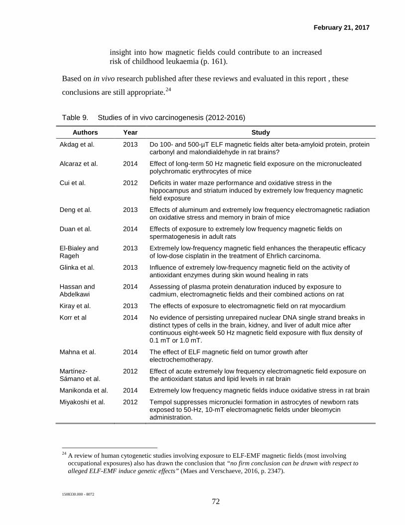
February 21, 2017
1508330.000 - 8072 72
insight into how magnetic fields could contribute to an increased risk of childhood leukaemia (p. 161).
Based on in vivo research published after these reviews and evaluated in this report , these
conclusions are still appropriate.24
Table 9. Studies of in vivo carcinogenesis (2012-2016)
Authors Year Study
Akdag et al. 2013 Do 100- and 500-µT ELF magnetic fields alter beta-amyloid protein, protein carbonyl and malondialdehyde in rat brains?
Alcaraz et al. 2014 Effect of long-term 50 Hz magnetic field exposure on the micronucleated polychromatic erythrocytes of mice
Cui et al. 2012 Deficits in water maze performance and oxidative stress in the hippocampus and striatum induced by extremely low frequency magnetic field exposure
Deng et al. 2013 Effects of aluminum and extremely low frequency electromagnetic radiation on oxidative stress and memory in brain of mice
Duan et al. 2014 Effects of exposure to extremely low frequency magnetic fields on spermatogenesis in adult rats
El-Bialey and Rageh
2013 Extremely low-frequency magnetic field enhances the therapeutic efficacy of low-dose cisplatin in the treatment of Ehrlich carcinoma.
Glinka et al. 2013 Influence of extremely low-frequency magnetic field on the activity of antioxidant enzymes during skin wound healing in rats
Hassan and Abdelkawi
2014 Assessing of plasma protein denaturation induced by exposure to cadmium, electromagnetic fields and their combined actions on rat
Kiray et al. 2013 The effects of exposure to electromagnetic field on rat myocardium
Korr et al 2014 No evidence of persisting unrepaired nuclear DNA single strand breaks in distinct types of cells in the brain, kidney, and liver of adult mice after continuous eight-week 50 Hz magnetic field exposure with flux density of 0.1 mT or 1.0 mT.
Mahna et al. 2014 The effect of ELF magnetic field on tumor growth after electrochemotherapy.
Martínez-Sámano et al.
2012 Effect of acute extremely low frequency electromagnetic field exposure on the antioxidant status and lipid levels in rat brain
Manikonda et al. 2014 Extremely low frequency magnetic fields induce oxidative stress in rat brain
Miyakoshi et al. 2012 Tempol suppresses micronuclei formation in astrocytes of newborn rats exposed to 50-Hz, 10-mT electromagnetic fields under bleomycin administration.
24 A review of human cytogenetic studies involving exposure to ELF-EMF magnetic fields (most involving
occupational exposures) also has drawn the conclusion that “no firm conclusion can be drawn with respect to alleged ELF-EMF induce genetic effects” (Maes and Verschaeve, 2016, p. 2347).
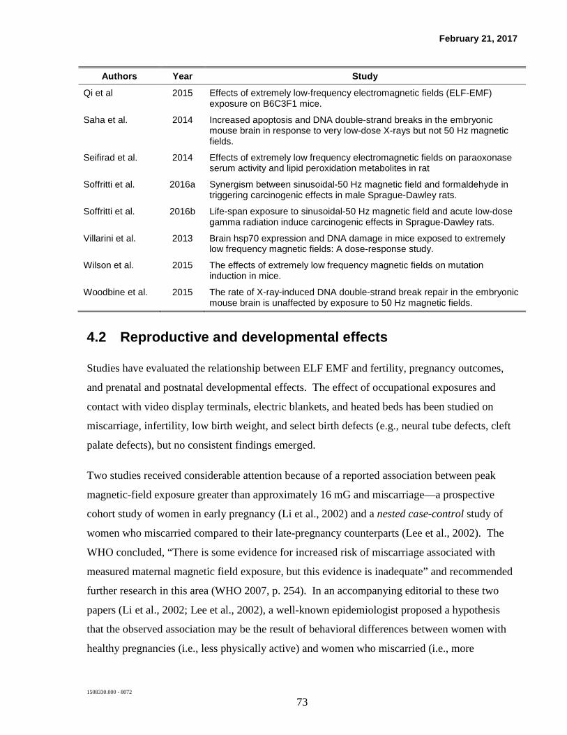
February 21, 2017
1508330.000 - 8072 73
Authors Year Study
Qi et al 2015 Effects of extremely low-frequency electromagnetic fields (ELF-EMF) exposure on B6C3F1 mice.
Saha et al. 2014 Increased apoptosis and DNA double-strand breaks in the embryonic mouse brain in response to very low-dose X-rays but not 50 Hz magnetic fields.
Seifirad et al. 2014 Effects of extremely low frequency electromagnetic fields on paraoxonase serum activity and lipid peroxidation metabolites in rat
Soffritti et al. 2016a Synergism between sinusoidal-50 Hz magnetic field and formaldehyde in triggering carcinogenic effects in male Sprague-Dawley rats.
Soffritti et al. 2016b Life-span exposure to sinusoidal-50 Hz magnetic field and acute low-dose gamma radiation induce carcinogenic effects in Sprague-Dawley rats.
Villarini et al. 2013 Brain hsp70 expression and DNA damage in mice exposed to extremely low frequency magnetic fields: A dose-response study.
Wilson et al. 2015 The effects of extremely low frequency magnetic fields on mutation induction in mice.
Woodbine et al. 2015 The rate of X-ray-induced DNA double-strand break repair in the embryonic mouse brain is unaffected by exposure to 50 Hz magnetic fields.
4.2 Reproductive and developmental effects
Studies have evaluated the relationship between ELF EMF and fertility, pregnancy outcomes,
and prenatal and postnatal developmental effects. The effect of occupational exposures and
contact with video display terminals, electric blankets, and heated beds has been studied on
miscarriage, infertility, low birth weight, and select birth defects (e.g., neural tube defects, cleft
palate defects), but no consistent findings emerged.
Two studies received considerable attention because of a reported association between peak
magnetic-field exposure greater than approximately 16 mG and miscarriage—a prospective
cohort study of women in early pregnancy (Li et al., 2002) and a nested case-control study of
women who miscarried compared to their late-pregnancy counterparts (Lee et al., 2002). The
WHO concluded, “There is some evidence for increased risk of miscarriage associated with
measured maternal magnetic field exposure, but this evidence is inadequate” and recommended
further research in this area (WHO 2007, p. 254). In an accompanying editorial to these two
papers (Li et al., 2002; Lee et al., 2002), a well-known epidemiologist proposed a hypothesis
that the observed association may be the result of behavioral differences between women with
healthy pregnancies (i.e., less physically active) and women who miscarried (i.e., more
February 21, 2017
1508330.000 - 8072 74
physically active), as opposed to a causal relationship between EMF and miscarriage (Savitz,
2002). Savitz proposed that physical activity is associated with higher likelihood of peak
magnetic-field exposures at any given cut-points, and nausea commonly experienced in early,
healthy pregnancies and the cumbersomeness of late, healthy pregnancies would reduce
physical activity levels, thereby decreasing the opportunity for exposure to peak magnetic fields.
Later studies that reported consistent associations between activity (mobility during the day) and
peak magnetic-field exposure metrics (Mezei et al., 2006; Savitz et al., 2006; Lewis et al., 2015)
provided empirical support to the notion that the associations observed in Lee et al. (2002) and
Li et al. (2002) were not due to a causal relationship but were likely due to behavioral
differences between cases and non-cases. Other criticisms of the two studies also considered the
timing of EMF measurements of the study subjects. In the Li et al. (2002) study, nearly half of
women who had miscarriages in the cohort had their magnetic-field measurements taken after
the miscarriage occurred, when changes in physical activity may have already occurred; in the
Lee et al. study (2002), all measurements occurred after the miscarriage.
The scientific panels that have considered these two studies concluded that the possibility of
bias in the studies precludes making any conclusions about the effect of magnetic fields on
miscarriage (NRPB, 2004; FPTRPC, 2005; WHO, 2007). With respect to epidemiologic
studies, the WHO concluded that “On the whole, epidemiological studies have not shown an
association between adverse human reproductive outcomes and maternal or paternal exposure to
ELF fields. There is some evidence for an increased risk of miscarriage associated with
maternal magnetic field exposure, but this evidence is inadequate” (WHO, 2007, pp. 8-9). The
WHO also concluded that, in general, experimental studies provide no consistent or convincing
evidence in support of a potential adverse effect of EMF on human reproductive and
developmental outcomes, and concluded that “Overall, the evidence for developmental and
reproductive effects is inadequate” (WHO, 2007, p. 9).
Relevant studies published since 2012
The relationship between ELF magnetic-field exposure and miscarriage or stillbirth was
examined in recently published epidemiologic studies from China, Iran and Canada. Wang et
al. (2013) included 413 pregnant women at 8 weeks of gestation in their study between 2010
February 21, 2017
1508330.000 - 8072 75
and 2012. Magnetic-field exposure of the study subjects was measured at the front door and the
alley in front of their homes. Study subjects were then followed up to determine their
pregnancy outcomes. The authors reported no statistically significant association between
miscarriage and average exposure at the front door; however, they reported an association for
miscarriage with maximum magnetic-field values measured in the alleys in front of the homes.
The study findings are difficult to interpret, and provide very limited, if any, contribution to the
scientific literature, because magnetic-field levels measured at the front door or on the street in
front of the house are very poor predictors of home and personal exposure.
A hospital-based case-control study in Iran included 58 women with spontaneous abortion
before 14 weeks of gestation and 58 pregnant women with more than 14 weeks of gestation
(Shamsi Mahmoudabadi et al., 2013). The authors reported a statistically significant increase in
measured magnetic-field levels among the cases compared to controls. The study, however,
provide little scientific contribution because of serious limitations and incomplete reporting of
subject recruitment and exposure assessment methods, the lack of description of exposure
metrics and potential confounders included in the analysis, and due to the small size of the
study.
The association between stillbirth and residential proximity to power lines was investigated in a
Canadian study (Auger et al., 2012). The authors determined the distance between postal code
at birth address and the closest transmission line for over 500,000 births and 2,033 stillbirths in
metropolitan areas of Québec between 1998 and 2007. They reported no consistent association
or trend between stillbirth and residential distance to power lines. Reliance on distance to
power lines and using the postal code for address information are major limitations of the
study’s exposure assessment resulting in substantial uncertainties in the interpretation of results.
Various birth outcomes in relation to ELF EMF exposure was evaluated from recent
epidemiologic studies reported from the United Kingdom, Iran, and Finland. Researchers from
the United Kingdom examined hospital records of over 140,000 births between 2004 and 2008
occurring in Northwest England and determined distance from birth addresses to the nearest
power lines by geographical information systems (de Vocht et al., 2014). The authors reported
moderately lower birth weight within 50 meters of power lines, but observed no statistically
February 21, 2017
1508330.000 - 8072 76
significant increase in risk of any adverse clinical birth outcomes (such as preterm birth, small
for gestational age, or low birth weight). The reliance on distance for exposure assessment and
the potential for confounding by socioeconomic status, as also discussed by the authors, are
among the main limitations of the study. A follow-up analysis of the same data suggested that
the observed association in the de Vocht et al. (2014) study was, at least partially, the result of
confounding and missing data (de Vocht and Lee, 2014).
Researchers from Iran reported no association between ELF EMF and pregnancy and
developmental outcomes, such as duration of pregnancy, birth weight and length, head
circumference, and congenital malformations (Mahram and Ghazavi, 2013). The study,
however, provided little information on subject selection and recruitment, thus it is difficult to
assess its quality.
Finnish scientists analyzed data on 373 mothers who gave birth between 1990 and 1994 in
Kuopio University Hospital (Eskelinen et al., 2016a). The study group was selected from the
birth register of the hospital. In the selection process, preference was given to mothers with
residences in close proximity to nearby sources (e.g., transmission lines, transformers) to
increase the prevalence of high EMF exposure and the exposure contrast in the study.
Magnetic-field exposure was assessed by spot measurements in the home and by a questionnaire
inquiring about potential occupational and residential EMF exposure sources (e.g., electrical
appliances and equipment). None of the EMF exposure metrics in the study was statistically
associated with measures of fetal growth or time to pregnancy. Consideration of various
metrics, including residential measurements, and availability of personal level information on
potential confounders were among the strengths of the study, while the relatively low number of
highly-exposed subjects limited the study’s statistical precision. These strengths and limitations
of the study were further discussed in subsequent correspondence (de Vocht and Burstyn, 2016;
Eskelinen et al., 2016b).
A small Italian study reported a statistically significant increase in blood melatonin levels
among 28 newborns 48 hours after being taken from incubators with assumed elevated ELF
EMF exposure, but not among 28 control newborns who were not in incubators (Bellieni et al.,
2012). Neither the before nor the after values, however, were statistically different from each
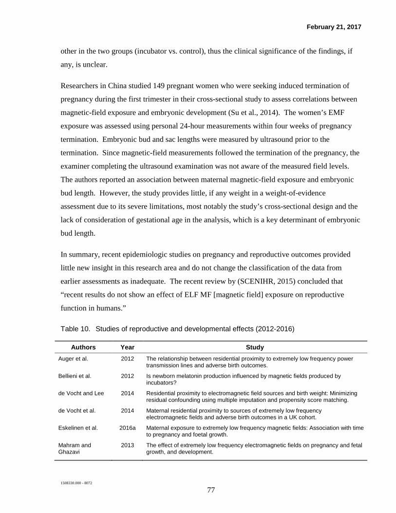
February 21, 2017
1508330.000 - 8072 77
other in the two groups (incubator vs. control), thus the clinical significance of the findings, if
any, is unclear.
Researchers in China studied 149 pregnant women who were seeking induced termination of
pregnancy during the first trimester in their cross-sectional study to assess correlations between
magnetic-field exposure and embryonic development (Su et al., 2014). The women’s EMF
exposure was assessed using personal 24-hour measurements within four weeks of pregnancy
termination. Embryonic bud and sac lengths were measured by ultrasound prior to the
termination. Since magnetic-field measurements followed the termination of the pregnancy, the
examiner completing the ultrasound examination was not aware of the measured field levels.
The authors reported an association between maternal magnetic-field exposure and embryonic
bud length. However, the study provides little, if any weight in a weight-of-evidence
assessment due to its severe limitations, most notably the study’s cross-sectional design and the
lack of consideration of gestational age in the analysis, which is a key determinant of embryonic
bud length.
In summary, recent epidemiologic studies on pregnancy and reproductive outcomes provided
little new insight in this research area and do not change the classification of the data from
earlier assessments as inadequate. The recent review by (SCENIHR, 2015) concluded that
“recent results do not show an effect of ELF MF [magnetic field] exposure on reproductive
function in humans.”
Table 10. Studies of reproductive and developmental effects (2012-2016)
Authors Year Study Auger et al. 2012 The relationship between residential proximity to extremely low frequency power
transmission lines and adverse birth outcomes.
Bellieni et al. 2012 Is newborn melatonin production influenced by magnetic fields produced by incubators?
de Vocht and Lee 2014 Residential proximity to electromagnetic field sources and birth weight: Minimizing residual confounding using multiple imputation and propensity score matching.
de Vocht et al. 2014 Maternal residential proximity to sources of extremely low frequency electromagnetic fields and adverse birth outcomes in a UK cohort.
Eskelinen et al. 2016a Maternal exposure to extremely low frequency magnetic fields: Association with time to pregnancy and foetal growth.
Mahram and Ghazavi
2013 The effect of extremely low frequency electromagnetic fields on pregnancy and fetal growth, and development.
February 21, 2017
1508330.000 - 8072 78
Authors Year Study Shamsi Mahmoudabadi et al.
2013 Exposure to Extremely Low Frequency Electromagnetic Fields during Pregnancy and the Risk of Spontaneous Abortion: A Case-Control Study.
Su et al. 2014 Correlation between exposure to magnetic fields and embryonic development in the first trimester.
Wang et al. 2013 Residential exposure to 50 Hz magnetic fields and the association with miscarriage risk: a 2-year prospective cohort study.
Comments on Eskelinen et al.
de Vocht and Burstyn
2016 Comments on "Maternal exposure to extremely low frequency magnetic fields: Association with time to pregnancy and foetal growth."
Eskelinen et al. 2016b Reply to Comment on "Maternal exposure to extremely low frequency magnetic fields: Association with time to pregnancy and foetal growth."
4.3 Neurodegenerative disease
Research into the possible effect of magnetic fields on neurodegenerative diseases began in
1995, and the majority of research since then has focused on Alzheimer’s disease and a specific
type of motor neuron disease called ALS, which is also known as Lou Gehrig’s disease. Based
on the initial findings on the Alzheimer’s disease the NRPB concluded in 2001 that there was
“only very weak evidence to suggest that it [ELF magnetic fields] could cause Alzheimer’s
disease” (NRPB, 2001b, p. 21). Early studies on ALS also reported an association between
ALS mortality among workers with certain electrical occupations. The review panels, however,
were hesitant to conclude that the associations provided strong support for a causal relationship
because they felt that an alternative explanation (i.e., electric shocks received at work) may be
the source of the observed association.
Also including more recent studies, the WHO panel concluded that there is “inadequate” data in
support of an association between magnetic fields and Alzheimer’s disease or ALS. They stated
that “When evaluated across all the studies, there is only very limited evidence of an association
between estimated ELF exposure and [Alzheimer’s or ALS] disease risk” (WHO 2007, p. 194).
While a subsequent meta-analysis also reported an association between occupational EMF
exposure and Alzheimer’s disease (Garcia et al., 2008), its conclusion was necessarily limited
by the quality of the studies included in the analysis. A Swiss study that was the first to
examine residential proximity to high-voltage power lines and neurodegenerative disease,
February 21, 2017
1508330.000 - 8072 79
reported an increase in Alzheimer’s disease mortality among people living with 50 meters of
transmission lines, but observed no association for ALS, Parkinson’s disease, and multiple
sclerosis (Huss et al., 2009) spurred interest in research on Alzheimer’s disease. Based on a
review of the evidence that also considered these studies, the Health Council of the Netherlands
EFHRAN review still considered the evidence as “inadequate” for all forms of
neurodegenerative diseases (EFHRAN, 2012).
Relevant studies published since 2012
In recent years, a number of epidemiologic studies have examined the potential association
between both occupational and residential EMF exposure and the development of
neurodegenerative diseases in Denmark, the Netherlands, Switzerland, the United States, and
Sweden. In Denmark, researchers conducted a population-based case-control study to examine
neurodegenerative diseases risk in relation to residential distance to power lines between 1994
and 2010 (Frei et al., 2013). Geographical information systems were used to determine distance
from the nearest power line for the residential addresses of all newly diagnosed cases and
matched controls. The authors reported no consistent associations for any of the investigated
diseases, including Alzheimer disease and other types of dementia, ALS, Parkinson’s disease, or
multiple sclerosis with residential proximity to power lines. The inclusion of newly-diagnosed
cases, identified through the Danish national hospital discharge database, represents a
significant methodological improvement over mortality studies (e.g., Huss et al., 2009). The
study, however, was limited by the methods used for the exposure assessment (i.e., residential
distance to high-voltage power lines).
Dutch researchers included 1,139 ALS cases diagnosed between 2006 and 2013 and 2,864
frequency-matched controls in their population-based case-control study (Seelen et al., 2014).
The shortest distance from the cases’ and controls’ addresses to the nearest high-voltage power
line (50 ‒ 380 kV) was determined by geocoding. The authors reported no statistically
significant associations between residential proximity to power lines with any of the included
voltages and ALS. An ad hoc analysis that combined the current results (Seelen et al., 2014)
with results from two previously published studies (Marcilio et al., 2011; Frei et al., 2013)
resulted in an overall OR of 0.9 (95% CI 0.7-1.1) for living within 200 meters of a high-voltage
February 21, 2017
1508330.000 - 8072 80
power line. Reconstruction of lifetime residential history represents a methodological
improvement of the current study. The main limitation, similarly to previous power-line
studies, is the use of distance to power lines as a surrogate for magnetic-field exposure.
Another case-control study conducted in the Netherlands identified 444 cases of Parkinson’s
disease and 876 matched controls from hospital records between 2006 and 2011(van der Mark
et al., 2015). Occupational exposure to EMF and electric shocks was ascertained from
questionnaire-based information on work history and corresponding job-exposure matrices. The
authors reported no associations between any of the exposure metrics and Parkinson’s disease.
Two publications from the previously described Netherlands Cohort Study that enrolled
approximately 120,000 men and women in the Netherlands in 1986 with a follow up until 2003
examined the occurrence of neurodegenerative diseases in relation to occupational exposure to
EMF (Koeman et al., 2015; Brouwer et al., 2015). In one study, the researchers identified 798
male and 1,171 female cases of non-vascular dementia (Koeman et al., 2015). Questionnaire
based information on lifetime occupational history and various job-exposure matrices on
occupational exposures to solvents, pesticides, metals, ELF magnetic fields, electric shocks, and
diesel exhaust were used for exposure assessment. The authors reported no association for
exposure to electric shocks, and reported moderate, statistically non-significant, associations for
the highest estimates of exposures to metals, chlorinated solvents, and ELF magnetic fields.
Based on no observed exposure-response relationship cumulative exposure, the authors
concluded that the association noted for ELF magnetic fields and solvents might be attributable
to confounding by exposure to metals. In the same cohort, Brouwer et al. (2015) identified 609
cases of Parkinson’s disease. Based on their results, the authors concluded that their findings do
not support the hypothesis that the investigated occupational exposures, including EMF,
increase mortality from Parkinson’s disease.
Researchers from Switzerland analyzed data of approximately 2.2 million subjects enrolled in
the Swiss National Cohort study to examine the potential relationship between occupational
exposure to EMF and electric shocks and ALS mortality from 2000 to 2008 (Huss et al., 2014).
Study subjects’ exposures were classified using job-exposure matrices and occupations reported
for the study subjects in the 1990 and 2000 censuses. A total of 278 cases of ALS were
February 21, 2017
1508330.000 - 8072 81
identified in the cohort. The authors reported an association with medium and high estimates of
ELF EMF exposure, but not with estimates of exposure to electric shocks.
In a cross-sectional study of 3,050 elderly subjects in the United States, the authors reported a
statistically significant association between estimated occupational magnetic-field exposure and
severe cognitive dysfunction (Davanipour et al., 2014). Information on occupational history,
and socio-demographic variables were obtained by in-person interviews. Occupational
exposure to magnetic fields was classified as low, medium, and high. The mini-mental state
exam was used to evaluate cognitive function. The reported association is, however, difficult to
interpret due to the number of severe limitations of the study; these limitations include the cross-
sectional nature of the study, the lack of clear clinical diagnosis for case-definition, the
rudimentary assessment of exposure to occupational EMF, and the reliance on questionnaire-
based information to assess exposure in a population with substantial cognitive decline. Yu et
al. (2014), also conducted in the United States, included 66 cases and 66 controls in a small
case-control study that examined various lifestyle, environmental, and work-related variables as
potential risk factors for ALS. Their results on occupational EMF exposure, however, cannot be
meaningfully interpreted because of a severe error of combining estimates of ionizing and non-
ionizing radiation exposures in their analysis.
Using mortality data in the United States between 1991 and 1999, Vergara et al. (2015)
conducted a case-control study of occupational exposure to electric shock and magnetic fields
and ALS. The researchers identified a total of 5,886 deaths due to ALS, and, for each ALS
death, they selected 10 controls from among other deaths, matched on sex, age, year of death,
and region. The occupation reported on the death certificates were linked to job exposure
matrices for electric shocks and magnetic fields, and classified as high, medium, and low.
Occupations classified as “electric occupations” were moderately associated with ALS (OR
1.23, 95% CI, 1.04-1.47). Electric shocks, however, were inversely related to ALS (OR 0.73,
95% CI, 0.67-0.79 in high exposure, and OR 0.90, 95% CI, 0.84-0.97 in medium exposure
compared to low exposure), and no statistically significant associations were reported between
EMF and ALS (OR 1.09, 95% CI, 1.00-1.19 in high exposure, and OR 1.09, 95% CI, 0.96-1.23
in medium exposure compared to low exposure). The authors concluded that their findings did
February 21, 2017
1508330.000 - 8072 82
not support that exposure to either electric shocks or magnetic fields explained the observed
association of ALS with “electric occupations.”
Fischer et al. (2015) conducted a population-based case-control study of occupational exposure
to electric shocks and magnetic fields and ALS in Sweden. The base population of the study
included all individuals born in Sweden between 1901 and 1970 who were enumerated during
the 1990 Swedish Census. All cases of ALS in the study population, newly diagnosed between
1990 and 2010, were identified by record linkages to the Swedish patient and death registries.
Five controls, individually matched to cases on birth year and sex, were selected for each case
from the study base. Census-based information on occupations was linked to multiple
previously developed job-exposure matrices to classify exposure to EMF and electric shocks for
cases and controls. A total of 4,709 cases and 23,335 controls were included in the study.
Overall, neither EMF nor electric shocks were related to ALS. Among subjects aged < 65 years,
statistically significant increases in ALS risk were reported with exposure to electric shocks. A
statistically non-significant decrease, however, was also observed among subjects 65 years and
older. The study has a number of strengths, which include its large sample size, population-
based design, inclusion of incidence cases, and the reliance on multiple job-exposure matrices
(three for EMF and two for electric shocks) for exposure assessment.
Recently published meta-analyses of occupational exposure to ELF magnetic fields and
neurodegenerative disease reported weak to no evidence of an association (Zhou et al., 2012;
Vergara et al., 2013; Capozzella et al., 2014; Huss et al., 2015). The authors of these meta-
analyses concluded that potential within-study biases, evidence of publication bias, and
uncertainties in the various exposure assessments greatly limit the ability to infer an association,
if any, between occupational exposure to magnetic fields and neurodegenerative disease.
Overall, these recent meta-analyses provide no convincing evidence of a relationship between
ELF magnetic fields and neurodegenerative disease.
The suggestion that the weak and inconsistent association between ELF EMF and ALS might be
explained by electric shocks encountered in occupational environments was not supported by
findings reported in recently published studies (Das et al., 2012; Grell et al., 2012; van der Mark
et al., 2015; Vergara et al., 2015; Fischer et al., 2015).
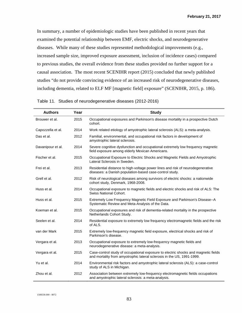
February 21, 2017
1508330.000 - 8072 83
In summary, a number of epidemiologic studies have been published in recent years that
examined the potential relationship between EMF, electric shocks, and neurodegenerative
diseases. While many of these studies represented methodological improvements (e.g.,
increased sample size, improved exposure assessment, inclusion of incidence cases) compared
to previous studies, the overall evidence from these studies provided no further support for a
causal association. The most recent SCENIHR report (2015) concluded that newly published
studies “do not provide convincing evidence of an increased risk of neurodegenerative diseases,
including dementia, related to ELF MF [magnetic field] exposure” (SCENIHR, 2015, p. 186).
Table 11. Studies of neurodegenerative diseases (2012-2016)
Authors Year Study Brouwer et al. 2015 Occupational exposures and Parkinson's disease mortality in a prospective Dutch
cohort.
Capozzella et al. 2014 Work related etiology of amyotrophic lateral sclerosis (ALS): a meta-analysis.
Das et al. 2012 Familial, environmental, and occupational risk factors in development of amyotrophic lateral sclerosis.
Davanipour et al. 2014 Severe cognitive dysfunction and occupational extremely low frequency magnetic field exposure among elderly Mexican Americans.
Fischer et al. 2015 Occupational Exposure to Electric Shocks and Magnetic Fields and Amyotrophic Lateral Sclerosis in Sweden.
Frei et al. 2013 Residential distance to high-voltage power lines and risk of neurodegenerative diseases: a Danish population-based case-control study.
Grell et al. 2012 Risk of neurological diseases among survivors of electric shocks: a nationwide cohort study, Denmark, 1968-2008.
Huss et al. 2014 Occupational exposure to magnetic fields and electric shocks and risk of ALS: The Swiss National Cohort.
Huss et al. 2015 Extremely Low Frequency Magnetic Field Exposure and Parkinson's Disease--A Systematic Review and Meta-Analysis of the Data.
Koeman et al. 2015 Occupational exposures and risk of dementia-related mortality in the prospective Netherlands Cohort Study.
Seelen et al. 2014 Residential exposure to extremely low frequency electromagnetic fields and the risk of ALS.
van der Mark 2015 Extremely low-frequency magnetic field exposure, electrical shocks and risk of Parkinson's disease.
Vergara et al. 2013 Occupational exposure to extremely low-frequency magnetic fields and neurodegenerative disease: a meta-analysis.
Vergara et al. 2015 Case-control study of occupational exposure to electric shocks and magnetic fields and mortality from amyotrophic lateral sclerosis in the US, 1991-1999.
Yu et al. 2014 Environmental risk factors and amyotrophic lateral sclerosis (ALS): a case-control study of ALS in Michigan.
Zhou et al. 2012 Association between extremely low-frequency electromagnetic fields occupations and amyotrophic lateral sclerosis: a meta-analysis.
February 21, 2017
1508330.000 - 8072 84
5 Electromagnetic hypersensitivity
The WHO 2007 report discussed anecdotal accounts of persons who reported that they could
perceive EMF at levels below accepted thresholds and accounts of persons who believed they
had developed a variety of symptoms including sleep disturbances, general fatigue, difficulty
concentrating, dizziness, and eyestrain due to EMF exposure. Based on double-blind studies of
human volunteers, office workers, and self-reported hypersensitive individuals, however, the
WHO review concluded that the perception of EMF and health complaints are not related to
exposure. Neither healthy volunteers nor self-identified hypersensitive individuals can reliably
distinguish field exposure from sham-exposure. Also, no exposure-related differences were
observed in levels of stress hormones or inflammatory mediators. The WHO proposed that
electromagnetic hypersensitivity should more appropriately be termed “idiopathic
environmental intolerance (IEI) with attribution to EMF” and explained that “[t]hese symptoms
are not explained by any known medical, psychiatric or psychological disorder, and the term IEI
has no medical diagnostic value. IEI individuals cannot detect EMF exposure any more
accurately than non-IEI individuals, and well-controlled and conducted double-blind studies
have consistently shown that their symptoms are not related to EMF exposure per se” (WHO,
2007, p. 137).
Studies published following the WHO review, overall, supported the conclusion that ELF EMF
is not detected by self-identified sensitive subjects or other subjects, and that symptoms are not
reliably elicited by exposure to ELF magnetic or electric fields over a range of exposure levels.
The 2012 EFHRAN report also concluded that the available evidence suggests the lack of an
effect on people with “electrical hypersensitivity” (EFHRAN, 2012).
Relevant studies published since 2012
A review of the literature related to IEI attributable to EMF (IEI-EMF) highlighted the poorly
defined nature of IEI-EMF and the considerable heterogeneity in criteria identifying people with
IEI-EMF across published studies (Baliatsas et al., 2012a). Development of a uniform
definition might be helpful for future research in this area, and may enable more active
February 21, 2017
1508330.000 - 8072 85
involvement of medical practitioners in assisting these individuals. A systematic review of
observational studies reported no association between actual EMF and non-specific symptoms
in the general population, but suggested some association with “perceived” exposure (Baliatsas
et al., 2012b). Two observational studies from the Netherlands also reported an association
between non-specific symptoms and “perceived” exposure; the authors, however, cautioned
against drawing causal conclusions based on their results (Baliatsas et al., 2015, Bolte et al.,
2015). Authors generally attribute the reported associations with perceived exposure to the
“nocebo” effect (i.e., symptoms explained by unconscious psychological reaction as a result of
the expectation of an effect, rather than the effect of the exposure itself), and report the co-
occurrence of other psychological symptoms among people with IEI-EMF (Nordin et al., 2014;
Domotor et al., 2016; Kjellqvist et al., 2016; Dieudonne, 2016; Porsius et al., 2016). A recent
double-blind randomized controlled trial provided further confirmation that subjects with self-
identified electromagnetic hypersensitivity were not able to detect exposure to EMF better than
chance (van Moorselaar et al., 2016). An Italian study of 30 IEI-EMF subjects and 25 control
subjects reported no differences in melatonin levels between the two groups, despite
significantly lower sleep quality scores among IEI-EMF individuals (Andrianome et al., 2016).
Another study reported statistically significant differences in certain metabolic parameters
among individuals with multiple-chemical sensitivity and electromagnetic hypersensitivity
compared to control subjects, however, the clinical significance of these differences is uncertain,
and the findings remain to be replicated by independent laboratories (De Luca et al., 2014).
Researchers from Hungary reported that individuals with IEI-EMF, as opposed to controls, were
able to detect the presence of 50 Hz magnetic fields to “some extent” or to “small extent,” but
reporting of symptoms by these individuals were related to perceived exposure (Koteles et al.,
2012; Szemerszky et al., 2015). Blinding was not clearly described in these experiments, and
the findings are yet to be replicated by other scientists.
In summary, recent studies did not provide new sufficient evidence to change the overall
conclusion that either self-identified individuals with electromagnetic hypersensitivity or
members of the general populations can detect EMF exposure encountered in our environment,
or that general non-specific symptoms are related to EMF exposure. The recent SCENIHR
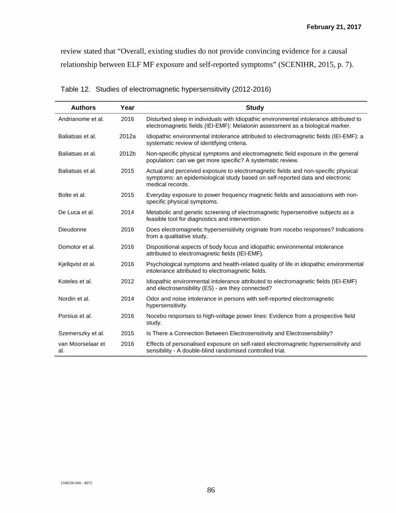
February 21, 2017
1508330.000 - 8072 86
review stated that “Overall, existing studies do not provide convincing evidence for a causal
relationship between ELF MF exposure and self-reported symptoms” (SCENIHR, 2015, p. 7).
Table 12. Studies of electromagnetic hypersensitivity (2012-2016)
Authors Year Study Andrianome et al. 2016 Disturbed sleep in individuals with Idiopathic environmental intolerance attributed to
electromagnetic fields (IEI-EMF): Melatonin assessment as a biological marker.
Baliatsas et al. 2012a Idiopathic environmental intolerance attributed to electromagnetic fields (IEI-EMF): a systematic review of identifying criteria.
Baliatsas et al. 2012b Non-specific physical symptoms and electromagnetic field exposure in the general population: can we get more specific? A systematic review.
Baliatsas et al. 2015 Actual and perceived exposure to electromagnetic fields and non-specific physical symptoms: an epidemiological study based on self-reported data and electronic medical records.
Bolte et al. 2015 Everyday exposure to power frequency magnetic fields and associations with non-specific physical symptoms.
De Luca et al. 2014 Metabolic and genetic screening of electromagnetic hypersensitive subjects as a feasible tool for diagnostics and intervention.
Dieudonne 2016 Does electromagnetic hypersensitivity originate from nocebo responses? Indications from a qualitative study.
Domotor et al. 2016 Dispositional aspects of body focus and idiopathic environmental intolerance attributed to electromagnetic fields (IEI-EMF).
Kjellqvist et al. 2016 Psychological symptoms and health-related quality of life in idiopathic environmental intolerance attributed to electromagnetic fields.
Koteles et al. 2012 Idiopathic environmental intolerance attributed to electromagnetic fields (IEI-EMF) and electrosensibility (ES) - are they connected?
Nordin et al. 2014 Odor and noise intolerance in persons with self-reported electromagnetic hypersensitivity.
Porsius et al. 2016 Nocebo responses to high-voltage power lines: Evidence from a prospective field study.
Szemerszky et al. 2015 Is There a Connection Between Electrosensitivity and Electrosensibility?
van Moorselaar et al.
2016 Effects of personalised exposure on self-rated electromagnetic hypersensitivity and sensibility - A double-blind randomised controlled trial.
February 21, 2017
1508330.000 - 8072 87
6 Possible Effects of ELF Electric and Magnetic Fields on Implanted Cardiac Devices
The sensing system of pacemakers and implanted cardioverter-defibrillators (ICD) is designed
to be responsive to the heart’s electrical signal. For this reason, other electrical signals can
potentially interfere with the normal functioning of pacemakers and ICDs, a phenomenon called
electromagnetic interference (EMI). Most sources of EMF are too weak to affect a pacemaker
or ICD; however, EMF from certain sources (e.g., some appliances and industrial equipment)
may cause interference. This section considers potential EMI associated with ELF EMF to
implanted cardiac devices such as pacemakers and defibrillators.
In the presence of electromagnetic fields, devices can respond in different ways, defined as
modes. The likelihood of interference occurring and the mode of the response depend on the
parameters (e.g., strength, frequency, duty cycle) of the interfering signal, the patient’s
orientation in the electromagnetic field, the exact location of the device, and the variable
parameters of the device that are specific to a patient. Modern devices incorporate various
technological safeguards (e.g., shielding by titanium casing and electrical filtering) to minimize
the potential for EMI (Dyrda and Khairy, 2008). Experimental research has been conducted to
assess whether interference may occur when currents are induced in the patient’s body by
environmental electric fields and magnetic fields.
In the absence of specific recommendations from medical device manufacturers, the American
Conference of Governmental Industrial Hygienists (ACGIH) suggested exposure levels to
prevent pacemaker EMI. For electric fields, the ACGIH suggested keeping exposures below 1
kV/m, and for magnetic fields, they recommended exposure not exceed 1 G (ACGIH, 2001;
ACGIH, 2009). These recommendations are general in nature and do not address that classes of
pacemakers from some manufacturers are quite immune to interference even at levels much
greater than these recommended guidelines. The ACGIH also recommended that patients
consult their physicians and the respective pacemaker manufacturers before following any
organizations’ guidelines.
February 21, 2017
1508330.000 - 8072 88
Manufacturers of pacemakers and other implantable devices will typically follow the AAMI
PC69:2007 or ANSI/AAMI/ISO 14117:2012 (North America) or IEC 45502-2-1:2003 / IEC
45502-2-2:2003 (Europe) standards. These standards require a test to verify that the function of
the cardiac device is not affected to at least a 2 millivolt peak-to-peak signal applied to the
sensing electrodes. This test verifies immunity of a cardiac device25 of at least 0.83 G (root-
mean-square magnetic field) at 60 Hz. At 60 Hz, the reference levels in EC 519/99 (also known
as 1999/519/EC) are 0.83 G and 4.167 kV/m—the standard assumes that only an electric or
magnetic field is present at any time (CEU, 1999).
Moreover, the standard procedure (EN 50527-1:2010) to assess EMF exposure for workers with
active implantable medical devices (AIMD) states that the “risk assessment is based on the
approach that AIMDs are expected to work uninfluenced as long as the General Public
Reference levels of 1999/519/EC (except for static magnetic fields) are not exceeded …, where
the AIMD has been implanted and programmed following good medical practice” (CENELEC,
2010). The procedure recommended by this standard contains steps for assessing that the field
levels of EC 519/99 are not exceeded and that AIMD patients do not have higher than normal
sensitivity settings on their device for clinical reasons.
Previous studies indicated occurrence of pacing abnormalities at magnetic-field levels that are
much higher than the levels a person would encounter on a daily basis. While electric fields did
produce interference at levels that can be produced by certain electrical sources (Toivonen et al.,
1991; Astridge et al., 1993; Scholten and Silny, 2001; Joosten et al., 2009), most pacemakers
were not affected by high levels of electric fields (up to 20 kV/m) and did not exhibit any pacing
abnormalities. Joosten et al. (2009) showed that the most sensitive unipolar pacemakers may be
affected by electric-field levels between 4.3 kV/m and 6.2 kV/m; however, most modern
pacemakers are bipolar devices, which are designed specifically to reduce the potential for EMI.
Joosten et al. (2009), for example, found that in Germany in 2007, only 6% of the pacemakers in
use had a unipolar sensing system.
25 The magnetic-field value is calculated using an average area (225 cm2) of a unipolar cardiac device. In rare
cases, such as for a large patient with a unipolar implant, the immunity may be lower. For a patient with a bipolar lead configuration, the immunity will be higher.
February 21, 2017
1508330.000 - 8072 89
Relevant studies published since 2012
Much of the recent scientific research that evaluated potential interference with pacemakers and
other implanted cardiac devices focused on possible interference from dental, medical, surgical,
diagnostic, and therapeutic equipment (e.g., Zaphiratos et al., 2013; Maheshwari et al., 2015;
Magnani et al., 2014; Yoshida et al., 2014) or personal electronic devices (e.g., Misiri et al.,
2012; Kozik et al., 2014). While some of these reports indicate the possibility of interference in
certain scenarios with these equipment devices, these interference scenarios are not relevant to
the electricity infrastructure environments due to differences in many factors, including, most
importantly, the proximity of the interfering signal sources and the intensity and frequency of
the interfering electromagnetic fields.
Tiikkaja et al. (2013a) tested 11 volunteers with pacemakers and 13 volunteers with ICDs in an
experimental setting at ELF magnetic-field levels up to 3,000 mG. Frequencies tested in the
experimental setting ranged from 2 to 200 Hz. No interference was observed with ICDs or
pacemakers with bipolar sensing, while three pacemakers with unipolar sensing experienced
some form of interference. The authors note that magnetic-field intensities used in their study
are rare even in industrial environments, and the public is unlikely to encounter such high
magnetic fields. The same research team also tested 11 volunteers with pacemakers in
environments near EMF sources, including overhead high-voltage transmission lines, an
electrically powered commuter train, and mobile phone base stations; none of the pacemakers
experienced interference in any of these exposure situations (Tiikkaja et al., 2013b). An earlier
report by the same research group also reported that, in most cases, no interference occurred at
magnetic field levels below the ICNIRP occupational safety limits (Tiikkaja et al., 2012).
Researchers in Germany and the Netherlands (Napp et al., 2014) evaluated interference
thresholds for 110 patients with ICDs in an experimental setting. Patients were exposed to
single and combined 50-Hz electric fields and magnetic fields with strengths of up to 30 kV/m
and 25,500 mG (25.5 G), respectively. Tests were conducted with ICD devices set to maximum
and normal sensitivities. No interference was detected for either electric fields or magnetic
fields below European Union (1999/519/EC) exposure limits for the general public (5 kV/m and
1,000 mG). With normal sensitivity, no interference was detected with any of the ICD devices
February 21, 2017
1508330.000 - 8072 90
in fields up to about 5,000 mG (5 G) and about 9‒10 kV/m. The authors concluded that ELF
EMF fields typically encountered in daily life do no not interfere with ICDs. High fields that
may be present in some occupational environments, however, may cause inappropriate sensing
in some ICD devices.
Researchers in Finland studied potential interference of ICDs of older designs (>10 years old) in
human shaped phantoms (Korpinen et al., 2014). They reported potential interference at
exposure levels above the current European Union limits for one unit out of the investigated 10
units. The authors were not able to replicate the interference in the following day with the same
unit. The use of phantoms instead of humans and testing of older designs limit the interpretation
of the authors’ findings.
Researchers in Germany investigated EMI events among 2,940 patients with ICDs between
2005 and 2013 (von Olshausen et al., 2016). They reported 48 out-of-hospital EMI events
occurring in 18 patients out of the total number of 2,940 ICD patients. Only one of the events
was clinically significant and was related to close proximity to the engine of a lawnmower; two
other events were deemed potentially significant (related to direct contact with electric current
and proximity to a mobile phone); while the remaining 45 events were of minor significance and
were not noticed by the patients. Another 97 events were also reported in the hospital
environment in the same cohort of patients; nearly all clinically significant events in hospitals
were related to electrocautery. None of the EMI events were reported to be related to proximity
to power lines or substations.
Table 13. Studies of EMI (2012-2016)
Authors Year Study
Korpinen et al. 2014 Implantable cardioverter defibrillators in electric and magnetic fields of 400 kV power lines.
Kozik et al. 2014 iPad2(R) use in patients with implantable cardioverter defibrillators causes electromagnetic interference: the EMIT Study.
Magnani et al. 2014 Lack of interference of electromagnetic navigation bronchoscopy to implanted cardioverter-defibrillator: in-vivo study.
Maheshwari et al. 2015 Evaluating the effects of different dental devices on implantable cardioverter defibrillators.
Misiri et al 2012 Electromagnetic interference and implanted cardiac devices: the nonmedical environment (part I).
February 21, 2017
1508330.000 - 8072 91
Authors Year Study
Napp et al. 2014 Electromagnetic interference with implantable cardioverter-defibrillators at power frequency: an in vivo study.
Tiikkaja et al. 2012 Interference of low frequency magnetic fields with implantable cardioverter-defibrillators.
Tiikkaja et al. 2013a Testing of common electromagnetic environments for risk of interference with cardiac pacemaker function.
Tiikkaja et al. 2013b Electromagnetic interference with cardiac pacemakers and implantable cardioverter-defibrillators from low-frequency electromagnetic fields in vivo.
von Olshausen et al.
2016 Electromagnetic interference in implantable cardioverter defibrillators: present but rare.
Yoshida et al. 2014 Electromagnetic interference of implantable cardiac devices from a shoulder massage machine.
Zaphiratos et al. 2013 Magnetic interference of cardiac pacemakers from a surgical magnetic drape.
February 21, 2017
1508330.000 - 8072 92
7 Fauna and Flora Research
7.1 Fauna
Previous Exponent reports reviewed the relevant research and concluded that the research to
date did not suggest that electric or magnetic fields result in any adverse effects on the health,
behavior, or productivity of fauna, including livestock such as cows, sheep, and pigs, a variety
of small mammals, deer, elk, birds, or bees. Results of studies published since 2012 have not
provided substantive new evidence that would alter previous conclusions.
7.2 Flora
Previous Exponent reports described the body of research on the possible effects of EMF on
forest species and agriculture crops, concluding that researchers have found no adverse effects
on plant responses at the levels of EMF produced by high-voltage transmission lines, excluding
some corona-related effects from high-voltage lines on the growth of nearby trees. Results of
studies published since 2012 have not provided substantive new evidence that would alter
previous conclusions.
February 21, 2017
1508330.000 - 8072 93
Glossary
Association – An association is a measure of how things vary together. They are measured by odds ratios and relative risks.
Basic restriction – The basic restriction is the electric field level or current density inside the body that is recommended as a limit to protect exposed populations. The term is used in standards or guidelines that recommend exposure limits.
Bias – Bias refers to any error in the design, conduct or analysis of a study that results in a distorted estimate of an exposure’s effect on the risk of disease. For example, the characteristics of persons selected by telephone calls to participate in a study may not accurately reflect those of the entire community and this can introduce error into the study’s findings.
Carcinogenesis – Carcinogenesis describes the process of the progression of normal cells to cancerous cells.
Causation or cause – A cause is an exposure or condition of the individual that has been proven through a sound weight-of-evidence review to increase risk of a disease.
Cause-and-effect relationship – A cause-and-effect relationship between an exposure and a disease is a statistically significant association that is determined through a weight-of-evidence review to be causal in nature.
Case-control study – A case-control study compares persons without a disease (controls) to persons with a disease (cases) to see if they differ on any factors or exposures of interest.
Chance – Chance refers to random sampling variation, like a coincidence. An association can be observed between an exposure and disease that is simply the result of a chance occurrence.
Cohort study – A cohort study follows a group of people over a long period of time to observe whether the occurrence of disease differs among exposed and unexposed persons in the group.
Confidence interval – A confidence interval is a range of values for an estimate of effect that has a specified probability (e.g., 95%) of including the “true” estimate of effect. A 95% confidence interval indicates that, if the study were conducted a very large number of times, 95% of the measured estimates would be within the upper and lower confidence limits.
Confounding – Confounding is a situation in which an association is distorted because the exposure is associated with other risk factors for the disease. For example, a link between coffee drinking in mothers and low birth weight babies has been reported in the past. However, some women who drink coffee also smoke cigarettes. It was found that when the smoking habits of the mothers are taken into account, coffee drinking was not associated with low birth weight babies because of the confounding effect of smoking.
February 21, 2017
1508330.000 - 8072 94
Dose-response assessment/relationship – Data from scientific research in which a change in amount, intensity, or duration of exposure is associated with a change in risk of a specified outcome. A pattern of a stronger association with increasing exposure, or dose.
Electric field – The electric field is a property of a location or point in space and its electrical environment, and describes the forces that would be experienced by a charged body in that space by virtue of its charge. The electric field is expressed in measurement units of volts per meter (V/m) or kilovolts per meter (kV/m); a kilovolt per meter is equal to 1,000 V/m. Electromagnetic spectrum – The range of wavelengths of electromagnetic energy, including visible light, arranged by frequency. Wavelength decreases with increasing frequency; the ELF range includes the power frequencies of 50/60-Hz.
Electromagnetic hypersensitivity – Self-reported responses to or perception of electromagnetic fields, including ELF-EMF, at levels far below exposure limits that may include a wide range symptoms, including sleep disturbances, general fatigue, difficulty in concentrating, dizziness, and eyestrain.
Epidemiology – The study of the frequency and distribution of disease and health events in human populations and the factors that contribute to disease and health events.
Exposure assessment – The step in risk assessment that characterizes the exposure circumstances of the situation under analysis.
Extremely low frequency (ELF) fields – Extremely low frequency refers to electromagnetic fields in the range of 0-300 Hz.
Hazard identification – The identification of adverse effects on health from a specific exposure based on a weight-of-evidence review of the scientific research.
In vitro – Laboratory studies of isolated cells that are artificially maintained in test tubes or culture dishes are called in vitro studies, literally “in glass.” Researchers expose isolated cells or groups of cells (tissues) to a specific agent under controlled conditions. These studies help explain the mechanisms by which exposures might affect biological processes.
In vivo – Studies in living animals or experimental studies of processes in whole living organisms are called in vivo studies. Scientists expose laboratory animals to a specific agent under controlled conditions and look for effects on body function, measures of health, or disease. Experience has shown that effects in laboratory animals can help to predict effects that occur in people.
Initiation – The first stage in the development of cancer, initiation typically results from exposure to an agent that can cause mutations in a cell. Initiation is believed to be irreversible, and increases the likelihood of cancer occurring.
Job-exposure matrix – A job-exposure matrix cross-classifies job titles and exposure estimates. Job-exposure matrices are used to estimate cumulative occupational exposure (e.g., magnetic field exposure) based on an individual’s job history.
February 21, 2017
1508330.000 - 8072 95
Magnetic fields – The magnetic field is a state of region in space, and describes the forces that would be experienced by a moving charge (or magnetic material) in proportion to its charge and velocity. The strength of magnetic fields is expressed as magnetic flux density in units called gauss (G), or in milligauss (mG), where 1 G = 1,000 mG. Meta-analysis – An analytic technique that combines the results of many studies into one summary estimate of the association between a particular exposure and disease.
Nested case-control study – A case-control study in which the cases and controls are drawn from a cohort study’s population.
Odds ratio – An odds ratio is a measure of association that describes the ratio of the odds of exposure among persons with a disease to the odds of exposure among persons without a disease. For example, an odds ratio of two would suggest that persons with the disease are two times more likely to have had exposure than persons without the disease.
Pooled analysis – A pooled analysis combines individual-level data across many studies and analyzes the data together to get a summary estimate of the association between a particular exposure and disease.
Precautionary principle – The precautionary principle refers to the idea that, when evidence does not support the suggestion that an exposure is a cause of a particular disease but where a risk is perceived, precautionary measures may be taken that are proportional to the perceived level of risk, with science as the basis for measuring that risk.
Promotion – Promotion is a later stage in cancer development, following initiation. If there is sufficient exposure to the agent, promoters increase the frequency of tumor formation that occurs after initiation.
Reference level – The reference level is a measurable level of electric or magnetic field outside of the body that is used as a screening value. It is a practical measure to determine whether the internal level identified as the basic restriction is likely to be exceeded.
Relative risk – A relative risk is an estimate that compares the risk of disease among persons who are exposed to the risk of disease among persons who are unexposed. For example, a relative risk of two means that that exposed persons in the study is two times more likely to develop the disease than unexposed persons.
Risk characterization – A quantitative estimation of the likelihood of adverse effects that may result from exposure to a specific agent in a specific situation.
Safety factor – A multiplicative factor (usually less than 1.0) incorporated into risk assessments or safety standards to allow for unpredictable types of variation, such as variability in responses from test animals to humans or person-to-person variability.
Selection bias – Selection bias occurs when there are differences in the type of person who participates in the study compared to the type of person who doesn’t participate in the study.
February 21, 2017
1508330.000 - 8072 96
Selection bias introduces systematic error into a study, and limits the conclusions and generalizations that can be drawn.
Spot measurement – A spot measurement is an instantaneous magnetic or electric field reading that is taken at one location as an estimate of exposure.
Statistically significant – An association is statistically significant if one can conclude (with an established level of confidence using standard statistical tests) that the association is not due to a chance occurrence.
Time-weighted average (TWA) - The average exposure over a given specified time period (i.e., an 8-hr workday or a 24-hr day) of a person’s exposure to a chemical or physical agent. The average is determined by sampling the exposure of interest throughout the time period.
Voltage – Voltage is the difference in electric potential between any two conductors of a circuit. It is the electric ‘pressure’ that exists between two points and is capable of producing the flow of current through an electrical conductor.
Weight-of-evidence review – A weight-of-evidence review critically evaluates the strength of the evidence for causality for a particular exposure and disease. It entails a comprehensive assessment of all relevant scientific research, in which each of the studies is critically evaluated, and more weight is given to studies of better quality.
Wire code categories – Wire coding categories are based on a classification system of homes using characteristics of power lines outside the home (e.g., thickness of the wires) and their distances from the home. This information is used to code the homes into categories based on their predicted magnetic field level.
February 21, 2017
1508330.000 - 8072 97
References
Ahlbom A, Day N, Feychting M, Roman E, Skinner J, Dockerty J, Linet M, McBride M, Michaelis J, Olsen JH, Tynes T, Verkasalo PK. A pooled analysis of magnetic fields and childhood leukaemia. Br J Cancer 83: 692-8, 2000.
Akdag MZ, Dasdag S, Cakir DU, Yokus B, Kizil G, Kizil M. Do 100- and 500-muT ELF magnetic fields alter beta-amyloid protein, protein carbonyl and malondialdehyde in rat brains? Electromagn Biol Med 32: 363-72, 2013.
Alcaraz M, Olmos E, Alcaraz-Saura M, Achel DG, Castillo J. Effect of long-term 50 Hz magnetic field exposure on the micronucleated polychromatic erythrocytes of mice. Electromagn Biol Med 33: 51-7, 2014.
American Conference of Government Industrial Hygienists (ACGIH). Documentation of the Threshold Limit Values and Biological Exposure Indices, 7th edition. Publication No. 0100. . Cincinnati, Ohio: ACGIH, 2001.
American National Standards Institute, Association for the Advancement of Medical Instrumentation (AAMI). Active implantable medical devices : electromagnetic compatibility : EMC test protocols for implantable cardiac pacemakers and implantable cardioverter defibrillators. Arlington, VA: Association for the Advancement of Medical Instrumentation, 2007.
Anderson LE, Boorman GA, Morris JE, Sasser LB, Mann PC, Grumbein SL, Hailey JR, McNally A, Sills RC, Haseman JK. Effect of 13 week magnetic field exposures on DMBA-initiated mammary gland carcinomas in female Sprague-Dawley rats. Carcinogenesis 20: 1615-20, 1999.
Andrianome S, Hugueville L, de Seze R, Hanot-Roy M, Blazy K, Gamez C, Selmaoui B. Disturbed sleep in individuals with Idiopathic environmental intolerance attributed to electromagnetic fields (IEI-EMF): Melatonin assessment as a biological marker. Bioelectromagnetics 2016 [epub].
Armstrong BG, Deadman J, McBride ML. The determinants of Canadian children's personal exposures to magnetic fields. Bioelectromagnetics 22: 161-9, 2001.
Astridge PS, Kaye GC, Whitworth S, Kelly P, Camm AJ, Perrins EJ. The response of implanted dual chamber pacemakers to 50 Hz extraneous electrical interference. Pacing Clin Electrophysiol 16: 1966-74, 1993.
Auger N, Joseph D, Goneau M, Daniel M. The relationship between residential proximity to extremely low frequency power transmission lines and adverse birth outcomes. J Epidemiol Community Health 65: 83-5, 2010.
Baldi I, Coureau G, Jaffre A, Gruber A, Ducamp S, Provost D, Lebailly P, Vital A, Loiseau H,
February 21, 2017
1508330.000 - 8072 98
Salamon R. Occupational and residential exposure to electromagnetic fields and risk of brain tumors in adults: a case-control study in Gironde, France. Int J Cancer 129: 1477-84, 2011.
Baliatsas C, Van Kamp I, Lebret E, Rubin GJ. Idiopathic environmental intolerance attributed to electromagnetic fields (IEI-EMF): a systematic review of identifying criteria. BMC Public Health 12: 643, 2012a.
Baliatsas C, Van Kamp I, Bolte J, Schipper M, Yzermans J, Lebret E. Non-specific physical symptoms and electromagnetic field exposure in the general population: can we get more specific? A systematic review. Environ Int 41: 15-28, 2012b.
Baliatsas C, Bolte J, Yzermans J, Kelfkens G, Hooiveld M, Lebret E, van Kamp I. Actual and perceived exposure to electromagnetic fields and non-specific physical symptoms: an epidemiological study based on self-reported data and electronic medical records. Int J Hyg Environ Health 218: 331-44, 2015.
Baum A, Mevissen M, Kamino K, Mohr U, Löscher W. A histopathological study on alterations in DMBA-induced mammary carcinogenesis in rats with 50 Hz, 100 mT magnetic field exposure. Carcinogenesis 16:119-125, 1995.
Bellieni CV, Tei M, Iacoponi F, Tataranno ML, Negro S, Proietti F, Longini M, Perrone S, Buonocore G. Is newborn melatonin production influenced by magnetic fields produced by incubators? Early Hum Dev 88: 707-10, 2012.
Belson M, Kingsley B, Holmes A. Risk factors for acute leukemia in children: a review. Environ Health Perspect 115: 138-45, 2007.
Berg H, Gunther B, Hilger I, Radeva M, Traitcheva N, Wollweber L. Bioelectromagnetic field effects on cancer cells and mice tumors. Electromagn Biol Med 29: 132-43, 2010.
Bernard N, Alberdi AJ, Tanguy ML, Brugere H, Helissey P, Hubert C, Gendrey N, Guillosson JJ, Nafziger J. Assessing the potential leukemogenic effects of 50 Hz magnetic fields and their harmonics using an animal leukemia model. J Radiat Res (Tokyo) 49: 565-77, 2008.
Bolte JF, Baliatsas C, Eikelboom T, van Kamp I. Everyday exposure to power frequency magnetic fields and associations with non-specific physical symptoms. Environ Pollut 196: 224-9, 2015.
Bonnet-Belfais M, Lambrozo J, Aurengo A. Comment: childhood leukaemia and power lines--the Geocap study: is proximity an appropriate MF exposure surrogate? Br J Cancer 109: 1382-3, 2013.
Boorman GA, Anderson LE, Morris JE, Sasser LB, Mann PC, Grumbein SL, Hailey JR, McNally A, Sills RC, Haseman JK. Effect of 26 week magnetic field exposures in a DMBA initiation-promotion mammary gland model in Sprague-Dawley rats. Carcinogenesis 20: 899-904, 1999a.
Boorman GA, McCormick DL, Findlay JC, Hailey JR, Gauger JR, Johnson TR, Kovatch RM,
February 21, 2017
1508330.000 - 8072 99
Sills RC, Haseman JK. Chronic toxicity/oncogenicity evaluation of 60 Hz (power frequency) magnetic fields in F344/N rats. Toxicol Pathol 27: 267-78, 1999b.
Brouwer M, Koeman T, van den Brandt PA, Kromhout H, Schouten LJ, Peters S, Huss A, Vermeulen R. Occupational exposures and Parkinson's disease mortality in a prospective Dutch cohort. Occup Environ Med 72: 448-55, 2015.
Buffler PA, Kwan ML, Reynolds P, Urayama KY. Environmental and genetic risk factors for childhood leukemia: appraising the evidence. Cancer Invest 23: 60-75, 2005.
Bunch KJ, Keegan TJ, Swanson J, Vincent TJ, Murphy MF. Residential distance at birth from overhead high-voltage powerlines: childhood cancer risk in Britain 1962-2008. Br J Cancer 110: 1402-8, 2014.
Bunch KJ, Swanson J, Vincent TJ, Murphy MF. Magnetic fields and childhood cancer: an epidemiological investigation of the effects of high-voltage underground cables. J Radiol Prot 35: 695-705, 2015.
Bunch KJ, Swanson J, Vincent TJ, Murphy MF. Epidemiological study of power lines and childhood cancer in the UK: further analyses. J Radiol Prot 36: 437-455, 2016.
Capozzella A, Sacco C, Chighine A, Loreti B, Scala B, Casale T, Sinibaldi F, Tomei G, Giubilati R, Tomei F, Rosati MV. Work related etiology of amyotrophic lateral sclerosis (ALS): a meta-analysis. Ann Ig 26: 456-72, 2014.
Chen Q, Lang L, Wu W, Xu G, Zhang X, Li T, Huang H. A Meta-Analysis on the Relationship between Exposure to ELF-EMFs and the Risk of Female Breast Cancer. PLoS One 8: e69272, 2013.
Cherian SB, Bairy KL, Rao MS. Induction of micronuclei in hippocampal neuarons exposed to chronic prenatal restraint stress in male and female wistar rat pups. Int J Adv Res 3: 1217-1222, 2015.
Chung MK, Kim YB, Ha CS, Myung SH. Lack of a co-promotion effect of 60 Hz rotating magnetic fields on N-ethyl-N-nitrosourea induced neurogenic tumors in F344 rats. Bioelectromagnetics 29: 539-48, 2008.
Chung MK, Yu WJ, Kim YB, Myung SH. Lack of a co-promotion effect of 60 Hz circularly polarized magnetic fields on spontaneous development of lymphoma in AKR mice. Bioelectromagnetics 31: 130-9, 2010.
Clavel J, Sermage-Faure C, Demoury C, Rudant J, Goujon-Bellec S, Guyot-Goubin A, Deschamps F, Hemon D. Reply: comment on 'Childhood leukaemia close to high-voltage power lines--the Geocap study, 2002-2007'--is proximity an appropriate MF exposure surrogate? Br J Cancer 109: 1383-4, 2013.
Coble JB, Dosemeci M, Stewart PA, Blair A, Bowman J, Fine HA, Shapiro WR, Selker RG, Loeffler JS, Black PM, Linet MS, Inskip PD. Occupational exposure to magnetic fields and the
February 21, 2017
1508330.000 - 8072 100
risk of brain tumors. Neuro Oncol 11: 242-9, 2009.
Council of the European Union (CEU). Council recommendation of 12 July 1999 on the limitation of exposure of the general public to electromagnetic fields (0 Hz to 300 GHz) - 1999/519/EC. Official Journal of the European Communities L199: 59-70, 1999.
Crespi CM, Vergara XP, Hooper C, Oksuzyan S, Wu S, Cockburn M, Kheifets L. Childhood leukaemia and distance from power lines in California: a population-based case-control study. Br J Cancer 115: 122-8, 2016.
Cui Y, Ge Z, Rizak JD, Zhai C, Zhou Z, Gong S, Che Y. Deficits in water maze performance and oxidative stress in the hippocampus and striatum induced by extremely low frequency magnetic field exposure. PLoS One 7: e32196, 2012.
Das K, Nag C, Ghosh M. Familial, environmental, and occupational risk factors in development of amyotrophic lateral sclerosis. N Am J Med Sci 4: 350-5, 2012.
Davanipour Z, Tseng CC, Lee PJ, Markides KS, Sobel E. Severe cognitive dysfunction and occupational extremely low frequency magnetic field exposure among elderly Mexican Americans. Br J Med Med Res 4: 1641-1662, 2014.
De Bont R and van Larebeke N. Endogenous DNA damage in humans: a review of quantitative data. Mutagenesis 19: 169-85, 2004.
De Luca C, Thai JC, Raskovic D, Cesareo E, Caccamo D, Trukhanov A, Korkina L. Metabolic and genetic screening of electromagnetic hypersensitive subjects as a feasible tool for diagnostics and intervention. Mediators Inflamm 2014: 924184, 2014.
de Vocht F. Adult cancers near high-voltage power lines. Epidemiology 24: 782, 2013.
de Vocht F, Hannam K, Baker P, Agius R. Maternal residential proximity to sources of extremely low frequency electromagnetic fields and adverse birth outcomes in a UK cohort. Bioelectromagnetics 35: 201-9, 2014.
de Vocht F and Lee B. Residential proximity to electromagnetic field sources and birth weight: Minimizing residual confounding using multiple imputation and propensity score matching. Environ Int 69: 51-7, 2014.
de Vocht F and Burstyn I. Comments on "Maternal exposure to extremely low frequency magnetic fields: Association with time to pregnancy and foetal growth". Environ Int 96: 190-191, 2016.
Deadman JE, Armstrong BG, McBride ML, Gallagher R, Theriault G. Exposures of children in Canada to 60-Hz magnetic and electric fields. Scand J Work Environ Health 25: 368-75, 1999.
Dechent D and Driessen S. Re: Role of Electromagnetic Field Exposure in Childhood Acute Lymphoblastic Leukemia and No Impact of Urinary Alpha- Amylase - a Case Control Study in Tehran, Iran. Asian Pac J Cancer Prev 17: 877-8, 2016.
February 21, 2017
1508330.000 - 8072 101
Deng Y, Zhang Y, Jia S, Liu J, Liu Y, Xu W, Liu L. Effects of aluminum and extremely low frequency electromagnetic radiation on oxidative stress and memory in brain of mice. Biol Trace Elem Res 156: 243-52, 2013.
Dieudonne M. Does electromagnetic hypersensitivity originate from nocebo responses? Indications from a qualitative study. Bioelectromagnetics 37: 14-24, 2016.
Domotor Z, Doering BK, Koteles F. Dispositional aspects of body focus and idiopathic environmental intolerance attributed to electromagnetic fields (IEI-EMF). Scand J Psychol 57: 136-43, 2016.
Draper G, Vincent T, Kroll ME, Swanson J. Childhood cancer in relation to distance from high voltage power lines in England and Wales: a case-control study. BMJ 330: 1290, 2005.
Duan W, Liu C, Wu H, Chen C, Zhang T, Gao P, Luo X, Yu Z, Zhou Z. Effects of exposure to extremely low frequency magnetic fields on spermatogenesis in adult rats. Bioelectromagnetics 35: 58-69, 2014.
Dyrda K, Khairy P. Implantable rhythm devices and electromagnetic interference: myth or reality? Expert Rev Cardiovasc Ther 6: 823-32, 2008.
Eden T. Aetiology of childhood leukaemia. Cancer Treat Rev 36: 286-97, 2010.
El-Bialy NS and Rageh MM. Extremely low-frequency magnetic field enhances the therapeutic efficacy of low-dose cisplatin in the treatment of Ehrlich carcinoma. Biomed Res Int 2013: 189352, 2013.
Elliott P, Shaddick G, Douglass M, de Hoogh K, Briggs DJ, Toledano MB. Adult cancers near high-voltage overhead power lines. Epidemiology 24: 184-90, 2013.
Eskelinen T, Roivainen P, Makela P, Juutilainen J. Reply to Comment on "Maternal exposure to extremely low frequency magnetic fields: Association with time to pregnancy and foetal growth". Environ Int 96: 192, 2016.
Eskelinen T, Roivainen P, Makela P, Keinanen J, Kauhanen O, Saarikoski S, Juutilainen J. Maternal exposure to extremely low frequency magnetic fields: Association with time to pregnancy and foetal growth. Environ Int 94: 620-5, 2016.
European Committee for Electrotechnical Standardization (CENELEC). Procedure for the assessment of the exposure to electromagnetic fields of workers bearing active implantable medical devices - Part 1: General (EN 50527-1). Brussels, Belgium: CENELEC, 2010
European Health Risk Assessment Network on Electromagnetic Fields Exposure (EFHRAN). Report on the Analysis of Risks Associated to Exposure to EMF: In Vitro and In Vivo (Animals) Studies. Milan, Italy 2010.
European Health Risk Assessment Network on Electromagnetic Fields Exposure (EFHRAN). Risk Analysis of Human Exposure to Electromagnetic Fields (Revised). Report D2 of the

February 21, 2017
1508330.000 - 8072 102
EFHRAN Project. Milan, Italy: EFHRAN, 2012.
Exponent, Inc. EMF and Health – Review and Update of the Scientific Research. Prepared for BC Hydro, Inc., Exponent, 2007.
Exponent, Inc. EMF and Health – Review and Update of the Scientific Research, September 2007 through January 2010. Prepared for BC Hydro, Inc., Exponent, 2010.
Exponent, Inc. EMF and Health – Comprehensive Review and Update of the Scientific Research, January 15, 2010 through March 1, 2012. \Prepared for BC Hydro, Inc., Exponent, 2012.
Fam WZ, Mikhail EL. Lymphoma induced in mice chronically exposed to very strong low-frequency electromagnetic field. Cancer Lett 105: 257-69, 1996.
Federal-Provincial-Territorial Radiation Protection Committee (FPTRPC). 2008. "Response Statement to Public Concerns Regarding Electric and Magnetic Fields (EMFs) from Electrical Power Transmission and Distribution Lines." Accessed January 24, 2017. http://www.hc-sc.gc.ca/ewh-semt/radiation/fpt-radprotect/emf-cem-eng.php.
Federal-Provincial-Territorial Radiation Protection Working Group. Health Effects and Exposure Guidelines Related to Extremely Low Frequency Electric and Magnetic Fields - An Overview. Ottawa: FPTRPC Committee, 2005.
Fedrowitz M and Löscher W. Exposure of Fischer 344 rats to a weak power frequency magnetic field facilitates mammary tumorigenesis in the DMBA model of breast cancer. Carcinogenesis 29: 186-93, 2008.
Fedrowitz M and Löscher W. Gene expression in the mammary gland tissue of female Fischer 344 and Lewis rats after magnetic field exposure (50 Hz, 100 muT) for 2 weeks. Int J Radiat Biol 88: 425-9, 2012.
Feychting M. Invited commentary: extremely low-frequency magnetic fields and breast cancer--now it is enough! Am J Epidemiol 178: 1046-50, 2013.
Fischer H, Kheifets L, Huss A, Peters TL, Vermeulen R, Ye W, Fang F, Wiebert P, Vergara XP, Feychting M. Occupational Exposure to Electric Shocks and Magnetic Fields and Amyotrophic Lateral Sclerosis in Sweden. Epidemiology 26: 824-30, 2015.
Flint M, McCarty K, Jenkins F, Conrads T, Sun M, Baum A. Psychological stress accelerates the onset of tumour formation and alters the type and location of tumours in a DMBA mouse carcinogenesis model. Stress and Health 27: e129-e138, 2011.
Foliart DE, Pollock BH, Mezei G, Iriye R, Silva JM, Ebi KL, Kheifets L, Link MP, Kavet R. Magnetic field exposure and long-term survival among children with leukaemia. Br J Cancer 94: 161-4, 2006.
Frei P, Poulsen AH, Mezei G, Pedersen C, Cronberg Salem L, Johansen C, Roosli M, Schuz J.
February 21, 2017
1508330.000 - 8072 103
Residential distance to high-voltage power lines and risk of neurodegenerative diseases: a Danish population-based case-control study. Am J Epidemiol 177: 970-8, 2013.
Gallastegi M, Guxens M, Jimenez-Zabala A, Calvente I, Fernandez M, Birks L, Struchen B, Vrijheid M, Estarlich M, Fernandez MF, Torrent M, Ballester F, Aurrekoetxea JJ, Ibarluzea J, Guerra D, Gonzalez J, Roosli M, Santa-Marina L. Characterisation of exposure to non-ionising electromagnetic fields in the Spanish INMA birth cohort: study protocol. BMC Public Health 16: 167, 2016.
Garcia AM, Sisternas A, Hoyos SP. Occupational exposure to extremely low frequency electric and magnetic fields and Alzheimer disease: a meta-analysis. Int J Epidemiol 37: 329-40, 2008.
Gift JS, Caldwell JC, Jinot J, Evans MV, Cote I, Vandenberg JJ. Scientific considerations for evaluating cancer bioassays conducted by the Ramazzini Institute. Environ Health Perspect 121: 1253-63, 2013.
Glinka M, Sieroń A, Birkner E, Cieślar G. Influence of extremely low-frequency magnetic field on the activity of antioxidant enzymes during skin wound healing in rats. Electromagn Biol Med 32:463-70, 2013.
Gobba F, Bravo G, Rossi P, Contessa GM, Scaringi M. Occupational and environmental exposure to extremely low frequency-magnetic fields: a personal monitoring study in a large group of workers in Italy. J Expo Sci Environ Epidemiol 21: 634-645, 2011.
Government of Canada, Privy Council Office. A framework for the application of precaution in science-based decision making about risk. Ottawa: Privy Council Office, 2003.
Greenland S, Sheppard AR, Kaune WT, Poole C, Kelsh MA. A pooled analysis of magnetic fields, wire codes, and childhood leukemia. Childhood Leukemia-EMF Study Group. Epidemiology 11: 624-34, 2000.
Greenland S and Kheifets L. Leukemia attributable to residential magnetic fields: results from analyses allowing for study biases. Risk Anal 26: 471-482, 2006.
Grell K, Meersohn A, Schuz J, Johansen C. Risk of neurological diseases among survivors of electric shocks: a nationwide cohort study, Denmark, 1968-2008. Bioelectromagnetics 33: 459-65, 2012.
Grellier J, Ravazzani P, Cardis E. Potential health impacts of residential exposures to extremely low frequency magnetic fields in Europe. Environ Int 62: 55-63, 2014.
Grundy A, Harris SA, Demers PA, Johnson KC, Agnew DA, Villeneuve PJ. Occupational exposure to magnetic fields and breast cancer among Canadian men. Cancer Med 5: 586-96, 2016.
Harris AW, Basten A, Gebski V, Noonan D, Finnie J, Bath ML, Bangay MJ, Repacholi MH. A test of lymphoma induction by long-term exposure of E mu-Pim1 transgenic mice to 50 Hz magnetic fields. Radiat Res 149:300-307, 1998.
February 21, 2017
1508330.000 - 8072 104
Hassan NS, Abdelkawi SA. Assessing of plasma protein denaturation induced by exposure to cadmium, electromagnetic fields and their combined actions on rat. Electromagn Biol Med 33: 147-53, 2014.
Health Canada. Human Health Risk Assessment for Priority Substances. Environmental Health Directorate. Canadian Environmental Protection Act. Ottawa: Health Canada, 1994.
Health Canada. Health Canada Decision-Making Framework for Identifying, Assessing, and Managing Health Risk. Ottawa, Canada: Health Canada, 2000.
Health Canada. Limits of Human Exposure to Radiofrequency Electromagnetic Energy in the Frequency Range from 3 kHz to 300 GHz. Safety Code 6 (2015). Ottawa, Ontario: Health Canada, 2015.
Health Council of the Netherlands (HCN) ELF Electromagnetic Fields Committee. Electromagnetic Fields: Annual Update 2001. Publication No. 2001/14. The Hague: Health Council of the Netherlands, 2001.
Health Council of the Netherlands (HCN) ELF Electromagnetic Fields Committee. Electromagnetic Fields: Annual Update 2003. Publication No. 2004/01. The Hague: Health Council of the Netherlands, 2004.
Health Council of the Netherlands (HCN) ELF Electromagnetic Fields Committee. Electromagnetic Fields: Annual Update 2005. Publication No. 2005/14. The Hague: Health Council of the Netherlands, 2005.
Health Council of the Netherlands (HCN) ELF Electromagnetic Fields Committee. Electromagnetic Fields: Annual Update 2006. Publication No. 2007/06. The Hague: Health Council of the Netherlands, 2007.
Health Council of the Netherlands (HCN) ELF Electromagnetic Fields Committee. Electromagnetic Fields: Annual Update. Publication No. 2009/02. The Hague: Health Council of the Netherlands, 2009.
Health Council of the Netherlands (HCN) ELF Electromagnetic Fields Committee. Advisory letter - Power lines and Alzheimer's disease to the Minister of Housing, Spatial Planning and the Environment. The Hague: Health Council of the Netherlands, 2009.
Health Protection Agency (HPA). Power frequency electromagnetic fields, melatonin and the risk of breast cancer: report of an independent advisory group on non-ionising radiation. Documents of the Health Protection Agency. Series B: Radiation, Chemical and Environmental Hazards. Oxfordshire: Health Protection Agency, 2006.
Hill AB. The Environment and Disease: Association or Causation? Proc R Soc Med 58: 295-300, 1965.
Huss A, Spoerri A, Egger M, Röösli M. Residence near power lines and mortality from neurodegenerative diseases: longitudinal study of the Swiss population. Am J Epidemiol 169:
February 21, 2017
1508330.000 - 8072 105
167-75, 2009.
Huss A, Spoerri A, Egger M, Kromhout H, Vermeulen R, for The Swiss National Cohort. Occupational exposure to magnetic fields and electric shocks and risk of ALS: The Swiss National Cohort. Amyotroph Lateral Scler Frontotemporal Degener: 1-6, 2014.
Huss A, Koeman T, Kromhout H, Vermeulen R. Extremely Low Frequency Magnetic Field Exposure and Parkinson's Disease--A Systematic Review and Meta-Analysis of the Data. Int J Environ Res Public Health 12: 7348-56, 2015.
International Agency for Research on Cancer (IARC). Mechanisms for Carcinogenesis in Risk Identification. Lyon, France: IARC Press, 1992.
International Agency for Research on Cancer (IARC). IARC Monographs on the Evaluation of Carcinogenic Risks to Humans. Volume 80: Static and Extremely Low-Frequency (ELF) Electric and Magnetic Fields. Lyon, France: IARC Press, 2002.
International Agency for Research on Cancer (IARC). IARC Monographs on the Evaluation of Carcinogenic Risks to Humans. Preamble. Lyon, France: IARC, Press, 2006.
International Commission for Non-Ionizing Radiation Protection (ICNIRP). ICNIRP Statement. General Approach to Protection Against Non-Ionizing Radiation. Health Phys 82: 540-548, 2002.
International Commission on Non-Ionizing Radiation Protection (ICNIRP). Guidelines for limiting exposure to time-varying electric, magnetic, and electromagnetic fields (up to 300 GHz). Health Phys 74: 494-522, 1998.
International Commission on Non-Ionizing Radiation Protection (ICNIRP). Exposure to Static and Low Frequency Electromagnetic Fields, Biological Effects and Health Consequences (0-100 kHz) – Review of the Scientific Evidence on Dosimetry, Biological Effects, Epidemiological Observations, and Health Consequences Concerning Exposure to Static and Low Frequency Electromagnetic Fields (0-100 kHz). Matthes R, McKinlay AF, Bernhardt JH, Vecchia P, Beyret B (eds.). Munich: ICNIRP, 2003.
International Commission on Non-ionizing Radiation Protection (ICNIRP). Guidelines for limiting exposure to time-varying electric and magnetic fields (1 Hz to 100 kHz). Health Phys 99: 818-36, 2010.
International Committee on Electromagnetic Safety (ICES). IEEE Standard for Safety Levels with Respect to Human Exposure to Electromagnetic Fields 0 to 3 kHz. Piscataway, NJ: IEEE, 2002.
International Electrotechnical Commission (IEC). Active Implantable Medical Devices Part 2-1: Particular Requirements for Active Implantable Medical Devices Intended to Treat Bradyarrhythmia (Cardiac Pacemakers). Geneva, Switzerland: IEC, 2003a.
International Electrotechnical Commission (IEC). Active Implantable Medical Devices Part 2-2:
February 21, 2017
1508330.000 - 8072 106
Particular Requirements for Active Implantable Medical Devices Intended to Treat Tachyarrhythmia (Includes Implantable Defibrillators). Geneva, Switzerland: IEC, 2003b.
Jeffers D. Comment on 'Residential distance at birth from overhead high-voltage powerlines: childhood cancer risk in Britain 1962-2008'. Br J Cancer 111: 2199-200, 2014.
Jeffers D. Comment on: Childhood cancer and exposure to corona ions from power lines: an epidemiological study. J Radiol Prot 35: 481-3, 2015.
Jiménez -Garcia MN, Robledo JA, Aparicio-Bautists DI, Rodriguez-Segura MA, Villa-Trevino S, Godina-Nava JJ. Anti-proliferative effect of extremely low frequency electromagnetic field on preneoplastic lesions formation in the rat liver. BMC Cancer 10: 1-12, 2010.
Jirik V, Pekarek L, Janout V, Tomaskova H. Association between childhood leukaemia and exposure to power-frequency magnetic fields in Middle Europe. Biomed Environ Sci 25: 597-601, 2012.
Johansen C, Raaschou Nielsen O, Olsen JH, Schüz J. Risk for leukaemia and brain and breast cancer among Danish utility workers: a second follow-up. Occup Environ Med 64: 782-4, 2007.
Johnson JC and Thaul SE (Eds.). An Evaluation of Radiation Exposure Guidance for Military Operations. Washington, D.C.: National Academies Press, 1997.
Joosten S, Pammler K, Silny J. The influence of anatomical and physiological parameters on the interference voltage at the input of unipolar cardiac pacemakers in low frequency electric fields. Phys Med Biol 54: 591-609, 2009.
Karipidis KK. Survey of residential power-frequency magnetic fields in Melbourne, Australia. Radiat Prot Dosimetry 163: 81-91, 2015.
Keegan TJ, Bunch KJ, Vincent TJ, King JC, O'Neill KA, Kendall GM, MacCarthy A, Fear NT, Murphy MF. Case-control study of paternal occupation and childhood leukaemia in Great Britain, 1962-2006. Br J Cancer 107: 1652-9, 2012.
Kennedy EK, McNamee JP, Lalonde LP, Jones T, Wilkinson D. Acellular comet assay: A tool for assessing variable influencing the akaline comet assay. Radiat Prot Dosim 148: 155-161, 2012.
Kheifets LI, Afifi AA, Buffler PA, Zhang ZW. Occupational electric and magnetic field exposure and brain cancer: a meta-analysis. J Occup Environ Med 37: 1327-41, 1995.
Kheifets L, Swanson J, Greenland S. Childhood leukemia, electric and magnetic fields, and temporal trends. Bioelectromagnetics 27: 545-52, 2006.
Kheifets L, Monroe J, Vergara X, Mezei G, Afifi AA. Occupational electromagnetic fields and leukemia and brain cancer: an update to two meta-analyses. J Occup Environ Med 50: 677-88, 2008.
February 21, 2017
1508330.000 - 8072 107
Kheifets L, Bowman JD, Checkoway H, Feychting M, Harrington JM, Kavet R, Marsh G, Mezei G, Renew DC, van Wijngaarden E. Future needs of occupational epidemiology of extremely low frequency electric and magnetic fields: review and recommendations. Occup Environ Med 66: 72-80, 2009.
Kheifets L, Ahlbom A, Crespi CM, Draper G, Hagihara J, Lowenthal RM, Mezei G, Oksuzyan S, Schüz J, Swanson J, Tittarelli A, Vinceti M, Wunsch Filho V. Pooled analysis of recent studies on magnetic fields and childhood leukaemia. Br J Cancer 103: 1128-35, 2010a.
Kheifets L, Ahlbom A, Crespi CM, Feychting M, Johansen C, Monroe J, Murphy MF, Oksuzyan S, Preston-Martin S, Roman E, Saito T, Savitz D, Schüz J, Simpson J, Swanson J, Tynes T, Verkasalo P, Mezei G. A pooled analysis of extremely low-frequency magnetic fields and childhood brain tumors. Am J Epidemiol 172: 752-61, 2010b.
Kheifets L, Crespi CM, Hooper C, Oksuzyan S, Cockburn M, Ly T, Mezei G. Epidemiologic study of residential proximity to transmission lines and childhood cancer in California: description of design, epidemiologic methods and study population. J Expo Sci Environ Epidemiol 25: 45-52, 2015.
Kiray A1, Tayefi H, Kiray M, Bagriyanik HA, Pekcetin C, Ergur BU, Ozogul C. The effects of exposure to electromagnetic field on rat myocardium. Toxicol Ind Health 29:418-25, 2013.
Kjellqvist A, Palmquist E, Nordin S. Psychological symptoms and health-related quality of life in idiopathic environmental intolerance attributed to electromagnetic fields. J Psychosom Res 84: 8-12, 2016.
Koeman T, van den Brandt PA, Slottje P, Schouten LJ, Goldbohm RA, Kromhout H, Vermeulen R. Occupational extremely low-frequency magnetic field exposure and selected cancer outcomes in a prospective Dutch cohort. Cancer Causes Control 25: 203-14, 2014.
Koeman T, Schouten LJ, van den Brandt PA, Slottje P, Huss A, Peters S, Kromhout H, Vermeulen R. Occupational exposures and risk of dementia-related mortality in the prospective Netherlands Cohort Study. Am J Ind Med 58: 625-35, 2015.
Korpinen L, Kuisti H, Elovaara J, Virtanen V. Implantable cardioverter defibrillators in electric and magnetic fields of 400 kV power lines. Pacing Clin Electrophysiol 37: 297-303, 2014.
Korr H, Angstman NB, Born TB, Bosse K, Brauns B, Demmler M, Fueller K, Kántor O, Kever BM, Rahimyar N, Salimi S, Silny J, Schmitz C. No evidence of persisting unrepaired nuclear DNA single strand breaks in distinct types of cells in the brain, kidney, and liver of adult mice after continuous eight-week 50 Hz magnetic field exposure with flux density of 0.1 mT or 1.0 mT. PLoS One 9:e109774, 2014.
Koteles F, Szemerszky R, Gubanyi M, Kormendi J, Szekrenyesi C, Lloyd R, Molnar L, Drozdovszky O, Bardos G. Idiopathic environmental intolerance attributed to electromagnetic fields (IEI-EMF) and electrosensibility (ES) - are they connected? Int J Hyg Environ Health 216: 362-70, 2013.
February 21, 2017
1508330.000 - 8072 108
Kozik TM, Chien G, Connolly TF, Grewal GS, Liang D, Chien W. iPad2(R) use in patients with implantable cardioverter defibrillators causes electromagnetic interference: the EMIT Study. J Am Heart Assoc 3: e000746, 2014.
Lai H and Singh NP. Magnetic-field-induced DNA strand breaks in brain cells of the rat. Environ Health Perspect 112: 687-94, 2004.
Lee GM, Neutra RR, Hristova L, Yost M, Hiatt RA. A nested case-control study of residential and personal magnetic field measures and miscarriages. Epidemiology 13: 21-31, 2002.
Leitgeb N. Childhood leukemia not linked with EMF magnetic fields. J Electromagn Anal 6: 174-183, 2014.
Leitgeb N. Synoptic Analysis Clarifies Childhood Leukemia Risk from ELF Magnetic Field Exposure. J Electromagn Anal 7: 245-258, 2015.
Lewis RC, Evenson KR, Savitz DA, Meeker JD. Temporal variability of daily personal magnetic field exposure metrics in pregnant women. J Expo Sci Environ Epidemiol 25: 58-64, 2015.
Li DK, Odouli R, Wi S, Janevic T, Golditch I, Bracken TD, Senior R, Rankin R, Iriye R. A population-based prospective cohort study of personal exposure to magnetic fields during pregnancy and the risk of miscarriage. Epidemiology 13: 9-20, 2002.
Li W, Ray RM, Thomas DB, Yost M, Davis S, Breslow N, Gao DL, Fitzgibbons ED, Camp JE, Wong E, Wernli KJ, Checkoway H. Occupational exposure to magnetic fields and breast cancer among women textile workers in Shanghai, China. Am J Epidemiol 178: 1038-45, 2013.
Linet MS, Hatch EE, Kleinerman RA, Robison LL, Kaune WT, Friedman DR, Severson RK, Haines CM, Hartsock CT, Niwa S, Wacholder S, Tarone RE. Residential exposure to magnetic fields and acute lymphoblastic leukemia in children. N Engl J Med 337: 1-7, 1997.
Liorni I, Parazzini M, Struchen B, Fiocchi S, Roosli M, Ravazzani P. Children's Personal Exposure Measurements to Extremely Low Frequency Magnetic Fields in Italy. Int J Environ Res Public Health 13, 2016.
Löscher W, Mevissen M, Lehmacher W, Stamm A. Tumor promotion in a breast cancer model by exposure to a weak alternating magnetic field. Cancer Lett 71: 75-81, 1993.
Löscher W, Wahnschaffe U, Mevissen M, Lerchl A, Stamm A. Effects of weak alternating magnetic fields on nocturnal melatonin production and mammary carcinogenesis in rats. Oncology 51: 288-295, 1994.
Löscher W and Mevissen M. Linear relationship between flux density and tumor co-promoting effect of prolonged magnetic field exposure in a breast cancer model. Cancer Lett 96: 175-80, 1995.
Löscher W, Mevissen M, Haussler B. Seasonal influence on 7,12-dimethylbenz[a]anthracene-

February 21, 2017
1508330.000 - 8072 109
induced mammary carcinogenesis in Spague-Dawley rats under controlled laboratory conditions. Pharmacol Toxicol 81: 265-270, 1997.
Ma X, Urayama K, Chang J, Wiemels JL, Buffler PA. Infection and pediatric acute lymphoblastic leukemia. Blood Cells Mol Dis 42: 117-20, 2009.
Maes A and Verschaeve L. Genetic damage in humans exposed to extremely low-frequency electromagnetic fields. Arch Toxicol 90: 2337-48, 2016.
Magnani A, Balbo P, Facchini E, Occhetta E, Marino P. Lack of interference of electromagnetic navigation bronchoscopy to implanted cardioverter-defibrillator: in-vivo study. Europace 16: 1767-71, 2014.
Magnani C, Mattioli S, Miligi L, Ranucci A, Rondelli R, Salvan A, Bisanti L, Masera G, Rizzari C, Zambon P, Cannizzaro S, Gafa L, Luzzatto LL, Benvenuti A, Michelozzi P, Kirchmayer U, Cocco P, Biddau P, Galassi C, Celentano E, Guarino E, Assennato G, de Nichilo G, Merlo DF, Bocchini V, Pannelli F, Mosciatti P, Minelli L, Chiavarini M, Cuttini M, Casotto V, Torregrossa MV, Valenti RM, Forastiere F, Haupt R, Lagorio S, Risica S, Polichetti A. SETIL: Italian multicentric epidemiological case-control study on risk factors for childhood leukaemia, non hodgkin lymphoma and neuroblastoma: study population and prevalence of risk factors in Italy. Ital J Pediatr 40: 103, 2014.
Maheshwari KR, Nikdel K, Guillaume G, Letra AM, Silva RM, Dorn SO. Evaluating the effects of different dental devices on implantable cardioverter defibrillators. J Endod 41: 692-5, 2015.
Mahna A, Firoozabadi SM, Shankayi Z. The effect of ELF magnetic field on tumor growth after electrochemotherapy. J Membr Biol 247: 9-15, 2014.
Mahram M and Ghazavi M. The effect of extremely low frequency electromagnetic fields on pregnancy and fetal growth, and development. Arch Iran Med 16: 221-4, 2013.
Malvandi AM, Haddad F, Moghimi A. Acute restraint stress increases the frequency of vinblastine-induced micronuclei in mouse bone marrow cells. Stress 13: 276-80, 2010.
Mandeville R, Franco E, Sidrac-Ghali S, Paris-Nadon L, Rocheleau N, Mercier G, Desy M, Gaboury L. Evaluation of the potential carcinogenicity of 60 Hz linear sinusoidal continuous-wave magnetic fields in Fisher F344 rats. FASEB Journal 11:1127-1136, 1997.
Manikonda PK, Rajendra P, Devendranath D, Gunasekaran B, Channakeshava, Aradhya SR, Sashidhar RB, Subramanyam C. Extremely low frequency magnetic fields induce oxidative stress in rat brain. Gen Physiol Biophys 33: 81-90, 2014.
Marcilio I, Gouveia N, Pereira Filho ML, Kheifets L. Adult mortality from leukemia, brain cancer, amyotrophic lateral sclerosis and magnetic fields from power lines: a case-control study in Brazil. Rev Bras Epidemiol 14: 580-8, 2011.
Mariucci G, Villarini M, Moretti M, Taha E, Conte C, Minelli A, Aristei C, Ambrosini MV. Brain DNA damage and 70-kDa heat shock protein expression in CD1 mice exposed to
February 21, 2017
1508330.000 - 8072 110
extremely low frequency magnetic fields. Int J Radiat Biol 86: 701-10, 2010.
Martínez-Sámano J, Torres-Durán PV, Juárez-Oropeza MA, Verdugo-Díaz L. Effect of acute extremely low frequency electromagnetic field exposure on the antioxidant status and lipid levels in rat brain. Arch Med Res 43: 183-9, 2012.
McBride ML, Gallagher RP, Theriault G, Armstrong BG, Tamaro S, Spinelli JJ, Deadman JE, Fincham S, Robson D, Choi W. Power-frequency electric and magnetic fields and risk of childhood leukemia in Canada. Am J Epidemiol 149: 831-42, 1999.
McCormick DL, Boorman GA, Findlay JC, Hailey JR, Johnson TR, Gauger JR, Pletcher JM, Sills RC, and Haseman JK. Chronic toxicity/oncogenicity evaluation of 60 Hz (power frequency) magnetic fields in B6C3F1 mice. Toxicol Pathol 27:279-85, 1999.
McNally RJ and Parker L. Environmental factors and childhood acute leukemias and lymphomas. Leuk Lymphoma 47: 583-98, 2006.
Mee T, Whatmough P, Broad L, Dunn C, Maslanyj M, Allen S, Muir K, McKinney PA, van Tongeren M. Occupational exposure of UK adults to extremely low frequency magnetic fields. Occup Environ Med 66: 619-27, 2009.
Mevissen M, Stamm A, Buntenkotter S, Zwingleberg R, Wahnschaffe U, Löscher W. Effects of magnetic fields on mmmary tumor development induced by 7,12-dimethylbenz(a)anthracene in rats. Bioelectromagnetics 14: 131-143, 1993a.
Mevissen M, Stamm A, Buntenkotter S, Zwingleberg R, Wahnschaffe U, Löscher W. Effects of AC Magnetic Field on DMBA-induced Mammary Carcinogenesis in Sprague-Dawley Rats. In Electricity and Magnetism in Biology and Medicine, edited by M. Blank. San Francisco, CA: San Francisco Press, 1993b.
Mevissen M, Lerchl A, Löscher W. Study on pineal function and DMBA-induced breast cancer formation in rats during exposure to a 100-mg, 50-Hz magnetic field. J Toxicol Environ Health 48: 169-185, 1996a.
Mevissen M, Lerchl A, Szamel M, Löscher W. Exposure of DMBA-treated female rats in a 50-Hz, 50 microTesla magnetic field: effects on mammary tumor growth, melatonin levels, and T lymphocyte activation. Carcinogenesis 17: 903-10, 1996b.
Mevissen M, Haussler M, Lerchl A, Löscher W. Acceleration of mammary tumorigenesis by exposure of 7,12-dimethylbenz[a]anthracene-treated female rats in a 50-Hz, 100-microT magnetic field: replication study. J Toxicol Environ Health 53: 401-418, 1998.
Mezei G, Bracken TD, Senior R, Kavet R. Analyses of magnetic-field peak-exposure summary measures. J Expo Sci Environ Epidemiol 16: 477-85, 2006.
Mezei G, Gadallah M, Kheifets L. Residential magnetic field exposure and childhood brain cancer: a meta-analysis. Epidemiology 19: 424-30, 2008.
February 21, 2017
1508330.000 - 8072 111
Misiri J, Kusumoto F, Goldschlager N. Electromagnetic interference and implanted cardiac devices: the nonmedical environment (part I). Clin Cardiol 35: 276-80, 2012.
Miyakoshi J, Koji Y, Wakasa T, Takebe H. Long-term exposure to a magnetic fields (5 mT at 60 Hz) increases X-ray-induced mutations. J Radiat Res 40: P13-21, 1999.
Miyakoshi Y, Kajihara C, Shimizu H, Yanagisawa H. Tempol suppresses micronuclei formation in astrocytes of newborn rats exposed to 50-Hz, 10-mT electromagnetic fields under bleomycin administration. Mutat Res 747: 138-41, 2012.
Mizuno K, Narita E, Yamada M, Shinohara N, Miyakoshi J. ELF magnetic fields do not affect cell survival and DNA damage induced by ultraviolet B. Bioelectromagnetics 35: 108-15, 2014.
Napp A, Joosten S, Stunder D, Knackstedt C, Zink M, Bellmann B, Marx N, Schauerte P, Silny J. Electromagnetic interference with implantable cardioverter-defibrillators at power frequency: an in vivo study. Circulation 129: 441-50, 2014.
National Institute of Environmental Health Sciences (NIEHS). Assessment of Health Effects from Exposure to Power-line Frequency Electric and Magnetic Fields: Working Group Report. Research Triangle Park, NC: National Institute of Environmental Health Sciences of the U.S. National Institute of Health, 1998.
National Institute of Environmental Health Sciences (NIEHS). Health Effects from Exposure to Power Line Frequency Electric and Magnetic Fields. NIH Publication No. 99-4493. Research Triangle Park, NC: National Institute of Environmental Health Sciences of the U.S. National Institute of Health, 1999.
National Institute of Environmental Health Sciences (NIEHS). EMF Questions & Answsers (NIH Publication 02-4493). Research Triangle Park, NC: National Institute of Environmental Health Sciences of the U.S. National Institute of Health, 2002.
National Radiological Protection Board (NRPB). Electromagnetic Fields and the Risk of Cancer. Report of an Advisory Group on Non-ionizing Radiation. Volume 3, Number 1. Chilton, UK: NRPB, 1992.
National Radiological Protection Board (NRPB). Electromagnetic Fields and the Risk of Cancer. Supplementary Report by the Advisory Group on Non-ionising Radiation. Volume 5, Number 2. Chilton, UK: NRPB, 1994a.
National Radiological Protection Board (NRPB). Health Effects Related to the Use of Visual Display Units. Report by the Advisory Group on Non-ionising Radiation. Volume 5, Number 2. Chilton, UK: NRPB, 1994b.
National Radiological Protection Board (NRPB). ELF Electromagnetic Fields and the Risk of Cancer: Report of an Advisory Group on Non-ionising Radiation. Volume 12, No. 1. Chilton: NRPB, 2001a.
National Radiological Protection Board (NRPB). ELF Electromagnetic Fields and
February 21, 2017
1508330.000 - 8072 112
Neurodegenerative Disease. Volume 12, Number 4. Chilton, UK: NRPB, 2001b.
National Radiological Protection Board (NRPB). Review of the Scientific Evidence for Limiting Exposure to Electromagnetic Fields (0-300 GHz). Chilton, UK: NRPB, 2004.
National Research Council (NRC). Possible Health Effects of Exposure to Residential Electric and Magnetic Fields. Washington, D.C.: National Academy Press, 1997.
National Toxicology Program (NTP). Toxicology and Carcinogenesis Studies of 60-Hz Magentic Fields in F344/N Rats and B6C3F1 Mice (Whole Body Exposure Studies), NIH Publication No. 99-3978. Research Triangle Park, NC: US Department of Health and Human Services, Public Health Service, National Institutes of Health, 1999.
Negishi T, Imai S, Shibuya K, Nishimura I, Shigemitsu T. Lack of promotion effects of 50 Hz magnetic fields on 7,12-dimethylbenz(a)anthracene-induced malignant lymphoma/lymphatic leukemia in mice. Bioelectromagnetics 29: 29-38, 2008.
Nordin S, Neely G, Olsson D, Sandstrom M. Odor and noise intolerance in persons with self-reported electromagnetic hypersensitivity. Int J Environ Res Public Health 11: 8794-805, 2014.
Okudan N, Celik I, Salbacak A, Cicekcibasi AE, Buyukmumcu M, Gokbel H. Effects of long-term 50 Hz magnetic field exposure on the micro nucleated polychromatic erythrocyte and blood lymphocyte frequency and argyrophilic nucleolar organizer regions in lymphocytes of mice. Neuro Endocrinol Lett 31: 208-14, 2010.
Otaka Y, Chida T, Yamagishi Y, Kitamura S. Carcinogenicity test in B6C3F1 mice after parental and prenatal exposure to 50 Hz magnetic fields. Bioelectromagnetics 23: 206-13, 2002.
Pedersen C, Raaschou-Nielsen O, Rod NH, Frei P, Poulsen AH, Johansen C, Schuz J. Distance from residence to power line and risk of childhood leukemia: a population-based case-control study in Denmark. Cancer Causes Control 25: 171-7, 2014a.
Pedersen C, Brauner EV, Rod NH, Albieri V, Andersen CE, Ulbak K, Hertel O, Johansen C, Schuz J, Raaschou-Nielsen O. Distance to high-voltage power lines and risk of childhood leukemia--an analysis of confounding by and interaction with other potential risk factors. PLoS One 9: e107096, 2014b.
Pedersen C, Johansen C, Schuz J, Olsen JH, Raaschou-Nielsen O. Residential exposure to extremely low-frequency magnetic fields and risk of childhood leukaemia, CNS tumour and lymphoma in Denmark. Br J Cancer 113: 1370-4, 2015.
Philips A, O'Carroll M, Henshaw D, Lamburn G. Adult cancers near high-voltage power lines. Epidemiology 24: 782-3, 2013.
Porsius JT, Claassen L, Woudenberg F, Smid T, Timmermans DR. Nocebo responses to high-voltage power lines: Evidence from a prospective field study. Sci Total Environ 543: 432-8, 2016.
February 21, 2017
1508330.000 - 8072 113
Qi G, Zuo X, Zhou L, Aoki E, Okamula A, Watanebe M, Wang H, Wu Q, Lu H, Tuncel H, Watanabe H, Zeng S, Shimamoto F. Effects of extremely low-frequency electromagnetic fields (ELF-EMF) exposure on B6C3F1 mice. Environ Health Prev Med 20: 287-93, 2015.
Ries L, Smith MA, Gurney JG, Linet M, Tamra T, Young JL, Bunin GR (Eds.) Cancer Incidence and Survival among Children and Adolescents: United States SEER Program 1975-1995 (NIH Publication No. 99-4649). Bethesda, MD: National Cancer Institute, 1999.
Rodriguez-Garcia JA and Ramos F. High incidence of acute leukemia in the proximity of some industrial facilities in El Bierzo, northwestern Spain. Int J Occup Med Environ Health 25: 22-30, 2012.
Rossig C and Juergens H. Aetiology of childhood acuted leukaemias: current statis of knowledge. Radiat Prot Dosimetry 132: 114-118, 2008.
Rothman KJ and Greenland S. Modern Epidemiology. Philadelphia: Lippencott-Raven Publishers, 1998.
Rudant J, Lightfoot T, Urayama KY, Petridou E, Dockerty JD, Magnani C, Milne E, Spector LG, Ashton LJ, Dessypris N, Kang AY, Miller M, Rondelli R, Simpson J, Stiakaki E, Orsi L, Roman E, Metayer C, Infante-Rivard C, Clavel J. Childhood acute lymphoblastic leukemia and indicators of early immune stimulation: a Childhood Leukemia International Consortium study. Am J Epidemiol 181: 549-62, 2015.
Saha S, Woodbine L, Haines J, Coster M, Ricket N, Barazzuol L, Ainsbury E, Sienkiewicz Z, Jeggo P. Increased apoptosis and DNA double-strand breaks in the embryonic mouse brain in response to very low-dose X-rays but not 50 Hz magnetic fields. JR Soc Interface 11: 20140783, 2014.
Salvan A, Ranucci A, Lagorio S, Magnani C. Childhood leukemia and 50 Hz magnetic fields: findings from the Italian SETIL case-control study. Int J Environ Res Public Health 12: 2184-204, 2015.
Savitz DA, Herring AH, Mezei G, Evenson KR, Terry JW, Jr., Kavet R. Physical activity and magnetic field exposure in pregnancy. Epidemiology 17: 222-5, 2006.
Schmitz C, Keller E, Freuding T, Silny J, Korr H. 50-Hz magnetic field exposure influences DNA repair and mitochondrial DNA synthesis of distinct cell types in brain and kidney of adult mice. Acta Neuropathol 107: 257-64, 2004.
Scholten A and Silny J. The interference threshold of cardiac pacemakers in electric 50 Hz fields. J Med Eng Technol 25: 1-11, 2001.
Schüz J, Grell K, Kinsey S, Linet MS, Link MP, Mezei G, Pollock BH, Roman E, Zhang Y, McBride ML, Johansen C, Spix C, Hagihara J, Saito AM, Simpson J, Robison LL, Dockerty JD, Feychting M, Kheifets L, Frederiksen K. Extremely low-frequency magnetic fields and survival from childhood acute lymphoblastic leukemia: an international follow-up study. Blood Cancer J 2: e98, 2012.
February 21, 2017
1508330.000 - 8072 114
Schüz J. Commentary: power lines and cancer in adults: settling a long-standing debate? Epidemiology 24: 191-2, 2013.
Schüz J, Dasenbrock C, Ravazzani P, Roosli M, Schar P, Bounds PL, Erdmann F, Borkhardt A, Cobaleda C, Fedrowitz M, Hamnerius Y, Sanchez-Garcia I, Seger R, Schmiegelow K, Ziegelberger G, Capstick M, Manser M, Muller M, Schmid CD, Schurmann D, Struchen B, Kuster N. Extremely low-frequency magnetic fields and risk of childhood leukemia: A risk assessment by the ARIMMORA consortium. Bioelectromagnetics 2016 [Epub].
Scientific Committee of Emerging and Newly Identified Health Risks (SCENIHR) for the Directorate-General for Health & Consumers of the European Commission. Possible Effects of Electromagnetic Fields (EMF) on Human Health. Brussels, Belgium: European Commission, 2007.
Scientific Committee on Emerging and Newly Identified Health Risks (SCENIHR). Health Effects of Exposure to EMF. Brussels, Belgium: European Commission, 2009.
Scientific Committee on Emerging and Newly Identified Health Risks (SCENIHR). Opinion on Potential Health Effects of Exposure to Electromagnetic Fields (EMF). Brussels, Belgium: European Commission, 2015.
Scientific Committee on Toxicity, Ecotoxicity, and the Environment (CSTEE). Opinion on Possible Effects of Electromagnetic Fields (EMF), Radio Frequency Fields (RF) and Microwave Radiation on Human Health. Brussels: European Commission, 2001.
Scientific Steering Committee of the European Commission (SSC). 1998. "Opinion on possible health effects from exposure to electromagnetic fields (0 Hz- 300 GHz) - Report and opinion adopted at the meeting of the Scientific Steering Committee of 25-26 June 1998." Accessed January 24, 2017. https://ec.europa.eu/food/sites/food/files/safety/docs/sci-com_ssc_out19_en.pdf.
Seelen M, Vermeulen RC, van Dillen LS, van der Kooi AJ, Huss A, de Visser M, van den Berg LH, Veldink JH. Residential exposure to extremely low frequency electromagnetic fields and the risk of ALS. Neurology 83: 1767-9, 2014.
Seifirad S, Farzampour S, Nourbakhsh M, Amoli MM, Razzaghy-Azar M, Larijani B. Effects of extremely low frequency electromagnetic fields on paraoxonase serum activity and lipid peroxidation metabolites in rat. J Diabetes Metab Disord 13: 85, 2014.
Sermage-Faure C, Demoury C, Rudant J, Goujon-Bellec S, Guyot-Goubin A, Deschamps F, Hemon D, Clavel J. Childhood leukaemia close to high-voltage power lines--the Geocap study, 2002-2007. Br J Cancer 108: 1899-906, 2013.
Shamsi Mahmoudabadi F, Ziaei S, Firoozabadi M, Kazemnejad A. Exposure to Extremely Low Frequency Electromagnetic Fields during Pregnancy and the Risk of Spontaneous Abortion: A Case-Control Study. J Res Health Sci 13: 131-4, 2013.
Slusky DA, Does M, Metayer C, Mezei G, Selvin S, Buffler PA. Potential role of selection bias
February 21, 2017
1508330.000 - 8072 115
in the association between childhood leukemia and residential magnetic fields exposure: a population-based assessment. Cancer Epidemiol 38: 307-13, 2014.
Soffritti M, Belpoggi F, Lauriola M, Tibaldi E, Manser M, Accurso D, Chiozzotto D, Giuliani L. Mega-experiments on the carcinogenicity of Extremely Low Frequency Magnetic Fields (ELFMF) on Sprague-Dawley rats exposed from fetal life until spontaneous death: plant of the project and early results on mammary carcinogenesis. In Non-Thermal Effects and Mechanisms of Interaction between Electromagnetic Fields and Living Matter-An ICEMS Monograph (Eur J Oncol - Library Vol 5), edited by L. Giuliani and M. Soffritti. Bologna, Italy: National Institute for the Study and Control of Cancer and Environmental Diseases "Bernardino Ramazzini", 2010.
Soffritti M, Tibaldi E, Bua L, Padovani M, Falcioni L, Lauriola M, Manservigi M, Manservisi F, Belpoggi F. Life-span carcinogenicity studies on Sprague-Dawley rats exposed to gamma-radiation: design of the project and report on the tumor occurrence after post-natal radiation exposure (6 weeks of age) delivered in a single acute exposure. Am J Ind Med 58: 46-60, 2015.
Soffritti M, Tibaldi E, Padovani M, Hoel DG, Giuliani L, Bua L, Lauriola M, Falcioni L, Manservigi M, Manservisi F, Belpoggi F. Synergism between sinusoidal-50 Hz magnetic field and formaldehyde in triggering carcinogenic effects in male Sprague-Dawley rats. Am J Ind Med 59: 509-21, 2016a.
Soffritti M, Tibaldi E, Padovani M, Hoel DG, Giuliani L, Bua L, Lauriola M, Falcioni L, Manservigi M, Manservisi F, Panzacchi S, Belpoggi F. Life-span exposure to sinusoidal-50 Hz magnetic field and acute low-dose gamma radiation induce carcinogenic effects in Sprague-Dawley rats. Int J Radiat Biol 92: 202-14, 2016b.
Sommer AM and Lerchl A. The risk of lymphoma in AKR/J mice does not rise with chronic exposure to 50 Hz magnetic fields (1 microT and 100 microT). Radiat Res 162:194-200, 2004.
Sorahan T. Cancer incidence in UK electricity generation and transmission workers, 1973-2008. Occup Med (Lond) 62: 496-505, 2012.
Sorahan T. Magnetic fields and brain tumour risks in UK electricity supply workers. Occup Med (Lond) 64: 157-65, 2014a.
Sorahan T. Magnetic fields and leukaemia risks in UK electricity supply workers. Occup Med (Lond) 64: 150-6, 2014b.
Struchen B, Liorni I, Parazzini M, Gangler S, Ravazzani P, Roosli M. Analysis of personal and bedroom exposure to ELF-MFs in children in Italy and Switzerland. J Expo Sci Environ Epidemiol 26: 586-596, 2016.
Su XJ, Yuan W, Tan H, Liu XY, Li D, Li DK, Huang GY, Zhang LW, Miao MH. Correlation between exposure to magnetic fields and embryonic development in the first trimester. PLoS One 9: e101050, 2014.
Su L, Fei Y, Wei X, Guo J, Jiang X, Lu L, Chen G. Associations of parental occupational
February 21, 2017
1508330.000 - 8072 116
exposure to extremely low-frequency magnetic fields with childhood leukemia risk. Leuk Lymphoma: 1-8, 2016.
Sun JW, Li XR, Gao HY, Yin JY, Qin Q, Nie SF, Wei S. Electromagnetic field exposure and male breast cancer risk: a meta-analysis of 18 studies. Asian Pac J Cancer Prev 14: 523-8, 2013.
Svendsen AL, Weihkopf T, Kaatsch P, Schüz J. Exposure to magnetic fields and survival after diagnosis of childhood leukemia: a German cohort study. Cancer Epidemiol Biomarkers Prev 16: 1167-71, 2007.
Swanson J. Residential mobility of populations near UK power lines and implications for childhood leukaemia. J Radiol Prot 33: N9-14, 2013.
Swanson J, Vincent TJ, Bunch KJ. Relative accuracy of grid references derived from postcode and address in UK epidemiological studies of overhead power lines. J Radiol Prot 34: N81-6, 2014a.
Swanson J, Bunch KJ, Vincent TJ, Murphy MF. Childhood cancer and exposure to corona ions from power lines: an epidemiological test. J Radiol Prot 34: 873-89, 2014b.
Swanson J, Bunch KJ, Vincent TJ, Murphy MF. Reply to 'Comment on: Childhood cancer and exposure to corona ions from power lines: an epidemiological study'. J Radiol Prot 35: 485-6, 2015.
Swedish Radiation Protection Authority (SSI). Fourth annual report from SSI’s Independent Expert Group on Electromagnetic Fields, 2006: Recent Research on EMF and Health Risks. SSI Rapport 2007:04. Stockholm, Sweden: SSI, 2007.
Swedish Radiation Protection Authority (SSI). Fifth annual report from SSI’s Independent Expert Group on Electromagnetic Fields, 2007. SSI Rapport 2008:12. Stockholm, Sweden: SSI, 2008.
Swedish Radiation Safety Authority (SSM). Recent Research on EMF and Health Risks: Seventh annual report from SSMs independent Expert Group on Electromagnetic Fields 2010. Report Number: 2010:44. Stockholm, Sweden: SSM, 2010.
Swedish Radiation Safety Authority (SSM). Research 2013:19. Eighth Report from SSM:s Scientific Council on Electromagnetic Fields. Stockholm, Sweden: SSM, 2013.
Swedish Radiation Safety Authority (SSM). Research 2014:16. Recent Research on EMF and Health Risk. Ninth report from SSM's Scientific Council on Electromagnetic Fields. Stockholm, Sweden: SSM, 2014.
Swedish Radiation Safety Authority (SSM). Research 2015:19. Recent Research on EMF and Health Risk - Tenth report from SSM's Scientific Council on Electromagnetic Fields. Stockholm, Sweden: SSM, 2015.
Swedish Radiation Safety Authority (SSM). Research 2016:15. Recent Research on EMF and

February 21, 2017
1508330.000 - 8072 117
Health Risk - Eleventh report from SSM's Scientific Council on Electromagnetic Fields, 2016. Including Thirteen years of electromagnetic field research monitored by SSM’s Scientific Council on EMF and health: How has the evidence changed over time? Stockholm, Sweden: SSM, 2016.
Szemerszky R, Gubanyi M, Arvai D, Domotor Z, Koteles F. Is There a Connection Between Electrosensitivity and Electrosensibility? A Replication Study. Int J Behav Med 22: 755-63, 2015.
Tabrizi MM and Bidgoli SA. Increased risk of childhood acute lymphoblastic leukemia (ALL) by prenatal and postnatal exposure to high voltage power lines: a case control study in Isfahan, Iran. Asian Pac J Cancer Prev 16: 2347-50, 2015.
Tabrizi MM and Hosseini SA. Role of Electromagnetic Field Exposure in Childhood Acute Lymphoblastic Leukemia and No Impact of Urinary Alpha- Amylase--a Case Control Study in Tehran, Iran. Asian Pac J Cancer Prev 16: 7613-8, 2015.
Talibov M, Guxens M, Pukkala E, Huss A, Kromhout H, Slottje P, Martinsen JI, Kjaerheim K, Sparen P, Weiderpass E, Tryggvadottir L, Uuksulainen S, Vermeulen R. Occupational exposure to extremely low-frequency magnetic fields and electrical shocks and acute myeloid leukemia in four Nordic countries. Cancer Causes Control 26: 1079-85, 2015.
Teepen JC and van Dijck JA. Impact of high electromagnetic field levels on childhood leukemia incidence. Int J Cancer 131: 769-78, 2012.
Tiikkaja M, Alanko T, Lindholm H, Hietanen M, Toivonen L, Hartikainen J. Interference of low frequency magnetic fields with implantable cardioverter-defibrillators. Scand Cardiovasc J 46: 308-14, 2012.
Tiikkaja M, Aro AL, Alanko T, Lindholm H, Sistonen H, Hartikainen JE, Toivonen L, Juutilainen J, Hietanen M. Testing of common electromagnetic environments for risk of interference with cardiac pacemaker function. Saf Health Work 4: 156-9, 2013a.
Tiikkaja M, Aro AL, Alanko T, Lindholm H, Sistonen H, Hartikainen JE, Toivonen L, Juutilainen J, Hietanen M. Electromagnetic interference with cardiac pacemakers and implantable cardioverter-defibrillators from low-frequency electromagnetic fields in vivo. Europace 15: 388-94, 2013b.
Toivonen L, Valjus J, Hongisto M, Metso R. The influence of elevated 50 Hz electric and magnetic fields on implanted cardiac pacemakers: The role of the lead configuration and programming of the sensitivity. Pacing Clin Electrophysiol 14: 2114-2122, 1991.
Turner MC, Benke G, Bowman JD, Figuerola J, Fleming S, Hours M, Kincl L, Krewski D, McLean D, Parent ME, Richardson L, Sadetzki S, Schlaefer K, Schlehofer B, Schuz J, Siemiatycki J, Van Tongeren M, Cardis E. Occupational exposure to extremely low frequency magnetic fields and brain tumour risks in the INTEROCC study. Cancer Epidemiol Biomarkers Prev, 2014.
February 21, 2017
1508330.000 - 8072 118
U.S. Environmental Protection Agency (USEPA). Guidelines for Carcinogen Risk Assessment and Supplemental Guidance for Assessing Susceptibility from Early-Life Exposure to Carcinogens. EPA/630/P-03/001F. Washington, D.C.: USEPA, 2005.
UK Childhood Cancer Study Investigators (UKCCS). Childhood cancer and residential proximity to power lines. Br J Cancer 83: 1573-80, 2000.
US Environmental Protection Agency (EPA). 2013. "Update of Ramazzini Institute Data in IRIS Assessments." US Environmental Protection Agency. https://www.epa.gov/iris/update-ramazzini-institute-data-iris-assessments.
van der Mark M, Vermeulen R, Nijssen PC, Mulleners WM, Sas AM, van Laar T, Kromhout H, Huss A. Extremely low-frequency magnetic field exposure, electrical shocks and risk of Parkinson's disease. Int Arch Occup Environ Health, 2014.
van Moorselaar I, Slottje P, Heller P, van Strien R, Kromhout H, Murbach M, Kuster N, Vermeulen R, Huss A. Effects of personalised exposure on self-rated electromagnetic hypersensitivity and sensibility - A double-blind randomised controlled trial. Environ Int, 2016.
Vergara X, Kheifets L, Greenland S, Oksuzyan S, Cho YS, Mezei G. Occupational exposure to extremely low-frequency magnetic fields and neurodegenerative disease: a meta-analysis. J Occup Environ Med 55: 135-46, 2013.
Vergara X, Mezei G, Kheifets L. Case-control study of occupational exposure to electric shocks and magnetic fields and mortality from amyotrophic lateral sclerosis in the US, 1991-1999. J Expo Sci Environ Epidemiol 25: 65-71, 2015.
Vergara XP, Kavet R, Crespi CM, Hooper C, Silva JM, Kheifets L. Estimating magnetic fields of homes near transmission lines in the California Power Line Study. Environ Res 140: 514-23, 2015.
Villarini M, Ambrosini MV, Moretti M, Dominici L, Taha E, Piobbico D, Gambelunghe C, Mariucci G. Brain hsp70 expression and DNA damage in mice exposed to extremely low frequency magnetic fields: a dose-response study. Int J Radiat Biol 89: 562-70, 2013.
von Olshausen G, Rondak IC, Lennerz C, Semmler V, Grebmer C, Reents T, Ammar-Busch S, Buiatti A, Bourier F, Deisenhofer I, Kolb C. Electromagnetic interference in implantable cardioverter defibrillators: present but rare. Clin Res Cardiol 105: 657-65, 2016.
Wang X, Zhao K, Wang D, Adams W, Fu Y, Sun H, Liu X, Yu H, Ma Y. Effects of exposure to a 50 Hz sinusoidal magnetic field during the early adolescent period on spatial memory in mice. Bioelectromagnetics 34: 275-84, 2013.
Weir E, Schabas R, Wilson K, Mackie C. Commentary: A Canadian framework for applying the precautionary principle to public health issues. Revue Canadienne de Santé Publique 101: 396-398, 2010.
Wen J, Jiang S, Chen B. The effect of 100 Hz magnetic field combined with X-ray on
February 21, 2017
1508330.000 - 8072 119
hepatoma-implanted mice. Bioelectromagnetics 32: 322-4, 2011.
Wilson JW, Haines J, Sienkiewicz Z, Dubrova YE. The effects of extremely low frequency magnetic fields on mutation induction in mice. Mutat Res 773: 22-6, 2015.
Wong O, Harris F, Wang Y, Fu H. A hospital-based case-control study of non-Hodgkin lymphoid neoplasms in Shanghai: analysis of personal characteristics, lifestyle, and environmental risk factors by subtypes of the WHO classification. J Occup Environ Med 52: 39-53, 2010.
Woodbine L, Haines J, Coster M, Barazzuol L, Ainsbury E, Sienkiewicz Z, Jeggo P. The rate of X-ray-induced DNA double-strand break repair in the embryonic mouse brain is unaffected by exposure to 50 Hz magnetic fields. Int J Radiat Biol 91: 495-9, 2015.
World Health Organization (WHO). Environmental Health Criteria 35: Extremely Low Frequency (ELF) Fields. Geneva, Switzerland: WHO, 1984.
World Health Organization (WHO). International Programme on Chemical Safety (IPCS). Environnemental Health Criteria 170 Assessing Human Health Risks of Chemicals: Derivation of Guidance Values for Health-based Exposure Limits. Geneva, Switzerland: WHO, 1994.
World Health Organization (WHO). WHO Handbook on: Establishing a dialogue on risks from electromagnetic fields. Geneva, Switzerland: WHO, 2002.
World Health Organization (WHO). Environmental Health Criteria 238: Extremely Low Frequency (ELF) Fields. Geneva, Switzerland: WHO, 2007.
Yasui M, Kikuchi T, Ogawa M, Otaka Y, Tsuchitani M, Iwata H. Carcinogenicity test of 50 Hz sinusoidal magnetic fields in rats. Bioelectromagnetics 18:531-540, 1997.
Yoshida S, Fujiwara K, Kohira S, Hirose M. Electromagnetic interference of implantable cardiac devices from a shoulder massage machine. J Artif Organs 17: 243-9, 2014.
Yu Y, Su FC, Callaghan BC, Goutman SA, Batterman SA, Feldman EL. Environmental risk factors and amyotrophic lateral sclerosis (ALS): a case-control study of ALS in Michigan. PLoS One 9: e101186, 2014.
Zaffanella LE. Survey of Residential Magnetic Field Sources. Volume 2: Protocol, Data Analysis, and Management. EPRI TR-102759-V2. Palo Alto, CA: EPRI, 1993.
Zaffanella LE, Kavet R, Pappa JR, Sullivan TP. Modeling magnetic fields in residences: validation of the resicalc program. J Expo Anal Environ Epidemiol 7: 241-259, 1997.
Zaphiratos V, Donati F, Drolet P, Bianchi A, Benzaquen B, Lapointe J, Fortier LP. Magnetic interference of cardiac pacemakers from a surgical magnetic drape. Anesth Analg 116: 555-9, 2013.
Zhang Y, Lai J, Ruan G, Chen C, Wang DW. Meta-analysis of extremely low frequency
February 21, 2017
1508330.000 - 8072 120
electromagnetic fields and cancer risk: a pooled analysis of epidemiologic studies. Environ Int 88: 36-43, 2016.
Zhao L, Liu X, Wang C, Yan K, Lin X, Li S, Bao H, Liu X. Magnetic fields exposure and childhood leukemia risk: a meta-analysis based on 11,699 cases and 13,194 controls. Leuk Res 38: 269-74, 2014a.
Zhao G, Lin X, Zhou M, Zhao J. Relationship between exposure to extremely low-frequency electromagnetic fields and breast cancer risk: a meta-analysis. Eur J Gynaecol Oncol 35: 264-9, 2014b.
Zhou H, Chen G, Chen C, Yu Y, Xu Z. Association between extremely low-frequency electromagnetic fields occupations and amyotrophic lateral sclerosis: a meta-analysis. PLoS One 7: e48354, 2012.
Ziegelberger G, Dehos A, Grosche B, Hornhardt S, Jung T, Weiss W. Childhood leukemia--risk factors and the need for an interdisciplinary research agenda. Prog Biophys Mol Biol 107: 312-4, 2011.