efficient talen-mediated gene targeting of chicken ... · techniques and resources research article...
TRANSCRIPT
TECHNIQUES AND RESOURCES RESEARCH ARTICLE
Efficient TALEN-mediated gene targeting of chicken primordialgerm cellsLorna Taylor1, Daniel F. Carlson2, Sunil Nandi1, Adrian Sherman1, Scott C. Fahrenkrug2 andMichael J. McGrew1,*
ABSTRACTIn this work we use TALE nucleases (TALENs) to target a reporterconstruct to the DDX4 (vasa) locus in chicken primordial germ cells(PGCs). Vasa is a key germ cell determinant in many animal speciesand is posited to control avian germ cell formation. We show thatTALENsmediate homology-directed repair of theDDX4 locus on theZsex chromosome at high (8.1%) efficiencies. Large genetic deletionsof 30 kbencompassing the entireDDX4 locuswere also created usinga single TALEN pair. The targeted PGCs were germline competentand were used to produce DDX4 null offspring. In DDX4 knockoutchickens, PGCs are initially formed but are lost during meiosis in thedeveloping ovary, leading to adult female sterility. TALEN-mediatedgene targeting in avian PGCs is therefore an efficient process.
KEY WORDS: TALEN, Primordial germ cell, Avian, Transgenicknockout, Chicken, DDX4
INTRODUCTIONThe chicken embryo is an established model for studying the geneticpathways regulating early patterning events and lineage commitmentand also provides a comparative model for mammalianembryogenesis (Stern, 2005). In addition, the chicken is a keyagricultural commodity, accounting for 30% of worldwide meatproduction and 1.2 billion eggs yearly [2012, Food and AgricultureOrganization of the United Nations (http://www.fao.org/faostat)].Therefore, the ability to precisely genetically edit the chickengenome will not only allow the investigation of key developmentalsignalling pathways in avian species but also the examination ofgenes involved in egg production, disease susceptibility andresistance with a view to promoting sustainability and biosecurityin both livestock and poultry production (Tizard et al., 2016; Whyteet al., 2016).Genetic manipulation of avian species has lagged behind that of
mammals owing to the complexity of the avian egg and the lack ofgermline-competent embryonic stem cell lines (Hunter et al., 2005).However, in sharp contrast to other vertebrate species, primordialgerm cells (PGCs) from chicken can be propagated in vitro insuspension and maintain germline competence when transplantedback into donor embryos (van de Lavoir et al., 2006).
Vasa, a DEAD box RNA helicase originally identified inDrosophila, is essential for proper germ cell formation in multiplespecies (Schupbach and Wieschaus, 1986; Komiya et al., 1994;Gruidl et al., 1996; Kawasaki et al., 1998; Knaut et al., 2000).Moreover, in Drosophila, C. elegans and D. rerio, Vasa is essentialfor oogenesis, whereas in mice and basally branching insects Vasahomologues are necessary for male germ cells to progress throughspermatogenesis (Kawasaki et al., 1998; Styhler et al., 1998; Tanakaet al., 2000; Kuramochi-Miyagawa et al., 2010; Ewen-Campenet al., 2013; Hartung et al., 2014). The divergent role of Vasabetween different species is likely to be due to the differingmechanisms of PGC specification adopted by these species. InDrosophila, C. elegans and D. rerio, germ cell fate is acquiredthrough the inheritance of maternal determinants, Vasa being one ofthese, and PGCs are present at the start of embryogenesis. Bycontrast, in mice, urodele amphibians and field crickets, PGCs arisefrom the mesoderm during mid-embryogenesis as a result ofsignalling cues (reviewed by Extavour and Akam, 2003).Expression of the chicken vasa homologue DDX4 (also known asCVH) marks the chicken germ cell lineage at the earliest stages ofembryonic development and therefore is hypothesised to be amaternal determinant for formation of the germ cell lineage(Tsunekawa et al., 2000). As a consequence, we would expectDDX4 to play a role in oogenesis in the chicken.
Here, we use transcriptional activator-like effector (TALE)nucleases (TALENs) to knockout the DDX4 locus in chickens,with the aim of demonstrating efficient targeting of genes importantfor development of the germ cell lineage. TALENs are synthetictranscription factors that can be modularly assembled intofunctional dimers that will target and cleave specific DNAsequences (usually 14-17 bp sequences for each module) in thetarget genome (Bogdanove and Voytas, 2011). Genomic cleavagecan result in non-homologous end joining (NHEJ) that can causesmall deletions or insertions (indels) at the target cleavage site.Introduction of a region of homology surrounding the cleavage sitecan result in homology-directed repair (HDR) that will lead to theincorporation of an exogenous DNA sequence into the target site ofthe genome. Classical gene targeting by homologous recombinationhas been demonstrated in cultured chicken PGCs (Schusser et al.,2013). Using CRISPR/Cas9, however, the frequency ofhomologous recombination in PGCs was greatly increased(Dimitrov et al., 2016). Both CRISPR and TALEN vectors havebeen used to generate indels in chicken PGCs and the resultingoffspring (Park et al., 2014; Oishi et al., 2016), and to modify thegenome of other vertebrate species (Tesson et al., 2011; Carlsonet al., 2012, 2016; Tan et al., 2013; Zu et al., 2013).
Using TALEN-mediated homologous recombination weefficiently targeted the chicken DDX4 locus. In contrast to thephenotype previously observed in mice, female chickens weresterile and contained no detectable follicles post-hatch. Examinationof early embryos revealed that the germ cell lineage was initiallyReceived 2 October 2016; Accepted 12 January 2017
1The Roslin Institute and Royal Dick School of Veterinary Studies, University ofEdinburgh, Easter Bush Campus, Midlothian EH25 9RG, UK. 2Recombinetics Inc,1246 University Avenue West, Suite 300, Saint Paul, MN 55104, USA.
*Author for correspondence ([email protected])
M.J.M., 0000-0001-8213-4632
This is an Open Access article distributed under the terms of the Creative Commons AttributionLicense (http://creativecommons.org/licenses/by/3.0), which permits unrestricted use,distribution and reproduction in any medium provided that the original work is properly attributed.
928
© 2017. Published by The Company of Biologists Ltd | Development (2017) 144, 928-934 doi:10.1242/dev.145367
DEVELO
PM
ENT
formed but female PGCs were subsequently lost during meiosis.This study demonstrates the utility of TALENs in genome targetingof poultry and the conserved function of theDDX4 gene in germ celldevelopment and oogenesis.
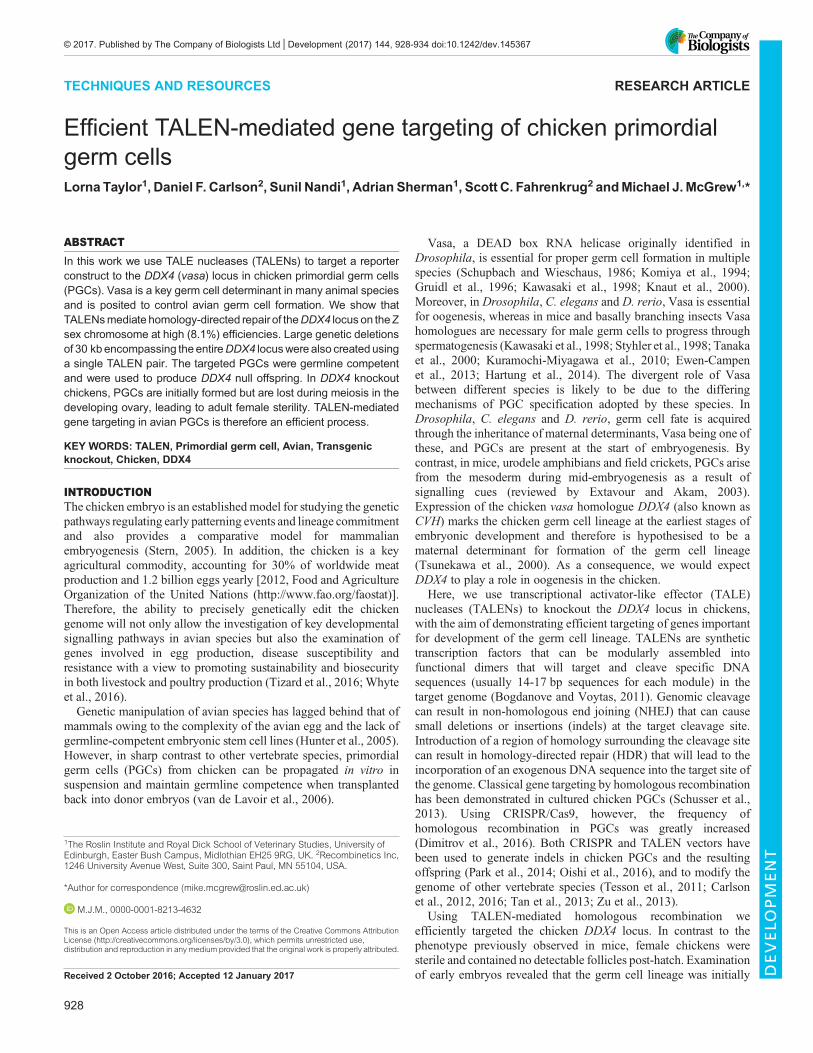
RESULTSTo target the DDX4 locus we utilised TALEN-stimulated HDR torecombine a GFP-2a-puromycin transgene targeting vector into theDDX4 locus, replacing exon 2 and 3 of the endogenous DDX4locus (Fig. 1A). A TALEN pair was designed that cleaved exon 2of the DDX4 locus immediately downstream of the ATG startcodon. The targeting vector contained homology arms of 2.9 and4.3 kb and fused the GFP-2a-puromycin reporter gene to theendogenous ATG codon. The correctly targeted locus expresses
GFP-2a-puromycin under control of the endogenous DDX4regulatory regions, and a poly(A) termination signal terminatestranscription prior to exon 4. The TALEN pair and the targetingvector were first transfected into PGCs and transiently selected(48 h) with puromycin to eliminate untransfected cells. PGCs weresubsequently propagated in culture for 2 weeks and examined byflow cytometry to identify cells stably expressing the GFPtransgene (Fig. 1B). PGCs transfected with the targeting vectoralone did not express GFP. This is consistent with previous resultsthat demonstrated that randomly integrated DNA vectors lackinginsulator elements could not be stably selected in cultured PGCs(Leighton et al., 2008; Macdonald et al., 2012b). By contrast, 8.1%of the PGCs stably expressed GFP when co-transfected with theTALEN pair.
Fig. 1. Targeting of the chicken DDX4 locus using TALENs. (A) Overview of targeting strategy to generate a knockout/knock-in DDX4 allele. The TALENpair cleavage site is indicated (red arrow). Protein-coding exons are indicated by black boxes. Vertical black arrows, MfeI sites. Red bar, external probe forSouthern blot. (B) Targeting efficiencies of cultured PGCs. PGCs (male) were transfected with the targeting vector with or without the TALEN constructs, selectedwith puromycin for 2 days to enrich for transfected cells, cultured for 2 weeks and analysed by flow cytometry. The data represent one of two independentexperiments with similar results. (C) Targeted GFP+ PGCs in culture after selection with puromycin. BF, bright-field. (D) Southern blot analysis of the GFP-purotargeted allele. For the external probe the expected fragment sizes are: wild-type, 6.7 kb; targeted; 11.7 kb. For the internal GFP probe the expected fragment sizeis: targeted, 11.7 kb. Genotype of cells is indicated by M (male) and F (female). Arrowhead, cell line used to generate targeted chicken (see text for details).
929
TECHNIQUES AND RESOURCES Development (2017) 144, 928-934 doi:10.1242/dev.145367
DEVELO
PM
ENT
To demonstrate that the GFP transgene was correctly integratedinto the DDX4 locus, a Southern blot analysis was performed.Several independent transfections of male and female PGC lineswere carried out and for each transfection the PGCs were selectedwith puromycin and genomic DNA was isolated (Fig. 1C). TheDDX4 gene is located on the Z chromosome and male (ZGFPZ)PGCs contain two chromosomal copies of the gene, whereas female(ZGFPW) PGCs are hemizygous and contain a single copy of theDDX4 gene. Multiple targeting events are likely to occur duringeach transfection as clonal colonies were not selected. The intensityof the bands in the Southern blot indicates that a single allele wastargeted in male cells and the female cells were hemizygousknockouts (Fig. 1D). Furthermore, RT-PCR analysis of a targetedfemale PGC line did not detect expression from the endogenousDDX4 locus (Fig. S1, see supplementary Materials and Methods).It has been reported that targeted insertion using site-specific
nucleases can produce a targeted gene locus and disruption of thesecond chromosomal locus by NHEJ (Merkle et al., 2015). Toaddress this concern, we sequenced the genomic locus of the non-targeted allele in the targeted male PGC lines and did not detect anyinstances of indels in the second chromosomal locus (Table S1). It ispossible that we did not detect any NHEJ in the second allelebecause bi-allelic ablation of DDX4 is lethal to male PGCs. This isunlikely as the targeted female PGC lines proliferated normally inculture (Fig. S1; data not shown). Accordingly, precise TALEN-mediated targeting of the DDX4 locus was achieved.We next asked if by varying the genomic location of the right
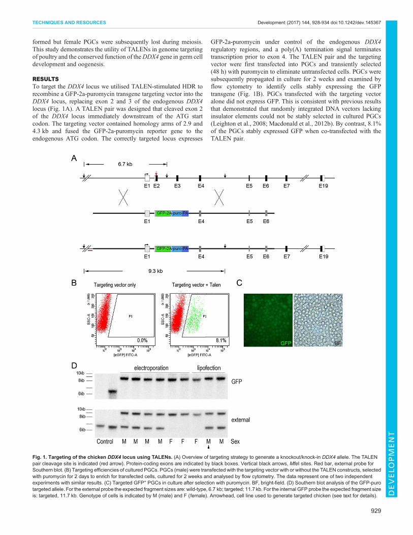
homology arm larger deletions of the DDX4 locus could be madewhile still co-transfecting with a single TALEN pair. The 4.3 kbright homology arm was replaced with 1.5 kb arms located afterexon 10 or after exon 19, the final protein-encoding exon, of theDDX4 gene. These targeting vectors would produce a genomic
deletion of 10.3 kb or 30.2 kb, respectively, after correct integrationinto the DDX4 locus (Fig. 2A,B, left). Following transfection,selection and expansion of PGCs, analysis of genomic DNA usingprimers located outside the homology arms revealed that the GFP-2a-puromycin transgene was precisely recombined into theendogenous DDX4 locus and that a series of deletion alleles wasproduced encompassing the entire locus (Fig. 2B, right).
Generation of targeted G1 offspringTargeted male cells (ZGFPZ) (arrowhead, lane 10, Fig. 1D) wereinjected into surrogate host chicken embryos (day 2.5, stage 16 HH),incubated until hatching, and raised to sexual maturity. Two hostmale cockerels were assayed for the presence of the GFP reportertransgene in their semen and mated to wild-type hens (Table 1). Onefounder male did not transmit the targeted allele to offspring,whereas the second male generated 17 G1 targeted offspring (6%;Fig. 3A, Table 1). Southern blot analysis of genomic DNA from theG1 transgenic offspring demonstrated that the male chicks wereheterozygous for the targeted allele and the female chicks werehemizygous mutant for DDX4 (Fig. 3B). The Southern blot patternexactly replicated the pattern seen in the PGC transfections(Fig. 1D), confirming that the PGCs were targeted at a single allele.
DDX4 is required for female fertility in birdsIn commercial egg-laying hens, ovulation begins at week 18 post-hatch (PH) from a single ovary originating from the left gonad. Themature ovary contains several thousand small white follicles and 30-100 yellow or small yolky follicles (Gilbert et al., 1983). As Ddx4knockout female mice exhibit normal reproductive capacity(Tanaka et al., 2000), the G1 chickens were raised to sexualmaturity with the intention to cross the females and males togenerate homozygous ZGFPZGFP males. Unexpectedly, the seven
Fig. 2. Large genomic deletions of the DDX4 locus using alternative homology arms. (A,B, left) Strategy to generate larger deletions of the DDX4locus. The TALEN dimer is indicated by the red arrow and the positions of the right targeting arms are shown. The 5′ primer sets are shown in red and the 3′ primersets in dark blue. (A, right) PCR genotype analysis of theDDX4 targeted nine-exon deletion using external and internal amplification primers. Expected products:left arm, 3.6 kb; right arm, 1.8 kb. (B, right) PCR genotype analysis of the DDX4 targeted 18-exon deletion using external and internal amplification primers.Expected products: left arm, 3.6 kb; right arm, 1.6 kb. Bl, blank; Ctr, control non-transgenic DNA; M1, M2, male cell line; F1, F2, female cell line.
930
TECHNIQUES AND RESOURCES Development (2017) 144, 928-934 doi:10.1242/dev.145367
DEVELO
PM
ENT

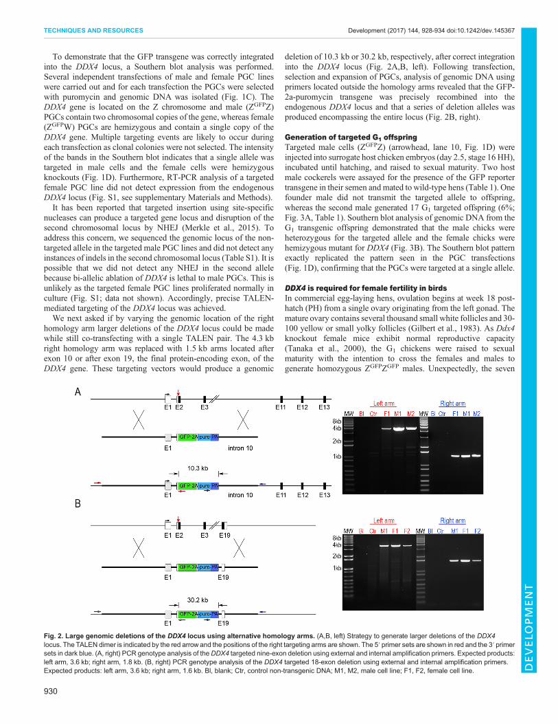
hemizygous ZGFPW female G1 chickens did not enter into lay by29 weeks PH. A morphological examination of the ovaries fromthese hens revealed that nowhite or yellow follicles were visible andno primary or secondary follicles were detected in sections (Fig. 4A,Fig. S2). To examine the earlier stages of ovarian development, aZGFPZ heterozygote male was mated to wild-type ZW hens andovaries of age-matched ZGFPW and ZW hatchlings were examinedpost-hatch. In the hatchlings, primary and secondary folliclessurrounded by layered granulosa cells were present at 2 and 4 weeksPH (n=6, ZW), whereas no follicles were detected in the ovaries ofthe ZGFPW chicks (n=6; Fig. 4B,C). Similarly, immunostaining forGFP and germ cell markers of mature oocytes (p63 and MLH1) didnot reveal any germ cells in the ovary at week 2 PH (Fig. 4D-G).To identify whether the defect in oogenesis was due to ablation of
the germ cell lineage, we examined early embryonic stages of theZGFPW embryos. DDX4 protein marks the germ cell lineage atcleavage stages of embryonic development and DDX4 RNA isexpressed during PGC migration to the gonad and at all subsequentdevelopmental stages (Tsunekawa et al., 2000).We found that GFP+
cells were present in ZGFPW gonads at day 6 and day 9 of incubation(Fig. S3A,B). These cells expressed the germ cell marker SSEA1.Immunostaining confirmed the lack of DDX4 protein in day 9female PGCs of ZGFPW embryos (Fig. S3B). We examined the
number of proliferative cells in the gonad of day 10.5 embryosto determine whether proliferation was compromised in thedeveloping gonad in the absence of DDX4. There was asignificant reduction in EdU+ PGCs in the developing cortex ofZGFPW gonads (Fig. S3C). These results show that germ cellsinitially form in ZGFPW chicken embryos and are lost at laterdevelopmental stages.
To determine if the ZGFPW germ cells entered meiosis, severalmeiotic markers were examined in the developing ovary. PGCs inthe female gonad enter meiosis by day 16 of embryonicdevelopment and most oocytes reach the diplotene stage ofmeiosis I by day 7 PH (Smith et al., 2008; del Priore and Pigozzi,2012). In control embryonic day 17 ZW gonads, germ cellscorrectly express the meiotic markers SCP3, γH2AX and MLH1(Fig. S4). By contrast, SCP3 was not detected at this stage, and germcell number was reduced in the ZGFPWovary. By embryonic day 19,ZGFPW germ cells expressed SCP3 at levels comparable to wild-type ZW germ cells, although visibly fewer germ cells were presentin the cortex of ZGFPW gonads (Fig. S4). The widespreadexpression of MLH1 indicated that many germ cells in bothZGFPW and ZW gonads had reached the pachytene stage ofmeiosis I. Expression of SCP1 was also detected in embryonic day19 ZGFPWembryos (Fig. S4). To determine if the remaining ZGFPWgerm cells progressed to later stages of meiosis, chicks wereexamined at 3 days PH. In chickens, the majority of germ cells haveentered the pachytene stage of meiosis at day 3 PH (Hughes, 1963;del Priore and Pigozzi, 2012). In control ZW day 3 hatchlings,SCP3+ MLH1+ germ cells were present in the cortex of the ovaryand SCP3 was expressed in a punctate manner indicative of thepachytene stage of meiosis (Fig. S4) (Guioli et al., 2012). Bycontrast, the majority of the GFP+ cells were located in the medullaof the ZGFPW day 3 PH ovary. The limited number of GFP+ cellsfound in the cortex were MLH1+, but very few expressed SCP3.These data are in agreement with a defect in progressing beyond thepachytene stage of meiosis, leading to a post-hatch loss of germcells in the ZGFPW ovary.
DISCUSSIONThese results demonstrate that TALEN-stimulated HDR in germcells is highly efficient and opens future avenues for investigation ofgene function in birds and for the introduction of production traits.Large genomic deletions have been achieved using two site-specificnucleases or CRISPR vectors, but to our knowledge this is the firstlarge genetic deletion produced using a single nuclease pair.Genetic deletions through altered placement of homology arms willbe useful to create a series of genetic deletions at loci of interest.CRISPR-stimulated HDR in chicken germ cells has been reportedbut the recombination efficiency was much lower than reported here(Dimitrov et al., 2016). As DDX4 is expressed in PGCs, it ispossible that this genomic locus may be highly accessible to site-specific nucleases and amenable to gene targeting. Additionally, thecell culture medium used in this report supports rapid cellproliferation (Whyte et al., 2015), which might lead to a muchhigher frequency of HDR. We did not observe a phenotype incultured female targeted PGCs or at early embryonic stages inZGFPWembryos. Early PGCs express the DEAD box genesDDX43and DDX25 (Jean et al., 2015), and it is possible that these proteinsreplace DDX4 at early developmental stages.
The precise role of DDX4 in meiosis remains unknown. ThemouseDdx4 knockout led to male sterility; male mouse PGCs entermeiosis but do not express diplotene markers and undergoapoptosis. Proliferation of Ddx4−/− PGCs was also severely
Table 1. Germline transmission frequency of donor ZGFPZmale PGCs insurrogate host chickens
Founderbirds ♂
Eggsset
Chickshatched (%)
Genomeequivalents insemen
DDX4+/−
offspring (%transmission)
Ddx 1-3 311 268 (86%) 10% 17 (6%)Ddx 4-12 143 104 (73%) 5% 0 (0%)
Fig. 3. Targeted knockout offspring produced from DDX4 targeted PGCs.(A) Targeted male and female G1 chicks. (B) Southern blot of control andindividual G1 offspring. Fragment sizes are equivalent to those in Fig. 1.Asterisk marks a larger DNA fragment generated by restriction fragment lengthpolymorphism in the control DNA sample.
931
TECHNIQUES AND RESOURCES Development (2017) 144, 928-934 doi:10.1242/dev.145367
DEVELO
PM
ENT
compromised in the early mouse gonad, similar to what wasobserved here in the forming ovary of ZGFPW embryos (Tanakaet al., 2000). Ddx4 is thought to function in amplifying thetranslation of a number of proteins needed for meiotic progressionand the assembly of cytoplasmic granules in germ cells, which arepotential RNA-processing centres (Aravin et al., 2009; Kuramochi-Miyagawa et al., 2010). Future experiments will address thefunction of DDX4 in chicken germ cell meiosis.
MATERIALS AND METHODSPGC culturePGC line derivation and culture were carried out as described (Whyte et al.,2015). Briefly, 1 μl blood isolated from a stage 16 HH embryo (Hamburgerand Hamilton, 1992) was placed in culture medium containing 1× B-27supplement (Thermo Fisher Scientific), 0.15 mM CaCl2, 2.0 mMGlutaMax (Thermo Fisher Scientific), 1× non-essential amino acids(Thermo Fisher Scientific), 0.1 mM β-mercaptoethanol, 1× EmbryoMaxnucleosides (Merck Millipore), 1.2 mM pyruvate (Thermo FisherScientific), 0.2% ovalbumin (Sigma) and 0.01% sodium heparin (Sigma).25 ng/ml activin A (Peprotech), 4 ng/ml FGF2 (R&D Systems) and 5 µg/mlovotransferrin (Sigma) were added to Avian Knockout DMEM (osmolality:250 mOsmol/kg, 12.0 mM glucose, calcium chloride free; Thermo FisherScientific, a custom modification of Knockout DMEM). Chicken serum at0.2% (Biosera) was added to this medium to produce FAOTcs medium. Amale and a female PGC line were derived in FAOTcs medium and expandedto 2.5×10⁵ cells in 5 weeks before use in targeting experiments.
TALEN design and constructionAll TALENs were designed using TALE-NT software and assembled usingmethods described in Cermak et al. (2011). Design and construction of
ggVASAe1.1 is described in Carlson et al. (2012) using thepC-GoldyTALEN (Addgene ID 38143). The TALEN pair creating acleavage site 15 bp 5′ to the ATG ofDDX4was designed (DDX1.1 targetingvector) with the following binding sites: left monomer (sense), GCTAAC-GTGCTCCTGGTCCT; right monomer (sense), ATTCGCTATGGAGGA-GG. A 2.9 kb genomic fragment upstream of exon 2 of the DDX4 gene wasPCR amplified from ISA Brown genomic DNA and cloned upstream of aGFP-2a-puromycin-poly(A) expression cassette (Hockemeyer et al., 2011)by converting the endogenous ATG to an NcoI site (outer primers, 5′-CTGGTAGAGAGCATTACAAAAGTC-3′ and 5′-GTGTCCCAGTCCT-CCTCCATAG-3′; inner NcoI primer, 5′-AACCATGGCGAATGCCAGC-AGCCCA-3′). A 4.3 kb downstream PCR fragment containing exons 4-6was cloned into a BamHI site downstream of the poly(A) site to createpddx4-GFP-polyA-exon4 using nested primers (outer primers, 5′-CTCC-TTGGCCCCATTAACAGA-3′ and 5′-GTTTTGTGCCATGACCACTG-3′; inner primers, 5′-GGGGCCCAGAAGTTCTCCTTA-3′ and 5′-TTGG-GCCCAAATCCACGGTGCAATATCC-3′). This right arm was replacedby digesting with BamHI and the following right arms were used: a 1.5 kbPCR fragment containing intron 10 termed pddx4-GFP-polyA-intron10(primers 5′-TAGTTGGATGCCTCAGACTTCA-3′ and 5′-ATTGCAAG-TGGAGCTTCAAGA-3′) and a 1.5 kb PCR fragment downstream of exon19 termed pddx4-GFP-polyA-3pUTR (primers 5′-GAAGGCAAAAGC-CATTTTCA-3′ and 5′-CCCTTCTAAACCCTGCAATTC-3′) usingPhusion HF polymerase (New England Biolabs).
PGC transfection and electroporation1 µg TALEN vector pair 1.1 (0.5 µg each of left and right) and 1 µgtargeting vector were co-transfected into PGCs using DIMRIEtransfection reagent (Thermo Fisher Scientific) as previously described(Macdonald et al., 2012a). Briefly, 1×10⁵ PGCs were washed inOptimem I (Thermo Fisher Scientific), mixed with the DNA and
Fig. 4. No follicles are present in the cortex of post-hatch ZGFPW hens. (A-C) H&E staining ofrepresentative post-hatch ZGFPW and ZW hens.Arrows indicate developing follicles. (C) ZGFPW,0 follicles/field; ZW, 26 follicles/field; average of tenfields from four ovaries/genotype. Scale bars: 100 µm.(D-G) Immunostaining of 2-week post-hatch folliclesfor GFP and the germ cell meiotic markers p63 andMLH1. Nuclear stain, white. Arrows indicatedeveloping follicles. Scale bars: 50 µm.
932
TECHNIQUES AND RESOURCES Development (2017) 144, 928-934 doi:10.1242/dev.145367
DEVELO
PM
ENT
transfection reagent and transfected in suspension for 6 h. PGCs werethen centrifuged and resuspended in FACS medium. For electroporation,1×105 PGCs were centrifuged and resuspended in 10 µl solution Rcontaining 1 μg DNA (0.5 µg TALEN vector pair 1.1 and 0.5 µgtargeting vector) and electroporated using a Neon electroporator (ThermoFisher Scientific) at 850 V, 50 ms pulse. To measure targetingefficiencies, PGCs were selected 24 h post-transfection with 0.6 µg/mlpuromycin for 48 h, then washed to remove all puromycin and furthercultured for 2 weeks to eliminate transient GFP fluorescence. To selectstably transfected cells, PGCs were selected at 4 days post-transfectionusing 0.3 µg/ml puromycin treatment over a 2 week period. PGCs fromeach individual transfection were then expanded in culture for 29 days to8×105 cells and cryopreserved using Avian Knockout DMEM/B-27supplement containing 5% DMSO and 4% chicken serum and frozen for9 months before use in germline transmission experiments. Genomic DNAwas isolated from each individual transfection and used for Southernblot analysis.
ImmunohistochemistryTissues were fixed in formalin for paraffin sections followed byHaematoxylin and Eosin (H&E) staining or cryo-embedded andprocessed for immunofluorescence (Whyte et al., 2015). The number offollicles per field for 2-week PH ovaries was determined by counting onemicroscope field per slide for four slides from four different ovaries for eachgenotype. Samples were incubated with primary antibody in 5% goat serumovernight at 4°C. Details of antibodies are provided in the supplementaryMaterials and Methods.
Flow cytometryFor flow cytometry analysis, PGCs were transiently selected withpuromycin at 24 h post-transfection to enrich for transfected cells.PGCs were treated for 48 h then washed to remove puromycin. Afterculture for an additional 3 weeks, PGCs were analysed for GFP fluorescenceusing a FACSAria II (BD Biosciences) to identify stably integrated cells.
Germline transmissionY25 male-targeted cells (Novagen Brown) were thawed from storage at−150°C for 9 months and cultured for 4-8 days before injection into stage16 HH surrogate host embryos in windowed eggs (Whyte et al., 2015).3000-5000 PGCs were injected into the dorsal aorta, the shells were resealedwith Parafilm and the eggs incubated at 37.7°C until hatching (McGrewet al., 2004; Nakamura et al., 2008). Four injection experiments were carriedout. PCR screening for the GFP transgene in the semen of two founder malecockerels was performed and these were then bred to wild-type hens.Offspring were screened by PCR for the presence of the GFP transgene(McGrew et al., 2004). Genomic DNAwas isolated from the blood of GFP+
G1 offspring and used for Southern blot analysis. Animal experiments wereconducted under UK Home Office license.
Southern blot analysisGenomic DNAwas isolated from blood samples or stably transfected PGCs(∼1×10⁶ cells) using a Flexigene Kit (Qiagen) and 5 µg was digestedovernight withMfeI. The DNA digests were resolved by gel electrophoresisand transferred via capillary action to Hybond N membrane (GEHealthcare). A GFP fragment (0.8 kb) or a 0.6 kb DDX4 genomic DNAfragment (amplified using primers 5′-GACAAGCCATCACATACAAAG-C-3′ and 5′-AAGGAAGCTGGGAGCTCTTC-3′) was labelled with[α-32P]dCTP using the DIG High Prime DNA Labelling and DetectionKit II (Roche) and used to hybridise the Southern blot.
To assay integration into the DDX4 locus by PCR, the following nestedprimers were used: for the common left arm, outer primers (5′-CAGCAC-TGTTAAAGGGCACA-3′ and 5′-AAGTCGTGCTGCTTCATGTG-3′)and inner primers (5′-GCGCGCTTTGACATATTTTT-3′ and 5′-GGTCA-CGAGGGTGGGCCAG-3′); 11 kb right arm (5′-GCCTGAAGAACGA-GATCAGC-3′ and 5′-TCCACTGCCATATGAGGACA-3′); 20 kb rightarm (5′-GCCTGAAGAACGAGATCAGC-3′ and 5′-GGGGTTGGACTT-AATCTCTGG-3′).
AcknowledgementsWe thank the members of the transgenic chicken facility (M. Hutchison andF. Thomson) for care and breeding of the chickens; and Denis Headon and JamesGlover for constructive criticism of the manuscript.
Competing interestsS.C.F. and D.F.C. are full-time employees of Recombinetics, Inc.
Author contributionsM.J.M. and S.C.F. conceived the experiments. L.T., D.F.C., A.S., S.N. and M.J.M.designed and performed experiments. L.T., D.F.C., S.N. and M.J.M. wrote themanuscript; all authors read and approved the manuscript.
FundingThis research was funded by Institute Strategic Grant funding from theBiotechnology and Biological Sciences Research Council (BB/J004316/1,BB/J004219/1). Deposited in PMC for immediate release.
Supplementary informationSupplementary information available online athttp://dev.biologists.org/lookup/doi/10.1242/dev.145367.supplemental
ReferencesAravin, A. A., van der Heijden, G. W., Castaneda, J., Vagin, V. V., Hannon, G. J.
and Bortvin, A. (2009). Cytoplasmic compartmentalization of the fetal piRNApathway in mice. PLoS Genet. 5, e1000764.
Bogdanove, A. J. and Voytas, D. F. (2011). TAL effectors: customizable proteinsfor DNA targeting. Science 333, 1843-1846.
Carlson, D. F., Tan, W., Lillico, S. G., Stverakova, D., Proudfoot, C., Christian,M., Voytas, D. F., Long, C. R., Whitelaw, C. B. A. and Fahrenkrug, S. C. (2012).Efficient TALEN-mediated gene knockout in livestock. Proc. Natl. Acad. Sci. USA109, 17382-17387.
Carlson, D. F., Lancto, C. A., Zang, B., Kim, E.-S., Walton, M., Oldeschulte, D.,Seabury, C., Sonstegard, T. S. and Fahrenkrug, S. C. (2016). Production ofhornless dairy cattle from genome-edited cell lines. Nat. Biotechnol. 34, 479-481.
Cermak, T., Doyle, E. L., Christian, M., Wang, L., Zhang, Y., Schmidt, C., Baller,J. A., Somia, N. V., Bogdanove, A. J. and Voytas, D. F. (2011). Efficient designand assembly of custom TALEN and other TAL effector-based constructs for DNAtargeting. Nucleic Acids Res. 39, e82.
del Priore, L. and Pigozzi, M. I. (2012). Chromosomal axis formation and meioticprogression in chicken oocytes: a quantitative analysis. Cytogenet. Genome Res.137, 15-21.
Dimitrov, L., Pedersen, D., Ching, K. H., Yi, H., Collarini, E. J., Izquierdo, S., vande Lavoir, M.-C. and Leighton, P. A. (2016). Germline gene editing in chickensby efficient CRISPR-mediated homologous recombination in primordial germcells. PLoS ONE 11, e0154303.
Ewen-Campen, B., Donoughe, S., Clarke, D. N. and Extavour, C. G. (2013).Germ cell specification requires zygotic mechanisms rather than germ plasm in abasally branching insect. Curr. Biol. 23, 835-842.
Extavour, C. G. and Akam, M. (2003). Mechanisms of germ cell specificationacross the metazoans: epigenesis and preformation. Development 130,5869-5884.
Gilbert, A. B., Perry, M. M., Waddington, D. and Hardie, M. A. (1983). Role ofatresia in establishing the follicular hierarchy in the ovary of the domestic hen(Gallus domesticus). J. Reprod. Fertil. 69, 221-227.
Gruidl, M. E., Smith, P. A., Kuznicki, K. A., McCrone, J. S., Kirchner, J.,Roussell, D. L., Strome, S. and Bennett, K. L. (1996). Multiple potential germ-line helicases are components of the germ-line-specific P granules ofCaenorhabditis elegans. Proc. Natl. Acad. Sci. USA 93, 13837-13842.
Guioli, S., Lovell-Badge, R. and Turner, J. M. A. (2012). Error-prone ZW pairingand no evidence for meiotic sex chromosome inactivation in the chicken germ line.PLoS Genet. 8, e1002560.
Hamburger, V. and Hamilton, H. L. (1992). A series of normal stages in thedevelopment of the chick embryo. Dev. Dyn. 195, 231-272.
Hartung, O., Forbes, M. M. and Marlow, F. L. (2014). Zebrafish vasa is required forgerm-cell differentiation and maintenance. Mol. Reprod. Dev. 81, 946-961.
Hockemeyer, D., Wang, H., Kiani, S., Lai, C. S., Gao, Q., Cassady, J. P., Cost,G. J., Zhang, L., Santiago, Y., Miller, J. C. et al. (2011). Genetic engineering ofhuman pluripotent cells using TALE nucleases. Nat. Biotechnol. 29, 731-734.
Hughes, G. C. (1963). The population of germ cells in the developing female chick.J. Embryol. Exp. Morphol. 11, 513-536.
Hunter, C. V., Tiley, L. S. and Sang, H. M. (2005). Developments in transgenictechnology: applications for medicine. Trends Mol. Med. 11, 293-298.
Jean, C., Oliveira, N. M. M., Intarapat, S., Fuet, A., Mazoyer, C., De Almeida, I.,Trevers, K., Boast, S., Aubel, P., Bertocchini, F. et al. (2015). Transcriptomeanalysis of chicken ES, blastodermal and germ cells reveals that chick ES cellsare equivalent to mouse ES cells rather than EpiSC. Stem Cell Res. 14, 54-67.
933
TECHNIQUES AND RESOURCES Development (2017) 144, 928-934 doi:10.1242/dev.145367
DEVELO
PM
ENT
Kawasaki, I., Shim, Y.-H., Kirchner, J., Kaminker, J., Wood,W. B. and Strome, S.(1998). PGL-1, a predicted RNA-binding component of germ granules, isessential for fertility in C. elegans. Cell 94, 635-645.
Knaut, H., Pelegri, F., Bohmann, K., Schwarz, H. and Nusslein-Volhard, C.(2000). Zebrafish vasa RNA but not its protein is a component of the germ plasmand segregates asymmetrically before germline specification. J. Cell Biol. 149,875-888.
Komiya, T., Itoh, K., Ikenishi, K. and Furusawa, M. (1994). Isolation andcharacterization of a novel gene of the DEAD box protein family which isspecifically expressed in germ cells of Xenopus laevis. Dev. Biol. 162, 354-363.
Kuramochi-Miyagawa, S., Watanabe, T., Gotoh, K., Takamatsu, K., Chuma, S.,Kojima-Kita, K., Shiromoto, Y., Asada, N., Toyoda, A., Fujiyama, A. et al.(2010). MVH in piRNA processing and gene silencing of retrotransposons.GenesDev. 24, 887-892.
Leighton, P. A., van de Lavoir, M.-C., Diamond, J. H., Xia, C. and Etches, R. J.(2008). Genetic modification of primordial germ cells by gene trapping, genetargeting, and phiC31 integrase. Mol. Reprod. Dev. 75, 1163-1175.
Macdonald, J., Taylor, L., Sang, H. M. and McGrew, M. J. (2012a). Geneticmodification of the chicken genome using transposable elements. TransgenicRes. 21, 912-913.
Macdonald, J., Taylor, L., Sherman, A., Kawakami, K., Takahashi, Y., Sang,H. M. and McGrew, M. J. (2012b). Efficient genetic modification and germ-linetransmission of primordial germ cells using piggyBac and Tol2 transposons. Proc.Natl. Acad. Sci. USA 109, E1466-E1472.
McGrew, M. J., Sherman, A., Ellard, F. M., Lillico, S. G., Gilhooley, H. J.,Kingsman, A. J., Mitrophanous, K. A. and Sang, H. (2004). Efficient productionof germline transgenic chickens using lentiviral vectors. EMBO Rep. 5, 728-733.
Merkle, F. T., Neuhausser, W. M., Santos, D., Valen, E., Gagnon, J. A., Maas, K.,Sandoe, J., Schier, A. F. and Eggan, K. (2015). Efficient CRISPR-Cas9-mediated generation of knockin human pluripotent stem cells lacking undesiredmutations at the targeted locus. Cell Rep. 11, 875-883.
Nakamura, Y., Yamamoto, Y., Usui, F., Atsumi, Y., Ito, Y., Ono, T., Takeda, K.,Nirasawa, K., Kagami, H. and Tagami, T. (2008). Increased proportion of donorprimordial germ cells in chimeric gonads by sterilisation of recipient embryos usingbusulfan sustained-release emulsion in chickens. Reprod. Fertil. Dev. 20,900-907.
Oishi, I., Yoshii, K., Miyahara, D., Kagami, H. and Tagami, T. (2016). Targetedmutagenesis in chicken using CRISPR/Cas9 system. Sci. Rep. 6, 23980.
Park, T. S., Lee, H. J., Kim, K. H., Kim, J.-S. and Han, J. Y. (2014). Targeted geneknockout in chickens mediated by TALENs. Proc. Natl. Acad. Sci. USA 111,12716-12721.
Schupbach, T. and Wieschaus, E. (1986). Germline autonomy of maternal-effectmutations altering the embryonic body pattern of Drosophila. Dev. Biol. 113,443-448.
Schusser, B., Collarini, E. J., Yi, H., Mettler Izquierdo, S., Fesler, J., Pedersen,D., Klasing, K. C., Kaspers, B., Harriman, W. D., van de Lavoir, M.-C. et al.(2013). Immunoglobulin knockout chickens via efficient homologousrecombination in primordial germ cells. Proc. Natl. Acad. Sci. USA 110,20170-20175.
Smith, C. A., Roeszler, K. N., Bowles, J., Koopman, P. and Sinclair, A. H. (2008).Onset of meiosis in the chicken embryo; evidence of a role for retinoic acid. BMCDev. Biol. 8, 85.
Stern, C. D. (2005). The chick; a great model system becomes even greater. Dev.Cell 8, 9-17.
Styhler, S., Nakamura, A., Swan, A., Suter, B. and Lasko, P. (1998). vasa isrequired for GURKEN accumulation in the oocyte, and is involved in oocytedifferentiation and germline cyst development. Development 125, 1569-1578.
Tan, W., Carlson, D. F., Lancto, C. A., Garbe, J. R., Webster, D. A., Hackett, P. B.and Fahrenkrug, S. C. (2013). Efficient nonmeiotic allele introgression inlivestock using custom endonucleases. Proc. Natl. Acad. Sci. USA 110,16526-16531.
Tanaka, S. S., Toyooka, Y., Akasu, R., Katoh-Fukui, Y., Nakahara, Y., Suzuki, R.,Yokoyama, M. and Noce, T. (2000). The mouse homolog of Drosophila Vasa isrequired for the development of male germ cells. Genes Dev. 14, 841-853.
Tesson, L., Usal, C., Menoret, S., Leung, E., Niles, B. J., Remy, S., Santiago, Y.,Vincent, A. I., Meng, X., Zhang, L. et al. (2011). Knockout rats generated byembryo microinjection of TALENs. Nat. Biotechnol. 29, 695-696.
Tizard, M., Hallerman, E., Fahrenkrug, S., Newell-McGloughlin, M., Gibson, J.,de Loos, F., Wagner, S., Laible, G., Han, J. Y., D’Occio, M. et al. (2016).Strategies to enable the adoption of animal biotechnology to sustainably improveglobal food safety and security. Transgenic Res. 25, 575-595.
Tsunekawa, N., Naito, M., Sakai, Y., Nishida, T. and Noce, T. (2000). Isolation ofchicken vasa homolog gene and tracing the origin of primordial germ cells.Development 127, 2741-2750.
van de Lavoir, M.-C., Diamond, J. H., Leighton, P. A., Mather-Love, C., Heyer,B. S., Bradshaw, R., Kerchner, A., Hooi, L. T., Gessaro, T. M., Swanberg, S. E.et al. (2006). Germline transmission of genetically modified primordial germ cells.Nature 441, 766-769.
Whyte, J., Glover, J. D., Woodcock, M., Brzeszczynska, J., Taylor, L., Sherman,A., Kaiser, P. and McGrew, M. J. (2015). FGF, insulin, and SMAD signalingcooperate for avian primordial germ cell self-renewal. Stem Cell Rep. 5,1171-1182.
Whyte, J., Blesbois, E. and McGrew, M. J. (2016). Increased sustainability inpoultry production: new tools and resources for genetic management. InSustainable Poultry Production in Europe (ed. E. Burton, J. Gatcliffe, H. MaseyO’Neill and D. Scholey), pp. 214-234. Cambridge, MA, USA: CABI Publishing.
Zu, Y., Tong, X., Wang, Z., Liu, D., Pan, R., Li, Z., Hu, Y., Luo, Z., Huang, P., Wu,Q. et al. (2013). TALEN-mediated precise genome modification by homologousrecombination in zebrafish. Nat. Methods 10, 329-331.
934
TECHNIQUES AND RESOURCES Development (2017) 144, 928-934 doi:10.1242/dev.145367
DEVELO
PM
ENT