efficacy of a combined drug therapy for ameliorating noise
TRANSCRIPT

Washington University School of MedicineDigital Commons@Becker
Independent Studies and Capstones Program in Audiology and CommunicationSciences
2011
Efficacy of a combined drug therapy forameliorating noise-induced hearing lossRandi Lynn Luxmore
Follow this and additional works at: http://digitalcommons.wustl.edu/pacs_capstones
Part of the Medicine and Health Sciences Commons
This Thesis is brought to you for free and open access by the Program in Audiology and Communication Sciences at Digital Commons@Becker. It hasbeen accepted for inclusion in Independent Studies and Capstones by an authorized administrator of Digital Commons@Becker. For moreinformation, please contact [email protected].
Recommended CitationLuxmore, Randi Lynn, "Efficacy of a combined drug therapy for ameliorating noise-induced hearing loss" (2011). Independent Studiesand Capstones. Paper 622. Program in Audiology and Communication Sciences, Washington University School of Medicine.http://digitalcommons.wustl.edu/pacs_capstones/622
EFFICACY OF A COMBINED DRUG THERAPY FOR AMELIORATING NOISE-INDUCED HEARING LOSS
by
Randi Lynn Luxmore
A Capstone Project submitted in partial fulfillment of the
requirements for the degree of:
Doctor of Audiology
Washington University School of Medicine Program in Audiology and Communication Sciences
May 18, 2012
Approved by: Jianxin Bao, Ph.D., Capstone Project Advisor
Brian Faddis, Ph.D., Second Reader
Abstract: Experiments investigated the median effective dose of antiepileptic drugs and synthetic glucocorticoids for the prevention and treatment of noise-induced hearing loss for C57BL/6J mice. We also tested the possible synergistic effects of combining drugs from the two
drug families.

copyright by
Randi L. Luxmore
2011

Luxmore
I would like to acknowledge the following individuals for their support throughout this project: Dr. Jianxin Bao: For supporting and encouraging me throughout this process. Your vast knowledge and kind personality were both an inspiration to me. Dr. Brian Faddis: For offering time and advice by serving as the second reader for this project. Dr. Debin Lei: For teaching me the lab skills I needed for finishing this project, and for volunteering extra assistance without even being asked. Michelle Hungerford: For helping with data collection and for being another avenue of support during this process. My family: For always making education a priority, and for supporting me in every way so that I could work toward my goals.
ii

Luxmore
TABLE OF CONTENTS
Acknowledgements………………………………………………..……………..ii
List of Tables and Figures……………………………………….…….…………2
Abbreviations……………………………………………………………..……...3
Introduction……………………………………………………………..………..4
Materials and Methods…………………………………………….…………….9
Results……………………………………………………………….………….12
Discussion………………………………………………………………………16
Conclusion..……………………………………………………………………..19
References………………………………………………………………………20
iii

Luxmore
LIST OF TABLES AND FIGURES
Figure 1: Prevention Protocol Example Figure 2: Treatment Protocol Example Figure 3: NIHL prevention by synthetic glucocorticoid drugs Figure 4: NIHL prevention by antiepeleptic drugs. Figure 5: NIHL prevention by low doses of methlprednisolone and zonisamide Figure 6: NIHL treatment by glucocorticoid and antiepeleptic drugs. Table 1: Pharmacological information of the drugs used in the study
iv

Luxmore
ABBREVIATIONS
ABR auditory brainstem response
CI combination index
cm centimeter
dB SPL decibel sound pressure level
GC glucocorticoid
GR glucocorticoid receptor
HPD hearing protective device
IHC inner hair cell
i.p. intraperitoneal
kg kilogram
kHz kilohertz
mg milligram
NIHL noise-induced hearing loss
OBN octave band of noise
OHC outer hair cell
PTS permanent threshold shift
sec second
SGN spiral ganglion neuron
SNHL sensorineural hearing loss
TTS temporary threshold shift
VGCC voltage-gated calcium channel
v

Luxmore
Introduction
Noise-induced hearing loss (NIHL) is the single predominant health hazard posed by
occupational and recreational settings (NIOSH, 2001). The incidence of NIHL has continued to
grow in recent years partly due to growing popularity of portable music players with highly
efficient headphones (Fligor and Cox, 2004; Serra et al., 2005). Although promising approaches
have been identified for reducing NIHL mainly based on the free radical pathway (Lynch and
Kil, 2005; Campbell et al., 2007; Kopke et al., 2007; Le Prell et al., 2007b), there are currently
no effective pharmacologic agents in the clinical field to diminish permanent hearing loss.
Development of an efficacious treatment has been hampered by the complex array of cellular and
molecular pathways involved in NIHL. This project tests whether NIHL could be prevented by a
synergistic pharmacologic intervention in multiple signaling pathways. Based on previous
studies (Canlon et al., 2007; Shen et al., 2007), we have developed a two-drug combination
therapy for NIHL with FDA-approved drugs from two different drug families: anti-epileptic
drugs blocking T-type calcium channels and synthetic steroid drugs up-regulating the GC
signaling pathway. The goal of this work was to explore this novel therapeutic direction for
treating NIHL.
NIHL and its pathogenesis
After the initial noise exposure, auditory brainstem response (ABR) testing can typically
detect two phases of hearing loss. These include a temporary threshold shift (TTS) that is most
prominent in the first 24 hours, and an improved but still elevated permanent threshold shift
(PTS) two to three weeks later (Clark, 1991; Quaranta et al., 1998; Nordmann et al., 2000).
However, the dynamics of noise-induced damages and subsequent recovery are dependent on the
intensity and duration of noise exposure (Wang et al., 2002; Harding and Bohne 2007). Three
1

Luxmore
distinct NIHL patterns have been well documented in CBA/CaJ mice (Wang et al., 2002). When
the exposure is an 8-16 kHz octave-band of noise (OBN) at 94 dB SPL for two hours, there is an
obvious TTS immediately post-exposure with a near complete recovery after two weeks (no
significant PTS). From 100 to 112 dB SPL, the exposure causes a monotonic increase of both
TTS and PTS, but partial recovery is observed (i.e., PTS is less than TTS). Above 116 dB SPL,
the recovery of PTS is minimal possibly due to the regional rupturing of the reticular lamina.
This noise level is defined as the “critical level” or the “inflection point” because the dramatic
injury leads to the regional rupture of the reticular lamina and disruption of the endolymphatic
compartment (Bohne, 1976, Wang et al., 2002; Harding and Bohne, 2007; Ohlemiller, 2008).
The critical level is above 125-130 dB SPL for most animal models although it may be a little
lower for mice, or when the exposure is temporally-skewed impact or impulse noise. In other
animal models, similar changes are observed for these three NIHL patterns (e.g., Slepecky, 1986;
Saunders et al., 1991; Lawner et al., 1997; Ohlemiller et al., 2000; Ohlemiller, 2008).
Histologically, above critical level, the most obvious pathological change is the rupture of the
reticular lamina, detachment of the organ of Corti from the basilar membrane and degeneration
of both inner and outer hair cells (IHCs and OHCs). Below critical level, injuries to the cochlea
include: the organ of Corti (stereocilia disarray, small to moderate hair cell losses, and
supporting cell damage), spiral ganglion neurons (SGNs; swelling of their terminals), stria
vascularis (edema), and spiral ligament (loss of type IV fibrocytes).
Mechanical destruction and decreased blood flow contribute to NIHL in some instances
(Spoendlin, 1971; Bohne, 1976; Ward et al., 1981; Quirk et al., 1991; Mulroy et al., 1998). One
major mechanism underlying NIHL is the increase of mitochondrial free radical formation due to
noise-induced intense metabolic activity in the cochlea (Lim and Melnick, 1971; Lynch and Kil,
2

Luxmore
2005; Henderson et al., 2006; Campbell et al., 2007; Darrat et al., 2007; Kopke et al, 2007; Le
Prell et al., 2007b). The involvement of this pathway in NIHL is strongly supported mainly by
three lines of evidence: First, a noise-induced increase of free radicals is observed in stria
vascularis, OHCs, supporting cells of the organ of Corti, and SGNs (Yamane et al., 1995;
Ohlemiller et al., 1999; Ohinata et al., 2000; Henderson et al., 2006), and this free radical insult
can continue up to 14 days post-exposure (Yamashita et al., 2004). Second, the depletion of
endogenous antioxidants and reduction of superoxide dismutase results in increased
susceptibility to NIHL (Ohlemiller et al., 1999; 2000; McFadden et al., 2001). Third, an
enhancement of antioxidants attenuates NIHL (Yamasoba et al., 1999; Ohinata et al., 2003; Duan
et al., 2004; Lynch et al., 2004; Kramer et al., 2006). Due to these changes, it is not surprising
that attempts to prevent NIHL by antioxidant agents have become the focus of much research in
this field (Seidman et al., 1993; Hight et al.; 2003; McFadden et al., 2005; Yamashita et al.,
2005; Bielefeld et al., 2007; Campbell et al., 2007; Kopke et al., 2007). Most of these
interventions with single chemicals are only partially effective in preventing NIHL. Therefore, a
few studies have started to intervene at multiple sites in the free radical pathway or in
combinations with other pathways. Synergistic effects have been seen in some but not all of
these studies (Yamasoba et al., 1999; Le Prell et al., 2007a; 2007b). These studies provide
compelling evidence for the role of free radicals in NIHL, but they also suggest that other
signaling mechanisms may contribute to NIHL.
T-type calcium channels and glucocorticoids
Among other main mechanisms contributing to NIHL such as the excitotoxic glutamate
at the initial phase or cell death at the end phase (Puel et al., 2002; Le Prell et al., 2003; Guitton
et al., 2004; Zine and Van De Water 2004), two new pathways have emerged: calcium and
3

Luxmore
glucocorticoid (GC) signaling pathways. Disturbance in calcium homeostasis has been
suspected to contribute to trauma-induced neuronal injury (Nikonenko et al., 2005;Werling et al.,
2007; Park et al., 2008). Calcium homeostasis in the cochlea can be regulated by several types
of calcium channels, including voltage-gated calcium channels (VGCCs) (Rodrigues-Contreraz
and Yamoah, 2001; Adamson et al., 2002; Fuchs 2002; Schnee and Ricci, 2003). VGCCs play a
key role in calcium entry into neurons and control various calcium-dependent functions. These
include neurotransmitter release, gene expression, synaptic plasticity, and neuronal excitability
(Mattson 1990; Zipfel et al., 2000). VGCCs can be divided into two groups: high-voltage
activated and low-voltage activated calcium channels (Igelmund et al., 1996; Lacinova et al.,
2000; Perez-Reyes, 2003; Yunker and McEnery, 2003). The family of low-voltage activated, or
T-type (Cav3), calcium channels is comprised of three members (Cav3.1, Cav3.2, and Cav3.3)
based on their respective main pore-forming alpha subunits: a1G, a1H, and a1I (Perez-Reyes,
2003; Yunker and McEnery, 2003). The expression of a1G and a1I is found in the hair cells and
supporting cells of the organ of Corti. A strong a1H and weak expression of the remaining two
subunits are found in SGNs of the mouse cochlea (Shen et al., 2007). The same work has found
that NIHL can be prevented by the administration of anticonvulsant drugs blocking T-type
calcium channels either before or after the noise exposure. Inhibition of T-type calcium channels
also protects neurons after stroke (Nikonenko et al., 2005). Thus, it is possible that
pharmacological modulation of T-type calcium channels can prevent injury-induced alterations
of calcium homeostasis, which may contribute to NIHL.
Another major molecular mechanism involved in NIHL is the GC signaling pathway.
Synthetic GCs such as methylprednisolone are still used for clinical therapy of neural trauma
such as spinal cord injury (Ahn and Fehlings, 2008; Xu et al., 2009). Synthetic GCs are also
4

Luxmore
used clinically to treat hearing loss in a variety of cochlear disorders such as autoimmune inner
ear disease, tinnitus, and Meniére’s disease (McCabe, 1979, Dodson and Sismanis, 2004;
Dodson et al., 2004; MacArthur et al., 2008). Although no current reports exist on the clinical
use of synthetic GCs for acoustic trauma, extensive evidence suggests an important role of GC
pathways in NIHL. First, stressful preconditioning such as restraint, heat exposure, or even low-
level sound in animal models has been found to be protective against NIHL (Paz et al., 2004;
Wang and Liberman, 2002; Yoshida et al., 1999). Second, because the noise exposure itself is a
stressful event, a pretreatment of blockers for GC signaling make animals more susceptible to
NIHL (Tahera et al., 2006a). Third, synthetic GCs such as dexamethasone and
methylprednisolone can protect against NIHL (Canlon et al., 2007; Henry, 1992; Lamm and
Arnold, 1998; Sendowski et al., 2006; Tabuchi et al., 2006; Tahera et al., 2006b; Tahera et al.,
2006c). Fourth, although GCs can bind to both GC receptors (GR) and minerocorticoid
receptors, antagonists against minerocorticoid receptors have no effect on NIHL (Tahera et al.,
2006a). Finally, a series of studies have systematically revealed the role of GR signaling
pathways in NIHL (Canlon et al., 2007; Tahera et al., 2006b; Tahera et al., 2006c). In addition,
the expression pattern of GR in the cochlea is known at both mRNA and protein levels. GR
mRNA is detected in the cells of the spiral ligament, spiral limbus, and SGNs (ten Cate et al.,
1993). GR immunoreactivity is also found in the organ of Corti (ten Cate et al., 1993; Zuo et al.,
1995, Shimazaki et al., 2002). In mice, the adult GR expression pattern in the cochlea is
achieved by postnatal day 14 (Erichsen et al., 1996), and can be up-regulated following acoustic
stress (Tahera et al., 2006b).
Similar to the NIHL intervention methods based on the free radical pathway, current
interventions based on synthetic GC drugs or anticonvulsants blocking T-type calcium channels
5

Luxmore
show limited success in preventing NIHL (Canlon et al., 2007; Shen et al., 2007). It must be
noted that these two current interventions are from two completely different drug families, which
most likely act on different molecular pathways underlying NIHL. The identification of specific
drug combinations from these two drug families that may act in synergistic ways against NIHL is
a logical next step for further study. Through this project, we validated this novel therapeutic
approach to effectively prevent or treat NIHL.
Methods
Animals
All animal procedures were approved by the Animal Studies Committee at Washington
University in St. Louis. The study included male and female C57BL/6J mice aged 2-6 months,
purchased from Jackson Laboratories (Bar Harbor, ME, USA). All mice were housed two to five
per cage in a noise-controlled environment on a 12h light/dark cycle with light onset at 6:00 a.m.
Noise exposure
Similar to the approaches described previously (Shen et al., 2007), noise exposures were
performed in a foam-lined, double-walled soundproof room (Industrial Acoustics). The noise
exposure apparatus consisted of a 21 x 21 x 11 cm wire cage mounted on a pedestal inserted into
a B&K 3921 turntable. The cage was rotated at 1 revolution per 80 sec within a 42 x 42 cm
metal bar frame. A Motorola KSN1020A piezo ceramic speaker (four total) was attached to each
side of the frame. Opposing speakers were oriented not concentrically, but parallel to the cage
and driven by independent channels of a Crown D150A power amplifier. Noise was generated
by two General Radio 1310 generators and bandpassed at 4.0-45.0 kHz by Krohn-Hite 3550
filters. The overall noise level was measured at the center of the cage using a B&K 4135 ¼ inch
6

Luxmore
microphone in a combination with a B&K 2231 sound level meter set at broadband (0.2 Hz-70
kHz). Mice were exposed in pairs to white noise at 110 dB SPL for 30 minutes.
Drug administration
Mice were randomly assigned to either experimental or control groups. For the
prevention protocol, the experimental mice were given the drugs in an intraperitoneal (i.p.)
injection and the control mice were i.p. injected with normal saline. The drugs were injected 2
hours before noise exposure.
Figure 1: Example of the prevention protocol used in this study
For the treatment protocol, the experimental mice were i.p. injected with the different drugs and
the control mice were i.p. injected with normal saline 24 hours after noise exposure. An
additional treatment protocol was performed in which the drugs were orally fed in a 1% sucrose
solution, and the control mice were fed with the 1% sucrose solution only. The drug
administrations began approximately 24 hours post noise exposure and were monitored for two
weeks in the drinking water.
Figure 2: Example of the treatment protocol used in this study
7

Luxmore
Auditory brainstem recording
The mouse cochlea typically responds to frequencies ranging from 2-100 kHz. The most
sensitive region of the audiogram is roughly 5-40 kHz. To cover this range, we tested at 5, 10,
20, 28.3, and 40 kHz. The “near field” sound stimulation and calibration were used in which the
speaker was near the ear (7 cm) within the range where the sound field was approximately
homogeneous within an imaginary cylinder surrounding the ear. To make sure sound stimuli
were constant from animal to animal, a B&K 4135 ¼ inch microphone was placed where the
mouse ear would normally be and calibrated before the experiment. Prior to testing, all mice
were anesthetized with pentobarbital (60 mg/kg, i.p.) and given atropine sulfate (0.5 mg/kg, i.p.)
to reduce respiratory distress. Core temperature was maintained at 37 +/- 1 oC using a
thermostatically controlled heating pad in conjunction with a rectal probe (Yellow Springs
Instruments Model 73A). Platinum needle electrodes (Grass) was inserted subcutaneously just
behind the right ear (active), and at the vertex (reference), and in the back (ground). Electrodes
were led to a Grass P15 differential amplifier (100-10,000Hz, x100), then to a custom amplifier
providing another x1,000 gain, finally digitized at 30 kHz using a Cambridge Electronic Design
Micro1401 in conjunction with SIGNALTM and custom signal averaging software operating on a
120 MHz Pentium PC. Sinewave stimuli generated by a Wavetek Model 148 oscillator were
shaped by a custom electronic switch to 5 ms total duration, including 1 ms rise/fall times. The
stimulus was amplified by a Crown D150A power amplifier and output to a KSN1020A piezo
ceramic speaker. Toneburst stimuli at each frequency and level were presented 1,000 times at
20/sec. The minimum sound pressure level required for a response (short-latency negative wave)
was determined at selected frequencies, using a 5 dB minimum step size.
8

Luxmore
Results
Because this was the first study attempting to establish the pharmacokinetics of these
drugs for protection from NIHL, we did not know whether first-order or higher-order dynamics
existed in the dose-effect curve for each drug. Thus, we began our study with the focus on the
median effective dose (ED50) determination by using the median-effect equation (Chou, 2006).
The median effect equation is the general equation for the dose-effect relationship derived from
the mass-action law principle that takes into account both the potency and the shape of the dose-
effect curve (Chou, 2006). The ED50 of a drug is the amount of drug that produces a response in
50% of the subjects taking it. Two steps decided the dosage range: (1) to obtain the known ED50
based on previous studies, and (2) to expand the dosage to determine the ED50 against NIHL.
The ABR shift curves for corticosteroid drugs, at multiple dosages are present in Figure 1, and
the ABR shift curves for anti-epileptic drugs are present in Figure 2.
Figure 3: NIHL prevention by the synthetic corticosteroid drugs. (A) ABR thresholds among the control and different dosages of methylprednisolone (n=8 for each group); (B) ABR thresholds among the control and different dosages of dexamethasone (n=8 for each group).
9

Luxmore
Figure 4: NIHL prevention by the antiepileptic drugs. (A) ABR thresholds among the control and different dosages of ethosuximide (n=8 for each group); (B) ABR thresholds among the control and different dosages of zonisamide (n=8 for each group)
After these observations, we entered these data into the CompuSyn software (ComboSyn,
Inc) to calculate ED50s based on the median-effect equation. The ED50s to prevent NIHL for
drugs from two different drug families were: methylprednisolone (525 mg/kg), dexamethasone
(39.4 mg/kg), and zonisamide (125 mg/kg). Because the unpredictable nature of ethosuximide
(Figure 2A), no ED50 could be obtained for this drug.
Because we only obtained the ED50 for zonisamide in the antiepileptic family, the
combination of zonisamide with either methylprednisolone or dexamethasone was tested at
different dosages to determine possible synergic effect between these combinations. A
combination of methylprednisolone and zonisamide at lower dosages (8 and 60 mg/kg
respectively, or their ED10 values) was found to effectively prevent NIHL (Figure 5). With the
same CompuSyn software, a synergic effect was found for these two drugs to prevent NIHL
based on the value of the combination index (CI), which was equal to 0.97.
10

Luxmore
Figure 5: NIHL prevention by a drug combination of methylprednisolone and zonisamide at their low dosages. ABR thresholds were about 10 dB lower across four frequencies between the control (blue line) and treated mice (red line, n=6 for each group). A synergistic effect was found due to the fact of CI <1
Similarly, the ED50s to treat NIHL 24 hours after the noise exposure were also
determined for all four drugs from the same two drug families (Figure 6). The exact ED50s were:
methylprednisolone (95.6 mg/kg), dexamethasone (96.3 mg/kg), zonisamide (2543 mg/kg), and
ethosuximide (243 mg/kg). However, little evidence existed for possible synergistic effects to
treat NIHL by i.p. injections with these two families of drugs. Interestingly, by oral
administration for two weeks, we did find a considerable effect to treat NIHL by the two-drug
treatment (Figure 6E). Thus, our data provided important preclinical drug information to prevent
and treat NIHL.
11

Luxmore
Figure 6: NIHL treatment by glucocorticoid and antiepileptic drugs. (A) ABR thresholds among the control and different dosages of methylprednisolone (n=8 for each group), (B) ABR thresholds among the control and different dosages of dexamethasone (n=9 for each group); (C) ABR thresholds among the control and different dosages of ethosuximide (n=8 for each group); (D) ABR thresholds among the control and different dosages of zonisamide (n=8 for each group); and (E) ABR thresholds between the control and a combination of methylprednisolone and ethosuximide fed in the drinking water over 24 hours after the noise exposures for two weeks (n=4 for each group)
12

Luxmore
Discussion
NIHL is a major health issue without effective medication, mainly due to the multiple
factors contributing to this complex disease. Based on previous studies in the advisor’s
laboratory, we have carried out the first detailed pharmacodynamic study examining a
combination therapy against NIHL. The first part of the study concentrated on the possible
prevention of NIHL, while the second part looked at a possible treatment. ED50s against NIHL
for drugs from two different families (two drugs per family) were derived based on ABR testing,
and a synergistic effect for one drug combination was discovered to prevent NIHL.
NIHL Prevention
Various studies have found that synthetic GCs such as methylprednisolone and
dexamethasone can protect against NIHL. This effect is in conjunction with its biologic function
of promoting neuronal adaptation and survival (McEwen, 2008). The advisor’s lab has
previously found that 5 mg/kg of methylprednisolone injected two hours before noise would
reduce NIHL in the mouse model. In addition, significant preservation of hair cells and SGNs
were found as compared to the control group. Drugs blocking T-type calcium channels were
also found to partially prevent NIHL (Shen et al., 2007). Therefore, it was not completely
surprising for this study to obtain different ED50 values for each drug from these two families.
However, it is considerable that we discovered a synergistic effect against NIHL with a
combination of methylprednisolone and zonisamide. This was based on the fact that the CI =
0.97. The CI is beneficial because it creates a quantitative measure of the degree of drug
interaction in terms of synergistic (CI<1), antagonistic (CI >1), and additive (CI=1) effects
(Chou, 2010). Therefore, the effects of methylprednisolone and zonisamide together were
greater than what would be expected when adding each of their individual effects together.
13

Luxmore
Based on this finding, drugs blocking T-type calcium channels and synthetic glucocorticoids
could be part of a promising new avenue of prevention.
Although these results are very encouraging, they are not without limits. First, these
results were obtained in a controlled laboratory environment with genetically identical animals.
Thus, these results need to be explored further in other animal models. If these results look
promising, then clinical trials with humans can begin. Another consideration is the fact that
many times, noise exposure is unexpected. Therefore, any compounds that prevent NIHL will be
unusable in these situations. This will be true especially for those in military operations.
Impulse noise is a huge concern for the military, combined with the fact that many soldiers
choose not to consistently wear hearing protective devices (HPDs) despite hearing conservation
programs stressing their usage. A therapy that can treat NIHL will benefit this population
greatly.
NIHL Treatment
The ED50s for the treatment of NIHL were found for all four drugs as well. Interestingly,
these values were quite different than those for the prevention of NIHL. With the exception of
methylprednisolone, all of the drugs had much higher ED50s for the treatment of NIHL. This
difference strongly suggests an optimal therapeutic time window for NIHL. In our NIHL
prevention diagram, each drug should have its effect most likely after the noise exposure due to
their long half-life (Table 1), although all drugs were applied two hours before the noise
exposure. Half-life is defined as the time required for half of the quantity of a drug to be
metabolized in a living organism. For example, it would take around 50-70 hours to lose half of
the potency of zonisamide after the injection. Therefore, its effects against NIHL before the noise
exposure could only account for 2/70 (about 3%). Because of this we found that drugs were
14
Luxmore
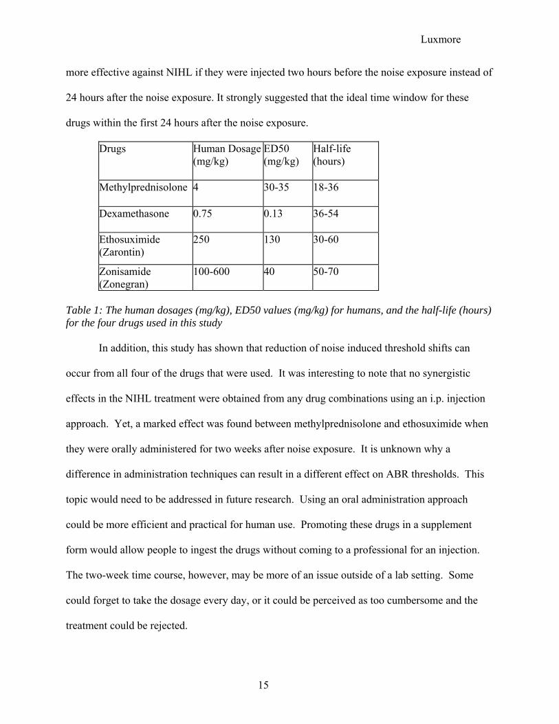
more effective against NIHL if they were injected two hours before the noise exposure instead of
24 hours after the noise exposure. It strongly suggested that the ideal time window for these
drugs within the first 24 hours after the noise exposure.
Drugs
Human Dosage (mg/kg)
ED50 (mg/kg)
Half-life (hours)
Methylprednisolone
4
30-35
18-36
Dexamethasone
0.75
0.13
36-54
Ethosuximide (Zarontin)
250
130
30-60
Zonisamide (Zonegran)
100-600
40
50-70
Table 1: The human dosages (mg/kg), ED50 values (mg/kg) for humans, and the half-life (hours) for the four drugs used in this study
In addition, this study has shown that reduction of noise induced threshold shifts can
occur from all four of the drugs that were used. It was interesting to note that no synergistic
effects in the NIHL treatment were obtained from any drug combinations using an i.p. injection
approach. Yet, a marked effect was found between methylprednisolone and ethosuximide when
they were orally administered for two weeks after noise exposure. It is unknown why a
difference in administration techniques can result in a different effect on ABR thresholds. This
topic would need to be addressed in future research. Using an oral administration approach
could be more efficient and practical for human use. Promoting these drugs in a supplement
form would allow people to ingest the drugs without coming to a professional for an injection.
The two-week time course, however, may be more of an issue outside of a lab setting. Some
could forget to take the dosage every day, or it could be perceived as too cumbersome and the
treatment could be rejected.
15

Luxmore
Future Research
For both the prevention and treatment protocols, further research is needed. Future
studies would need to investigate the most effective dosages of each drug in combination with
another. The time course and method of drug administration both need to be investigated further
as well.
Conclusion
In this study, we set out to investigate the effects that FDA approved drugs from two
different families could have on ameliorating NIHL. These two families, T-type calcium
channel blockers and synthetic glucocorticoids, affect two different pathways known to be
involved in NIHL. No such study has been attempted with these compounds before. This study
had two phases: one investigating the prevention of NIHL, and one regarding the treatment of
NIHL. We found that the administration of each of these drugs before noise exposure has an
effect on PTS. In addition, the low dosage combination of two of these compounds can also
have an effect by reducing PTS compared to controls. The second phase illustrated that each of
these drugs also has a positive effect on PTS when administered 24 hours after noise exposure.
However, this effect is markedly less than that of the prevention protocol. A synergism between
two of the drugs was found when they were orally administered for two weeks after noise
exposure. This novel investigation demonstrated that pharmaceutical intervention before or after
noise exposure could ameliorate some effects of NIHL.
16

Luxmore
References
Adamson CL, Reid MA, Davis RL. (2002) Opposite actions of brain-derived neurotrophic factor
and neurotrophin-3 on firing features and ion channel composition of murine spiral
ganglion neurons. J Neurosci, 22(4), 1385-96.
Ahn H, Fehlings MG. (2008) Prevention, identification, and treatment of perioperative spinal
cord injury. Neurosurg Focus, 25(5), E15.
Bielefeld EC, Kopke RD, Jackson RL, Coleman JK, Liu J, Henderson D. (2007) Noise
protection with N-acetyl-l-cysteine (NAC) using a variety of noise exposures, NAC
doses, and routes of administration. Acta Otolaryngol, 127(9), 914-9.
Bohne BA. In: Henderson D, Hamernik RP, Mills JH, Dosanjh DS Eds. (1976) Mechanisms of
noise damage in the inner ear. Effects of Noise on Hearing. NY, Raven Press, 41-68.
Campbell KC, Meech RP, Klemens JJ, Gerberi MT, Dyrstad SS, Larsen DL, Mitchell DL, El-
Azizi M, Verhulst SJ, Hughes LF. (2007) Prevention of noise- and drug-induced hearing
loss with D-methionine. Hear Res, 226(1-2), 92-103.
Canlon B, Meltser I, Johansson P, Tahera Y. (2007) Glucocorticoid receptors modulate auditory
sensitivity to acoustic trauma. Hear Res, 226(1-2), 61-9.
Chou, T.C. (2006) Theoretical basis, experimental design, and computerized simulation of
synergism and antagonism in drug combination studies. Pharmacol Rev, 58(3), 621-81.
Chou, T.C. (2010) Drug comination studies and their synergy quanificaition using the Chou-
Talalay method. Cancer Res, 70(2), 440-446.
Clark WW. (1991) Recent studies of temporary threshold shift (TTS) and permanent threshold
shift (PTS) in animals. J Acoust Soc Am, 90(1), 155-63.
17

Luxmore
Darrat I, Ahmad N, Seidman K, Seidman MD. (2007) Auditory research involving antioxidants.
Curr Opin Otolaryngol Head Neck Surg, 15(5), 358-63.
Dodson, K.M., Sismanis, A. (2004) Intratympanic perfusion for the treatment of tinnitus.
Otolaryngol Clin North Am, 37, 991-1000.
Dodson, K.M., Woodson, E., Sismanis, A. (2004) Intratympanic steroid perfusion for the
treatment of Meniere's disease: a retrospective study. Ear Nose Throat J, 83, 394-8.
Duan M, Qiu J, Laurell G, Olofsson A, Counter SA, Borg E. (2004) Dose and time-dependent
protection of the antioxidant N-L-acetylcysteine against impulse noise trauma. Hear Res,
192(1-2), 1-9.
Erichsen, S., Bagger-Sjoback, D., Curtis, L., Zuo, J., Rarey, K., Hultcrantz, M. (1996)
Appearance of glucocorticoid receptors in the inner ear of the mouse during
development. Acta Otolaryngol, 116, 721-5.
Fligor BJ, Cox LC. (2004) Output levels of commercially available portable compact disc
players and the potential risk to hearing. Ear Hear, 25(6), 513-27.
Fuchs P. (2002) The synaptic physiology of cochlear hair cells. Audiol Neurootol. 2002, 7(1),
40-4.
Guitton MJ, Wang J, Puel JL. (2004) New pharmacological strategies to restore hearing and treat
tinnitus. Acta Otolaryngol, 124(4), 411-5.
Harding GW, Bohne BA. (2007) Distribution of focal lesions in the chinchilla organ of Corti
following exposure to a 4-kHz or a 0.5-kHz octave band of noise. Hear Res, 225(1-2),
50-9.
18

Luxmore
Henderson D, Bielefeld EC, Harris KC, Hu BH. (2006) The role of oxidative stress in noise-
induced hearing loss. Ear Hear, 27(1), 1-19.
Henry, K.R. (1992) Noise-induced auditory loss: influence of genotype, naloxone and methyl-
prednisolone. Acta Otolaryngol, 112, 599-603.
Hight NG, McFadden SL, Henderson D, Burkard RF, Nicotera T. (2003) Noise-induced hearing
loss in chinchillas pre-treated with glutathione monoethylester and R-PIA. Hear Res, 179,
21-32.
Igelmund P, Zhao YQ, Heinemann U. (1996) Effects of T-type, L-type, N-type, P-type, and Q-
type calcium channel blockers on stimulus-induced pre- and postsynaptic calcium fluxes
in rat hippocampal slices. Exp. Brain Res, 109(1), 22-32.
Kopke RD, Jackson RL, Coleman JK, Liu J, Bielefeld EC, Balough BJ. (2007) NAC for noise:
from the bench top to the clinic. Hear Res, 226(1-2), 114-25.
Kramer S, Dreisbach L, Lockwood J, Baldwin K, Kopke R, Scranton S, O'Leary M. (2006)
Efficacy of the antioxidant N-acetylcysteine (NAC) in protecting ears exposed to loud
music. J Am Acad Audiol, 17(4), 265-78.
Lacinova L, Klugbauer N, Hofmann F. (2000) Low voltage activated calcium channels: from
genes to function. Gen. Physiol. Biophys, 19(2), 121-36.
Lamm, K., Arnold, W. (1998) The effect of prednisolone and non-steroidal anti-inflammatory
agents on the normal and noise-damaged guinea pig inner ear. Hear Res, 115, 149-61.
Lawner BE, Harding GW, Bohne BA. (1997) Time course of nerve-fiber regeneration in the
noise-damaged mammalian cochlea. Int J Dev Neurosci, 15(4-5), 601-17.
19

Luxmore
Le Prell CG, Dolan DF, Schacht J, Miller JM, Lomax MI, Altschuler RA. (2003) Pathways for
protection from noise induced hearing loss. Noise Health, 5(20), 1-17
Le Prell CG, Hughes LF, Miller JM. (2007a) Free radical scavengers vitamins A, C, and E plus
magnesium reduce noise trauma. Free Radic Biol Med, 42(9), 1454-63.
Le Prell CG, Yamashita D, Minami SB, Yamasoba T, Miller JM. (2007b) Mechanisms of noise-
induced hearing loss indicate multiple methods of prevention. Hear Res, 226(1-2), 22-43.
Lim DJ, Melnick W. (1971) Acoustic damage of the cochlea. A scanning and transmission
electron microscopic observation. Arch Otolaryngol, 94(4), 294-305.
Lynch ED, Kil J. (2005) Compounds for the prevention and treatment of noise-induced hearing
loss. Drug Discov Today, 10(19), 1291-8.
Lynch ED, Gu R, Pierce C, Kil J. (2004) Ebselen-mediated protection from single and repeated
noise exposure in rat. Laryngoscope, 114(2), 333-7.
MacArthur CJ, Kempton JB, DeGagne J, Trune DR. (2008) Control of chronic otitis media and
sensorineural hearing loss in C3H/HeJ mice: glucocorticoids vs mineralocorticoids.
Otolaryngol Head Neck Surg, 139(5), 646-53.
Mattson MP. (1990) Excitatory amino acids, growth factors, and calcium: a teeter-totter model
for neural plasticity and degeneration. Adv. Exp. Med. Biol, 268, 211-20.
McCabe, B.F. (1979) Autoimmune sensorineural hearing loss. Ann Otol Rhinol Laryngol, 88,
585-9.
McEwen, B.S. (2008) Central effects of stress hormones in health and disease. Eur J Pharm 583:
174-185.
20
Luxmore
McFadden SL, Ohlemiller KK, Ding D, Shero M, Salvi RJ. (2001) The Influence of Superoxide
Dismutase and Glutathione Peroxidase Deficiencies on Noise-Induced Hearing Loss in
Mice. Noise Health, 3(11), 49-64.
McFadden SL, Woo JM, Michalak N, Ding D. (2005) Dietary vitamin C supplementation
reduces noise-induced hearing loss in guinea pigs. Hear Res, 202(1-2), 200-8.
Mulroy MJ, Henry WR, McNeil PL. (1998) Noise-induced transient microlesions in the cell
membranes of auditory hair cells. Hear Res, 115(1-2), 93-100.
Nikonenko I, Bancila M, Bloc A, Muller D, Bijlenga P. (2005) Inhibition of T-type calcium
channels protects neurons from delayed ischemia-induced damage. Mol Pharmacol,
68(1), 84-9.
NIOSH (National Institute for Occupational Safety and Health). Work-Related Hearing Loss.
Pub 2001-103.
Nordmann AS, Bohne BA, Harding GW. (2000) Histopathological differences between
temporary and permanent threshold shift. Hear Res, 139(1-2), 13-30.
Ohinata Y, Miller JM, Altschuler RA, Schacht J. (2000) Intense noise induces formation of
vasoactive lipid peroxidation products in the cochlea. Brain Res, 878(1-2), 163-73.
Ohinata Y, Miller JM, Schacht J. (2003) Protection from noise-induced lipid peroxidation and
hair cell loss in the cochlea. Brain Res, 966(2), 265-73.
Ohlemiller KK. (2008) Recent findings and emerging questions in cochlear noise injury. Hear
Res, 245(1-2), 5-17.
Ohlemiller KK, McFadden SL, Ding DL, Flood DG, Reaume AG, Hoffman EK, Scott RW,
Wright JS, Putcha GV, Salvi RJ. (1999) Targeted deletion of the cytosolic Cu/Zn-
21

Luxmore
superoxide dismutase gene (Sod1) increases susceptibility to noise-induced hearing loss.
Audiol Neurootol, 4(5), 237-46.
Ohlemiller KK, McFadden SL, Ding DL, Lear PM, Ho YS. (2000) Targeted mutation of the
gene for cellular glutathione peroxidase (Gpx1) increases noise-induced hearing loss in
mice. J Assoc Res Otolaryngol, 1(3), 243-54.
Park E, Bell JD, Baker AJ. (2008) Traumatic brain injury: can the consequences be stopped?
CMAJ, 178(9), 1163-70.
Paz, Z., Freeman, S., Horowitz, M., Sohmer, H. (2004) Prior heat acclimation confers protection
against noise-induced hearing loss. Audiol Neuro-otol, 9, 363-9.
Perez-Reyes E. (2003) Molecular physiology of low-voltage-activated t-type calcium channels.
Physiol. Rev, 83(1), 117-61.
Puel JL, Ruel J, Guitton M, Pujol R. (2002) The inner hair cell afferent/efferent synapses
revisited: a basis for new therapeutic strategies. Adv. Otorhinolaryngol, 59, 124-30.
Quaranta A, Portalatini P, Henderson D. (1998) Temporary and permanent threshold shift: an
overview. Scand Audiol Suppl, 48, 75-86.
Quirk WS, Shapiro BD, Miller JM, Nuttall AL. (1991) Noise-induced changes in red blood cell
velocity in lateral wall vessels of the rat cochlea. Hear Res, 52(1), 217-23.
Rodriguez-Contreras A, Yamoah EN. (2001) Direct measurement of single-channel Ca(2+)
currents in bullfrog hair cells reveals two distinct channel subtypes. J Physiol, 534(Pt 3),
669-89.
22

Luxmore
Saunders JC, Cohen YE, Szymko YM. (1991) The structural and functional consequences of
acoustic injury in the cochlea and peripheral auditory system: a five year update. J Acoust
Soc Am, 90, 136-46.
Schnee ME, Ricci AJ. (2003) Biophysical and pharmacological characterization of voltage-gated
calcium currents in turtle auditory hair cells. J Physiol, 549(Pt 3), 697-717.
Seidman MD, Shivapuja BG, Quirk WS. (1993) The protective effects of allopurinol and
superoxide dismutase on noise-induced cochlear damage. Otolaryngol Head Neck Surg,
109, 1052-6.
Sendowski, I., Abaamrane, L., Raffin, F., Cros, A., Clarencon, D. (2006) Therapeutic efficacy of
intra-cochlear administration of methylprednisolone after acoustic trauma caused by
gunshot noise in guinea pigs. Hear Res, 221, 119-27.
Serra MR, Biassoni EC, Richter U, Minoldo G, Franco G, Abraham S, Carignani JA, Joekes S,
Yacci MR. (2005) Recreational noise exposure and its effects on the hearing of
adolescents. Part I: an interdisciplinary long-term study. Int J Audiol, 44(2), 65-73.
Slepecky N. (1986) Overview of mechanical damage to the inner ear: noise as a tool to probe
cochlear function. Hear Res, 22, 307-21.
Shen H, Zhang B, Shin JH, Lei D, Du Y, Gao X, Wang Q, Ohlemiller KK, Piccirillo J, Bao J.
(2007) Prophylactic and therapeutic functions of T-type calcium blockers against noise-
induced hearing loss. Hear Res, 226(1-2), 52-60.
Shimazaki T, Ichimiya I, Suzuki M, Mogi G. (2002) Localization of glucocorticoid receptors in
the murine inner ear. Ann Otol Rhinol Laryngol, 111(12 Pt 1), 1133-8.
23

Luxmore
Spoendlin H. (1971) Primary structural changes in the organ of Corti after acoustic
overstimulation. Acta Otolaryngol, 71(2), 166-76.
Tabuchi, K., Murashita, H., Sakai, S., Hoshino, T., Uemaetomari, I., Hara, A. (2006) Therapeutic
time window of methylprednisolone in acoustic injury. Otol Neurotol, 27, 1176-9.
Tahera, Y., Meltser, I., Johansson, P., Bian, Z., Stierna, P., Hansson, A.C., Canlon, B. (2006a)
NF-kappaB mediated glucocorticoid response in the inner ear after acoustic trauma. J
Neurosci Res, 83, 1066-76.
Tahera, Y., Meltser, I., Johansson, P., Canlon, B. (2006b) Restraint stress modulates
glucocorticoid receptors and nuclear factor kappa B in the cochlea. Neuroreport, 17, 879-
82.
Tahera, Y., Meltser, I., Johansson, P., Hansson, A.C., Canlon, B. (2006c) Glucocorticoid
receptor and nuclear factor-kappa B interactions in restraint stress-mediated protection
against acoustic trauma. Endocrinology, 147, 4430-7.
ten Cate, W.J., Curtis, L.M., Small, G.M., Rarey, K.E. (1993) Localization of glucocorticoid
receptors and glucocorticoid receptor mRNAs in the rat cochlea. Laryngoscope, 103,
865-71.
Wang Y, Hirose K, Liberman MC. (2002) Dynamics of noise-induced cellular injury and repair
in the mouse cochlea. J Assoc Res Otolaryngol, 3(3), 248-68.
Ward WD, Santi PA, Duvall AJ 3rd, Turner CW. (1981) Total energy and critical intensity
concepts in noise damage. Ann Otol Rhinol Laryngol, 90(6 Pt 1), 584-90.
Werling LL, Lauterbach EC, Calef U. (2007) Dextromethorphan as a potential neuroprotective
agent with unique mechanisms of action. Neurologist, 13(5), 272-93.
24

Luxmore
Xu J, Chen S, Chen H, Xiao Q, Hsu CY, Michael D, Bao J. (2009) STAT5 mediates
antiapoptotic effects of methylprednisolone on oligodendrocytes. J Neurosci, 29(7),
2022-6.
Yamane H, Nakai Y, Takayama M, Iguchi H, Nakagawa T, Kojima A. (1995) Appearance of
free radicals in the guinea pig inner ear after noise-induced acoustic trauma. Eur Arch
Otorhinolaryngol, 252(8), 504-8.
Yamashita D, Jiang HY, Le Prell CG, Schacht J, Miller JM. (2005) Post-exposure treatment
attenuates noise-induced hearing loss. Neuroscience, 134(2), 633-42.
Yamashita D, Jiang HY, Schacht J, Miller JM. (2004) Delayed production of free radicals
following noise exposure. Brain Res, 1019(1-2), 201-9.
Yamasoba T, Schacht J, Shoji F, Miller JM. (1999) Attenuation of cochlear damage from noise
trauma by an iron chelator, a free radical scavenger and glial cell line-derived
neurotrophic factor in vivo. Brain Res, 815(2), 317-25.
Yoshida, N., Kristiansen, A., Liberman, M.C., 1999. Heat stress and protection from permanent
acoustic injury in mice. J Neurosci. 19, 10116-24.
Yunker AM, McEnery MW. (2003) Low-voltage-activated ("T-Type") calcium channels in
review. J. Bioenerg. Biomembr, 35(6), 533-75.
Zine A, van de Water TR. (2004) The MAPK/JNK signalling pathway offers potential
therapeutic targets for the prevention of acquired deafness. Curr Drug Targets CNS
Neurol Disord, 3(4), 325-32.
Zipfel GJ, Babcock DJ, Lee JM, Choi DW. (2000) Neuronal apoptosis after CNS injury: the
roles of glutamate and calcium. J Neurotrauma, 17(10), 857-69.
25

Luxmore
26
Zuo J, Curtis LM, Yao X, ten Cate WJ, Bagger-Sjöbäck D, Hultcrantz M, Rarey KE. (1995)
Glucocorticoid receptor expression in the postnatal rat cochlea. Hear Res, 87(1-2), 220-7.