effectsofintravenoushumanumbilicalcordblood cd34
TRANSCRIPT

Effects of intravenous human umbilical cord bloodCD34+ stem cell therapy versus levodopain experimentally induced Parkinsonism in mice
Noha Abo-Grisha1, Soha Essawy2, Dina M. Abo-Elmatty3, Zenab Abdel-Hady4
Ab s t r a c t
IInnttrroodduuccttiioonn:: Parkinsonism is a neurodegenerative disease with impaired motorfunction. The current research was directed to investigate the effect of CD34+stem cells versus levodopa in 1-methyl-4-phenyl-1,2,3,6-tetrahydropyridine(MPTP)-induced Parkinsonism. MMaatteerriiaall aanndd mmeetthhooddss:: Mice were divided into 4 groups; saline-injected, MPTP:received four MPTP injections (20 mg/kg, i.p.) at 2 h intervals, MPTP groupstreated with levodopa/carbidopa (100/10 mg/kg/twice/day for 28 days) or sin-gle intravenous injection of 106 CD34+ stem cells/mouse at day 7 and allowedto survive until the end of week 5. RReessuullttss:: Levodopa and stem cells improved MPTP-induced motor deficits; theyabolished the difference in stride length, decreased percentage of foot slip errorsand increased ambulation, activity factor and mobility duration in parkinsonianmice (p < 0.05). Further, they significantly (p < 0.05) increased striatal dopamine(85.3 ±4.3 and 110.6 ±5.3) and ATP levels (10.6 ±1.1 and 15.5 ±1.14) compared toMPTP (60.1 ±3.9 pmol/g and 3.6 ±0.09 mmol/g, respectively) (p < 0.05). More-over, mitochondrial DNA from mice treated with levodopa or stem cells was inintact form; average concentration was (52.8 ±3.01 and 107.8 ±8.6) and no appre-ciable fragmentation of nuclear DNA was found compared to MPTP group.Regarding tyrosine hydroxylase (TH) immunostaining, stem cell group showeda marked increase of percentage of TH-immunopositive neurons (63.55 ±5.2)compared to both MPTP (37.6 ±3.1) and levodopa groups (41.6 ±3.5).CCoonncclluussiioonnss:: CD34+ cells ameliorated motor, biochemical and histological deficitsin MPTP-parkinsonian mice, these effects were superior to those produced bylevodopa that would be promising for the treatment of PD.
KKeeyy wwoorrddss:: CD34+ stem cells, levodopa, mice, 1-methyl-4-phenyl-1,2,3,6-tetra -hydropyridine, Parkinsonism.
Introduction
Parkinson’s disease (PD) is a progressive neurodegenerative diseasewith impaired motor function including bradykinesia, rigidity, posturalinstability and resting tremors [1]. The development of clinically identifi-able PD symptoms results from loss of more than 50% of the dopamin-ergic neurons in the SNpc and a marked reduction in the release ofdopamine by those neurons that project into the neostriatum [2].
CCoorrrreessppoonnddeennccee aauutthhoorr:: Soha Essawy MDPharmacology DepartmentFaculty of MedicineSuez Canal University41522 Ismailia, Egypt Phone: 0201127586084E-mail: [email protected]
Experimental research
1Department of Physiology, Faculty of Medicine, Suez Canal University, Suez, Egypt2Pharmacology Department, Faculty of Medicine, Suez Canal University, Suez, Egypt3Department of Biochemistry, Faculty of Pharmacy, Suez Canal University, Suez, Egypt4Department of Histology, Faculty of Medicine, Suez Canal University, Suez, Egypt
SSuubbmmiitttteedd:: 3 June 2012AAcccceepptteedd:: 23 August 2012
Arch Med Sci 2013; 9, 6: 1138–1151DOI: 10.5114/aoms.2013.39237Copyright © 2013 Termedia & Banach

Arch Med Sci 6, December / 2013 1139
The causes of the majority of PD cases remainundefined, but multiple factors such as age-relat-ed neurodegeneration, genetic constitution and tox-in exposure may play a role [3]. In the absence ofa genetic cause, environmental influences appearto play an important role in PD [4]. Whether causedby toxin exposure or genetic susceptibility, evidencesuggests that subsequent oxidative stress, inflam-mation, and apoptosis play a major role in promo -ting neurodegeneration [5]. In addition, mitochon-drial dysfunction has been implicated as de creasedactivity of complex I of the electron transport chain(ETC) and the more recent description of mtDNAdeletions was reported in PD patients [6].
The MPTP mouse model replicates many neu-ropathologic and neurochemical features of PD. 1-Methyl-4-phenylpyridinium ion (MPP+), the meta -bolite of MPTP, inhibits complex I of the mito-chondrial ETC [6]. 1-Methyl-4-phenyl-1,2,3,6-tetra -hydro pyridine causes selective loss of dopaminergicneurons in the SNpc leading to dopamine depletionin the caudate putamen and selective motor impair-ment in rodents [7].
Levodopa has had an established position in the treatment of Parkinson's disease for the last 40 years. The treatment is relatively easy in the firstuncomplicated stage of PD, but at the late stagesthe levodopa treatment is a challenge due to thechanges in reactivity to single doses as a result ofchanges in its pharmacokinetics, reactivity of do -pamine receptors and progressive loss of dopamin-ergic neurons [8]. Surgical treatment is not betterthan drugs [9]. Cell transplantation has emerged asa therapeutic alternative. In order to make cell ther-apy in PD successful, the cells must produce do -pamine in appropriate amounts, survive and makeconnections with the host tissue and they shouldbe effective in reducing PD symptoms [10].
Human umbilical cord blood (HUCB) is a majorsource for hematopoietic stem cells used in regen-erative medicine to repair loss of organ function.Possible targets for regenerative medicine includeCNS applications [11]. Human umbilical cord bloodcells present multiple benefits including their sim-ple and economical retrieval and their immaturity,which may play a significant role when trans-planted into a mismatched host [12]. Further, theyare considered to be an accessible and lessimmunogenic source for mesenchymal, unrestrict-ed somatic and for other stem cells with pluri/mul-tipotent properties [13]. Using rodent models, intra-venous HUCB stem cells have been reported tomigrate and accumulate in injured areas of spinalcord, amyotrophic lateral sclerosis, heat and cere-bral strokes, and to differentiate into neural cells,and thus improve functional recovery of these ani-mals [14–17].
To the best of our knowledge studies of poten-tial use of stem cells in the setting of PD mostly use
embryonic stem cells [18]. Furthermore, relativelyfew studies have used HUCB stem cells and thosestudies examined the effect of mesenchymal stemcells but not of hematopoietic stem cells. Therefore,the present study aimed to evaluate the amelio-rating effect of intravenous HUCB CD34+ stem celltherapy on experimentally induced Parkinsonismin mice.
Material and methods
AAnniimmaallss
Forty-eight female Swiss mice weighing 20–25 gwere used. Mice were purchased from the Nation-al Centre of Research (Cairo, Egypt), housed in cleancages under hygienic conditions and allowed toacclimatize for 7 days before starting the experi-ment. Mice were kept on standard chow and waterad libitum with reversed dark-light cycle. The behav-ioral tests were done daily at 4 p.m. to minimize thecircadian influence on animal behavior. All experi-mental protocols were approved by the InstitutionalAnimal Care and Use Committee at Suez Canal Uni-versity. All efforts were made to minimize animalsuffering and to reduce the number of animalsused.
DDrruuggss aanndd cchheemmiiccaallss
1-Methyl-4-phenyl-1,2,3,6-tetrahydropyridinehydrochloride (Sigma-Aldrich, MO, USA) was dis-solved in sterile saline. Levodopa/carbidopa was kind-ly provided by Global Napi Pharmaceuticals (Cairo,Egypt) and dissolved in distilled water. mtDNA iso-lation kit (Bio Vision, USA) and Wizard® nDNA purifi-cation kit (Promega Corporation, Madison, WI, USA)were used according to the manufacturer's instruc-tions. CD34 separation kit (Dynabeads M-450 CD34,DETACHaBEAD CD34, Dynal Biotech ASA, Oslo, Nor-way). The biotinylated rabbit anti-mouse tyrosinehydroxylase antibodies were purchased from R&DSystems® (Minneapolis, USA), whereas horseradishperoxidase (HRP) was purchased from DakoCy-tomation (Carpinteria, USA). All other chemicals wereobtained from Sigma-Aldrich (St. Louis, MO, USA).
IInndduuccttiioonn ooff eexxppeerriimmeennttaall PPaarrkkiinnssoonniissmm
Experimental Parkinsonism was induced ingroups II, III and IV by injections of four doses ofMPTP hydrochloride (20 mg/kg/2 h, i.p.) in a vol-ume of 2 ml/kg [19].
EExxppeerriimmeennttaall pprroottooccooll
Mice were randomly allocated into four groups,twelve mice each: group I (saline group): mice re -ceived 4 i.p. injections of sterile saline (2 ml/kg) parallel to MPTP. Group II (MPTP group): 1 weekafter MPTP injection, mice received distilled water
Effects of intravenous human umbilical cord blood CD34+ stem cell therapy versus levodopa in experimentally induced Parkinsonism in mice

1140 Arch Med Sci 6, December / 2013
(1 ml/kg/twice a day, p.o.) parallel to levodopa treat-ment. Group III (Levodopa group): 1 week afterMPTP injection, mice received levodopa/carbidopa(100/10 mg/kg/twice a day, p.o.) in a volume of 1 ml/kg [20]. Distilled water or levodopa was admin-istered by oral gavage starting at day 7 – after thebaseline evaluation of motor function – and con-tinued until the end of week 5. Group IV (Stem cellgroup): 1 week after MPTP injection, mice receiveda single intravenous injection of 106 CD34+ stemcells/mouse at day 7 and allowed to survive untilthe end of week 5 [21].
TTeecchhnniiqquuee ooff ccoolllleeccttiioonn,, sseeppaarraattiioonn aanndd ttrraannssppllaannttaattiioonn ooff CCDD3344++ sstteemm cceellllss
CCDD3344++ cceellll ccoolllleeccttiioonn
The HUCB was used as the source of CD34+cells. After obtaining informed consent, HUCB wascollected ex-utero from healthy full-term pregnantwomen with a male fetus during normal vaginaldelivery. The HUCB was collected from four volun-teers in four 50 ml graded sterile plastic Falcontubes (Greiner Bio-One, Germany) containing 7 mlof citrate phosphate dextrose adenine-1 (CPDA-1)anticoagulant. Immediately after the cord cutting,and before the placental separation, the maternalend of the umbilical cord was sterilized with ethylalcohol (70%) and the blood was allowed to flowinto the tube. Blood samples were kept at a tem-perature of 4°C during the transport and storageprocedures until being processed within 24 h aftercollection [22].
CCDD3344++ cceellll sseeppaarraattiioonn aanndd ppuurriiffiiccaattiioonn
Isolation of low density mononuclear cells (MNC) UCB was collected in presence of anticoagulant
and was diluted 1 : 1 in isolation buffer (100 mlphosphate buffered saline (PBS) + 10 ml richen pla-cental medium (RPMI) + 1 ml 10% fetal calf serum(FCS) (Lonza Bioproducts, Belgium). Seven millilitresof diluted UCB sample was layered drop by dropusing sterile Pasteur pipettes into a sterile cen-trifuge tube (50 ml, Greiner Bio-One, Germany) con-taining 15 ml of lymphocyte separation me dium,Ficoll-Hypaque solution (Sigma Aldrich, Cat# H8889),and submitted to density gradient centrifugationat 2500 rpm for 20 min at room temperature inorder to isolate the buffy coat containing the low-density MNC. The MNC were transferred carefullyto a new sterile tube using sterile Pasteur pipettesand washed twice with PBS through centrifugationat 2000 rpm for 10 min. Then the pellet was resus-pended in cold isolation buffer. The total volumewas 1 ml and was maintained at 2–8°C [23].
Dynabeads washing procedureThe Dynabeads M-450 CD34, supplied as a sus-
pension of 4 × 10⁸ beads/ml in phosphate buffered
saline (PBS), pH 7.4, containing 0.1% human serumalbumin (HSA) and 0.02% (NaN3), were resus-pended thoroughly in the vial. The desired amountof Dynabeads was transferred into a washing tubethat was placed in a Dynal MPC for 1 min and thefluid was pipetted off. The tube was removed fromthe Dynal MPC and 2 ml of washing buffer (PBS/0.1% BSA) was added and resuspended, the previ-ous step was repeated, and the washed Dynabeadswere resuspended in an equal volume of washingbuffer that was originally pipetted from the vial.
Positive selection and characterization of CD34+ cellsThe optimal concentration of Dynabeads M-450
CD34 was 4 × 10⁷ beads/ml. The cells were resus-pended thoroughly with a pipette with a narrow tipto prevent cells from aggregating before addingthem to the beads; the cells were added to thebeads then the mixture was vortexed for 2–3 s andthe cell-bead suspension was incubated for 30 minat 2–8°C with gentle tilt rotation. The cell-bead com-plexes were resuspended then vortexed for 2–3 s,the tube was placed in a DYNAL MPC for 2 min toseparate Dynabeads M-450 CD34-rosetted cellsfrom non-target cells, the isolation buffer wasadded to the height of the magnet, and the super-natant containing non-rosetted cells was aspirat-ed while the tube was still exposed to the magnet.This step was repeated. After the final wash, therosettes were resuspended in 100 µl of isolationbuffer per 4 × 10⁷ beads used.
Detachment of beads from purified cells One hundred microlitres DETACHaBEAD CD34 was
added per 4 × 10⁷ Dynabeads M-450 CD34 in a vol-ume of 100 µl and the mixture was vortexed for 2–3 s then incubated for 15 min at 37°C with gentle tiltrotation. After incubation, 2 ml of isolation bufferwas added and vortexed for 2–3 s to enhancedetachment of Dynabeads M-450 CD34 from thecells; the tube was placed on the DYNAL MPC andthe Dynabeads M-450 CD34 were allowed to accu-mulate on the tube wall for 2 min. The released cellswere transferred to a new tube and the previous stepwas repeated three times then pooling released cellsinto a single tube. The tube with the pooled isolat-ed cells was placed on the DYNAL MPC for 2 min toremove any residual beads and the supernatant con-taining released cells was transferred to a fresh con-ical tube. Finally 50 µl of NH3Cl was added to thesupernatant to hemolyse any red blood cells [24].
AAsssseessssmmeenntt ooff tthhee qquuaannttiittyy aanndd qquuaalliittyy ooff tthheesseeppaarraatteedd CCDD3344++ cceellllss
The quantity of the isolated CD34+ cells wasassessed by putting the sample on automated cellcounters (Hitachi auto analyzer, Japan) [25]. Thequality of the isolated CD34+ cells was determined
Noha Abo-Grisha, Soha Essawy, Dina M. Abo-Elmatty, Zenab Abdel-Hady

Arch Med Sci 6, December / 2013 1141
by using the trypan blue dye exclusion test: theviable cells were not stained [26].
TTrraannssppllaannttaattiioonn ooff CCDD3344++ cceellllss
A single dose of 106 CD34+ stem cells/mousewere injected intravenously in the tail vein afterinducing venodilatation by rubbing the tail withalcohol [21].
FFuunnccttiioonnaall aasssseessssmmeenntt
At the end of week 1, mice were screened for themotor function in the following tests and revaluat-ed again at the end of the therapeutic period (atthe end of week 5).
SSttrriiddee lleennggtthh qquuaannttiittaattiivvee ggaaiitt aannaallyyssiiss tteesstt
Mice were habituated to the apparatus for 3 days prior to starting the experiments. The appa-ratus was composed of an open field (60 cm × 60 cm× 40 cm) illuminated by a light (80 lx), in which a runway (4.5 cm wide, 42 cm long, borders 12 cmhigh) was arranged to lead out into a dark wood-en box (20 cm × 17 cm × 10 cm). Stride lengths weremeasured by wetting animals’ fore- and then hindpaws with pencil black ink and letting them trot ona strip of paper (4.5 cm wide, 40 cm long) down thebrightly lit runway towards the dark goal box. Theforelimb stride lengths were first measured for allanimals, then the hind-limbs on a new strip ofpaper, once the forelimb inked paws had dried.Stride lengths were measured manually as the dis-tance between two paw-prints. The mean of thethree longest stride lengths (corresponding to max-imal velocity) were measured from each run. Paw-prints made at the beginning (7 cm) and the end(7 cm) of the run were excluded because of veloc-ity changes. Runs in which the mice made stops orobvious decelerations observed by the experimenterwere excluded from the analysis [27].
GGrriidd wwaallkkiinngg tteesstt
It assesses the ability to accurately place theforepaws during spontaneous exploration of an ele-vated grid by analyzing the frequency the micefailed to accurately grasp the rungs. Mice wereplaced on a wire circular grid (330 mm in diameterwith 15 mm × 15 mm grid squares) and allowed tofreely explore for 3 min. The mice were videotapedand at a later date an experimenter blinded to thetreatment group scored the percentage of foot slipsout of the first 50 steps taken with the left and rightfore and hind paws. A foot slip was scored eitherwhen the paw completely missed a rung and thusthe limb fell between the rungs and the animal lostbalance, or when the paw was correctly placed onthe rung but slipped off during weight bearing. The
animals required no pretraining but were placed onthe grid twice prior to MPTP injection for habitua-tion and to obtain baseline scores [28].
OOppeenn ffiieelldd tteesstt
The open-field arena (113 cm × 113 cm × 44 cm)was made of dark glass. The floor was painted withwhite lines that formed a 5 cm × 5 cm pattern.Mice were introduced individually in the open fieldarena and behavioral parameters were observedfor 5-min sessions; the test was performed underhigh-light conditions (80 lx). Mice were observedfor ambulation (the number of squares crossed),number of stops and mobility duration. An experi-enced observer, who was blinded to the treatmentgroups, quantified the open-field locomotivebehavior. In addition, an activity factor (number ofsquares divided by the total number of stops) wascalculated [29].
PPrroocceessssiinngg ooff tthhee bbrraaiinn
After assessment of the motor performance,mice were anesthetized by injection of thiopen-tal sodium (50 mg/kg) [30] and killed by decapi-tation. Brains were quickly dissected and washedwith ice-cold saline and prepared as 300 µm slicesusing Vibratome 1000 Plus (United States). Thecompact part of the SN was dissected from thesesections [31]. The SN from the right hemisphereof each mouse was immediately frozen at –80°Cand used to detect the male-specific SRY gene (inthe stem cell group), dopamine, ATP and extrac-tion of mtDNA and nDNA (in all groups) asdescribed later. The SN from the left hemispherewas prepared for histopathological staining withhematoxylin + eosin (H + E) and cresyl violet forNissl staining as well as for immunohistochemi-cal staining for TH.
RReeaall--ttiimmee ppoollyymmeerraassee cchhaaiinn rreeaaccttiioonn aannaallyyssiissooff tthhee mmaallee--ssppeecciiffiicc SSRRYY ggeennee
At the end of 5 weeks, the SRY gene (on the Ychromosome) was used to identify male donor-derived cells in female recipients. Genomic DNA wasextracted from the SNpc of female recipients byJena Bioscience (Germany) according to the manu-facturer’s guidelines. Real-time polymerase chainreaction (RT-PCR) was done using (Biocompare,Light Cycler, Germany). The primer sequences usedwere 5'-CAGCTAACACTGATCTTTTC-3' and 5'-TTA C -TGAGCCAGAATCATAG-3' and concentrations wereoptimized according to the manufacturer’s guide-lines (Light Cycler-DNA Amplification Kit SYBR Green I). DNA from normal human females andmales was used as negative and positive controls,respectively [32].
Effects of intravenous human umbilical cord blood CD34+ stem cell therapy versus levodopa in experimentally induced Parkinsonism in mice

1142 Arch Med Sci 6, December / 2013
DDeetteerrmmiinnaattiioonn ooff ddooppaammiinnee lleevveell
SSaammppllee pprreeppaarraattiioonn
Frozen tissue was homogenized for 20 s usinga tapered motorized pestle in 500 µl of ice-coldmobile phase (50 mM sodium acetate buffer, pH 4.3, containing 35 mM citric acid, 0.13 mM dis-odium EDTA, 0.45 mM 1-octanesulfonate and 10%methanol) containing a known amount of 3,4-dihy-droxybenzylamine as an internal standard. Thehomogenized tissue suspension was centrifugedat 10 000 × g for 1 min, and the supernatant wasremoved and filtered through a 0.2 µm Teflon sy -ringe filter for HPLC analysis.
HHPPLLCC aannaallyyssiiss
Levels of dopamine were measured in the re -verse-phase HPLC coupled to ECD. First, the mobilephase was oxidized by a guard cell (V = +/450 mV).The guard cell serves to perform a partial clean upof the mobile phase prior to sample injection. Thesamples were introduced into the autosampler car-rousel (CMA 200, CMA Microdialysis, Solna, Swe-den) with the flow rate of mobile phase being 0.8 ml/min (ESA 2200 Analytic Pump, Bischoff,Leonberg, Germany). Separation of the neuro-transmitter was carried out by means of a reverse-phase column (Prontosil 33 × 4.0 mm, pore diame-ter: 3.0 mm; Bischoff). The substrates were detectedby an ESA detector (ESA Coulochem 5100A, Ana-lytical cell ESA 5010). Typical values of applied po -tential used in the present experiment were ±20 mV at electrode 1 and ±320 mV at electrode 2.
DDeetteerrmmiinnaattiioonn ooff AATTPP
ATP was extracted from the tissues with 10 mlof 0.6 M perchloric acid in the ice bath for 1 min[33]. The extraction mixture was centrifuged for 10 min at 6000 × g and 4°C, and 6 ml of the super-natant was taken and quickly neutralized to pH =6.5 with 1 M KOH solution. The neutralized super-natant was then allowed to stand for 30 min in anice bath to precipitate most of the potassium per-chlorate, which was removed by paper filtration.The filtrate solution was filtered again througha 0.45 µm filter. The final filtrate solution was madeup to 8 ml and then stored at –30°C prior to theanalysis. The concentration of ATP was determinedas described previously [34].
AAsssseessssmmeenntt ooff mmttDDNNAA iinntteeggrriittyy
IIssoollaattiioonn ooff mmiittoocchhoonnddrriiaa
Mitochondria were extracted by differential cen-trifugations [35]. One part (100 mg) of tissue washomogenized in 0.25 M sucrose in 0.7 M Tris-HClbuffer (pH = 7.4) at 1 g tissue : 9 ml of Tris-sucrose.EDTA was added to aid disruption of cells. Tissue
homogenate was spun at 2500 × g for 10 min toremove nuclei and unbroken cells. Supernatant flu-id was decanted into centrifuge tubes and spun at10000 × g for 10 min to form a primary mitochon-drial pellet. Supernatant fluid was decanted andthe pellet was gently resuspended in 10 ml Tris-sucrose for washing. The pellet was recentrifugedand supernatant fluid was decanted. This washingcycle was repeated several times to improve thedegree of mitochondrial purity. The final mito-chondrial pellet was resuspended (1 ml Tris-su -crose/1 g of original sample).
IIssoollaattiioonn ooff mmttDDNNAA
The mtDNA samples were subjected to 1% aga -rose gel electrophoresis at 4 V/cm using TAE solu-tion (40 mM Tris-acetate, pH 8.0, 1 mM EDTA) asa running buffer. The gel was stained with 0.5 µg/ml ethidium bromide and photographed bythe G:Box Gel Documentation system (Syngene,USA) [36]. Furthermore, mtDNA quantity and puri-ty were determined using NanoDrop™ 1000 spec-trophotometer V3.7 (Thermo Fisher Scientific Inc.,Wilmington, DE, USA).
DDNNAA llaaddddeerriinngg aassssaayy
Endogenous endonucleases, a group of enzymesthat cleave double-stranded DNA in the linkerregion between nucleosomes, generating mono-and oligonucleosomes of 180 bp or multiples, areone of the characteristics of apoptosis. To assessendonuclease-dependent ladder-like DNA frag-mentation by gel electrophoresis, genomic DNA wasextracted from the tissue by Jena Bioscience (Ger-many) according to the manufacturer’s guidelinesthen loaded onto agarose gel (15 µg/lane). DNA lad-dering was determined by constant voltage modeelectrophoresis (in a large submarine at 4 V/cm, for4 h) on 1.5% agarose gel containing 0.5 µg/ml ethid-ium bromide. A 1 kbp ladder served as a DNA basepair marker [37]. Gels were visualized by the G:BoxGel Documentation system (Syngene, USA).
HHiissttooppaatthhoollooggyy aanndd iimmmmuunnoohhiissttoocchheemmiissttrryy
The SN of the left hemispheres from each brainwas fixed with 4% paraformaldehyde in 0.1 M phos-phate buffer (pH = 7.4) by immersion for 48 h at 4°C.Tissue samples were processed to prepare 5-µm thickparaffin sections. Sections were stained with H + Eand cresyl violet stain. Immunostaining was per-formed using antibodies against neuronal TH. Briefly,sections were incubated in 0.3% H2O2/0.3% goatserum/PBS for 30 min to block endogenous peroxi-dase. After washing in PBS, sections were incubat-ed in 10% goat serum/0.3% Triton X 100/PBS for 1 hand then incubated in the primary antibodies (rab-bit anti-tyrosine hydroxylase, 1 : 1000) at 4°Covernight. Sections were washed in PBS and then
Noha Abo-Grisha, Soha Essawy, Dina M. Abo-Elmatty, Zenab Abdel-Hady

Arch Med Sci 6, December / 2013 1143
incubated in the secondary antibody (biotinylatedgoat antimouse IgG, 1 : 500) for 1 h. The avidin-biotincomplex method was used to detect the secondaryantibody (ABC elite kit, Vector laboratories, Burlin -game, CA) and the reaction product was visualizedby 3,3’-diaminobenzidine tetrachloride. Finally sec-tions were counterstained in Mayer's hematoxylin,dehydrated, cleared and mounted. Control sectionsfor TH were incubated with mouse IgG (0.2 µg/ml,Sigma). Using the light microscope, histological sec-tions of SNpc were examined for quantitative assess-ment of neuronal population and percentage in THimmunostained sections. The number of TH positiveneurons with distinct nuclei was estimated by man-ual counting and then divided by the total numberof neurons in the examined fields to obtain the per-centage of positive cells. Five high power fields wereassessed per section and 3 sections were examinedper mouse.
SSttaattiissttiiccaall aannaallyyssiiss
Data were expressed as mean ± SEM and ana-lyzed using the Statistical Package of Social Sciences(SPSS program, version 17, SPSS Inc., Chicago, IL,USA). The difference of mean values among groupswas assessed by using one-way analysis of variance(ANOVA) followed by Bonferroni’s multiple compar-isons test. All p values reported are two-tailed andp < 0.05 was considered significant. All possible com-parisons were made among groups, amounting tosix multiple comparisons for each variable.
Results
FFuunnccttiioonnaall aasssseessssmmeenntt
SSttrriiddee lleennggtthh qquuaannttiittaattiivvee ggaaiitt aannaallyyssiiss tteesstt
The results of the current study showed thatthere was a difference between the stride length
of forelimbs and hind limbs of mice at week 1 ingroup II, III and IV (Figure 1 A). However, at week 5,a difference in mean stride length was observedonly in group II (MPTP group) (p < 0.05, Figure 1 B).Treatment with levodopa/carbidopa or stem cellsabolished the difference in the stride length com-pared to the MPTP group (Figure 1 B).
GGrriidd wwaallkkiinngg tteesstt
At the end of week 1, group II, III and IV showedmore foot slip errors in the grid walking test as com-pared with the saline group. At the end of week 5,mice treated with levodopa/carbidopa or stem cellsdecreased the percentage of foot slip errors com-pared to the MPTP group at the end of week 5.Importantly, mice treated with stem cells showed
Forelimbs Hindlimbs
8
6
4
2
0
AA
SSaalliinnee MMPPTTPP LLeevvooddooppaa SStteemm cceellllss
* * *
SSttrriiddee lleennggtthh [[ccmm]]
8
6
4
2
0
BB
SSaalliinnee MMPPTTPP LLeevvooddooppaa SStteemm cceellllss
*
SSttrriiddee lleennggtthh [[ccmm]]
FFiigguurree 11.. Forelimbs and hind limbs stride length (cm) in the experimental groups. AA – Stride length measured at theend of week 1. BB – Stride length measured at the end of week 5. MPTP induced a difference between the forelimbsand hind limbs mean stride length at the end of week 1 and 5. Levodopa or stem cell therapy ameliorated the dif-ference between the forelimbs and hind limbs stride length compared to the MPTP group at week 5. Results aremean ± SEM and were analyzed using one-way ANOVA and Bonferroni post-hoc test. Six multiple comparisons weremade among groups*p < 0.05 compared to forelimbs stride length in the MPTP group, MPTP – 1-methyl-4-phenyl-1,2,3,6-tetrahydropyridine
40
30
20
10
0SSaalliinnee MMPPTTPP LLeevvooddooppaa SStteemm cceellllss
Week 1 Week 5
PPeerrcceennttaaggee ffoooott sslliipp eerrrroorrss
FFiigguurree 22.. The percentage of foot slips in the experi-mental groups in the grid walking test. The MPTPinduced higher foot slip errors compared to the salinegroup at week 1 and 5. Treatment with levodopa orstem cells decreased the percentage of foot slip errorsat the end of week 5 compared to the MPTP group.Results are mean ± SEM and were analyzed usingone-way ANOVA and Bonferroni post-hoc test. Sixmultiple comparisons were made among groups†p < 0.05 compared to saline group, ‡p < 0.05 compared toMPTP group, $p < 0.05 compared to levodopa group, MPTP – 1-methyl-4-phenyl-1,2,3,6-tetrahydro pyridine
Effects of intravenous human umbilical cord blood CD34+ stem cell therapy versus levodopa in experimentally induced Parkinsonism in mice

1144 Arch Med Sci 6, December / 2013
fewer errors as compared to the levodopa group (p < 0.05, Figure 2).
OOppeenn ffiieelldd tteesstt
Mice treated with MPTP showed poor mobilityin the open field test. At the end of week 1, theambulation (Figure 3 A), activity index (Figure 3 C)and mobility duration (Figure 3 D) were decreased,whereas the number of stops did not differ signif-icantly compared to the saline group (Figure 3 B).At the end of week 5, the ambulation, the activityfactor and the mobility duration were greater,whereas the number of stops was lower in lev-odopa and stem cell groups compared to the MPTPgroup. Stem cell therapy was superior compared tolevodopa/carbidopa in improving all the above-mentioned parameters, with the exception of thenumber of stops (p < 0.05, Figures 3 A–D).
RReeaall--ttiimmee PPCCRR aannaallyyssiiss ffoorr iiddeennttiiffiiccaattiioonn ooff hhuummaann SSrryy ggeennee iinn ffeemmaallee mmiiccee
Genomic DNA was isolated from the SNpc offemale mice recipients, of the stem cell group, atthe end of the study and HUCB CD34+ stem celltransplantation. DNA from normal human femalesand males was used as negative and positive con-trols, respectively.
Within the stem cell injected group (12 mice),the PCR product could be amplified from the regen-erating SNpc of nine female mice, so 75% of SNpcsamples were positive for presence of the Sry genewhile 25% of SNpc samples were negative for pres-ence of the Sry gene.
The PCR product was also detected in DNA fromnormal human males but was absent in DNA fromnormal human untransplanted females, demon-strating the specificity of the assay (Figure 4).
DDooppaammiinnee aanndd AATTPP lleevveellss
MPTP-treated mice showed a decrease indopamine and ATP levels compared to the salinegroup. Treatment with levodopa/carbidopa or stemcells increased these parameters compared to theMPTP group. Dopamine and ATP levels are lessreduced by MPTP after CD34+ stem cell injection(p < 0.05, Figures 5 A, B).
MMiittoocchhoonnddrriiaall DDNNAA iinntteeggrriittyy aanndd ffrraaggmmeennttaattiioonn ooff nnuucclleeaarr DDNNAA
In saline-treated mice, the intact form of mtD-NA was electrophoresed as a major band of approx-imately 16.5 kb (lanes 1, 2). On the other hand,administration of MPTP markedly decreased theamounts of intact mtDNA in the brain tissue (lanes
800
600
400
200
0
AA
SSaalliinnee MMPPTTPP LLeevvooddooppaa SStteemm cceellllss
AAmmbbuullaattiioonn
80
60
40
20
0
BB
SSaalliinnee MMPPTTPP LLeevvooddooppaa SStteemm cceellllss
NNuumm
bbeerr ooff ssttooppss
FFiigguurree 33.. The motor performance of mice in the open field test. Mice were observed for the ambulation (AA), num-ber of stops (BB), activity factor (CC) and mobility duration (DD) in a 5-minute session. The MPTP group showed a decreasein ambulation, activity factor and mobility duration compared to the saline group at week 1 and 5. Treatment withlevodopa or stem cells improved these parameters as compared to the MPTP group at the end of week 5. Resultsare mean ± SEM and were analyzed using one-way ANOVA and Bonferroni post-hoc test. Six multiple comparisonswere made among groups for each variable†p < 0.05 compared to saline group, ‡p < 0.05 compared to MPTP group, $p < 0.05 compared to levodopa group, MPTP – 1-me thyl -4-phenyl-1,2,3,6-tetrahydropyridine
15
10
5
0
CC
SSaalliinnee MMPPTTPP LLeevvooddooppaa SStteemm cceellllss
AAccttiivviittyy iinnddeexx
400
300
200
100
0
DD
SSaalliinnee MMPPTTPP LLeevvooddooppaa SStteemm cceellllss
MMoobbiilliittyy dduurraattiioonn
Week 1 Week 5
Noha Abo-Grisha, Soha Essawy, Dina M. Abo-Elmatty, Zenab Abdel-Hady

Arch Med Sci 6, December / 2013 1145
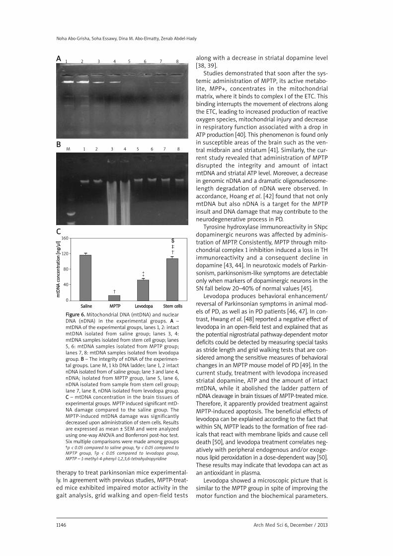
5, 6). However, mtDNA from mice treated with lev-odopa/carbidopa (lanes 7, 8) and stem cells (lanes3, 4) was electrophoresed in its intact form (singleband) (Figure 6 A). The MPTP administration result-ed in a marked decrease in the amount of intactnDNA in brain tissues (Figure 6 B). Moreover, a dra-matic oligonucleosome-length degradation of DNAwas observed, characterized by mixed smearingand laddering (lane 5). No appreciable fragmenta-tion of nDNA was found to occur in animals treat-ed with levodopa/carbidopa (lanes 6, 7, 8) and stemcells (lanes 2, 3, 4). However, only 5 mice treatedwith levodopa/carbidopa showed mild smearing(data not shown), while the other 7 mice of thisgroup showed total ladder and smear negativity.Such mild smearing may be attributed to the resid-ual damage in the heavily injured cells associatedwith the ongoing recovery, repair and renovationprocesses. In the MPTP-treated group, a decline wasobserved in the brain tissue content of mtDNA, ascompared to that of the saline group (p < 0.05, Fig-ure 6 C). However, improvement of the mtDNA con-tent was observed upon treatment with stem cells.The integrity of mtDNA from the levodopa/car-bidopa group (lanes 7, 8) was apparently damaged.
HHiissttooppaatthhoollooggiiccaall eexxaammiinnaattiioonn aanndd iimmmmuunnoossttaaiinniinngg
H + E stained sections in the SNpc of the salinegroup showed a cell-rich region with abundant largeand medium sized neurons with vesicular nuclei. Sec-tions from MPTP and levodopa groups showed sim-ilar neuronal degeneration. In the CD34+ stem celltreated animals, the reduction of the dopamine cellsproduced by the MPTP is counteracted (Figure 7 A).
With cresyl violet stain, abundant purple Nissl gran-ules were detected in the perikarya and dendrites ofnigral neurons in the saline group. The MPTP grouprevealed decreased purple Nissl granules. Treatmentwith levodopa/carbidopa resulted in increased pur-ple Nissl granules; however, stem cell therapy revealed
purple Nissl granules of nigral neurons more or lesssimilar to those of the saline group (Figure 7 B).
Regarding TH immunostaining, the saline grouprevealed abundant TH-immunopositive neurons in theSNpc. The MPTP group showed a marked loss in TH-immunopositive neurons (Figure 8 A) witha decrease in the mean percentage of TH-positive neu-rons compared to the saline group (Figure 8 B). TH-immunopositive neurons in the levodopa groupshowed a microscopic picture similar to the MPTPgroup with a mean percentage of 38.5 ±3.14% (Fig-ures 8 A, B), while in the stem cell group there wasamarked increase of TH-immunopositive neurons witha mean percentage of 56.6 ±4.18% that was greatercompared to the levodopa group (p < 0.05, Figure 8 B).
Discussion
The current study aimed to explore the cura-tive effect of intravenous HUCB CD34+ stem cell
2220181614121086420
1 6 11 16 21 26 31 36
CCyycclleess
M1: –Ve Cont M2 M3 M4
M5: +Ve Cont M6 M7 M8
M9 M10 M11 M12 M13
FFlluuoorreesscceennccee ((448833--553333))
FFiigguurree 44.. Real-time PCR analysis for identification ofhuman Sry gene in the female mice brain of thestem cell group. The PCR product could be amplifiedfrom the regenerating SNpc of nine female mice, so75% of SNpc samples were positive for presence ofthe Sry gene while 25% of SNpc samples were neg-ative for presence of the Sry gene
150
100
50
0
AA
SSaalliinnee MMPPTTPP LLeevvooddooppaa SStteemm cceellllss
DDooppaamm
iinnee lleevveell [[ppmmooll//gg]]
24
20
16
12
8
4
0
BB
SSaalliinnee MMPPTTPP LLeevvooddooppaa SStteemm cceellllss
AATTPP ccoonncceennttrraattiioonn [[mmmmooll//gg]]
FFiigguurree 55.. Dopamine (AA) and ATP (BB) levels in the experimental groups at the end of week 5 dopamine and ATP lev-els were markedly decreased in the MPTP group compared to the saline group. Treatment with levodopa or stemcells increased the dopamine and ATP levels compared to the MPTP group. Results are expressed as mean ± SEMand were analyzed using one-way ANOVA and Bonferroni post-hoc test. Six multiple comparisons were made amonggroups for each variable†p < 0.05 compared to saline group, ‡p < 0.05 compared to MPTP group, $p < 0.05 compared to levodopa group, MPTP – 1-methyl-4-phenyl-1,2,3,6-tetrahydropyridine
Effects of intravenous human umbilical cord blood CD34+ stem cell therapy versus levodopa in experimentally induced Parkinsonism in mice
1146 Arch Med Sci 6, December / 2013
therapy to treat parkinsonian mice experimental-ly. In agreement with previous studies, MPTP-treat-ed mice exhibited impaired motor activity in thegait analysis, grid walking and open-field tests
along with a decrease in striatal dopamine level[38, 39].
Studies demonstrated that soon after the sys-temic administration of MPTP, its active metabo-lite, MPP+, concentrates in the mitochondrialmatrix, where it binds to complex I of the ETC. Thisbinding interrupts the movement of electrons alongthe ETC, leading to increased production of reactiveoxygen species, mitochondrial injury and decreasein respiratory function associated with a drop inATP production [40]. This phenomenon is found onlyin susceptible areas of the brain such as the ven-tral midbrain and striatum [41]. Similarly, the cur-rent study revealed that administration of MPTPdisrupted the integrity and amount of intact mtDNA and striatal ATP level. Moreover, a decreasein genomic nDNA and a dramatic oligonucleosome-length degradation of nDNA were observed. Inaccordance, Hoang et al. [42] found that not onlymtDNA but also nDNA is a target for the MPTPinsult and DNA damage that may contribute to theneurodegenerative process in PD.
Tyrosine hydroxylase immunoreactivity in SNpcdopaminergic neurons was affected by adminis-tration of MPTP. Consistently, MPTP through mito-chondrial complex 1 inhibition induced a loss in THimmunoreactivity and a consequent decline indopamine [43, 44]. In neurotoxic models of Parkin-sonism, parkinsonism-like symptoms are detectableonly when markers of dopaminergic neurons in theSN fall below 20–40% of normal values [45].
Levodopa produces behavioral enhancement/reversal of Parkinsonian symptoms in animal mod-els of PD, as well as in PD patients [46, 47]. In con-trast, Hwang et al. [48] reported a negative effect oflevodopa in an open-field test and explained that asthe potential nigrostriatal pathway-dependent motordeficits could be detected by measuring special tasksas stride length and grid walking tests that are con-sidered among the sensitive measures of behavioralchanges in an MPTP mouse model of PD [49]. In thecurrent study, treatment with levodopa increasedstriatal dopamine, ATP and the amount of intactmtDNA, while it abolished the ladder pattern ofnDNA cleavage in brain tissues of MPTP-treated mice.Therefore, it apparently provided treatment againstMPTP-induced apoptosis. The beneficial effects oflevodopa can be explained according to the fact thatwithin SN, MPTP leads to the formation of free rad-icals that react with membrane lipids and cause celldeath [50], and levodopa treatment correlates neg-atively with peripheral endogenous and/or exoge-nous lipid peroxidation in a dose-dependent way [50].These results may indicate that levodopa can act asan antioxidant in plasma.
Levodopa showed a microscopic picture that issimilar to the MPTP group in spite of improving themotor function and the biochemical parameters.
AA
BB
1 2 3 4 5 6 7 8
M 1 2 3 4 5 6 7 8
160
120
80
40
0
CC
SSaalliinnee MMPPTTPP LLeevvooddooppaa SStteemm cceellllss
mmttDDNNAA ccoonncceennttrraattiioonn [[nngg//µµll]]
FFiigguurree 66.. Mitochondrial DNA (mtDNA) and nuclearDNA (nDNA) in the experimental groups. AA – mtDNA of the experimental groups, lanes 1, 2: intactmtDNA isolated from saline group; lanes 3, 4: mtDNA samples isolated from stem cell group; lanes5, 6: mtDNA samples isolated from MPTP group;lanes 7, 8: mtDNA samples isolated from levodopagroup. BB – The integrity of nDNA of the experimen-tal groups. Lane M, 1 kb DNA ladder; lane 1, 2 intactnDNA isolated from of saline group; lane 3 and lane 4,nDNA; isolated from MPTP group, lane 5, lane 6,nDNA isolated from sample from stem cell group;lane 7, lane 8, nDNA isolated from levodopa group.CC – mtDNA concentration in the brain tissues ofexperimental groups. MPTP induced significant mtD-NA damage compared to the saline group. TheMPTP-induced mtDNA damage was significantlydecreased upon administration of stem cells. Resultsare expressed as mean ± SEM and were analyzedusing one-way ANOVA and Bonferroni post-hoc test.Six multiple comparisons were made among groups†p < 0.05 compared to saline group, ‡p < 0.05 compared toMPTP group, $p < 0.05 compared to levodopa group, MPTP – 1-methyl-4-phenyl-1,2,3,6-tetrahydropyridine
Noha Abo-Grisha, Soha Essawy, Dina M. Abo-Elmatty, Zenab Abdel-Hady

Arch Med Sci 6, December / 2013 1147
Levodopa led to an increase in striatal dopaminelevel and this was reflected by motor improvementwithout a beneficial effect on the damaged do -paminergic neurons. This can be explained as lev-odopa is a catechol-containing compound that mayundergo autoxidation in vivo in the striatal extra-
cellular compartment, with the consequent gener-ation of quinone derivatives and superoxide anionthat cause oxidative stress and lead to neuronaldestruction by necrosis [51]. Moreover, patients withearly PD often respond well to dopaminergic med-ications with few clinical fluctuations. This is likely
SStteemm
cceellllss
FFiigguurree 77.. Sections from the substantia nigra in experimental groups stained with hematoxylin and eosin (H + E;400×) (AA) or cresyl fast violet (600×) (BB). AA – H + E staining of substantia nigra from saline-treated mice showeda cell-rich region full of abundant large and medium sized neurons with vesicular nuclei, substantia nigra pars com-pacta (SNpc) and a cell-poor substantia nigra pars reticularis (SNpr). The MPTP group showed neuronal degenera-tion in SNpc. The levodopa group showed neuronal degeneration in the SNpc similar to the MPTP group. The stemcell group showed a marked increase of the neuronal population in SNpc. BB – Cresyl fast violet staining indicatedthat SNpc taken from the saline group showed large abundant purple Nissl granules (arrows) in the perikarya anddendrites of nigral neurons. The MPTP group showed decreased purple Nissl granules (arrows) in the perikarya anddendrites of nigral neurons. The levodopa group showed purple Nissl granules (arrows) in the perikarya and den-drites of a few nigral neurons. The stem cell group showed purple Nissl granules (arrows) in the perikarya and den-drites of nigral neurons similar to that of the saline groupMPTP – 1-methyl-4-phenyl-1,2,3,6-tetrahydropyridine
SSaalliinnee
LLeevvooddooppaa
MMPPTTPP
AA BB
123.79 µm 35.87 µm
Effects of intravenous human umbilical cord blood CD34+ stem cell therapy versus levodopa in experimentally induced Parkinsonism in mice

1148 Arch Med Sci 6, December / 2013
AA BBSStteemm
cceellllss
FFiigguurree 88.. AA – Tyrosine hydroxylase (TH) immunostaining in thesubstantia nigra in the experimental groups. The saline groupshowed abundant TH-immunopositive neurons in the substan-tia nigra pars compacta (SNpc). The MPTP group showed markedloss of TH-immunopositive neurons in the SNpc. The levodopagroup showed marked loss of TH-immunopositive neurons inthe SNpc similar to the MPTP group. The stem cell group showedmarked increase of TH-immunopositive neurons in the SNpc.DAB and Mayer's hematoxylin counterstaining (200×, left col-umn and 400×, right column). BB – The mean percentage of TH-immunopositive neurons in the SNpc in the experimentalgroups. The mean percentage of TH-positive neurons in theMPTP group was significantly decreased compared to the salinegroup. Treatment with stem cells increased this mean percent-age compared to the MPTP group. Results are mean ± SEM andwere analyzed using one-way ANOVA and Bonferroni post-hoctest. Six multiple comparisons were made among groups†p < 0.05 compared to saline group, ‡p < 0.05 compared to MPTP group,$p < 0.05 compared to levodopa group, MPTP – 1-methyl-4-phenyl-1,2,3,6-tetrahydropyridine
SSaalliinnee
LLeevvooddooppaa
MMPPTTPP
100
80
60
40
20
0
CC
SSaalliinnee MMPPTTPP LLeevvooddooppaa SStteemm cceellllss
TTHH--iimmmmuunnooppoossiittiivvee nneeuurroonnss
161.87 µm 82.81 µm
Noha Abo-Grisha, Soha Essawy, Dina M. Abo-Elmatty, Zenab Abdel-Hady

Arch Med Sci 6, December / 2013 1149
due to the up-regulation of nigropallidal dopamin-ergic projection to the globus pallidus internus (GPi),reducing inhibitory output from GPi to the thala-mus. In more advanced Parkinson’s, loss of nigropal-lidal upregulation may result in alteration of the fir-ing pattern of GPi from tonic to burst firing,heralding the onset of motor complications [52].
In the present study, treatment with CD34+ stemcells improved locomotor activity, dopamine andATP levels, as well as mtDNA and nDNA integrity. Inaccordance, I.V. HUCB stem cell therapy was report-ed to restore dopaminergic function in humans [53]and delayed the onset of symptoms and death ofParkinsonian mice [54].
DNA damage and cell death after injection ofMPTP lead to a dramatic increase in the levels ofsecreted stress signals – chemokines, cytokines,and proteolytic enzymes – in many organs as partof the regeneration and repair process, which haveprofound impacts on stem cell migration and repop-ulation [55]. Stem cells were reported to grow in thepresence of different tissue types such as heart,lung and nerve tissue. The stem cells differentiateinto the same type of cells that were surroundingthem [56].
The beneficial effects of stem cells in neuronaldamage can be explained also as they produce neu-rotrophic factors, enhance angiogenesis, secretenumerous angiogenic factors, and reduce inflam-mation, apoptotic protection and nerve fiber reor-ganization [57, 58]. However, neuroregenerativetherapies should be tested for their potential clin-ical use [59, 60].
In the current study, stem cell therapy improvedMPTP-induced dopaminergic degeneration andinduced a marked increase of TH-immunopositiveneurons. Similarly, stem cells can differentiate intodopaminergic neurons that reinnervate the dener-vated striatum and become functionally integrat-ed, restoring striatal dopamine release [61]. Stemcell transplantation and activation of endogenousneurogenesis represents currently one of the mostpromising therapeutic approaches to CNS repair inneuroregenerative medicine [62]. In the presentstudy, functional assessment agreed with the bio-chemical and histopathologic assessment forCD34+ stem cell therapy. The beneficial effect ofstem cells over levodopa was also in accordancewith Lindvall and Björklund [61], who stated thattransplantation of human embryonic stem cells hasprovided proof of the principle that neuronal re -placement can work in PD patients.
In conclusion, CD34+ stem cells improved themotor function, ameliorated striatal dopamine andATP levels, and restored mtDNA and nDNA integrity,in addition to increasing TH immunostaining in theSNpc of MPTP-parkinsonian mice. These effects weresuperior to those produced by levodopa. Stem cells
as an alternative neuronal source can be employedto generate mature dopaminergic neurons to inner-vate the affected striatum in PD. Further studies areneeded to explore other possible mechanisms in -volved in the neuroprotective effect of CD34+ stemcells in experimental models of PD and to determinethe clinical benefit of using such therapy.
Re f e r e n c e s1. Kramberger MG, Stukovnik V, Cus A, et al. Parkinson'sdisease dementia: clinical correlates of brain spect perfusionand treatment. Psychiatr Danub 2010; 22: 446-9.
2. Liu B. Modulation of microglia pro-inflammatory andneurotoxic activity for the treatment of Parkinson’sdisease. AAPS J 2006; 8: 606-21.
3. Lang AE, Lozano AM. Parkinson’s disease (first of twoparts). N Engl J Med 1998; 339: 1044-53.
4. Fernandez-Espejo E. Pathogenesis of Parkinson’s disease:prospects of neuroprotective and restorative therapies.Mol Neurobiol 2004; 29: 15-30.
5. Hunot S, Hirsch EC. Neuroinflammatory processes inParkinson's disease. Ann Neurol 2003; 3: 49-58.
6. Lin MT, Beal MF. Mitochondrial dysfunction and oxidativestress in neurodegenerative diseases. Nature 2006; 443:787-93.
7. Dehay B, Bezard E. New animal models of Parkinson'sdisease. Mov Disord 2011; 26: 1198-205.
8. Sławek J. Levodopa in the treatment of Parkinson disease:yesterday and today [Polish]. Neurol Neurochir Pol 2012;46: 63-75.
9. Nishida N, Murakami T, Kadoh K, et al. Subthalamic nu -cleus deep brain stimulation restores normal rapid eyemovement sleep in Parkinson's disease. Mov Disord 2011;26: 2418-22.
10. Lindvall O, Kokaia Z. Stem cells in human neurode -generative disorders: time for clinical translation? J ClinInvest 2010; 120: 29-40.
11. Chen N, Hudson JE, Walczak P, et al. Human umbilical cordblood progenitors: the potential of these hematopoieticcells to become neural. Stem Cells 2005; 23: 1560-70.
12. Rosenkranz K, Meier C. Umbilical cord blood cell trans -plantation after brain ischemia: from recovery of functionto cellular mechanisms. Ann Anat 2011; 193: 371-9.
13. Arien-Zakay H, Lazarovici P, Nagler A. Tissue regenerationpotential in human umbilical cord blood. Best Pract ResClin Haematol 2010; 23: 291-303.
14. Chen CT, Foo NH, Liu WS, Chen SH. Infusion of humanumbilical cord blood cells ameliorates hind limb dys -function in experimental spinal cord injury through anti-inflammatory, vasculogenic and neurotrophic me -chanisms. Pediatr Neonatol 2008; 49: 77-83.
15. Garbuzova-Davis S, Willing AE, Zigova T, et al. Intravenousadministration of human umbilical cord blood cells ina mouse model of amyotrophic lateral sclerosis: di -stribution, migration, and differentiation. J HematotherStem Cell Res 2003; 12: 255-70.
16. Liu WS, Chen CT, Foo NH, et al. Human umbilical cordblood cells protect against hypothalamic apoptosis andsystemic inflammation response during heatstroke in rats.Pediatr Neonatol 2009; 50: 208-16.
17. Ou Y, Yu S, Kaneko Y, et al. Intravenous infusion of GDNFgene-modified human umbilical cord blood CD34+ cellsprotects against cerebral ischemic injury in spontaneouslyhypertensive rats. Brain Res 2010; 1366: 217-25.
Effects of intravenous human umbilical cord blood CD34+ stem cell therapy versus levodopa in experimentally induced Parkinsonism in mice

1150 Arch Med Sci 6, December / 2013
18. Lunn JS, Sakowski SA, Hur J, Feldman EL. Stem cell tech -nology for neurodegenerative diseases. Ann Neurol 2011;70: 353-661.
19. Serra PA, Stefano P, Bianca M, Maria SD, Egidio M. TheMPTP mouse model: cues on DA release and neural stemcell restorative role. Parkinsonism Relat Disord 2008; 14:189-93.
20. Paillé V, Brachet P, Damier P. Role of nigral lesion in thegenesis of dyskinesias in a rat model of Parkinson'sdisease. Neuroreport 2004; 15: 561-4.
21. Kim JH, Auerbach JM, Rodríguez-Gómez JA, et al. Do -pamine neurons derived from embryonic stem cellsfunction in an animal model of Parkinson's disease. Nature2002; 418: 50-6.
22. Wagner JE, Kernan NA, Steinbuch M, et al. Allogeneicsibling cord blood transplantation in forty four childrenwith malignant and non malignant disease. Lancet 1995;346: 214-9.
23. Erices A, Conget P, Minguell JJ. Mesencymalprogenito cellsin human umbilical cord blood. Br J Haematol 2000; 109:235-42.
24. Miltenyi S, Mueller W, Weichel W, Radbruch A. High gra -dient magnetic cell separation with MACS. Cytometry1990; 11: 231-8.
25. Armitage S, Hargreaves R, Samson D, Brennan M, Kanfer E,Navarrete C. CD34 counts to predict the adequate col -lection of peripheral blood progenitor cells. BM Transplant1997; 20: 587-91.
26. Strober W. Trypan blue exclusion test of cell viability. CurrProtoc Immunol 2001; 21: A.3B.1-2.
27. Fernagut OP, Elsa D, Bertrand L, Francois T. A simple me -thod to measure stride length as an index of nigrostriataldysfunction in mice. J Neurosci Methods 2002; 113: 123-30.
28. Menet V, Prieto M, Privat A, Ribotta M. Axonal plasticityand functional recovery after spinal cord injury in micedeficient in both glial fibrillary acidic protein and vimentingenes. Proc Natl Acad Sci 2003; 100: 8999-9004.
29. El-Awady EE, Zaitone SA, Abd El-Hamied AS. Effect ofcelecoxib, meloxicam, pentoxyphillin and ramipril onexperimental Parkinson's disease. Bull Fac Pharm CairoUniv 2007; 45: 355-75.
30. Vogler GA. Anesthesia and analgesia. In: The laboratoryrat. Suckow MA, Weisbroth SH, Franklin CL (eds.). ElsevierAcademic Press, New York 2006; 627-95.
31. Kozina EA, Khaindrava VG, Kudrin VS, et al. Experimentalmodeling of functional deficiency of the nigrostriataldopaminergic system in mice. Ross Fiziol Zh Im I M Se -chenova 2010; 96: 270-82.
32. Wang LJ, Chen YM, George D, et al. Engraftment assess -ment in human and mouse liver tissue after sex-mismatched liver cell transplantation by real-timequantitative PCR for Y chromosome sequences. LiverTranspl 2002; 8: 822-8.
33. Yang NC, Ho WM, Chen YH, Hu ML. A convenient one-stepextraction of cellular ATP using boiling water for theluciferin-luciferase assay of ATP. Anal Biochem 2002; 306:323-7.
34. Liu H, Jiang Y, Luo Y, Jiang W. A simple and rapid de -termination of ATP, ADP and AMP concentrations in pericarptissue of litchi fruit by high performance liquid chro -matography. Food Technol Biotechnol 2006; 44: 531-4.
35. Chappel JB, Hansford RG. Subcellular components.Butterworths, London 1969.
36. Chang BJ, Nishikawa M, Sato E, Utsumi K, Inoue M. L-Car -nitine inhibits cisplatin-induced injury of the kidney andsmall intestine. Arch Biochem Biophys 2002; 405: 55-64.
37. Sambrook J, Maniatis T, Fritsch F. Molecular cloning,a laboratory manual. Cold Spring Harbor Laboratory Press,Cold Spring Harbor, New York 2001.
38. Jackson-Lewis V, Przedborski S. Protocol for the MPTPmodel of Parkinson’s disease. Nat Protoc 2007; 2: 141-51.
39. Kim SN, Kim ST, Doo AR, et al. Phosphatidylinositol 3kinase/Akt signaling pathway mediates acupuncture-induced dopaminergic neuron protection and motorfunction improvement in a mouse model of Parkinson'sdisease. Int J Neurosci 2011; 121: 562-9.
40. Singh N, Pillay V, Choonara YE. Advances in the treatmentof Parkinson’s disease. Prog Neurobiol 2007; 81: 29-44.
41. Khan FH, Sen T, Maiti AK, Jana S, Chatterjee U, Chakrabar -ti S. Inhibition of rat brain mitochondrial electron transportchain activity by dopamine oxidation products duringextended in vitro incubation: implications for Parkinson’sdisease. BBA Mol Bas Dis 2005; 1741: 65-74.
42. Hoang T, Choi DK, Nagai M, et al. Neuronal NOS andcyclooxygenase-2 contribute to DNA damage in a mousemodel of Parkinson disease. Free Radic Biol Med 2009;47: 1049-56.
43. Aoki E, Yano R, Yokoyama H, Kato H, Araki T. Role ofnuclear transcription factor kappa B (NF-kappaB) for MPTP(1-methyl-4-phenyl-1,2,3,6-tetrahyropyridine)-inducedapoptosis in nigral neurons of mice. Exp Mol Pathol 2009;86: 57-64.
44. Lane El, Carlsson T, Kirik D, Dunnett SB. Animals modelsof Parkinsonism. In: source book of models for biomedicalresearch. Conn PM (ed.), Human Press. Inc., Totowa, N J.2008; 313-20.
45. Breit S, Bouali-Benazzouz R, Popa RC, Gasser T, Bena bid AL,Benazzouz A. Effects of 6-hydroxydopamine-inducedsevere or partial lesion of the nigrostriatal pathway onthe neuronal activity of pallido-subthalamic network inthe rat. Exp Neurol 2007; 205: 36-47.
46. Hutter-Saunders JA, Kosloski LM, McMillan JM, et al. BL-1023 improves behavior and neuronal survival in 1-methyl-4-phenyl-1,2,3,6-tetrahydropyridine-intoxicated mice.Neuroscience 2011; 180: 293-304.
47. O'Sullivan SS, Johnson M, Williams DR, et al. The effectof drug treatment on neurogenesis in Parkinson's disease.Mov Disord 2011; 26: 45-50.
48. Hwang DY, Fleming SM, Ardayfio P, et al. 3,4-Dihydro -xyphenylalanine reverses the motor deficits in Pitx3-deficient Aphakia mice: behavioral characterization ofa novel genetic model of Parkinson’s disease. J Neurosci2005; 25: 2132-7.
49. Lewis GN, Byblon WD, Walt SE. Stride length regulationin Parkinson’s disease: the use of extrinsic, visual cues.Brain 2000; 123: 2077-90.
50. Agil A, Durán R, Barrero F, et al. Plasma lipid peroxida -tion in sporadic Parkinson’s disease. Role of the l-dopa. J Neurol Sci 2006; 240: 31-6.
51. Serra PA, Esposito G, Enrico P, et al. Manganese increasesL-DOPA auto-oxidation in the striatum of freely movingrats: potential implications to L-DOPA long-term therapyof Parkinson’s disease. Br J Pharmacol 2000; 130: 937-45.
52. Schaller B, Cornelius JF, Sandu N. Molecular medicinesuccesses in neuroscience. Mol Med 2008; 14: 361-4.
53. Arenas E. Towards stem cell replacement therapies forParkinson's disease. Biochem Biophys Res Commun 2010;396: 152-6.
54. Ende N, Chen R. Parkinson's disease mice and humanumbilical cord blood. J Med 2002; 33: 173-80.
55. Ponomaryov T, Peled A, Petit I, et al. Induction of thechemokine stromal-derived factor-1 following DNA da -mage improves human stem cell function. J Clin Invest2000; 106: 1331-9.
56. Adult stem cells can produce a wealth of cell types,science authors report. Science Daily Magazine (adaptedfrom news release by American Association for the
Noha Abo-Grisha, Soha Essawy, Dina M. Abo-Elmatty, Zenab Abdel-Hady

Arch Med Sci 6, December / 2013 1151
Advancement of Science) Jun 2000: http://sciencedaily.com/releases/2000/06/000602072837.html.
57. Fan CG, Zhang QJ, Tang FW, Han ZB, Wang GS, Han ZC.Human umbilical cord blood cells express neurotrophicfactors. Neurosci Lett 2005; 380: 322-5.
58. Ding DC, Shyu WC, Chiang MF, et al. Enhancement ofneuroplasticity through upregulation of beta1-integrin inhuman umbilical cord-derived stromal cell implantedstroke model. Neurobiol Dis 2007; 27: 339-53.
59. Matusik E, Wajgt A, Janowska J, et al. Cell adhesion mo -lecular markers in ischaemic stroke patients: correlationwith clinical outcome and comparison with primaryautoimmune disease. Arch Med Sci 2009; 5: 182-9.
60. Schaller B. The role of endothelin in stroke: experimentaldata and underlying pathophysiology. Arch Med Sci 2006;2: 146-58.
61. Lindvall O, Björklund A. Cell therapy in Parkinson’s disease.Neuro Rx 2004; 1: 382-93.
62. Sandu N, Momen-Heravi F, Sadr-Eshkevari P, Schaller B.Molecular imaging for stem cell transplantation in neuro -regenerative medicine. Neurodegener Dis 2012; 9: 60-7.
Effects of intravenous human umbilical cord blood CD34+ stem cell therapy versus levodopa in experimentally induced Parkinsonism in mice