effects of phosphorylation on the nlrp3 inflammasome
TRANSCRIPT

University of Calgary
PRISM: University of Calgary's Digital Repository
Cumming School of Medicine Cumming School of Medicine Research & Publications
2019-03-05
Effects of phosphorylation on the NLRP3
inflammasome
Sandall, Christina F.; MacDonald, Justin Anthony
Sandall, C. F., & MacDonald, J. A. (2019). Effects of phosphorylation on the NLRP3 inflammasome,
1-33. http://dx.doi.org/10.1016/j.abb.2019.02.020
http://hdl.handle.net/1880/110193
unknown
Downloaded from PRISM: https://prism.ucalgary.ca

1
Effects of Phosphorylation on the NLRP3 Inflammasome
Christina F. Sandall and Justin A. MacDonald*
Department of Biochemistry & Molecular Biology, Cumming School of Medicine, University of Calgary, Calgary, AB, T2N 4Z6, Canada
*Correspondence to: Prof. Justin A. MacDonald, Libin Cardiovascular Institute of Alberta, Cumming School of Medicine, University of Calgary, 3280 Hospital Drive NW, Calgary, AB, Canada
(Tel.) 403-210-8433 (Email) [email protected]
Keywords: nucleotide-binding domain and leucine-rich repeat-containing receptors, NLR, NLRP3, NACHT, PKA, cAMP, molecular modeling, nucleotide-binding, inflammasome assembly, phosphorylation
1234567891011121314151617181920212223242526272829303132333435363738394041424344454647484950515253545556

2
Abstract
The pyrin domain containing Nod-like receptors (NLRPs) are a family of pattern recognition
receptors known to regulate an array of immune signaling pathways. Emergent studies
demonstrate the potential for regulatory control of inflammasome assembly by phosphorylation,
notably NLRP3. Over a dozen phosphorylation sites have been identified for NLRP3 with many
more suggested by phosphoproteomic studies of the NLRP family. Well characterized NLRP3
phosphorylation events include Ser198 by c-Jun terminal kinase (JNK), Ser295 by protein kinase
D (PKD) and/or protein kinase A (PKA), and Tyr861 by an unknown kinase but is
dephosphorylated by protein tyrosine phosphatase non-receptor 22 (PTPN22). Since the PKA-
and PKD-dependent phosphorylation of NLRP3 at Ser295 is best characterized, we provide
detailed review of this aspect of NLRP3 regulation. Phosphorylation of Ser295 can attenuate
ATPase activity as compared to its dephosphorylated counterpart, and this event is likely unique
to NLRP3. In silico modeling of NLRP3 is useful in predicting how Ser295 phosphorylation
might impact upon the structural topology of the ATP-binding domain to influence catalytic
activity. It is important to gain as complete understanding as possible of the complex
phosphorylation-mediated mechanisms of regulation for NLRP3 in part because of its
involvement in many pathological processes.
1. Introduction
1.1 Pattern recognition receptors in innate immunity
Vertebrates rely on two highly conserved and specialized defense strategies: the innate and
adaptive immune systems. The innate immune system functions as the first line of defense in reconciling
injury and infection through the early detection of impending threats, and consequential triggering of
proinflammatory responses1. These recognition and response mechanisms are mediated by the pattern
recognition receptors (PRRs) during both microbial infection and anomalous endogenous signaling,
including the aberrant localization of danger signals, the formation of abnormal molecular complexes
and other indicators of cellular stress2. Evolutionarily conserved microbial motifs recognized by PRRs
are referred to as pathogen-associated molecular patterns (PAMPs) and irregular host-derived signals as
danger-associated molecular patterns (DAMPs). PRRs are expressed primarily in immune and
inflammatory cells such as monocytes, macrophages, neutrophils, epithelial and dendritic cells3.
57585960616263646566676869707172737475767778798081828384858687888990919293949596979899100101102103104105106107108109110111112

3
Members of the PRR superfamily are broadly localized, functioning in the extracellular milieu, plasma
membrane, endosomal compartments and the cytosol. Five classes of PRR families have been identified:
the membrane-bound Toll-like receptors (TLRs) and C-type lectin receptors (CLRs), the cytoplasmic
retinoic acid-inducible gene-I-like receptors (RLRs), nucleotide-binding domain and leucine-rich repeat-
containing receptors (NLRs) and the absent in melanoma 2-like receptors (ALRs)4. The comparable
structural domain composition of the PRR families reflect their collective role in the recognition of
danger signals and the subsequent activation of pro-inflammatory signal transduction pathways. These
pathways often converge on the activation of the nuclear factor kappa-light-chain-enhancer of activated
B cells (NF-κB) or antiviral type I interferon (IFN) pathways, or in the case of NLRs on the maturation
and secretion of pro-inflammatory cytokines5.
1.2 NLRs Nucleate Signaling Complexes in Response to PAMPs or DAMPs
Over the past decade, certain PRRs, including the NLRs and ALRs, have emerged as key sensors
of intracellular danger signals. These receptors exhibit the capacity to nucleate multimeric signaling
complexes, termed inflammasomes, in response to diverse stimuli6. The first NLR inflammasome
(NLRP3) was characterized as a caspase-1-activating complex in which proximity-induced auto-
proteolysis activated the protease to cleave and maturate the pro-inflammatory interleukin (IL)-1β
precursor, pro-IL-1β7. The majority of the inflammasome forming sensors described to date are NLR
receptors8. The NLR protein family is comprised of 22 members, characterized by a central nucleotide-
binding domain (NACHT - [NAIP (neuronal apoptosis inhibitory protein), CIITA (MHC class II
transcription activator), HET-E (in-compatibility locus protein from Podospora anserina) and TP1
(telomerase-associated protein)]), and discernable by their N-terminal effector domains9. The family can
be subdivided into the NLRA, NLRB, NLRC or NLRP subfamilies based on whether the N-terminus
contains an acidic domain, Baculovirus IAP repeat (BIR), caspase recruitment domain (CARD), or pyrin
domain (PYD), respectively10. NLRP1, NLRP3 and NLRC4 constitute the best-characterized
inflammasomes11; however, evidence also supports the existence of inflammasome complexes nucleated
by NLRP612, NLRP713, NLRP1214 and AIM215,16. Although the potential inflammasome assembly of the
remaining NLRP proteins is largely uncharacterized, rapidly developing models suggest these proteins
play a role of paramount importance in initiating and directing the inflammatory response to cellular
injury17. Regrettably, critical aspects pertaining to the underlying biology of this family and their
signaling complexes remain to be elucidated.
113114115116117118119120121122123124125126127128129130131132133134135136137138139140141142143144145146147148149150151152153154155156157158159160161162163164165166167168
4
1.2 NLRPs are STAND ATPase family members
Comparative sequence analyses of the 14 NLRP genes reveal conserved ATP-binding motifs
within the central NACHT domain that have designated them as members of the Signal Transduction
ATPases with Numerous Domains (STAND) clade, related to the larger ATPases-Associated with
various cellular Activities (AAA+ ATPase) superfamily of proteins18. The majority of STAND P-Loop
ATPases are modular proteins, containing three or more domains involved in DNA or protein binding,
signal transduction and scaffolding19. Structural data and biochemical investigations of several family
members have revealed a common mechanism of regulation. STAND proteins have a conserved core
containing ATPase activity, as well as key effector domains involved in sensing of stimuli and in
downstream signaling19. A conserved mechanism of activation has been proposed, based on the structural
data of four STAND ATPases20. Briefly, the integrated data suggest that these ATPases function as
regulated molecular switches, which undergo structural reorganizations corresponding to monomeric,
ADP-bound forms associated with the “off position”, and oligomeric, ATP-bound forms associated with
the “on position”, the latter of which can initiate downstream signal transduction pathways via protein-
protein interactions mediated by the effector domains.
To date, only NLRP321 ,NLRP722 and NLRP1223 have been empirically demonstrated to possess
ATP-binding potential and intrinsic ATPase activity. While it seems evident that NLRP1 binds ATP24,
the capacity of NLRP1 to hydrolyze ATP is not yet clear. Although the seminal paper linked the
contribution of nucleotide-binding to the functional role of NLRP1, a soluble fragment of the protein
containing the NACHT domain and the LRR exhibited negligible ability to hydrolyze triphosphate
nucleotides25. The basic biochemistry of these proteins remains elusive and under-characterized, so
further description of the enzymology and the role of ATP in driving inflammasome activation and the
ensuing inflammatory signaling pathways will be critical for a comprehensive understanding of this
family and their role in human disease.
1.3 Conserved Structural Motifs Present in the NLRP-NACHT Domain
Several distinct motifs cooperate to enable nucleotide-binding and/or hydrolysis. These motifs,
including their functional importance and conservation among the various NLRP family members, have
been previously reviewed in detail20,26,9, but the specific function of each in the context of NLRP3 is
briefly summarized here.
The Walker A motif, containing the consensus sequence GXXGXGK(S/T), forms an integral P
loop that is highly conserved across the entire family of STAND ATPases, including a conserved Lys
residue (Lys232 in NLRP3) that provides a direct stabilizing interaction with the terminal -phosphate
169170171172173174175176177178179180181182183184185186187188189190191192193194195196197198199200201202203204205206207208209210211212213214215216217218219220221222223224

5
of the nucleotide. Mutation of Gly231, Lys232 and Thr233 to Ala residues was associated with reduced
ATP binding and disrupted NLRP3 inflammasome-dependent signaling, including a reduction in
caspase-1 activation, IL-1β maturation, cell death, macromolecular complex formation, self-association,
and association with the inflammasome component ASC, all indicating the importance of ATP-binding
in NLRP3 function21,27.
The Walker B motif is characterized by the consensus sequence DGX(D/E)E and contains
conserved acidic residues that coordinate Mg2+ ion binding and aid in the priming of an H2O molecule
thought to be important in the hydrolysis of ATP (Asp302, Asp305 and Glu306 in NLRP3). However,
mutations of this motif in NLRP12 did not abolish nucleotide hydrolysis23, suggesting that ATP-binding
and hydrolysis is driven primarily by other key nucleotide-binding regions of NLRPs. Homology
modeling suggests that key regions of the NLRP3 Walker B site are not located in close proximity to the
predicted nucleotide-binding site, which could clarify the lack of global influence of Walker B mutations
on function.
The Arginine finger motif is characterized by the conserved CRE sequence. The positive charge
of the aspartic acid residue acts to stabilize the negatively charged phosphate moieties present in ATP.
The net effect is to render the -phosphate a better electrophile, thereby enabling the effective hydrolysis
of ATP. In most AAA+ ATPases, this arginine residue is contributed in trans from a neighboring subunit
present in a homo-oligomeric complex28.
The Sensor 1 motif is comprised of three hydrophobic residues followed by two Ser/Thr residues
and a conserved Arg (Leu346 to Arg351 in NLRP3), all of which contribute to intramolecular
interactions with elements of the Walker A and B motifs in the NACHT domain in addition to binding
the nucleotide.
The Sensor 2 motif is the final conserved functional element of the NACHT domain. It is
characterized by a conserved Arg or Lys residue (Arg366 in NLRP3) that also assists in coordinating
nucleotide-binding, hydrolysis, and coordinating global conformational changes between subunits.
1.4 NLR Phosphorylation is an Emerging Regulatory Mechanism
In addition to transcriptional priming, which has been meticulously reviewed29,30,31, post-
translational modifications of inflammasome components have emerged as key checkpoints in the
highly-regulated processes of inflammasome activation. These broadly regulated mechanisms shape
distinct biochemical responses and have been shown to alter NLRP3 protein-protein interactions,
catalytic activity, sub-cellular localization, NLRP3 nucleation, and thus activation status and
inflammatory signaling32. These modifications include ubiquitination, alkylation, S-nitrosylation,
225226227228229230231232233234235236237238239240241242243244245246247248249250251252253254255256257258259260261262263264265266267268269270271272273274275276277278279280
6
proteolytic processing, ADP-ribosylation, and phosphorylation33. Protein phosphorylation is the most
common mechanism in the regulation of enzymatic output and functionality; indeed, it has been
speculated that one-third of all proteins in the human proteome are substrates for phosphorylation34.
Among other processes, phosphorylation can transiently alter the intrinsic biological activity, structural
conformation, subcellular localization, half-life and protein-protein docking interactions of the enzyme35.
Recent evidence supports a significant role for phosphorylation events in both the positive and negative
regulation of various NLR inflammasomes (Table 1). Indeed, evidence is quickly amassing to suggest
that protein phosphorylation events are pervasive in inflammasome regulation across the NLR family, as
well as in concomitant signaling pathways (reviewed in 36). While many phosphorylation events have
been characterized (with both high throughput and low throughput methods), it is clear that we do not
yet have a full appreciation of how these important regulatory steps work synergistically to regulate
inflammation. Various kinases and phosphatases have been identified to associate with different NLRs,
yet not all phosphorylation events have been linked with an effector kinase/phosphatase system. In
addition, not all phosphorylation events have been linked with clear functional effects on inflammasome
regulation. Thorough biochemical assessments are still required to comprehensively identify all
phosphorylation events and to assess the functional implications on NLR enzymology.
2. Phosphorylation and Regulation of the NLRP3 Inflammasome NLRP3 is the best studied NLRP protein, and is considered to be the prototypical inflammasome-
forming member of the family. In the canonical activation pathway, two distinct and critical steps are
essential for assembly of the NLRP3 inflammasome: an initial transcriptional priming step, and a second,
oligomerization and assembly step37 (Figure 1). Each stage is meticulously controlled and has been
extensively profiled in several publications38,39,40,41. As a result, this review will detail the recent
advances made regarding phosphorylation events and their regulation of NLRP3 inflammasome
signaling, with an additional specific focus on cAMP-dependent protein kinase (PKA)-dependent
phosphorylation of NLRP3.
2.1 NLRP3 Phosphorylation Events
Current research into NLRP3 phosphorylation has revealed 13 unique sites (Table 2), reported as
outputs of both high throughput methods (i.e., identification of sites using only discovery mass
spectrometry) and low throughput methods (i.e., sites were identified and then validated with other
biochemical methods). These NLRP3 phosphorylation events have been linked to the induction or
281282283284285286287288289290291292293294295296297298299300301302303304305306307308309310311312313314315316317318319320321322323324325326327328329330331332333334335336

7
inhibition of inflammasome activity (Table 2), changes in NLRP3 protein conformation, the regulation
of other molecular associations, as well as to the occurrence of other post-translational modifications
(such as ubiquitination).
In 2016, a mechanism of NLRP3 inflammasome inhibition by tyrosine phosphorylation of
NLRP3 at Tyr861 was reported by Spalinger and colleagues42. Loss of the protein tyrosine phosphatase
non-receptor 22 (PTPN22) was associated with augmented phosphorylation at Tyr861, and decreased
caspase-1 activation and IL-1 maturation. Phosphomimetic mutant NLRP3-Tyr861Glu attenuated
inflammasome activity, while the NLRP3-Tyr861Phe mutant resulted in enhanced NLRP3 activation.
The investigators also showed that PTPN22 could directly interact with NLRP3 in the presence of ASC.
Six phosphorylation sites (Tyr13, Ser163, Ser198, Ser334, Ser728 and Ser975) were identified in
2017 using a HEK293T reconstitution cell system in which FLAG-tagged NLRP3, ASC, pro-caspase-1
and pro-IL-1 were expressed. The NLRP3 inflammasome was captured by immunoprecipitation during
priming and analyzed with discovery mass spectrometry (LC-MS/MS)43. From this survey, only Ser198
(Ser194 in mouse) was found to have any demonstrable impact on inflammasome activation and was
subjected to further interrogation. Through site-directed alanine replacement scanning, the investigators
found that only NLRP3-Ser198Ala exhibited an obvious impact on inflammasome signaling, as
evidenced by decreased pro-IL-1 maturation. Subsequent expression of NLRP3-Ser198Ala in
immortalized human bone marrow-derived macrophages or knock-in mice harboring the Nlrp3
Ser194Ala allele (Nlrp3S194A/S194A) provided confirmation of results. The phosphorylation event had a
positive priming effect on NLRP3 that was essential for inflammasome activation. Interestingly, the
phosphorylation of S194 was only observed after priming and was not induced by PAMP or DAMP
stimulation of unprimed cells. Further kinase profiling and analyses with selective small molecule
inhibitors revealed that c-Jun terminal kinase 1 (JNK1) could directly phosphorylate NLRP3 at Ser194
which in turn facilitated the self-association and activation of the NLRP3 inflammasome. Ultimately, the
phosphorylation of Ser194 by JNK1 was shown to play a critical role in deubiquitination of NLRP3
during priming, an effect linked to the molecular interaction of NLRP3 with BRCC3 (a Lys63-specific
deubiquitinase). Inactive, monomeric NLRP3 is maintained in a ubiquitinated state, thereby impeding
oligomerization and inflammasome activation until a priming signal activates the BRCC3 ubiquitinase.
This Zn2+-dependent metalloprotease promotes the deubiquitination of the NLRP3-LRR domain44.
Recent studies have suggested that the deubiquitination of NLRP3 is indispensable for the activation of
the inflammasome, as pan-inhibition of deubiquitinases can block NLRP3 activation45.
337338339340341342343344345346347348349350351352353354355356357358359360361362363364365366367368369370371372373374375376377378379380381382383384385386387388389390391392
8
In another 2017 study, Zhang and colleagues demonstrated a link between protein kinase D (PKD)
and NLRP3 inflammasome activation46. The investigators established diacylglycerol (DAG) and PKD
enrichment in the Golgi fraction in response to NLRP3 inflammasome activation by various
DAMPs/PAMPs. Subsequent investigations with PKD knockouts and small molecule inhibitors
corroborated the requirement of PKD activity for effective inflammasome activation, potentially by
mediating the recruitment of ASC to NLRP3. Through mutational analysis and the use of an antibody
with immunoreactivity toward PKD consensus sites (i.e., [I/L]-X-[R]-X-[S/T]), a phosphorylation site
was identified within the NACHT domain of mouse NLRP3 at murine Ser291 (corresponding to Ser295
in human NLRP3). We note an apparent discrepancy in the numbering of residues in the Zhang et al.
publication, the authors designate the phosphorylated serine in the mouse NLRP3 protein as Ser293;
however, in verifying the surrounding sequence (displayed in Figure S4D of the Zhang et al. 2017
publication) against UniProtKB Q8R4B8-1, we determined that the authors were referring to sequence
corresponding to residues 281-303, not 283-308. We also verified that no other murine NLRP3 isoforms
listed in the UniProtKB or NCBI databases are numbered as listed in the Zhang publication.
Consequently, the following descriptions will reference PKD-dependent phosphorylation of Ser295
(human) and Ser291 (mouse). The phosphorylation site was confirmed with mass spectrometry and the
use of a phosphospecific anti-[pSer291]-NLRP3 antibody. The authors also revealed that Ser295
phosphorylation was detected in the mature inflammasome downstream of NLRP3 self-oligomerization
and suggested that the PKD-mediated phosphorylation could release NLRP3 from mitochondria-
associated ER membranes, allowing for the assembly of a mature inflammasome. Furthermore, the
oligomerization of a phosphomimetic NLRP3 (i.e., Ser291Glu) was abolished in a reconstitution system,
whereas the oligomerization of either Ser291Ala or wild-type NLRP3 was unaffected.
Also in 2017, Stutz and colleagues independently mapped additional NLRP3 phosphorylation
events using quantitative proteomics47. The phosphorylation of Ser5, Ser161 and Ser728 residues was
identified following overexpression of FLAG-NLRP3 in a murine immortalized macrophage cell line
and stimulation with various DAMPs/PAMPs. Significantly, the phosphorylation of the Ser5 site within
the PYD was linked to inflammasome inhibition. In this regard, structural modeling suggested that Ser5
phosphorylation could neutralize a charged helical surface that acts as the interface for homotypic PYD-
PYD interactions between NLRP3 molecules as well as heterotypic interactions between NLRP3 and
ASC.
Three additional NLRP3 phosphorylation events (Ser233, Ser387, Ser436) were annotated as a
result of other high-throughput discovery mass spectrometry studies48,49,50. These phosphorylations have
yet to be validated, and it is unknown if they are linked to any regulatory roles in priming or assembly.
393394395396397398399400401402403404405406407408409410411412413414415416417418419420421422423424425426427428429430431432433434435436437438439440441442443444445446447448
9
Finally, two reports published in 2016 described the actions of cAMP-dependent protein kinase (PKA)
toward NLRP351,52. The impact of PKA phosphorylation on NLRP3 is reviewed in detail in Sections 3
and 4 of this review. Briefly, PKA was found to phosphorylate human NLRP3 on Ser295 (murine residue
Ser291) in the NACHT domain, resulting in an inhibition of inflammasome signaling.
2.2 Protein Phosphatases and the Dephosphorylation of NLRP3
The protein kinase catalyzed addition of a phosphoryl group to a substrate protein is a reversible
modification that is subsequently removed by the hydrolytic activity of protein phosphatases53. Thus,
confirming, characterizing and identifying the mechanisms of action of NLRP3-associated phosphatase
holoenzymes will provide an essential understanding of NLRP3 functionality and inflammasome
regulation. To date, a limited number of phosphatases have been identified to drive NLRP3
dephosphorylation under certain contexts. The afore mentioned protein tyrosine phosphatase PTPN22 is
responsible for dephosphorylation of the pTyr861 residue42. The pSer5 residue, which inhibits
inflammasome activation during priming47, is suggested to be the target of protein phosphatase type-2A
(PP2A). Pretreatment with okadaic acid, a PP2A and PP2A-like phosphatase inhibitor, resulted in
increased Ser5 phosphorylation of NLRP3 in primed macrophages. These results were substantiated with
knockdown of the PP2A catalytic subunit (PPP2AC). As with other fields of investigation, the definition
of the cognate protein phosphatases responsible for dephosphorylation of specific NLRP sites has lagged
behind the discovery of the phosphorylation events driven by the protein kinases. While investigations
have advanced understanding of phosphatase involvement, (e.g., by linking PPP2AC and PTPN22 to the
Ser5 and Tyr861 residues of NLRP3, respectively), additional focused studies are still required. As an
example, it is not yet known how the regulatory/targeting subunits of the PP2A holoenzyme complex
integrate with NLRP3 to control inflammasome activity. Furthermore, studies have yet to define the
protein phosphatase responsible for maintaining Ser295 in the dephosphorylated state. Thus, identifying
and characterizing the mechanism of action of the specific NLRP3-associated phosphatase
holoenzyme(s) will add essential understanding to NLRP3 functionality and inflammasome regulation.
Moreover, the comprehensive examination of the specific phosphatases involved in the regulation of all
the various NLRPs will be a worthy research pursuit.
3. PKA-dependent phosphorylation and the regulation of NLRP inflammasomes
449450451452453454455456457458459460461462463464465466467468469470471472473474475476477478479480481482483484485486487488489490491492493494495496497498499500501502503504

10
3.1 Regulation of the NLRP3 Inflammasome by cAMP
A link between cyclic 3’,5’-adenosine monophosphate (cAMP) and NLRP3 inflammasome
activity has been observed by multiple groups. To date, four mechanisms of cAMP-dependent regulation
have been studied (Figure 2A): (1) prostaglandin E2 (PGE2) signaling via the PGE2 receptors EP4 and
EP251, (2) bile acid signaling via the transmembrane G-coupled receptor-5 (TGR5)52, dopamine binding
to the dopamine D1 receptor (DRD1)54, and extracellular Ca2+ ions signaling via the G protein-coupled
calcium-sensing receptor (CASR)55. In each case, activation of the receptor influences the activity of
adenylyl cyclase (ADCY), which catalyzes the conversion of ATP to cAMP. In some cases, the receptors
may stimulate the phospholipase C (PLC)-dependent cleavage of phospholipid membrane component
phosphatidylinositol 4,5-bisphosphate (PIP2) to inositol trisphosphate (IP3) and DAG. IP3 interaction
with its receptor (IP3R) on the endoplasmic reticulum induces Ca2+ ion release and an increase in
cytoplasmic concentration. Increased cytosolic Ca2+ concentration is a known activating stimulus of the
NLRP3 inflammasome, while enhanced cAMP levels drove NLRP3 inhibition. Additionally, Yan and
colleagues suggested that direct cAMP binding (as demonstrated by co-IP) could trigger K48 linked
ubiquitination by the Membrane Associated Ring-CH-Type Finger 7 (MARCH7) E3 ubiquitin ligase,
driving NLRP3 ubiquitination and autophagy mediated NLRP3 degradation54. These studies highlight
the complexity of cAMP-dependent modulation of inflammasome activity, and offer a possible
explanation for the wide breadth of NLRP activating ligands.
3.2 Identification of PKA-dependent phosphorylation of NLRP3
Two recent studies provide clear evidence that NLRP3 is a direct target of cAMP-dependent
protein kinase (PKA). In this regard, the kinase was demonstrated to phosphorylate human NLRP3 at
Ser295 (murine residue Ser291) in the NACHT domain and provide inhibition of NLRP3-dependent
inflammasome signaling. This phosphorylation was in response to cellular exposures that activate
adenylyl cyclase to elevate intracellular [cAMP] and trigger the subsequent activation of PKA, namely
prostaglandin E2 signaling via the E-prostanoid (EP)-4 receptor51 and bile acid signaling via the
transmembrane G protein-coupled receptor (TGR)-552.
Through mutational analysis and the use of an antibody with immunoreactivity toward
phosphorylated PKA consensus sites (i.e., [R/K](2)-X-[S/T]), Mortimer and colleagues revealed PKA-
dependent phosphorylation of NLRP3 at Ser295 that resulted in the rapid inhibition of NLRP3
inflammasome activity51. The stimulation of PKA signaling prior to and following activation of the
inflammasome could abrogate pro-caspase-1 processing and IL-1 maturation of murine bone marrow-
derived macrophages (BMDMs) and human PMA-differentiated THP-1 cells. Moreover, PKA itself was
505506507508509510511512513514515516517518519520521522523524525526527528529530531532533534535536537538539540541542543544545546547548549550551552553554555556557558559560
11
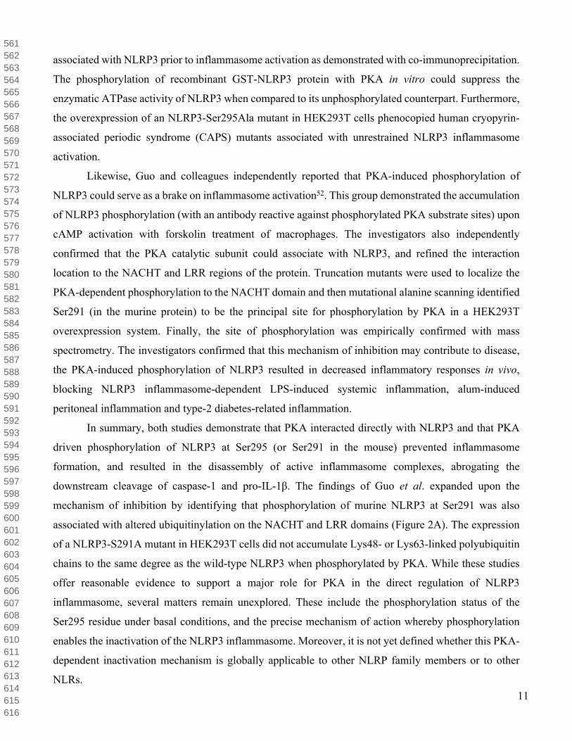
associated with NLRP3 prior to inflammasome activation as demonstrated with co-immunoprecipitation.
The phosphorylation of recombinant GST-NLRP3 protein with PKA in vitro could suppress the
enzymatic ATPase activity of NLRP3 when compared to its unphosphorylated counterpart. Furthermore,
the overexpression of an NLRP3-Ser295Ala mutant in HEK293T cells phenocopied human cryopyrin-
associated periodic syndrome (CAPS) mutants associated with unrestrained NLRP3 inflammasome
activation.
Likewise, Guo and colleagues independently reported that PKA-induced phosphorylation of
NLRP3 could serve as a brake on inflammasome activation52. This group demonstrated the accumulation
of NLRP3 phosphorylation (with an antibody reactive against phosphorylated PKA substrate sites) upon
cAMP activation with forskolin treatment of macrophages. The investigators also independently
confirmed that the PKA catalytic subunit could associate with NLRP3, and refined the interaction
location to the NACHT and LRR regions of the protein. Truncation mutants were used to localize the
PKA-dependent phosphorylation to the NACHT domain and then mutational alanine scanning identified
Ser291 (in the murine protein) to be the principal site for phosphorylation by PKA in a HEK293T
overexpression system. Finally, the site of phosphorylation was empirically confirmed with mass
spectrometry. The investigators confirmed that this mechanism of inhibition may contribute to disease,
the PKA-induced phosphorylation of NLRP3 resulted in decreased inflammatory responses in vivo,
blocking NLRP3 inflammasome-dependent LPS-induced systemic inflammation, alum-induced
peritoneal inflammation and type-2 diabetes-related inflammation.
In summary, both studies demonstrate that PKA interacted directly with NLRP3 and that PKA
driven phosphorylation of NLRP3 at Ser295 (or Ser291 in the mouse) prevented inflammasome
formation, and resulted in the disassembly of active inflammasome complexes, abrogating the
downstream cleavage of caspase-1 and pro-IL-1β. The findings of Guo et al. expanded upon the
mechanism of inhibition by identifying that phosphorylation of murine NLRP3 at Ser291 was also
associated with altered ubiquitinylation on the NACHT and LRR domains (Figure 2A). The expression
of a NLRP3-S291A mutant in HEK293T cells did not accumulate Lys48- or Lys63-linked polyubiquitin
chains to the same degree as the wild-type NLRP3 when phosphorylated by PKA. While these studies
offer reasonable evidence to support a major role for PKA in the direct regulation of NLRP3
inflammasome, several matters remain unexplored. These include the phosphorylation status of the
Ser295 residue under basal conditions, and the precise mechanism of action whereby phosphorylation
enables the inactivation of the NLRP3 inflammasome. Moreover, it is not yet defined whether this PKA-
dependent inactivation mechanism is globally applicable to other NLRP family members or to other
NLRs.
561562563564565566567568569570571572573574575576577578579580581582583584585586587588589590591592593594595596597598599600601602603604605606607608609610611612613614615616

12
The consensus sequence of PKA is [R/K](2)-X-[S/T]-[Hydrophobic], due to two glutamic acid
residues in the catalytic pocket of PKA that form ionic binding sites for the P-3 and P-2 position of the
substrate, as well as a hydrophobic pocket that favours a hydrophobic residue in the P+1 position56,57,58.
This sequence conforms to the catalytic site of PKA and confers specificity to the kinase. The sequence
surrounding the Ser295 residue of NLRP3 is highly conserved among different species (Figure 3A), yet
differs from the canonical PKA consensus sequence at the P+1 position, where a basic residue is found
in all mammalian NLRP3 orthologues examined, and 12 of the 14 human NLRP family members (Figure
3B). Another interesting feature of the sequence surrounding Ser295 is the proline residue located at the
P-1 position, which while conserved in orthologous mammalian NLRP3 proteins, is unusual for PKA
phosphorylation sites. Nonetheless, the high level of conservation in this region across mammals suggests
the importance of this site for the regulation of NLRP3 activity.
3.3 Protein kinase D can also phosphorylate NLRP3 at Ser295
A series of investigations revealed that PKD could effectively target both mouse and human
NLRP3 at the Ser295 residue. The sequence surrounding the Ser295 site in human NLRP3 conforms to
the prototypical PKD consensus sequence (i.e., [I/L]-X-[R]-X-[S/T]), where a hydrophobic Ile/Leu
residue at the P-5 position and a basic Arg residue at P-3 define the optimal sequence for PKD-mediated
phosphorylation87. While the Ser phosphorylation site and Ile/Leu residues are conserved among all but
one of the mammalian NLRP3 orthologues examined, the Arg residue at P-3 is absent in all but mouse
and hominid NLRP3 proteins (Figure 3A). Taken together, these observations suggest that the PKD-
dependent mechanism for NLRP3 regulation is narrowly restricted to only a few organisms. Additional
biochemical analyses will be required to determine if other NLRP3 homologues that lack the consensus
sequence are effective targets of PKD. For example, it is possible that the conservation of a basic residue
(Lys/Arg) at the P-2 position among the majority of NLRP3 mammalian homologues may accommodate
the lack of Arg at P-3.
In contrast to the inhibitory effect reported in the studies of PKA-dependent regulation described
in Section 3.2, the phosphorylation of the same Ser295 site by PKD was necessary for inflammasome
activation (Figure 2B)46. In response to inflammasome activators including ATP, nigericin, as well
particulates Alum and Nano-SiO2, mitochondria-associated ER membranes (MAMs) clustered around
Golgi membranes, where diacylglycercol (DAG) production was enhanced, thereby activating PKD in
MAMs. A Golgi integrity disruption agent, Brefeldin A, attenuated NLRP3 activation, indicating the
importance of Golgi signaling in close proximity to MAMs for NLRP3 activation. NLRP3 agonists also
resulted in NLRP3 translocation to MAMs, where the proximate PKD phosphorylated NLRP3 at Ser295
617618619620621622623624625626627628629630631632633634635636637638639640641642643644645646647648649650651652653654655656657658659660661662663664665666667668669670671672
13
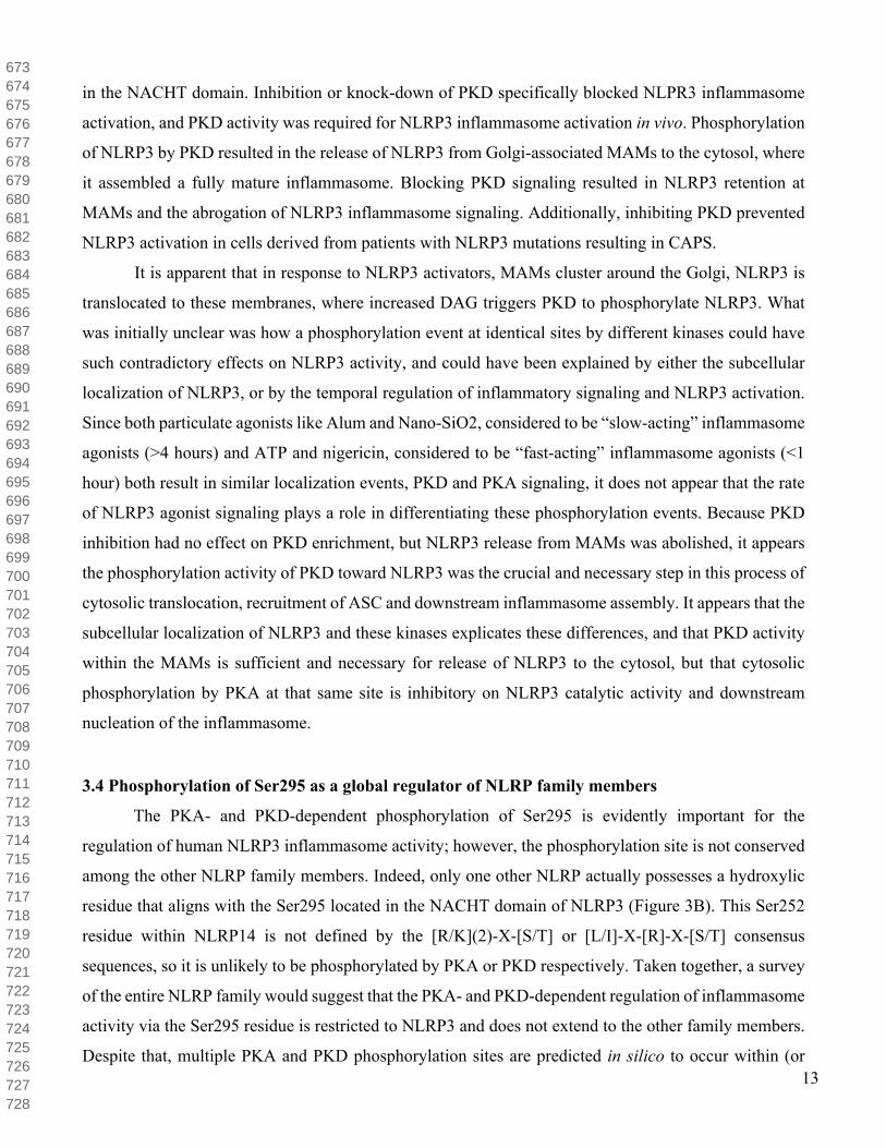
in the NACHT domain. Inhibition or knock-down of PKD specifically blocked NLPR3 inflammasome
activation, and PKD activity was required for NLRP3 inflammasome activation in vivo. Phosphorylation
of NLRP3 by PKD resulted in the release of NLRP3 from Golgi-associated MAMs to the cytosol, where
it assembled a fully mature inflammasome. Blocking PKD signaling resulted in NLRP3 retention at
MAMs and the abrogation of NLRP3 inflammasome signaling. Additionally, inhibiting PKD prevented
NLRP3 activation in cells derived from patients with NLRP3 mutations resulting in CAPS.
It is apparent that in response to NLRP3 activators, MAMs cluster around the Golgi, NLRP3 is
translocated to these membranes, where increased DAG triggers PKD to phosphorylate NLRP3. What
was initially unclear was how a phosphorylation event at identical sites by different kinases could have
such contradictory effects on NLRP3 activity, and could have been explained by either the subcellular
localization of NLRP3, or by the temporal regulation of inflammatory signaling and NLRP3 activation.
Since both particulate agonists like Alum and Nano-SiO2, considered to be “slow-acting” inflammasome
agonists (>4 hours) and ATP and nigericin, considered to be “fast-acting” inflammasome agonists (<1
hour) both result in similar localization events, PKD and PKA signaling, it does not appear that the rate
of NLRP3 agonist signaling plays a role in differentiating these phosphorylation events. Because PKD
inhibition had no effect on PKD enrichment, but NLRP3 release from MAMs was abolished, it appears
the phosphorylation activity of PKD toward NLRP3 was the crucial and necessary step in this process of
cytosolic translocation, recruitment of ASC and downstream inflammasome assembly. It appears that the
subcellular localization of NLRP3 and these kinases explicates these differences, and that PKD activity
within the MAMs is sufficient and necessary for release of NLRP3 to the cytosol, but that cytosolic
phosphorylation by PKA at that same site is inhibitory on NLRP3 catalytic activity and downstream
nucleation of the inflammasome.
3.4 Phosphorylation of Ser295 as a global regulator of NLRP family members
The PKA- and PKD-dependent phosphorylation of Ser295 is evidently important for the
regulation of human NLRP3 inflammasome activity; however, the phosphorylation site is not conserved
among the other NLRP family members. Indeed, only one other NLRP actually possesses a hydroxylic
residue that aligns with the Ser295 located in the NACHT domain of NLRP3 (Figure 3B). This Ser252
residue within NLRP14 is not defined by the [R/K](2)-X-[S/T] or [L/I]-X-[R]-X-[S/T] consensus
sequences, so it is unlikely to be phosphorylated by PKA or PKD respectively. Taken together, a survey
of the entire NLRP family would suggest that the PKA- and PKD-dependent regulation of inflammasome
activity via the Ser295 residue is restricted to NLRP3 and does not extend to the other family members.
Despite that, multiple PKA and PKD phosphorylation sites are predicted in silico to occur within (or
673674675676677678679680681682683684685686687688689690691692693694695696697698699700701702703704705706707708709710711712713714715716717718719720721722723724725726727728

14
proximal to) the NACHT domain of other NLRP family members (Figure 3C), as well as in other regions
of the proteins, such as the LRR or PYD. This observation implies that other NLRPs could be
phosphorylated by PKA or PKD, so additional biochemical analyses will be required to identify if
prospective consensus sites for phosphorylation in all human NLRP proteins represent a conserved
mechanism of regulation amongst the family.
Intriguingly, 8 of the 14 NLRPs display acidic amino acid substitutions at the Ser295 site (Figure
3B), suggesting that the incorporation of negative charge in this region of the NACHT domain may have
a functional role in the regulation of inflammasome activity. It is possible to infer that most NLRP
proteins are, therefore, held in an inhibitory conformation with the glutamic and aspartic acid residues
providing similar molecular properties to that of the phosphorylated serine. While it is unclear if
phosphorylation blocks ATP binding, Mortimer et al. demonstrated that Ser295 phosphorylation
attenuated ATP hydrolysis and inflammasome activity, so the introduction of negative charge near the
Walker B motif clearly impacts upon the active site. At least two NLRP proteins that contain a negative
residue at the Ser295 site were previously reported to bind and/or hydrolyze ATP (i.e., NLRP124 and
NLRP1223), so the presence of a negative residue in place of the serine does not support the complete
abrogation of catalytic function. These findings suggest that while phosphorylation of Ser295 appears to
play an important role in the regulation of certain NLRPs, it is likely that distinct enzymatic properties
and regulatory mechanisms will be identified for the different family members.
4. The Phosphorylation of Ser295 and Its Impact on NLRP3 Structure
4.1 Molecular Modeling of the NLRP3-NACHT Structure
To further examine the inhibitory effects of PKA phosphorylation of Ser295 on NLRP3 activity,
a structural homology model of the NLRP3 NACHT domain was assembled with the Phyre2 Protein
Fold Recognition Server59. Despite concerted efforts to crystalize different NLRP proteins or close
family members, no 3-dimensional structure of any NLRP-NACHT domain has been published, and this
knowledge gap represents a critical impediment to understanding the activation mechanisms of these
inflammatory platforms. Despite this obstruction, significant advances in computational techniques have
resulted in more accurate protein structure predictions60, and we have applied in silico modelling
approaches to predict the effects of Ser295 phosphorylation on the topology of the nucleotide-binding
site. Phyre2 uses remote homology detection methods to build structural models by matching the
sequence of interest to libraries of known folds, minimizing the more error-prone aspects of simulated-
folding approaches59. In an NLRP3-NACHT structural model derived with Phyre2 (Figure 4), 98% of
729730731732733734735736737738739740741742743744745746747748749750751752753754755756757758759760761762763764765766767768769770771772773774775776777778779780781782783784

15
NLRP3 NACHT residues were modeled at over a 90% ‘confidence’ level (i.e., represents the probability
that the match between sequence and the constructed model is a true homology). Residues modeled at
lower scores were the first seven residues and the last residue of the submitted sequence string for the
NLRP3 NACHT domain (residues 190-197 and 536, respectively). This is likely due to the expansion of
flexibility given these residues were removed from any neighboring structural constraints.
Three protein structures were selected based on heuristics to model the NLRP3-NACHT domain,
including: NLRC4 (PDB 4KXF), also a caspase-1 activating inflammasome protein, NAIP5 (PDB 5YUD), a sensor component of the NLRC4 inflammasome, and a NAIP5-NLRC4 complex (PDB 6B5B).
Ultimately, the NLRP3 NACHT model was predicted to contain the following secondary structure: 52%
-helices, 11% -strands, 5% transmembrane helices, and the remaining 15% intrinsically
disordered/unstructured. Given the close alignment of gene ontology between the template structures and
NLRP3 sequence in the modeled regions, we place reasonable confidence in the accuracy of predicted
model; however, empirical data will obviously be required to define the native structure.
To determine the reliability of the NLRP3-NACHT model with respect to the core nucleotide-
binding site structure, we directly compared the NLRP3-NACHT model to the published x-ray crystal
structure of apoptotic protease-activating factor (APAF)-1, the protein closest to NLRP3-NACHT in
sequence similarity with high-quality 2-3 Å rmsd published structure and reliable biochemical data on
nucleotide binding and hydrolysis (PDB: 1Z6T, 2.21Å)61,62,63. APAF-1 is also a nucleotide-binding,
inflammasome forming receptor, and it shares conservation of key functional motifs with NLRP3 (Figure
4A-B). Several residues of APAF-1 known to coordinate the bound nucleotide are not present within the
NLRP3-NACHT model. However, most key residues were situated in close proximity within the
alignment, and the overall topology of the catalytic pocket, which in turn plays a key stabilization role in
nucleotide-binding, appears to be reasonably conserved given the differences in sequence (Figure 4C-E).
In the empirical APAF-1 structure, the specific binding of the nucleotide is achieved with direct
hydrogen bonding from Val127, Gly157, Gly159, Lys160, Ser161, Val162 and His438. Direct hydrogen
bonds from the amide and carbonyl of Val127 coordinate the N1 and N6 atoms of the adenine base
(respectively), while the amides of Gly157, Gly159, Lys160 and Val162, as well as the amino group of
Lys160 and the τ-nitrogen of the His438 imidazole group provide hydrogen bonding with the β-
phosphate. The final direct H-bond is between the amide of Val162 and the α-phosphate. Water-mediated
H-bonds between the carbonyl groups of Gly159, Val125 and the side-chain of Arg129 stabilize the
adenine base, while the carbonyl of Ser422 provides the final water-mediated H-bond with the ribose
785786787788789790791792793794795796797798799800801802803804805806807808809810811812813814815816817818819820821822823824825826827828829830831832833834835836837838839840

16
moiety. Lastly, van der Waals interactions with both the adenine and ribose groups are provided by
Val127, Gly159, Val162, Arg129, Pro321 and Leu322.
Most of the direct hydrogen bonding residues in APAF-1 do align with conserved or similar
residues in NLRP3 (i.e., Tyr381, Gly229, Gly231, Lys232, Thr233, Ile234 and Arg351). Likewise,
APAF-1 residues that may confer structural stability in the active site also align well with the NLRP3
model. These include the residues which provide water-mediated hydrogen bonds, Val125, Arg129 and
Ser422 in APAF-1 (aligning with NLRP3 Tyr381, Lys377, and Thr430, respectively), and those which
provide only van der Waals interactions: Pro321 and Leu322 in APAF-1 (aligning with NLRP3 Pro412
and Leu413, respectively). According to the widely-used amino acid substitution matrix BLOSUM62,
two of these substitutions possess low scores: the Tyr substitutions for Val (i.e., APAF-1 Val127 and
Val125 replaced with Tyr381 and Tyr385 in NLRP3), and Arg substitutions for His (i.e, APAF-1 His438
replaced with Arg351 in NRLP3). It is possible that the homology model of NLRP3 could still function
to provide equivalent bonding patterns, since both interactions from Val127 and Val125 in APAF-1 are
provided by the amino-acid backbone (carbonyl and main chain, respectively) of the NLRP3 model, and
the hydrogen-bond between APAF-1 His438 and the β-phosphate of the nucleotide could be substituted
by the NLRP3 Arg351 side-chain.
Properties inherent to the NACHT domain, namely ATP-binding and ATP-hydrolysis, are
indispensable for NLRP3 oligomerization and inflammasome assembly. Since all of the aforementioned
motifs (Section 1.3) play key functional roles in the regulation of substrate binding and catalysis, we
postulated that PKA-dependent phosphorylation of Ser295 could result in conformational fluctuations
within these structured regions. A phosphoryl group was introduced at the Ser295 residue within the
NLRP3-NACHT model structure (Figure 5A, Ser295 in cyan; Figure 5B, pSer295 in green) using the
Vienna-PTM Server64,65. Vienna-PTM utilizes several different widely-used and extensively-tested
molecular dynamics (MD) simulation force fields, as well as integrates newly-derived parameters for
phosphate ions within the GROMACS 4.5 force field parameter sets selected66. To minimize the energy
and identify the most stable conformation of our phosphorylated model structure, we applied the
obminimize program in openbabel67,68. The structure was minimized using the steepest descent
algorithm. The phosphorylated NLRP3-NACHT model was aligned with the unphosphorylated structure,
resulting in an overall RMSD of 0.913 over 345 of 347 residues (Figure 5C). As expected, pairwise
RMSD analysis suggests the largest shifts in fold conformation and residue position immediately
preceding Ser295, but the introduction of the post-translational modification also resulted in higher
energy deviations between key residues in the Sensor 1 and 2 regions, even affecting secondary structure
in those areas, as well as in other concentrated areas throughout the structure (Figure 5D-F). These
841842843844845846847848849850851852853854855856857858859860861862863864865866867868869870871872873874875876877878879880881882883884885886887888889890891892893894895896

17
deviations are representative of the impact that Ser295 phosphorylation has on secondary structure
composition within these areas and suggests a disordering of regions previously composed of -strands.
As noted above, these regions play key roles in the activity of the NACHT domain as a whole, and these
results could explain why phosphorylation of Ser295 results in both the abrogation of NLRP3 ATPase
activity as well as an impediment to any future activation.
Lastly, we examined the effect of phosphorylation on the nucleotide-binding site using the
pSer295 NLRP3-NACHT model derived with Vienna-PTM. Subtle, yet potentially important, deviations
were observed (Figure 6A-B), including a positional shift in the location of the Lys232 residue of the
Walker A motif and the Pro412 residue (Figure 6C-D). This proline is conserved in most of the NLRPs
(all except NLRP2 and NLRP7), and mutation of this residue disrupts downstream signaling in NOD269.
Due to its known interaction with the adenine moiety of ATP in NOD2, and the known hydrophobicity
of this region being essential in facilitating the binding of ATP, this shift could be significant in
explicating the effects of phosphorylation on the inhibition of NLRP3 activity.
It is difficult to predict how small changes within the catalytic pocket could impact upon overall
inflammasome activity, or whether conformational changes instigated by Ser295 phosphorylation even
need to be conveyed within the catalytic pocket to result in inactivation. A subtle adjustment in Lys232
or Pro412 positioning, as observed with modelling in Figure 6, could result in impaired nucleotide-
binding, but it is equally possible that the subtle conformational shifts within the catalytic pocket have
no functional impact, and the inhibitory effect of pSer295 actually occurs in other regions distal to the
core motifs of the ATP-binding pocket. Experiments to individually interrogate the impacts of Ser295
phosphorylation on ATP-binding, ATP-hydrolysis and ADP-release properties of the NLRP3
inflammasome will be essential to understand the role of ATP in regulating NLRP3 activity. While ATP-
binding appears indispensable for NLRP3 activation, it remains unclear if it is ATP-binding, the cleavage
of the - phosphate bond, the release of the phosphate or the release of the ADP moiety that drives the
conformational changes necessary to expose docking regions for NLRP3 receptor oligomerization and/or
other inflammasome-binding partners (such as ASC). In addition, empirical structural data to determine
where each of these key residues are positioned will be important in understanding exactly how the
phosphorylation of Ser295 impacts upon the conformation of the domain, therefore negatively regulating
inflammasome activity.
897898899900901902903904905906907908909910911912913914915916917918919920921922923924925926927928929930931932933934935936937938939940941942943944945946947948949950951952
18
5. Conclusions and Future OpportunitiesWhile studies offer reasonable evidence to support a major role for PKA (and PKD) in NLRP3
regulation, several matters remain unexplored. Atomic resolution structural data are available in the PDB
for several domains of the NLR family, including structures for the PYD of NLRPs 1,3,7,10,12 and 14,
the LRR of NLRP1, the CARD of NOD1 and NLRP1, the BIR of NAIP, as well as the RNA-binding
element of NLRX1. Unfortunately, no structures have been solved for the central catalytic NACHT
domain of any NLRP protein. So, additional investigations of the NACHT domain structural architecture,
the conformational status of the domain in relation to nucleotide-binding and/or phosphorylation state,
and the residues involved in oligomerization will be essential for a complete understanding of NLRP
inflammasome function.
It has been widely assumed that the enzymatic properties of the nucleotide-binding NACHT
domain are fundamental to the functionality of all 14 NLRP members, inflammasome assembly and
otherwise, but ATP-binding and/or hydrolysis has only been demonstrated with NLRP321, NLRP722 and
NLRP1223. This lack of experimental data for NLRP3-NACHT ATP binding, hydrolysis and
oligomerization has to date obstructed the ability to develop an overall understanding of the biochemical
mechanisms of inflammasome activation. Multiple publications concerning inflammasome activation
appear to have misconstrued results in the seminal 2007 publication by Duncan by reporting that
hydrolysis was established as a critical step in inflammasome function, whereas the data only
demonstrated that ATP binding was indispensable for downstream signaling and did not conclusively
link ATP hydrolysis with inflammasome activation21. While investigators have stated that it is probable
that ATP binding and/or hydrolysis in the NACHT domain is/are responsible for a conformational
remodeling that is transferred to other NLRP domains, overall there is much to learn regarding the
functionality of the NACHT activity its role in inflammasome oligomerization, activation, and
downstream signaling.
NLRP3 protein can be maintained in an inactive yet signaling competent state by protein-protein
interactions with the ubiquitin ligase–associated protein (SGT1) and heat-shock protein 90 (HSP90)70
(Figure 7A). Knockdown of SGT1 by small interfering RNA or pharmacological inhibition of HSP90
with geldanamycin was demonstrated to abrogate inflammasome activity and reduce NLRP3-mediated
disease symptoms in mice. Additionally, SGT1 and HSP90 form similar complexes with several other
NLRs (NLRP2,4,12, NOD1,2 and IPAF), and are indispensable for NOD2 and IPAF inflammasome
activation as well. Notably, HSP90 but not SGT1 was crucial for the maintenance of stable NLRP3
protein levels in the cell. Treatment with geldanamycin resulted in a substantial decrease in endogenous
THP-1 NLRP3 levels, and also provided complete depletion of NLRP3 by 8h of treatment, and thus to
953954955956957958959960961962963964965966967968969970971972973974975976977978979980981982983984985986987988989990991992993994995996997998999100010011002100310041005100610071008

19
the inhibition of downstream inflammasome activity, in ‘Flp-In T-rex’ HEK293T cells with inducible
NLRP3 expression. These effects were blocked by the proteasome inhibitor lactacystin, strongly
suggesting that the loss of HSP90 function resulted in the proteasome-dependent degradation of NLRP3.
Hydrolysis of ATP in NLRP3 could aid in driving a conformational change that exposes docking sites
for nucleation of the inflammasome and regulatory partners, but which binding partners are essential,
and the order in which they bind remains unknown. Furthermore, the integration of phosphorylation into
these schemes remains to be considered.
In consideration of the data available for phosphorylation of Ser295, we propose a scheme in
which NLRP3 is serially phosphorylated and dephosphorylated to regulate inflammasome signaling in
response to priming (Figure 7A), activating stimuli (Figure 7B), and assembly (Figure 7C). This
mechanism of regulation is not unique to NLRP3; the pyrin inflammasome is also regulated by
phosphorylation at Ser242 and bound by inhibitory 14-3-3 proteins, which must be relieved by
phosphatase activity prior to inflammasome assembly71,72,73,74. PKA stimulation and the ensuing
phosphorylation of Ser295 is inhibitory prior to activation, and can also initiate inflammasome
disassembly of the mature inflammasome. PKD phosphorylation of that same residue is essential for the
progression of NLRP3 through the activation mechanism, and drives the subcellular relocation of NLRP3
from Golgi-associated MAMs to the cytosol where the inflammasome complex is assembled. The
intrinsic phosphorylation status of Ser295 in the priming stage remains unclear, but PKA was found to
be associated with NLRP3 at that stage49. Additionally, NLRP3 is ubiquitinated with mixed K48 and
K68 chains at this phase, and Ser295 phosphorylation is known to play a key role in ubiquitination50.
Therefore, we propose that NLRP3 is likely phosphorylated at Ser295 in the priming stage, driving
ubiquitination and blocking oligomerization at resting state. Following the sensing of a signal two
agonist, it is unclear whether NLRP3 would be dephosphorylated by some unknown phosphatase prior
to migrating to Golgi-associated MAMs where PKD activity is enhanced, or if PKD and PKA activities
on this site are redundant. PKD phosphorylation of NLRP3 is plainly indispensable for NLRP3 release
from these membranes to the cytosol, although dephosphorylation of Ser295 is obligatory for NLRP3
inflammasome assembly. The co-localization of PKA and NLRP3 was not considered in any study of
Ser295 phosphorylation. The seemingly specific inhibition of PKD in MAMs did abolish NLRP3
signaling; and therefore, each kinase may regulate NLRP3 in distinctive cellular compartments along the
activation pathway. Following activation, the experimental stimulation of PKA and NLRP3
phosphorylation could induce dissociation of the inflammasome, but whether this mechanism of action
is biologically relevant remains uncertain.
10091010101110121013101410151016101710181019102010211022102310241025102610271028102910301031103210331034103510361037103810391040104110421043104410451046104710481049105010511052105310541055105610571058105910601061106210631064

20
Because the bulk of investigations into NLRP3 phosphorylation mechanisms were performed in
vitro, determining what mechanisms are germane to intrinsic NLRP3 activation in vivo will require
further exploration in the relevant model systems. Furthering our understanding of the biochemical and
biophysical properties of NLRP3 and other inflammasome forming proteins will be fundamental to the
development of treatments for the myriad of human diseases in which these proteins play central roles,
including CAPS, multiple sclerosis, lupus, the crystalline deposit-related diseases of silicosis and gout,
inflammatory bowel disease, infectious colitis, metabolic diseases (including obesity, type II diabetes,
and atherosclerosis), Parkinson’s disease, and renal disease (acute kidney injury and chronic kidney
disease)75,76,77. Multiple NLRP3 inhibitory compounds are under investigation78,79,80, and it is hoped that
these research endeavors will ultimately guide the attainment of treatment for many chronic
inflammatory pathologies.
6. Acknowledgements
We would like acknowledge those authors whose studies on NLRP3 were not presented due to space
limitations. This work was supported by the Canadian Institutes of Health Research (CIHR) Health
Challenges in Chronic Disease Signature Initiative (#THC-13523). C.F.S. was supported by a Natural
Sciences and Engineering Research Council (NSERC) Postgraduate Scholarship and a Cumming School
of Medicine Doctoral Scholarship. The authors appreciate the technical assistance of Tayeb Kakeshpour
(Department of Chemistry, Michigan State University) in rendering the energy minimization and
optimization of the phosphorylated NLRP3 model with Open Babel.
7. Author Contributions
CFS completed the molecular modelling of NLRP3 NACHT and Ser295 phosphorylation, wrote the
manuscript, and prepared the figures. JAM edited the manuscript, supervised trainees and provided
intellectual contributions to the project. Both authors reviewed the results and approved the final version
of the manuscript.
8. Authors’ Competing Interests Statement
JAM is cofounder and holds an equity position in Arch Biopartners Inc. All other authors declare no
conflicts of interest.
10651066106710681069107010711072107310741075107610771078107910801081108210831084108510861087108810891090109110921093109410951096109710981099110011011102110311041105110611071108110911101111111211131114111511161117111811191120

21
Figure and Table Legends
Table Legends
Table 1. Phosphorylation of the NLR Family Members. The nucleotide-binding domain and leucine-
rich repeat-containing (NLR) family members are subdivided based on the presence of one of four N-
terminal effector domains: NLRAs with an acidic domain, NLRBs with a Baculovirus IAP repeat,
NLRCs with a caspase recruitment domain, or NLRPs with a pyrin domain. Empirical NLR
phosphorylation events are listed and, if known, linked with the protein kinase associated with each site
in the last column. The naming system approved by the HUGO Gene Nomenclature Committee
(HGNC)10 is used. Domain abbreviations: AD, acidic domain; BIR, Baculovirus inhibitor of apoptosis
repeat; CARD, caspase activation and recruitment domain; CT, C-terminal; FIIND, function to find
domain; LRR, leucine rich repeat; NACHT, nucleotide triphosphatase (NTPase) domain [NAIP
(neuronal apoptosis inhibitory protein), CIITA (MHC class II transcription activator), HET-E (in-
compatibility locus protein from Podospora anserina) and TP1 (telomerase-associated protein)]; NT, N-
terminal; PYD, pyrin domain. Empirical phosphorylation and kinase data obtained from iPTMnet and
PhosphoSite Plus81,82.
Table 2. NLRP3 Phosphorylation Sites. The impact of several phosphorylation events on NLRP3
inflammasome assembly and downstream maturation of caspase-1 and IL-1 have been defined.
Empirical NLRP3 phosphorylation sites and the corresponding human (UniProtKB: Q96P20) or mouse
(UniProtKB: Q8R4B8) residues are listed, as is the domain in which the site is found. Dashed lines in
the domain column represent disordered, linker regions. Results of the phosphorylation event, as well
as the kinase or phosphatase associated with the event are listed with the corresponding reference in the
low or high-throughput experiment column (LTP/HTP, respectively). Abbreviations used: LTP, low
throughput methods (i.e., sites were identified and then validated with other biochemical methods), HTP,
high throughput methods (i.e., sites were identified using only discovery mass spectrometry), PYD, pyrin
domain; NACHT, [NAIP (neuronal apoptosis inhibitory protein), CIITA (MHC class II transcription
activator), HET-E (in-compatibility locus protein from Podospora anserina) and TP1 (telomerase-
associated protein)] domain; LRR, leucine-rich repeat domain; PKA (PRKACA), cAMP-dependent
protein kinase; PKD (PRKD), serine/threonine protein kinase D; PTPN22, tyrosine-protein phosphatase
non-receptor type 22; JNK1 (MAPK8), c-Jun N-terminal kinase 1; PP2A (PPP2CA), protein phosphatase
type-2A catalytic subunit.
11211122112311241125112611271128112911301131113211331134113511361137113811391140114111421143114411451146114711481149115011511152115311541155115611571158115911601161116211631164116511661167116811691170117111721173117411751176
22
Figure Legends
Figure 1. The Canonical Two-Signal Model for NLRP3 Inflammasome Activation. The first
transcriptional priming step (i.e., Signal One) in the assembly of the NLRP3 inflammasome is initiated
when microbial molecules (e.g., LPS) or endogenous cytokines (not shown) lead to the upregulation of
NLRP3 and pro-IL-1β transcription through Toll-like receptor (TLR)-dependent activation of the NF-κB
signaling pathway. Other elements of the NLRP3 inflammasome, including ASC, caspase-1 and pro-IL-
18 are constitutively expressed. NF-κB signaling also results in the activation of BRCC3, a
metalloprotease that deubiquitinates NLRP3 prior to activation. In the second, oligomerization and
assembly step, several Signal Two stimuli, including but not limited to ATP, ionophores like nigericin,
and particulate matter can result in activation of NLRP3 and inflammasome assembly. Most of these
stimuli induce K+ efflux, and/or calcium flux which is essential and sufficient for NLRP3 activation, but
mitochondrial dysfunction, including reactive oxygen species (ROS), oxidized mitochondrial DNA or
externalization of cardiolipin (mitochondrial dysfunction) can also act as the second signal. Upon
detection of the second signal in the cytosol, the NLRP3 inflammasome is activated in an ATP-dependent
manner with oligomerization of multiple NLRP3 molecules and recruitment of ASC and pro-caspase-1.
Proximity-induced proteolytic cleavage and activation of caspase-1 enables the subsequent proteolysis
and maturation of pro-inflammatory cytokines (i.e., pro-IL-1β and pro-IL-18) to their secreted forms,
which can induce acute inflammatory responses in the surrounding tissue.
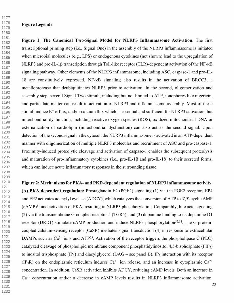
Figure 2: Mechanisms for PKA- and PKD-dependent regulation of NLRP3 inflammasome activity.
(A) PKA dependent regulation: Prostaglandin E2 (PGE2) signaling (1) via the PGE2 receptors EP4
and EP2 activates adenylyl cyclase (ADCY), which catalyzes the conversion of ATP to 3',5'-cyclic AMP
(cAMP)51 and activation of PKA; resulting in NLRP3 phosphorylation. Comparably, bile acid signaling
(2) via the transmembrane G-coupled receptor-5 (TGR5), and (3) dopamine binding to its dopamine D1
receptor (DRD1) stimulate cAMP production and induce NLRP3 phosphorylation52,54. The G protein-
coupled calcium-sensing receptor (CaSR) mediates signal transduction (4) in response to extracellular
DAMPs such as Ca2+ ions and ATP55. Activation of the receptor triggers the phospholipase C (PLC)
catalyzed cleavage of phospholipid membrane component phosphatidylinositol 4,5-bisphosphate (PIP2)
to inositol trisphosphate (IP3) and diacylglycerol (DAG – see panel B). IP3 interaction with its receptor
(IP3R) on the endoplasmic reticulum induces Ca2+ ion release, and an increase in cytoplasmic Ca2+
concentration. In addition, CaSR activation inhibits ADCY, reducing cAMP levels. Both an increase in
Ca2+ concentration and/or a decrease in cAMP levels results in NLRP3 inflammasome activation.
11771178117911801181118211831184118511861187118811891190119111921193119411951196119711981199120012011202120312041205120612071208120912101211121212131214121512161217121812191220122112221223122412251226122712281229123012311232

23
Additionally, two of these studies suggested that cAMP can directly bind NLRP3, triggering in the K48-
chain ubiquitination of the LRR and NACHT domains by the Membrane Associated Ring-CH-type finger
7 (MARCH7) E3 ubiquitin ligase, aggregation and finally autophagy mediated NLRP3 degradation (3,4).
(B) PKD dependent regulation: In response to inflammasome activators, including ATP, nigericin, and
particulates Alum and Nano-SiO2 (5), mitochondria associated ER membranes (MAMs) localize adjacent
to Golgi membranes, where diacylglycercol (DAG) production was enhanced, thereby activating PKD
in MAMs. Brefeldin A (BFA), a Golgi integrity disruption agent, attenuated NLRP3 activation and
signaling, indicating the importance of Golgi signaling close to MAMs in NLRP3 activation. These
agonists also resulted in NLRP3 translocation to MAMs, where the proximate PKD phosphorylated
NLRP3 at Ser295 in the NACHT domain. Phosphorylation of NLRP3 by PKD resulted in the release of
NLRP3 from Golgi-associated MAMs to the cytosol, where it assembled into a mature inflammasome.
NLRP3 is predicted to be dephosphorylated upon activation, by a yet unknown phosphatase. Ultimately,
caspase-1 is activated by the mature inflammasome and catalyzes the cleavage and processing of pro-IL-
1β and IL-18. These mature cytokines are, in turn, secreted from the cell to stimulate inflammatory
responses in the surrounding tissue.
Figure 3. Conservation of the Ser295 phosphorylation site and predicted PKA-dependent
phosphorylation of NLRP family members. (A) Conservation of Ser295 among NLRP3 orthologues.
Orthologous genes are listed in descending order by shared sequence identity. The conventional PKA
consensus sequence and Walker B motif are labeled above the alignment, with the phosphorylated
residue indicated (Ser295 in human NLRP3). Sequence conservation among species is denoted by the
coloured highlighting when conservation of the amino acid class was retained among 4 or more species.
Hydrophobic residues (A,I,L,M,F,W,V,C,G,P) are coloured in yellow; polar residues in green;
hydroxylic (S,T,Y) in dark green and other polar (N,Q) in light green; acidic (D,E) in red; and basic
(K,R,H) in blue. (B) Conservation of Ser295 among different NLRP family members. The PKA
consensus sequence and Walker B motif are labeled above the alignment. The sequence logo below the
alignment indicates sequence conservation among species, from increased conservation of amino acids
and function (larger letters) to when amino acids were different and no conservation was retained (smaller
letters). (C) Occurrence of PKA- and PKD-dependent phosphorylation sites based on Group-based
Prediction System (GPS; http://gps.biocuckoo.org) analysis of the primary sequences of NLRP family
members. The GPS 3.0 Species Specific Tool (Homo sapiens) was used to predict kinase-specific
phosphorylation sites in hierarchy with medium threshold (6% false-positive rate)88.
12331234123512361237123812391240124112421243124412451246124712481249125012511252125312541255125612571258125912601261126212631264126512661267126812691270127112721273127412751276127712781279128012811282128312841285128612871288

24
Figure 4. Structural modeling of the NACHT domain of NLRP3. A homology model of an extended
NLRP3-NACHT domain (I190-L536) was generated in silico using Phyre2 and aligned with the X-ray
crystal structure of APAF-1 (PDB: 1Z6T)59,61. The Phyre2 algorithm indicates that 98% of residues were
modelled at >90% confidence (i.e., residues 190-196 and 536 were modeled ab initio, which can be
unreliable, while the rest were modeled with high confidence, percentage indentity and alignment
coverage). Both the NLRP3-NACHT model and the empirical APAF-1 structure were visualized using
Pymol v2.2.1. The APAF-1 structure and the NLRP3-NACHT model display conserved topology within
the nucleotide-binding domain. In (A, ribbon diagram) and (B, cartoon representation), APAF-1 is
represented in gray, while the newly generated NLRP3 model is shown in purple. An ADP molecule
empirically identified within the NACHT fold of APAF-1 is shown in red. Experimentally defined
nucleotide-binding residues in APAF-1 are shown in orange, and the corresponding NLRP3 residues
expected to interact with the nucleotide are shown in yellow. In (C), an alignment of key nucleotide-
binding residue side chains between APAF-1 and the NLRP3-NACHT model is indicative of
conservation within the nucleotide-binding domain of APAF-1 (D) and NLRP3 (E).
Figure 5. Predicted effect of Ser295 phosphorylation on the NLRP3-NACHT structure. Conserved
sequence motifs that are important for nucleotide-binding and inflammasome activation are highlighted
in cartoon diagrams for unphosphorylated (A, model in blue, Ser295 residue in cyan) and phosphorylated
(B, model in dark red, pSer295 residue in dark green) NLRP3-NACHT domains. These include the
Walker A (WA), Walker B (WB), Sensor 1 (S1) and Sensor 2 (S2) motifs, coloured as listed in the table
at the bottom right hand corner of panel F). These regions play important roles in stabilizing
intramolecular interactions, coordinating nucleotide-binding, nucleotide-hydrolysis, and protein
conformational changes during oligomerization. In (C), an alignment of NLRP3-NACHT and pSer295-
NLRP3-NACHT is coloured by RMSD. The overall RMSD of both structures is 0.913. Dark blue
indicates close structural alignment, while higher energy deviations are shown in red. In (D), the NLRP3-
NACHT sequence is coloured by RMSD as shown in panel C, indicating the predicted structural
differences between the models. Key motifs are indicated above the sequence, and nucleotide-binding
residues are labeled with black dots. In (E), an alignment of NLRP3-NACHT and pSer295-NLRP3-
NACHT model structures illustrates conformational and positional changes of key motifs (F) upon
phosphorylation of Ser295.
Figure 6. Potential impact of Ser295 phosphorylation on residues that coordinate nucleotide-
binding. Representative structural models of NLRP3-NACHT (A) and pSer-NLRP3-NACHT (B) are
12891290129112921293129412951296129712981299130013011302130313041305130613071308130913101311131213131314131513161317131813191320132113221323132413251326132713281329133013311332133313341335133613371338133913401341134213431344

25
provided with active site residues shown in yellow and green for the unphosphorylated (coloured in blue)
and phosphorylated NLRP3 (coloured in dark red), respectively. An ADP molecule (in red) was docked
within the active site of each. The Ser295 and phosphorylated pSer295 residues are highlighted in cyan
and dark green, respectively. Phosphorylation of the NLRP3 model in silico was performed with Vienna
PTM, and the resulting structure was minimized with openbabel, using the steepest descent algorithm of
obminimize. An alignment of NLRP3-NACHT and pSer295-NLRP3-NACHT structures (C) suggests
gross conformational alterations of the ATP-binding pocket upon phosphorylation. Small positional and
conformational changes are apparent for key residue side chains implicated in nucleotide-binding (D),
which could contribute to the known disparities in function between unphosphorylated and
phosphorylated NLRP3.
Figure 7. Involvement of Ser295 phosphorylation in the activation of NLRP3 inflammasomes. (A)
NLRP3 Priming (Signal One). Inflammasome components ASC, caspase-1 and IL-18 are constitutively
expressed, while basal NLRP3 and pro-IL-1β transcription are inadequate for inflammasome
activation83. Priming or transcriptional activation is facilitated by NF-κB signaling, including but not
limited to NOD1/NOD2 activation of NF-κB, TLR signaling, and activation of the cytokine receptors
TNFR1 and/or IL-1R1. Following transcription, the resting NLRP3 protein localizes adjacent to
endoplasmic reticulum (ER) structures84,85. At this stage, NLRP3 may be held in an inactive but signaling
competent complex with HSP90 and SGT170. NLRP3 association with HSP90 blocks the proteasomal
degradation of NLRP3. Inactive NLRP3 carries mixed K48 and K63 ubiquitin chains primarily on the
LRR and NACHT domains. NF-κB signaling activates BRCC3 deubiquitination activity during priming,
a requisite for NLRP3 activation86,44. Ca2+ signaling can inhibit NLRP3 activation through the
CaSR/ADCY/cAMP axis since direct binding of cAMP to NLRP3 blocks activation52. Stimulation of the
EP2/EP4 or TGR5 receptors activates protein kinase A (PKA) and induces phosphorylation of Ser295 in
the NACHT domain of NLRP3, driving ubiquitination and inflammasome inhibition. PKA can associate
with NLRP3 at this juncture as well, although the basal phosphorylation state of NLRP3 is still
unresolved.
(B) NLRP3 Activation (Signal Two). NLRP3 inflammasome assembly is likely triggered by cellular
stresses that converge upon K+ efflux and Ca2+ flux rather than by direct PAMP or DAMP binding37,87.
Pore-forming toxins, ionophores like nigericin, and the P2X7 receptor facilitate K+ efflux and NLRP3
activation. Cell swelling can initiate mitochondrial dysfunction and Ca2+ flux through the transient
receptor potential vanilloid 2 (TRPV2) receptor and/or voltage-dependent anion channel 1 (VDAC1)
88,89. This culminates in release of intracellular NLRP3 agonists, including mitochondrial reactive oxygen
13451346134713481349135013511352135313541355135613571358135913601361136213631364136513661367136813691370137113721373137413751376137713781379138013811382138313841385138613871388138913901391139213931394139513961397139813991400
26
species (ROS), mitochondrial DNA (mtDNA) and cardiolipin. Crystalline DAMPs also impact on
mitochondrial dysfunction, K+ Efflux and Ca2+ flux via release of lysosomal cathepsins B and L. Both
K+ efflux and cathepsin B release are linked to dissociation of the HSP90-SGT1 complex from NLRP370.
P2Y2 receptor activation of PLC/IP3/IP3R signaling can trigger Ca2+ release from the ER, and coincident
diacylglycerol (DAG) acts at mitochondria associated ER membranes (MAMs), where it provides
downstream activation of protein kinase D (PKD). NLRP3 agonists also result in NLRP3 translocation
to MAMs, and the proximate PKD phosphorylates NLRP3 at Ser295 in the NACHT domain.
(C) NLRP3 Inflammasome Assembly. Phosphorylation of NLRP3 by PKD results in the release of
NLRP3 from Golgi-associated MAMs to the cytosol. NLRP3 is dephosphorylated upon activation by a
yet unknown phosphatase. Active NLRP3 nucleates ASC, caspase-1 and other NLRP3 adapter proteins
(not shown), before forming the mature inflammasome complex. Ultimately, caspase-1 activation
catalyzes the processing and maturation of pro-IL-1β and IL-18. Protein kinase A (PKA) signaling
downstream of EP2/EP4 or TGR5 receptors induces NLRP3 phosphorylation of Ser295 in the NACHT
domain. The Ser295 phosphorylation may inhibit inflammasome activation or lead to inflammasome
disassembly, depending upon the timing of the PKA stimulus. Inflammasome activation is presumed to
require enhanced phosphatase (?) activity to ensure dephosphorylation of the inhibitory Ser295 site.
14011402140314041405140614071408140914101411141214131414141514161417141814191420142114221423142414251426142714281429143014311432143314341435143614371438143914401441144214431444144514461447144814491450145114521453145414551456

27
References
1 Pasare, C. & Medzhitov, R. Toll-like receptors: Linking innate and adaptive immunity. Microbes Infect. 6, 1382–1387 (2004).
2 Mogensen, T. H. Pathogen recognition and inflammatory signaling in innate immune defenses. Clin. Microbiol. Rev. 22, 240–273 (2009).
3 Schroder, K. & Tschopp, J. The inflammasomes. Cell 140, 821–832 (2010).
4 Jang, J. H. et al. An overview of pathogen recognition receptors for innate immunity in dental pulp. Mediators Inflamm. 2015, 794143 (2015).
5 Martinon, F. & Tschopp, J. NLRs join TLRs as innate sensors of pathogens. Trends Immunol. 26, 447–454 (2005).
6 Sharma, D. & Kanneganti, T. D. The cell biology of inflammasomes: Mechanisms of inflammasome activation and regulation. J. Cell Biol. 213, 617–629 (2016).
7 Martinon, F., Burns, K. & Tschopp, J. The inflammasome: A molecular platform triggering activation of inflammatory caspases and processing of proIL-β. Mol. Cell 10, 417–426 (2002).
8 Latz, E., Ts, X. & Stutz, A. Activation and regulation of the inflammasomes. Rev. Immunol. 13, 397-411 (2013).
9 Proell, M., Riedl, S. J., Fritz, J. H., Rojas, A. M. & Schwarzenbacher, R. The Nod-like receptor (NLR) family: A tale of similarities and differences. PLoS One 3, 1–11 (2008).
10 Ting, J. P. Y. et al. The NLR gene family: A standard nomenclature. Immunity 28, 285–287 (2008).
11 Vanaja, S. K., Rathinam, V. A. K. & Fitzgerald, K. A. Mechanisms of inflammasome activation: Recent advances and novel insights. Trends Cell Biol. 25, 308–315 (2015).
12 Grenier, J. M. et al. Functional screening of five PYPAF family members identifies PYPAF5 as a novel regulator of NF‐κB and caspase‐1. FEBS Lett. 530, 73–78 (2002).
13 Khare, S. et al. An NLRP7-containing inflammasome mediates recognition of microbial lipopeptides in human macrophages. Immunity 36, 464–476 (2012).
14 Wang, L. et al. PYPAF7, a novel PYRIN-containing Apaf1-like protein that regulates activation of NF-κB and caspase-1-dependent cytokine processing. J. Biol. Chem. 277, 29874–29880 (2002).
15 Bürckstümmer, T. et al. An orthogonal proteomic-genomic screen identifies AIM2 as a cytoplasmic DNA sensor for the inflammasome. Nat. Immunol. 10, 266 (2009).
16 Fernandes-Alnemri, T., Yu, J.-W., Datta, P., Wu, J. & Alnemri, E. S. AIM2 activates the inflammasome and cell death in response to cytoplasmic DNA. Nature 458, 509 (2009).
14571458145914601461146214631464146514661467146814691470147114721473147414751476147714781479148014811482148314841485148614871488148914901491149214931494149514961497149814991500150115021503150415051506150715081509151015111512

28
17 Mason, D. R., Beck, P. L. & Muruve, D.A. Nucleotide-binding oligomerization domain-like receptors and inflammasomes in the pathogenesis of non-microbial inflammation and diseases. J. Innate Immun. 4, 16–30 (2011).
18 Hughes, A. L. Evolutionary relationships of vertebrate NACHT domain-containing proteins. Immunogenetics 58, 785–791 (2006).
19 Leipe, D. D., Koonin, E. V & Aravind, L. STAND, a class of P-loop NTPases including animal and plant regulators of programmed cell death: multiple, complex domain architectures, unusual phyletic patterns, and evolution by horizontal gene transfer. J. Mol. Biol. 343, 1–28 (2004).
20 Danot, O., Marquenet, E., Vidal-Ingigliardi, D. & Richet, E. Wheel of life, wheel of death: A mechanistic insight into signaling by STAND proteins. Structure 17, 172–182 (2009).
21 Duncan, J. A. et al. Cryopyrin/NALP3 binds ATP/dATP, is an ATPase, and requires ATP binding to mediate inflammatory signaling. Proc. Natl. Acad. Sci. U. S. A. 104, 8041–8046 (2007).
22 Radian, A. D., Khare, S., Chu, L. H., Dorfleutner, A. & Stehlik, C. ATP binding by NLRP7 is required for inflammasome activation in response to bacterial lipopeptides. Mol. Immunol. 67, 294–302 (2015).
23 Ye, Z. et al. ATP binding by monarch-1/NLRP12 is critical for its inhibitory function. Mol. Cell. Biol. 28, 1841–1850 (2008).
24 Faustin, B. et al. Reconstituted NALP1 inflammasome reveals two-step mechanism of caspase-1 activation. Mol. Cell 25, 713–724 (2007).
25 Martino, L. et al. The biophysical characterisation and SAXS analysis of human NLRP1 uncover a new level of complexity of NLR proteins. PLoS One 11, 1–15 (2016).
26 MacDonald, J. A., Wijekoon, C. P., Liao, K. C. & Muruve, D. A. Biochemical and structural aspects of the ATP-binding domain in inflammasome-forming human NLRP proteins. IUBMB Life 65, 851–862 (2013).
27 Bracey, N. A. et al. Mitochondrial NLRP3 protein induces reactive oxygen species to promote Smad protein signaling and fibrosis independent from the inflammasome. J. Biol. Chem. 289, 19571–19584 (2014).
28 Zhang, X. & Wigley, D. B. The ‘glutamate switch’ provides a link between ATPase activity and ligand binding in AAA+ proteins. Nat. Struct. Mol. Biol. 15, 1223 (2008).
29 Jo, E.-K., Kim, J. K., Shin, D.-M. & Sasakawa, C. Molecular mechanisms regulating NLRP3 inflammasome activation. Cell. Mol. Immunol. 13, 148 (2015).
30 Patel, M. N. et al. Inflammasome priming in sterile inflammatory disease. Trends Mol. Med. 23, 165–180 (2017).
15131514151515161517151815191520152115221523152415251526152715281529153015311532153315341535153615371538153915401541154215431544154515461547154815491550155115521553155415551556155715581559156015611562156315641565156615671568

29
31 Groslambert, M. & Py, B. F. NLRP3, un inflammasome sous contrôle. [Regulation of the NLRP3 inflammasome.] Medecine/Sciences 34, 47–53 (2018).
32 Shim, D. W. & Lee, K. H. Posttranslational regulation of the NLR family pyrin domain-containing 3 inflammasome. Front. Immunol. 9, 17–19 (2018).
33 Yang, Y., Wang, H., Kouadir, M., Song, H. & Shi, F. Recent advances in the mechanisms of NLRP3 inflammasome activation and its inhibitors. Cell Death Dis. 10, 128 (2019).
34 Cohen, P. The regulation of protein function by multisite phosphorylation--a 25-year update. Trends Biochem. Sci. 25, 596–601 (2000).
35 Ardito, F., Giuliani, M., Perrone, D., Troiano, G., & Lo Muzio, L. The cruicial role of protein
phosphorylation in cell signaling and its use as targeted therapy (review). Int. J. Mol. Med. 40, 271–280 (2017).
36 Gong, T., Jiang, W. & Zhou, R. Control of inflammasome activation by phosphorylation. Trends Biochem. Sci. 43, 685–699 (2018).
37 Sutterwala, F. S., Haasken, S. & Cassel, S. L. Mechanism of NLRP3 inflammasome activation. Ann. N. Y. Acad. Sci. 1319, 82–95 (2014).
38 Song, N. & Li, T. Regulation of NLRP3 inflammasome by phosphorylation. 9, 1–9 (2018).
39 He, Y., Hara, H. & Núñez, G. Mechanism and regulation of NLRP3 inflammasome activation. Trends Biochem. Sci. 41, 1012–1021 (2016).
40 Broz, P. & Dixit, V. M. Inflammasomes: mechanism of assembly, regulation and signalling. Nat. Rev. Immunol. 16, 407 (2016).
41 Lu, A. & Wu, H. Structural mechanisms of inflammasome assembly. FEBS J. 282, 435-444 (2015).
42 Spalinger, M. R. et al. NLRP3 tyrosine phosphorylation is controlled by protein tyrosine phosphatase PTPN22. J. Clin. Invest. 126, 1783–1800 (2016).
43 Song, N. et al. NLRP3 phosphorylation is an essential priming event for inflammasome activation. Mol. Cell 68, 185–197.e6 (2017).
44 Py, B. F., Kim, M. S., Vakifahmetoglu-Norberg, H. & Yuan, J. Deubiquitination of NLRP3 by BRCC3 Critically Regulates Inflammasome Activity. Mol. Cell 49, 331–338 (2013).
45 Lopez-Castejon, G. et al. Deubiquitinases regulate the activity of caspase-1 and interleukin-1β secretion via assembly of the inflammasome. J. Biol. Chem. 288, 2721–2733 (2013).
46 Zhang, Z. et al. Protein kinase D at the Golgi controls NLRP3 inflammasome activation. J. Exp. Med. 214, 2671–2693 (2017).
15691570157115721573157415751576157715781579158015811582158315841585158615871588158915901591159215931594159515961597159815991600160116021603160416051606160716081609161016111612161316141615161616171618161916201621162216231624
30
47 Stutz, A. et al. NLRP3 inflammasome assembly is regulated by phosphorylation of the pyrin domain. J. Exp. Med. 214, 1725–1736 (2017).
48 Phosphosite CST Curation Set CST Curation Set: 6967; Ren H (2009); Biosample/Treatment: tissue, lung/untreated; Disease: esophageal cancer; SILAC: -; Specificities of Antibodies Used to Purify Peptides prior to LCMS: pY Antibodies Used to Purify Peptides prior to LCMS:Phospho-Tyrosine Mouse mAb (P-Tyr-100) Cat#: 9411, PTMScan(R) Phospho-Tyr Motif (Y*) Immunoaffinity Beads Cat#: 1991
49 Phosphosite CST Curation Set 7749; Tucker M (2009); Biosample/Treatment: tissue, liver/untreated; Disease: liver cancer; SILAC: -; Specificities of Antibodies Used to Purify Peptides prior to LCMS: pY Antibodies Used to Purify Peptides prior to LCMS: Phospho-Tyrosine Mouse mAb (P-Tyr-100) Cat#: 9411, PTMScan(R) Phospho-Tyr Motif (Y*) Immunoaffinity Beads Cat#: 1991
50 Phosphosite CST Curation Set 8345; Possemato A (2009); Biosample/Treatment: cell line, Jurkat/calyculin_A & pervanadate; Disease: T cell leukemia; SILAC: -; Specificities of Antibodies Used to Purify Peptides prior to LCMS: p[ST]P
51 Mortimer, L., Moreau, F., MacDonald, J. A. & Chadee, K. NLRP3 inflammasome inhibition is disrupted in a group of auto-inflammatory disease CAPS mutations. Nat. Immunol. 17, 1176-1186 (2016).
52 Guo, C. et al. Bile Acids Control Inflammation and Metabolic Disorder through inhibition of NLRP3 inflammasome. Immunity 45, 802–816 (2016).
53 Mustelin, T. A brief introduction to the protein phosphatase families. Methods Mol. Biol. 365, 9–22 (2007).
54 Yan, Y. et al. Dopamine controls systemic inflammation through inhibition of NLRP3 inflammasome. Cell 160, 62–73 (2015).
55 Lee, G.-S. et al. The calcium-sensing receptor regulates the NLRP3 inflammasome through Ca2+ and cAMP. Nature 492, 123–7 (2012).
56 Zheng, J. et al. 2.2 Å refined crystal structure of the catalytic subunit of cAMP-dependent protein kinase complexed with MnATP and a peptide inhibitor. Acta Crystallogr. D 49, 362–365 (1993).
57 Taylor, S. S. et al. Dynamics of signaling by PKA. Biochim. Biophys. Acta 1754, 25–37 (2005).
58 Ubersax, J. A. & Ferrell Jr, J. E. Mechanisms of specificity in protein phosphorylation. Nat. Rev. Mol. cell Biol. 8, 530 (2007).
59 Kelley, L. A., Mezulis, S., Yates, C. M., Wass, M. N. & Sternberg, M. J. E. The Phyre2 web portal for protein modeling, prediction and analysis. Nat. Protoc. 10, 845–858 (2015).
16251626162716281629163016311632163316341635163616371638163916401641164216431644164516461647164816491650165116521653165416551656165716581659166016611662166316641665166616671668166916701671167216731674167516761677167816791680

31
60 Maharana, J., Panda, D. & De, S. Deciphering the ATP-binding mechanism(s) in NLRP-NACHT 3D models using structural bioinformatics approaches. PLoS One 13, e0209420 (2018).
61 Riedl, S. J., Li, W., Chao, Y., Schwarzenbacher, R. & Shi, Y. Structure of the apoptotic protease-activating factor 1 bound to ADP. Nature 434, 926–933 (2005).
62 Reubold, T. F., Wohlgemuth, S. & Eschenburg, S. Crystal structure of full-length Apaf-1: how the death signal is relayed in the mitochondrial pathway of apoptosis. Structure 19, 1074–1083 (2011).
63 Zhou, M. et al. Atomic structure of the apoptosome: mechanism of cytochrome c-and dATP-mediated activation of Apaf-1. Genes Dev. 29, 2349-2361 (2015).
64 Margreitter, C., Petrov, D. & Zagrovic, B. Vienna-PTM web server: a toolkit for MD simulations of protein post-translational modifications. Nucleic Acids Res. 41, W422–W426 (2013).
65 Petrov, D., Margreitter, C., Grandits, M., Oostenbrink, C. & Zagrovic, B. A systematic framework for molecular dynamics simulations of protein post-translational modifications. PLOS Comput. Biol. 9, e1003154 (2013).
66 Margreitter, C., Reif, M. M. & Oostenbrink, C. Update on phosphate and charged post-translationally modified amino acid parameters in the GROMOS force field. J. Comput. Chem. 38, 714–720 (2017).
67 O’Boyle, N. M. et al. Open Babel: An open chemical toolbox. J. Cheminform. 3, 33 (2011).
68 Open Babel Package, version 2.4.0 http://openbabel.org (accessed Sept 2018)
69 Zurek, B., Proell, M., Wagner, R. N., Schwarzenbacher, R. & Kufer, T. A. Mutational analysis of human NOD1 and NOD2 NACHT domains reveals different modes of activation. Innate Immun. 18, 100–111 (2012).
70 Mayor, A., Martinon, F., De Smedt, T., Petrilli, V. & Tschopp, J. A crucial function of SGT1 and HSP90 in inflammasome activity links mammalian and plant innate immune responses. Nat. Immunol. 8, 497–503 (2007).
71 Masters, S. L. et al. Familial autoinflammation with neutrophilic dermatosis reveals a regulatory mechanism of pyrin activation. Sci. Transl. Med. 8, 332ra45 (2016).
72 Akula, M. K. et al. Control of the innate immune response by the mevalonate pathway. Nat. Immunol. 17, 922 (2016).
73 Gao, W., Yang, J., Liu, W., Wang, Y. & Shao, F. Site-specific phosphorylation and microtubule dynamics control Pyrin inflammasome activation. Proc. Natl. Acad. Sci. 113, E4857–E4866 (2016).
74 Park, Y. H., Wood, G., Kastner, D. L. & Chae, J. J. Pyrin inflammasome activation and RhoA signaling in the autoinflammatory diseases FMF and HIDS. Nat. Immunol. 17, 914 (2016).
16811682168316841685168616871688168916901691169216931694169516961697169816991700170117021703170417051706170717081709171017111712171317141715171617171718171917201721172217231724172517261727172817291730173117321733173417351736

32
75 Guo, H., Callaway, J. B. & Ting, J. P. Y. Inflammasomes: mechanism of action, role in disease, and therapeutics. Nat. Med. 21, 677 (2015).
76 Hutton, H. L., Ooi, J. D., Holdsworth, S. R. & Kitching, A. R. The NLRP3 inflammasome in kidney disease and autoimmunity. Nephrology (Carlton) 21, 736-744 (2016).
77 Conforti-Andreoni, C., Ricciardi-Castagnoli, P. & Mortellaro, A. The inflammasomes in health and disease: From genetics to molecular mechanisms of autoinflammation and beyond. Cell. Mol. Immunol. 8, 135-145 (2011).
78 Shao, B. Z., Xu, Z. Q., Han, B. Z., Su, D. F. & Liu, C. NLRP3 inflammasome and its inhibitors: A review. Front. Pharmacol. 6, 262 (2015).
79 Bertinaria, M., Gastaldi, S., Marini, E. & Giorgis, M. Development of covalent NLRP3 inflammasome inhibitors: Chemistry and biological activity. Arch. Biochem. Biophys. (2018). doi: 10.1016/j.abb.2018.11.013. [Epub ahead of print]
80 Yang, Y., Wang, H., Kouadir, M., Song, H. & Shi, F. Recent advances in the mechanisms of NLRP3 inflammasome activation and its inhibitors. Cell Death Dis. 10, 128 (2019).
81 Ross, K. E. et al. iptmnet: Integrative bioinformatics for studying ptm networks. Protein Bioinformatics 333–353 (Springer, 2017).
82 Hornbeck, P. V et al. PhosphoSitePlus, 2014: mutations, PTMs and recalibrations. Nucleic Acids Res. 43, D512–D520 (2014).
83 Schroder, K. et al. Acute lipopolysaccharide priming boosts inflammasome activation independently of inflammasome sensor induction. Immunobiology 217, 1325–1329 (2012).
84 Zhou, R., Yazdi, A. S., Menu, P. & Tschopp, J. A role for mitochondria in NLRP3 inflammasome activation. Nature 469, 221–225 (2011).
85 Yang, C. S. et al. Small heterodimer partner interacts with NLRP3 and negatively regulates activation of the NLRP3 inflammasome. Nat. Commun. 6, 6115 (2015).
86 Juliana, C. et al. Non-transcriptional priming and deubiquitination regulate NLRP3 inflammasome activation. J. Biol. Chem. 287, 36617–36622 (2012).
87 Murakami, T. et al. Critical role for calcium mobilization in activation of the NLRP3 inflammasome. Proc. Natl. Acad. Sci. 109, 11282–11287 (2012).
88 Compan, V. et al. Cell volume regulation modulates NLRP3 inflammasome activation. Immunity 37, 487–500 (2012).
17371738173917401741174217431744174517461747174817491750175117521753175417551756175717581759176017611762176317641765176617671768176917701771177217731774177517761777177817791780178117821783178417851786178717881789179017911792

33
89 Camara, A. K. S., Zhou, Y., Wen, P.-C., Tajkhorshid, E. & Kwok, W.-M. Mitochondrial VDAC1: A key gatekeeper as potential therapeutic target. Front. Physiol. 8, 460 (2017).
87 Rozengurt, E. Protein kinase D signaling: Multiple biological function in health and disease. Physiology (Bethesda). 26, 23-33 (2011).
88 Xue, Y., Ren, J., Gao, X., Jin, C., Wen, L. & Yao, X. GPS 2.0, a tool to predict kinase-specific phosphorylation sites in hierarchy. Mol. Cell. Prot. 7, 1598-1608 (2008).
17931794179517961797179817991800180118021803180418051806180718081809181018111812181318141815181618171818181918201821182218231824182518261827182818291830183118321833183418351836183718381839184018411842184318441845184618471848

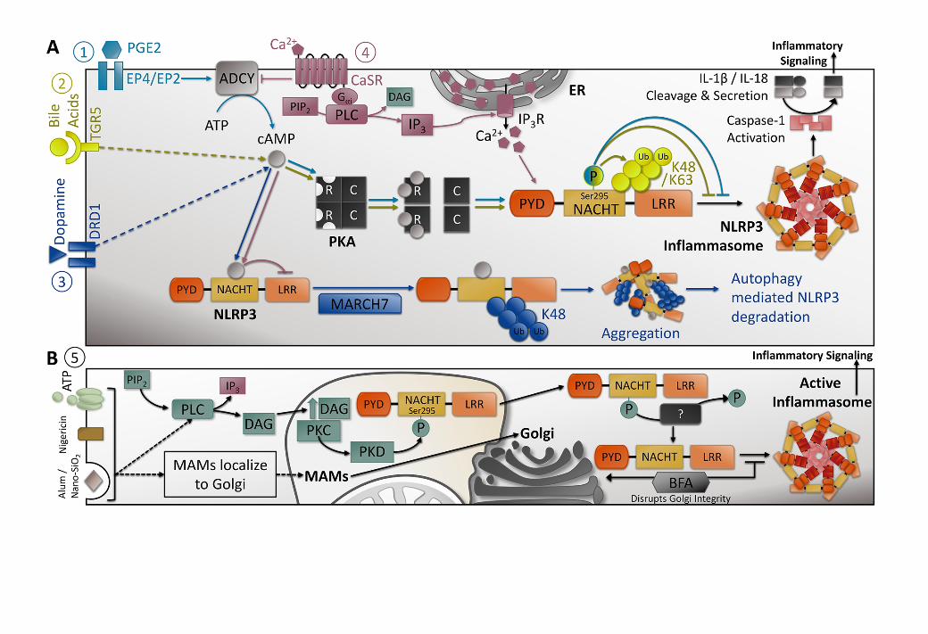


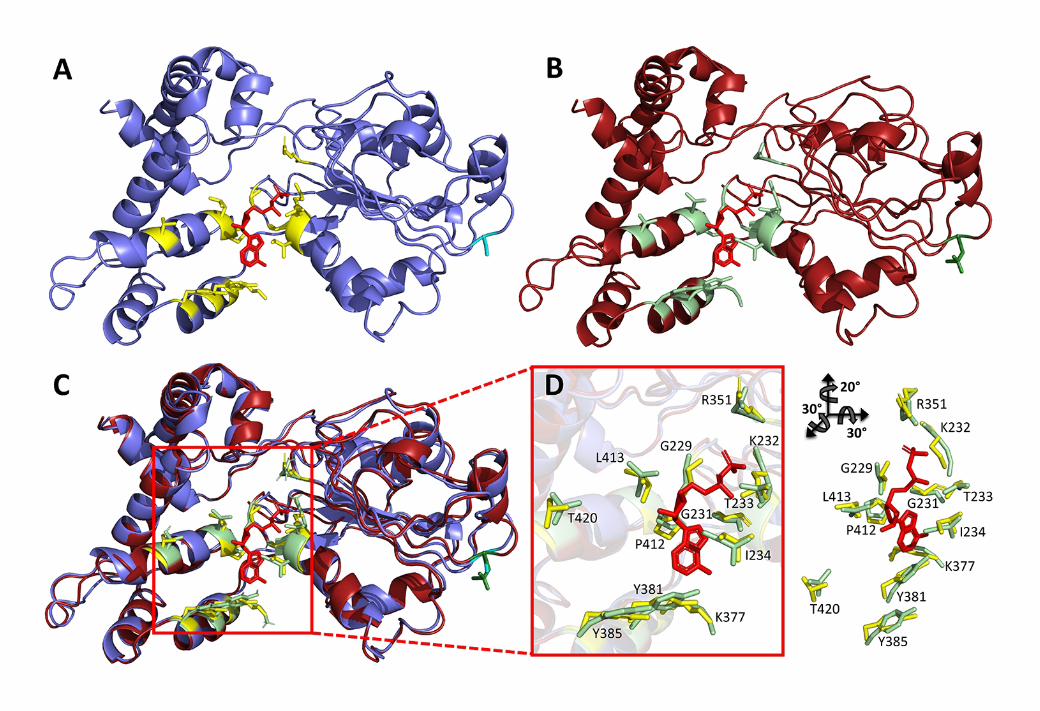
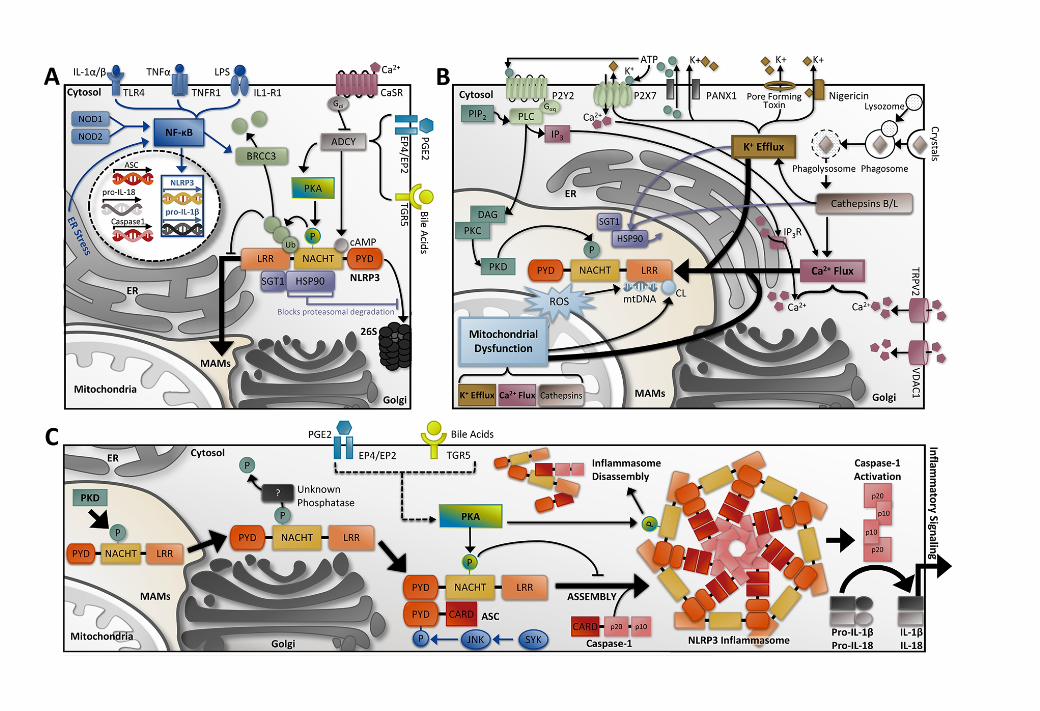

12345678910111213141516171819202122232425262728293031323334353637383940414243444546474849505152535455

123456789101112131415161718192021222324252627282930313233343536