activation of nlrp3 inflammasome complex potentiates ...activation of nlrp3 inflammasome complex...
TRANSCRIPT

Activation of NLRP3 inflammasome complexpotentiates venous thrombosis in response to hypoxiaNeha Guptaa, Anita Sahua, Amit Prabhakara, Tathagata Chatterjeeb, Tarun Tyagia, Babita Kumaria, Nilofar Khana,Velu Nairc, Nitin Bajajd, Manish Sharmaa,1, and Mohammad Zahid Ashrafa,1
aGenomics Division, Defence Institute of Physiology and Allied Sciences, Defence Research and Development Organization, Timarpur, Delhi 110054, India;bArmy Hospital (Research & Referral), New Delhi 110010, India; cArmed Forces Medical College, Pune 411040, India; and dCommand Hospital Chandimandir,Chandigarh 134107, India
Edited by Gregg L. Semenza, Johns Hopkins University School of Medicine, Baltimore, MD, and approved March 24, 2017 (received for review December14, 2016)
Venous thromboembolism (VTE), caused by altered hemostasis,remains the third most common cause of mortality among allcardiovascular conditions. In addition to established genetic andacquired risk factors, low-oxygen environments also predisposeotherwise healthy individuals to VTE. Although disease etiologyappears to entail perturbation of hemostasis pathways, the keymolecular determinants during immediate early response remainelusive. Using an established model of venous thrombosis, wehere show that systemic hypoxia accelerates thromboembolicevents, functionally stimulated by the activation of nucleotidebinding domain, leucine-rich-containing family, pyrin domain contain-ing 3 (NLRP3) inflammasome complex and increased IL-1β secretion.Interestingly, we also show that the expression of NLRP3 is mediatedby hypoxia-inducible factor 1-alpha (HIF-1α) during these conditions.The pharmacological inhibition of caspase-1, in vivo knockdown ofNLRP3, or HIF-1α other than IL-1β-neutralizing antibodies attenuatedinflammasome activation and curtailed thrombosis under hypoxicconditions. We extend the significance of these preclinical findingsby studying modulation of this pathway in patients with altitude-induced venous thrombosis. Our results demonstrate distinctive, in-creased expression of NLRP3, caspase-1, and IL-1β in individuals withclinically established venous thrombosis. We therefore propose thatan early proinflammatory state in the venous milieu, orchestrated bythe HIF-induced NLRP3 inflammasome complex, is a key determinantof acute thrombotic events during hypoxic conditions.
thrombosis | hypoxia | HIF-1α | NLRP3 inflammasome | IL-1β
Epidemiological studies during recent years have unprece-dentedly highlighted venous thromboembolism (VTE) as a
key comorbidity factor during several life-threatening medicalconditions. In addition to clinical complications such as cancer(1), cardiovascular diseases (2), surgery (3), and trauma (4),hypoxia as experienced during ascent to high altitude has emergedas another predisposing factor for VTE (5–8). A significantlyhigher incidence of deep vein thrombosis and pulmonary embolism(8, 9), portal vein thrombosis (10), cerebral venous thrombosis(11), transient ischemic attacks, and stroke (12) has been observedat high to extreme altitude (13). Despite clinical relevance, a caveatin our basic understanding of early molecular events underlyinghypoxia-induced venous thrombosis poses a major bottleneck foreffective design of interventional approaches.Recent studies have highlighted a strong link between hypoxia
responses and inflammation, involving activation of multiple celltypes including lymphocytes, platelets, and endothelium (14, 15).Plausibly, in addition to direct mechanism involving hypoxia-induced modulation of hemostasis and coagulation factors (5, 16),pleiotropic modalities such as sterile inflammation could be in-volved. The role of this axis and key mediators in hypoxia-inducedhypercoagulation, however, remains to be experimentally validated.The activation of coagulation pathways per se has been shown topromote inflammation in the system (17, 18). This secondary in-flammation can engage in a positive feed-forward loop to aggravatethe prothrombotic phenotype. Thus, the cause or consequence
relationship between inflammation and hypercoagulation duringhypoxia remains far from being resolved.We recently showed that hypobaric hypoxia promotes a pro-
thrombotic propensity through the involvement of a crucialcysteine protease, calpain (19). In continuing work, we blendedan unbiased systems-level approach with targeted pharmaco-logical inhibition and in vivo siRNA-mediated knockdownstrategies to show that the activation of nucleotide binding do-main, leucine-rich-containing family, pyrin domain containing 3(NLRP3) inflammasome complex augments thrombus formationin response to hypoxia. We reinforced significance of this keypreclinical finding from the animal model by demonstrating theactivation of the NLRP3 gene in patients, who developedthrombosis at high altitudes. We also present evidence that theactivation of this complex is an early response to hypoxia and iscritically regulated by hypoxia-inducible factor 1-alpha (HIF-1α).
ResultsFor all experiments described here, we have used uniform groupnomenclature. After inferior vena cava (IVC) ligation (also referto SI Materials and Methods), the group of animals kept in normalatmosphere conditions was designated as thrombotic (T), whereasthe ligated ones kept under simulated hypobaric hypoxia wereidentified as hypoxia thrombotic (HT). Sham surgery controlskept under normal environmental conditions were designated asnormoxic (N), and those kept in simulated hypoxia were iden-tified as hypoxic (H).
Significance
Hypoxia predisposes otherwise healthy individuals to venousthrombosis, but the underlying mechanism has been unclear.Our study revealed a causal role for nucleotide binding do-main, leucine-rich-containing family, pyrin domain containing 3(NLRP3) inflammasome and IL-1β during hypoxia-induced ve-nous thrombosis. We further show a direct association be-tween NLRP3 and hypoxia-inducible factor 1-alpha (HIF-1α)during these conditions. Specific interventions within thehypoxia–HIF-1α–NLRP3–IL-1β axis in the venous milieu signifi-cantly reduced venous thrombosis in our animal model. Notably,we also observed modulation of similar pathways in patientsdiagnosed with altitude-induced venous thrombosis. Our studythus revealed thrombosis at high altitude to be centrallyregulated by a complex network of coagulatory and in-flammatory processes, critically linked through HIF-1α.
Author contributions: M.S. and M.Z.A. designed research; N.G., A.S., A.P., T.C., T.T., B.K.,and N.B. performed research; N.K. and V.N. contributed new reagents/analytic tools; N.G.,A.S., A.P., M.S., and M.Z.A. analyzed data; and N.G., M.S., and M.Z.A. wrote the paper.
The authors declare no conflict of interest.
This article is a PNAS Direct Submission.1To whom correspondence may be addressed. Email: [email protected] [email protected].
This article contains supporting information online at www.pnas.org/lookup/suppl/doi:10.1073/pnas.1620458114/-/DCSupplemental.
www.pnas.org/cgi/doi/10.1073/pnas.1620458114 PNAS | May 2, 2017 | vol. 114 | no. 18 | 4763–4768
MED
ICALSC
IENCE
S
Dow
nloa
ded
by g
uest
on
Feb
ruar
y 22
, 202
0
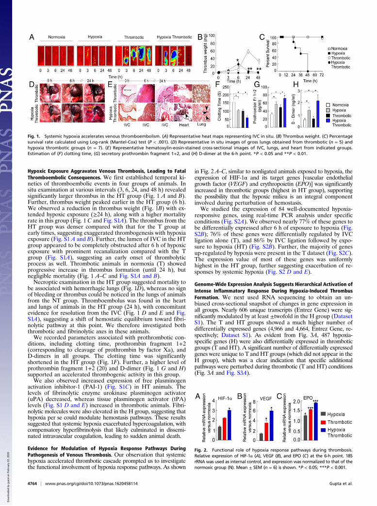
Hypoxic Exposure Aggravates Venous Thrombosis, Leading to FatalThromboembolic Consequences. We first established temporal ki-netics of thromboembolic events in four groups of animals. Insitu examination at various intervals (3, 6, 24, and 48 h) revealedsignificantly larger thrombus in the HT group (Fig. 1 A and B).Further, thrombus weight peaked earlier in the HT group (6 h).We observed a reduction in thrombus weight (Fig. 1B) with ex-tended hypoxic exposure (≥24 h), along with a higher mortalityrate in this group (Fig. 1 C and Fig. S1A). The thrombus from theHT group was denser compared with that for the T group atearly times, suggesting exaggerated thrombogenesis with hypoxiaexposure (Fig. S1 A and B). Further, the lumen of IVC in the HTgroup appeared to be completely obstructed after 6 h of hypoxicexposure with prominent recanalization compared with the Tgroup (Fig. S1A), suggesting an early onset of thrombolyticprocess as well. Thrombotic animals in normoxia (T) showedprogressive increase in thrombus formation (until 24 h), butnegligible mortality (Fig. 1 A–C and Fig. S1A and B).Necroptic examination in the HT group suggested mortality to
be associated with hemorrhagic lungs (Fig. 1D), whereas no signof bleeding or thrombus could be noticed in the lungs of animalsfrom the NT group. Thromboembolus was found in the heartand lungs of animals in the HT group (24 h), with concomitantevidence for resolution from the IVC (Fig. 1 D and E and Fig.S1A), suggesting a shift of hemostatic equilibrium toward fibri-nolytic pathway at this point. We therefore investigated boththrombotic and fibrinolytic axes in these animals.We recorded parameters associated with prothrombotic con-
ditions, including clotting time, prothrombin fragment 1+2(corresponding to cleavage of prothrombin by factor Xa), andD-dimers in all groups. The clotting time was significantlyshortened in the HT group (Fig. 1F). Further, a higher level ofprothrombin fragment 1+2 (20) and D-dimer (Fig. 1 G and H)supported an accelerated thrombogenic activity in this group.We also observed increased expression of free plasminogen
activation inhibitor-1 (PAI-1) (Fig. S1C) in HT animals. Thelevels of fibrinolytic enzyme urokinase plasminogen activator(uPA) decreased, whereas tissue plasminogen activator (tPA)levels (Fig. S1 D and E) increased in thrombotic animals. Fibri-nolytic molecules were also elevated in the H group, suggesting thathypoxia per se could modulate hemostasis pathways. These resultssuggested that systemic hypoxia exacerbated hypercoagulation, withcompensatory hyperfibrinolysis that likely culminated in dissemi-nated intravascular coagulation, leading to sudden animal death.
Evidence for Modulation of Hypoxia Response Pathways DuringPathogenesis of Venous Thrombosis. Our observation that systemichypoxia accelerated thrombotic cascade prompted us to investigatethe functional involvement of hypoxia response pathways. As shown
in Fig. 2 A–C, similar to nonligated animals exposed to hypoxia, theexpression of HIF-1α and its target genes [vascular endothelialgrowth factor (VEGF) and erythropoietin (EPO)] was significantlyincreased in thrombotic groups (highest in HT group), supportingthe possibility that the hypoxic milieu is an integral componentinvolved during perturbation of hemostasis.We studied the expression of 84 well-documented hypoxia-
responsive genes, using real-time PCR analysis under specificconditions (Fig. S2A). We observed nearly 77% of these genes tobe differentially expressed after 6 h of exposure to hypoxia (Fig.S2B); 76% of these genes were differentially regulated by IVCligation alone (T), and 86% by IVC ligation followed by expo-sure to hypoxia (HT) (Fig. S2B). Further, the majority of genesup-regulated by hypoxia were present in the T dataset (Fig. S2C).The expression value of most of these genes was uniformlyhighest in the HT group, further suggesting exacerbation of re-sponses by systemic hypoxia (Fig. S2 D and E).
Genome-Wide Expression Analysis Suggests Hierarchical Activation ofIntense Inflammatory Response During Hypoxia-Induced ThrombusFormation. We next used RNA sequencing to obtain an un-biased cross-sectional snapshot of changes in gene expression inall groups. Nearly 606 unique transcripts (Entrez Gene) were sig-nificantly modulated by at least ±twofold in the H group (DatasetS1). The T and HT groups showed a much higher number ofdifferentially expressed genes (4,966 and 4,664, Entrez Gene, re-spectively; Dataset S1). As evident from Fig. 3A, 487 hypoxia-specific genes (H) were also differentially expressed in thromboticgroups (T and HT). A significant number of differentially expressedgenes were unique to T and HT groups (which did not appear in theH group), which was a clear indication that specific additionalpathways were perturbed during thrombotic (T and HT) conditions(Fig. 3A and Fig. S3A).
Fig. 1. Systemic hypoxia accelerates venous thromboembolism. (A) Representative heat maps representing IVC in situ. (B) Thrombus weight. (C) Percentagesurvival rate calculated using Log-rank (Mantel-Cox) test (P < .001). (D) Representative in situ images of gross lungs obtained from thrombotic (n = 5) andhypoxia thrombotic groups (n = 7). (E) Representative hematoxylin-eosin-stained cross-sectional images of IVC, lungs, and heart from indicated groups.Estimation of (F) clotting time, (G) secretory prothrombin fragment 1+2, and (H) D-dimer at the 6-h point. *P < 0.05 and **P < 0.01.
Fig. 2. Functional role of hypoxia response pathways during thrombosis.Relative expression of HIF-1α (A), VEGF (B), and EPO (C) at the 6-h point. 18SrRNA was used as internal control, and expression was normalized to that of thenormoxic group (N). Mean ± SEM (n = 6) is shown. *P < 0.05; ***P < 0.001.
4764 | www.pnas.org/cgi/doi/10.1073/pnas.1620458114 Gupta et al.
Dow
nloa
ded
by g
uest
on
Feb
ruar
y 22
, 202
0
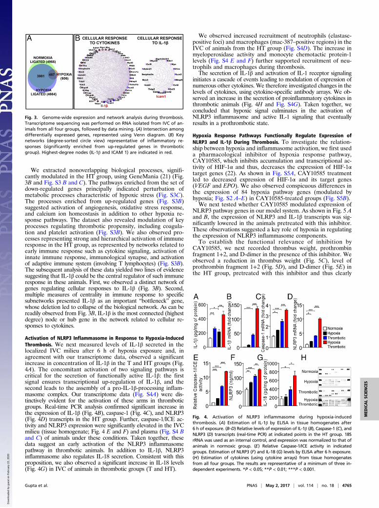
We extracted nonoverlapping biological processes, signifi-cantly modulated in the HT group, using GeneMania (21) (Fig.3B and Fig. S3 B and C). The pathways enriched from the set ofdown-regulated genes principally indicated perturbation ofmetabolic processes characteristic of hypoxic stress (Fig. S3C).The processes enriched from up-regulated genes (Fig. S3B)suggested activation of angiogenesis, oxidative stress response,and calcium ion homeostasis in addition to other hypoxia re-sponse pathways. The dataset also revealed modulation of keyprocesses regulating thrombotic propensity, including coagula-tion and platelet activation (Fig. S3B). We also observed pro-cesses representing strong and hierarchical activation of immuneresponse in the HT group, as represented by networks related toearly immune response such as cytokine signaling, activation ofinnate immune response, immunological synapse, and activationof adaptive immune system (involving T lymphocytes) (Fig. S3B).The subsequent analysis of these data yielded two lines of evidencesuggesting that IL-1β could be the central regulator of such immuneresponse in these animals. First, we observed a distinct network ofgenes regulating cellular responses to IL-1β (Fig. 3B). Second,multiple measures of centrality in immune response to specificsubnetworks presented IL-1β as an important “bottleneck” gene,whose deletion led to collapse of the biological network. As can bereadily observed from Fig. 3B, IL-1β is the most connected (highestdegree) node or hub gene in the network related to cellular re-sponses to cytokines.
Activation of NLRP3 Inflammasome in Response to Hypoxia-InducedThrombosis. We next measured levels of IL-1β secreted in thelocalized IVC milieu after 6 h of hypoxia exposure and, inagreement with our transcriptome data, observed a significantincrease in concentration of IL-1β in the T and HT groups (Fig.4A). The concomitant activation of two signaling pathways iscritical for the secretion of functionally active IL-1β: the firstsignal ensures transcriptional up-regulation of IL-1β, and thesecond leads to the assembly of a pro-IL-1β-processing inflam-masome complex. Our transcriptome data (Fig. S4A) were dis-tinctively evident for the activation of these arms in thromboticgroups. Real-time PCR analysis confirmed significant increase inthe expression of IL-1β (Fig. 4B), caspase-1 (Fig. 4C), and NLRP3(Fig. 4D) transcripts in the HT group. Further, caspase-1/ICE ac-tivity and NLRP3 expression were significantly elevated in the IVCmilieu (tissue homogenate; Fig. 4 E and F) and plasma (Fig. S4 Band C) of animals under these conditions. Taken together, thesedata suggest an early activation of the NLRP3 inflammasomepathway in thrombotic animals. In addition to IL-1β, NLRP3inflammasome also regulates IL-18 secretion. Consistent with thisproposition, we also observed a significant increase in IL-18 levels(Fig. 4G) in IVC of animals in thrombotic groups (T and HT).
We observed increased recruitment of neutrophils (elastase-positive foci) and macrophages (mac-387–positive regions) in theIVC of animals from the HT group (Fig. S4D). The increase inmyeloperoxidase activity and monocyte chemotactic protein-1levels (Fig. S4 E and F) further supported recruitment of neu-trophils and macrophages during thrombosis.The secretion of IL-1β and activation of IL-1 receptor signaling
initiates a cascade of events leading to modulation of expression ofnumerous other cytokines. We therefore investigated changes in thelevels of cytokines, using cytokine-specific antibody arrays. We ob-served an increase in the secretion of proinflammatory cytokines inthrombotic animals (Fig. 4H and Fig. S4G). Taken together, weconcluded that hypoxic signal culminates in the activation ofNLRP3 inflammasome and active IL-1 signaling that eventuallyresults in a prothrombotic state.
Hypoxia Response Pathways Functionally Regulate Expression ofNLRP3 and IL-1β During Thrombosis. To investigate the relation-ship between hypoxia and inflammasome activation, we first useda pharmacological inhibitor of hypoxia response pathway,CAY10585, which inhibits accumulation and transcriptional ac-tivity of HIF-1α and thus, decreases the expression of HIF-1αtarget genes (22). As shown in Fig. S5A, CAY10585 treatmentled to decreased expression of HIF-1α and its target genes(VEGF and EPO). We also observed conspicuous differences inthe expression of 84 hypoxia pathway genes (modulated byhypoxia; Fig. S2 A–E) in CAY10585-treated groups (Fig. S5B).We next tested whether CAY10585 modulated expression of
NLRP3 pathway genes in our model system. As shown in Fig. 5 Aand B, the expression of NLRP3 and IL-1β transcripts was sig-nificantly lowered in the animals pretreated with this inhibitor.These observations suggested a key role of hypoxia in regulatingthe expression of NLRP3 inflammasome components.To establish the functional relevance of inhibition by
CAY10585, we next recorded thrombus weight, prothrombinfragment 1+2, and D-dimer in the presence of this inhibitor. Weobserved a reduction in thrombus weight (Fig. 5C), level ofprothrombin fragment 1+2 (Fig. 5D), and D-dimer (Fig. 5E) inthe HT group, pretreated with this inhibitor and thus clearly
Fig. 3. Genome-wide expression and network analysis during thrombosis.Transcriptome sequencing was performed on RNA isolated from IVC of an-imals from all four groups, followed by data mining. (A) Intersection amongdifferentially expressed genes, represented using Venn diagram. (B) Keynetworks (degree-sorted circle view) representative of inflammatory re-sponses (significantly enriched from up-regulated genes in thromboticgroup). Highest-degree nodes (IL-1β and ICAM 1) are indicated in red.
Fig. 4. Activation of NLRP3 inflammasome during hypoxia-inducedthrombosis. (A) Estimation of IL-1β by ELISA in tissue homogenates after6 h of exposure. (B–D) Relative levels of expression of IL-1β (B), Caspase-1 (C), andNLRP3 (D) transcripts (real-time PCR) at indicated points in the HT group. 18SrRNA was used as an internal control, and expression was normalized to that ofanimals in normoxic group. (E) Relative Caspase-1/ICE activity in indicatedgroups. Estimation of NLRP3 (F) and IL-18 (G) levels by ELISA after 6 h exposure.(H) Estimation of cytokines (using cytokine arrays) from tissue homogenatesfrom all four groups. The results are representative of a minimum of three in-dependent experiments. *P < 0.05; **P < 0.01; ***P < 0.001.
Gupta et al. PNAS | May 2, 2017 | vol. 114 | no. 18 | 4765
MED
ICALSC
IENCE
S
Dow
nloa
ded
by g
uest
on
Feb
ruar
y 22
, 202
0

supporting a functional role of hypoxia response pathways in theactivation of the NLRP3 inflammasome and thrombogenesis.
Evidence for a Functional Role of HIF-1α During Hypoxia-InducedNLRP3 Expression. We next tested whether HIF-1α, the centralregulator of hypoxia responses (23), was involved in hypoxia-induced expression of NLRP3 and thrombogenesis. We usedan in vivo siRNA approach. As shown in Fig. 6A, animals in theHT group (6 h), treated with HIF-1α siRNA, showed a signifi-cant reduction in the accumulation of the HIF transcript. Theexpression of NLRP3 (Fig. 6B), IL-1β (Fig. 6C), and caspase-1(Fig. S6A) transcripts was significantly reduced in these groups.Further, caspase-1/ICE activity (Fig. S6B) and NLRP3 proteinlevels (Fig. S6C) were also diminished in the plasma of thesegroups. The animals in siRNA-treated groups showed significantreduction in thrombus weight (Fig. 6D), with an increase inclotting (Fig. 6E) and prothrombin time (PT; Fig. S6D). Theknock-down of HIF-2α in our experiments led to a significantincrease in HIF-1α expression, a likely compensatory response(Fig. 6A). Concurrent with an increase in expression of HIF-1α,the expression of NLRP3, IL-1β, and caspase-1 was also ele-vated, along with increase in thrombus weight, clotting time, andPT (Fig. 6 A–E and Fig. S6D).We next analyzed the NLRP3 promoter for putative HIF-
responsive elements/sites. Our in silico analysis returned three se-quences closely matching HIF-responsive element consensus (Fig.S6E). We therefore performed chromatin immunoprecipitation
experiments, using two different HIF-1α antibody clones and probesspanning NLRP3 promoter. As shown in Fig. 6F, we consistentlyobserved recruitment of HIF-1α at one of these sites [−975 w.r.ttranscription start site (TSS)], implicating functional involvement ofHIF-1α in regulating NLRP3 expression during HT conditions.
NLRP3 Inflammasome Axis Inhibition Curtails Hypoxia-InducedThrombosis. To establish whether the activation of NLRP3inflammasome played a causal role in the initiation and propa-gation of thrombosis in our model system, we used three dif-ferent inhibition strategies and, subsequently, performed in situthrombus examination, recorded thrombus weight and length,and checked in vivo levels of prothrombin fragment 1+2 andD-dimer, in addition to monitoring the aggregation of plateletsisolated from these animals.We first knocked down NLRP3 transcript in the animals, using
in vivo grade siRNA (10 mg/kg body weight), aiming to check theassembly of the NLRP3 inflammasome complex. We observed asignificant reduction in caspase-1/ICE activity (Fig. S7A), inaddition to reduced thrombus (Fig. 7 A–C), in IVC of the HTgroup treated with NLRP3 siRNA. We also observed a signifi-cant reduction in the levels of prothrombin fragment 1+2 andthe D-dimer (Fig. 7 D and E, respectively). These results sug-gested a causal role for NLRP3 during thrombosis. The plateletsisolated from animals pretreated with NLRP3 siRNA, beforeIVC ligation, also showed reduced aggregation in response toADP (used as physiological agonist) (Fig. 7F). This observationsuggested an upstream role of NLRP3 inflammasome to plateletactivation, which is a vital step in the thrombogenic cascade.In the next set of experiments, we used SML0499 to inhibit the
catalytic activity of caspase-1, required for the production ofactive IL-1β from its proform. The in situ thrombus examination,thrombus weight and length, prothrombin fragment 1+2, D-dimer,and ex vivo platelet aggregation assay using ADP are shown inFig. S7 B–G. The cumulative results from all these assays showedthat inhibition of caspase-1 activity reduced thrombogenesis underhypoxic conditions.Finally, we injected specific antibodies against active IL-1β and
thus limited its bioavailability, essential for signaling via cognateIL-1 receptors. As presented in Fig. S7 H–M, these animals alsoshowed a reduction in thrombus formation, as evident from asimilar set of parameters described earlier. Taken together, theseresults (Fig. 7 A–F and Fig. S7 B–M) demonstrated an indispensablerole for NLRP3 inflammasome-mediated active IL-1β generation inhypoxia-induced thrombus formation. The expression analysis inspecific cell types [peripheral blood mononuclear cells (PBMNs),platelets, and vessel wall] suggested that both NLRP3 and IL-1βincreased significantly in the PBMNs, apart from thrombus isolatedfrom a localized (ligated) venous site (Fig. S8).
Fig. 5. Hypoxia-induced proinflammatory state is regulated by transcrip-tional activity of HIF-1α. HIF inhibitor, CAY10585 (100 μg/kg) was adminis-tered (intravenous) before IVC ligation and hypoxic challenge for 6 h.Relative expression of (A) NLRP3 and (B) 1L-1β transcripts (real-time PCR).(C) Dot plot showing medians of thrombus weight, (D) Levels of prothrombinfragment 1+2, (E ) D-dimer (ELISA) estimation in plasma samples. Mean ±SEM is shown (n ≥ 6). *P < 0.05; **P < 0.01; ***P < 0.001.
Fig. 6. HIF-1α regulates NLRP3 expression and thrombogenesis. Animals were treated with in vivo grade HIF-1α and HIF-2α siRNA. After RNA isolation fromindicated groups, real-time PCR was performed for (A) HIF-1α, (B) NLRP3, (C ) IL-1β. Thrombus weight (D) and clotting time (E ) were also recorded.(F) Chromatin immunoprecipitation with two different HIF-1α antibody (indicated) and primer pairs spanning putative sites (indicated). The enrichment ofNLRP3 promoter region in chromatin immunoprecipitation experiments was quantitated and plotted to obtain the bar graph (mean ± SEM) shown in thefigure. *P < 0.05; **P < 0.01; ***P < 0.001.
4766 | www.pnas.org/cgi/doi/10.1073/pnas.1620458114 Gupta et al.
Dow
nloa
ded
by g
uest
on
Feb
ruar
y 22
, 202
0

Evidence for the Involvement of the NLRP3 Inflammasome in HumanPatients with Altitude-Induced Thrombosis. We next sought to in-vestigate the potential involvement of the NLRP3 inflammasomein clinically confirmed cases of VTE (n = 18) occurring in re-sponse to the hypoxic environment. The demographic, clinical,and specific genetic parameters of patients with VTE are pre-sented in Fig. S9. We observed a relatively higher number ofpatients lacking thrombophilic traits [including deficiency ofprotein C, protein S, and ATIII, in addition to activated proteinC (APC) resistance], major SNPs [factor V Leiden, prothrombin,tissue factor pathway inhibitor (TFPI), fibrinogen-β, methylenetetrahydrofolate reductase (MTHFR), and PAI-1], and otheradditional risk factors (including lipid profile, homocysteine,and blood glucose levels) known to be associated with a pre-disposition to VTE (Fig. S9). These observations likely suggestedthat VTE episodes in these individuals were potentially triggeredby environmental conditions (hypoxia) prevailing at altitudes.To test the likely involvement of the NLRP3 inflammasome
pathway, we next studied relative expression of key genes of thispathway in these patients. As shown in Fig. 8 A–D, we observedan increase in NLRP3, caspase-1, IL-1β, and IL-18 mRNA ex-pression in patients compared with healthy age-matched controls.Furthermore, caspase-1/ICE activity was significantly elevated alongwith increased levels of NLRP3 (protein) and IL-1β in the patientsamples (plasma, Fig. 8 E–G). This dataset supported involvementof the NLRP3 inflammasome pathway in the pathogenesis of VTEin individuals exposed to the hypoxic challenge.Fig. 8H schematically depicts the scheme of events causally
underlying activation of thrombosis in hypoxic environments.
DiscussionThe present study revealed a causal role of strong inflammatoryresponse involving NLRP3 and IL-1β in activating hypoxia-induced thrombogenic cascade in the venous milieu. Of criticalnote is the fact that HIF-1α, known to regulate a plethora ofhuman diseases (24), emerged as the key node connecting hyp-oxia responses to proinflammatory state via its ability to regulatethe expression of NLRP3 (transcript) under these conditions.Conceivably, the evidence for a direct connection between HIF-1α and NLRP3 is likely to have general implications, especiallyas a target for intervention in other pathological conditionsemanating from hypoxia and the proinflammatory state.The biological activation of IL-1β requires parallel activation
of pathways, culminating in transcriptional up-regulation of IL-1β, increased expression of NLRP3, and enzymatic activation ofcaspase-1 (25). Some recent reports suggested platelets as alikely source of IL-1β (26–29), produced by virtue of a storedrepertoire of molecules (mRNA, inflammasome components)and cue-dependent processing/secretion during thrombogenesis.Thus, an important question pertaining to the possibility ofplatelet-origin IL-1β in sustaining an intense phenotype, such asthat observed in our study, remains paradigmatic. Our presentdataset provides some additional information in this regard. Weobserved up-regulation of IL-1β transcript in addition to other
inflammasome components in mononuclear cells (PBMNs), apartfrom thrombus isolated from localized (ligated) venous site, andthis up-regulation could be prevented using HIF inhibitor,CAY10585, or HIF-1α-specific siRNA. It thus is reasonable to as-sume that the immune cells are likely to play an important role inregulating the intensity of venous thrombosis, likely through denovo cue-dependent transcriptional up-regulation of IL-1β andinflammasome pathway genes (NLRP3, caspase-1). Finally, thefact that we also observed a concomitant increase in the relativeexpression of NLRP3, caspase-1, IL-1β, and IL-18 transcripts inperipheral blood cells of volunteers who developed VTE athigh altitudes lends strong support to this proposition.The hypoxic milieu in vivo has been proposed as a critical reg-
ulator of sterile inflammation and consequent pathological ef-fects. The mechanistic basis appears to include modulation ofintrinsic mitochondrial redox homeostasis (30), in addition toactivation of toll-like-receptors and various danger signals such asATP release from necrotic cells (31). The issue of hypoxia-induced
Fig. 7. Knock-down of NLRP3 curtails hypoxia-Induced thrombosis. NLRP3 was knock-down using in vivo grade siRNA complexes and specific parameterstudied. (A) Photomicrographs of thrombosed IVC; (B) thrombus weight; (C) thrombus length (median indicated); (D) Prothrombin fragments 1+2;(E) D-dimer, and (F) platelet aggregation assay in groups (indicated in figure), 6 h postinduction. All datasets are representative of a minimum of threeindependent experiments. *P < 0.05; **P < 0.01; ***P < 0.001.
Fig. 8. Evidence for involvement of NLRP3 inflammasome components inpatients with altitude-induced venous thrombosis. (A–D) RNA was isolatedfrom PBMNs from the blood samples of patients (n = 18), and real-time PCRfor indicated genes was performed. β-actin was used as an internal control.The relative expression of NLRP3 (A), caspase-1 (B), 1L-1β (C), and IL-18 (D)transcripts. (E) Caspase-1/ICE activity in plasma samples from patients andcontrols (n = 12). Estimation of NLRP3 (F) and IL-1β (G) levels in plasmasamples from patients and controls (n ≥ 8) Median values for individualgroups are also shown. *P < 0.05; **P < 0.01. (H) Diagrammatic representation ofan inferred scheme of events during hypoxia-induced thrombosis.
Gupta et al. PNAS | May 2, 2017 | vol. 114 | no. 18 | 4767
MED
ICALSC
IENCE
S
Dow
nloa
ded
by g
uest
on
Feb
ruar
y 22
, 202
0

inflammasome activation and the proinflammatory state breed-ing pathophysiological outcomes at high altitude encompassesconflicting studies and opinions (15, 32). Our transcriptome datarevealed dense gene networks related to strong proinflammatoryresponses, involving both innate and adaptive immune cells(Fig. 3). Further, as described here, hypoxia-induced throm-bosis could be circumvented by inflammasome inhibition, sug-gesting an early role of inflammation in this process. We alsoshowed that inhibition of hypoxia response pathways (usingpharmacological inhibitor or siRNA) prevented transcriptionalup-regulation of NLRP3 and IL-1β with significant antith-rombotic effects. Taken together, these results posit that hyp-oxia regulates significant pathological effects via its ability topromote the proinflammatory state.Virtually all mechanistic understanding, elucidated to date,
pertaining to specific forms of thrombosis keep complying withVirchow’s Triad, although in somewhat kaleidoscopic molecularpatterns regulating individual hypercoagulable states (33). Inkeeping with the essence of this fact, venous and arterialthrombosis also appears to entail principally similar events, butconspicuously divergent origins. Although early endothelial in-jury is an established modus operandi of arterial thrombosis, itappears to be dispensable during early stages of venous formsthat precipitate under diverse conditions and stimuli. A recentstudy showed that the NLRP3 inflammasome inhibitor, Argla-bin, curtailed the atherogenic effect of high-fat diet in ApoE2.Kimice (34). In view of such information, it is tempting to speculatethat activation of NLRP3 inflammasome complex could consti-tute a unifying molecular cornerstone between diverse patho-logical states and various forms of thrombosis.Hypercoagulable state is also known to predispose an indi-
vidual to elevated risk for pulmonary embolism, which is a biggerclinical challenge and often more fatal, arising from increasedthrombus dissemination. We too observed a somewhat similarphenomenon in our animal model with disseminated intravas-cular coagulation or hypercoagulation concomitant with elevatedfibrinolytic activity, under hypoxic condition (Fig. 1). Conversely,a thrombotic state could also manifest as a result of skewing ofhomeostasis toward hypercoagulation due to a less-effective
fibrinolytic system. Taken together, such arguments define anapparent paradigm for clinical significance and an area of futureinvestigation. In view of our results, we posit that the strength ofbiological cues propagating individual pathways (coagulatory-fibrinolytic) critically regulate resultant effects with phenotypicmanifestation such as localized thrombosis, consumptive coa-gulopathy, or pulmonary embolism.In summary, our study revealed an important target, NLRP3
inflammasome, for hypoxia-induced venous thrombosis in additionto reinforcing an intriguing complexity involving intricately inter-acting coagulatory, thrombolytic, and inflammatory hubs at its core.
Materials and MethodsDetailed materials and methods are included in SI Materials and Methods.
Animal Experiments. All experiments were conducted in compliance withguidelines of the Committee for Purpose of Control and Supervision of Ex-periments on Animals, Government of India. Male Sprague–Dawley rats,weighing 250–300 g, were used and exposed to hypobaric hypoxia, usingenvironmental chamber simulating 429 torr. The IVC ligation model forin vivo thrombosis was used as previously described by us (19).
Human Studies. Human studies were conducted in strict compliance with theethical standards of Indian Council of Medical Research. Informed consentwas obtained from the subjects as per Declaration of Helsinki. Young malepatients with VTE (n = 18) evacuated from high-altitude regions to tertiarycare facilities (Command Hospital Chandimandir, Chandigarh or Army Hos-pital, New Delhi) were enrolled. Equal numbers of healthy, age-matchedmale subjects with no prior history for VTE were included as controls.
Statistics. Data are presented as mean ± SEM. The statistical significance ofdifferences was evaluated using unpaired t test or Mann-Whitney test.Bonferroni post hoc test was done for multiple group comparison, usingPrism 5 (GraphPad) software. The statistical significance of differences wererepresented as *P < 0.05, **P < 0.01, and ***P < 0.001.
ACKNOWLEDGMENTS. We acknowledge Dr. Shashi B Singh, DirectorateGeneral Armed Forces Medical Services, Dr. Srishti Gupta, Dr. Iti Garg, andDr. R. J. Tirpude for their support. This study was funded by Defence Re-search and Development Organization Project SL-10/DIP-255.
1. Zwicker JI, et al. (2009) Tumor-derived tissue factor-bearing microparticles are associatedwith venous thromboembolic events in malignancy. Clin Cancer Res 15:6830–6840.
2. Zhou X, et al. (2010) Incidence and risk factors of venous thromboembolic events inlymphoma. Am J Med 123:935–941.
3. Chan MY, Andreotti F, Becker RC (2008) Hypercoagulable states in cardiovasculardisease. Circulation 118:2286–2297.
4. Demers C, et al. (1998) Incidence of venographically proved deep vein thrombosisafter knee arthroscopy. Arch Intern Med 158:47–50.
5. Bendz B, Rostrup M, Sevre K, Andersen TO, Sandset PM (2000) Association between acutehypobaric hypoxia and activation of coagulation in human beings. Lancet 356:1657–1658.
6. Lapostolle F, et al. (2001) Severe pulmonary embolism associated with air travel. NEngl J Med 345:779–783.
7. Smallman DP, McBratney CM, Olsen CH, Slogic KM, Henderson CJ (2011) Quantifica-tion of the 5-year incidence of thromboembolic events in U.S. Air Force Academycadets in comparison to the U.S. Naval and Military Academies. Mil Med 176:209–213.
8. Ward M (1975) Mountain medicine: A clinical study of cold and high altitude (CrosbyLockwood Staples, London).
9. Zangari M, et al. (2013) Could hypoxia increase the prevalence of thrombotic com-plications in polycythemia vera? Blood Coagul Fibrinolysis 24:311–316.
10. Anand AC, Saha A, Kumar R, Sharma V, Jha SK (2000) Portal system thrombosis: Anew dimension of high altitude illnesses. Trop Gastroenterol 21:172–173.
11. Cheng S, Chng SM, Singh R (2009) Cerebral venous infarction during a high altitudeexpedition. Singapore Med J 50:e306–e308.
12. Jha SK, Anand AC, Sharma V, Kumar N, Adya CM (2002) Stroke at high altitude: Indianexperience. High Alt Med Biol 3:21–27.
13. Gupta N, Ashraf MZ (2012) Exposure to high altitude: A risk factor for venousthromboembolism? Semin Thromb Hemost 38:156–163.
14. Mannucci PM, Gringeri A, Peyvandi F, Di Paolantonio T, Mariani G (2002) Short-termexposure to high altitude causes coagulation activation and inhibits fibrinolysis.Thromb Haemost 87:342–343.
15. Eltzschig HK, Carmeliet P (2011) Hypoxia and inflammation. N Engl J Med 364:656–665.16. Rider P, et al. (2012) The transcription of the alarmin cytokine interleukin-1 alpha is con-
trolled by hypoxia inducible factors 1 and 2 alpha in hypoxic cells. Front Immunol 3:290.17. Mileno MD, et al. (1995) Coagulation of whole blood stimulates interleukin-1 beta
gene expression. J Infect Dis 172:308–311.
18. Reitsma PH, Rosendaal FR (2004) Activation of innate immunity in patients with ve-nous thrombosis: The Leiden Thrombophilia Study. J Thromb Haemost 2:619–622.
19. Tyagi T, et al. (2014) Altered expression of platelet proteins and calpain activitymediate hypoxia-induced prothrombotic phenotype. Blood 123:1250–1260.
20. Páramo JA (2010) Prothrombin fragments in cardiovascular disease. Adv Clin Chem51:1–23.
21. Warde-Farley D, et al. (2010) The GeneMANIA prediction server: Biological network in-tegration for gene prioritization and predicting gene function.Nucleic Acids Res 38:W214–220.
22. Lee K, et al. (2007) (Aryloxyacetylamino)benzoic acid analogues: A new class ofhypoxia-inducible factor-1 inhibitors. J Med Chem 50:1675–1684.
23. Semenza GL (2007) Life with oxygen. Science 318:62–64.24. Semenza GL (2000) HIF-1 and human disease: One highly involved factor. Genes Dev
14:1983–1991.25. Martinon F, Mayor A, Tschopp J (2009) The inflammasomes: Guardians of the body.
Annu Rev Immunol 27:229–265.26. Lindemann S, et al. (2001) Activated platelets mediate inflammatory signaling by
regulated interleukin 1beta synthesis. J Cell Biol 154:485–490.27. Denis MM, et al. (2005) Escaping the nuclear confines: Signal-dependent pre-mRNA
splicing in anucleate platelets. Cell 122:379–391.28. Hottz ED, et al. (2013) Platelets mediate increased endothelium permeability in
dengue through NLRP3-inflammasome activation. Blood 122:3405–3414.29. Brown GT, Narayanan P, Li W, Silverstein RL, McIntyre TM (2013) Lipopolysaccharide
stimulates platelets through an IL-1β autocrine loop. J Immunol 191:5196–5203.30. Usui F, et al. (2015) Inflammasome activation by mitochondrial oxidative stress in
macrophages leads to the development of angiotensin II-induced aortic aneurysm.Arterioscler Thromb Vasc Biol 35:127–136.
31. Weinberg SE, Sena LA, Chandel NS (2015) Mitochondria in the regulation of innateand adaptive immunity. Immunity 42:406–417.
32. Wanderer AA (2011) Hypoxia and inflammation. N Engl J Med 364(20):1976.33. López JA, Chen J (2009) Pathophysiology of venous thrombosis. Thromb Res 123:S30–S34.34. Abderrazak A, et al. (2015) Anti-inflammatory and antiatherogenic effects of the
NLRP3 inflammasome inhibitor arglabin in ApoE2.Ki mice fed a high-fat diet. Circulation131:1061–1070.
35. Trapnell C, et al. (2012) Differential gene and transcript expression analysis of RNA-seq experiments with TopHat and Cufflinks. Nat Protoc 7:562–578.
4768 | www.pnas.org/cgi/doi/10.1073/pnas.1620458114 Gupta et al.
Dow
nloa
ded
by g
uest
on
Feb
ruar
y 22
, 202
0