effects of cytomegalovirus infection in human neural precursor...
TRANSCRIPT

Effects of cytomegalovirus infection in human neural precursorcells depend on their differentiation state
H. M. González-Sánchez & A. Monsiváis-Urenda & C. A. Salazar-Aldrete &
A. Hernández-Salinas & D. E. Noyola & M. E. Jiménez-Capdeville &
A. Martínez-Serrano & C. G. Castillo
Received: 18 August 2014 /Revised: 31 December 2014 /Accepted: 9 January 2015 /Published online: 8 April 2015# Journal of NeuroVirology, Inc. 2015
Abstract Cytomegalovirus (CMV) is the most commoncause of congenital infection in developed countries and amajor cause of neurological disability in children. AlthoughCMV can affect multiple organs, the most important sequelaeof intrauterine infection are related to lesions of the centralnervous system. However, little is known about the pathogen-esis and the cellular events responsible for neuronal damage ininfants with congenital infection. Some studies have demon-strated that neural precursor cells (NPCs) show the greatestsusceptibility to CMV infection in the developing brain. Wesought to establish an in vitro model of CMV infection of thedeveloping brain in order to analyze the cellular events asso-ciated with invasion by this virus. To this end, we employedtwo cell lines as a permanent source of NPC, avoiding thecontinuous use of human fetal tissue, the human SK-N-MCneuroblastoma cell line, and an immortalized cell line of hu-man fetal neural origin, hNS-1. We also investigated the effectof the differentiation stage in relation to the susceptibility ofthese cell lines by comparing the neuroblastoma cell line with
the multipotent cell line hNS-1. We found that the effects ofthe virus were more severe in the neuroblastoma cell line.Additionally, we induced hNS-1 to differentiate and evaluatedthe effect of CMVin these differentiated cells. Like SK-N-MCcells, hNS-1-differentiated cells were also susceptible to infec-tion. Viability of differentiated hNS-1 cells decreased afterCMV infection in contrast to undifferentiated cells. In addi-tion, differentiated hNS-1 cells showed an extensive cytopath-ic effect whereas the effect was scarce in undifferentiated cells.We describe some of the effects of CMV in neural stem cells,and our observations suggest that the degree of differentiationis important in the acquisition of susceptibility.
Keywords Cytomegalovirus . Neural stem cells . Neuralprogenitor cells . Cytophatic effect . Differentiation
Introduction
Human cytomegalovirus (HCMV), a member of the herpesvirus family, is a common pathogen in immunocompromizedpatients and newborns. Excretion of viral particles in manybody fluids, including urine and saliva, among others, is re-sponsible for HCMV spreading which results in 40 % sero-positive adults in developed countries. The incidence of infec-tion in developing countries is even higher, where almost100 % of the population has been infected by adulthood(Wynn and Khanna 2006; Ornoy 2007; Noyola et al.2010). Congenital infection by HCMV has an incidencebetween 0.8 and 2.6 % worldwide, and this infection isthe most frequent cause for developmental brain disor-ders, including mental retardation and hearing loss.
Although most of the research about pathogenic mecha-nisms of CMV in the brain has been performed on animals,
H. M. González-Sánchez : C. A. Salazar-Aldrete :M. E. Jiménez-Capdeville :C. G. Castillo (*)Department of Biochemistry, Facultad de Medicina, UniversidadAutónoma de San Luis Potosí, Av. Venustiano Carranza No. 2405,Colonia Los Filtros, 78210 San Luis Potosí, SLP, Méxicoe-mail: [email protected]
A. Monsiváis-UrendaDepartment of Immunology, Facultad de Medicina, UniversidadAutónoma de San Luis Potosí, San Luis Potosí 78210, SLP, México
A. Hernández-Salinas :D. E. NoyolaDepartment of Microbiology, Facultad de Medicina, UniversidadAutónoma de San Luis Potosí, San Luis Potosí 78210, SLP, México
A. Martínez-SerranoDepartment of Molecular Biology, Center of Molecular BiologySevero Ochoa, Autonomous University of Madrid-CSIC CampusCantoblanco, Madrid, Spain
J. Neurovirol. (2015) 21:346–357DOI 10.1007/s13365-015-0315-5
an important limitation has been the strict host specificity ofCMVs, which requires the utilization of the correspondinganimal virus (Wynn and Khanna 2006). Furthermore, the clin-ical manifestations on some animal models are different fromthose observed in the human central nervous system (CNS)(Woolf et al. 2007). Nevertheless, these animal models haveprovided evidence that the highest CMV susceptibility inCNS is presented by neural precursor cells (NPCs) (Tsutsuiet al. 2002). Infection of these cells might be the cause ofmicrocephaly and mental retardation since it has been ob-served that murine CMV inhibits NPC proliferation and mi-gration (Shinmura et al. 1997; Tsutsui et al. 2008). Further-more, studies of functional neural cells after CMV infection,though scarce, reveal that infected cells have altered intercel-lular communication and deficient response to neurotransmit-ters (Ho and van den Pol 2007).
In order to study NPC in vitro, it is necessary to isolatethem from embryonic tissue and maintain them in cultureunder specific conditions to favor neural proliferation.In vitro studies in which these cells were infected with humanCMV demonstrated that NPCs are highly permissive to CMVinfection and all types of viral proteins are expressed. In ad-dition, cytopathic effect (CPE) in these cells at the multiplicityof infection (MOI) of three plaque formation units (PFU) percell consisted in formation of syncytia, which can be the resultof viral cell to cell dissemination (Luo et al. 2008). Infection ofthese cells may increase the apoptosis frequency and inhibitneuronal and glia differentiation; however, this effect remainscontroversial since it has also been reported that CMV infec-tion may cause a spontaneous, premature, and abnormal NPCdifferentiation (Odeberg et al. 2006, 2007; Luo et al. 2010).
Recently, the importance of the differentiation degree relat-ed to the capacity of the virus to effectively infect the cellsduring the first steps of the viral cycle was described. It wasshown that the concentration of heparan sulfate in the cellsurface, the presence of β1-integrin and vimentin, and thenuclear pore density is important for the attachment, penetra-tion, and internalization to the nucleus (Kawasaki et al. 2011).In mouse models, it has been observed that embryonic stemcells cannot be infected with CMVand they acquire suscepti-bility through differentiation (Matsukage et al. 2006; Kawa-saki et al. 2011). However, differentiated neurons are alsorefractory to the infection and this may suggest that only dur-ing a precise period during differentiation, the cells can beinfected (Cheeran et al. 2005).
Taking this into account, we aimed to establish an in vitromodel of CMV infection in NPC that could help clarify theseissues. In order to do so, we employed, as a permanent sourceof NPC, two cell lines, avoiding the continuous use of humanfetuses, the human SK-N-MC neuroblastoma cell line and animmortalized cell line of fetal origin hNS-1. The human neuralstem cell line hNS-1 was isolated from a 10.5-week-old hu-man fetal tissue, immortalized using a retroviral vector
encoding v-myc fusion protein, and they proliferate in thepresence of growth factors (EGF and bFGF) (Villa et al.2000, 2004). These cells are multipotent, and upon removalof growth factors, they readily differentiated into neurons (β-tubulin III+14.3±1.35 %), astrocytes (glial fibrillary acidicprotein (GFAP)+, 33.4±1.85), and oligodendrocytes(Gal C <1 %) (Martinez-Serrano et al. 2001; Navarro-Galve et al. 2005; Rubio et al. 2000; Villa et al. 2000,2001, 2002). We also investigated the effect of the dif-ferentiation degree related to the susceptibility of thesecell lines by comparing the neuroblastoma cell line withthe multipotent cell line hNS-1. We found that the ef-fects of the virus were more severe in the neuroblasto-ma cell line. Additionally, we induced hNS-1 to differ-entiate and evaluated the effect of HCMV in these dif-ferentiated cells. Like SK-N-MC cells, hNS-1-differentiated cells were also susceptible, although notin the same extent. We describe some effects of HCMVin NPC, showing the importance of the differentiationdegree in the acquisition of susceptibility.
Methods
Cell culture and media The human NPC cell line hNS-1 wasgenerously donated from Centro de Biología MolecularBSevero Ochoa,^ Madrid, Spain. These cells were grown inpoly-L-lysine (Sigma-Aldrich, St. Louis, MO)-coated plasticflasks using a cell culture medium that contained DMEM-F12(1:1, Gibco, Invitrogen Life Technologies, Carlsbad, CA) sup-plemented with 0.6 % glucose, N2 (Life Technologies), 0.5 %AlbuMAX I (Gibco), and human epidermal and fibroblastgrowth factors (EGF/FGF-2, R&D Systems) as previouslydescribed (Villa et al. 2000). For differentiation experiments,hNS-1 cells were cultured 5 days without FGF-2 and EFG andin the presence of fetal bovine serum 0.5 % (FBS, ATCC,Rockville, MD). SK-N-MC cells (ATCC, Lot 4399800, Ma-nassas, VA) were grown in MEM (ATCC) supplemented with10 % FBS. Human foreskin fibroblasts were obtained fromprimary culture and were grown in DMEM medium supple-mented with 10 % calf serum (Gibco). MDCK cells (ATCC)were grown in the same conditions as fibroblasts. Penicillinand streptomycin were used (100 U/mL and 100 μg/mL) toprevent culture contamination. For cells requiring serum, amaintenance medium was used where the serum concentra-tion was decreased to 2 % after CMV inoculation. All cellcultures were grown at 37 °C in a 95 % humidity and a 5 %CO2 atmosphere. Cell karyotyping and mycoplasma contam-ination were assessed periodically.
Virus strain and infection Human CMV AD169 laboratorystrain was propagated in fibroblast cultures; cell-free viralstocks were obtained from supernatants, frozen, and stored
J. Neurovirol. (2015) 21:346–357 347
at −70 °C until use. Viral titers were determined by plaqueassay as previously described (Wentworth and French 1970).Real-time quantification (LightCycler CMV LC Set vers 2.0)of the viral stock was carried out, and cell lines and fibroblastswere serum-starved and infected by virus adsorption for 2 to24 h; then, maintenance medium was added. For differentia-tion experiments, infection was carried out after 5 days ofgrowth factor starving.
Experimental design In all experimental approaches, the 0.01MOI was administrated and a lower or a higher MOI, depend-ing on the technique, was employed. In order tomonitor CMVinfection in NPC, we followed three procedures. First, weperiodically analyzed morphological changes through lightmicroscopy observation after cell infection at 0.01 and 0.1MOI of CMV for 2 h. In addition, we confirmed infectionusing a nested PCR to determine viral production in NPC(SK-N-MC and hNS-1); we used MDCK and fibroblasts asnegative and positive controls of viral production, respective-ly. In this case, we employed 2 h of infection with 10 DNAviral copies per cell and supernatant aliquots of these infectedcells were drawn at 1, 4, 7, 11 and 14 days post-infection (dpi)to test for the presence of CMV through PCR. Finally, wedemonstrated the presence of CMV within the infected cells(0.1 MOI of CMV for 24 h) by detection of the viral glyco-protein B (gB) by immunocytochemistry at 7 dpi. In this case,a higher MOI was used because of the low detection of pos-itive cells. We also analyzed cellular viability by trypan blueassays and estimated proliferation using 3-(4,5-dimethyl-th iazol -2-yl ) -5- (3-carboxymethoxyphenyl) -2-(4-sulfophenyl)-2H-tetrazolium, inner salt (MTS)/phenazinemethosulfate (PMS) assay (Promega, Fitchburg, WI) at 1, 4,and 7 dpi. NPC and fibroblasts were infected at 0.01 and 0.001MOI of CMV for 24 h, controls corresponded to mock-infected cells, and cisplatin (Sigma-Aldrich)-intoxicated cellswere used as a death control and inhibitor of proliferation(2.5 μM for NPC and 50 μM for fibroblasts). Additionally,carboxyfluorescein (CFSE, Invitrogen) assay was used to an-alyze proliferation at 1, 4, and 7 dpi (0.01 MOI).
CPE on NPC Generally, the term CPE is attributed to diversepathological alterations in cells triggered by eukaryotic viruses(Agol 2012). It includes the ability to injure or kill the infectedcells. In the case of CMV, intranuclear inclusions described likea central nuclear body surrounded by a halo termed owl’s eyesare the typical CPE (Ho 2008). In order to identify the CPE,cells were seeded at 1×10⁵ cells/well and, after attachment,were infected for 2 h and kept in maintenance medium until10–14 dpi. Cells were observed to evaluate morphologicalchanges including cytomegalic inclusions (owl’s eyes), vacu-oles, cellular size changes, cellular soma granulations, nuclearappearance variation, and the length of cellular prolongations.We analyzed at least five random fields per well using an
inverted microscope Nikon TMS at×10 and×20 magnifica-tions and performed at least three independent experiments.
Immunocytochemical evaluation NPC (5×10⁴ cells/well) wereseeded in coverslips, and after attachment, they were infectedwith 0.01 MOI of CMV. Coverslips were washed withphosphate-buffered saline (PBS) and fixed with paraformalde-hyde 4 % for 10 min; they were washed three times with awashing buffer (1 % FBS and 0.025 % Triton X-100 in PBS)for 10min and sodium borohydride (Sigma-Aldrich) for 10min,followed by three washes with the washing buffer and incuba-tion with a blocking buffer (10% FBS, 0.025%Triton X-100 inPBS) for 2 h, and left overnight at 4 °C with the mouse mono-clonal primary antibodies against viral gB (1:800, Serotec, Ra-leigh, NC), nestin (1:1000, Santa Cruz Biotechnology, Dallas,TX), and microtubule-associated protein 2 (MAP-2) (1:1000,Sigma) or with the rabbit polyclonal primary antibody againstGFAP (1:1000, Dako, Mexico, DF). Coverslips were washedthree times with the washing buffer and incubated with thepolyclonal goat anti-mouse Cy3-conjugated (1:300, Abcam),rabbit anti-mouse FITC-conjugated (1:300, Serotec) or goatanti-rabbit Alexa-546-conjugated (1:300, Invitrogen) antibodiesfor 2 h. Cell nuclei were counterstained with Hoechst 33258(0.2 μg/mL, Invitrogen) or propidium iodide (5 μg/mL, Sigma)for 5 min. Microscopic examination and photography of speci-mens were performed in a Olympus BX41 epifluorescence mi-croscope. The quantification of CMV-infected cells was carriedout in a BD FACSCalibur flow cytometer by detecting the per-centage of gB-immunoreactive cells.
Viral production assay Onto six-well plates, 2×10⁵ cells/wellwere seeded, supernatant aliquots (two wells for each studycondition, 200 μL each) were taken, and cells were infected.After 2 h of incubation, cells were washed with PBS and 4 mLof maintenance medium was added. Aliquots were taken atdifferent dpi and preserved at −70 °C until use. Medium re-position was done immediately before each sample was ob-tained. Cells were trypsinized at 14 dpi and were processedwith the supernatants. Each sample was spiked with 5×10⁴
SK-N-MC cells to homogenize the actin control. DNA extrac-tion and nested PCR (described below) were carried out, andelectrophoretic products were photographed and analyzed bydensitometry with the program ImageJ (Wayne Rasband,HIMH, NIH, free access http://rsbweb.nih.gov/ij/).
Nested PCR Viral DNA was extracted from culture superna-tants (0.2 μL) as previously described (Monsivais-Urendaet al. 2010). Outer (forward 5′-GTC AAA CAG ATT AAGGTT CGA GTG G-3′, reverse 5′-TGTACT CAT TAC ACATTG TTT CCA CAC-3′) and inner (forward 5′-ACT GGCGCC TTT AAT ATG ATG GG-3′, reverse 5′-GAG CACTGA GGC AAG TTC TGC-3′) primers were aimed to theimmediate early protein 1 gene of CMVand amplified a final
348 J. Neurovirol. (2015) 21:346–357

380-bp fragment (Monsivais-Urenda et al. 2010). Another setof primers (forward 5′-ACGGCT CCG GCATGT GCA AG-3′ and reverse 5′-TGA CGA TGC CGT GCT CGA TG-3′)were used in the same reactions as an internal control whichamplified β-actin gene (240 pb, Köhler 1995). For both PCRs,0.06 UI/μLTaq (Vivantis Technologies Sdn Bhd, Selangor DE,Malaysia) was utilized in a final volume of 12.5 μL per reaction.In the first PCR, 0.5μmol/L of CMVouter primers, 0.075μmol/L of β-actin primers, and 1 μL of DNAwere used, whereas inthe second reaction, 0.5 μmol/L of CMV inner primers,0.1 μmol/L of β-actin primers, and 1 μL of the first reactionwere employed. PCR conditions were the same for both reac-tions, beginning with DNA denaturalization at 94 °C for 5 minand 30 amplification cycles (94 °C for 1 min, 60 °C for 1 min,and 72 °C for 1 min) followed by a final extension of 10 min at72 °C. Nested PCR products were observed after electrophoresisin a 1.5 % agarose gel stained with ethidium bromide andphotographed using a Kodak Gel Logic 100 transilluminator.The detection limit of this assay is 1×10⁴ DNA copies/mL.
Trypan blue assay NPC and fibroblasts were seeded at 5×10⁴
cells/well in 24-well plates. After attachment, the cells wereinfected with CMVor exposed to cisplatin for 24 h. Viabilityassays were performed using 0.05 % trypan blue in a cellularsuspension of 1 mL. After incubation for 5 min, dead (blue)and live (colorless) cells were counted in a Neubauer chamber.The percentage of viable cells was obtained from four repli-cates of each condition, using three independent experiments.
MTS/PMS assay The proliferation assaywas performed using akit which determines the overall amount of metabolically activecells through conversion of a tetrazolium salt to formazan whichis solubilized and determined spectrophotometrically at 490 nm.In 96-well plates, 5×10⁴ NPC and 2.5×10⁴ fibroblasts wereseeded, and after attachment, the cells were infected withCMV or exposed to cisplatin. At 1 and 7 dpi, 20 μL of aMTS/PMS mix was added to each well and incubated at37 °C for 2 h for NPC and 1 h for fibroblasts, and then, absor-bance was measured in a BioTek ELx800 spectrophotometer.Metabolic activity percentage was calculated through compari-son with the average absorbance of the mock-infected control.At least four replicates were performed for each condition inthree independent experiments.
CFSE assay NPCs were seeded at 2×10⁵ cell/well in six-wellplates. Culture mediumwas removed, and 500 μL of PBS and1 μL of CFSE (5 mM) were added. Cells in these conditionswere incubated in the dark for 10 min, and after this time,2 mL of medium was added and removed immediately andcells were maintained in incubation with growth medium untilthe indicated times. Then, at the indicated days, cells weretrypsinized and suspended in p-formaldehyde 1 % and ana-lyzed in a BD FACSCalibur flow cytometer.
Statistical analysis Data were expressed as mean±standarddeviation (SD) or median±interquartile range (IQR). If thedata meet the specifications, parametric assays were per-formed, Student t test or ANOVA for multiple comparisonswith Bonferroni post hoc test. In order to analyze nonparamet-ric data, Mann-Whitney U was used. A level of <0.05 wasconsidered significant. All data were analyzed in GraphPadPrism 5.01.
Results
Productive infection rate in SK-N-MC is higher than in hNS-1cells
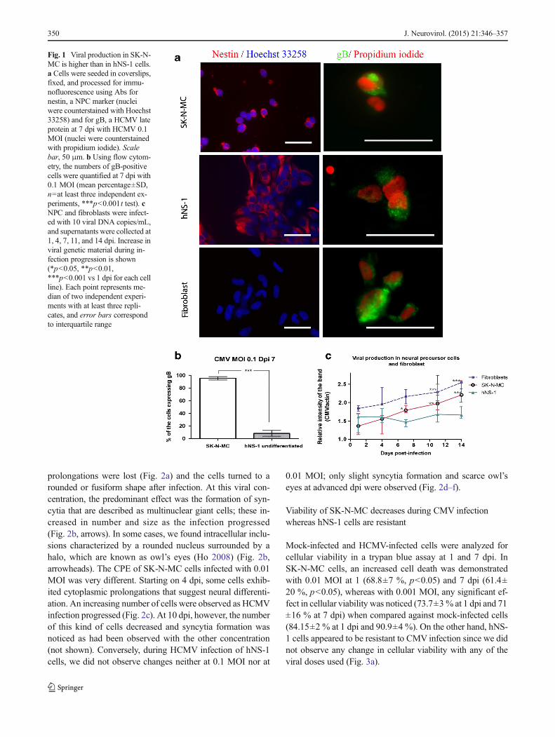
In order to demonstrate HCMV protein expression in NPCwith the AD169 HCMV strain, infected cells in coverslipswere stained for the presence of the late viral gB at 7 dpi. Inaddition, nestin antibody was used to show the NPC identity(Fig. 1a). Quantification of the percentage of gB-immunoreactive cells at 7 dpi showed that SK-N-MC cells(95.4±2.7 %, n=4) are much more susceptible to be infectedwith HCMV than hNS-1 cells (8.4±4.5 %, n=7) at an MOI of0.1 (Fig. 1b). As gB results fromHCMV late gene expression,this assay is an indirect verification of viral immediate-earlyand early gene expression, since viral replication was efficientand complete viral particle formation was inferred in thesecells. Additionally, we sought to investigate if these NPCscan produce viral particles, so we performed a comparativeassay using fibroblasts in which viral propagation in vitro nor-mally occurs (positive control). On the other hand, MDCKcells were used as a negative control of viral production basedon its canine origin. The media from such cells were collectedat different dpi, and viral production was quantified by anested PCR. Aswe expected, inMDCK cells, viral productionwas null (not shown), whereas SK-N-MC cells followed thesame behavior as fibroblasts. Even though viral productionwas slightly lower at 1 dpi, it was similar to that of fibroblastsin the last time points studied. On the contrary, viral produc-tion was almost null in hNS-1-infected cells, showing thatthese can be infected but they cannot actively replicate thisvirus (Fig. 1c).
CPE of human CMV in NPCs
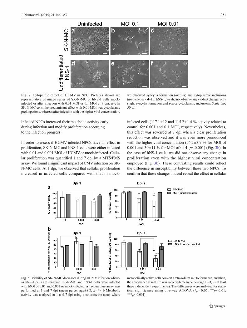
After HCMV infection in SK-N-MC cells, we observed animportant cellular loss and the development of viralconcentration-dependent CPE. In these cells, vacuole forma-tion and presence of granulation were not as evident as theywere seen during fibroblast infection (not shown). SK-N-MCcells infected with 0.1 MOI initially showed a loss of normalmorphology, since their multiple short-length cytoplasmic
J. Neurovirol. (2015) 21:346–357 349
prolongations were lost (Fig. 2a) and the cells turned to arounded or fusiform shape after infection. At this viral con-centration, the predominant effect was the formation of syn-cytia that are described as multinuclear giant cells; these in-creased in number and size as the infection progressed(Fig. 2b, arrows). In some cases, we found intracellular inclu-sions characterized by a rounded nucleus surrounded by ahalo, which are known as owl’s eyes (Ho 2008) (Fig. 2b,arrowheads). The CPE of SK-N-MC cells infected with 0.01MOI was very different. Starting on 4 dpi, some cells exhib-ited cytoplasmic prolongations that suggest neural differenti-ation. An increasing number of cells were observed as HCMVinfection progressed (Fig. 2c). At 10 dpi, however, the numberof this kind of cells decreased and syncytia formation wasnoticed as had been observed with the other concentration(not shown). Conversely, during HCMV infection of hNS-1cells, we did not observe changes neither at 0.1 MOI nor at
0.01 MOI; only slight syncytia formation and scarce owl’seyes at advanced dpi were observed (Fig. 2d–f).
Viability of SK-N-MC decreases during CMV infectionwhereas hNS-1 cells are resistant
Mock-infected and HCMV-infected cells were analyzed forcellular viability in a trypan blue assay at 1 and 7 dpi. InSK-N-MC cells, an increased cell death was demonstratedwith 0.01 MOI at 1 (68.8±7 %, p<0.05) and 7 dpi (61.4±20 %, p<0.05), whereas with 0.001 MOI, any significant ef-fect in cellular viability was noticed (73.7±3% at 1 dpi and 71±16 % at 7 dpi) when compared against mock-infected cells(84.15±2 % at 1 dpi and 90.9±4 %). On the other hand, hNS-1 cells appeared to be resistant to CMV infection since we didnot observe any change in cellular viability with any of theviral doses used (Fig. 3a).
Fig. 1 Viral production in SK-N-MC is higher than in hNS-1 cells.a Cells were seeded in coverslips,fixed, and processed for immu-nofluorescence using Abs fornestin, a NPC marker (nucleiwere counterstained with Hoechst33258) and for gB, a HCMV lateprotein at 7 dpi with HCMV 0.1MOI (nuclei were counterstainedwith propidium iodide). Scalebar, 50 μm. b Using flow cytom-etry, the numbers of gB-positivecells were quantified at 7 dpi with0.1 MOI (mean percentage±SD,n=at least three independent ex-periments, ***p<0.001t test). cNPC and fibroblasts were infect-ed with 10 viral DNA copies/mL,and supernatants were collected at1, 4, 7, 11, and 14 dpi. Increase inviral genetic material during in-fection progression is shown(*p<0.05, **p<0.01,***p<0.001 vs 1 dpi for each cellline). Each point represents me-dian of two independent experi-ments with at least three repli-cates, and error bars correspondto interquartile range
350 J. Neurovirol. (2015) 21:346–357
Infected NPCs increased their metabolic activity earlyduring infection and modify proliferation accordingto the infection progress
In order to assess if HCMV-infected NPCs have an effect inproliferation, SK-N-MC and hNS-1 cells were either infectedwith 0.01 and 0.001 MOI of HCMVor mock-infected. Cellu-lar proliferation was quantified 1 and 7 dpi by a MTS/PMSassay.We found a significant impact of CMVinfection on SK-N-MC cells. At 1 dpi, we observed that cellular proliferationincreased in infected cells compared with that in mock-
infected cells (117.1±12 and 115.2±1.4 % activity related tocontrol for 0.001 and 0.1 MOI, respectively). Nevertheless,this effect was reversed at 7 dpi when a clear proliferationreduction was observed and it was even more pronouncedwith the higher viral concentration (56.2±3.7 % for MOI of0.001 and 30±11 % for MOI of 0.01, p<0.001) (Fig. 3b). Inthe case of hNS-1 cells, we did not observe any change inproliferation even with the highest viral concentrationemployed (Fig. 3b). These contrasting results could reflectthe difference in susceptibility between these two NPCs. Toconfirm that these changes indeed reveal the effect in cellular
Fig. 3 Viability of SK-N-MC decreases during HCMV infection where-as hNS-1 cells are resistant. SK-N-MC and hNS-1 cells were infectedwith MOI of 0.01 and 0.001 or mock-infected. a Trypan blue assay wasperformed at 1 and 7 dpi (mean percentage±SD, n=4). b Metabolicactivity was analyzed at 1 and 7 dpi using a colorimetric assay where
metabolically active cells convert a tetrazolium salt to formazan, and then,the absorbance at 490 nmwas recorded (mean percentage±SD, n=at leastthree independent experiments). The differences were analyzed for statis-tical significance using one-way ANOVA (*p<0.05, **p<0.01,***p<0.001)
Fig. 2 Cytopathic effect of HCMV in NPC. Pictures shown arerepresentative of image series of SK-N-MC or hNS-1 cells mock-infected or after infection with 0.01 MOI or 0.1 MOI at 7 dpi. a–c InSK-N-MC cells, the predominant effect with 0.01 MOI was cytoplasmicprolongations, whereas after infection with the higher viral concentration,
we observed syncytia formation (arrows) and cytoplasmic inclusions(arrowheads). d–f In hNS-1, we did not observe any evident change, onlyslight syncytia formation and scarce cytoplasmic inclusions. Scale bar,50 μm
J. Neurovirol. (2015) 21:346–357 351

proliferation and not only in metabolic activity, we also eval-uated this parameter by CFSE dilution in cells infected withthe MOI of 0.01 and mock-infected cells. However, in infect-ed SK-N-MC cells, we only found an increase in proliferationat 4 dpi and any evident effect at 1 or 7 dpi. As the MTS/PMSassay showed, the metabolic activity was higher at 1 dpi andresults in cellular division at 4 dpi as it is revealed in CFSEassay; nevertheless, at 7 dpi, the cellular activity was dimin-ished, but it does not affect proliferation (Fig. 4a). On the otherhand, an enhanced proliferation was evidenced in hNS-1 cellsat 4 and 7 dpi, although no effect was observed in the MTS/PMS assay (Fig. 4b, c). This may be related to the lessersusceptibility of these cells to the infection because after4 dpi, the effects start to emerge.
Differentiation of hNS-1 cells makes them more susceptibleto HCMV infection
Since SK-N-MC has a more committed fate than hNS-1 cells,it may explain their distinct susceptibility, and given that hNS-1 cell line is multipotent, it can be induced to differentiate andanalyze if the differentiation degree modifies their capacity tobe infected and the effects of infection. Subsequently, hNS-1cells were induced to differentiate during 5 days, in order toproduce some neural and glial progenitors, and then, theywere infected with HCMV at MOI of 0.01 and 0.1 or mock-infected. At 7 dpi, it became evident that the CPE was morepronounced in the differentiated cells, and we noticed syncytiaformation, some cytoplasmatic inclusions, and vacuoles(Fig. 5a). Differentiated hNS-1 cells begin to express MAP-
2 (6.6±4.5 %) and GFAP (4.9±2.3 %), cellular markers ofneurons and astrocytes, respectively (Fig. 5b). At this timepoint, the cells were infected and the percentage of cells ex-pressing gB were higher (20.6±4.6 %, n=3, p<0.01) than inthe undifferentiated cells (Fig. 5c) with the MOI of 0.1 at7 dpi. In contrast with the undifferentiated hNS-1, the viabilityof the differentiated hNS-1 cells decreases with the progres-sion of HCMV infection even at 1 (72.5±15 %) and 7 dpi(61.5±6.7 %) (Fig. 5d). Whereas, the metabolic activity ofthese infected cells decreases at 1 dpi (74.1±6.5 %, p<0.05)and the effect is reverted at 7 dpi (Fig. 5e). Similarly, theseeffects were observed in the proliferation assay, and we founda decline in cellular division at 1 dpi (5±7.4 %), but this effectis lost according to the infection progress (Fig. 5f). Indeed, wedid not expect a strong effect in proliferation, since these cellswere differentiating and only a remaining percentage con-tinues in cell division.
Discussion
In congenital HCMV infection, the brain is the main infectiontarget and may cause significant damage to the developingbrain. It has been recently described that the most susceptiblecells inside the CNS are located in the ventricular andsubventricular zones containing NPCs (Cheeran et al. 2009;Tsutsui 2009). Infection of NPCs may cause malformationssuch as microcephaly and cerebellar hypoplasia and function-al disorders like mental retardation and epilepsy (Tsutsui
Fig. 4 Proliferation of NPCincreases slightly duringinfection. a SK-N-MC and bhNS-1 cells were incubated in thedark with CFSE 20 μM for10 min; then, the cells were in-fected with the MOI of 0.01 ormock-infected. NPCs were col-lected at 1, 4, and 7 dpi, and thequantification of CFSE chargewas analyzed by flow cytometry(median±IQR, n=4, **p<0.01,***p<0.001 Mann Whitney Utest). c Representative histogramsof hNS-1 cells indicating the per-centage of cellular division ateach time of infection for cellsinfected with MOI 0.01 andmock-infected. The median per-centage is indicated in each plot
352 J. Neurovirol. (2015) 21:346–357

Fig. 5 Differentiation of hNS-1cells makes them more suscepti-ble to HCMV infection. hNS-1cells were induced to differentiateduring 5 days; then, they wereinfected with HCMVat MOI of0.01 and 0.1 or mock-infected. aDifferentiated and undifferentiat-ed hNS-1 cells infected withHCMVat 7 dpi showed CPE thatwas more notable in differentiatedcells (cytoplasmic inclusionsshown by arrow heads and vacu-oles shown by asterisks). b Dif-ferentiated hNS-1 cells begin toexpress MAP-2 and GFAP, cellu-lar markers of neurons and astro-cytes, respectively. Scale bar,50 μm. c Expression of gB indifferentiated hNS-1 cells at 7 dpi(mean percentage±SD, n=at leastthree independent experiments,**p<0.01t test). d In contrastwith the undifferentiated hNS-1,the viability of the differentiatedhNS-1 cells decreases with theprogression of HCMV infection(mean percentage±SD, n=4, *p<0.05 t test). e By MTS/PMS assay, it was determined thatthe metabolic activity of these in-fected cells decreases at 1 dpi andthe effect is null at 7 dpi (meanpercentage±SD, n=5, *p<0.05 ttest). f In the proliferation assayusing CFSE, we found a declinein cellular division at 1 dpi, butthis effect is lost according to theinfection progress (median±IQR,n=4, *p<0.05 Mann Whitney Utest)
J. Neurovirol. (2015) 21:346–357 353
2009). The molecular mechanisms underlying pathogenic re-sponses to HCMV infection of NPCs are poorly understood.Increasing knowledge about how HCMV interferes with thenormal function of NPC is therefore of considerable interestbecause of NPC’s role during development. Here, we haveused an in vitro model based on human NPC infection toinvestigate these subjects. We employed the human cell linesSK-N-MC and hNS-1 as a source of NPC. The SK-N-MC is anaturally immortal cell line because it derives from a neuro-blastoma, while hNS-1 cell line was genetically perpetuatedby transfection of the avian oncogen v-myc and it is condi-tionally immortalized since it has a strict requirement for mi-togens EGF and FGF-2 (Villa et al. 2000). The SK-N-MC cellline is more committed to the neuronal fate than hNS-1 cellswhich are multipotent and can produce neurons and gliathrough differentiation (Villa et al. 2000). We confirmedHCMV infection in these cell lines and analyzed cellular via-bility and proliferation. In SK-N-MC cells, we found out thatlow viral doses stimulated proliferation, increased their meta-bolic activity, and induced differentiation; we also noticed thatCPE is dose-dependent in this cell line. In contrast, at the sameMOIs, hNS-1 cells could only be infected to a lower extent.Scarce evidence of the infection was obtained since the im-munoreactivity for gB of HCMV inside the cells was muchweaker than in SK-N-MC cells. In addition, no evidence ofcell death or modifications in metabolic activity were found,although the proliferation rate increased at late days after in-fection. Interestingly, after 5 days of differentiation, hNS-1cells were highly infected showing thus an increased suscep-tibility. The CPE was more pronounced, and viability, mito-chondrial activity, and proliferation decreased while a higherpercentage of cells were found infected. These results suggestthat neurotoxicity mechanisms associated to HCMV infectionmay be dependent on the differentiation stage of these NPCs.
The observation of syncytia formation after HCMV infec-tion in SK-N-MC with the MOI of 0.1 is in agreement withearlier reported findings (Luo et al. 2008). However, the tem-poral course of these changes was different, which could beattributed to HCMV doses. Syncytia formation has also beenreported in other cellular types, and it seems to be related to cellfusion induced by viral glycoproteins present in the plasmamembrane (Belec et al. 1990; van Den Pol et al. 1999; Kinzlerand Compton 2005). On the other hand, when we infected SK-N-MC with the MOI of 0.01, cellular prolongations were ob-served suggesting cellular differentiation to a neuron-like mor-phology, an unexpected finding since we did not add any otherstimulus that may account for these changes, and this normallydoes not occur in an spontaneous way (Higgins et al. 2009).This induction of differentiation is in agreement with the recentobservation of abnormal, spontaneous, and premature differen-tiation of NPC induced by HCMV (Odeberg et al. 2006; Luoet al. 2010). The virus concentrations that we used were muchlower than those in previous studies, what may explain the
delayed effect observed. In contrast with those reports, wemonitored the CPE in the cells for a more prolonged periodof time and observed syncytia formation as the infectionprogressed. Altogether, these observations suggest that cellulardifferentiation is stimulated in early stages of infection and thatcells tend to fuse forming syncytia as viral concentration in-creases or time after infection progresses. A possible explana-tion for this latter effect can be a distinct regulation of transcrip-tion factors such as SOX2 which is related to embryonic de-velopment and stem cell self-renewal and expression of somereceptors like EGFR as well as cytoskeleton components(Jafferji et al. 2009; Luo et al. 2010). Nevertheless, the rele-vance of HCMV-induced differentiation in the context of CNSdevelopment and finding why it is reversed when viral concen-tration increases are a matter for further investigation. In con-trast with our findings in SK-N-MC cells, previous reportsshowed a decrease in NPC differentiation to neurons and gliaafter HCMV infection (Odeberg et al. 2006, 2007). Indeed, theexperimental methods used in these studies were different, andsince we did not induce differentiation, it occurred spontane-ously. In addition, the virus concentrations employed werehigher in the studies of Odeberg et al. than the ones used inthe present work. In the case of hNS-1, we noticed a slight CPEwith some owl’s eyes and syncytia formation at advancedstages post-infection, indicating that these cells are less suscep-tible to HCMV infection which may be a consequence of theirfully undifferentiated state (hNS-1 is composed of neural stemcells).
Viral particle production was monitored by the viral pro-duction assay, through the increased presence of viral DNA inthe cell supernatants at different times post-infection. The lev-el of detection of viral particles in the cell lines used here wasconsistent with the results obtained using a different source ofNPC (Luo et al. 2008). However, it is dissimilar to those ofothers who have reported that NPCs are fully permissive to theinfection, but few viral particles are released (Odeberg et al.2006). We observed that viral production in SK-N-MC wassimilar to the one in fibroblasts but not detectable in hNS-1.This might reflect the variability of results obtained whenworking with different NPC cultures. On the one hand, SK-N-MC cells may resemble one cell type used (Luo et al. 2008)whereas hNS-1may bemore similar to the other one (Odeberget al. 2006). Also, when we studied gB expression, we foundthe majority of SK-N-MC cells being positive in immunocy-tochemistry; however, only a low percentage of hNS-1 cellswere positively stained for gB. This might reflect their lowsusceptibility to infection and may explain that viral produc-tion could not be detected. Since gB is a product of late viralgenes, its presence reflects the capacity of the virus and cellsfor viral replication.
HCMV infection induces cell death of SK-N-MC cells butnot of hNS-1 ones. Cell death induced by HCMV could be theresult of a necrotic or an apoptotic process since this assay
354 J. Neurovirol. (2015) 21:346–357

does not indicate the involved mechanism (Cheeran et al.2005; Odeberg et al. 2006). In addition, proliferation assaysshowed a strong reduction in SK-N-MC proliferation at 7 dpiwith both HCMV concentrations. However, paradoxically, at1 dpi, an increased metabolism was observed after HCMVinfection; this observation could also derive from an increasein cellular activity due to viral stimulation of macromoleculesynthesis during viral replication, rather than an actual in-crease in the number of cells. Indeed, in the proliferation assayusing CFSE, we only detected an increase in cellular divisionat 4 dpi. It is consistent with a previous report of no change inproliferation at 7 dpi using the MOI of 0.1 by the detection ofPCNA, a marker for proliferation; however, we showed aprevious contradictory induction of proliferation and metabol-ic activity (Odeberg et al. 2007). It is believed that virusesavoid cellular macromolecule production to elude immunesystem activation. However, in contrast with other viruses, ithas been observed that HCMV stimulates cellular DNA,RNA, and protein synthesis, perhaps because HCMVrequiresa protein that cannot be produced byHCMV (DeMarchi 1983;Griffiths and Grundy 1987). As expected, cellular metabolismdecreased at 7 dpi because although immediate-early proteinsstimulate cellular function, early proteins interrupt this process(Griffiths and Grundy 1987). In contrast, hNS-1 cells ap-peared more resistant even than fibroblast in this MTS/PMSassay and did not show any change in cellular viability, con-sistent with the lack of viral production, as evidenced by DNAdetection. Nevertheless, even though some changes in meta-bolic activity were detected, cellular proliferation increased at4 and 7 dpi. This may represent a delayed effect which may bea product of the low rate of infection of these cells. The re-duced susceptibility to HCMV infection may be a matter ofcellular differentiation since hNS-1 cells can be consideredmore undifferentiated cells than SK-N-MC ones. Of note,some reports have shown that embryonic stem cells are resis-tant to HCMV infection and that embryos acquire susceptibil-ity during development (Tsutsui 2009). In order to test thishypothesis, hNS-1 cells were differentiated for 5 days andthey were HCMV-infected. At this stage, some cells begin toexpress GFAP and MAP-2, cellular markers of astroglia andneurons, respectively. Interestingly, a more marked CPE wasevidenced in differentiated cells, and cytomegalic inclusions,syncytia formation, vacuoles, and granulations were clearer. Ahigher percentage of dead cells were found in the viabilityassay even at 1 and 7 dpi, and the positivity for gB increasedin comparison with nondifferentiated hNS-1 cells.
The increased susceptibility to infection after differentia-tion induction may be a consequence of changes in one ormore steps of the infection process, such as attachment, entry,trafficking, nuclear entry, and promoter activity (Kawasakiet al. 2011). It has been reported that the susceptibility ofmouse stem cells to CMV may be determined by the concen-tration of heparan sulfate, presence of β1-integrin and
vimentin, and nuclear pore density (Kawasaki et al. 2011). Itis possible that one of these factors may be related to theacquisition of susceptibility to HCMV in human cells, includ-ing hNS-1 cells, which will be the object of further investiga-tion. Interestingly, as the mouse embryonic stem cells are re-fractory to the infection, differentiated neurons also haveshown an apparent refractivity (Cheeran et al. 2009). Thiseffect has been suggested to be explained by the expressionof the transcription factor C/EBP β and its dominant negativeisoform which binds to the enhancer region and inhibits tran-scription from the HCMVMIEP (Cheeran et al. 2005). By thisreason, we assessed the role of differentiation in hNS-1 cells atan early stage when some progenitors begin to differentiateand not when the differentiated neurons can be found.
Because our main objective was to describe the earliesteffects of HCMV infection, we have used significantly lowerMOI than those employed in other reports (Cheeran et al.2005; Odeberg et al. 2006, 2007; Luo et al. 2008, 2010).Instead of showing the deepest impact of the infection, wedecided to monitor the progression in different time points,in order to follow slight effects. This scheme allows us toshow that during a congenital infection, even when a low viraldose reaches the CNS, it eventually may be able to produceeffects in the NPC.
The mechanisms that produce CNS damage during con-genital HCMV infection have not been clearly elucidated.However, recent efforts through in vivo and in vitro modelshave allowed a better understanding of the pathogenesis ofthis viral infection. It is well known that the major neurolog-ical damage occurs during intrauterine infection by HCMVatearly pregnancy stages. In addition, some brain localizationslike ventricular and subventricular zones are particularly im-portant; therefore, it is possible that NPCs play a substantialrole during neurological damage. Previous studies haveshown that CMV infection in NPC results in differentiationinhibition, reduced proliferation, cellular signaling interfer-ence, and cell death; together, these effects may produce mi-gration defects during brain development (Kawasaki andTsutsui 2003; Cheeran et al. 2005; Odeberg et al. 2006; Hoand van den Pol 2007). We developed an in vitro model usingtwo different NPCs promoting the use of these cell lines in-stead of primary cultures. Permissibility to infection and di-minished cellular viability were corroborated in the most dif-ferentiated cell line; however, in contrast with other studies,we found out that low viral concentrations induced cellularproliferation in a transitory way in addition to possible differ-entiation stimulation. Also, we verified that the permissibilityto HCMVinfection is closely related to cellular differentiationsince cells that more closely resemble neural stem cells wereless vulnerable to HCMV and they acquired susceptibilitythrough differentiation.
In conclusion, these experimental results can be interpretedin the context of the observed abnormalities during a
J. Neurovirol. (2015) 21:346–357 355
congenital infection. It is important to underline the impor-tance of NPC susceptibility to HCMV because infection ofthese cells may have a significant effect on fetal brain devel-opment. Thus, both cell death and spontaneous and prematuredifferentiation of NPC can affect the normal proliferation andmigration patterns of neurons during fetal development lead-ing to altered brain architecture and function. Therefore, inorder to elucidate the mechanisms and clinical consequencesof the observed effects, further investigations are required.
Acknowledgments This work was supported by grant fromCONACYT (CB16782 and #120452), PROMEP (103.5/10/7697), andFAI-UASLP (C12-FAI-03-62.62). Hilda Gonzalez and Claudia Salazarwere recipients of scholarships from CONACYT (CVU 265757 and265189).
Conflict of interests The authors declare that there is no conflict ofinterests regarding the publication of this paper.
References
Agol VI (2012) Cytopathic effects: virus-modulated manifestations ofinnate immunity? Trends Microbiol 20(12):570–576
Belec L, Mhiri C, Belghiti D, Geny C, Boudes P, Gray F (1990)Cytomegalovirus (CMV) and human immunodeficiency virus(HIV) co-infection, of multinucleated giant cells in acquired immu-nodeficiency syndrome (AIDS) encephalopathy. Arch Anat CytolPathol 38:189–197
Cheeran MC, Hu S, Ni HT, Sheng W, Palmquist JM, Peterson PK,Lokensgard JR (2005) Neural precursor cell susceptibility to humancytomegalovirus diverges along glial or neuronal differentiationpathways. J Neurosci Res 82:839–850
Cheeran MC, Lokensgard JR, Schleiss MR (2009) Neuropathogenesis ofcongenital cytomegalovirus infection: disease mechanisms andprospects for intervention. Clin Microbiol Rev 22:99–126,Table of Contents
DeMarchi JM (1983) Nature of the block in the expression of some earlyvirus genes in cells abortively infected with human cytomegalovi-rus. Virology 129:287–297
Griffiths PD, Grundy JE (1987) Molecular biology and immunology ofcytomegalovirus. Biochem J 241:313–324
Higgins S, Wong SH, Richner M, Rowe CL, Newgreen DF, Werther GA,Russo VC (2009) Fibroblast growth factor 2 reactivates G1 check-point in SK-N-MC cells via regulation of p21, inhibitor of differen-tiation genes (Id1-3), and epithelium-mesenchyme transition-likeevents. Endocrinology 150:4044–4055
Ho M (2008) The history of cytomegalovirus and its diseases. MedMicrobiol Immunol 197:65–73
Ho WS, van den Pol AN (2007) Bystander attenuation of neuronal andastrocyte intercellular communication by murine cytomegalovirusinfection of glia. J Virol 81:7286–7292
Jafferji I, Bain M, King C, Sinclair JH (2009) Inhibition of epidermalgrowth factor receptor (EGFR) expression by human cytomegalovi-rus correlates with an increase in the expression and binding ofWilms’ Tumour 1 protein to the EGFR promoter. J Gen Virol 90:1569–1574
Kawasaki H, Tsutsui Y (2003) Brain slice culture for analysis of devel-opmental brain disorders with special reference to congenital cyto-megalovirus infection. Congenit Anom (Kyoto) 43:105–113
Kawasaki H, Kosugi I, Arai Y, Iwashita T, Tsutsui Y (2011) Mouseembryonic stem cells inhibit murine cytomegalovirus infectionthrough a multi-step process. PLoS One 6:e17492
Kinzler ER, Compton T (2005) Characterization of human cytomegalo-virus glycoprotein-induced cell-cell fusion. J Virol 79:7827–7837
Köhler TH (1995) Quantitative RT-PCR. In: Quantitation of mRNA bypolymerase chain reaction (Springer-Verlag, ed), pp 71-80 BerlinHeidelberg
Luo MH, Schwartz PH, Fortunato EA (2008) Neonatal neural progenitorcells and their neuronal and glial cell derivatives are fully permissivefor human cytomegalovirus infection. J Virol 82:9994–10007
Luo MH, Hannemann H, Kulkarni AS, Schwartz PH, O’Dowd JM,Fortunato EA (2010) Human cytomegalovirus infection causes pre-mature and abnormal differentiation of human neural progenitorcells. J Virol 84:3528–3541
Martinez-Serrano A, Rubio FJ, Navarro B, Bueno C, Villa A (2001)Human neural stem and progenitor cells: in vitro and in vivo prop-erties, and potential for gene therapy and cell replacement in theCNS. Curr Gene Ther 1(3):279–299
Matsukage S, Kosugi I, Kawasaski H, Miura K, Kitani H, Tsutsui Y(2006) Mouse embryonic stem cells are not susceptible to cytomeg-alovirus but acquire susceptibility during differentiation. BirthDefects Res A Clin Mol Teratol 76:115–125
Monsivais-Urenda A, Noyola-Cherpitel D, Hernandez-Salinas A, Garcia-Sepulveda C, Romo N, Baranda L, Lopez-Botet M, Gonzalez-Amaro R (2010) Influence of human cytomegalovirus infection onthe NK cell receptor repertoire in children. Eur J Immunol 40:1418–1427
Navarro-Galve B, Villa A, Bueno C, Thompson L, Johansen J, Martinez-Serrano A (2005) Gene marking of human neural stem/precursorcells using green fluorescent proteins. J Gene Med 7(1):18–29
Noyola DE, Jimenez-Capdeville ME, Demmler-Harrison GJ (2010)Central nervous system disorders in infants with congenital cyto-megalovirus infection. Neurol Res 32:278–284
Odeberg J,Wolmer N, Falci S,WestgrenM, Seiger A, Soderberg-NauclerC (2006) Human cytomegalovirus inhibits neuronal differentiationand induces apoptosis in human neural precursor cells. J Virol 80:8929–8939
Odeberg J, Wolmer N, Falci S, Westgren M, Sundtrom E, Seiger A,Soderberg-Naucler C (2007) Late human cytomegalovirus(HCMV) proteins inhibit differentiation of human neural precursorcells into astrocytes. J Neurosci Res 85:583–593
Ornoy A (2007) Fetal effects of primary and non-primary cytomegalovi-rus infection in pregnancy: are we close to prevention? Isr MedAssoc J 9:398–401
Rubio FJ, Bueno C, Villa A, Navarro B, Martinez-Serrano A (2000)Genetically perpetuated human neural stem cells engraft and differ-entiate into the adult mammalian brain. Mol Cell Neurosci 16:1–13
Shinmura Y, Kosugi I, Aiba-Masago S, Baba S, Yong LR, Tsutsui Y(1997) Disordered migration and loss of virus-infected neuronalcells in developing mouse brains infected with murine cytomegalo-virus. Acta Neuropathol 93:551–557
Tsutsui Y (2009) Effects of cytomegalovirus infection on embryogenesisand brain development. Congenit Anom (Kyoto) 49:47–55
Tsutsui Y, Kawasaki H, Kosugi I (2002) Reactivation of latent cytomeg-alovirus infection in mouse brain cells detected after transfer to brainslice cultures. J Virol 76:7247–7254
Tsutsui Y, Kosugi I, Kawasaki H, Arai Y, Han GP, Li L, Kaneta M (2008)Roles of neural stem progenitor cells in cytomegalovirus infection ofthe brain in mouse models. Pathol Int 58:257–267
van Den Pol AN, Mocarski E, Saederup N, Vieira J, Meier TJ (1999)Cytomegalovirus cell tropism, replication, and gene transfer inbrain. J Neurosci 19:10948–10965
Villa A, Snyder EY, Vescovi A, Martinez-Serrano A (2000)Establishment and properties of a growth factor-dependent,
356 J. Neurovirol. (2015) 21:346–357

perpetual neural stem cell line from the human CNS. Exp Neurol161:67–84
Villa A, Rubio FJ, Navarro B, Bueno C, Martinez-Serrano A (2001)Human neural stem cells in vitro. A focus on their isolation andperpetuation. Biomed Pharmacother 55:91–95
Villa A, Navarro B, Martinez-Serrano A (2002) Genetic perpetu-ation of in vitro expanded human neural stem cells: cellularproperties and therapeutic potential. Brain Res Bull 57:789–794
Villa A, Navarro-Galve B, Bueno C, Franco S, Blasco MA, Martinez-Serrano A (2004) Long-term molecular and cellular stability of hu-man neural stem cell lines. Exp Cell Res 294:559–570
Wentworth BB, French L (1970) Plaque assay of cytomegalovirus strainsof human origin. Proc Soc Exp Biol Med 135:253–258
Woolf NK, Jaquish DV, Koehrn FJ (2007) Transplacental murine cyto-megalovirus infection in the brain of SCID mice. Virol J 4:26
Wynn KK, Khanna R (2006) Models of CMV infection. Drug DiscovToday 3:91–96
J. Neurovirol. (2015) 21:346–357 357