effects of antifibroblast antiserum on cells derived from
TRANSCRIPT

Portland State University Portland State University
PDXScholar PDXScholar
Dissertations and Theses Dissertations and Theses
7-29-1975
Effects of Antifibroblast Antiserum on Cells Derived Effects of Antifibroblast Antiserum on Cells Derived
from Fibroblast Outgrowth of Human Prostatic from Fibroblast Outgrowth of Human Prostatic
Tissues Tissues
Eva Shang-Lian King Portland State University
Follow this and additional works at: https://pdxscholar.library.pdx.edu/open_access_etds
Part of the Biology Commons
Let us know how access to this document benefits you.
Recommended Citation Recommended Citation King, Eva Shang-Lian, "Effects of Antifibroblast Antiserum on Cells Derived from Fibroblast Outgrowth of Human Prostatic Tissues" (1975). Dissertations and Theses. Paper 2380. https://doi.org/10.15760/etd.2377
This Thesis is brought to you for free and open access. It has been accepted for inclusion in Dissertations and Theses by an authorized administrator of PDXScholar. Please contact us if we can make this document more accessible: [email protected].

AN ABSTRACT OF THE THESIS OF Eva Shang-Lian King for the
Master of Science pres~nted 29 July 1975.
Title: Effects of Antifibroblast Antiserum on Cells
Derived from Fibroblast Outgrowth of Human
Prostatic Tissues
APPROVED BY MEMBERS OF THE THESIS COMMITTEE:
Thelma N. Fisher, Chairman ~
The purpose of this investigation was to provide pure
cultures of normal human prostatic epithelium free of fibro-
blasts in order to study malignant conversion by chemical
carcinogens. Normal epithelial cells were needed because
this was the cell type implicated in prostatic malignancies
of human subjects. Unfortunately fibroblasts grew faster
than epithelial cells so that cultures were always overgrown
with connective tissue elements. It was considered impor-
tant to find a method which would eliminate fibroblasts so
that normal epithelial cells could grow out in pure culture.

2
Fibroblast cultures we ·e obtained from human prostatic
tissues fed with Eagle's medium containing 20% fetal bovine
serum. To prepare antifibroblast antiserum rabbits were in
jected intravenously with human prostatic fibroblasts which
had been transferred through at least 14 tissue culture
passages. Complement fixation tests were used successfully
to measure reactions of rabbit antifibroblast antiserum with
prostatic fibroblasts. It was shown that in the rabbit anti
fibroblast antiserum reaction with complement and specific
antigen, fibroblasts were lysed while prostatic epithelium
was unaffected in mixed cell type cultures obtained from
explant outgrowth of prostatic tissues. Control tissue
cultures treated with normal rabbit serum remained unaffected
when rabbit antifibroblast antiserum diluted 1:2 and comple
ment diluted 1:5 were added to test systems.
Ammonium sulfate precipitation followed by DEAE cellu
lose column chromatography was used to isolate IgG from
rabbit antifibroblast antiserum and normal rabbit serum.
Only IgG purified from rabbit antifibroblast antiserum and
added in the presence of complement had the ability to de
stroy fibroblasts and not epithelial cells. IgG obtained
from normal rabbit serum had no effect.
When rabbit antifibroblast antiserum absorbed with H.
Ep. 2 cells and complement were added to cultures of mixed
cell types obtained from cellular outgrowth of human pro
static explants similar results were achieved: cells ex
hibiting fibroblast-like morphology were destroyed while

3
epithelial cells persisted and continued to multiply.
Thus fibroblasts were lyzed after treatment with
rabbit antifibroblast antiserum and complement. Epithelial
cells remained and proliferated in pure culture therby pro
viding a model tissue culture system of normal human pro
static epithelium which was available for group studies de
signed to measure initial events in malignant conversion
processes of prostatic tissue.

EFFECTS OF ANTIFIBROBI...AST ANTISERUM ON CELLS
DERIVED FROM FIBROBLAST OUTGROWTH OF
HUMAN PROSTATI~ TISSUES
by
EVA SHANG-LIAN KING
A thesis submitted in partial fulfillment of the requirements for the degree of
MASTER OF SCIENCE
in
BIOLOGY
Portland State University 1975

TO THE OFFICE OF GRADUATE STUDIES AND RESEARCH:
The members of .the Committee approve the thesis of
Eva Shang-Lian King presented 29 July 1975.
Thelma N. Fisher, Chairman
GOr<fonii
APPROVED:
t of Biology
~avidT. Clcii'k> Dean of Graduate Studies and Research

ACKNOWLEDGEMENTS
Give thanks to God for blessing me here . I am
grateful to my family for letting me have a chance to
study here . I am really thankful for the instruction
of my advisor , Dr . Thelma N. Fisher and committee members
Drs . Earl Fisher, Jr . and Gordon Kilgour . I greatly
appreciate Mrs . Donna Ann Fisher for her instruction in
tissue culture maintenance techniques .

TABLE OF CONTENTS
PAGE
ACKNOWLEDGEMENTS . . • • . • . . . . . • . • . • • • . • • • . • • • • • • • • • . • . • • • . iii
LIST OF TABLES......................................... vii
LIST OF FIGURES . ..................•.........•.. · . · · · · · · ix
INTRODUCTION. . . . . . . . . . . . . . . . . . . . . . . . . . . . . • . • . . . . . • . • . . . 1
MATERIALS AND .METHODS . . . . . . . . . . . . . . • . . . . . • . . . . . • . . . • • . . 3
Cell Culttires.................................... 3
Procedures for Isolation, Feeding and Harvesting Cell Cultures and the Media Used.............. 3
Procedures for Rabbit Inoculation and Serum Harvest. . . . . . . . . . . . . . . . . . . . . . . . . . . . . . . . . . . . . . . 6
Complement. . . . . . . . . . . . . . . . . . . . . . . . . . . . . . . . . . . . . . . 6
Complement Fixation Tests Employing Rabbit Antifibroblas t Antiserum and Fibroblast Cells. . . . . . . . . . . . . . . . . . . . . . . . . . . . . . . . . . . . . . . . . 7
Indicator System............................ 7
Test System................................. 7
Procedure for Measuring the Effect of Non-HeatInacti vated Rabbit Antifibroblast Antiserum on Fibroblasts and H. Ep. 2 Cells............. 9
Determination of the Most Effective Concentration of Heat Inactivated Rabbit.Antifibroblast Antiserum and Complement Necessary to Lyse Fibroblasts Observed in Mixed Fibroblast and EpitheliaJ Cellular Outgrowth from Human Prostatic Explants............................ 9
Purification of IgG Obtained from Normal Rabbit Serum and Rabbit Antifibroblast Antiserum..... 10
Ammonium Sulfate Precipitation (2).......... 10
DEAE Cellulose Column Chromatography (2).... 12
Complement Fixation Tests Using IgG Obtained from Rabbit Antifibroblast Antiserum and Fibroblast Cells....... . . . . . . . . . . . . . . . . . . . . . . . . . . . . . . . . . . 14

Indicator System ..•...•.••..•.•.•.•••.•.•.•.
Test System ................................ .
A Comparison of Activies of IgG Obtained from Normal Rabbit Serum and from Rabbit Antifibroblast Antiserum on Human Prostatic Cultures Consisting of Mixed Fibroblast and Epithelial Cell Types ................................... .
An Attempt to Remove Antibodies of Epithelial Cells from Non-Heat-Inactivated Rabbit Antifibroblast Antiserum by Treating
v
PAGE
14
14
14
Antiserum with H. Ep. 2 Cells................. 15
RESULTS. . . . . . . . . . . . . . . . . . . . . . . . . . . . . . . . . . . . . . . . . . . . . . . . 17
Complement Fixation Tests with Rabbit Antifibroblast Antiserum and Fibroblast Cell Cultures Derived from Human Prostatic Ti ·s sues . • . • . . . . . • . • . . . . . . • • . . . . . . • . • • . . . • . . . • • 1 7
Visual Effects of Non-Heat-Inactivated Rabbit Antifibroblast Antiserum on Fibroblasts and H. Ep. 2 Cells................................ 17
Determination of Effective Concentrations of Heat Inactivated Rabbit Antifibroblast Antiserum and Complement as Used to Lyse Fibroblasts Present in Cultures of Mixed Cell Types Derived from Human Prostatic Explants... 19
Purification of IgG Recovered from Rabbit Antifibroblast Antiserum and Normal Rabbit Serum. . . . . . . . . . . . . . . . . . . . . . . . . . . . . . . . . . . . . . . . . 24
Purification of Gamma Globulin Isolated from Rabbit Antifibroblast Antiserum by DEAE Cellulose Column Chromatography............... 43
Analytical Ultracentrifugation Information Used to Characterize Gamma Globulin Isolated from Rabbit Antifibroblast Antiserum by DEAE Cellulose Column Chromatography............... 46
Purification of Gamma Globulin Isolated from Normal Rabbit Serum by DEAE Cellulose Column Chromatography. . . . . . . . . . . . . . . . . . . . . . . . . . . . . . . . 49

Analytical Ultracentrifugation Data Used to Characterize Gamma Globulin Isolated from Normal Rabbit Serum by DEAE Cellulose
vi
PAGE
Column Chromatography......................... 49
Complement Fixation Tests Using IgG Isolated from Rabbit Antifibroblast Antiserum as Antibody and Human Prostatic Fibroblasts as Antigen. . . . . . . . . . . . . . . . . . . . . . . . . . . . . . . . . . . . 53
Indicator System............................ 53
Test System..... . . . . . . . . . . . . . . . . . . . . . . . . . . . . 54
Effects of IgG Obtained from Rabbit Antifibroblast Antiserum and Normal Rabbit Serum on mixed Human Prostatic Cultures Composed of Fibroblasts and Epithelial Cells.............. 54
Effects of Non-Heat Inactivated Rabbit Antifibroblast Antiserum Absorbed with H. Ep. 2 Cells on Human Prostatic Cultures Composed of Fibro-blasts and Epithelial Cells................... 54
DISCUSSION. . . . . . . . . . . . . . . . . . . . . . . . . . . . . . . . . . . . . . . . . . . . . 62
SU.MMARY. . • • . • • • • • . • • • • • • • • • • • • • • • • • • • • • • • • • • • • • • • • • • • • • 6 5
BIBLIOGRAPHY. . . . . . . . . . • . • . . . . . . . • . . . . . . . . . . . . . • . . . • . . . . 67

LIST OF TABLES
TABLE PAGE
I Formula of Hanks' Balanced Salt Solution,
(lOX) Stock Solution......................... 4
II Constituents of a Modified Eagle's Medium as
Used to Cultivate Human Prostatic Tissues.... 5
III Lowry (Folin-Ciocalteau) Method for Protein
Estimation................................... 13
IV An Estimation of the Amount of Rabbit Anti
fibroblast Antiserum required to fix
Complement in the Presence of Varying
Dilutions of Fibroblast Cell Suspensions..... 18
V Results of Control Systems and Different
Dilutions of Rabbit Antifibroblast
Antiserum with Complement on Cultures
Composed of Mixed Fibroblasts and
Epithelial Cells Derived from Human
Pros ta tic Tissues............................ 23
VI Data Required for the Calculation of -the
Sedimentation Coefficient of IgG Isolated
from Rabbit Antifibroblast Antiserum......... 47
VII Data Required for the Calculation of the
_Sedimentation Coefficient of IgG Isolated
from Normal Rabbit Serum..................... 51

viii
TABLE PAGE
VIII A Complement Fixation Reaction with IgG
Isolated from Rabbit Antifibroblast
Antiserum and Suspensions of Human
Prostatic Fibroblasts Serving as Test
Reactants......... . . . . . . . . . . . . . . . . . . . . . . . . . . . 55

FIGURE
1
LIST OF FIGURES
A comparison of fibroblast and H. Ep. 2
cell cultures before and after treatment
with antifibroblast antiserum and
PAGE
complement................................. 20
2 Photographic demonstration of selective
effects of antifibroblast antiserum and
complement on cultures of mixed cell types
arising from explants of human prostatic
tissues... . . . . . . . . . . . . . . . . . . . . . . . . . . . . . . . . . 25
3 Standard curve as prepared by the Lowry
method for protein estimation and used to
calculate protein content of rabbit anti
fibroblast antiserum and that of fractions
derived from it............................ 42
4 Standard curve as prepared by the Lowry
method for protein estimation and used to
calculate protein content of normal rabbit
serum and that of fractions derived
from it . . . . . . . . . . . . . . . . . . . . . . . . . . . . . . . . . . . . 4 4
5 Tracing of a partially purified peak of
gaITL.~a globulin isolated from rabbit anti
fibroblast antiserum by DEAE cellulose
column chromatography .........•............ 45

FIGURE
6 Plot of log10x versus t as used for the cal
culation of a sedimentation coefficient
PAGE
for gamma globulin (IgG) isolated from
antifibroblast antiserum. . . . . . . . . • . . . . . . . . . 48
7 Tracing of a partially purified peak of
gamma globulin isolated from normal rabbit
serum by DEAE cellulose column chromato-
graphy. . . . . . . . . . . . . . . . . . . . . . . . . . . . . . . . . . . . . 5 o
8 Plot of log10x versus t as used for the cal-
culation of a sedimentation coefficient
for gamma globulin (IgG) isolated from
normal rabbit serum ..........•............. 52
9 Observation of lytic effects by purified
IgG isolated from rabbit antifibroblast
antiserum on fibroblasts derived from
human prostatic explant outgrowth
composed of fibroblasts and epithelial
cells. . . . . . . . . . . . . . . . . . . . . . . . . . . . . . . . . . . . . . 56
10 Effect of complement and non-heat-
inac ti va ted, H. Ep. 2-cell absorbed,
rabbit antifibroblast antiserum on cellu
lar outgrowth of mixed cell types obtained
from human prostatic explant tissue ........ 60
x
INTRODUCTION
Fibroblasts derived from human fetal tissue (21, 27,
15), human skin (8, 25), chick embryo (26), and murine
embryo (12), etc., have been studied extensively by others
in order to determine hexosaminidase activity (10), to
assess the effects of pH (8) and amino acid composition (15)
on growth, to measure dihydrotestosterone formation (25), to
study adhesion properties (16, 17), and to isolate glyco
proteins from cell surfaces (26). Fibroblasts derived from
numerous human prostatic tissues were tested in our study.
For the preparation of antiserum many investigators
immunized rabbits via the intravenous route (18, 24), by
intramuscular and intravenous injections (7, 15), by intra
dermal inoculation (22, 9, 18), by alternating intraperi
toneal with subcutaneous type injections (5), or by foot pad
inoculations (4, 6). Intravenous procedures via marginal
ear veins of rabbits provided a satisfactory method for
preparing antifibroblast antiserum used in our research.
Because it was necessary to prepare pure cultures of
normal prostatic epithelium free of fibroblasts in order to
study malignant conversion by chemical carcinogens an
effective method to elJ.minete fibroblast o utgrowth from
prostatic explants was required. Recovery of epithelium
was essential since this cell type was thought to ·undergo
malignant transformation in human subjects. Fibroblasts
multiplied more rapidly than epithelial cells thereby over
growing epithelial cultures. Our purpose was to destroy

2
fibroblasts so that normal epithelial cells would prolifer
ate freely in pure culture. Subsequently, a cancer research
group hoped to study initial events in the malignant con
version process by treating known normal human prostatic
epithelial cells with chemical carcinogens. It was obvious
to all investigators involved in this field of research
that the first enormous problem was to control overgrowth of
epithelium by fibroblasts. To make antifibroblast anti
serum in rabbits via intravenous inoculation and to test
the effects of antibody in the presence of complement on
fibroblasts derived from human prostatic tissue, was the
purpose of this study. We hoped to demonstrate that such a
system destroyed cells which resembled fibroblasts, thereby
allowing typical epithelial cell types to persist and
flourish.

MATERIALS AND METHODS
Cell Cultures
Fibroblast cultures were isolated from human prostatic
tissues removed surgically from patients at Providence
Hospital, Portland, Oregon. H. Ep. 2 cells, human epithe
lium obtained orig inally from a pat~ent with carcinoma of
the larynx, were carried in continuous culture in our labo
ratory through 280 transfers. Often mixed cultures of
fibroblast and epithelial cell types were obtained from
prostatic explant outgrowth. These cultures were subjected
to antifibroblast antiserum treatment also.
Procedures for Isolation, Feeding and Harvesting Cell
Cultures and the Media Used
Hanks' Balanced Salt Solution containing antibiotics
(penicillin and streptomycin) was used to wash prostatic
tissues 3 times. Tissues were cut into small 2 mm2 pieces
when held in the third wash solution. Explants were placed
in small plastic petri dishes (60 x 15 mm) and allowed to
remain at room temperature for approximately 1 hr., after
which time Eagle's medium containing 30% fetal bovine serum
(Flow Laboratories) was added. Cultures were held in a 95%
air-5% C02 water-saturated environment at 37°. Hanks'
Balanced Salt Solution and Eagle's medium as in Table I and
Table II respectively were prepared as indicated in the
formulae.
Fibroblast cultures were fed 3-5 times per week with

TABLE I
FORMULA OF HANKS' BALANCED SALT SOLUTION, (lOX) STOCK SOLUTION
Materials
Solution A
NaCl
KCl
Amount
80 gms
4 gms
2 gms
The above reagents were dissolved in about 300 ml distilled
water.
Solution B
KH2P04
Na2HP04·2H20
Glucose
0.6 gms
o.6 gms
10 gms
4
These compounds were dissolved in approximately 300 ml dis-
tilled water.
Solution C
1.4 gms
Calcium chloride was dissolved in 200 ml distilled water and
added to Solutions A and B.
Solution D
Phenol Red 0.2%, 100 ml
The final volume was brought to 1,000 ml and the solution
was sterilized by Sietz filtration. Ten times concentrated
Hanks' solution was stored at 4° until dilute Hanks' pre-
paration was required.

TABLE II
CONSTITUENTS OF A MODIFIED EAGLE'S MEDIUM AS USED TO CULTIVATE HUMAN PROSTATIC TISSUES
Materials
Hanks' Balanced Salt Solution
Fetal Bovine Serum
(inactivated at 56° for 30 min)
Non Essential Amino Acids (lOOx, Flow
Laboratories)
Vitamins (lOOx, Flow Laboratories)
Amino Acids (lOOx, Flow Laboratories)
Penicillin [Stock Solution: 20,000 units
per ml (Pfizer Laboratories)]
Streptomycin [Stock Solution: 20 mg per
ml (Pfizer Laboratories)]
5% NaHC03
Putrescine (1 mg/ml, Sigma Laboratories)
Glutamine (lOOx, Flow Laboratories)
Amount
85 ml
20 ml or 30 ml
1 ml
1 ml
1 ml
1 ml
1 ml
2 ml
0.25 ml
1 ml
5

6
Eagle's medium containing 20% fet~l bovine serum. Trypsin
(0.2~%, Difeo) was used to subculture fibroblasts. Tissues
we~e cultivated in 30 ml plastic tissue culture flasks or in
200 ml milk dilution bottles.
Human prostatic fibroblasts were subcultured for at
least 14 passages before they were used experimentally. Cells
(approximately 2.35 x 10⁶ per ml) were washed twice with 10
ml each of saline, and harvested in a small amount of saline.
These cells were frozen and thawed 3 times then inactivated
in a water bath at 56° for 30 min. This crude preparation
served as antigen which was used to immunize rabbits.
Procedures for Rabbit Inoculation and Serum Harvest
The prepared antigen (0.1 ml per inoculation) was in
jected intravenously into ear marginal veins of rabbits, 3
times per week for 3 weeks. A booster inoculation was given
one week later. After a few days, rabbits were bled from
the heart in order to obtain 15 ml blood from each animal.
Rabbit blood was refrigerated overnight at 4° in a centrifuge
tube. Clots were rimmed carefully 24 hr. later and blood
was centrifuged twice in order to obtain large serum yields
free of red blood cells. Serum was inactivated at 56° for
30 min.
Complement
The complement employed throughout consisted of lyophi
lized guinea pig serum obtained commercially (Flow

7
Laboratories). Just before use complement was dissolved, in
the cold, in 7 ml diluent of which the latter contained 6%
sodium acetate and 2% boric acid.
Complement Fixation Tests Employing Rabbit Antifibroblast
Antiserum and Fibroblast Cells
Indicator System. The titration of hemolysin as des
cribed by Burrell (1) was achieved by using 0.5 ml hemolysin
dilutions ranging from 1:1,000 ~o 1:16,000 with 0.3 ml of a
solution made up of coIT.plement diluted 1:30, 0.5 ml of a 2%
suspension of sheep erythrocytes and 1.7 ml of 0.01 mg%MgS04
dissolved in physiological saline (Mag saline).
According to Burrell (1) complement was titrated using
the following components: two units of an adequate dilution
of hemolysin contained in 0.5 ml, 0.1 to 0.5 ml of a fresh
solution made up of complement diluted 1:30, 0.5 ml of a 2%
suspension of sheep erythrocytes and Mag saline added to pro
vide a final volume of 2.5 ml per tube were included in the
titration.
Test System. It was considered necessary to determine
adequate concentrations of antigen and antibody required to
observe lysis of fibroblasts. A master titration of fibro
blasts was achieved by first preparing 2 doubling dilutions
starting with a 1:5 dilution of suspended fibroblasts. Rab
bit antifibroblast antiserum, heat inactivated 30 min at 56°,
was diluted 1:5 and 1:20. Four rows of 13 x 100 mm test

8
tubes were arranged so that there were 3 tubes per row.
Inocula were pipetted as follows: tube l of each row---0.5
ml of 1:5 inoculum, tube 2 of each row---0.5 ml of 1:10 ino
culum, tube 3 of each row---0.5 ml of 1:20 inoculum. Rabbit
antifibroblast antiserum was pipetted as follows: two-tenths
ml of undilute serum was added to each tube in the first row;
0.2 ml of serum diluted 1:5 was added to each tube in the
second row; 0.2 ml of serum diluted 1:10 was added to each
tube in the third row and 0.2 ml of serum was diluted 1:20
and added to each tube in the fourth row. The following
controls were included: rabbit antifibroblast antiserum con
trol (0.2 ml of undiluted serum and 0.5 ml of Mag saline),
fibroblast antigen control (0.5 ml of fibroblasts diluted
1:5 and 0.2 ml of Mag saline), and a hemolytic system control
(0.7 ml of Mag saline). Preparations were incubated 15 min
at room temperature, one ml of complement (2 units per ml)
was added to each tube. Preparations were held in the re
frigerator for 15-18 hr. Tubes were placed in a 37° water
bath for 10 min after which time 0.5 ml of hemolysin (2
units) and 0.5 ml of a 2% suspension of sheep erythrocytes
were added to each tube. A sheep erythrocytes control con
taining 0.5 ml of sheep red blood cells suspended in 2.2 ml
of Mag saline was included in each titration. Contents of
each tube were mixed well and preparations were incubated at
37° for 30 min.
Procedure for Measuring the Effect of Non-Heat-Inactivated
Rabbit Antifibroblast Antiserum on Fibroblasts and H. Ep. 2
Cells
9
Initially growth fluids of fibroblast and H. Ep. 2 cell
cultures were removed and appropriate photographs were made.
Following photography cultures were fed with Eagle's medium
containing 20% fetal bovine serum. To these preparations 0.1
ml of undiluted complement, and 0.1 ml of undiluted rabbit
antifibroblast antiserum were added to fibroblast and H. Ep.
2 cell cultures. Treated cultures were incubated in a 95%
air-5% C02 water-saturated environment at 37°. The next day
cultures were examined for evidence of cell lysis and photo
graphs were made to record the results.
Determination of the Most Effective Concentration of Heat
Inactivated Rabbit Antifibroblast Antiserum and Comolement
Necessary to Lyse Fibroblasts Observed in Mixed Fibroblast
and Epithelial Cellular Outgrowth from Human Prostatic
Explants
Growth fluids were removed and cultures were photo
graphed initially. Then cultures were fed with Eagle's
medium containing 20% fetal bovine serum. Undiluted rabbit
antifibroblast antiserum (0.2 ml) was added to cultures dis
playing mixed cell type (fibroblasts and epithelial cells)
outgrowth. Other cultures showing outgrowth of mixed cell
types were treated with a 0.2 ml rabbit antifibroblast anti
serum diluted 1:2. Test cultures were held in a 95% air-5%

10
co 2 water-saturated envirofiment at 37° for 15 min, after
which time 1 ml of different dilutions of complement (un
diluted, 1:5, 1:10, 1:20, 1:30, 1:45; 1:60, 1:75, 1:150) was
added to appropriate cultures. Controls consisted of un
treated cultures, cultures treated with 0.2 ml undiluted rab
bit antifibroblast antiserum, cultures treated with 1 ml
complement diluted 1:20, cultures treated with 0.2 ml un
diluted heat inactivated normal rabbit serum and cultures
treated with 1 ml of complement diluted 1:5. Control and test
systems were incubated in a 95% air-5% C02 water-saturated
environmentat 37° overnight then examined and photographed
the following day.
Purification of IgG Obtained from Normal Rabbit Serum and
Rabbit Antifibroblast Antiserum
Twenty-two ml aliquots of normal rabbit serum and rab
bit antifibroblast antiserum were processed in order to ob
tain IgG fractions from each serum sample. The procedure
followed for purification of IgG was as follows:
Ammonium Sulfate Precipitation (2)
1. Saturated (NH4)2S04 (11 ml) was adjusted to
approximately pH 7.8 by the addition of 2 N NaOH. This ad
justment was made just prior to precipitation of gamma
globulin in order to prevent ammonia release.
2. With constant stirring the 11 ml of saturated
(NH4) 2S04 solution, pH 7.8, was added slowly, in dropwise

11
fashion to 22 ml of serum. This procedure salted out those
components which were insoluble in a 33.3% (NH4) 2so 4 solu
tion.
3. Upon completion of (NH4)2S04 addition, suspensions
were stirred for 2 to 3 hr.
4. Subsequently, suspensions were centrifuged at room
temperature for 30 min at 1,465 x g.
5. Precipitates were dissolved in enough saline to
restore volumes of solutions to those of original serum
samples.
6. Gamma globulin fractions were purified by a second
and a third precipitation. For the second precipitation
steps l through 5 were repeated. For the third precipita
tion, steps 1 through 4 were followed.
7. Precipitates from third precipitations were dis
solved in borate-buffered saline to a final volume half that,
or less than that of original serum samples.
8. Ammonium sulfate was removed from precipitates by
dialyzing against borate-buffered s~line for several days at
4°. Dialyzates were changed mornings and evenings, and at
these intervals a few drops of 5% BaCl2 and 3.7% HCl were
used to check for sulfate ions.
9. After dialysis was completed, solutions were re
moved from dialysis tubing then centrifuged 35 min 9,750 x g,
at 4°.
10. The Lowry (Folin-Ciocalteu) method for protein

12
estimation was followed by adding standard solutions, saline,
sample, reagent A, and reagent B in amounts and sequences as
shown in Table III.
Absorbance of reaction mixtures were observed at 500 run
as measured against a distilled water blank. A standard
curve was prepared and protein concentrations of samples were
read directly from the calibration curve.
DEAE Cellulose Column Chromatography ( 2)
1. A DEAE cellulose column equilibrated in 0.01 M
phosphate buffer, pH 7.5 was prepared.
2. Crude gamma globulin fractions were dialyzed
against 0.01 M phosphate buffer, pH 7.5, for 24 hr at 4°.
3. Gamma globulin fractions were removed from
dialysis bags, centrifuged for 25 min at 1,085 x g, and sedi
ments were discarded.
4. Protein content of supernatant fluids was esti
mated by the Lowry Method.
5. Nine ml samples of crude gamma globulin were
fractionated by DEAE cellulose column chromatography.
6. Samples which displayed the highest protein con
tent were pooled and lyophilized.
7. Dried protein powders were dissolved in adequate
amounts of distilled water in order to bring the final pro
tein concentration to 4-5 mg/ml. Protein solutions were
dialyzed against 0.01 M phosphate buffer, pH 7.5, for 2 days
at 4°.

Tube Number:
mg or protein. atandard/tube:
ml standard (0.5 rng/ml)
ml saline
ml sample
ml Reagent A
•l Re~.lgent B
TABLE III
LOWRY (FOLIN·CIOCALTEAU) METHOD FOR PROTEIN ESTIMATION
Reagent Blank
l
,40
2
• 01
.02
.38
3
.02
• 04
.36
2.0 (ALL TUBES)
4
.03
• 06
,34
Standards
5
.05
.10
,30
6
.10
.20
.20
7 8
.15 .20
.30
.10
.110
mix, let stand at room temperature tor 10 min
, 20 (ALL TUBES)
Sample
9-1• 9-2•
• i. 0 .110
mix, let stand at room temperature tor 30 min, read at 500 nm
Reagent blanks, standard dilutions and sample3 were tested in duplicate.
• indicated that 0,1 ml sample solution was diluted in 0.9 ml saline (9-l) and 0.1 ml 9-l sample
solution was diluted in 0.9 ml saline (9-2).
Reagent ~
10
.110
...... VJ

8. After completing dialysis procedures, solutions
were centrifuged 25 min at 1,085 x g. Sedimentation co
efficients of protein solutions were determined following
analytical centrifugation in a Model E ultracentrifuge.
Complement Fixation Tests Using IgG Obtained from Rabbit
Antifibroblast Antiserum and Fibroblast Cells _
14
Indicator System. Methods for titrating hemolysin and
complement (1) as described earlier were followed.
Test System. It was necessary to determine appropriate
concentrations of reactants for this system. IgG obtained
from rabbit antifibroblast antiserum was diluted 1:5 and 1:10
and added to suspensions of human prostatic fibroblasts
diluted 1:5, and 1:10 in the presence of complement (2 units
per ml), as indicated earlier.
A Comparison of Activities of IgG Obtained from Normal
Rabbit Serum and from Rabbit Antifibroblast Antiserum on
Human Prostatic Cultures Consisting_ of Mixed Fibroblast
and Epithelial Cell Types
Growth fluids were removed and initial photographs were
made of all cell cultures. Cultures were fed with Eagle's
medium containing 20% fetal bovine serum to which 0.2 ml of
undiluted IgG purified from rabbit antifibroblast antiserum
was added. Test systems were held in a 95% air-5% C02 water
saturated environment at 37° for 15 min. One ml of
15
complement diluted 1:5 was added to each culture. A con
trol cell culture received 0.2 ml undiluted IgG obtained from
normal rabbit serum and 1 ml of a solution made up of comple
ment diluted 1:5. Preparations were examined and photo
graphed.
An Attempt to Remove Antibodies of Epithelial Cells from Nor.
Heat-Inactivated Rabbit Antifibroblast Antiserum by Treating
Antiserum with H. Ep. 2 Cells
For absorption studies 2-200 ml bottle cultures of H.
Ep. 2 cells approximately 5 days old were used for these
experiments. Growth fluids were removed and 3.5 ml rabbit
antifibroblast antiserum were added to each culture. Cells
were scraped from glass by means of rubber spatulas. Pre
parations were held 30 min in a 95% air-5% co 2 water-satu
rated environment at 37°. Cells were centrifuged 15 min at
3,020 x g. Supernatant fluids were retained while sedi
mented fractions were discarded. Presumably this super
natant fluid contained antifibroblast antiserum from which
epithelial components of H. Ep. 2 cells had been removed by
the absorption process. To test the effect of absorbed anti
fibroblast antiserum on cell cultures, growth fluids were
removed from cultures of prostatic explants displaying mixed
cell type outgrowth. Preparations were photographed then fed
with Eagle's medium containing 20% fetal bovine serum and
0.2 ml undiluted but absorbed antifibroblast antiserum.
After 15 min incubation at 37° 1 ml of a solution made up of

16
complement diluted 1:5 was added to preparations. Test
systems were cultivated overnight in a 95% air-5% co 2 water
saturated environment at 37e. The following day cultures
were examined. and photogr aphed in order to document observed
effects.

RESULTS
Complement Fixation Tests with Rabbit Antifibroblast
Antiserum and Fibroblast Cell Cultures Derived from Human
Prostatic Tis s ues
It was determined by titration that 0.5 ml of a 1:500
dilution of hemolysin contained 2 units of complement. This
amount proved to be the highest hemolysin C.ilution which gave
c.omplete sheep red blood cell hemolysis. Complement was
titrated also. Two full units of a solution of complement
diluted 1:75 and contained in 1 ml was found to be the least
amount of complement which provided complete lysis of sheep
red blood cells.
The test system was carried out as described in Table
IV.
When a 1:5 dilution of suspended fibroblast cells and
a 1:10 dilution of rabbit antifibroblast antiserum were in
cluded in test systems> the greatest amount of complement
fixation was observed. From such observations it was evident
that antibody specific for fibroblasts was present in the
rabbit antiserum.
Visual Effects of Non-Heat-Inactivated Rabbit Antifibroblast
Antiserum on Fibroblasts and H. Ep. 2 Cells
After treating human prostatic fibroblasts with rabbit
antifibroblast antiserum in the presence of complement>
growth of fibroblasts was limited and cell lysis occurred so
that fibroblasts disappeared from cultures in 24 to 72 hr.

TABLE IV
AN ESTIMATION OF THE AMOUNT OF RABBIT ANTIFIBROBLAST ANTISERUM REQUIRED TO FIX COMPLEMENT IN THE
PRESENCE OF VARYING DILUTIONS OF FIBROBLAST CELL SUSPENSIONS
Rabbit Antifibroblast Fibroblast Cell Suspension
Antiserum Dilutions 1:5 1:10
Undiluted + +
l: 5 ++ ' +
1:10 ++++ +++
1:20 ++
Symbols: ·
+ = no hemolysis
.... = hemolysis
·Dilutions
1:20
+
+
I-' en

19
Rabbit antifibroblast antiserum and complement had no visible
effect on H. Ep. 2 cells. The latter were known to be malig
nant epitheloid cell types. These findings suggested that
rabbit antifibroblast antiserum added in the presence of
complement brought about a specific reaction with fibroblasts
which caused them to lyse; however, such activity was not
demonstrable for H. Ep. 2 cells. Photographs displaying
these effects were documented and recorded in Figure 1.
Determination of Effective Concentrations of Heat Inactivated
Rabbit Antifibroblast Antiserum and Complement as Used to
Lyse Fibroblasts Present in Cultures of Mixed Cell Types
Derived from Human Prostatic Explants
As seen in Table V, 1 ml of complement diluted 1:5 and
0.2 ml of antifibroblast antiserum diluted 1:2 provided the
best conditions for fibroblast cell lysis when these reagents
were used. Antifibroblast antiserum represented pooled
serum collected from 6 immunized rabbits as obtained early
during these studies.
Depending on controls, rabbit antifibroblast antiserum
alone or complement alone or normal rabbit serum with comple
ment gave negative results throughout. It was apparent that
it was necessary to add complement in the reaction mixture
in order to achieve lysis of fibroblasts when antigen reacted
with antibody. Also, it appeared that rabbit antifibroblast
antiserum was specific for cells demonstrating fibroblast
like morphology but not for cells resembling epithelium.

20
Figure 1. A comparison of fibroblast and H. Ep. 2
cell cultures before and after treatment with antifibroblast
antiserum and complement.
Fibroblast cultures (12th passage) were obtained
initially from human prostatic tissue while H. Ep. 2 cells
(epithelial like) were isolated originally from a patient
known to have carcinoma of the larynx. Photographs, taken
with a Zeiss Standard W. L. Research Microscope equipped
with Polaroid attachments, were made just before treatment
of cultures (Figs. la and le) with antifibroblast antiserum
(O.l ml of undiluted preparation) and complement (0.1 ml of
undiluted preparation). Twenty-five hours following treat
ment with antifibroblast antiserum plus complement, photo
graphs were recorded again (Figs. lb and ld). Photographs
shown in Figs. la and lb were made of fibroblast cultures
before and after antifibroblast treatment respectively while
photographs displayed in Figs. le and ld were made of H. Ep.
2 cells before and after antifibroblast treatment respec
tively. Magnification x205.


TABLE V
RESUV.t'S o:P CONTROL SYS'l'EMS AND DIFFERENT DILUTIONS OF RABBI'!' ANTIFIBROBLAST ANTISERUM WITH GOMPLEMEN'l' ON CULTURES COMPOSED OF MIXED FIBROBLASTS AND
EPI'l'HELIAL CELLS DERIVED FROM HUMAN PROSTATIC TISSUES
~----·-~~~~-
1.rES STEMS CONTROLS SYSTEMS .. -. ... implement Dilutions
Rab bit _,_l __ Ant1fibroblaat . Rabbit Normal Rabbit .
Antiserum Cells Antifibroblast Complement Serum + Dilutions U~d ed 1:5 1:10 1:20 Alone Antiserum Alone Alone Complement
'
Undiluted + + ++ +++ - - - -Diluted 1:2 +++ ++++ ++ ++ - - - -
..
+ to ++++ indl1.:at<>d degree of c1:l.l lysis when 0, 2 ml of undiluted or rabbit antifibroblast antiserum
diluted 1: 2 and 1 ml cumplement •.!oncentrations ranging from undiluted to l: 20 were added to the mixed
cultures of fibroblasts and epithelium obtained from human prostatic tissues. Higher dilutions of
reactants were not made because antiserum and cell cultures were limiting.
--
ru w

24
Many epithelial cells remained in cultures following treat
ment of mixed cell type outgrowth with antifibroblast anti
serum and complement. These results were observed and re
corded in photographs shown in Figure 2.
After many experimental assays using different dilu
tions of rabbit antifibroblast antiserum and complement, it
was concluded that undiluted rabbit antifibroblast antiserum
plus complement diluted 1:150 or rabbit antifibroblast anti
serum diluted 1:5 plus complement diluted 1:75 provided very
little evidence of fibroblast cell lysis. Because quantities
of mixed cell cultures consisting of fibroblasts and epithe
lial cells and the supply of rabbit antifibroblast antiserum
were limiting other dilutions of reactants could not be
carried out in these studies. Of the various dilutions of
rabbit antiribroblast antiserum andcomplement, rabbit
antifibroblast antiserum diluted 1:2 s~pplemented wit_h_
· complement diluted 1:5 provided the best and m9st consistent
effects in terms of promoting lysis of fibroblasts.
Purification of IgG Recovered from Rabbit Antifibroblast
Antiserum and Normal Rabbit Serum
By reference to the standard curve for protein esti
mation, prepared according to the Lowry method and shown in
Figure 3, the protein content of the reconstituted fraction
of antifibroblast antiserum after (NH4) 2so4 precipitation
was calculated to be 0.071 mg protein per 0.4 ml reaction
mixture or 0.18 mg per ml. The protein concentration of

25
Figure 2. Photographic demonstration of selective
effects of antifibroblast antiserum and complement on
cultures of mixed cell types arisjng from explants of human
prostatic tissues.
In these studies va.rious concentrations of antifibro
blast antiserum and complement were employed in order to
find the most effective reagent proportions necessary to
achieve fibroblast destruction. Procedures for culture
treatment, including control systems, were recorded in the
Materials and Methods section. All cells were grown in
Eagle's medium containing 20% fetal bovine serum. Mixed
cell type cultures were composed of cells exhibiting epithe
lial and fibroblast-like morphologies. All antifibroblast
antiserum was heat inactivated at 56° for 30 min. Magnifi
cation xl60.
Identification of photographs: All photographs de
signated with the letter "a" as from (la) through (14a) were
those cultures composed of mixed cell types which were
photographed at zero time before treament. Photographs de
signated with the letter "b" were made of preparations
treated in the various ways listed below.
(lb) Untreated control culture of mixed cell types
photographed 23 hr after zero time.
(2b) Mixed cell type culture photographed 23 hr
after treatment with undiluted antifibroblast antiserum only:

26
an antiserum control.
(3b) Mixed cell type culture photographed 23 hr after
treatment with complement only, diluted 1:20: a complement
control.
(4b) Mixed cell type culture photographed 23 hr after
treatment with undiluted normal rabbit serum and complement
diluted 1:5: a normal rabbit serum control.
(5b) Mixed cell type culture photographed 23 hr after
treatement with undiluted antifibroblast antiserum and un
diluted complement.
(6b) Mixed cell type culture photographed 28 hr after
treatment with undiluted antifibroblast antiserum and comple
ment diluted 1:5.
(7b) Mixed cell type culture photographed 28 hr after
treatment with undiluted antifibroblast antiserum and comple
ment diluted 1:10.
(8b) Mixed cell type culture photographed 28 hr after
treatment with undiluted antifibroblast antiserum and comple
ment diluted 1:20.
(9b) Mixed cell type culture photographed 28 hr after
treatment with undiluted antifibroblast antiserum and comple
ment diluted 1:30.
(lOb) Mixed cell ~ype culture photographed 23 hr after
treatment with undiluted antifibroblast antiserum and comple
ment diluted 1:45.
(llb) Mixed cell type culture photographed 23 hr after
27
treatment with undiluted antifibroblast antiserum and comple-
ment diluted 1:60.
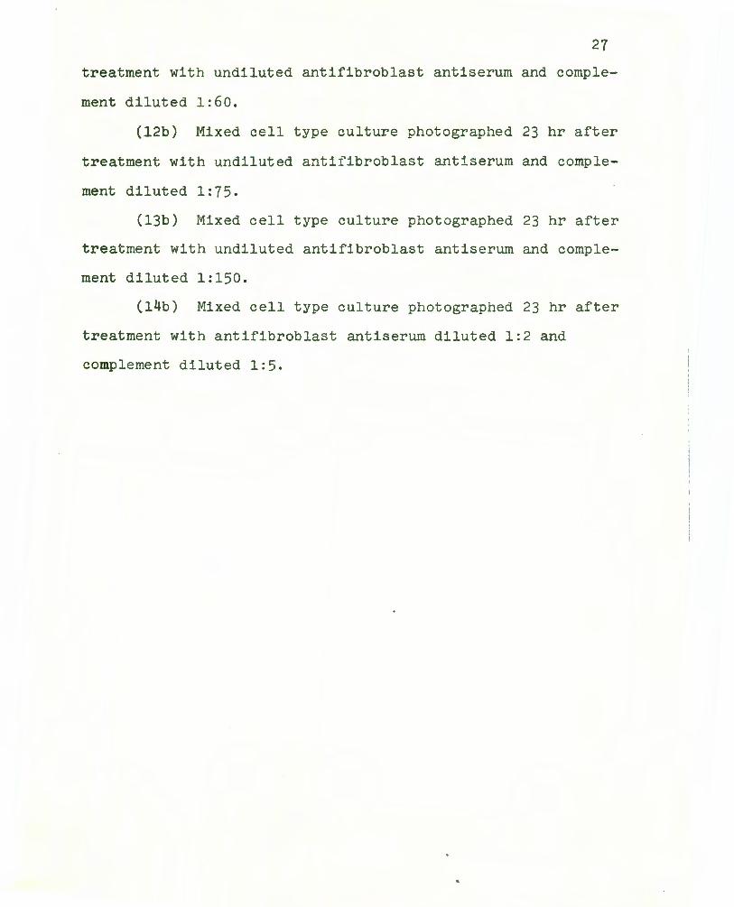
(12b) Mixed cell type culture photographed 23 hr after
treatment with undiluted antifibroblast antiserum and comple
ment diluted 1:75.
(13b) Mixed cell type culture photographed 23 hr after
treatment with undiluted antifibroblast antiserum and comple
ment diluted 1:150.
(14b) Mixed cell type culture photographed 23 hr after
treatment with antifibroblast antiserum diluted 1:2 and
complement diluted 1:5.


42
.60 o/
. 55 /~
/ . 50 /
a / . 45 j/
. 40 / ,.-...
/ s ~
0 . 35 / 0 L[\ 0 / '---'
Q) . 30 ;y C)
~
/ cd .0 . 25 H
/ 0 rn ..0 ~ . 20
0
. 15
. 10
. 05
0 .01 . 02 . 03 . 05 . 07 . 1 0 .15 . 20
mg of protein
Figure 3. Standard curve as prepared by the Lowry
method for protein estimation and used to calculate protein
content of rabbit antifibroblast antiserum and t hat of
fractions derived from it .

purified IgG recovered from antifibroblast antiserum was
found to be 18 mg per ml.
43
As calculated from an appropriate Lowry standard curve
for protein estimation shown in Figure 4 the protein con
centration of the reconstituted fraction of normal rabbit
serum after (NH4 ) 2S04 precipitation used in these experiments
was found to be 0.066 mg per 0.4 ml reaction mixture or 0.17
mg per ml. Purified IgG isolated from normal rabbit serum
contained 17 mg protein per ml.
Purification of Gamma Globulin Isolated from Rabbit
Antifibroblast Antiserum by DEAE Cellulose Column
Chromatography
As seen in Figure 5, a single peak representing parti
ally purified gamma globulin was separated from rabbit anti
fibroblast antiserum and removed from the antiserum by DEAE
cellulose column chromatography. High ultraviolet (UV) ab
sorbing (280 nm) spectrophotometric readings suggested that
significant amounts of protein were present in samples re
covered from the peak area displayed in Figure 5. The
average protein concentration of eluted solutions showing UV
absorption at 280 nm was calculated to be 0.29 mg per ml.

44
. 65 i
. 60 I /
55 I
.50 a/ /
. 45 / I
;---... . 40 s I s:: 0 0 'f. / 0 . 35 U\
I Q)
I C) . 30 s:: crj
I ..0 ~ 0 .25 I (/)
..0 c:i:
. 20
. 15
. 10 0
. 0
. 01 . 02 . 03 . 05 . 06 . 10 . 15 .20
mg of protein
Figure 4. Standard curve as prepared by the Lowry
method for protein estimation and used to calculate protein
content of normal rabbit serum and that of fractions derived
from it .

45
1.0
0.9
0.8
0 . 7 ,--....
s s:: 0 o . 6 ro (\J ..._.,
Q) 0.5 C)
s:: cd .0
0.4 H 0 ff)
.0 ~
0.3
0.2
O.l
0 5 10 15 20 25 30 35 40 45 50
Tube Number (10 ml/tube)
Figure 5. Tracing of a partially purified peak of
gamma globulin isolated from rabbit antifibroblast antiserum
by DEAE cellulose column chromatography .
Data recorded in Figure 5 represented UV absorption
values (read at 280 nm) vs volume. DEAE cellulose column
was equilibrated in 0 . 01 M phosphate buffer , pH 7.5 .

46
Analytical Ultracentrifugation Information Used to
Characterize Gamma Globulin Isolated from Rabbit Antifibro-
blast Antiserum by DEAE Cellulose Column Chromatography
Partially purified gawma globulin recovered from anti-
fibroblast antiserum was centrifuged in the Model E Spinco
Analytical Ultracentrifuge as stated in the Materials and
Methods section in order to obtain sufficient information
for sedimentation coefficient calculations. Photographs
were made at 8 min intervals and scale readings were meas-
ured in cm as recorded in Table VI. Log10x values were
determined by using appropriate log10 tables.
Using log10 values vs time in min, as recorded in
Table VI it was possible to determine the slope of the line.
A plot of the slope was recorded in Figure 6.
1 dr 2.303 dlogx Equation: s = x = ( ) (3)
dt 60 w2 dt'
dlogx =
0.8338 - 0.7850
80 6 -4 = .1 x 10
dt'
2.303 = 1.116 x lo-9 (3) at 56,000 rpm,
60 w2
The sedimentation coefficient of IgG obtained from
normal rabbit serum was reported by others to be approxi-
mately 6.6. However such measurements were made under
totally standard conditions. Observations for the rabbit
antifibroblast antiserum IgG fraction were not made under

Frame Number
1-1
1-2
1-3
1-4
1-5
2-1
2-2
2-3
2.4
2-5
TABLE VI
DATA REQUIRED FOR THE CALCULATION OF THE SEDIMENTATION COEFFICIENT OF IgG ISOLATED FROM RABBIT
ANTIFIBROBLAST ANTISERUM
Time (interval in min) Scale Reading (cm)
8 6.095
8 6.17
- -----8 6.32
8 6.42
8 6.57
8 6,67
- -----8 6.72
8 6.82
Log10x -
0.7850
0.7903
------0.8007
0.8075
0.8176
0.8241
------0.8274
0.8338
.i::~

48
1. 0
0 . 9
0 0 0 0 0 D.8 0 0 0
Slope = 6 . 1 x lo - 4 0 . 7
0 . 6
>< 0 rl 0 . 5
bO 0
.....:i
0. 4
0.3
0 . 2
0 . 1
0 8 16 24 32 40 48 56 64 72 80
Time in Minutes
Figure 6 . Plot of log10x versus t as used for the
calcula tion of a s edime nt a t i on coefficient for gamma globulin
(IgG) isolated from antifibroblast antiserum .
49
rigidly controlled conditions which probably accounted for
the small difference in S values. From these data it was
evident that the gamma globulin fraction obtained from rab
bit antifibroblast antiserum was IgG.
Purification of Gamma Globulin Isolated from Normal Rabbit
Serum by DEAE Cellulose Column Chromatography
Again a single peak of partially purified gamma glob
ulin, as shown in Figure 7, was obtained from normal rabbit
serum. High UV absorbing values when measured spectrophoto
metrically at 280 nm were obtained. The average concentra
tion of UV absorbing substance in peak solutions was cal
culated to be 0.27 mg per ml.
Analytical Ultracentrifugation Data Used to Characterize
Gamma Globulin Isolated from Normal Rabbit Serum by DEAE
Cellulose Column Chromatography
As reported for similiar studies with IgG obtained from
rabbit antifibroblast antiserum partially purified gamma
globulin recovered from normal rabbit serum was centrifuged
in the Model E Spinco Analytical Ultracentrifuge. Photo
graphs were made at 8 min intervals and scale readings were
measured in cm as recorded in Table VII. Log10x values were
determined by using appropriate log10 tables.
Using log10 values vs time in min, as recorded in
Table VII the slope of the line was determined. A plot of
the slope was recorded in Figure 8 and the S value was

50
1.1
1.0
0.9
o. 8
......... 0 . 7 s s:::
0 o.6 ro (\J ..._,
(l) 0 . 5 C)
s::: cd .a H 0.4 0 U)
.0 ~
0.3
0.2
0 . 1
0
5 lO 15 20 25 30 35 40 45 50
Tube Number (10 m/tube)
Figure 7. Tracing of a partially purified peak of
gamma globulin isolated from normal rabbit serum by DEAE
cellulose column chromatography .
Data recorded in Figure 7 represented UV absorption
values (read at 280 nm) vs volume. DEAE cellulose column was
equilibrated in 0 . 01 M phosphate buffer , pH 7.5.

Frame Number
1-1
1-2
1-3
1-4
1-5
2-1
2-2
2-3
2-4
2-5
TABLE VII
DATA REQUIRED FOR THE CALCULATION OF THE SEDIMENTATION COEFFICIENT OF IgG I SOLATED FROM NORMAL
RABBIT SERUM
Time (interval in min) Scale Reading (cm)
8 5.995
8 6.08
8 6.15
8 6.22
8 6.295
8 6.38
8 6. 117
8 6.52
8 6.62
8 6.695
Log10x
-0.7778
0.7839
0.7889
0.7938
0.7990
0.8048
0.8109
0.8142
0.8209
0.8257
\JI .....

52
1. 0
0 . 9
0 0 o-o 0 0. 8 0- 0 0 0 0
0. 7
0. 6 K
0 rl 0. 5 bO
0 rl
0. 4
0. 3
0.2
0 . 1
8 16 24 32 40 48 56 64 72 80
Time in Minutes
Figure 8 . Plot of log10x versus t as used for the
calculation of a sedimentation coefficient for gamma
globulin (IgG) isolated from normal rabbit serum .

53
determined as follows:
dlog x _ 4 = 5.99 x 10 dt'
2.303 d log x ( )
s = 60 w2 dt 1
= (1.116 x 10⁻⁹) (5.99 x lo-4)
= 6.68 x lo-13
= 6.68 s
The data indicated taht the purified gamma globulin
fraction obtained from normal rabbit serum was IgG, as was
the fraction characterized from rabbit antifibroblast anti-
serum.
Complement Fixation Tests Using IgG Isolated from Rabbit
Antifibroblast Antiserum as Antibody and Human Prostatic
Fibroblasts as Antigen
Indicator System. Two units of hemolysin contained in
0.5 ml of a 1:500 dilution gave complete hemolysis of sheep
red blood cells as used in these assays. Two full units of
complement diluted 1:75 and contained in a 1 ml volume was
the least amount of complement which could be used in the
reaction mixture which gave complete lysis of sheep erythro-
cytes.

54
Test System. The complement fixation test with IgG
and fibroblasts was carried out by the same procedure as
that described for antifibroblast antiserQm and fibroblasts
using concentrations of constituents of the indicator system
as detailed above. Results were recorded in Table VIII.
In the experiment reported in Table VIII, undiluted
fibroblast antigen suspension and undiluted IgG purified
from rabbit antifibroblast antiserum provided the greatest
amount of complement fixation. It was evident that IgG
possessed the function of antibody which could react with
fibroblast cells to promote cell lysis.
Effects of IgG Obtained from Rabbit Antifibroblast Antiserum
and Normal Rabbit Serum on Mixed Human Prostatic Cultures
Composed of Fibroblasts and Epithelial Cells
As shown in Figure 9 only IgG isolated from rabbit
antifibroblast antiserum had the ability to destroy fibro
blasts, while epithelial cells persisted. IgG from normal
rabbit serum like untreated normal rabbit serum did not have
any effect on fibroblasts or epithelium. Thus formation of
IgG in antifibroblast antiserum must have occurred in spec~
ific response to the fibroblast antigen.
Effects of Non-Heat Inactivated Rabbit Antifibroblast Anti
serum Absorbed with H. Ep. 2 Cells on Human Prostatic
Cultures Composed of Fibroblasts and Epithelial Cells
Photographs taken before and after treatment as shown

IgG Dilutions
Undiluted
1:5
1:10
Symbols:
TABLE VIII
A COMPLEMENT FIXATION REACTION WITH IgG ISOLATED FROM RABBIT ANTIFIBROBLAST ANTISERUM AND SUSPENSIONS OF HUMAN PROSTATIC
FIBROBLASTS SERVING AS TEST SYSTEM REACTANTS
Fibroblast Antigen Dilutions
Undiluted 1:5
++++ +++
neg.
neg.
+ to ++++: Degree of complement fixed in the test syst em neg. : Negative or no complement fixed in test system
1:10
++
neg.
neg.
----: Not done at these dilutions because of insufficient amount of fibroblast antigen preparation.
Undiluted fibroblast antigen contained 2.35 x 10⁶ cells per ml. Undiluted IgG antibody
contained 0.29 mg protein per ml.
Ul Ul

56
Figure 9. Observation of lytic effects by purified
IgG isolated from rabbit antifibroblast antiserum on fibro
blasts derived from human prostatic explant outgrowth com
posed of fibroblasts and epithelial cells.
Mixed cultures of fibroblasts and epithelial cells de
rived from explant outgrowth of human prostatic tissues were
photographed (Magnification xl60) at zero time before treat
ment and 23 hr following treatment. In the interim prepara
tions were cultivated in Eagle's medium containing 20% fetal
bovine serum at 37° in a 95% air-5% C02 water-saturated en
vironment. Photographs la and 2a represent both preparations
before treatment.
lb: This culture was allowed to react with comple
ment and IgG isolated from rabbit antifibroblast antiserum
according to routine procedures described in Materials and
Methods. The preparation was photographed 23 hr after
treatement.
2b: The culture was allowed to react with complement
and IgG isolated from normal rabbit serum, according to
procedures described. The preparation was photographed 23
hr after treatment.


59
in Figure 10 indicated that rabbit antifibroblast antiserum
absorbed earlier with H. Ep. 2 cells (derived from human
epidermoid carcinoma of the larynx) destroyed cells which
resembled fibroblasts morphologically but had no effect on
cells displaying characteristics of epithelium. Antiserum
appeared to remain specific for fibroblasts. It was of con
siderable interest to speculate as to whether or not there
were certain different receptor site components on cell sur
faces which accounted for the different antigenic responses
of fibroblasts and epithelial-like-cells.

60
Figure 10. Effect of complement and non-heat-inacti
vated, H. Ep. 2-cell absorbed, rabbit antifibroblast anti
serum on cellular outgrowth of mixed cell types obtained from
human prostatic explant tissue.
The preparation was photographed (Magnification xl60)
before (la) and 23 hr after (lb) treatment with reactants.
Cells were cultivated in Eagle's medium containing 20% fetal
bovine serum.
lb: Photograph of culture 23 hr after treatment with
complement diluted 1:5 and undiluted-H. Ep. 2-cell-absorbed
rabbit antifibroblast antiserum.

DISCUSSION
Experimental results presented in this thesis showed
that rabbit antifibroblast antiserum in the presence of
complement had the capacity to destroy human prostatic cells
resembling fibroblasts morphologically, while cells display
ing characteristic epithelial-like morphology were un
affected. Control systems containing normal rabbit serum
supplemented with complement or rabbit antifibroblast anti
serum alone had no effect on fibroblasts and epithelium de
rived from prostatic tissues. These data suggested that
antifibroblast antiserum was specific for the fibroblast
antigen. The observation that fibroblasts and epithelial
cells might have certain differences in antigenicity was
extended further when it was shown that antifibroblast anti
serum absorbed in the presence of H. Ep. 2 epithelial-like
cells lysed fibroblasts only and not epithelial cells found
growing in mixed cell type cultures derived from human
prostate tissue. Concentrations of rabbit antifibroblast
antiserum diluted 1:2 in the presence of complement diluted
1:5 provided the most effective reaction mixture for ob
serving lysis of fibroblasts. When fibroblasts and epithe
lial cells found in cultures of prostatic explant outgrowth
were treated with antibody plus complement, fibroblasts
lysed and disappeared, while epithelium remained intact and
multiplied.
In general, ammonium sulfate precipitation followed by
DEAE cellulose column chromatography were found to be very

useful methods for isolation of IgG from various serum
sources (20, 23); these methods were frequently used in
enzyme purification studies also (19, 14). IgG isolated
from rabbit antifibroblast antiserum caused fibroblast lysis.
Concentration studies were not carried out to determine
whether purifie d IgG or crude antifibroblast antiserum was
most effective in promoting fibroblast lysis. It was not
known if IgG was partially denatured in the purification pro
cess. Such questions require further investigation.
According to data reported in this thesis, quantities
of IgG purified from rabbit antifibroblast antiserum and
normal rabbit serum were very close in value, while sedi
mentation coefficients varied slightly. Variability must be
due to the fact that standard conditions were not maintained
when such determinations were made. In regard to IgG acti
vity, perhaps responses obtained with IgG as compared with
crude rabbit antifibroblast antiserum differed as a result
of partial i nactivation of IgG protein during purification
processes of gamma globulin. Our findings demonstrated the
usefulness of the complement fixation test in observing lysis
of fibroblasts when subjected to antifibroblast antiserum.
The test was shown t o be rapid and specific. The mechanism
of complement f ixation has to do with the fact that an an'ti
body-complement complex binds to surface receptors of anti
gens (ll) after which the reaction, such as lysis, occurs.
Complement binding sites are located on heavy chains of

64
antibody molecules found just before and after the s-s
regions of antibody. It is known that IgG combines with
complement in this way al5o. Thus fibroblasts are lyzed and
disappear after treatment of cells with antifibroblast anti
serum and complement while epithelial cells are maintained
and continue to proliferate in pure culture. This procedure
allows investigators working in the field of cancer carcino
genesis to use such epithelial cultures as model systems in
order to study initial events of the malignant conversion
process.

SUMMARY
Antiflbroblast antiserum was prepared in rabbits by
immunizing 6 animals with human prostatic fibroblasts culti
vated and transferred through 14 passages in tissue culture.
By routin complement fixation procedures complement was fixed
by antifibroblast antiserum complexed with s uspensions of
fibroblast cells. Likewise, when rabbit antifibroblast an~i
serum and comp l ement were added to cultures of mixed cell
types obtained from cellular outgrowth of prostatic explant
tissues, fibroblasts were destroyed but epithelial cells re
mained intact in cultur8 and continued to multiply. Normal .
rabbit serum used for controls had no effect on tissue
cultures.
Ammonium sulfate precipitation followed by DEAE cellu
lose column chromatography was adopted to purify IgG from
rabbit antifibroblast antiserum and normal rabbit serum.
IgG obtained from rabbit antifibroblast antiserum and supple
mented with complement eliminated growth of fibroblasts but
not epithelium. IgG obtained from normal rabbit serum and
tested in the presence of complement had no effect on
cultures of prostatic fibroblasts and epithelial cells.
Following rabbit antifibroblast antiserum absorption
with H. Ep. 2 cells, absorbed antiserum in the presence of
complement retained the capacity to lyse fibroblasts but not
epithelial cells.
Thus fibroblasts were destroyed and after treatment
with .rabbit antifibroblast antiserum and complement,

66
epithelial cells persisted intact and multiplied in pure
culture. As a result of these research efforts a model
system was provided so that other investigators could study
initial events in the conversion of normal prostatic epithe
lium to malignant cells.
BIBLIOGRAPHY
1. Burrell, Robert. 1974. Experimental Immunology, 4th ed. Burgess Publishing Company. p. 44-47.
2. Campbell, Dan. H., Fustine S . Garvey, Natalie E. Cremer, and Dieter H. Sussdorf. 1970. Methods in Immunology, 2nd ed. W. A. Ben jamin, Inc., New York. p. 76-79, 45-48, 189-191, 193-197, 104-110.
3. Chervenka, C. H. 1969. A Manual of Methods for the
4.
Analytical Ul t racentrifuge. Sp inco Division of Beckman Instruments Inc., Palo Alto, California. p. 26-29.
Cho-Chung, Yoon Sang, and Pietro M. Gullino. Mammary Tumor Regression. J. Biol. Chern. 4743-4749.
1973. 248(13):
5. Cohen, Max H., Allan D. Bernstein, and Paul H. Levine. 1974. Hematological and Serological Effects of Rauscher Leukemia Virus and Epstine-Barr Virus on Irnmunosuppressed Newborn Submhuman Primates. Oncology. 29:353-363.
6. Ennis, Francis. 1973. Host Defense Mechanisms against Herpes Simplex Virus. I. Control of Infection in vitro by Sensitized Spleen Cells and Antibody. Infect. Immunity. 7(6):898-904.
7. Epstin, M. A., and B. G. Achong. 1968. Specific Irnmunofluorescence Test for the Herpes-Type EB Virus of Burkitt Lymphoblasts, Authenticated by Electron Microscopy. J. Nat. Cancer Inst. 40:593-607.
8. Froehlich, Jeffrey E., and Tassos P . . Anastassiades.
9.
1974. Role of pH in Fibroblast Proliferation. J. Cell. Physiol. 84(2):253-260.
Garfinkle, B., and B. R. McAuslan. formation of Cultured Mammalian Herpes Simplex Virus Subtypes 1 Nat. Acad. Sci. 71(1):220-224.
1974. TransCells by Viable and 2. Proc.
10. Hickman, Scot , Larry J. Shapiro, and Elizabeth F. Neufeld. 1974. A Recognition Marker Required ·for Uptake of a Lysosomal Enzyme by Cultured Fibroblasts. Biochem. Biophys. Res. Comm. 57(1):55-61.

11.
12.
13.
14.
15.
16.
17.
18.
19.
20.
21.
Irie Kenji, Reiko F. Irie, and Donald L. Morton. 1§74. Evidence for in vivo Reaction of Antibody and Complement to Surface Antigens of Human Cancer Cells. Science. 186:454-456.
Kim, Kwang Soo, and Richard I. Ca.rp. 1973. Effect of Proteolytic Enzymes on the Infectivity of a Number of Herpesviruses. J. Infect. Dis. 128(6): 788-790.
Leslie, R. G. Q., and S. Cohen. 1974. Cytophilic Activity of IgG2 from Sera of Unimrnunized Guineapigs. Immunology. 27:577-587.
Lin, Marie C. M., and Conrad Wagner. 1975. Purification and Characterization of N-Methyalanine Dehydrogenase. J. Biol. Chem. 250(10):3746-3751.
Litwin, J. 1974. Growth of Human Diploid Fibroblasts in Media with Different Amino Acid Composition. J. Cell Sci. 14:671-680.
Maroudas, N. G. 1973. Chemical and Mechanical Requirements for Fibroblast Adhesion. Nature. 224:353-354.
Niewiarowski, Stefan, Erwin Regoeczi, and J. Fraser Mustard. 1972. Adhesion of Fibroblasts to Polymerizing Fibrin and Retraction of Fibrin Induced by Fibroblasts. Proc. Soc. Exp. Biol. Med. 140: 199-204.
Rosenberg, Gary L., and Abner Louis Notkins. 1974. Induction of Cellular Immunity to Herpes Simplex Virus: Relationship to the Humoral Immune Response. J. Immunol. 112(3):1019-1025.
Schwartz, Lawrence B., and Robert G. Roeder. 1975. Purification and Subunit Structure of Deoxyribonucleic Acid-dependent Ribonucleic Acid Polymerase II . from the Mouse Plasmacytoma, MOPC 315. J. Biol. Chem. 250(9):3221-3228.
Seon, B. K., and D. Pressman. 1975. Isolation and Characterization of a CH2 Domain Fragment of Human IgG. Immunochemistry. 121:333-337.
Tomkins, Graeme A., Eric J. Stanbridge, and Leonard Hayflick. 1974. Viral Probes of Aging in the Human Diploid Cell Strain WI-38. Proc. Soc. Exp. Biol. Med. 146:385-390.

22. Verjans, H. L., B. A. Cooz e, F. H. De Jong, C. M. M. De Jong, and H. J. Van De r Molen. 1973. Evaluation of a Radioirmnunoassay for Testosterone Estimation. J. Steroid Biochem. b:665-676.
23. Vidal, Glorietta P., Tc:.iji Kato, and Makoto Sasaki. 1975. Cleavage Sit~s of Human IgG by Soluble and Water-Insoluble St8n1 Br ome la.in. Immunochemistry. 12:403-409.
69
24. Wallis, Craig, a nd J oseph L. Melni ck. 1971. Herpesvirus Neutralization: The Role of Complement. J. Imrnunol. 107(5):1235-1242.
25. Wilson, Jean D. 1975. Dihydrotestosterone Formation in Cultured Human Fibroblasts. J. Biol. Chem. 250:3498-3504.
26. Yamada, Kenneth M., and James A. Weston. 1974. Isolation of a Major Cell Surface Glycoprotein from Fibroblasts. Proc. Nat. Acad. Sci. 71(9): 3492-3496.
27. Yamanaka, N., and D. Deamer. 1974. Superoxide Dismutase Activity in WI-38 Cell Cultures: Effects of Age, Trypsinization and Sv-40 Transformation. Physiol. Chem. Physics. 6:95-106.