effects cyclosporin a fk506 fc8 i-initiated · effects ofcyclosporin aandfk506onfc8...
TRANSCRIPT
Proc. Nati. Acad. Sci. USAVol. 89, pp. 8542-8546, September 1992Immunology
Effects of cyclosporin A and FK506 on Fc8 receptor type I-initiatedincreases in cytokine mRNA in mouse bone marrow-derivedprogenitor mast cells: Resistance to FK506 is associatedwith a deficiency in FK506-binding protein FKBP12
(immunophilin/cyclophilln/cacineurin/cytokine/exocytosis)
RANDALL E. KAYE*t, DAVID A. FRUMANt, BARBARA E. BIERER*t§, MARK W. ALBERS¶, LYNNE D. ZYDOWSKYIISUSANNA I. Ho II, YONG-JIU JIN**t, MARIA C. CASTELLS*t, STUART L. SCHREIBERI, CHRISTOPHER T. WALSH",STEVEN J. BURAKOFFttt, K. FRANK AUSTEN*t, AND HOWARD R. KATZ*tDepartments of *Medicine, **Pathology, ttPediatrics, and 1Biological Chemistry and Molecular Pharmacology, Harvard Medical School, Boston, MA 02115;tDepartment of Rheumatology and Immunology, and §Hematology-Oncology Division, Brigham and Women's Hospital, Boston, MA 02115; *Division ofPediatric Oncology, Dana-Farber Cancer Institute, Boston, MA 02115; and IDepartment of Chemistry, Harvard University, Cambridge, MA 02138
Contributed by K. Frank Austen, June 1, 1992
ABSTRACT The inhibitory effects of cyclosporin A (CsA)and FK506 on Fc. receptor type I-initiated increases in cyto-kine mRNA and the expression of their intracellular bindingproteins were studied in interleukin 3 (IL-3)-dependent, mousebone marrow-derived mast cells (BMMCs). In BMMCs sensi-tized with IgE anti-trinitrophenyl, CsA inhibited trinitrophe-nylated bovine serum albumin-induced increases in mRNA forIL-1/3, tumor necrosis factor a (TNF-a), and IL-6 in adose-related manner (IC5o values of 4, 65, and 130 nM,respectively). FK506 did not inhibit hapten-specific increases ofmRNA for TNF-a or IL-6, and for IL-1fi the IC,5 was >50-foldhigher than that of CsA. Neither agent inhibited exocytosis ofthe endogenous secretory granule mediators .3-hexosaminidaseand histamine at the ICSo values for inhibition of increases incytokine mRNA. BMMCs expressed cyclophilin, and CsAinhibited the phosphatase activity of cellular calcineurin withan IC5. of -8 nM. That CsA inhibited IL-lS mRNA accumu-lation in IgE-activated BMMCs with an ICso similar to that forinhibition of calcineurin activity, whereas the IC5, values were-20-fold higher for the inhibition of TNF-a and IL-6 mRNA,suggests that the induction ofTNF-a and IL-6 is less dependentupon calcineurin activity than is the induction of IL-1p.BMMCs were deficient in the 12-kDa FK506-binding proteinFKBP12, but not FKBP13, as assessed byRNA and protein blotanalyses. FK506 did not inhibit calcineurin phosphatase activ-ity in BMMCs, even at drug concentrations of 1000 nM. Theresistance of BMMCs to inhibition of Fce receptor type I-me-diated increases in cytokine mRNA by FK506 is most ikely dueto their deficiency ofFKBP12 and the related inability to inhibitthe activity of calcineurin.
The immunosuppressive drugs cyclosporin A (CsA) andFK506 bind to families of intracellular proteins, termedcyclophilins (CyPs) and FK506-binding proteins (FKBPs),respectively, that have peptidyl-prolyl cis-trans isomeraseactivity; these molecules are collectively termed immuno-philins. CsA and FK506 inhibit calcium-dependent cell re-sponses, such as T-cell receptor-mediated and lectin-mediated transcription of interleukin 2 (IL-2) by human andmouse T lymphocytes (1). The drugs also inhibit stimulus-induced exocytosis of endogenous serine esterase activityfrom cytotoxic T lymphocytes (2), lactoferrin from humanneutrophils (3), and histamine from human basophils (4, 5),but suppression of these functions generally requires higher
concentrations ofCsA than those which suppress productionof IL-2 by T lymphocytes. On a molar basis, FK506 is 10-100times more potent than CsA in inhibiting graft rejection invivo (6) and IL-2 production in vitro by T lymphocytes withsubsequent cell proliferation (7). Based upon studies ofprotein associations (8, 9), it has been proposed that impor-tant effects of CsA and FK506 are mediated via binding ofdrug-immunophilin complexes to the calcium- and calmod-ulin-dependent protein phosphatase calcineurin. Both CsAand FK506 added individually to T lymphocytes inhibit thecalcineurin phosphatase activity in cell extracts (10).
In this study, we have assessed the effects of CsA andFK506 on the IgE-dependent, hapten-specific increases incytokine mRNA levels and on the release of secretorygranule mediators by mouse IL-3-dependent, bone marrow-derived mast cells (BMMCs) (11), which are immature pro-genitors for the major mast cell subclasses (12, 13). CsAinhibits calcineurin activity and the augmentation of IL-1pmRNA in BMMCs with an IC50 (<10 nM) similar to that forthe inhibition of IL-2 expression by T lymphocytes (14),whereas the IC50 for inhibition of induced tumor necrosisfactor a (TNF-a) and IL-6 mRNA requires -20-fold moreCsA. We have also demonstrated that BMMCs are deficientin the expression of the 12-kDa FKBP (FKBP12) and thatconcomitantly, FK506 is unable to inhibit calcineurin activityand increases in cytokine mRNA.
MATERIALS AND METHODSCell Cultures. Mouse BMMCs were obtained by culturing
bone marrow cells from the femurs and tibias of 6- to16-week-old BALB/c mice (The Jackson Laboratory) for 3-8weeks in the presence of 50%o WEHI-3 [American TypeCulture Collection (ATCC)] cell-conditioned medium(WCM) and 50% enriched medium as described (15). After 3weeks, >96% of the nonadherent cells in culture were mastcells as assessed by metachromatic staining with toluidineblue (16).
Jurkat J77 cells were obtained from K. Smith, DartmouthMedical School, and the rat basophilic leukemia cell lineRBL-1 was obtained from ATCC.
Activation ofBMMCs. For IgE-dependent activation, BM-MCs (107 per ml) were incubated for 1 hr at 370C with a
Abbreviations: CsA, cyclosporin A; CyP, cyclophilin; FKBP,FK506-binding protein; IL, interleukin; BMMC, mouse bone mar-row-derived mast cell; TNF-a, tumor necrosis factor a; WCM, 50%OWEHI-3 cell-conditioned medium; TNP, trinitrophenyl; BSA, bo-vine serum albumin.
8542
The publication costs of this article were defrayed in part by page chargepayment. This article must therefore be hereby marked "advertisement"in accordance with 18 U.S.C. §1734 solely to indicate this fact.

Proc. Natl. Acad. Sci. USA 89 (1992) 8543
saturating concentration (50 kkg/ml) ofmouse IGEL a2 mono-clonal IgE anti-trinitrophenyl (TNP) (17) (ATCC) in 50%oWCM. The cells were washed twice in 50%6 WCM bycentrifugation, suspended in WCM at a concentration of 1.25x 10⁷ per ml, and incubated for 10 min at 370C. Incrementalconcentrations of CsA (Merck), FK506 (Sandoz), or vehiclecontrol (50% methanol diluted in 50%6 WCM) were added at30-sec intervals to tubes in duplicate. After each 10-miinterval from the start of the drug/vehicle additions, 50%6WCM or an optimal concentration (100 ng/ml) of TNP-bovine serum albumin (BSA) was added to parallel tubes induplicate. After incubation for 10 min more at 370C, samplesof the cells were removed, diluted with equal volumes of 0. 15M ethylenediaminetetraacetic acid (EDTA), and sedimentedby centrifugation at 250 x g for 5 min at 4(C. The superna-tants were decanted and retained, and the pellets weresuspended to their original volumes with a 1:1 (vol/vol)mixture of 50%o WCM/0.15 M EDTA and sonicated on ice./-Hexosaminidase was quantitated by spectrophotometric
analysis of the hydrolysis of p-nitrophenyl ,(D-2-acetamido-2-deoxyglucopyranoside (18). Histamine was determinedwith a commercial RIA kit (AMAC, Westbrook, ME). Thepercent release values were calculated by the formula [S/(S+ P)] x 100, where S and P are the respective mediatorcontents of the samples of each supernatant and cell pellet.The net percent release values were obtained by subtractingthe percent release of replicate sensitized cells incubated inmedium alone from that of cells challenged with TNP-BSA.The percent inhibition was defined as [(V - D)/V] x 100,where V is the net percent antigen-induced release in buffercontaining vehicle and D is the net percent release in thepresence of drug.mRNA Analysis. For assessment of levels of mast cell
cytokine mRNA, sensitized BMMCs were sedimented at 250x g for 5 min at 4°C one hour after the addition of eithermedium or TNP-BSA, and their total cellular RNA wasisolated by the acid guanidinium thiocyanate-phenol/chloroform extraction method (19). RNA was precipitatedwith 2-propanol, washed with 80%6 ethanol, and quantitatedby absorbance at 260 nm. For RNA blots, portions (10-20 ,g)of each RNA sample were electrophoresed in a 1.3% aga-rose/6% formaldehyde gel and transferred to a charged nylonmembrane (Zeta-Probe, Bio-Rad) by capillary transfer over-night in 20x standard saline citrate (SSC). The membraneswere baked at 80°C for 2 hr, washed in 0.2x SSC/0.2% SDSat 65°C for 30 min, and prehybridized in glass bottles at 43°Covernight in 50%o formamide/5x SSC/0.1% SDS/1 mMEDTA/10 mM sodium phosphate/5x Denhardt's solutioncontaining denatured herring sperm DNA (100 ug/ml; Sig-ma). Blots were hybridized with random primer-labeled(Boehringer Mannheim) cDNA probes [mouse IL-1,B (20),TNF-a (21), IL-6 (22), and actin (23)] at 430C overnight in 50%6formamide/Sx SSC/0.1% SDS/1 mM EDTA/10 mM sodiumphosphate/i x Denhardt's solution (Sigma) containing dena-tured herring sperm DNA (100 pyg/ml) and 10%o dextransulfate. The RNA blots were washed twice in 1x SSC/0.2%SDS/10mM sodium phosphate at room temperature and thentwice in 0.2x SSC/0.2% SDS/10 mM sodium phosphate for15-20 min at 650C. The blots were wrapped in plastic wrap,and autoradiography was performed on Kodak XAR-5 film.The radioactivity associated with each band was quantitatedwith a Betascope 603 blot analyzer (Betagen, Waltham, MA).The counts for each cytokine mRNA were divided by thecounts for 3-actin mRNA in the same lane. The actin-adjusted values for cytokine mRNA from sensitized cellsmaintained in buffer were subtracted from the actin-adjustedvalues of replicates that were activated with TNP-BSA, toprovide an actin-adjusted, agonist-induced net increment foreach cytokine-specific transcript for each experimental treat-ment. The effects of each drug treatment were calculated as
percent inhibition of the value obtained for cells exposed tovehicle.For assessment of FKBP mRNA content, samples (10 ,ug)
of cytoplasmic RNA were electrophoresed through a 1%agarose/6% formaldehyde gel, transferred to a nitrocellulosefilter by capillary blotting, crosslinked by UV to the filter,and hybridized as described (24). Full-lengthcDNA probes tohuman FKBP12 (350 base pairs) (25) and FKBP13 (600 basepairs) (26) were labeled with 32P by random priming and foundto hybridize predominantly with species of 1.8 kilobases and0.6 kilobase, respectively, on RNA blots of Jurkat cells.Hybridization was carried out at 420C overnight in 50%oformamide/Sx SSC/2.5 mM sodium phosphate, pH 6.5/5xDenhardt's solution/0.2% SDS containing denatured salmonsperm DNA at 500 ug/ml and 1 ,uCi (37 kBq) of 32P-labeledprobe per lane. Blots were washed at a final stringency of0.1x SSC/0.1% SDS at 650C and exposed to Kodak X-Omatfilm at -700C with intensifying screens.
Protein Blots. Detergent extracts of cells were prepared asdescribed (10) and protein concentrations were determinedusing Bradford reagent (Bio-Rad) with BSA as a standard.Proteins were separated by SDS/15% PAGE and electro-blotted to poly(vinylidene difluoride) (Immobilon, Millipore)membranes with a Bio-Rad minigel system. Membranes wereblocked for either 1 hr or overnight in Tris-buffered saline(TBS) containing 1.5% BSA, 1.5% ovalbumin, and 0.02%sodium azide. Membranes were then incubated for 2-3 hrwith rabbit IgG specific for a human FKBP12 peptide (gift ofMatthew Harding, Vertex Pharmaceuticals, Cambridge,MA) or a human FKBP13 peptide-specific rabbit IgG (gen-erated by Berkeley Antibody, Richmond, CA) diluted inTBScontaining 0.15% BSA and 0.15% ovalbumin, washed threetimes in TBS containing 0.05% Tween 20, and developed withan alkaline phosphatase-conjugated anti-rabbit IgG system(Promega).For analysis of CyPs, BMMCs were pelleted and resus-
pended in SDS/PAGE sample buffer (27) containing 0.5 mMbenzamidine, 1 mM phenylmethylsulfonyl fluoride, aprotinin(10 pg/ml), chymostatin (10 ug/ml), leupeptin (10 ,ug/ml),pepstatin A (10 ,ug/ml), and BSA (0.5 mg/ml). Samples wereboiled for 5 min, flash frozen in liquid nitrogen, and stored at-70°C. Proteins were resolved by SDS/15% PAGE (27) andelectroblotted to nitrocellulose at 150 mA in 50 mM Tris/600mM glycine/20%o methanol over 6-8 hr. The membraneswere treated with blocking solution (20 mM Tris HCl, pH7.5/150 mM NaCl/5% Carnation nonfat dry milk) for 2 hr atroom temperature. The blocking solution was removed, andthe membranes were incubated overnight at 40C with antisera(diluted 1:100 in blocking solution) from rabbits immunizedwith recombinant human CyPA or human CyPB (gifts fromKim McIntyre, Hoffmann-La Roche). Membranes werewashed four times (once for 1 min, twice for 20 min, and oncefor 1 min) with the blocking solution and treated with 20 ,Ciof 12-5I-labeled donkey anti-rabbit IgG (Amersham) for 1 hr atroom temperature. Membranes were washed once for 5 minwith blocking solution and four times for 5 min with phos-phate-buffered saline (pH 7.2), dried on blotting paper, andexposed to x-ray film for 5-20 hr at -70°C with intensifyingscreens.Binding and Elution of Immunophilins to Solid-Phase CsA.
BMMCs were washed three times by centrifugation in phos-phate-buffered saline at 4°C. The cell pellet was suspended ata concentration of 107 cells per ml in lysis buffer [50 mMTris-HCl, pH 7.5/1% Nonidet P-40/150 mM NaCl/1 mMEDTA/50 mM NaF/300 AM sodium pyrophosphate/5 mM2-mercaptoethanol/1 mM phenylmethylsulfonyl fluoride/leupeptin (10 ug/ml)/pepstatin (10 .g/ml)], and the suspen-sion was incubated for 20 min at 40C and homogenized in aDounce homogenizer with a type B pestle. The homogenatewas centrifuged at 25,000 x g for 30 min at 40C, and the
Immunology: Kaye et al.
Proc. Natl. Acad. Sci. USA 89 (1992)
supernatant was centrifuged at 100,000 x g for 1 hr at 40C.The protein concentration of the resultant supernatant wasdetermined by the Bradford assay.
Fifty-microliter columns of CsA- and ethanolamine-capped Affi-Gel 10 (Bio-Rad) (28) were equilibrated with lysisbuffer, and 5-ml portions ofBMMC extract were passed overeach resin two times at a flow rate of 12 ml/hr at 40C. Thecolumns were washed twice with 1 ml of lysis buffer, oncewith 1 ml of lysis buffer containing 650 mM NaCl, and twicewith 1 ml of 20 mM Tris HCl, pH 7.5/0.1% Nonidet P-40.Each resin was transferred to a separate Eppendorf tube with~1 ml of 50 mM Tris HCl (pH 7.5), and the tubes werecentrifuged at 4000 x g for 2 min at 40C. The supernatantswere withdrawn and discarded, and 250 td of50 mM Tris-HCl(pH 7.5) and 25 A.l of 6 mM CsA in methanol were added toeach resin. The samples were mixed for 12 hr at 40C andcentrifuged at 4000 x g for 2 min at 40C. The eluates werelyophilized, resuspended in water, and analyzed by SDS/PAGE with silver staining (29).
Calcineurin Assay. Cells were cultured with immunosup-pressive agents or vehicle for 1 hr, hypotonic lysates wereprepared, and calcineurin activity was measured as described(10). Assays were performed in duplicate, and the cpmmeasured in assay tubes lacking the source of calcineurinwere subtracted from the cpm in samples obtained in thepresence of the enzyme to obtain a net value.
RESULTSEffects ofCsA and FK506 on IgE-Mediated, Hapten-Specific
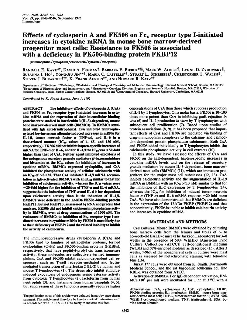
Increases in Cytokine mRNA and Exocytosis of SecretoryGranule Mediators. BMMCs sensitized with IgE were incu-bated with vehicle, CsA, or FK506 for 10 min and activatedwith antigen in the continued presence of the drugs. Sampleswere taken at 10 min for measurement of f-hexosaminidaseand histamine release and at 1 hr for assessment of mRNAlevels for three cytokines by RNA blot analysis. The meanIC50 values for CsA-induced inhibition of the increasedexpression of IL-1p, TNF-a, and IL-6 mRNA and for therelease of (3-hexosaminidase (Fig. 1) and histamine (data notshown) were 4, 65, 130, >2000, and >2000 nM, respectively.Preincubation of IgE-sensitized BMMCs with CsA for up to60 min before antigen challenge did not decrease the IC50values for inhibition of (3-hexosaminidase release or cytokinemRNA levels (data not shown).
-.-.---,---t ...,,--- ----------1.2 2 20 200 2000
Cyclosporin A (nM)
FIG. 1. Dose-response analysis of the effects of CsA on mRNAlevels for IL-1(3 (o), TNF-a (m), and IL-6 (o) and on the release of,B-hexosaminidase (A) by BMMCs. Cells were sensitized with mousemonoclonal IgE anti-TNP, incubated for 10 min with vehicle ordrugs, and incubated with medium or TNP-BSA for 10 min ((-hexosaminidase) or 1 hr (cytokines). Data are expressed as mean +SD, n = 3 for IL-1,B and P-hexosaminidase, and n = 5 for TNF-a andIL-6.
ABMMC
kb9.5-7.5-4.4-2.4-,
l1.4--
BBMMC
p
0.24-
18'
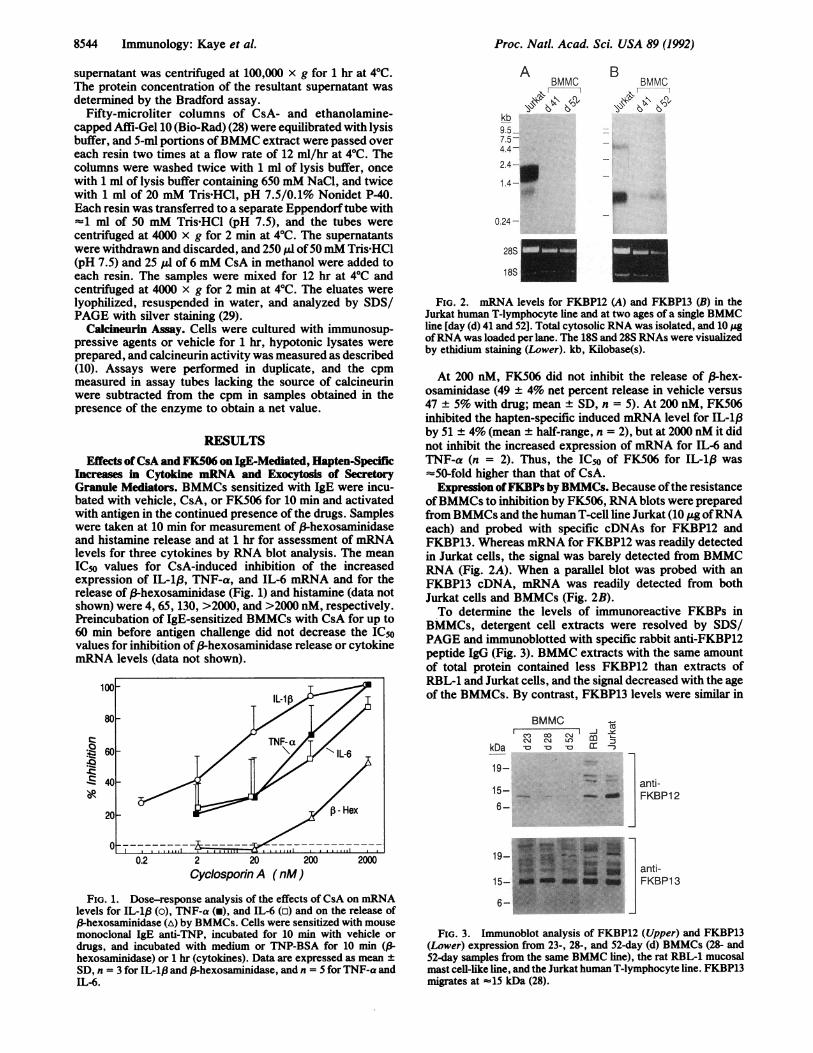
FIG. 2. mRNA levels for FKBP12 (A) and FKBP13 (B) in theJurkat human T-lymphocyte line and at two ages of a single BMMCline [day (d) 41 and 52]. Total cytosolic RNA was isolated, and 10 ;.gofRNA was loaded per lane. The 18S and 28S RNAs were visualizedby ethidium staining (Lower). kb, Kilobase(s).
At 200 nM, FK506 did not inhibit the release of 3-hex-osaminidase (49 ± 4% net percent release in vehicle versus47 ± 5% with drug; mean ± SD, n = 5). At 200 nM, FK506inhibited the hapten-specific induced mRNA level for IL-1,Bby 51 ± 4% (mean + half-range, n = 2), but at 2000 nM it didnot inhibit the increased expression of mRNA for IL-6 andTNF-a (n = 2). Thus, the IC50 of FK506 for IL-1,B was=50-fold higher than that of CsA.Expression ofFKBPs by BMMCs. Because ofthe resistance
ofBMMCs to inhibition by FK506, RNA blots were preparedfrom BMMCs and the human T-cell line Jurkat (10 jg ofRNAeach) and probed with specific cDNAs for FKBP12 andFKBP13. Whereas mRNA for FKBP12 was readily detectedin Jurkat cells, the signal was barely detected from BMMCRNA (Fig. 2A). When a parallel blot was probed with anFKBP13 cDNA, mRNA was readily detected from bothJurkat cells and BMMCs (Fig. 2B).To determine the levels of immunoreactive FKBPs in
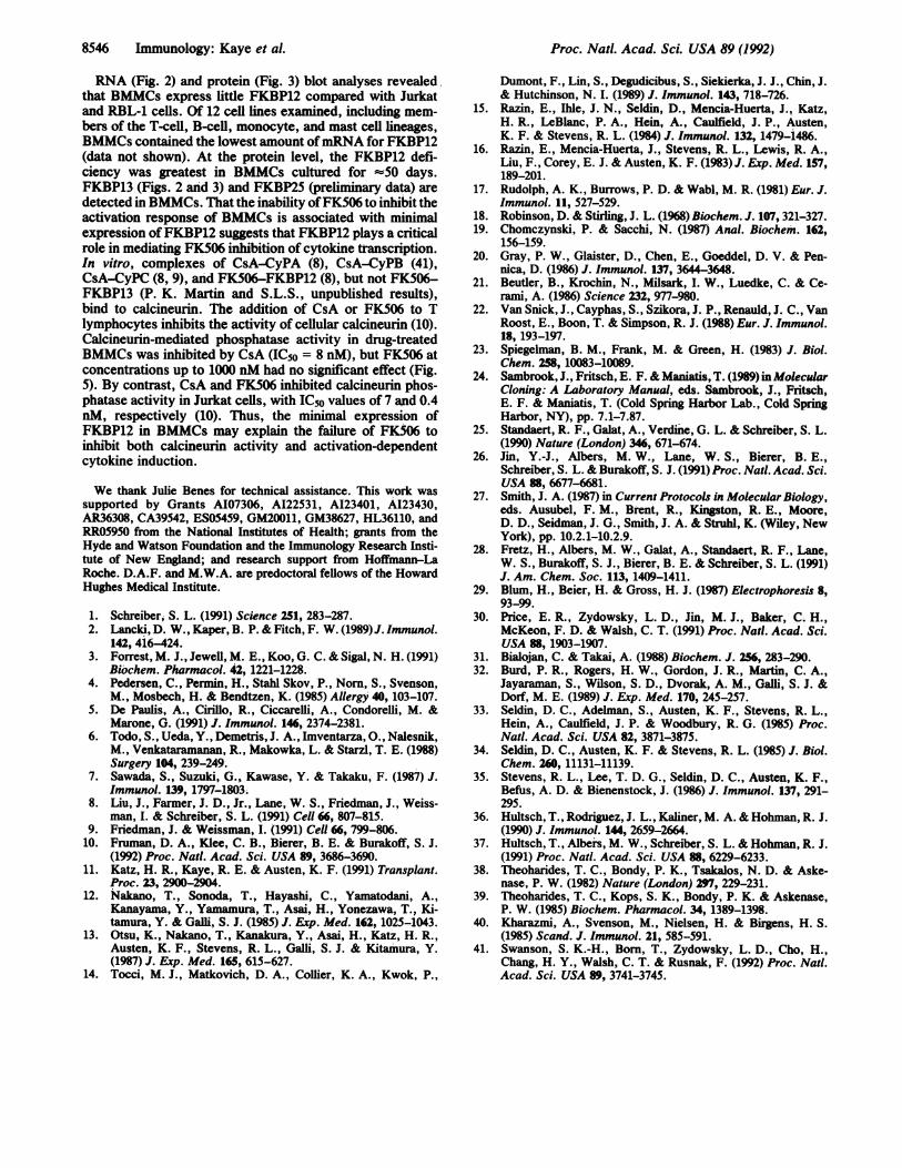
BMMCs, detergent cell extracts were resolved by SDS/PAGE and immunoblotted with specific rabbit anti-FKBP12peptide IgG (Fig. 3). BMMC extracts with the same amountof total protein contained less FKBP12 than extracts ofRBL-1 and Jurkat cells, and the signal decreased with the ageof the BMMCs. By contrast, FKBP13 levels were similar in
kDa
BMMC[I
I C C'aCJ C'J tf)
19-
15-6-
19-
15-
6-
-J
cc
FKBPl 2
anti-FKBP13
FIG. 3. Immunoblot analysis of FKBP12 (Upper) and FKBP13(Lower) expression from 23-, 28-, and 52-day (d) BMMCs (28- and52-day samples from the same BMMC line), the rat RBL-1 mucosalmast cell-like line, and the Jurkat human T-lymphocyte line. FKBP13migrates at '15 kDa (28).
8544 Immunology: Kaye et al.

Proc. Nati. Acad. Sci. USA 89 (1992) 8545
A B97
66
45
31
X.'.#-<CyP14----,--:14
FIG. 4. CyP isolated from a BMMC extract by affinity chroma-tography on an immobilized CsA column (lane B); eluate from a
control resin is shown in lane A. Eluates were analyzed by SDS/PAGE with silver staining.
the three types of cells and did not decrease with age inBMMCs (Fig. 3).
Expression of Cyclophilins by BMMCs. When extracts ofBMMCs were bound to and specifically eluted from a CsA-affinity matrix, a CyP species of Mr 18,000 was recovered
from the CsA affinity matrix, but not from a control matrix,suggesting that BMMCs express CyPA (Fig. 4). As assessedby immunoblotting of BMMC extracts with anti-CyPA andanti-CyPB polyclonal antibodies (each of which also weaklycrossreacted with the other CyP), BMMCs contained twoimmunoreactive species of Mr 17,000 and 20,000 (data notshown), indicative of CyPA and CyPB, respectively (30).
Calcineurn Phosphatase Activity. To determine whetherthe level of immunophilin expression in BMMCs related tothe ability of CsA and FK506 to inhibit calcineurin activity,intact cells were exposed to the drugs for 1 hr, and cell lysateswere then assessed for calcineurin activity in the presence of500 nM okadaic acid, which inhibits phosphatases 1 and 2A(10, 31). CsA inhibited BMMC calcineurin phosphatase ac-tivity with an IC50 of -8 nM (n = 2), whereas FK506 atconcentrations up to 1000 nM gave no appreciable inhibition(Fig. 5). Decreasing the incubation time of BMMCs withdrugs to 10 min did not change the IC50 values (data notshown).
DISCUSSIONThe addition ofCsA to BMMCs sensitized with IgE anti-TNPbefore Fc6 receptor type I perturbation with TNP-BSAresulted in a dose-related decrease in the augmentation ofmRNA levels for IL-113, TNF-a, and IL-6, with IC50 values
FK506
~~~~~~~~~CsA
Q0 0
0.1 1 10 100 1000Drug Concentration (nM)
FIG. 5. Dose-response analysis of the effects of CsA (c) andFK506 (-) on calcineurin-mediated phosphatase activity. Cells wereincubated with the indicated concentrations of drugs or vehicle,cytoplasmic extracts were prepared and incubated with a [32P]phos-phorylated peptide substrate, and liberated phosphate was mea-sured. Cells exposed to vehicle released 743 ± 49 pmol of phosphateper min per mg of protein. Data are mean + half-range, n = 2.
of 4, 65, and 130 nM, respectively (Fig. 1). The IC50 forinhibition of IL-1(3 is similar to that obtained for the inhibitionof calcineurin phosphatase activity in BMMCs by CsA (Fig.5), suggesting that calcineurin plays a critical role in the signaltransduction pathway leading to increased IL-1f3 mRNA aftercell activation with IgE and antigen. The higher IC50 valuesforTNF-a and IL-6 suggest that calcineurin inhibition may beinsufficient to explain the effects of CsA on these cytokinesin BMMCs and that there may be additional intracellularsignals that are inhibited at a higher drug concentration.Inhibition ofIL-6mRNA stimulated by IgE plus antigen in anIL-3-dependent, BMMC-like continuous mouse mast cell linehas been reported to be incomplete at -1700 nM CsA (32). Inhuman T lymphocytes activated by concanavalin A plusphorbol myristate acetate, the IC50 for CsA inhibition ofTNF-a mRNA augmentation is -100 nM, whereas the IC50for inhibition of IL-2 mRNA augmentation is A5 nM, reflect-ing a 20-fold difference (14). Thus, cytokines in both mastcells and T lymphocytes differ in their sensitivity to inhibitionby CsA.
In contrast to its effect on cytokine mRNA levels, CsA inconcentrations up to 2000 nM inhibited the release of thesecretory granule mediators ,3-hexosaminidase (Fig. 1) andhistamine by <50%, and the refractoriness of the BMMCswas not reduced by incubating them for up to 60 min with CsAbefore challenge (data not shown). The resistance ofBMMCsto the inhibition of exocytosis by CsA is a characteristicshared by human blood basophils, which exhibit an IC50 of800-1000 nM for the inhibition of histamine release mediatedby IgE plus antigen (4, 5). By contrast, the IgE-mediatedrelease ofexogenously incorporated [3H]serotonin into RBL-2H3, which is a transformed cell line with several secretorygranule characteristics of rat mucosal mast cells (33-35), isinhibited by CsA with an IC50 of 200 nM (36, 37). However,there is uncertainty whether exogenously added serotonin isreleased from the same intracellular compartment as endog-enous secretory granule components such as ,3-hexosamini-dase and histamine (38, 39). The IC50 for CsA-mediatedinhibition of esterase exocytosis from clones of mouse cyto-toxic T lymphocytes activated with anti-T cell receptor orphorbol myristate acetate and calcium ionophore is 50-150nM (2). Neutrophil exocytosis elicited by complement com-ponent C5a, f-Met-Leu-Phe, or phorbol myristate acetate isnot inhibited by CsA (3), and for exocytosis elicited withcalcium ionophore, the IC50 of CsA is about 35 nM, with amaximum inhibition of 70% at 100 nM. In a separate report(40), calcium ionophore-stimulated exocytosis from humanneutrophils was reduced by only 50% with 4000 nM CsA.Thus, exocytosis of endogenous preformed mediators bymultiple cell types is more resistant to inhibition by CsA thanis cytokine gene transcription and the resultant production ofbioactive protein by T cells. This distinction is now presentedin a single cell type activated with a physiologic stimulus (Fig.1).
In contrast to the CsA-mediated inhibition of BMMCcytokine mRNA levels, no significant inhibition for TNF-a orIL-6 was obtained with FK506, and the IC5o of FK506 forinhibition of IL-1.8 (200 nM) was 50-fold higher than that forCsA, and considerably higher than conventional concentra-tions. By contrast, FK506 inhibits the transcription of IL-2and TNF-a in human T lymphocytes with IC5o values of 0.06and -10 nM, respectively (14). Because IL-1/3 is particularlysensitive to inhibition by CsA in BMMCs, with an IC50 (Fig.1) identical to that for inhibition of calcineurin activity byCsA (Fig. 5), the ability of high concentrations of FK506 toinhibit IL-1f3 in BMMCs may reflect the interaction withminimal amounts ofFKBP12 in some BMMCs (Fig. 3). In oneexperiment with 7-week-old BMMCs, 2000 nM FK506 failedto inhibit the IgE-dependent increment in IL-1,8mRNA (datanot shown).
Immunology: Kaye et al.
Proc. Nadl. Acad. Sci. USA 89 (1992)
RNA (Fig. 2) and protein (Fig. 3) blot analyses revealedthat BMMCs express little FKBP12 compared with Jurkatand RBL-1 cells. Of 12 cell lines examined, including mem-bers of the T-cell, B-cell, monocyte, and mast cell lineages,BMMCs contained the lowest amount ofmRNA for FKBP12(data not shown). At the protein level, the FKBP12 defi-ciency was greatest in BMMCs cultured for =50 days.FKBP13 (Figs. 2 and 3) and FKBP25 (preliminary data) aredetected in BMMCs. That the inability ofFK506 to inhibit theactivation response of BMMCs is associated with minimalexpression ofFKBP12 suggests that FKBP12 plays a criticalrole in mediating FK506 inhibition of cytokine transcription.In vitro, complexes of CsA-CyPA (8), CsA-CyPB (41),CsA-CyPC (8, 9), and FK506-FKBP12 (8), but not FK506-FKBP13 (P. K. Martin and S.L.S., unpublished results),bind to calcineurin. The addition of CsA or FK506 to Tlymphocytes inhibits the activity of cellular calcineurin (10).Calcineurin-mediated phosphatase activity in drug-treatedBMMCs was inhibited by CsA (IC5o = 8 nM), but FK506 atconcentrations up to 1000 nM had no significant effect (Fig.5). By contrast, CsA and FK506 inhibited calcineurin phos-phatase activity in Jurkat cells, with IC5o values of 7 and 0.4nM, respectively (10). Thus, the minimal expression ofFKBP12 in BMMCs may explain the failure of FK506 toinhibit both calcineurin activity and activation-dependentcytokine induction.
We thank Julie Benes for technical assistance. This work wassupported by Grants A107306, A122531, AI23401, A123430,AR36308, CA39542, ES05459, GM20011, GM38627, HL36110, andRRO59SO from the National Institutes of Health; grants from theHyde and Watson Foundation and the Immunology Research Insti-tute of New England; and research support from Hoffmann-LaRoche. D.A.F. and M.W.A. are predoctoral fellows of the HowardHughes Medical Institute.
1. Schreiber, S. L. (1991) Science 251, 283-287.2. Lancki, D. W., Kaper, B. P. & Fitch, F. W. (1989) J. Immunol.
142, 416-424.3. Forrest, M. J., Jewell, M. E., Koo, G. C. & Sigal, N. H. (1991)
Biochem. Pharmacol. 42, 1221-1228.4. Pedersen, C., Permin, H., Stahl Skov, P., Norn, S., Svenson,
M., Mosbech, H. & Bendtzen, K. (1985) Allergy 40, 103-107.5. De Paulis, A., Cirillo, R., Ciccarelli, A., Condorelli, M. &
Marone, G. (1991) J. Immunol. 146, 2374-2381.6. Todo, S., Ueda, Y., Demetris, J. A., Imventarza, O., Nalesnik,
M., Venkataramanan, R., Makowka, L. & Starzl, T. E. (1988)Surgery 104, 239-249.
7. Sawada, S., Suzuki, G., Kawase, Y. & Takaku, F. (1987) J.Immunol. 139, 1797-1803.
8. Liu, J., Farmer, J. D., Jr., Lane, W. S., Friedman, J., Weiss-man, I. & Schreiber, S. L. (1991) Cell C, 807-815.
9. Friedman, J. & Weissman, I. (1991) Cell 66, 799-806.10. Fruman, D. A., Klee, C. B., Bierer, B. E. & Burakoff, S. J.
(1992) Proc. Natl. Acad. Sci. USA 89, 3686-3690.11. Katz, H. R., Kaye, R. E. & Austen, K. F. (1991) Transplant.
Proc. 23, 2900-2904.12. Nakano, T., Sonoda, T., Hayashi, C., Yamatodani, A.,
Kanayama, Y., Yamamura, T., Asai, H., Yonezawa, T., Ki-tamura, Y. & Galli, S. J. (1985) J. Exp. Med. 162, 1025-1043.
13. Otsu, K., Nakano, T., Kanakura, Y., Asai, H., Katz, H. R.,Austen, K. F., Stevens, R. L., Galli, S. J. & Kitamura, Y.(1987) J. Exp. Med. 165, 615-627.
14. Tocci, M. J., Matkovich, D. A., Collier, K. A., Kwok, P.,
Dumont, F., Lin, S., Degudicibus, S., Siekierka, J. J., Chin, J.& Hutchinson, N. I. (1989) J. Immunol. 143, 718-726.
15. Razin, E., IhIe, J. N., Seldin, D., Mencia-Huerta, J., Katz,H. R., LeBlanc, P. A., Hein, A., Caulfield, J. P., Austen,K. F. & Stevens, R. L. (1984) J. Immunol. 132, 1479-1486.
16. Razin, E., Mencia-Huerta, J., Stevens, R. L., Lewis, R. A.,Liu, F., Corey, E. J. & Austen, K. F. (1983) J. Exp. Med. 157,189-201.
17. Rudolph, A. K., Burrows, P. D. & Wabl, M. R. (1981) Eur. J.Immunol. 11, 527-529.
18. Robinson, D. & Stirling, J. L. (1968) Biochem. J. 107, 321-327.19. Chomczynski, P. & Sacchi, N. (1987) Anal. Biochem. 162,
156-159.20. Gray, P. W., Glaister, D., Chen, E., Goeddel, D. V. & Pen-
nica, D. (1986) J. Immunol. 137, 3644-3648.21. Beutler, B., Krochin, N., Milsark, I. W., Luedke, C. & Ce-
rami, A. (1986) Science 232, 977-980.22. Van Snick, J., Cayphas, S., Szikora, J. P., Renauld, J. C., Van
Roost, E., Boon, T. & Simpson, R. J. (1988) Eur. J. Immunol.18, 193-197.
23. Spiegelman, B. M., Frank, M. & Green, H. (1983) J. Biol.Chem. 258, 10083-10089.
24. Sambrook, J., Fritsch, E. F. & Maniatis, T. (1989) in MolecularCloning: A Laboratory Manual, eds. Sambrook, J., Fritsch,E. F. & Maniatis, T. (Cold Spring Harbor Lab., Cold SpringHarbor, NY), pp. 7.1-7.87.
25. Standaert, R. F., Galat, A., Verdine, G. L. & Schreiber, S. L.(1990) Nature (London) 346, 671-674.
26. Jin, Y.-J., Albers, M. W., Lane, W. S., Bierer, B. E.,Schreiber, S. L. & Burakoff, S. J. (1991) Proc. Natl. Acad. Sci.USA 88, 6677-6681.
27. Smith, J. A. (1987) in Current Protocols in Molecular Biology,eds. Ausubel, F. M., Brent, R., Kingston, R. E., Moore,D. D., Seidman, J. G., Smith, J. A. & Struhl, K. (Wiley, NewYork), pp. 10.2.1-10.2.9.
28. Fretz, H., Albers, M. W., Galat, A., Standaert, R. F., Lane,W. S., Burakoff, S. J., Bierer, B. E. & Schreiber, S. L. (1991)J. Am. Chem. Soc. 113, 1409-1411.
29. Blum, H., Beier, H. & Gross, H. J. (1987) Electrophoresis 8,93-99.
30. Price, E. R., Zydowsky, L. D., Jin, M. J., Baker, C. H.,McKeon, F. D. & Walsh, C. T. (1991) Proc. Natl. Acad. Sci.USA 88, 1903-1907.
31. Bialojan, C. & Takai, A. (1988) Biochem. J. 256,283-290.32. Burd, P. R., Rogers, H. W., Gordon, J. R., Martin, C. A.,
Jayaraman, S., Wilson, S. D., Dvorak, A. M., Galli, S. J. &Dorf, M. E. (1989) J. Exp. Med. 170, 245-257.
33. Seldin, D. C., Adelman, S., Austen, K. F., Stevens, R. L.,Hein, A., Caulfield, J. P. & Woodbury, R. G. (1985) Proc.Natl. Acad. Sci. USA 82, 3871-3875.
34. Seldin, D. C., Austen, K. F. & Stevens, R. L. (1985) J. Biol.Chem. 260, 11131-11139.
35. Stevens, R. L., Lee, T. D. G., Seldin, D. C., Austen, K. F.,Befus, A. D. & Bienenstock, J. (1986) J. Immunol. 137, 291-295.
36. Hultsch, T., Rodriguez, J. L., Kaliner, M. A. & Hohman, R. J.(1990) J. Immunol. 144, 2659-2664.
37. Hultsch, T., Albers, M. W., Schreiber, S. L. & Hohman, R. J.(1991) Proc. Natl. Acad. Sci. USA SS, 6229-6233.
38. Theoharides, T. C., Bondy, P. K., Tsakalos, N. D. & Aske-nase, P. W. (1982) Nature (London) 297, 229-231.
39. Theoharides, T. C., Kops, S. K., Bondy, P. K. & Askenase,P. W. (1985) Biochem. Pharmacol. 34, 1389-1398.
40. Kharazmi, A., Svenson, M., Nielsen, H. & Birgens, H. S.(1985) Scand. J. Immunol. 21, 585-591.
41. Swanson, S. K.-H., Born, T., Zydowsky, L. D., Cho, H.,Chang, H. Y., Walsh, C. T. & Rusnak, F. (1992) Proc. Natl.Acad. Sci. USA 89, 3741-3745.
8546 Immunology: Kaye et al.