effect transcutaneous electrical nerve stimulation on...
TRANSCRIPT

694
Effect of Transcutaneous Electrical NerveStimulation on Coronary Blood Flow
Anoop Chauhan, MRCP; Paul A. Mullins, MRCP; Suren I. Thuraisingham, MRCP; Ged Taylor;Michael C. Petch, MD, FRCP; Peter M. Schofield, MD
Background Although neurostimulation has been shown tobe of benefit in angina pectoris, the exact mechanism of itsaction is not clear. This study was performed to examine theeffect of transcutaneous electrical nerve stimulation on coro-nary blood flow.Methods and Results The effect of transcutaneous electrical
nerve stimulation was studied in 34 syndrome X patients(group 1), 15 coronary artery disease patients (group 2), and16 heart transplant patients (group 3). Coronary blood flowvelocity (CBFV) in the left coronary system was measured atrest and after a 5-minute stimulation period with a JudkinsDoppler. There was a significant increase in the resting CBFVin group 1 (from 6.8±4.1 to 10.5±5.7 cm/s, P<.001) and group2 (from 6.8±4.1 to 10.5±5.7 cm/s, P<.001). However, there
Du uring the last decade, electrical neurostimula-tion has developed as a means of therapy forcontrol of pain and has been shown to be
useful in a variety of chronic painful conditions.'-3Recently, it has also been shown to have a beneficialeffect in the treatment of ischemic pain.4-7 Transcuta-neous electrical nerve stimulation (TENS) treatmenthas also been shown to be of benefit in angina.8-1" Inpatients treated with TENS there was a reduction inchest pain, an increase in work capacity, and a decreasein ST segment depression at a comparable workload.This improvement persisted during the posttreatmentfollow-up period of 2 weeks. Improved tolerance topacing and improved lactate metabolism also have beendemonstrated after TENS treatment.The connection between ischemia and pain is com-
plex, and it is not known how neurostimulation influ-ences this. One of the several possibilities is that TENSmay increase coronary blood flow, thus reducing isch-emia. Certainly, it has been shown that epidural neuro-stimulation improves microvascular blood flow in pe-ripheral vascular disease.4-7The aim of this study was totest the hypothesis that TENS treatment may relieve theischemia of angina by increasing coronary blood flow.
MethodsPatientsGroup 1. This group comprised 34 patients with typical
symptoms of angina and completely normal coronary arterieson angiography as reviewed by two independent observers. All
was no significant change in the resting CBFV in group 3.There were no significant changes in the coronary arterialdiameters as a result of neurostimulation. There was a signif-icant decrease in the epinephrine levels in group 1 (from79.6+17.8 to 58.5±17.5 ng/L, P=.01) and group 2 (from102.2±27.2 to 64.1± 19.1 ng/L, P=.01).
Conclusions Transcutaneous electrical nerve stimulationcan increase resting coronary blood flow velocity. The findingssuggest that the site of action is at the microcirculatory leveland that the effects may be mediated by neural mechanisms.(Circulation. 1994;89:694-702.)Key Words * transplantation * coronary disease a
norepinephrine
the patients had continued to remain symptomatic despitereassurance after their initial cardiac catheterization. Theduration of symptoms was greater than 6 months in allpatients. None of the patients had hypertension, diabetesmellitus, lung disease, or valvular heart disease. All patientshad stable symptoms of angina for the previous 2 months.There was no evidence of coronary spasm or myocardialbridging in these patients.Group 2. This group comprised 15 patients who had docu-
mented significant coronary artery disease on angiographyaffecting the right coronary artery. Significant coronary arterydisease was defined as >50% reduction in intraluminal diam-eter of the right coronary artery. The left coronary artery wasnormal in all patients. These patients were selected so thatcoronary flow velocity measurements could be undertaken bya Judkins-type Doppler catheter in the normal left coronarysystem in the absence of any stenoses that may affect measure-ments. One patient had previous cardiac surgery for an atrialseptal defect and had developed angina subsequently. Theduration of symptoms was >6 months in all patients, andsymptoms of angina had been stable for the previous 2 months.None of the patients had hypertension, diabetes mellitus, lungdisease, or valvular heart disease.Group 3. This group comprised 16 patients with heart
transplants and completely normal coronary arteries on angi-ography. None of these patients had chest pain. These patientswere undergoing their routine follow-up cardiac catheteriza-tion. Coronary occlusive disease is the main cause of morbidityand mortality more than 1 year after orthotopic cardiactransplantation.12 Clinical monitoring for the detection ofcoronary occlusive disease is dependent on serial coronaryangiography. Currently, we follow our transplant patientsannually with repeat coronary angiography; the patients stud-ied were recruited over a period of 3 months. The durationafter transplantation of the patients studied was variable andwas determined by the patients scheduled for repeat coronaryangiography and giving consent for the study during thisperiod. The minimum duration after transplantation was 12months and the maximum 100 months. None of the patients
Submitted May 20, 1993; revision accepted October 19, 1993.From the Regional Cardiac Unit, Papworth Hospital, Papworth
Everard, Cambridge, England.Correspondence to Dr A. Chauhan, Regional Cardiac Unit,
Papworth Hospital, Papworth Everard, Cambridge, UK.
by guest on July 13, 2018http://circ.ahajournals.org/
Dow
nloaded from
Chauhan et al Transcutaneous Electrical Nerve Stimulation and Coronary Blood Flow 695
TABLE 1. Patient Variables
Group 1 Group 2 Group 3(n=34) (n=15) (n=16)
Age, y 55±9 57±10 49+11Sex
Male 15 9 15
Female 19 6 1
Weight, kg 72.3±12.3 77.4±11 67.3±10.2
Hemoglobin, g/dL 13.7±1.3 12.9±1.1 10.7+0.94ESR, mm/h 14±10 9±6 11±6
Urea, mmol/L 5.6±0.98 6.2±0.9 15.3±10.3
Glucose, mmol/L 5.6±0.8 5.4±0.6 6.5±2.2
Creatinine, rtmol/L 101±14 111±12 222±177
Cholesterol, mmol/L 6.38±0.83 6.4±0.8 6.61 ±0.34
LVEDP, mm Hg 10.5±4.0 11±3 9±2
Median time after operation, mo ... ... 47 (range, 12-100)
Antianginal drugs
Nitrates 18 15 0
Calcium antagonists 23 12 0
3-Blockers 19 6 0
Azathioprine 0 0 16
Cyclosporine 0 0 16
Steroids 0 0 6
ESR indicates erythrocyte sedimentation rate; LVEDP,expressed as mean±SD where appropriate.
had hypertension, diabetes mellitus, lung disease, or valvularheart disease. All patients were receiving cyclosporine andazathioprine immunosuppression with or without steroid treat-ment (Table 1).
EchocardiographyEchocardiographic assessment was performed in all pa-
tients. Cross-sectional and M-mode assessment of the leftventricular posterior wall and septal thickness was made in allpatients. Patients with a diastolic septal or posterior wallthickness of >11 mm were excluded from the study to mini-mize any effect of left ventricular hypertrophy on coronaryblood flow measurements. Patients with evidence of mitral oraortic valve disease were also excluded.
Electric Nerve Stimulator and TechniqueA commercially available transcutaneous nerve stimulator
(Compact TENS, Neurotechnics Ltd, Aylesbury, England) wasused in the study. This has two electric channels that areelectrically isolated. Each channel is capable of a current outputof 0 to 60 mA into 1 kQl. The two channels can be used andcontrolled separately. The stimulator allows modulation andadjustment of both pulse width and pulse rate. The pulse widthis adjustable from 30 to 300 ,um/s. The pulse rate is adjustablefrom 1 to 200 Hz. The stimulator has a 9-V alkaline battery asits power source. For our study, the stimulator was set to deliverconstant-current pulses of 300 milliseconds. The pulse repeti-tion frequency was kept at 150 Hz. The intensity of thestimulation was individually adapted to a level immediatelybelow that producing pain (10 to 60 mA).For the study, electrode paste was applied to the contact
surface to lower the skin impedance. Electrodes (50x35 mm)were placed 10 to 30 cm apart on the chest of the patient at theusual site of their most intense pain (groups 1 and 2). The
left ventricular end-diastolic pressure. Values are
interelectrode distance was 20 cm in all patients except for 2patients (10 cm) in group 1 in whom the electrode position wasaltered because of difficulties with electrode application ontothe chest wall and 1 patient (30 cm) in group 2 in whom theelectrodes were placed to avoid lesions of psoriasis. In thetransplant group (group 3), the electrodes were placed 20 cmapart on the anterior chest wall over the precordium. Theelectrodes were connected to the stimulator, and the stimula-tion was adjusted to a level immediately below that producingpain. The level of stimulation was noted, and the stimulatorthen was turned off. Cardiac catheterization was then per-formed, and baseline measurements were recorded. TheTENS treatment then was given for 5 minutes, and themeasurements were repeated immediately after.
Cardiac Catheterization StudyThe patients were fasted overnight before cardiac catheter-
ization. All vasoactive cardiac medications were stopped for atleast 48 hours. Coronary angiography was performed by theJudkins technique using the right femoral approach in allpatients. Coronary injections were performed manually withup to 8 mL of intracoronary radiopaque contrast (Urograffinor Niopam), and cinefilm recordings were made in multipleprojections. To eliminate any vasoactive effects of the contrastmedium, at least 10 minutes were allowed to lapse beforecoronary blood flow velocity measurements were recorded.A Judkins-type 8F Doppler-tipped coronary catheter (Cor-
dis Corp, Miami, Fla) was advanced to the ascending aortaover a 0.035-in. J-tipped guide wire and then positioned at theleft coronary ostium. This catheter has been validated beforefor measurements of coronary flow velocity and coronary flowreserve, and the technique is described in detail elsewhere.13,14The catheter was then flushed and filled with saline. Velocitysignal generation and processing was achieved with a standard
by guest on July 13, 2018http://circ.ahajournals.org/
Dow
nloaded from
696 Circulation Vol 89, No 2 February 1994
TABLE 2. Systemic Hemodynamics and Coronary BloodFlow Velocity Measurements
After TENSResting Data Treatment
Group 1 (n=34)
HR, beats per minute 69+16 69+17
Systolic BP, mm Hg 130+21 132±25
CBFV, cm/s 6.8±4.1 10.5±5.7*
RPP 8919±2352 8974±2760
Group 2 (n=15)
HR, beats per minute 72±15 73±15
Systolic BP, mm Hg 135±18 133±16
CBFV, cm/s 6.4±2.5 11.3±6.7*
RPP 9627±2162 9694±2214
Group 3 (n= 16)HR, beats per minute 91±9 90±11
Systolic BP, mm Hg 142±21 140±20
CBFV, cm/s 7.7±4.1 7.5±4.1
RPP 12 807±2061 12 656±2037
TENS indicates transcutaneous electrical nerve stimulation;HR, heart rate; BP, blood pressure; CBFV, coronary blood flowvelocity; and RPP, rate-pressure product. Values are given asmean±SD.*P<.001.
velocimeter (model MDV-20, Millar Instruments, Houston,Tex). The velocimeter was range-gated and calibrated with anarbitrary internally set calibration of 0 to 100 cm/s (1kHz=3.75 cm/s) for full-scale deflection (0 to 10 cm on therecorder). The Judkins Doppler was connected to the Millarvelocimeter, and the catheter position was adjusted to obtain astable position with good quality phasic and mean coronaryblood flow velocity signals. Injection of contrast through theDoppler catheter lumen fully opacified the coronary arteryand identified the relative catheter-artery position. The leftcoronary system was centered for optimal viewing, and theangiograms were reviewed to select the two best views forrepeating the angiogram after the period of TENS.
Resting coronary blood flow velocity, heart rate, and meanarterial and systolic blood pressures were recorded on aMingograf recorder (Siemens-Elema, Sweden). The transcu-taneous nerve stimulator was then turned on for 5 minutes,and the measurements were repeated immediately after thetreatment period. The coronary angiograms were repeated inthe preselected views immediately after recording the abovedata.
Quantitative MeasurementsQuantitative measurements of the coronary artery diame-
ters were performed using digital electronic calipers (SandhillScientific Inc). This method has been used previously to assessthe arterial diameter of coronary vessels and is reproduciblewith minimal interobserver and intraobserver variation.15-17The digital caliper is an application of the concept firstdescribed by Gensini et al.18 The device has a variability(standard deviation of multiple estimates) approximating±6% on estimates of "percent stenosis" and +0.18 mm onestimates of minimal lumen diameter.'9 The mean error of thecaliper estimates is not different from zero.'9 It has beenshown previously in animal experiments that resting coronaryflow is not affected until the coronary arterial diameter isreduced by 85%, and the capacity to increase flow over restingbasal levels in response to a vasodilatory stimulus does not
30
25-
20
15
10
5-
0
Pre TENS Post TENS
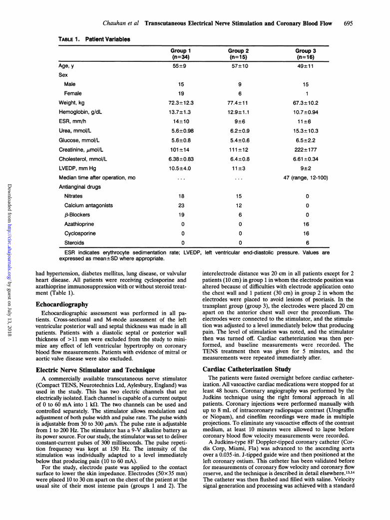
FIG 1. Plot shows the effect of transcutaneous electrical nervestimulation (TENS) on coronary blood flow velocity (CBFV) insyndrome X patients. Coincident points are not shown.
disappear until constriction of the arterial diameter of>85%.20 Studies in humans have suggested that a 50% to 70%reduction in coronary diameter is necessary before coronaryflow reserve is affected.2' The digital caliper provides adequateresolution and accuracy to detect any significant changes incoronary arterial diameter that may lead to changes in coro-nary blood flow.Two selected views were taken and projected by a Tagarno
system onto a sheet of paper. The arterial diameters of the leftanterior descending coronary artery and the circumflex arterywere measured from tracings of the projected images indiastole, at a distance of 3 cm from the tip of the Dopplercatheter. The diameter for each artery was calculated as themean of the measurements of the two views. The measure-ments were performed by two independent observers so thatinterobserver and intraobserver variation could also becalculated.
ReproducibilityTo assess the reproducibility of the effects of TENS, we
repeated the study in 10 chest pain patients (group 1), 10coronary artery disease patients (group 2) in whom there wasat least a 25% increase in the resting coronary blood flowvelocity, and in 10 transplant patients (group 3). In all patients,the coronary flow velocity was allowed to return to the originalresting level after the initial TENS treatment before continu-ing with this part of the study. In addition, in these patients wealso measured epinephrine and norepinephrine levels in theaortic root before and after the application of TENS.
Catecholamine MeasurementsThe effects of neurostimulation have been likened to those
of sympathectomy, and it has been suggested previously thatneurostimulation may inhibit sympathetic outflow.'0 To studythe effect of TENS on serum catecholamine levels in our study,blood samples were taken from the aortic root before and after
by guest on July 13, 2018http://circ.ahajournals.org/
Dow
nloaded from

Chauhan et al Transcutaneous Electrical Nerve Stimulation and Coronary Blood Flow 697
20 -
15 -
W
;A
Z
=LVi
10 -
5-
o n
Pre TENS Post TENS
Pre TENS Post TENSFIG 2. Plot shows the effect of transcutaneous electrical nervestimulation (TENS) on coronary blood flow velocity (CBFV) incoronary artery disease patients. Coincident points are notshown.
the application of TENS. Epinephrine and norepinephrinelevels were determined by a high-performance liquid chroma-tography technique.22
Statistical MethodsValues are reported as mean+SD. All comparisons were
performed using the Wilcoxon matched pairs test. A value ofP<.05 was considered to be statistically significant.
Ethical ApprovalThe study was approved by the Huntingdon District Health
Authority Ethical Committee, and full informed consent was
obtained from all patients before the study.
ResultsTable 1 shows relevant patient information, blood
results, and drug treatment details. Of the transplantedpatients, 10 originally underwent transplantation forischemic heart disease. The remaining 6 patients were
transplanted for dilated cardiomyopathy.
Systemic Hemodynamic EffectsIn all three groups there were no significant systemic
hemodynamic changes induced by TENS treatment.There were no significant differences in heart rate,systolic blood pressure, or mean arterial pressure. As a
result, there was no significant difference in the rate-pressure product (see Table 2).
Coronary Blood Flow Velocity ChangesThere was a significant increase in the resting coro-
nary flow velocity in group 1 patients (6.8±4.1 to10.5+±5.7 cm/s, Fig 1) and group 2 patients (6.4±2.5 to
FIG 3. Plot shows the effect of transcutaneous electrical nervestimulation (TENS) on coronary blood flow velocity (CBFV) intransplant patients. Coincident points are not shown.
11.3+6.7 cm/s, Fig 2) after TENS treatment. However,TENS treatment did not increase the resting coronaryflow velocity in group 3 (7.7+4.1 versus 7.5+±4.1 cm/s,Fig 3). There was no difference in the resting coronary
flow velocity or in the response to TENS between menand women in groups 1 and 2 (see Tables 2 and 3).
Coronary Artery Diameter MeasurementsArterial diameter measurements were reproduced
with minimal interobserver (r=.90) and intraobservervariation (r=.91). There was no significant difference inthe arterial diameters of the left coronary system as a
result of TENS in all three groups (Table 4).
ReproducibilityThere was again a significant increase in the coronary
blood flow velocity in group 1 and group 2 patients butnot in group 3 patients after the application of TENS.Once again, there were no significant hemodynamicchanges induced by TENS (Table 5).
Catecholamine MeasurementsIn group 1 patients, there was a significant decrease in
the arterial epinephrine concentration (P=.01) afterTENS application. However, there was no significantdifference in the norepinephrine levels. Similarly, therewas a significant decrease in the arterial epinephrineconcentration (P=.01) in group 2, and there was no
significant difference in the norepinephrine levels. Ingroup 3 patients, both the epinephrine and norepineph-rine levels showed no significant change after TENSapplication.
DiscussionCoronary flow measurements are being used increas-
ingly in clinical cardiology. Until recently, inaccuracies
35 -
30
25-
;0)
E;
20 -
1i
10 -
5-
0 | w X w w w w X w x by guest on July 13, 2018
http://circ.ahajournals.org/D
ownloaded from

698 Circulation Vol 89, No 2 February 1994
TABLE 3. Coronary Blood Flow Velocity Measurements (cm/s) Before and After TENS
Group 1 Group 2 Group 3
Patient Before TENS After TENS Before TENS After TENS Before TENS After TENS1 3.0 5.0 8.0 12.0 5.5 5.02
34
567
8
9
10
11
121314
1516171819202122232425262728293031323334
5.0
10.5
2.0
13.0
3.014.0
3.07.0
5.09.0
16.0
5.07.03.5
7.0
3.04.04.04.0
7.04.02.04.04.5
13.012.09.0
12.08.03.0
15.02.56.0
12.0
12.0
5.0
13.0
6.5
20.0
6.0
24.010.0
12.0
15.0
11.0
7.03.5
18.0
4.0
6.0
5.0
10.0
9.0
4.012.06.0
8.0
4.0
6.0
5.06.03.0
8.0
9.0
9.0
16.0
9.0
4.0
20.0
30.0
14.0
9.0
10.0
8.0
6.0
5.0
9.0
6.0
3.0
2.0
6.0
14.0
10.0
15.0
3.015.07.0
7.0
6.0
8.06.0
9.0
5.5
3.0
2.0
6.0
13.0
9.0
16.0
3.0
15.0
7.0
7.0
6.0
7.06.0
6.511.07.0
8.010.07.07.06.04.513.011.09.0
11.0
9.0
7.030.06.013.0
TENS indicates transcutaneous electrical nerve stimulation.
had limited the methods of assessing coronary bloodflow. The use of small-diameter intracoronary Dopplerflow catheters has now allowed subselective estimationsof coronary blood flow velocity in individual coronaryvessels and has become the most extensively used andvalidated method in the measurement of coronary flowvelocity. However, the major limitation of this tech-nique is that instrumentation of the coronary artery isrequired with the potential of serious complicationssuch as coronary dissection, vasospasm, and thrombosis.Also, the very nature of their use in individual coronaryvessels means that only the response of one coronaryvessel may be studied at any one time. For assessment of
the left coronary circulation, we used the Judkins-typeDoppler catheter in our study. These catheters havebeen validated previously and have been shown to offera safe, simple, and accurate alternative to the conven-tional intracoronary Doppler catheters.'3.14 The use ofJudkins Doppler catheters also allowed us to examinethe physiological and pharmacological responses of theentire left coronary system, which supplies the leftventricle. If TENS treatment does influence coronaryblood flow, these catheters would therefore be moresuited to assess the coronary circulation. Since we choseto study patients with angiographically normal left cor-onary arteries, the subselective intracoronary measure-
by guest on July 13, 2018http://circ.ahajournals.org/
Dow
nloaded from

Chauhan et al Transcutaneous Electrical Nerve Stimulation and Coronary Blood Flow 699
TABLE 4. Coronary Artery Diameter Measurements
Group 1 Group 2 Group 3Resting Cx diameter, mm 4.1±0.9 4.0±1.0 4.3±1.2
After TENS 4.0±1.0 4.0±1.0 4.4±1.1
Resting LAD diameter, mm 4.2±0.7 3.9±1.1 4.4±0.7
After TENS 4.3±0.6 3.9±1.1 4.3±0.5
Cx indicates circumflex coronary artery; TENS, transcutane-ous electrical nerve stimulation; and LAD, left anterior descend-ing coronary artery. Values are given as mean±SD.
ments that would have been required in vessels withsignificant coronary artery disease were not necessary.The aim of our study was to investigate whether
TENS can influence coronary blood flow. Our study hasshown that TENS can increase coronary blood flowvelocity significantly in the chest pain group of patients(groups 1 and 2), although it had no significant effect onthe transplant patients (group 3). The effect of TENSon the coronary blood flow velocity in these patientswas highly reproducible. Moreover, TENS did not sig-nificantly affect systemic hemodynamics. The level ofarterial epinephrine decreased significantly after TENStreatment in the chest pain patients (groups 1 and 2),although the level of norepinephrine was unchanged.The arterial catecholamine levels were unchanged inthe transplant group (group 3).How does neurostimulation work? The understand-
ing of the effects of neurostimulation in patients withpain is based on the theory of segmental pain inhibitionpostulated by Melzack and Wall.23 Their gate controltheory states that inhibition of the flow of pain impulsesoccurs at the first synaptic station in the spinal cord bymeans of a presynaptic neuron system. They assumedthat this system was fed by collateral nerves from bothlarge afferent A fibers, which do not transmit pain, andsmall myelinated and unmyelinated C fibers. The Cfibers inhibit the system and thus prepare the way forincreased transmission (the gate was opened), whereasactivity in A fibers excited the system and thus resultedin suppression of the transmission to the next neuronchain (the gate was closed). This theory gives a crudeexplanation for the variability of pain sensation. High-frequency electrical stimulation with pulse widths ofbetween 100 and 200 milliseconds, which stimulates thelarge A fibers but not the C fibers, reduced the sensationof pain.1 There is additional evidence that TENS mayact by releasing endorphins and inhibiting transmission
of noxious stimuli on various levels.24,25 TENS may exertits pain-reducing effect on anginal pain by activatingenkephalinergic systems either at segmental levels inthe spinal cord or possibly locally in the heart.26'27The effects of neurostimulation have been likened to
sympathectomy. Although the relief of the pain ofangina alone may indirectly reduce ischemia by reduc-ing reflex sympathetic nervous activity, stimulation ofthe dorsal column may have a direct effect. Stimulationof the dorsal column may lead to a spread of currentsinto the intermediate columns of the gray area, releas-ing segmental spinal reflexes that tonically inhibit sym-pathetic discharge, and this may cause vasodilatation. Ithas been shown that prostaglandins also may play a rolein mediating the increase in skeletal muscle blood flowcaused by activity in the afferent fibers.28 A similarmechanism may be active in the heart. It is postulatedthat TENS may produce its analgesic effects by inhibit-ing sympathetic outflow by influencing the connectionbetween pain fibers and sympathetic neurons in thedorsal horn of the spinal cord as a result of activation ofsegmental reflexes.
Previous studies have shown that treatment withTENS in patients with angina reduces pain, increasestolerance to pacing, improves myocardial lactate metab-olism, and is associated with less pronounced ST seg-ment depression.8-11 In long-term studies, the grouptreated with TENS demonstrated an increased workcapacity as assessed by repeated bicycle ergometrytests, had reduced frequency of angina attacks, and hada reduced consumption of nitrates.8 It has been sug-gested that this beneficial effect of TENS is due to anantianginal as well as an anti-ischemic effect. Onemechanism of action suggested in these studies has beenthat there may be left ventricular afterload reductioncaused by arteriolar dilatation, indicated by a drop insystemic vascular resistance and systolic blood pressureand a rise in cardiac index leading to a decrease inmyocardial oxygen demand. That these effects may beassociated with decreased sympathetic activity wasthought to be indicated by a drop in arterial concentra-tion of norepinephrine and epinephrine in patients inwhom angina disappeared during treatment at corre-
sponding pacing rates.10TENS has been shown to cause a significant decrease
in the blood pressure of hypertensive rats.29 In humans,TENS treatment to control the pain of parturition hasbeen shown to increase placental blood flow.30 TENSalso causes vasodilatation in patients with peripheral
TABLE 5. Reproducibility of the Effects of TENS
Group 1 (n=10) Group 2 (n=10) Group 3 (n=10)
First Study Second Study First Study Second Study First Study Second StudyPre-TENS HR, bpm 64±8 64±8 68±9 68±10 94+11 95±10Post-TENSHR, bpm 63+10 64±10 68±10 66±10 94±8 95±9
Pre-TENS MAP, mm Hg 94±12 95±11 96±11 95±12 104±8 103±7
Post-TENSMAP, mmHg 96±12 95±12 96±12 95±12 103±8 103±6
Pre-TENS CBFV, cm/s 6.0±1.8 6.3+1.5 6.5±1.8 6.0±1.8 7.1±1.7 7.0±1.8
Post-TENS CBFV, cm/s 13.7±5.2 13.8±5.5 13.7±5.2 13.7±5.2 6.8±1.5 6.8±1.5
TENS indicates transcutaneous electrical nerve stimulation; HR, heart rate; bpm, beats per minute; MAP, mean arterial pressure; andCBFV, coronary blood flow velocity. Values are given as mean±SD.
by guest on July 13, 2018http://circ.ahajournals.org/
Dow
nloaded from

700 Circulation Vol 89, No 2 February 1994
TABLE 6. Catecholamine Levels
Before TENS After TENS
Group 1 (n= 10)
Epinephrine, ng/L 89.3+25.9 60.6+18.4*
Norepinephrine, ng/L 300±146.2 284.5±144.5
Group 2 (n= 10)
Epinephrine, ng/L 102.2±27.2 64.1 ± 19.1*
Norepinephrine, ng/L 341.8±158.8 334.6±157.6
Group 3 (n= 10)
Epinephrine, ng/L 68.7±32.2 67.6±32.1
Norepinephrine, ng/L 338.3±95.2 332±94.1
TENS indicates transcutaneous electrical nerve stimulation.*P<.05. Values are given as mean±SD.
arterial disease.31 Recently, Kaada et a132 have shownthat TENS can cause a significant lowering of meanfemoral arterial pressure and systemic vascular resis-tance. This effect was considered to be due to anincrease in peripheral microcirculation as a result ofinhibition of the sympathetic system.Does neurostimulation increase coronary blood flow?
Noninvasive studies have suggested that there is lessischemia in some patients.8-11,33 However, other studieshave shown that although there was a reduction inangina, an increase in the time to onset of ischemia, anda higher perfusion at rest during stimulation than incontrols, there was no significant increase of perfusionto abnormal segments after maximal exercise as as-sessed by positron emission tomography.34 It has beensuggested that any changes in the perfusion caused byneurostimulation are small, occur mainly in the ischemicareas, and will be difficult to detect in the heart. It maybe that neurostimulation can result in the adjustment oflocal flow without any increase in total flow, for exam-ple, the so-called "Robin Hood effect" of aminophyl-line.35 However, our study has demonstrated that TENScan increase the resting coronary flow velocity, and thismay support the findings of Landsheere et al,34 whoreported an increased perfusion during stimulation thanin controls.
Similar to most other previous studies, our study doesnot demonstrate any effect of TENS at rest on systemichemodynamics.8-10 In the study reported by Kaada etal,32 which showed that TENS application can signifi-cantly lower mean femoral arterial pressure, the dura-tion of TENS treatment was 20 minutes, and low-frequency electrical nerve stimulation (2 Hz) was used.In our study, high-frequency (150 Hz) electrical nervestimulation was used, and the duration of stimulationwas only 5 minutes. It has been claimed previously thathigh-frequency electroacupuncture (100 Hz) causesmainly segmental analgesia, whereas the effects of low-frequency stimulation (< 10 Hz) are more generalized inboth humans and animals.36 It may be that high-fre-quency TENS given for the short duration in our studydid have a predominantly segmental effect, and this mayexplain the lack of any effect on the mean arterialpressure. If the duration had been prolonged, then wemay have seen a systemic effect.One possible mechanism by which TENS treatment
may influence coronary blood flow may be by affecting
the coronary vascular tone as a result of changes in theneural tone. We studied the transplant group becausethe heart in these patients is denervated and theyprovide an in vivo model of a denervated heart. It isinteresting to note that TENS treatment did not affectthe resting coronary blood flow velocity in the trans-plant group, although there was a significant increase inthe chest pain groups. This would also suggest that themechanism by which TENS may affect coronary bloodflow may have an underlying neural basis.There was necessarily a substantial variability associ-
ated with physical application of the electrical stimula-tion dependent on patient tolerance to the stimulationand the positioning of the electrodes over the painfulareas. However, the technique used is similar to otherreported studies.8-11 The interelectrode distance was thesame (20 cm) in most patients and is unlikely to haveinfluenced the difference seen in the response to TENS.Also, in each case, the patient acted as their owncontrol. It may be argued that there was no effect seenin the transplant group because of their previous sur-gery and the resulting sternotomy scars, which may haveresulted in differences in sensitivity to TENS and painperception or a failure of TENS transmission. In all ourtransplant patients, the electrodes were placed wellclear of the sternotomy scar on either side of themidline. In the only patient with a previous sternotomyscar from cardiac surgery in our study (patient 11, group2, Table 3), there was a significant increase in coronaryflow velocity in response to TENS, which suggests thatsternotomy scars do not lead to failure of TENS trans-mission. Indeed, one would not expect sternotomy scarsto interfere with TENS transmission on anatomicgrounds because the innervation of the anterior chestwall is symmetrically segmental. Furthermore, TENStreatment has been shown to be effective in relievingpain after thoracotomy and to reduce the incidence oflung atelectases.37The heart rate, systolic blood pressure, and the
rate-pressure product were higher in group 3. It hasbeen shown in heart transplant patients that increasesin heart rate produce an increase in resting coronaryflow velocity without a proportionate increase in thehyperemic coronary flow velocity.38 Consequently, cor-onary flow reserve measurements decrease with in-creasing heart rate. However, there is little reduction inthe hyperemic coronary flow velocity in absolute termswith increasing heart rates of up to 120 beats perminute.38 A similar study in patients undergoing electivecoronary angiography for the evaluation of chest painhas demonstrated that sudden increases in heart ratebut not mean arterial pressure lead to a substantialreduction in maximal coronary flow reserve-from3.7±0.9 at the heart rate of 71±18 beats per minute atrest to 2.6±0.5 during pacing at 120 beats per minute.39It is therefore possible that the higher resting heart ratein the transplant group may have resulted in the failureof the transplant patients to respond to TENS. How-ever, it is of note that even at the heart rate of 120 beatsper minute in Rossen and Winniford's study,39 therewas still a mean increase in coronary flow velocity by2.6-fold from the resting values, indicating that theability of the coronary microcirculation to increasecoronary blood flow toward hyperemia is still preservedalthough it may be impaired. It would not be unreason-
by guest on July 13, 2018http://circ.ahajournals.org/
Dow
nloaded from

Chauhan et al Transcutaneous Electrical Nerve Stimulation and Coronary Blood Flow 701
able to assume that the higher resting heart rate (94±11beats per minute) in the transplant group comparedwith the other groups in our study would not lead to atotal lack of response to TENS because the increasedheart rate would not completely abolish the ability ofthe coronary circulation to increase coronary flow,although this possibility cannot be entirely excluded.Increases in arterial pressure increase both the restingand hyperemic coronary flow velocities.38 Consequently,the coronary flow reserve remains unaffected. Wewould not expect the increased systolic pressures toaffect the ability of the coronary circulation in trans-plant patients to respond to TENS.The increase in coronary flow velocity in the chest
pain groups, in the absence of a significant difference inarterial pressure and heart rate, may have resulted froma change in the coronary artery diameter. However, thequantitative measurements of the artery diameters be-fore and after TENS have shown that there was nosignificant change in the vessel diameters. If TENS doesindeed affect the coronary circulation, then the possiblemechanisms of its actions are not clear. It may eitheralter the neural tone of the coronary microvasculaturewithout affecting the large epicardial vessels, thus re-sulting in increased blood flow, or may produce a similareffect by causing the local release of substances that areable to affect the microcirculation. There has been someinterest previously in the search for the mediators ofvasodilation peripherally produced by TENS, but noactive vasodilator has been identified in the heart.32There was variability in the response to TENS in
groups 1 and 2 that was independent of thresholdstimulation and the site of electrode placement. Thismay suggest individual differences in sensitivity to neu-rostimulation and underlines the complex mechanismsthat may be responsible for the effects of TENS.The results of the catecholamine measurements are
in keeping with previous studies and suggest sym-pathoinhibition.10,32 Emanuelsson et a110 suggested thatthe reduction in the sympathetic activity during TENSwas mainly secondary to pain relief rather than a directinfluence on the sympathetic system. However, there isother evidence supporting the theory that, in addition toits pain-relieving effect, TENS may influence autonomicsystems by suppressing sympathetic overactivity.31 Ourstudy also supports this because none of the patientsduring the study experienced any chest pain, and thereduction in arterial epinephrine level occurred as aresult of the effect of TENS at rest in the absence of anyischemia.
Study LimitationsOur study was open and therefore was exposed to
bias. It is almost impossible to design a blind study oftreatment with TENS because there is no placeboequivalent for the sensation of transcutaneous neuro-stimulation. However, in our study, the patients acted astheir own control, and the results of the reproducibilitystudy demonstrated consistent responses in the chestpain and transplant patients.
Evidence now exists for spontaneous reinnervation ofthe transplanted heart and restoration of sinus arrhyth-mia and baroreceptor function.40 None of the transplantpatients studied demonstrated sinus arrhythmia onECG recordings. However, it has been shown recently
that the response of the sinus node to stimulatorymaneuvers cannot be taken as evidence for or lack ofreinnervation of the remainder of the heart becausereinnervation after cardiac transplantation is regionallyheterogenous.41 We did not perform tyramine studies inthe transplant patients for the presence of reinnerva-tion, which may have helped us to partition the attenu-ated response of these patients to a peripheral sensorreception problem or a central effect.
All the transplant patients were on immunosuppres-sion therapy, which was continued. It is possible thatthese drugs may have affected pain threshold andcoronary vasoreactivity. However, we are not aware ofany evidence to suggest this possibility. Coronary mi-crovascular vasodilatory responses after cardiac trans-plantation do not seem to be affected by cyclosporine.42ConclusionsTENS can reproducibly increase resting coronary
blood flow velocity. The lack of any effect in thetransplant patients suggests that the effect of TENS maybe mediated by neural mechanisms. The lack of anychanges in the large epicardial coronary artery diametersuggests that the site of vascular action is at the micro-circulatory level. These may vasodilate, causing anincrease in coronary blood flow as a result of a localproduction of vasodilatory substances or directly by areduction in sympathetic activity or both. Our studydoes suggest a decreased sympathetic activity locally inthe heart, although any accompanying production ofvasodilator substances cannot be ruled out.
AcknowledgmentsThis work was presented in part at the 42nd Annual
Scientific Sessions of the American College of Cardiology,Anaheim, Calif, March 1993, and at the XVth Congress of theEuropean Society of Cardiology, Nice, France, August 1993.Dr Chauhan was supported by a British Heart FoundationJunior Research Fellowship.
References1. Wall PD, Sweet WH. Temporary abolition of pain in man. Science.
1967;155:108-109.2. Shealy CN, Mortimer JT, Hagfors NR. Dorsal column electroan-
algesia. J Neurosurg. 1970;32:560- 564.3. Friedman H, Nashold BS, Somjen G. Physiological effects of dorsal
column stimulation. Adv Neurol. 1974;4:769-771.4. Tallis RC, Illis LS, Sedgewick EM, Hardwidge C, Garfield JS.
Spinal cord stimulation in peripheral vascular disease. J NeurolNeurosurg Psychiatry. 1983;46:478-484.
5. Augustinsson LE, Holm J, Carlsson C-A, Jivegard L. Epiduralelectrical stimulation in severe limb ischemia: evidences of painrelief: increased blood flow and a possible limb saving effect. AnnSurg. 1985;202:104-111.
6. Sciacca V, Tamorri M, Rocco M. Modification of transcutaneousoxygen tension in lower limb peripheral arterial occlusive diseasepatients treated with spinal cord stimulation. Ital J Surg Sci. 1986;16:279-282.
7. Jacobs M, Jorning PJG, Joshi SR, Kitslaar P, Slaaf DW, RenemanRS. Epidural spinal cord electrical stimulation improves micro-vascular blood flow in severe limb ischemia. Ann Surg. 1988;207:65-69.
8. Mannheimer C, Carlsson C-A, Emanuelsson H, Vendin A,Waagstein F, Wilhelmsson C. The effects of transcutaneous electricnerve stimulation in patients with severe angina pectoris. Circulation.1985;71:308-316.
9. Mannheimer C, Carlsson C-A, Vendin A, Wilhelmsson C. Trans-cutaneous electric nerve stimulation (TENS) in angina pectoris.Pain. 1986;26:291-300.
10. Emanuelsson A, Mannheimer C, Waagstein F, Wilhelmsson C.Catecholamine metabolism during pacing-induced angina pectoris
by guest on July 13, 2018http://circ.ahajournals.org/
Dow
nloaded from

702 Circulation Vol 89, No 2 February 1994
and the effects of transcutaneous electrical nerve stimulation. AmHeart J. 1987;114:1360-1366.
11. Mannheimer C, Emanuelsson H, Waagstein F, Wilhelmsson C.Influence of naloxone on the effects of high frequency transcu-taneous electric nerve stimulation in angina pectoris induced byatrial pacing. Br Heart J. 1989;62:36-42.
12. Kreitt JM, Kayne MP. The registry of the international hearttransplant registry seventh official report - 1990. J Heart LungTransplant. 1990;9:323-336.
13. Kern JM. A simplified method to measure coronary blood flowvelocity in patients: validation and application of a Judkins-styleDoppler-tipped angiographic catheter. Am Heart J. 1990;120:1202-1212.
14. Chauhan A, Mullins P, Thuraisingham S, Taylor G, Petch M,Schofield P. A validation study of a Doppler-tipped angiographiccatheter for measurement of coronary flow reserve. Am J Cardiol.1993;71:1119-1121.
15. Kern MJ, Deligonul U, Vandormael M, Labovitz A, Gudipati CV,Gabliani G, Bodet J, Shah Y, Kennedy HL. Impaired coronaryvasodilator reserve in the immediate postcoronary angioplastyperiod: analysis of coronary artery flow velocity indexes andregional cardiac venous efflux. JAm Coll Cardiol. 1989;13:860-872.
16. Mullins PA, Chauhan A, Sharples L, Cary NR, Large SR,Wallwork J, Schofield PM. Impairment of coronary flow reserve inorthotopic cardiac transplant recipients with minor coronaryocclusive disease. Br Heart J. 1992;68:266-271.
17. Chauhan A, Mullins P, Thuraisingham S, Taylor G, Petch M,Schofield P. The effect of hyperventilation and mental stress oncoronary blood flow in Syndrome X patients. Br Heart J. 1993;69:516-524.
18. Gensini GG, Kelly AE, Da Costa BCB, Huntington PP. Quanti-tative angiography: the measurement of coronary vasomotion inthe intact animal and man. Chest. 1971;60:522-530.
19. Scoblionko DP, Brown G, Mitten S, Caldwell JH, Kennedy W,Bolson EL, Dodge HT. A new digital electronic caliper for mea-surement of coronary arterial stenosis: comparison with visualestimates and computer-assisted measurements. Am J Cardiol.1984;53:689-693.
20. Gould KL, Lipscomb K, Hamilton GW. Physiological basis forassessing critical coronary stenosis: instantaneous flow responseand regional distribution during coronary hyperemia as measuresof coronary flow reserve. Am J Cardiol. 1974;33:87-94.
21. Wilson RF, Marcus ML, White CW. Prediction of the physiologicsignificance of coronary arterial lesions by quantitative lesiongeometry in patients with limited coronary arterial disease. Circu-lation. 1987;75:723-732.
22. Frayn KN, Maycock PF. Sample preparation with ion-exchangeresin before liquid chromatographic determination of plasma cat-echolamines. Clin Chem. 1983;29:1426-1428.
23. Melzack R, Wall PD. Pain mechanisms: a new theory. Science.1965;150:971-979.
24. Oliveras JL, Hosobuchi Y, Redjemi F, Guilbad G, Besson JM.Opiate antagonist naloxone strongly reduces analgesia by stimu-lation of a raphe nucleus (centralis inferior). Brain Res. 1977;120:221-227.
25. Sjolund BH, Eriksson MBE. The influence of naloxone onanalgesia produced by peripheral conditioning stimulation. BrainRes. 1979;173:295-301.
26. Lang RE, Hermann K, Dietz R, Gaida W, Ganten D, Kraft K,Unger TH. Evidence for the presence of enkephalins in the heart.Life Sci. 1983;32:399-406.
27. Faccini E, Uzumala H, Govoni S. Afferent fibers mediate theincrease of metenkephalin elicited in rat spinal cord by localizedpain. Pain. 1984;18:25-31.
28. Hilton SM, Marshall JM. Dorsal root vasodilatation in cat skeletalmuscle. J Physiol (Lond). 1980;299:277-288.
29. Yao T, Andersson S, Thoren P. Long-lasting cardiovasculardepressor response following sciatic stimulation in spontaneouslyhypertensive rats: evidence for the involvement of centralendorphin and serotonin systems. Brain Res. 1982;244:295-303.
30. Kubista E, Philipp K. Der Einfluss der TNS (transkutane Nerven-stimulierung) auf die plazentare Durchstromung. Z GeburtshilfePerinatol. 1979;183:30-34.
31. Kaada B. Vasodilation induced by transcutaneous nerve stimu-lation in peripheral ischaemia (Raynaud's phenomenon anddiabetic polyneuropathy). Eur Heart J. 1982;3:303-314.
32. Kaada B, Vik-mo H, Rosland G, Woie L, Opstad PK. Transcu-taneous nerve stimulation in patients with coronary arterialdisease: haemodynamic and biochemical effects. Eur Heart J. 1990;11:447-453.
33. Mannheimer C, Augustinsson L-E, Carlsson C-A, Manhem K,Wilhelmsson C. Epidural spinal electrical stimulation in severeangina pectoris. Br Heart J. 1988;59:56-61.
34. Landsheere C, Habets A, Mannheimer C, Guillamme M, BourgeoisI, Augustinsson L, Lamotte D, Kulbertus H, Rigo P. Epidural stim-ulation in angina pectoris: any effect on regional myocardial per-fusion? Circulation. 1988;78(suppl II):II-322. Abstract.
35. Crea F, Pupita G, Galassi AR, Tammimi HE, Kaski JC, Davies GJ,Maseri A. Effects of theophylline on exercise induced myocardialischemia. Lancet. 1989;1:683-686.
36. Han JS, Zhou ZF, Xuan YT. Acupuncture has an analgesic effectin rabbits. Pain. 1983;15:83-87.
37. Tyler E, Caldwell C, Ghia JN. Transcutaneous electrical nervestimulation: an alternative approach to the management of post-operative pain. Anesth Analg. 1982;61:449-455.
38. McGinn AL, White CW, Wilson RF. Interstudy variability ofcoronary flow reserve: influence of heart rate, arterial pressure,and ventricular preload. Circulation. 1990;81:1319-1330.
39. Rossen JD, Winniford MD. Effect of increase in heart rate andarterial pressure on coronary flow reserve in humans. JAm CollCardiol. 1993;21:343-348.
40. Stark RP, McGinn AL, Wilson RF. Chest pain in cardiactransplant recipients: evidence of sensory reinnervation aftercardiac transplantation. N Engl J Med. 1991;324:1791-1794.
41. Wilson RF, Laxson DD, Christensen BV, McGinn AL, Kubo SH.Regional differences in sympathetic reinnervation after humanorthotopic cardiac transplantation. Circulation. 1993;88:165-171.
42. Mullins PA, Chauhan A, Large SR, Wallwork J, Schofield PM.Coronary microvascular vasodilation and cyclosporin levels aftercardiac transplantation. Eur Heart J. 1992;13:9. Abstract.
by guest on July 13, 2018http://circ.ahajournals.org/
Dow
nloaded from

A Chauhan, P A Mullins, S I Thuraisingham, G Taylor, M C Petch and P M SchofieldEffect of transcutaneous electrical nerve stimulation on coronary blood flow.
Print ISSN: 0009-7322. Online ISSN: 1524-4539 Copyright © 1994 American Heart Association, Inc. All rights reserved.
is published by the American Heart Association, 7272 Greenville Avenue, Dallas, TX 75231Circulation doi: 10.1161/01.CIR.89.2.694
1994;89:694-702Circulation.
http://circ.ahajournals.org/content/89/2/694World Wide Web at:
The online version of this article, along with updated information and services, is located on the
http://circ.ahajournals.org//subscriptions/
is online at: Circulation Information about subscribing to Subscriptions:
http://www.lww.com/reprints Information about reprints can be found online at: Reprints:
document. Permissions and Rights Question and Answer this process is available in the
click Request Permissions in the middle column of the Web page under Services. Further information aboutOffice. Once the online version of the published article for which permission is being requested is located,
can be obtained via RightsLink, a service of the Copyright Clearance Center, not the EditorialCirculationin Requests for permissions to reproduce figures, tables, or portions of articles originally publishedPermissions:
by guest on July 13, 2018http://circ.ahajournals.org/
Dow
nloaded from