research article vagal nerve stimulation therapy… nerve stimulation... · research article vagal...
TRANSCRIPT
RESEARCH ARTICLE
Vagal Nerve Stimulation Therapy: What IsBeing Stimulated?Guy Kember1, Jeffrey L. Ardell2, John A. Armour2, Mair Zamir3,4*
1. Department of Engineering Mathematics, Dalhousie University, Halifax, Nova Scotia, Canada, 2.Department of Biomedical Sciences, East Tennessee State University’s Quillen College of Medicine, JohnsonCity, Tennessee, United States of America, 3. Department of Applied Mathematics, Western University,London, Canada, 4. Department of Medical Biophysics, Western University, London, Canada
Abstract
Vagal nerve stimulation in cardiac therapy involves delivering electrical current to
the vagal sympathetic complex in patients experiencing heart failure. The therapy
has shown promise but the mechanisms by which any benefit accrues is not
understood. In this paper we model the response to increased levels of stimulation
of individual components of the vagal sympathetic complex as a differential
activation of each component in the control of heart rate. The model provides
insight beyond what is available in the animal experiment in as much as allowing
the simultaneous assessment of neuronal activity throughout the cardiac neural
axis. The results indicate that there is sensitivity of the neural network to low level
subthreshold stimulation. This leads us to propose that the chronic effects of vagal
nerve stimulation therapy lie within the indirect pathways that target intrinsic cardiac
local circuit neurons because they have the capacity for plasticity.
Introduction
Vagal nerve stimulation is currently being used to treat epilepsy and is being
explored for the treatment of heart disease and other ailments [1]. Specifically, it
is being utilized to treat patients experiencing cardiac arrhythmias [2, 3] and heart
failure [4, 5]. This procedure involves delivering electrical stimuli to the cervical
vagal sympathetic complex (VSC) [6, 7]. VNS therapy, which involves delivering
electrical current to the VSC, has been shown to impart some benefit to cardiac
arrhythmia and heart failure patients [3, 4, 8, 9]. The mechanisms by which this
benefit is obtained are not fully understood [3, 8, 10, 11]. Furthermore, the level
and mode of current delivery required to obtain optimum clinical outcome is not
known [4].
OPEN ACCESS
Citation: Kember G, Ardell JL, Armour JA, ZamirM (2014) Vagal Nerve Stimulation Therapy: WhatIs Being Stimulated? PLoS ONE 9(12): e114498.doi:10.1371/journal.pone.0114498
Editor: Marcus Gray, The University ofQueensland, Australia
Received: June 1, 2014
Accepted: November 10, 2014
Published: December 5, 2014
Copyright:� 2014 Kember et al. This is an open-access article distributed under the terms of theCreative Commons Attribution License, whichpermits unrestricted use, distribution, and repro-duction in any medium, provided the original authorand source are credited.
Data Availability: The authors confirm that all dataunderlying the findings are fully available withoutrestriction. All relevant data are within the paper.
Funding: Theoretical work was supported by theNatural Sciences and Engineering ResearchCouncil of Canada via two individual discoverygrants to GK and MZ. Experimental work wassupported by two grants to JLA, one from theNational Institute of Health 348 (HL71830) andanother from Cyberonics, Inc. (Houston, Texas).The funders had no role in study design, datacollection and analysis, decision to publish, orpreparation of the manuscript.
Competing Interests: The experimental part ofthis study was funded in part by Cyberonics, Inc.The theoretical part was funded fully by TheNatural Sciences and Engineering ResearchCouncil of Canada. Additionally, JLA and JAA arepaid consultants of Cyberonics, Inc. There are nopatents, products in development or marketedproducts to declare. This does not alter theauthors’ adherence to all the PLOS ONE policieson sharing data and materials. In particular: (a)there are no other competing interests relating toemployment, consultancy, patents, products indevelopment, marketed products, etc., and (b) theabove does not alter the authors’ adherence toPLOS ONE policies on sharing data and materials,to the effect that it places no restrictions on sharingof data and/or materials. (c) GK and MZ declarethat they have no competing interests.
PLOS ONE | DOI:10.1371/journal.pone.0114498 December 5, 2014 1 / 18
The VSC contains axons that carry cardiovascular sensory feedback to the
medulla along with both parasympathetic and sympathetic motor axons that
project to neurons on the heart [12]. These parasympathetic preganglionic
efferent axons synapse with (1) intrinsic cardiac parasympathetic efferent
postganglionic neurons that innervate the cardiac musculature and (2) intrinsic
cardiac local circuit neurons [13]. The second are much more numerous than the
first [13]. For the purpose of the present paper we shall refer to the first of these as
a ‘‘direct’’ parasympathetic pathway because its axons synapse directly with
cardiac cholinergic postganglionic neurons. The second shall be referred to as an
‘‘indirect’’ parasympathetic pathway because its axons synapse with intrinsic
cardiac local circuit neurons that in turn modulate cardiac motor neurons. The
main feature of the direct pathway is that it involves single pre-to-postganglionic
synapses, while the indirect pathway involves multiple synapses organized as a
neural network.
When the complex structure of the VSC is stimulated, all of its elements may be
affected. Afferent axons along with efferent cholinergic and adrenergic axons may
become activated. As a consequence, the corresponding alteration in cardiac
indices elicited in the process are multifaceted. These processes are at present
difficult to resolve in the intact state. Thus the questions of what is being affected
by vagal stimulation therapy and how to interpret the resulting changes in cardiac
indices, particularly heart rate, remain unresolved. The prospects for resolving
these questions in an experiment are clearly limited. Added to these difficulties is
the fact that the neural control system of the heart is a multilevel network [14]. As
such, it is not known how effects of VNS therapy influence the dynamics of
cardiac control. Similar questions arise in cranial nerve stimulation which is a
growing therapeutical strategy for treating epilepsy and various psychiatric
disorders [15–22].
In this paper we present a model in which the direct and the indirect pathways
of VNS therapy on individual elements of the cardiac hierarchy can be isolated.
This permits understanding the putative complexity of cardiac responsiveness to
such therapy. By selectively activating the different elements of the VSC and
observing their individual impact on the cardiac control hierarchy, our aim is to
establish a relationship between stimulation of the various components of the
VSC and the ensuing effects within the neural control hierarchy of the heart.
Neural Control of the Heart
In the classical view, neural control of the heart was explained mainly in terms of
central neural command, specifically in terms of medullary and spinal cord
autonomic efferent preganglionic neurons targeting efferent postganglionic
neurons that innervate the heart [23]. In recent years it has become clear that
there is a 3-level hierarchy of cardiac control, two of these residing outside the
central nervous system, specifically (1) within the intrinsic cardiac nervous system
and (2) within intrathoracic extracardiac ganglia. The results we present in this
Vagal Nerve Stimulation Therapy
PLOS ONE | DOI:10.1371/journal.pone.0114498 December 5, 2014 2 / 18

paper are based on a model of this 3-level control system which has previously
been shown to explain heart rate phenomena which could not be explained with
the classical view of cardiac control by central command [14, 24, 25].
Neural Network
A key feature of this model is that each of the three levels of control is assumed to
consist of a population of neurons which influence and are influenced by each other at
their own level of control as well as at adjacent levels. We refer to this collectively as
the ‘‘neural network’’.
The network we use in this paper has 3N neurons equally divided among three
levels of control which we shall refer to as levels 1,2,3, ‘‘bottom’’, ‘‘middle’’, ‘‘top’’,
or ‘‘cardiac’’, ‘‘intrathoracic’’, ‘‘central’’, respectively. Two indices, j,k are used to
identify the kth neuron at the jth level. The state of activity (: level of discharge)
of neuron j,k at time interval t(n) is denoted by Xs(n)
� �j,k for sympathetic control,
Xp(n)
� �j,k for indirect parasympathetic control, and Xr
(n)� �
j,k for direct
parasympathetic control as explained in more detail below. All neural activity is
scaled to range between 1.0 when a neuron is most active and 0.0 when it is
inactive.
Heart Rate Control Algorithm
Broadly speaking, the neural network receives continuous neuronal updates of current
demand for blood flow and current heart rate, and processes this state of the system at
each time interval to produce an appropriate change in heart rate. The main result of
this process, which is a key feature of the model, is that demand for blood flow does
not proceed directly to the heart or to central command but to the neural network as a
whole. The way this occurs is described briefly below, more details can be found in
[14, 24].
Heart rate is constrained to lie between a prescribed base value and a prescribed
maximum value. A scaled heart rate H is used such that H51.0 when heart rate is
maximum and H50.0 when heart rate is at base value.
The dynamics of the neural network unfold piecewise at consecutive time
intervals t(n), n~1,2, . . ., where demand for blood flow and current heart rate are
used as inputs and an incremental ‘‘move’’ DM(n)(t) is produced as output.
The average activity of sympathetic efferent postganglionic neurons at the
cardiac level is used as an external input to motor neurons at the cardiac level such
that the efferent sympathetic neural input Es to the heart satisfies
tsdEs
dtzEs~Ks(F
(n)
s {as) ð1Þ
where Ks is a sympathetic gain, ts is a sympathetic time constant, as is a
sympathetic reference level and
Vagal Nerve Stimulation Therapy
PLOS ONE | DOI:10.1371/journal.pone.0114498 December 5, 2014 3 / 18

F(n)
s ~
PNs1
k~1 Xs(n)
� �1,k
Ns1
ð2Þ
where Ns1 is the number of sympathetic neurons at the cardiac level.
Indirect parasympathetic efferent preganglionic neural activity Xp(n)
� �j,k passes
through the neural network and appears as input to parasympathetic efferent
postganglionic motor neurons at the cardiac level such that the efferent indirect
parasympathetic neural input Ep to the heart satisfies
tpdEp
dtzEp~Kp(F
(n)
p {ap) ð3Þ
where Kp is an indirect parasympathetic gain, tp is indirect parasympathetic time
constant, ap is the indirect parasympathetic reference level and
F(n)
p ~
PNp1
k~1 Xp(n)
� �1,k
Np1
ð4Þ
where Np1 is the number of indirect parasympathetic neurons at the cardiac level.
Direct parasympathetic activity Xr(n) bypasses the neural network and proceeds
directly from central command to the heart such that efferent direct
parasympathetic neural input Er to the heart satisfies
trdEr
dtzEr~Kr hr|H(n){dr|
D(n)
Dmax
� �ð5Þ
where Kr is a direct parasympathetic gain, tr is direct parasympathetic time
constant, ar is direct parasympathetic reference level, H(n) and D(n) are prevailing
heart rate and blood demand at time interval n and hr,dr are ‘‘sensitivities’’ of
direct parasympathetic neurons to heart rate and blood demand, respectively.Finally, the change in heart rate or ‘‘move’’ DM(n) at time interval n is the net of
the above three efferent contributions
DM(n)~asEs{apEp{arEr ð6Þ
where as,as,ap are constants.Within each time interval tn, heart rate is a continuous function of time
governed by a first order linear system
tHdH(n)(t)
dtzH(n)(t)~M(n)(t) ð7Þ
where tH is a time constant and
M(n)(t)~Xi~n{1
i~0
DM(i)(t) ð8Þ
Vagal Nerve Stimulation Therapy
PLOS ONE | DOI:10.1371/journal.pone.0114498 December 5, 2014 4 / 18

For simplicity, this time dependence will not be shown explicitly in what
follows but will be implicit within each time interval t(n).
Neural Discharge
In general, the level of activity of a neuron (neural discharge) is determined by the
demand for blood flow but is also affected by current heart rate and by the level of
activity of neighboring neurons.
Specifically, the action potential generated by a neuron j,k within the network
in time interval t(n) is represented by the state of activity X(n)� �
j,k of that neuron. A
change in the state of activity of the neuron due to these effects shall be denoted
respectively by d1 X(n)� �
j,k, d2 X(n)� �
j,k, d3 X(n)� �
j,k, and total change by D X(n)� �
j,k.
We distinguish between two types of neurons [26, 27]: ‘‘heart-rate neurons’’ which
are affected by only current heart rate and the activity of neighboring neurons, and
‘‘blood-demand neurons’’ which are affected by only demand for blood flow and the
activity of neighboring neurons.
Thus the change in the state of activity of a sympathetic neuron j,k is given by
D X(n)s
� �j,k~d1 X(n)
s
� �j,kzd3 X(n)
s
� �j,k heart-rate neurons ð9Þ
D X(n)s
� �j,k~d2 X(n)
s
� �j,kzd3 X(n)
s
� �j,k blood-demand neurons ð10Þ
and the corresponding change in the state of activity of an indirect
parasympathetic neuron is similarly given by
D X(n)p
� �j,k
~{d1 X(n)p
� �j,k
zd3 X(n)p
� �j,k
heart-rate neurons ð11Þ
D X(n)p
� �j,k
~{d2 X(n)p
� �j,k
zd3 X(n)p
� �j,k
blood-demand neurons ð12Þ
The extent to which heart rate H and blood demand D affect the state of activity
of a sympathetic or parasympathetic efferent neuron (j,k) depend on heart rate
and blood demand ‘‘sensitivities’’ (hs)j,k and (ds)j,k such that for a sympathetic
neuron
d1 X(n)s
� �j,k~{
hsð Þj,kh
|H(n{L) ð13Þ
d2 X(n)s
� �j,k~
dsð Þj,kd
|D(n)
Dmaxð14Þ
and for a parasympathetic neuron
Vagal Nerve Stimulation Therapy
PLOS ONE | DOI:10.1371/journal.pone.0114498 December 5, 2014 5 / 18
d1 X(n)p
� �j,k
~hp� �
j,k
h|H(n{L) ð15Þ
d2 X(n)p
� �j,k
~{dp� �
j,k
d|
D(n)
Dmaxð16Þ
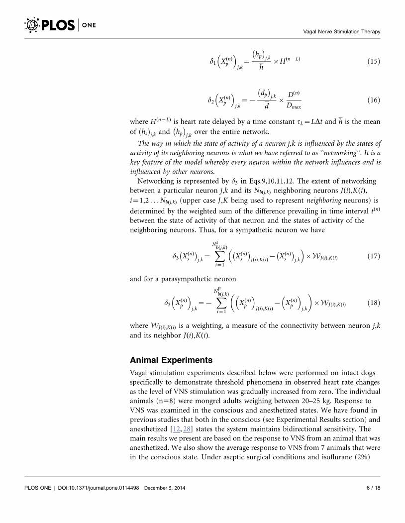
where H(n{L) is heart rate delayed by a time constant tL~LDt and h is the mean
of hsð Þj,k and hp� �
j,k over the entire network.
The way in which the state of activity of a neuron j,k is influenced by the states of
activity of its neighboring neurons is what we have referred to as ‘‘networking’’. It is a
key feature of the model whereby every neuron within the network influences and is
influenced by other neurons.
Networking is represented by d3 in Eqs.9,10,11,12. The extent of networking
between a particular neuron j,k and its Nb(j,k) neighboring neurons J(i),K(i),i~1,2 . . . Nb(j,k) (upper case J,K being used to represent neighboring neurons) is
determined by the weighted sum of the difference prevailing in time interval t(n)
between the state of activity of that neuron and the states of activity of the
neighboring neurons. Thus, for a sympathetic neuron we have
d3 X(n)s
� �j,k~
XNsb(j,k)
i~1
X(n)s
� �J(i),K(i){ X(n)
s
� �j,k
� �|WJ(i),K(i) ð17Þ
and for a parasympathetic neuron
d3 X(n)p
� �j,k
~{XNpb(j,k)
i~1
X(n)p
� �J(i),K(i)
{ X(n)p
� �j,k
� �|WJ(i),K(i) ð18Þ
where WJ(i),K(i) is a weighting, a measure of the connectivity between neuron j,kand its neighbor J(i),K(i).
Animal Experiments
Vagal stimulation experiments described below were performed on intact dogs
specifically to demonstrate threshold phenomena in observed heart rate changes
as the level of VNS stimulation was gradually increased from zero. The individual
animals (n58) were mongrel adults weighing between 20–25 kg. Response to
VNS was examined in the conscious and anesthetized states. We have found in
previous studies that both in the conscious (see Experimental Results section) and
anesthetized [12, 28] states the system maintains bidirectional sensitivity. The
main results we present are based on the response to VNS from an animal that was
anesthetized. We also show the average response to VNS from 7 animals that were
in the conscious state. Under aseptic surgical conditions and isoflurane (2%)
Vagal Nerve Stimulation Therapy
PLOS ONE | DOI:10.1371/journal.pone.0114498 December 5, 2014 6 / 18
anesthesia, the latter group received VNS therapy system implant (Demipulse 103
implantable stimulator with Model 304 bipolar helical cuff; Cyberonics, Houston,
TX) involving the right cervical vagus nerve. Following a two week recovery
period, animals were trained to the Pavlov stand. Following an initial 4 week
titration period, VNS response curves were determined at 10 Hz, 500 msec pulse
width, with a 18% duty cycle (14 sec on, 66 sec off) and with current randomized
between 0.25 to 3.50 mA. Heart rate responses were quantified by the percent
change from the baseline in response to VNS as shown in the results.
All experiments were performed in accordance with the guidelines for animal
experimentation described in the ‘‘Guide for the Care and Use of Laboratory
Animals: Eighth Edition, 2010’’. The Institutional Animal Care and Use
Committee of East Tennessee State University approved these experiments.
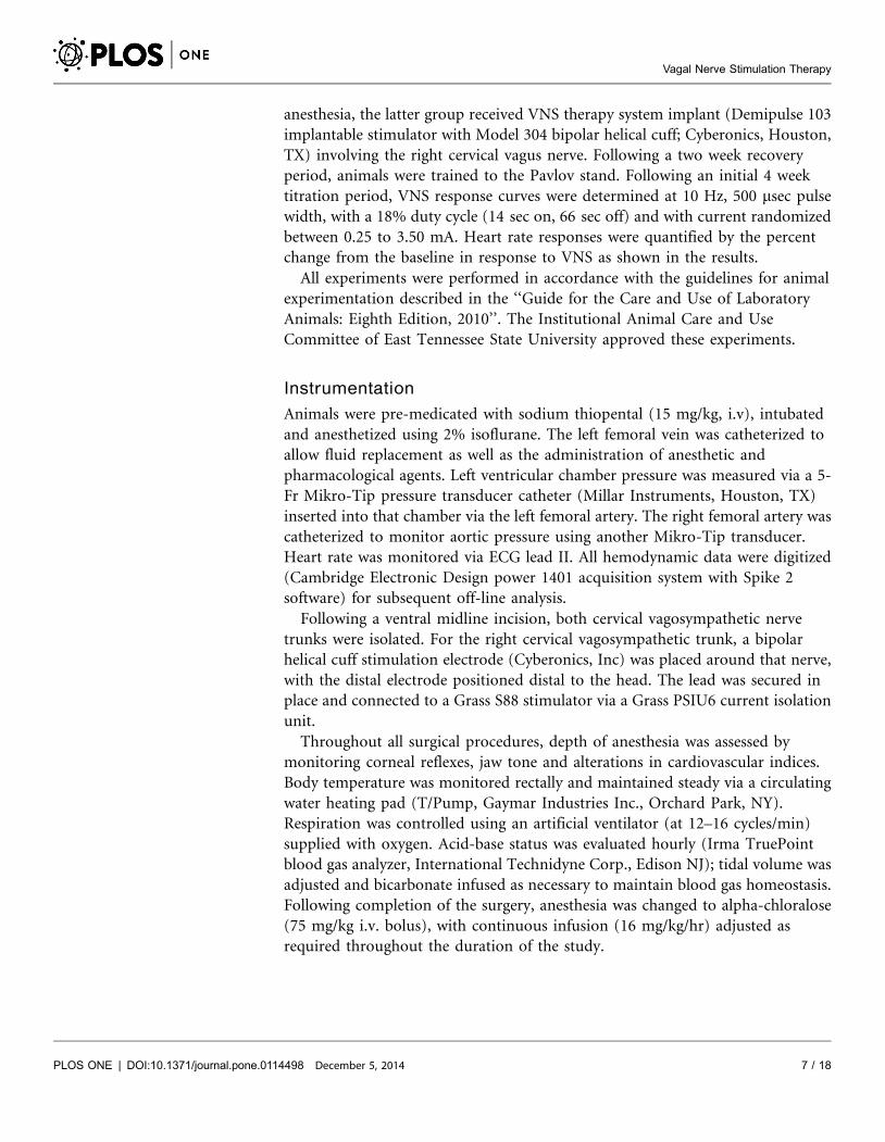
Instrumentation
Animals were pre-medicated with sodium thiopental (15 mg/kg, i.v), intubated
and anesthetized using 2% isoflurane. The left femoral vein was catheterized to
allow fluid replacement as well as the administration of anesthetic and
pharmacological agents. Left ventricular chamber pressure was measured via a 5-
Fr Mikro-Tip pressure transducer catheter (Millar Instruments, Houston, TX)
inserted into that chamber via the left femoral artery. The right femoral artery was
catheterized to monitor aortic pressure using another Mikro-Tip transducer.
Heart rate was monitored via ECG lead II. All hemodynamic data were digitized
(Cambridge Electronic Design power 1401 acquisition system with Spike 2
software) for subsequent off-line analysis.
Following a ventral midline incision, both cervical vagosympathetic nerve
trunks were isolated. For the right cervical vagosympathetic trunk, a bipolar
helical cuff stimulation electrode (Cyberonics, Inc) was placed around that nerve,
with the distal electrode positioned distal to the head. The lead was secured in
place and connected to a Grass S88 stimulator via a Grass PSIU6 current isolation
unit.
Throughout all surgical procedures, depth of anesthesia was assessed by
monitoring corneal reflexes, jaw tone and alterations in cardiovascular indices.
Body temperature was monitored rectally and maintained steady via a circulating
water heating pad (T/Pump, Gaymar Industries Inc., Orchard Park, NY).
Respiration was controlled using an artificial ventilator (at 12–16 cycles/min)
supplied with oxygen. Acid-base status was evaluated hourly (Irma TruePoint
blood gas analyzer, International Technidyne Corp., Edison NJ); tidal volume was
adjusted and bicarbonate infused as necessary to maintain blood gas homeostasis.
Following completion of the surgery, anesthesia was changed to alpha-chloralose
(75 mg/kg i.v. bolus), with continuous infusion (16 mg/kg/hr) adjusted as
required throughout the duration of the study.
Vagal Nerve Stimulation Therapy
PLOS ONE | DOI:10.1371/journal.pone.0114498 December 5, 2014 7 / 18
Experimental Protocol
The right cervical vagus was stimulated electrically with current intensities ranging
from 0.25 mA to 3.5 mA in increments of 0.25 mA. We employed a stimulus
isolation unit (Grass model PSIU6 photoelectric isolation unit) which was
connected to the Grass stimulator to active the vagosympathetic complex with
constant current for anesthetized studies. For conscious animals, we used a
Cyberonics Demipulse 103 implantable stimulator to deliver VNS. Each 122 s
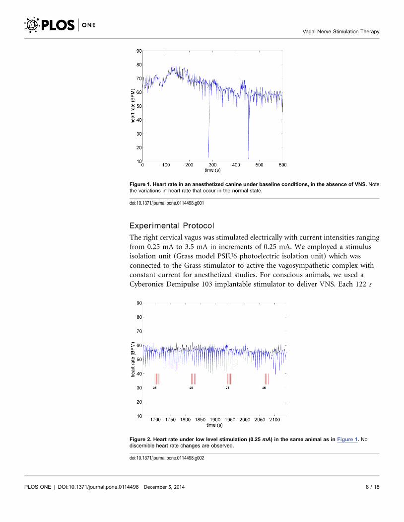
Figure 1. Heart rate in an anesthetized canine under baseline conditions, in the absence of VNS. Notethe variations in heart rate that occur in the normal state.
doi:10.1371/journal.pone.0114498.g001
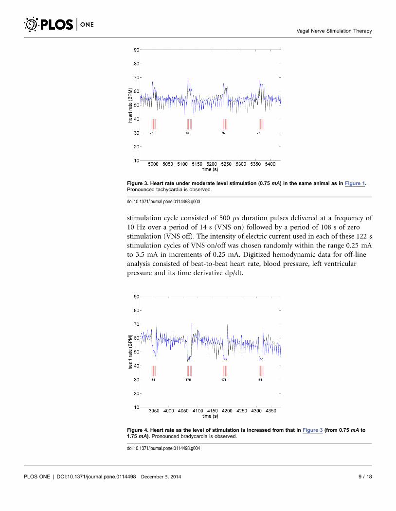
Figure 2. Heart rate under low level stimulation (0.25 mA) in the same animal as in Figure 1. Nodiscernible heart rate changes are observed.
doi:10.1371/journal.pone.0114498.g002
Vagal Nerve Stimulation Therapy
PLOS ONE | DOI:10.1371/journal.pone.0114498 December 5, 2014 8 / 18
stimulation cycle consisted of 500 ms duration pulses delivered at a frequency of
10 Hz over a period of 14 s (VNS on) followed by a period of 108 s of zero
stimulation (VNS off). The intensity of electric current used in each of these 122 s
stimulation cycles of VNS on/off was chosen randomly within the range 0.25 mA
to 3.5 mA in increments of 0.25 mA. Digitized hemodynamic data for off-line
analysis consisted of beat-to-beat heart rate, blood pressure, left ventricular
pressure and its time derivative dp/dt.
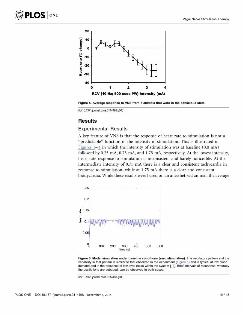
Figure 3. Heart rate under moderate level stimulation (0.75 mA) in the same animal as in Figure 1.Pronounced tachycardia is observed.
doi:10.1371/journal.pone.0114498.g003
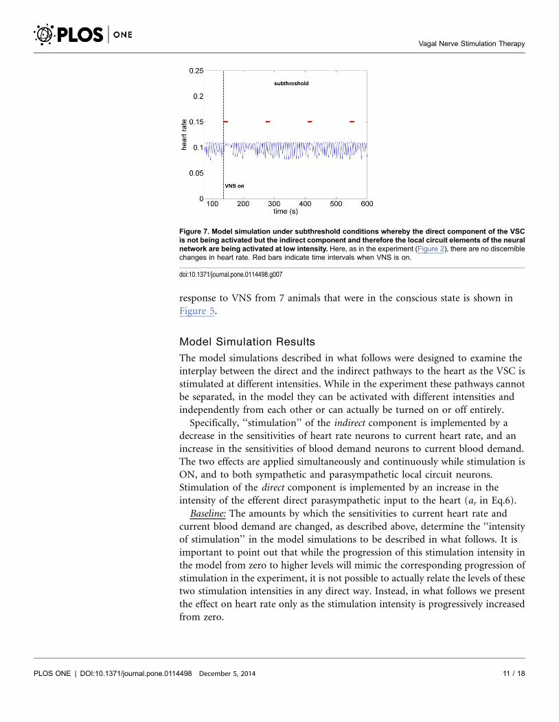
Figure 4. Heart rate as the level of stimulation is increased from that in Figure 3 (from 0.75 mA to1.75 mA). Pronounced bradycardia is observed.
doi:10.1371/journal.pone.0114498.g004
Vagal Nerve Stimulation Therapy
PLOS ONE | DOI:10.1371/journal.pone.0114498 December 5, 2014 9 / 18
Results
Experimental Results
A key feature of VNS is that the response of heart rate to stimulation is not a
‘‘predictable’’ function of the intensity of stimulation. This is illustrated in
Figures 1– 4 in which the intensity of stimulation was at baseline (0.0 mA)
followed by 0.25 mA, 0.75 mA, and 1.75 mA, respectively. At the lowest intensity,
heart rate response to stimulation is inconsistent and barely noticeable. At the
intermediate intensity of 0.75 mA there is a clear and consistent tachycardia in
response to stimulation, while at 1.75 mA there is a clear and consistent
bradycardia. While these results were based on an anesthetized animal, the average
Figure 5. Average response to VNS from 7 animals that were in the conscious state.
doi:10.1371/journal.pone.0114498.g005
Figure 6. Model simulation under baseline conditions (zero stimulation). The oscillatory pattern and thevariability in that pattern is similar to that observed in the experiment (Figure 1) and is typical at low blooddemand and in the presence of low level noise within the system [14]. Brief intervals of resonance, wherebythe oscillations are subdued, can be observed in both cases.
doi:10.1371/journal.pone.0114498.g006
Vagal Nerve Stimulation Therapy
PLOS ONE | DOI:10.1371/journal.pone.0114498 December 5, 2014 10 / 18
response to VNS from 7 animals that were in the conscious state is shown in
Figure 5.
Model Simulation Results
The model simulations described in what follows were designed to examine the
interplay between the direct and the indirect pathways to the heart as the VSC is
stimulated at different intensities. While in the experiment these pathways cannot
be separated, in the model they can be activated with different intensities and
independently from each other or can actually be turned on or off entirely.
Specifically, ‘‘stimulation’’ of the indirect component is implemented by a
decrease in the sensitivities of heart rate neurons to current heart rate, and an
increase in the sensitivities of blood demand neurons to current blood demand.
The two effects are applied simultaneously and continuously while stimulation is
ON, and to both sympathetic and parasympathetic local circuit neurons.
Stimulation of the direct component is implemented by an increase in the
intensity of the efferent direct parasympathetic input to the heart (ar in Eq.6).
Baseline: The amounts by which the sensitivities to current heart rate and
current blood demand are changed, as described above, determine the ‘‘intensity
of stimulation’’ in the model simulations to be described in what follows. It is
important to point out that while the progression of this stimulation intensity in
the model from zero to higher levels will mimic the corresponding progression of
stimulation in the experiment, it is not possible to actually relate the levels of these
two stimulation intensities in any direct way. Instead, in what follows we present
the effect on heart rate only as the stimulation intensity is progressively increased
from zero.
Figure 7. Model simulation under subthreshold conditions whereby the direct component of the VSCis not being activated but the indirect component and therefore the local circuit elements of the neuralnetwork are being activated at low intensity. Here, as in the experiment (Figure 2), there are no discerniblechanges in heart rate. Red bars indicate time intervals when VNS is on.
doi:10.1371/journal.pone.0114498.g007
Vagal Nerve Stimulation Therapy
PLOS ONE | DOI:10.1371/journal.pone.0114498 December 5, 2014 11 / 18
The pattern of heart rate with zero stimulation is shown in Figure 6. The
oscillatory pattern and the variability in that pattern is similar to that observed in
the experiment (Figure 1) and is typical at low blood demand and in the presence
of low level noise within the system [14]. Brief intervals of resonance whereby the
oscillations are subdued can be observed in both cases.
Subthreshold: As the level of stimulation is increased from zero in the model the
effect on heart rate is barely visible, as observed in Figure 7. Here the direct
component of the VSC is turned off and the indirect component is minimally
stimulated as described above. We refer to this set of conditions as ‘‘subthreshold’’
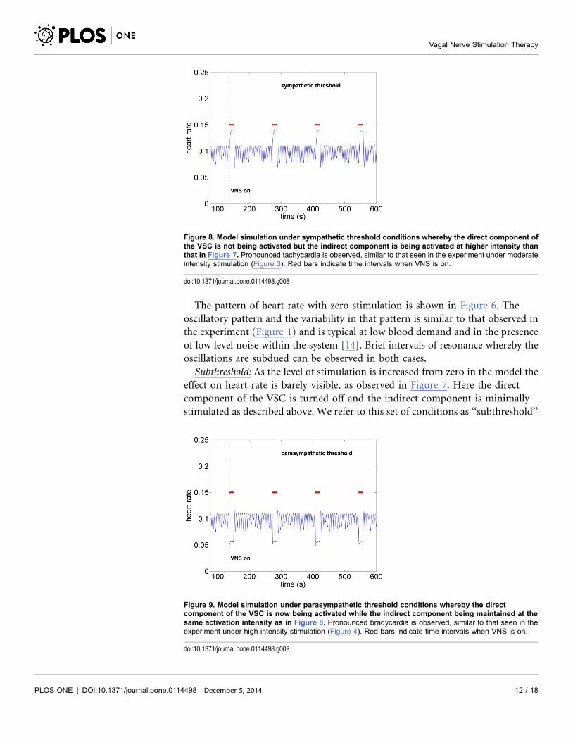
Figure 8. Model simulation under sympathetic threshold conditions whereby the direct component ofthe VSC is not being activated but the indirect component is being activated at higher intensity thanthat in Figure 7. Pronounced tachycardia is observed, similar to that seen in the experiment under moderateintensity stimulation (Figure 3). Red bars indicate time intervals when VNS is on.
doi:10.1371/journal.pone.0114498.g008
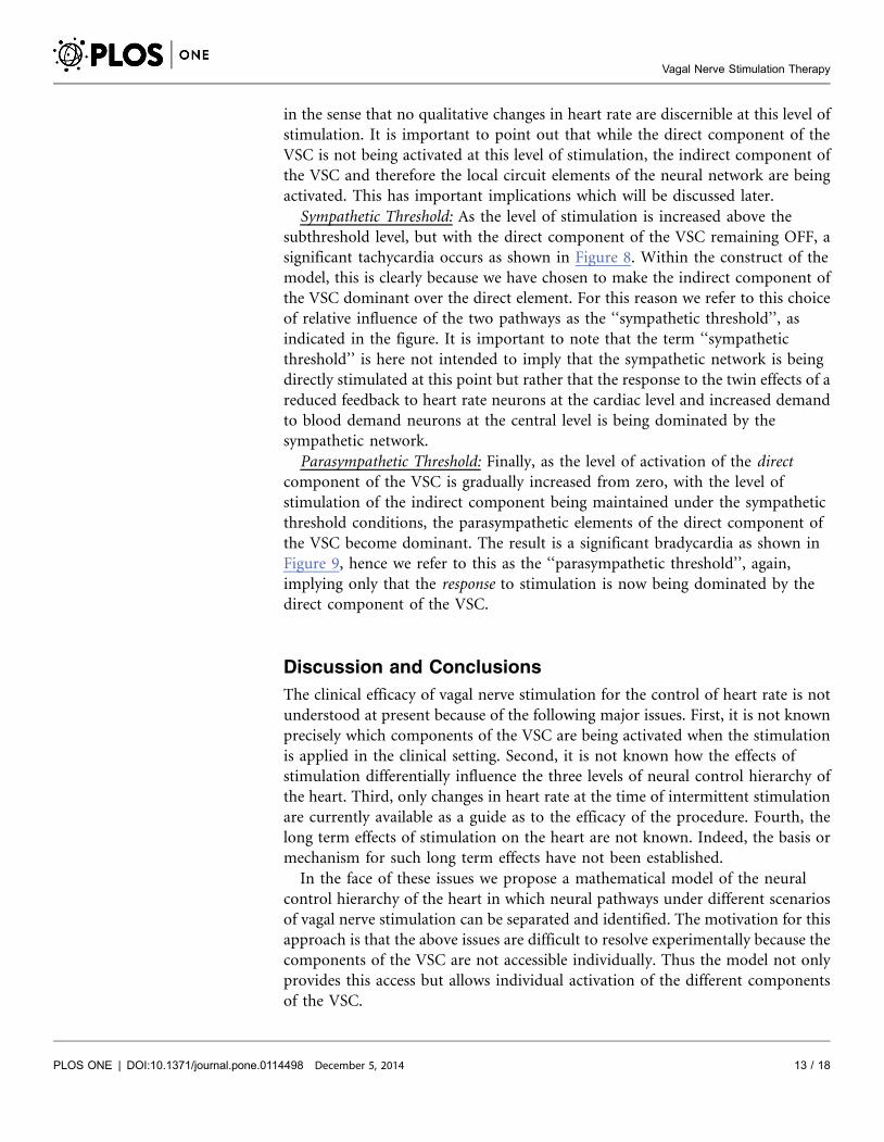
Figure 9. Model simulation under parasympathetic threshold conditions whereby the directcomponent of the VSC is now being activated while the indirect component being maintained at thesame activation intensity as in Figure 8. Pronounced bradycardia is observed, similar to that seen in theexperiment under high intensity stimulation (Figure 4). Red bars indicate time intervals when VNS is on.
doi:10.1371/journal.pone.0114498.g009
Vagal Nerve Stimulation Therapy
PLOS ONE | DOI:10.1371/journal.pone.0114498 December 5, 2014 12 / 18
in the sense that no qualitative changes in heart rate are discernible at this level of
stimulation. It is important to point out that while the direct component of the
VSC is not being activated at this level of stimulation, the indirect component of
the VSC and therefore the local circuit elements of the neural network are being
activated. This has important implications which will be discussed later.
Sympathetic Threshold: As the level of stimulation is increased above the
subthreshold level, but with the direct component of the VSC remaining OFF, a
significant tachycardia occurs as shown in Figure 8. Within the construct of the
model, this is clearly because we have chosen to make the indirect component of
the VSC dominant over the direct element. For this reason we refer to this choice
of relative influence of the two pathways as the ‘‘sympathetic threshold’’, as
indicated in the figure. It is important to note that the term ‘‘sympathetic
threshold’’ is here not intended to imply that the sympathetic network is being
directly stimulated at this point but rather that the response to the twin effects of a
reduced feedback to heart rate neurons at the cardiac level and increased demand
to blood demand neurons at the central level is being dominated by the
sympathetic network.
Parasympathetic Threshold: Finally, as the level of activation of the direct
component of the VSC is gradually increased from zero, with the level of
stimulation of the indirect component being maintained under the sympathetic
threshold conditions, the parasympathetic elements of the direct component of
the VSC become dominant. The result is a significant bradycardia as shown in
Figure 9, hence we refer to this as the ‘‘parasympathetic threshold’’, again,
implying only that the response to stimulation is now being dominated by the
direct component of the VSC.
Discussion and Conclusions
The clinical efficacy of vagal nerve stimulation for the control of heart rate is not
understood at present because of the following major issues. First, it is not known
precisely which components of the VSC are being activated when the stimulation
is applied in the clinical setting. Second, it is not known how the effects of
stimulation differentially influence the three levels of neural control hierarchy of
the heart. Third, only changes in heart rate at the time of intermittent stimulation
are currently available as a guide as to the efficacy of the procedure. Fourth, the
long term effects of stimulation on the heart are not known. Indeed, the basis or
mechanism for such long term effects have not been established.
In the face of these issues we propose a mathematical model of the neural
control hierarchy of the heart in which neural pathways under different scenarios
of vagal nerve stimulation can be separated and identified. The motivation for this
approach is that the above issues are difficult to resolve experimentally because the
components of the VSC are not accessible individually. Thus the model not only
provides this access but allows individual activation of the different components
of the VSC.
Vagal Nerve Stimulation Therapy
PLOS ONE | DOI:10.1371/journal.pone.0114498 December 5, 2014 13 / 18
At the core of our findings, based on the mathematical model, is a distinction
that must be made between direct parasympathetic pathways whereby cardiac
motor postganglionic neurons are targeted directly, and indirect sympathetic and
parasympathetic pathways which interact indirectly with a population of local
circuit neurons on the heart.
The application of VNS in the model is implemented differently for the direct
and indirect pathways. For the direct pathway, stimulation is implemented by
simply increasing the intensity of activation. By contrast, stimulation of the
indirect pathway is implemented by (i) a reduction in sensitivity of heart rate
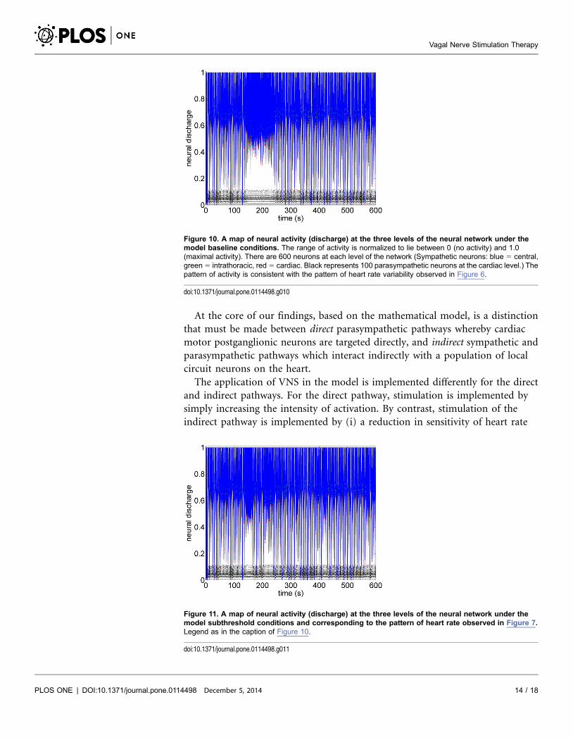
Figure 10. A map of neural activity (discharge) at the three levels of the neural network under themodel baseline conditions. The range of activity is normalized to lie between 0 (no activity) and 1.0(maximal activity). There are 600 neurons at each level of the network (Sympathetic neurons: blue 5 central,green 5 intrathoracic, red 5 cardiac. Black represents 100 parasympathetic neurons at the cardiac level.) Thepattern of activity is consistent with the pattern of heart rate variability observed in Figure 6.
doi:10.1371/journal.pone.0114498.g010
Figure 11. A map of neural activity (discharge) at the three levels of the neural network under themodel subthreshold conditions and corresponding to the pattern of heart rate observed in Figure 7.Legend as in the caption of Figure 10.
doi:10.1371/journal.pone.0114498.g011
Vagal Nerve Stimulation Therapy
PLOS ONE | DOI:10.1371/journal.pone.0114498 December 5, 2014 14 / 18
neurons and (ii) an increase in sensitivity of blood demand neurons. In addition,
in the model, heart rate neurons are located mainly at the level of the intrinsic
nervous system while blood demand neurons are located mainly at the level of the
central nervous system. Thus, VSC stimulation of the indirect pathway in the
model is represented as a complex combination of a reduction in afferent
feedback and an increase in efferent drive caused by an increase in central
sensitivity to blood demand.
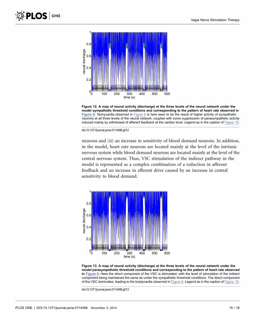
Figure 12. A map of neural activity (discharge) at the three levels of the neural network under themodel sympathetic threshold conditions and corresponding to the pattern of heart rate observed inFigure 8. Tachycardia observed in Figure 8 is here seen to be the result of higher activity of sympatheticneurons at all three levels of the neural network, coupled with some suppression of parasympathetic activityinduced mainly by withdrawal of afferent feedback at the cardiac level. Legend as in the caption of Figure 10.
doi:10.1371/journal.pone.0114498.g012
Figure 13. A map of neural activity (discharge) at the three levels of the neural network under themodel parasympathetic threshold conditions and corresponding to the pattern of heart rate observedin Figure 9. Here the direct component of the VSC is stimulated, with the level of stimulation of the indirectcomponent being maintained the same as under the sympathetic threshold conditions. The direct componentof the VSC dominates, leading to the bradycardia observed in Figure 9. Legend as in the caption of Figure 10.
doi:10.1371/journal.pone.0114498.g013
Vagal Nerve Stimulation Therapy
PLOS ONE | DOI:10.1371/journal.pone.0114498 December 5, 2014 15 / 18
The model results indicate that at very low levels of stimulation, if only the
indirect pathways through the VSC are activated, no discernible change in heart
rate is produced. As the level of stimulation is gradually increased beyond a
certain threshold, a clear tachycardia is observed. Then, as the intensity of the
direct pathway is gradually increased, while the level of activation of the indirect
pathways are maintained, another threshold emerges where the net effect on heart
rate is reversed and a clear bradycardia becomes evident.
We have thus modeled the response to increased levels of activation of the VSC
as the differential activation of different components of the VSC. To the extent
that the model results exhibit features similar to those observed in the
anesthetized animal, we are led to conclude that different components of the VSC
may respond differentially to different levels of stimulation.
In particular, our interpretation of the bradycardia observed at higher levels of
stimulation in the experiment is that the effect of stimulation is now dominated
by the direct parasympathetic elements of the VSC whose intensity has been
increased. On the other hand, our interpretation of the tachycardia observed at
lower levels of stimulation in the experiment is that now the indirect elements of
the VSC are primarily active. As stated earlier, in this case VNS stimulation in the
model is represented as a complex combination of a reduction in afferent
feedback and an increase in efferent drive caused by an increase in central
sensitivity to blood demand. The net result is the observed increased heart rate.
What the mathematical model also provides beyond what is available in the
animal experiment is the simultaneous assessment of neuronal activity
throughout the entire cardiac neuronal hierarchy. This is illustrated in Figures 10–
13 which correspond to the baseline and threshold conditions seen in Figures 6–
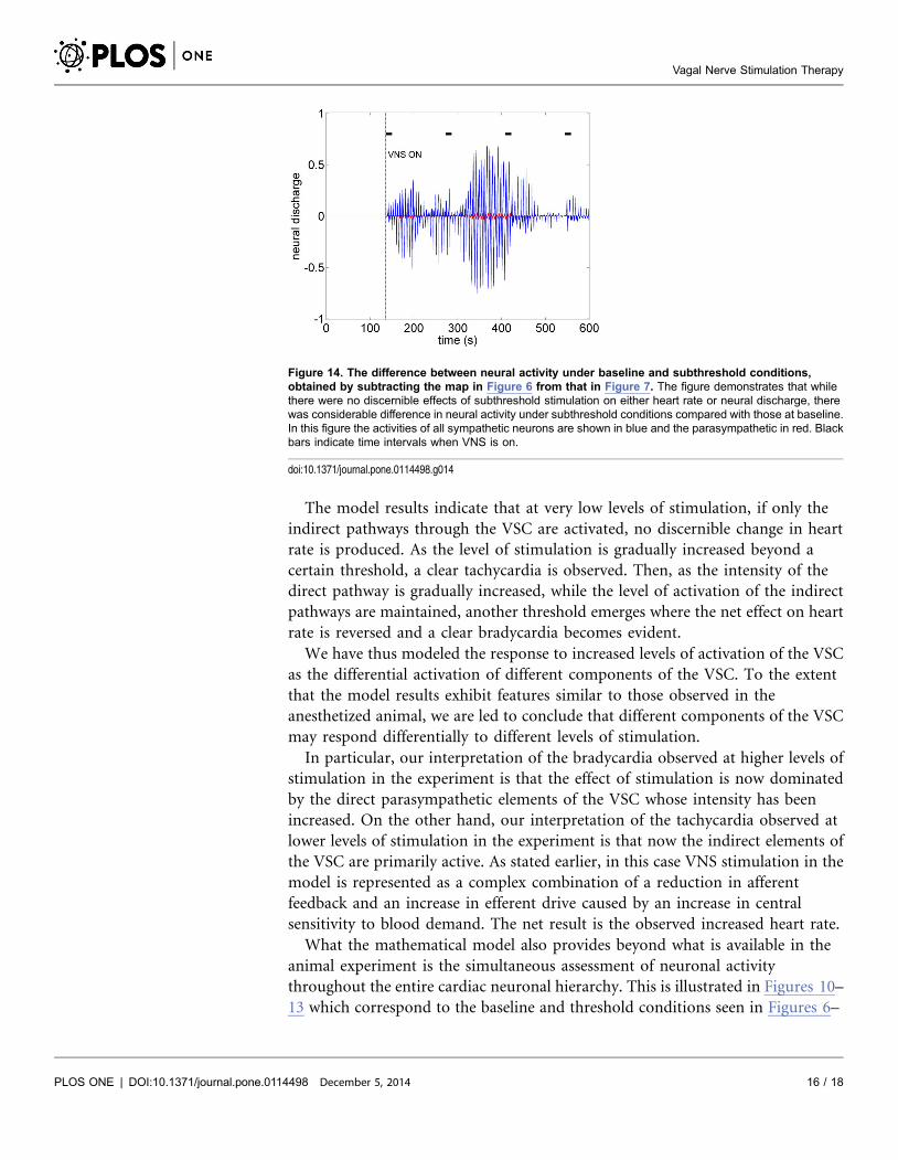
Figure 14. The difference between neural activity under baseline and subthreshold conditions,obtained by subtracting the map in Figure 6 from that in Figure 7. The figure demonstrates that whilethere were no discernible effects of subthreshold stimulation on either heart rate or neural discharge, therewas considerable difference in neural activity under subthreshold conditions compared with those at baseline.In this figure the activities of all sympathetic neurons are shown in blue and the parasympathetic in red. Blackbars indicate time intervals when VNS is on.
doi:10.1371/journal.pone.0114498.g014
Vagal Nerve Stimulation Therapy
PLOS ONE | DOI:10.1371/journal.pone.0114498 December 5, 2014 16 / 18

9. These figures show clearly that under subthreshold conditions, while there are
no discernible changes in heart rate (Figure 7), there is considerable neural
activity at all levels of the neural network (Figure 11). That this neural activity is
different from baseline, is confirmed in Figure 14 which shows the difference
between the level of discharge of each neuron at baseline and at subthreshold
conditions.
The sensitivity of the neural network to low level subthreshold stimulation leads
us to propose that the chronic effects of VNS therapy lie within the indirect
pathways because plasticity and the potential for remodeling reside within the
local circuit neurons of the intrinsic cardiac system and within the neural network
comprising the indirect pathways [24]. By inference, the prospects for long term
effects of VNS therapy lie in low level, ‘‘subthreshold’’, activation where the
indirect ‘‘plastic’’ components of the control system are involved.
Author Contributions
Conceived and designed the experiments: GK JLA JAA MZ. Performed the
experiments: GK JLA JAA MZ. Analyzed the data: GK JLA JAA MZ. Contributed
reagents/materials/analysis tools: GK JLA JAA MZ. Wrote the paper: GK JLA JAA
MZ.
References
1. Groves DA, Brown VJ (2005) Vagal nerve stimulation: a review of its applications and potentialmechanisms that mediate its clinical effects. Neuroscience and Biobehavioral Reviews 29: 493–500.
2. Ando M, Katare RG, Kakinuma Y, Zhang D, Yamasaki F, et al. (2005) Efferent vagal nerve stimulationprotects heart against ischemia-induced arrhythmias by preserving connexin43 protein. Circulation 112:164–170.
3. Zhang Y, Mazgalev TN (2011) Arrhythmias and vagus nerve stimulation. Heart Fail Rev 16: 147–161.
4. De Ferrari GM, Crijns HJ, Borggrefe M, Milasinovic G, Smid J, et al. for the CardioFit Multicenter TrialInvestigators (2011) Chronic vagus nerve stimulation: a new and promising therapeutic approach forchronic heart failure. European Heart Journal 32: 847–855.
5. DiCarlo L, Libbus I, Amurthur B, KenKnight BH, Anand IS (2013) Autonomic regulation therapy forthe improvement of left ventricular function and heart failure symptoms: the ANTHEM-HF study. J CardFail 19: 655–660.
6. Foley JO, DuBois F (1937) Quantitative studies of the vagus nerve in the cat: I. The ratio of sensorymotor studies. J Comp Neurol 67: 49–67.
7. Bailey P, Bremer FA (1938) Sensory cortical representation of the vagus nerve. J Neurophysiol 1: 405–412.
8. Schwartz PJ, De Ferrari GM, Sanzo A, Landolina M, Rordorf R, et al. (2008) Long term vagalstimulation in patients with advanced heart failure: first experience in man. Eur J Heart Fail 10: 884–891.
9. Zhang Y, Popovic ZB, Bibevski S, Fakhry I, Sica DA, et al. (2009) Chronic vagus nerve stimulationimproves autonomic control and attenuates systemic inflammation and heart failure progression in acanine high-rate pacing model. Circ Heart Fail 2: 692–699.
10. Osman F, Kundu S, Tuan J, Jeilan M, Stafford PJ, et al. (2010) Ganglionic plexus ablation duringpulmonary vein isolationpredisposing to ventricular arrhythmias? Indian Pacing and Electrophysiol J 10:104–107.
Vagal Nerve Stimulation Therapy
PLOS ONE | DOI:10.1371/journal.pone.0114498 December 5, 2014 17 / 18
11. Kasanuki H, Ohnishi S, Ohtuka M, Matsuda N, Nirei T, et al. (1997) Idiopathic ventricular fibrillationinduced with vagal activity in patients without obvious heart disease. Circulation 95: 2277–2285.
12. Randall WC, Armour JA (1977) Gross and microscopic anatomy of the cardiac innervation. In: NeuralRegulation of the Heart. Randall WC, Editor, Oxford Univ Press, N.Y.
13. Beaumont E, Salavatian S, Southerland EM, Vinet A, Jacquemet V, et al. (2013) Network interactionswithin the canine intrinsic cardiac nervous system: Implications for reflex control of regional cardiacfunction. J Physiol 591: 4515–4533.
14. Kember G, Armour J, Zamir M (2011) Neural control of heart rate: the role of neuronal networking.J Theor Biol 277: 41–47.
15. Cook IA, Espinoza R, Leuchter AF (2014). Neuromodulation for Depression: Invasive and Noninvasive(Deep Brain Stimulation, Transcranial Magnetic Stimulation, Trigeminal Nerve Stimulation).Neurosurgery Clinics of North America 25: 103–116.
16. Howland RH (2014) Vagus Nerve Stimulation. Current Behavioral Neuroscience Reports 1: 64–73.
17. Shiozawa P, Enokibara da Silva M, Cristina de Carvalho T, Cordeiro Q, Brunoni AR, et al. (2014)Transcutaneous vagus and trigeminal nerve stimulation for neuropsychiatric disorders: a systematicreview. Arquivos de Neuro-Psiquiatria 72: 542–7.
18. Zare M, Salehi M, Mahvari J, Najafi MR, Moradi A, et al. (2014) Trigeminal nerve stimulation: A newway of treatment of refractory seizures. Advanced Biomedical Research 3: 81.
19. Koo B, Ham SD, Sood S, Tarver B (2001) Human vagus nerve electrophysiologya guide to vagus nervestimulation parameters. J Clin Neurophysiol 18: 429–433.
20. Krahl SE, Senanayake SS, Handforth A (2001) Destruction of peripheral C-fibers does not altersubsequent vagus nerve stimulation induced seizure suppression in rats. Epilepsia 42: 586–589.
21. Nahas Z, Marangell LB, Husain MM, Rush AJ, Sackeim HA, et al. (2005) Two-year outcome of vagusnerve stimulation (VNS) for treatment of major depressive episodes. J Clin Psychiatry 66(9): 1097–104.
22. Woodbury DM, Woodbury JW (1990) Effects of vagal stimulation on experimentally induced seizures inrats. Epilepsia 31 (Suppl 2): S7–S19.
23. Williamson JW, Fadel JP, Mitchell JH (2005) New insights into central cardiovascular control duringexercise in humans: a central command update. Exp Physiol 91: 51–58.
24. Kember G, Armour J, Zamir M (2012) Dynamic neural networking as a basis for plasticity in the controlof heart rate. J Theor Biol 317: 39–46.
25. Kember G, Armour JA, Zamir M (2013) Neural control hierarchy of the heart has not evolved to dealwith myocardial ischemia. Physiol. Genomics 45: 638–644.
26. Turrigiano G, Nelson S (2004) Homeostatic plasticity in the developing nervous system. Nat RevNeurosci 5: 97–107.
27. Turrigiano G, Nelson S (2000) Hebb and homeostasis in neuronal plasticity. Curr Opin Neurbiol 10:358–364.
28. Randall WC, Ardell JL (1985) Selective parasympathectomy of automatic and conductile tissues of thecanine heart. Am J Physiol 248: H61–H68.
Vagal Nerve Stimulation Therapy
PLOS ONE | DOI:10.1371/journal.pone.0114498 December 5, 2014 18 / 18