effect of morphology on densification study of nano...
TRANSCRIPT

EFFECT OF MORPHOLOGY ON DENSIFICATION
STUDY OF NANO – HYDROXYAPATITE
Presented by :-
Kunal Kumar Tanty
108CR054
NIT Rourkela
PROJECT THESISSUBMITTED IN PARTIAL FULFILMENT OF THE REQUIREMENTS
FOR THE DEGREE OF
BACHELOR OF TECHNOLOGY
Department of Ceramic Engineering National Institute of Technology, Rourkela, Orissa - 769008

i
Certificate
This is to certify that this thesis entitled , “EFFECT OF MORPHOLOGY ON DENSIFICATION
STUDY OF NANO – HYDROXYAPATITE”, submitted by Mr Kunal Kumar Tanty in partial
fulfilment of the requirement of the award of Bachelor of Technology Degree in Ceramic
Engineering at National Institute of Technology, Rourkela is an authentic work carried out by
him under my supervision and guidance.
To the best of my knowledge, the matter embodied in the thesis has not been submitted to any other
university/institute for the award of Degree or Diploma.
Dr.DebasishSarkar
Date: Department of Ceramic Engineering
National Institute of Technology,
Rourkela, Orissa - 769008

ii
Acknowledgements
I would like to express my heartfelt thanks and gratitude to Professor Debasish Sarkar, Department
of Ceramic Engineering, NIT Rourkela, my guide and my mentor, who was with me during every
stage of the work; and whose guidance and valuable suggestions has made this work possible.
Further, I would also like to thank all the faculty members and staff of Department of Ceramic
Engineering, NIT Rourkela for their invaluable support and help during the entire project work.
I would further like to thank all the research scholars, especially Mr. Sanjay Swain, Mr. Ganesh
kumar sahoo and last but not the least Miss Geetanjali parida, for their round the clock help and
support during the entire project work.
Last but not the least I want to thank almighty lord for the successful completion of the project
work.
Kunal Kumar Tanty
Roll No- 108CR054

iii
Abstract
Spherical, rod and fibrous morphology of Hydroxyapatite (HA) nanoparticles were considered to
understand the densification behaviour at high temperature. Thermal analysis of as-received HA
nanoparticles confirmed the phase transformation temperature for different morphologies. The
phase stability was strictly deepened on morphology of HA nanoparticles. X – ray diffraction
(XRD) pattern and Fourier transformation Infrared (FTIR) was studied to evaluate the crystal
structure, phase and purity for different calcined powders and their sintered specimens. Spherical,
rod and fibrous nano-HA morphology were stable up to 1200oC, 700
oC and 1000
oC, respectively,
which were decomposed to beta tricalcium phosphate ( – TCP) beyond these critical temperatures.
Dilatometric study of all green specimens prepared from such nanoparticles was conducted up to
1250oC to synchronize the sintering temperature. The densification behaviour was dramatically
changed with respect to morphology when sintered at different temperatures; such as
700oC,900
oC,1000
oC, 1250
oC, respectively. Scanning electron microscopic (SEM) image, apparent
porosity and relative density were studied to justify the densification behaviour of HA
nanoparticles.

iv
Table of Contents
EFFECT OF MORPHOLOGY ON DENSIFICATION STUDY OF NANO –
HYDROXYAPATITE ........................................................................................................................ i
Acknowledgements ............................................................................................................................ ii
Abstract .............................................................................................................................................. iii
Table of Contents .............................................................................................................................. iv
LIST OF FIGURES .......................................................................................................................... vi
LIST OF TABLES ............................................................................................................................ vi
Chapter 1 ............................................................................................................................................ 1
INTRODUCTION .............................................................................................................................. 1
1.1 HYDROXYAPATITE. ......................................................................................................... 2
1.1.1 STRUCTURE ........................................................................................................................................... 3
1.1.2 APPLICATIONS ...................................................................................................................................... 4
Chapter 2 ............................................................................................................................................ 5
LITERATURE REVIEW.................................................................................................................. 5
2.1 DENSIFICATION OF HYDROXYAPATITE. .................................................................... 6
2.2 MORPHOLOGICAL STUDIES ........................................................................................... 6
2.3 STRUCTURE EFFECTS ...................................................................................................... 7
2.4 OBJECTIVE .......................................................................................................................... 8
Chapter 3 ............................................................................................................................................ 9
EXPERIMENTAL ............................................................................................................................. 9
3.1 PELLETIZATION .............................................................................................................. 10
3.2 SINTERING AT DIFFERENT TEMPERATURES:- ........................................................ 10
3.3 CHARACTERISATION ..................................................................................................... 11
3.3.1 DSC/TG .................................................................................................................................................. 11
3.3.2 XRD ....................................................................................................................................................... 11
3.3.3 FTIR ....................................................................................................................................................... 11
3.3.4 DILATOMETRIC ANALYSIS ............................................................................................................. 12
3.3.5 SEM ANALYSIS .................................................................................................................................. 12
3.3.6 BULK DENSITY AND APPARENT POROSITY MEASUREMENT ................................................ 12
Chapter 4 .......................................................................................................................................... 13
4.1 THERMAL ANALYSIS OF HYDROXYAPATITE ......................................................... 14
4.1.1 THERMAL ANALYSIS OF NANO SPHERICAL HYDROXYAPATITE ......................................... 14
4.1.2 THERMAL ANALYSIS OF NANO FIBRE HYDROXYAPATITE ................................................... 15

v
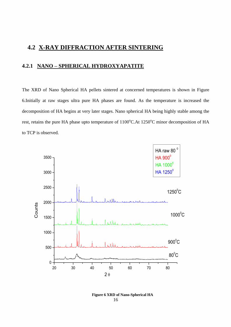
4.2 X-RAY DIFFRACTION AFTER SINTERING ................................................................. 16
4.2.1 NANO – SPHERICAL HYDROXYAPATITE ..................................................................................... 16
4.2.2 NANO – ROD HYDROXYAPATITE ................................................................................................... 17
4.2.3 NANO-FIBRE HYDROXYAPATITE .................................................................................................. 18
4.3 FTIR SPECTROSCOPY TO CHECK PURITY................................................................. 19
4.4 DILATOMETRIC STUDY OF HYDROXYAPATITE ..................................................... 20
4.5 SEM ANALYSIS ................................................................................................................ 21
4.5.1 NANO –SPHERICAL HYDROXYAPATITE ...................................................................................... 21
4.5.2 NANO ROD HYDROXYAPATITE ...................................................................................................... 22
4.5.3 NANO FIBRE HYDROXYAPATITE ................................................................................................... 23
4.6 BULK DENSITY OF DIFFERENT MORPHOLOGIES ................................................... 24
4.7 APPARENT POROSITY OF DIFFERENT MORPHOLOGIES ....................................... 25
4.8 BULK DENSITY AND APPARENT POROSITY AS A FUNCTION OF
TEMPERATURE........................................................................................................................... 26
4.8.1 NANO SPHERICAL HYDROXYAPATITE ........................................................................................ 26
4.8.2 NANO ROD HYDROXYAPATITE ...................................................................................................... 26
4.8.3 NANO FIBRE HA ................................................................................................................................. 27
Chapter 5 .......................................................................................................................................... 28
CONCLUSION ................................................................................................................................ 28
REFERENCES ................................................................................................................................. 30

vi
LIST OF FIGURES
Figure 1 HA crystal on matrix [1] ........................................................................................................ 2
Figure 2 Crystal Structure [2] .............................................................................................................. 3
Figure.3 Hydroxyapatite morphologies : (a) spherical (b) rod shaped (c) fibre shaped [3,4,5] ........ 3
Figure 4 DSC/TG of Nano Spherical HA .......................................................................................... 14
Figure 5 DSC/TG of Nano fibre HA . ................................................................................................ 15
Figure 6 XRD of Nano-Spherical HA................................................................................................ 16
Figure 7 XRD of Nano Rod HA . ..................................................................................................... 17
Figure 8 XRD of Nano fibre HA ...................................................................................................... 18
Figure 9 IR Spectrum composite of nano - (a) spherical (b) rod (c) fibre ........................................ 19
Figure 10 Dilatometric study of Nano HA......................................................................................... 20
Figure 11 SEM of Sintered Nano-spherical HA at 1250oC .............................................................. 21
Figure 12 SEM of sintered Nano-spherical HA ................................................................................. 22
Figure 13 (a) SEM of calcined Nano-fibre HA (b) SEM of raw Nano-fibre HA .............................. 23
Figure 14 BD composite of HA ......................................................................................................... 24
Figure 15 AP composite of HA.......................................................................................................... 25
Figure 16 BD vs AP of Nano-spherical HA ...................................................................................... 26
Figure 17 BD vs AP of Nano-rod HA................................................................................................ 27
Figure 18 BD vs AP of Nano-fibre HA ............................................................................................. 27
LIST OF TABLES
Table 1 Sintering temperatures .......................................................................................................... 10

1
Chapter 1
INTRODUCTION

2
1.1 HYDROXYAPATITE.
Hydroxyapatite(HA) is a naturally occurring mineral form of calcium apatite with the formula
Ca5(PO4)3(OH) which can be written as Ca10(PO4)6(OH)2 to denote that the crystal unit cell
comprises of two entities. HA material has been clinically applied in many areas of density and
orthopaedics because of its excellent osteoconductive and bio active properties. It is the main
inorganic constituent of bones in humans. Synthetic HA is successful in hard tissue surgery and
undergoes bonding osteogenesis. However it has low load bearing application as because of its poor
mechanical properties. In present scenario morphologies are playing a vital role in the mechanical
properties of the body, also the microstructure. Nano structured HA with different morphologies
like spherical, rod and fibre are found to characterise HA in different application.
Figure 1 HA crystal on matrix [1]
HA on sintering decomposes to anhydrous calcium phosphate such as Tri calcium
phosphate(TCP),at higher temperature as a result of dehydroxylation beyond a critical point. An
important property of HA is that it is chemically stable for long periods of time. Different
densification behaviour of HA particles were observed in the temperature range of 1100 – 1300oC.
While porous HA is advantageous promoting bone in growth and dense HA is used in load bearing
application.

3
1.1.1 STRUCTURE
Figure 2 Crystal Structure [2]
Hydroxylapatite is the hydroxyl end member of the complex apatite group. The OH- ion can be
replaced by fluoride, chlorideor carbonate, producing flouroapatite or chloroapatite. It crystallises in
the hexagonal crystal system. Pure HA powder is white. Naturally occurring apatites can, however
,also have brown, yellow, or green colorations, comparable to the discolorations of dental flourosis.
We are concerned with the following three morphologies as shown in figure 3.
Figure.3Hydroxyapatite morphologies : (a) spherical (b) rod shaped (c) fibre shaped[3,4,5]

4
1.1.2 APPLICATIONS
HA can be found in teeth and bones within the human body. Commonly used as a filler to replace
amputated bone or as a coating to promote bone in growth into prosthetic implants, although many
other phases exist with similar or even identical chemical makeup, the body responds much
differently to them. Coral skeletons can be transformed into HA by high temperatures. Their porous
structure allows relatively rapid growth at the expense of initial mechanical strength. The high
temperature also burns away any organic molecule such as proteins, preventing an immune
response and rejection.

5
Chapter 2
LITERATURE REVIEW

6
2.1 DENSIFICATION OF HYDROXYAPATITE.
S.laasri et al presented report on the effect of densification on the mechanical properties of HA.
[6]The densification study was done at range of 1000 0C -1300
0C.Further XRD and SEM were used
to verify the micro structural transformation. It was found that the sintering temperature and particle
size influence the densification and mechanical properties strongly. This showed the mechanical
property of HA being sensitive to densification
CY tan et al followed the method of microwave rapid sintering which was found to be beneficial in
terms of densification and mechanical properties. [7] This property achieved a theoretical density of
96%at a sintering temperature of 1100oC maintaining its microstructure. However further sintering
the body were not governed by density effect but were associated with grain size effect. The
sinterability of nano HA was studied. The xrd patterns of the sintered HA compacts fired between
1000 o C and 1250
oC were studied. The decomposition of HA was not observed throughout the
sintering process. However grain coarsening was observed above 1200 oC. Regardless of sintering
methods there existed a co-relation between hardness & grain size in HA. The hardness of HA
would start decreasing at certain critical grain size limit despite high bulk density.
2.2 MORPHOLOGICAL STUDIES
Ashis Banerjee et al studied the effect of powder morphology on densification at 1250 0 c with
different amount of rod shaped and spherical nano –powders. [8]It was observed that an increase in
high aspect ratio powder content in the compacts decreased sintered density under pressure less
sintering conditions. Sometimes no nano scale morphology could be retained in sintered

7
microstructure. However in case of simulated body fluid showed formation of apatite layer on entire
surface of compacts made with spherical and rod-shaped particles.
Microhardness was decreased due to the addition of rod-shaped particle and fracture toughness
increased with rod-shaped particle additive to spherical nanopowder. It was also found that due to
grain growth no rod shaped particles were pressed after high temperature sintering with the
formation of dense apatite layer.
DyCheang et al examined the effect of powder morphology on the tensile properties of polymer-HA
composite [9]. Experimental findings showed that surface morphology and structural integrity of
HA have considerable influence on properties of composites. HA with rough surfaces promoted
mechanical interlocking and also improved the interfacial bonding. Relatively lower tensile strength
was achieved by porous agglomerated HA powder.
2.3 STRUCTURE EFFECTS
Hala Zreiqat et al studied the influence of hydroxylapatite nanoparticle shape and size on biphasic
calcium phosphate scaffold and found that highly porous scaffold showed maximum compressive
strength and the nano composite coated scaffold of needle shaped HA showed strong osteoblast
profile, which shows that scaffold possessed improved mechanical and biological properties which
proves essential in application of bone tissue regeneration[10].
E.Landi et al prepared and characterised HA powders with different crystallinity degrees. [11]Now
densification and densification mechanism were studied at a range of 700-1250 oC .The effect of
different powder features with calcinations effect were evaluated .powder characterised by lowest
crystallinity had highest densification. The physio-chemical features of different powders affect the

8
densification behaviour and grain growth is stimulated due to grain boundary diffusion process,
which improves densification. The whole process is time and temperature dependent.
Irina et al characterised the composite by FTIR spectroscopy with X-ray diffraction and scanning
electronmicroscopy, which showed typical absorption bands for ACand HA confirming the
formation of composite. [12]
The XRD patterns revealed the characteristics peaks of low crystalline HA. The SEM micrographs
shared the morphology of pre mineralised and composite samples and hence estimated the HA
particles.
G.Muralitharan et al studied the effects of sintering temperature on properties of HA. [13]It showed
that the sintering of HA resulted in microstructure and the properties were influenced by
characterisation and impurities of raw material and also depended on the thermal history during the
fabrication process. This work was concerned on effect of grain size on relative density and
hardness .Here is iso-statically pressed HA was sintered at temperature ranging from 1000 oC to
1450 o C and it was found that upto 1250
oC the material composed pure HA phase. Porosity and
grain size played an important role in determining the properties of sintered HA.
2.4 OBJECTIVE
To understand the phase stability of different morphologies at a particular level of
temperature
To understand the densification behaviour with respect to temperature, morphologies and
phase stability.

9
Chapter 3
EXPERIMENTAL

10
3.1 PELLETIZATION
0.5 gm each of nano spherical and nano fibre HA and 0.3 gm of nano rod HA were weighed for
preparing pellets. A few drops of poly-vinyl alcohol (PVA 2%) in an agate mortar was mixed with each
composition. Keeping the dwell time to 120 seconds, powder compositions were then pressed in a
circular die of diameter 12mm at 4 ton pressure. For dilatometric test 1.0 gm each of nano spherical
and nano fibre HA and 0.5 gm of nano rod HA were weighed for preparing pellets. Keeping the
dwell time upto 120 second and pressure upto 4 ton pressing was done to form bars.
3.2 SINTERING AT DIFFERENT TEMPERATURES:-
After pressing the green samples were fired at 400oC, 700
oC,900
o C,1000
oC,1250
oC for 2 hrs. Firing
were done in electric chamber furnace ,pit furnace, initially kept on hold at 650oC for 1 hr for
complete binder burnout and then soaked at firing temperature for 2 hr. Heating rate was kept to
3oCmin
-1.The fired product was then cooled at room temperature and bulk density was calculated.
Table 1Sintering temperatures
Morphology
Spherical
Rod
Fibre
Sintering temperatures
Raw(800C) 400
oC 700
oC 900
oC 1000
oC 1250
0C
Y N N Y Y Y
Y Y Y Y Y Y
Y N Y Y Y Y

11
3.3 CHARACTERISATION
3.3.1 DSC/TG
The DSC/ TG thermal analysis were conducted in a Netzsch 449C Thermal Analyser. The
samples were heated in flowing argon (Ar) gas atmosphere keeping the heating rate of
5ºC/min and10ºC/min in the temperature range of 0oC to 1250
oC . The weight loss
measurements were also done in the same instrument.
3.3.2 XRD
The X=Ray Diffraction pattern of raw and sintered samples at 400oC,700
o C,900
o
C,1000oC,1250
oC of each nano spherical,nano rod and nano fibre morphology HA were
obtained in Philips X-Ray Diffractometer(pan-analytical PW 1730) with nickel filtered Cu
Kα radiation(λ=1.5406 Ᾰo)at 40 kV and 30 mA. The scanning rate was kept at 0.04 sec
-1
within a continuous scanning range of 20o to 80
o.
3.3.3 FTIR
The presence of the concerned functional groups at raw stages was studied by Fourier
Transform Infrared Spectroscopy, FTIR(perkin Elmer US).The FTIR spectra were taken on
the sample powders. The binders were pressed to a circular-disc (10 mm Ø) by mixing small
quantity of powder with Potassium bromide (KBr). The samples scanning wave number
range was kept at 4000-400 cm-1
.

12
3.3.4 DILATOMETRIC ANALYSIS
The shrinkage behaviour were obtained by NETZSCH dilatometer model DIL 402 C with a
sample length of 10mm and thickness of 2mm bar pellet. The heating rate was kept constant
at 10oCmin
-1 with firing temperature beginning from room temperature to 1300
oC. The
atmosphere inside the dilatometer was N2 atmosphere
3.3.5 SEM ANALYSIS
Raw samples in form of powder and sintered pellets at 400 o C,700
oC,900
oC,1000
o C,
1250 o C for 2 hrs were characterised in order to determine the morphological changes and
micro structural changes as a result of densification after sintering.
.
3.3.6 BULK DENSITY AND APPARENT POROSITY MEASUREMENT
To measure bulk density and apparent porosity of sintered HA pellets, the dry weight of
pellets was first measured. Then they were soaked in kerosene inside a beaker and were
evacuated till all the air bubbles vanished. After that they were kept inside dessicator
vacuum for few hours. After removing from the vacuum the suspended weight and soaked
weight of the samples were calculated. In order to obtain bulk density (B.D.) the following
formula was used:
B.D. = (dry weight)*(0.813)/(soak weight-suspended weight)
To obtain apparent porosity (A.P.) in % the formula used is:
A.P. = (soak weight-dry weight)*100/(soak weight-suspended weight)

13
Chapter 4
RESULTS AND DISCUSSION

14
4.1 THERMAL ANALYSIS OF HYDROXYAPATITE
4.1.1 THERMAL ANALYSIS OF NANO SPHERICAL HYDROXYAPATITE
Figure 4 shows the DSC/ TG study of the nano spherical HA at 5ºC/min showing significant weight
loss taking place only upto around 1250ºC. The plotted curve shows endothermic peaks at 100 oC
due to dehydroxylation of absorbed moisture leading to mass loss. Broad exothermic peaks at
600oC corresponds to the decomposition of carbonate resulting in significant weight loss and also
these exothermic peak at higher temperature corresponds to the crystallisation and oxidation of the
residual mass .TG curve shows weight loss of 12%, as a result of PVA burnout and moisture loss
followed by the weight loss added by decomposition of phases.
0 200 400 600 800 1000 1200 1400
-22
-20
-18
-16
-14
-12
-10
-8
-6
-4
-2
0
2
4
6
8
10
12
14
Temperature oC
DS
C
86
88
90
92
94
96
98
100
102
MA
SS
%
Figure 4 DSC/TG of Nano Spherical HA

15
4.1.2 THERMAL ANALYSIS OF NANO FIBRE HYDROXYAPATITE
Figure 5 shows the DSC/ TG study of the nano spherical HA at 5ºC/min showing significant weight
loss taking place only upto around 1250ºC. The plotted curve shows endothermic peaks at 100 oC
due to dehydroxylation of absorbed moisture leading to mass loss.
Broad exothermic peaks at 600oC and 1000
oC corresponds to decomposition of carbonate resulting
in significant weight loss and also these exothermic peak at higher temperature corresponds to the
crystallisation and oxidation of the residual mass .TG curve shows weight loss of 18% as a result of
PVA burnout and moisture loss followed by the weight loss added by decomposition of phases.
0 200 400 600 800 1000 1200 1400
-10
-8
-6
-4
-2
0
2
4
6
TEMPERATURE OC
MA
SS
%
80
82
84
86
88
90
92
94
96
98
100
102
DS
C
Figure 5 DSC/TG of Nano fibre HA .
16
4.2 X-RAY DIFFRACTION AFTER SINTERING
4.2.1 NANO – SPHERICAL HYDROXYAPATITE
The XRD of Nano Spherical HA pellets sintered at concerned temperatures is shown in Figure
6.Initially at raw stages ultra pure HA phases are found. As the temperature is increased the
decomposition of HA begins at very later stages. Nano spherical HA being highly stable among the
rest, retains the pure HA phase upto temperature of 1100oC.At 1250
oC minor decomposition of HA
to TCP is observed.
20 30 40 50 60 70 80
0
500
1000
1500
2000
2500
3000
3500
2
12500C
10000C
9000C
800C
Co
un
ts
HA raw 80 0
HA 9000
HA 10000
HA 12500
Figure 6 XRD of Nano-Spherical HA

17
4.2.2 NANO – ROD HYDROXYAPATITE
The XRD of Nano Rod HA pellets sintered at concerned temperatures is shown in Figure 7.Initially
at raw stages ultra pure HA phases are found. As the temperature is increased the decomposition of
HA begins at laterstages. Nano rod HA being stable upto1000oC, retains the pure HA phase upto
temperature of 1000oC and then HA starts decomposing to TCP.
20 30 40 50 60 70 80
0
200
400
600
800
1000
1200
1400
1600
1800
2000
2200
2400
2600
2800
Inte
nsity(a
u)
2
raw 80o
400o
700o
900o
1000o
1250o
Figure 7XRD of Nano Rod HA .

18
4.2.3 NANO-FIBRE HYDROXYAPATITE
The XRD Nano Fibre HA pellets sintered at 900 o C ,1000
o C ,1250
0 C temperatures is shown in
Figure 8.Initially at raw stages ultra pure HA phases are found. As the temperature is increased the
decomposition of HA begins at laterstages .Nano fibre HA being stable upto700oC, retains the pure
HA phase upto temperature of 700oC and then HA starts decomposing to TCP. First β TCP is
obtained then α TCP follows.
20 30 40 50 60 70 80
0
500
1000
1500
2000
2500
3000
Inte
nsity(
au
)
2
raw 80o
700o
900o
1000o
1250o
Figure 8 XRD of Nano fibre HA

19
4.3 FTIR SPECTROSCOPY TO CHECK PURITY
The Figure 9 shows the FTIR spectra of the three nano morphologies HA, showing all the
characteristics bands for HA in its raw stages before thermal treatment. The asymmetric stretching
and bending modes of PO43-
ion were detected at around 650cm-1
for nano-spherical,nano-rod and
nano fibre HA.PO43-
ions were also detected at 1000cm-1
respectively. The liberation and stretching
mode oh OH- were detected at 600cm
-1 and 3500 cm
-1 respectively. The stretching vibrations
ascribed to CO32-
ions at around 1500cm-1
.This indicates that carbonate group is incorporated into
the apatite structure. However nano-fibre HA is found to liberate less CO32-
.This study shows the
different stages of decomposition completion. Broad peaks are found showing the presence of water
or alcohol, this part contain excess of OH group.
4000 3500 3000 2500 2000 1500 1000 500
0
200
Fibre
Rod
Spherical
CO3
2-
PO4
3-
OH-
Fibre
Rod
Spherical
%tr
an
smitt
an
ce
wave number (cm-1)
Figure 9 IR Spectrum composite of nano - (a) spherical (b) rod (c) fibre

20
4.4 DILATOMETRIC STUDY OF HYDROXYAPATITE
Figure 10 shows the shrinkage behaviour of the three concerned morphologies of HA operated upto
a temperature of 1300 o C. Nano spherical HA started shrinking at an on set temperature of 900
o C
and continued to shrink upto 1300 o C. In cases of nano rod HA shrinking starts at an on set
temperature of 1200 o C and continues upto later stages. For nano fibre HA shrinking starts at an on
set temperature of 1100 o C. The initial shrinkages occurring are due to the decomposition of PVA
and moisture loss. The shrinkage in case of nano-spherical HA is found to be 0.16, that for nano-rod
HA is 0.06 and for nano-fibre HA is 0.03.
0 200 400 600 800 1000 1200 1400
-0.18
-0.16
-0.14
-0.12
-0.10
-0.08
-0.06
-0.04
-0.02
0.00
0.02
dL
/L0
temperatureoC
SPHERICAL
ROD
FIBRE
Figure 10 Dilatometric study of Nano HA

21
4.5 SEM ANALYSIS
4.5.1 NANO –SPHERICAL HYDROXYAPATITE
Figure 11 SEM of Sintered Nano-spherical HA at 1250oC
Figure 11 shows the SEM picture of sintered Nano-spherical HA at 1250oC.The microstructure is
studied and grain size of the sintered pellet could be estimated to be around 2-3 µm. Grains
developed after sintering are found to be with very less pores because of thermal curing. Finally
grains can be categorised as dense and with less pores. However pores are arranged and well
developed at an average diameter of 2µm.
22
4.5.2 NANO ROD HYDROXYAPATITE
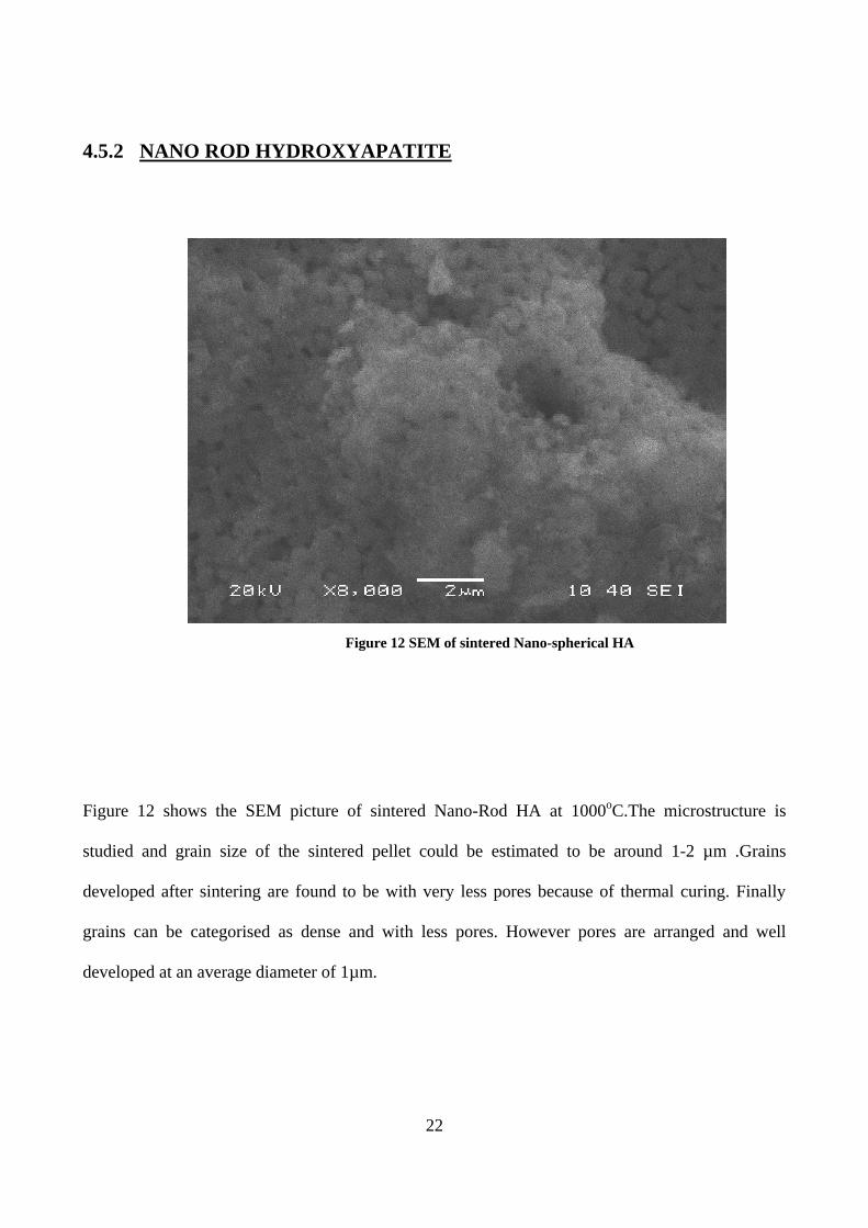
Figure 12 SEM of sintered Nano-spherical HA
Figure 12 shows the SEM picture of sintered Nano-Rod HA at 1000oC.The microstructure is
studied and grain size of the sintered pellet could be estimated to be around 1-2 µm .Grains
developed after sintering are found to be with very less pores because of thermal curing. Finally
grains can be categorised as dense and with less pores. However pores are arranged and well
developed at an average diameter of 1µm.

23
4.5.3 NANO FIBRE HYDROXYAPATITE
Figure 13 (a) SEM of calcined Nano-fibre HA (b) SEM of raw Nano-fibre HA
Figure 13(a) shows the SEM picture of calcined Nano-fibre HA at 700oC.The microstructure is
studied and grain size of the sintered pellet could be estimated to be around 0.5-1 µm .Grains
developed after sintering are found to be with very less pores because of thermal curing. Finally
grains can be categorised as dense and with less pores when compared to the figure 13(b) of raw
nano-fibre HA which is found to be less dense and more porous. However pores are arranged and
well developed at an average diameter of 0.5µm after calcination.
a b

24
4.6 BULK DENSITY OF DIFFERENT MORPHOLOGIES
Figure 14 shows the relative densification behaviour of the three concerned morphologies
(a)Nano-spherical HA:-At 700oC the density is 1.37gm/cc densification is fast at initial stages upto
a temperature of 1000oC.then the rate of densification slows down and final density of 2.88gm/cc is
achieved that is 92.02% of the theoretical density.
(b)Nano-rod HA: -At 700oC the density is 1.484 gm/cc densification is slow at initial stages and the
rate of densification remains almost constant achieving a density of 2.76gm/cc that is 88.17 % of
theoretical density.
(c)Nano-fibre HA :-At 700oC the density is 1.25 gm/cc densification is at a relatively constant rate
and finally a density of 2.07gm/cc is achieved that is 66.13 % of theoretical density.
700 800 900 1000 1100 1200 1300
1.2
1.4
1.6
1.8
2.0
2.2
2.4
2.6
2.8
3.0
Bul
k de
nsity
temperature
Spherical
Rod
Fibre
Figure 14 BD composite of HA

25
4.7 APPARENT POROSITY OF DIFFERENT MORPHOLOGIES
Figure 15 shows the pore formation behaviour of the three concerned morphologies
(a)Nano-spherical HA :-densification is fast at all stages upto a temperature of 1250oC reducing the
porosity, and final porosity of 5 % is achieved.
(b)Nano-rod HA :- densification is slow at initial stages and the rate of densification remains almost
constant which hardly affects the rate of % porosity achieving a porosity of 5%.
(c)Nano-fibre HA :- Initial porosity being 58% densification goes at a relatively constant rate and
finally a porosity of 30% is achieved.
700 800 900 1000 1100 1200 1300
0
10
20
30
40
50
60
Bulk
de
nsity
temperature
Spherical
Rod
Fibre
Figure 15 AP composite of HA

26
4.8 BULK DENSITY AND APPARENT POROSITY AS A FUNCTION OF
TEMPERATURE
4.8.1 NANO SPHERICAL HYDROXYAPATITE
Figure 16 shows that the bulk density is found to increase with increasing sintering temperature.
This is due to the increase in densification of samples with increase in temperature. Simultaneously
the apparent porosity is found to show reverse properties against bulk density and decreases with
increase in sintering temperature.
700 800 900 1000 1100 1200 1300
1.0
1.5
2.0
2.5
3.0
3.5
4.0
temp
de
nsity
0
10
20
30
40
50
60
A p
oro
sity
Figure 16 BD vs AP of Nano-spherical HA
4.8.2 NANO ROD HYDROXYAPATITE
Figure 17 shows that the bulk density is found to increase with increasing sintering temperature.
This is due to the increase in densification of samples with increase in temperature. Simultaneously
the apparent porosity is found to show reverse properties against bulk density and decreases with
increase in sintering temperature.

27
700 800 900 1000 1100 1200 1300
1.0
1.5
2.0
2.5
3.0
temp
de
nsi
ty
0
10
20
30
40
50
60
A p
oro
sity
Figure 17 BD vs AP of Nano-rod HA
4.8.3 NANO FIBRE HA
Figure 18 shows that the bulk density is found to increase with increasing sintering temperature.
This is due to the increase in densification of samples with increase in temperature.simultaneously
the apparent porosity is found to show reverse properties against bulk density and decreases with
increase in sintering temperature
700 800 900 1000 1100 1200 1300
1.2
1.4
1.6
1.8
2.0
2.2
temp
BD
10
20
30
40
50
60
AP
Figure 18 BD vs AP of Nano-fibre HA

28
Chapter 5
CONCLUSION

29
A brief conclusion could be summarized as follows:
[1] Presence of exothermic and endothermic peaks during thermal analysis shows the weight
loss of 12 % for nano-spherical and 18% for nano-fibre.
[2] Hydroxyapatite phase stability was strictly depended on morphology.
[3] FTIR data reveals the presence of carbonate incorporated which tends to form broad
exothermic peaks on thermal analysis. Presenceof PO42-
are also present with OH-.
[4] Shrinkage study by dilatometric test up to 1250oC showed 16%, 3% and 2% shrinkage for
nano-spherical, nano-rod and nano-fibre, respectively.
[5] SEM analysis showed the microstructural changes occurring after sintering on all the three
morphologies
[6] Nano-spherical HA is being the most stable towards decomposition and 92.01 % relative
density achieved at temperature of 900oC and porosity of 5%.
[7] Nano-rod HA was moderate stable, which was decomposed and densified to 88.1 % of
theoretical density at temperature of 1000oC and porosity of 5%.
[8] Nano-fibre HA was relatively unstable and decomposed to TCP at early stage and densified
to 66.13% of theoretical density at 700oC and porosity of 30 %.
[9] As the sintering temperature increases nano-spherical HA shows the maximum densification
followed by nano-rod and nano-fibre.

30
REFERENCES
[1] en.wikipedia.org/wiki/File:Mineraly.sk_-_hydroxylapatit.jpg
[2] Elliot, J. C. Structure and Chemistry of the Apatites and Other Calcium Orthophosphates,
Elsevier, 1994.
[3] V.S. Komlev,S.M. Barinov,E. Girardin,S. Oscarsson, F. Rustichelli,,V.P. Orlovskii, Porous
Spherical HA granules,processing and characterization.Volume 4, Issue 6, November 2003,
Pages 503–508
[4] P.Ramnarayanan,University of Rochester,Dept of Chemical Engineering,OPT 407 Practical
Electron Microscopy Spring 2010.
[5] Dr. Jon Binner and Mr. Rod Sambrook,Ceramic foams Processing and applications as filter,
inter penetrating composites and biomedical M
[6] S. Laasri, M. Taha, A. Laghzizil,E.K. Hlil, J. Chevalier d, The affect of densification and
dehydroxylation on the mechanical properties of stoichiometric HA bioceramics, Materials
Research Bulletin 45 (2010) 1433–1437.
[7]` S. Ramesh,C.Y. Tan , S.B. Bhaduri , W.D. Teng , Rapid densification of nanocrystalline HA
for biomedical applications, Ceramics International 33 (2007) 1363–1367
[8] A. Banerjee, A.Bandyopadhyay, Susmita Bose, HAnanopowders: Synthesis, densification
and cell–materials interaction, Mater. Sci. Engg. C 27 (2007) 729–735
[9] P. Cheang , K.A. Khor, Effect of particulate morphology on the tensile behaviour of
polymer/HA composites, Materials Science and Engineering A345 (2003) 47-54.

31
[10] S.ImanRoohani-Esfahani, Saied Nouri-Khorasani ,Zufu Lu, Richard Appleyard,HalaZreiqat,
The influence HA nanoparticle shape and size on the properties of biphasic calcium
phosphate scaffolds coated with HA PCL Composites, Biomaterials 31 (2010) 5498-5509.
[11] E. Landi, A. Tampieri , G. Celotti, S. Sprio, Densification behaviour and mechanisms of
synthetic HAs, Journal of the European Ceramic Society 20 (2000) 2377-2387
[12] I H. Arita, David S. Wilkinson, Maria Antonieta Mondragnt and Victor M. Castafio,
Chemistry and sintering behaviour of thin HA ceramics with controlled
porosity,Biomoteriols16(1995) 403-408.
[13]. G. Muralithran, S. Ramesh. The effects of sintering temperature on the properties of HA.
Ceramics International 26 (2000) 221-230
[14]. S.K.Swain.S.V.Dorozkin,D. Sarkar,Synthesis and dispersion of HAnanopowders, Materials
Science and Engineering: Available online 30 March 2012
[15]. V. Jokanovi, B. Jokanovi , D. Markovi , V. Zivojinovi, S. Pasali , D. Izvonar, M. Plavsi,
Kinetics and sintering mechanisms of hydro-thermally obtained HA, Mater. Chem. Phy. 111
(2008) 180–185.
[16]. Gultekin Go ller ,FaikNuzhetOktar , Sintering effects on mechanical properties of
biologically derived dentine HA, Mater. Lett. 56 (2002) 142–147
[17] Kaneda, K. et al. J. Am. Chem. Soc. 2002, 124, 11572.