ebola clinical care guidelines-2014-8-28a 2sep2014.pdf · ebolaclinical!care!guidelines! 3!...
TRANSCRIPT

Organized by the Public Health Agency of Canada
Canadian Critical Care Society Canadian Assoc. of Emergency Physicians Assoc. of Medical Microbiology & Infectious Diseases Canada
Ebola Clinical Care Guidelines A guide for clinicians in Canada
Interim Report August 29, 2014

Ebola Clinical Care Guidelines 2
CCCS – CAEP – AMMI 2014 2014
Ebola Virus Photo
Courtesy of CDC/ Frederick A. Murphy

Ebola Clinical Care Guidelines 3
CCCS – CAEP – AMMI 2014 2014
Forward
In response to the WHO declaration of the Ebola outbreak in West Africa as a Public Health Emergency of International Concern (PHEIC), the Public Health Agency of Canada (PHAC) invited the Canadian Critical Care Society (CCCS) and the Association of Medical Microbiology and Infectious Disease Canada (AMMI) to consider embarking upon a collaboration to develop clinical care guidelines to support clinicians in Canada who may be required to treat a patient suffering from the Ebola Virus Disease (EVD or “Ebola”). Both the CCCS and AMMI were fully supportive of collaborating on this important initiative. In order to ensure that the guidance developed was applicable to clinicians in all of the areas of hospitals most likely to be required to provide care, the CCCS and AMMI requested that the Canadian Association of Emergency Physicians (CAEP) also be invited by PHAC to participate in the development of the guidelines. As a result the current guidelines have been produced via a collaboration of efforts from all three of the societies.
The current iteration of these guidelines is intended to focus on the management of isolated cases of Ebola in persons arriving in or returning to Canada having been exposed to the virus during travel outside of Canada, Future updates of these guidelines will be developed as information evolves or if there is a need for clinical guidance regarding the management of larger volumes of cases of Ebola in Canadian hospitals. The focus of this document is on the clinical care and patient management (e.g. processes & patient flow, bedside care, etc). With the exception of Inter-‐facility transport, this document will not discuss the specifics of infection prevention and control (IPAC) and EVD diagnostic testing as these are covered in separate guidelines from PHAC. Although the specific guidance regarding IPAC will not be provided by this group, the clinical societies which represent those working on the front lines of patient management are uniquely positioned to provide insight on the human factors associated with applying IPAC guidelines in the delivery of patient care.
It must be recognized that much of the guidance within this document derives from expert opinion. There is scant high-‐grade evidence regarding the either the pathophysiology or optimal specific clinical management of EVD. The aim of this collaborative was to develop a simple, easy-‐to-‐use guide for the clinicians who face treating their first case of EVD. Having the luxury of time for contemplation and debate over some of the issues at present, rather than in the heat of the moment, we hope that by articulating our rationale and discussing the pros and cons of various options clinicians will find our ‘expert opinion’ beneficial.

Ebola Clinical Care Guidelines 4
CCCS – CAEP – AMMI 2014 2014
Table of Contents Forward ......................................................................................................................................................... 3
Contributions ................................................................................................................................................. 7 Core Working Group Membership ........................................................................................................................................ 7
CCCS ................................................................................................................................................................................... 7 CAEP .................................................................................................................................................................................. 7 AMMI ................................................................................................................................................................................. 7
Other Contributors ................................................................................................................................................................ 7
Overview of Ebola Virus Disease .................................................................................................................... 8 Epidemiology ......................................................................................................................................................................... 8 Transmission .......................................................................................................................................................................... 8 Clinical .................................................................................................................................................................................... 8 Diagnosis ................................................................................................................................................................................ 9 Treatment .............................................................................................................................................................................. 9
Clinical Leadership ....................................................................................................................................... 10
Point of First Contact – Emergency Department ........................................................................................... 12 Preparedness ....................................................................................................................................................................... 12 Case Identification ............................................................................................................................................................... 12 Infection Control .................................................................................................................................................................. 13 Testing guidelines ................................................................................................................................................................ 14
Where To Provide Care For An In-‐Patient With Ebola ................................................................................... 16
Clinical Care of Ebola Infected Patients ........................................................................................................ 18 Clinical Examination & Assessment ..................................................................................................................................... 18 Monitoring (Invasive/Non-‐invasive) .................................................................................................................................... 20
Non-‐invasive cardiorespiratory monitoring ..................................................................................................................... 20 Urine Output .................................................................................................................................................................... 20 Invasive arterial blood pressure monitoring .................................................................................................................... 21 Central line access and central venous pressure (CVP) monitoring ................................................................................. 21 End-‐tidal carbon dioxide (ETCO2) monitoring .................................................................................................................. 21 Other invasive monitoring ............................................................................................................................................... 22
Body Fluid Control ............................................................................................................................................................... 22 Airway Management & Ventilation ..................................................................................................................................... 22
Non-‐invasive ventilation .................................................................................................................................................. 22 Endotracheal intubation .................................................................................................................................................. 23

Ebola Clinical Care Guidelines 5
CCCS – CAEP – AMMI 2014 2014
Mechanical ventilation .................................................................................................................................................... 24 Fluid Resuscitation & Electrolytes ....................................................................................................................................... 24
Oral fluid and electrolyte replacement ............................................................................................................................ 25 Intravenous access .......................................................................................................................................................... 25 Intravenous fluid replacement and resuscitation ............................................................................................................ 25 Electrolyte replacement ................................................................................................................................................... 26
Vasopressors ........................................................................................................................................................................ 27 Antibiotics & Antivirals ........................................................................................................................................................ 27 Organ Support (IHD, CRRT, ECMO) ...................................................................................................................................... 28 Cardiopulmonary Resuscitation ........................................................................................................................................... 29 Symptom Management ....................................................................................................................................................... 30 Prophylaxis and Preventative Measures for the Critically Ill ............................................................................................... 30 Nutrition .............................................................................................................................................................................. 31 Experimental Antiviral Medications ..................................................................................................................................... 31 Palliative Care ...................................................................................................................................................................... 31
Key Messages: ................................................................................................................................................................. 31 Secretions ........................................................................................................................................................................ 32 Dyspnea (from pulmonary hemorrhage or other complication) ...................................................................................... 32 Sudden Terminal Events (Massive hemoptysis, asphyxiation) ......................................................................................... 33
Pregnancy & Obstetrics ....................................................................................................................................................... 33 Paediatric Considerations .................................................................................................................................................... 34 Impact on Health Care Worker’s Caring for EVD Patients ................................................................................................... 34 Psychological Support (Patients, Families & Providers) ....................................................................................................... 34
Inter-‐facility Transport of Patients with EVD ................................................................................................ 36 General Considerations for Aeromedical Evacuation .......................................................................................................... 36
Aircraft Selection ............................................................................................................................................................. 36 Logistical Pre-‐flight Planning and Post-‐flight Procedures ................................................................................................ 37 Emergency procedures .................................................................................................................................................... 37 Waste disposal ................................................................................................................................................................ 38 Cleaning and disinfection ................................................................................................................................................ 38
Regional Aeromedical Evacuation ....................................................................................................................................... 38 Patient Isolation & PPE .................................................................................................................................................... 38
National & International Aeromedical Evacuation .............................................................................................................. 39 Patient Isolation & PPE .................................................................................................................................................... 40
Research ...................................................................................................................................................... 43
Appendix 1 – ED Screening for & Response to Possible EVD Cases ............................................................... 48

Ebola Clinical Care Guidelines 6
CCCS – CAEP – AMMI 2014 2014
APPENDIX 2 – Bedside Nursing Care ............................................................................................................. 49

Ebola Clinical Care Guidelines 7
CCCS – CAEP – AMMI 2014 2014
Contributions
Each of the societies contributed participants for the working group who could provide particular knowledge, skills, and/or experience relevant to the management of patients with Ebola. Among the working group members there is representation from a variety of clinical backgrounds including both adult and paediatric providers as well as members of the group who have experience in treating patients with Ebola in Africa during the current outbreak as well as prior outbreaks. In addition to the core working group members, many other members of the societies and other clinical experts contributed content and comments during the development process. This trilateral collaboration was facilitated by PHAC (Dr. Thomas Wong, Dominique Baker, Margie Lauzon, Althea House, Eva Wong and Shamir Nizar Mukhi)
Core Working Group Membership
CCCS
� Dr Mike Christian – lead
� Dr Rob Fowler
� Dr Niranjan “Tex” Kissoon
� Dr Anand Kumar
� Dr Francois Lamontange
� Dr Srinivas Murthy
� Dr Randy Wax
CAEP
� Dr Laurie Mazurik – lead
� Dr Jill McEwen
AMMI
� Dr Michael Libman – lead
� Riccarda Galioto
� Dr. Gary Garber
� Dr Jay Keystone
� Dr Allison McGeer
� Dr Otto Vanderkooi
Other Contributors
� Dr James Downer
� Dr Stephen Lapinsky
� Dr Russell MacDonald
Please send correspondence or comments to: [email protected]

Ebola Clinical Care Guidelines 8
CCCS – CAEP – AMMI 2014 2014
Overview of Ebola Virus Disease
Epidemiology
Ebola virus is member of the filoviridae, enveloped non-‐segmented, negative stranded RNA viruses. There are five identified subtypes of Ebola virus of which four have caused human disease in Africa,: Ebola Zaire, Ebola Sudan, Ebola Ivory Coast, and Ebola Bundibugyo. The fifth virus, Ebola Reston has caused disease in nonhuman primates but not in humans. The reservoir of Ebola virus is most likely to be fruit bats in Central Africa but no human cases have been contracted directly from bats. In the wild, Ebola virus likely spreads from fruit bats to other animals such as rodents, monkeys and chimpanzees, following which humans acquire the infection from eating poorly cooked animals or handling raw animal meat (Bushmeat).
The first case of Ebola virus disease (EVD) was detected in 1976 in a large outbreak in southern Sudan and northern Democratic Republic of Congo (DRC). Twenty subsequent outbreaks have been recorded in Africa with a total of fewer than 1000 deaths until the current 2014 outbreak. The mortality rate from EVD varies from 30 to 90% depending on the subtype of the virus; the highest mortality is seen from Ebola Zaire (which is the cause of the 2014 West African outbreak) and the lowest from Ebola Bundibugyo infections. The 2014 outbreak began in January 2014 and is the largest on record with more than 2,000 cases and 1,000 deaths (~60% mortality) as of August, 2014, and cases reported in Guinea, Sierra Leone and Liberia, with limited spread in Nigeria.
Transmission
Most human infections occur by direct contact with secretions and excretions of infected patients or cadavers and not by aerosol spread. The virus enters the host through mucosal surfaces, breaks and abrasions in the skin or by the parenteral route. Health care workers are frequently infected from needle sticks or breaks in personal protection techniques. Asymptomatic individuals in the incubation period have not been documented to transmit the infection.
Clinical
The clinical picture of Ebola virus disease (EVD) begins with an abrupt onset of fever following an incubation period of 2 to 21 (mean 4-‐10 days) and is characterized initially by a nonspecific flulike illness with fever,

Ebola Clinical Care Guidelines 9
CCCS – CAEP – AMMI 2014 2014
headache, malaise, myalgia , sore throat and gastrointestinal symptoms such as nausea vomiting, diarrhoea and abdominal pain. Although cough can occur, it is not a primary feature of this illness. A maculopapular rash and conjuctival injection may occur. Postural hypotension, confusion and coma precede death. Haemorrhagic manifestations, occurring in fewer than 50 % of clinical cases, arise toward the end of the first week of illness and include petechiae, blood loss from venipuncture sites, bruising and gastrointestinal bleeding.
Diagnosis
Laboratory abnormalities associated with EVD include early leukopenia, lymphopenia and atypical lymphocytosis. AST and ALP are often elevated. Prothrombin and partial thromboplastin times are increased, and fibrin split products are detectable indicating disseminated intravascular coagulation. Patients who die often do so between the sixth and 16th day in hypovolemic shock and multi-‐organ failure. The definitive diagnosis of EVD is made by RT-‐PCR or antigen detection.
Treatment
The mainstay of treatment is supportive care, which includes careful attention to intravascular volume status and oral or intravenous fluid therapy, correction of electrolyte and metabolic abnormalities, correction of coagulation abnormalities, nutritional support, and antibiotics for secondary bacterial infections. Experimental therapies, including monoclonal antibody administration, small inhibitory RNA molecules, Ebola virus-‐specific convalescent plasma therapy and hyper-‐immune globulin, pre-‐ and post-‐exposure immunization, are still experimental without proven benefit in humans. Although some of these treatments have recently been used in a small number of individuals, the available data remains insufficient to determine their clinical efficacy and safety in humans. Efforts to launch clinical trials are underway and the World Health Organization has issued ethical guidance on use of such agents1.
1 http://www.who.int/csr/resources/publications/ebola/ethical-‐considerations/en/
2 http://www.phac-aspc.gc.ca/id-mi/vhf-fvh/ebola-ipc-pci-eng.php
Ebola Clinical Care Guidelines 10
CCCS – CAEP – AMMI 2014 2014
Clinical Leadership
Health facilities leadership should provide direction, be engaged and ensure that the institution has the ability to detect and provide initial appropriate levels of care for all communicable diseases, including those that are:
1. Rare or occur only by importation 2. Have high rates of infectivity 3. Have high mortality
Healthcare Institutions should be alert for the possibility of rare and severe diseases, and the screening of patients for such illness should be routine. The consequences of these diseases are as follows: 1. For patients – care of an unfamiliar disease may be suboptimal, and fear, even when the diagnosis has
not been confirmed, may contribute to patients receiving lower quality of care. 2. For staff regarding their own health and health of their families – they need to have confidence in the
medical system that they and their families will be protected from nosocomial transmission during health care.
3. For the entire health system – while the Emergency Department may be the portal of entry to the institution for many patients with infectious diseases, such patients often also receive care from many other services including: medical wards, ICUs, operating rooms, diagnostic imaging, laboratory and pathology services, and pharmacy.
4. Messaging to the media to inform citizens – it is likely that there will be members of the general public who present for assessment (“worried-‐well”) after hearing of certain novel communicable diseases and this has the potential to place additional stress on the capacity of the ED and hospital generally, and hence we need to inform while being prepared for the consequences of increased public concern and attention.
Administration Leaders within the institution should ensure that they are prepared for these diseases as part of routine care. Thus administration and senior leaders should ensure that resources are available such as personal protective equipment (PPE) and experts in patient management, infection prevention and control to help guide processes of care delivery, general staffing, equipment, medical supplies, etc..

Ebola Clinical Care Guidelines 11
CCCS – CAEP – AMMI 2014 2014
Clinical Leaders should ensure that a team is in place to coordinate the overall response. The team may vary from institution to institution but should include a senior nurse, senior physician, infection control officer, and employee safety officer as well as a communications officer. Should there be the possibility of a EVD case discovered in ED, or other area of the hospital, the staff of that clinical area should promptly notify appropriate personnel through their usual reporting channels who will then activate the hospital specific team through which will then use predefined protocols and prepared messaging that will need to be deployed on short notice.

Ebola Clinical Care Guidelines 12
CCCS – CAEP – AMMI 2014 2014
Point of First Contact – Emergency Department
Preparedness
It is key for Emergency Departments (ED) to prepare for responding to a potential patient with EVD before their first possible case presents to their department. This involves developing plans to screen patients and the response should a patient screen positive. Ideally, ED will have a single process which allows them to screen for and appropriately respond to any patient presenting with a highly infectious disease be it EVD, MERS CoV, H7N9, or any other novel emerging pathogen. However, this document will focus solely on issues related to EVD. Appendix 1 provides examples of the type of approach recommended within the ED to screen for and respond to a potential patient with EVD. Below a brief description of the key aspects of case identification, infection control and testing in the ED is provided however HCWs should refer to the most up-‐to-‐date guidance for these areas provided on-‐line by the National and Provincial public health agencies2.
Case Identification
Early identification of potential Ebola cases is essential for implementing transmission prevention measures, including patient isolation and personal protective equipment for health care workers. Early identification is based on: A) clinical presentation, AND B) epidemiologic risk. Please refer to the PHAC website for the most up-‐to-‐date case definition3. Case definitions can evolve over time based upon the changing epidemiology of an outbreak in addition to advancements in our scientific understanding.
Case identification requires a high index of suspicion, routine screening of all patients, knowledge of clinical features of EVD, and frequently updated knowledge of relevant exposure risks for EVD.
EVD is almost always associated with high fever – usually at least 38.6 degrees Celsius. Although a single temperature taken at triage may be normal, a history of fever or feverishness will be present. Prostration and headache may be useful very early indicators. Rash, conjunctival injection, nausea and vomiting usually start several days into illness. Because these symptoms are non-‐specific, knowledge of and screening for travel history and other exposures is critical.
EVD should be suspected in a febrile patient whose onset of symptoms is within 2 to 21 days of: 2 http://www.phac-aspc.gc.ca/id-mi/vhf-fvh/ebola-ipc-pci-eng.php
3 http://www.phac-aspc.gc.ca/id-mi/vhf-fvh/national-case-definition-nationale-cas-eng.php

Ebola Clinical Care Guidelines 13
CCCS – CAEP – AMMI 2014 2014
• residence in or travel to an area where EVD transmission is active4 • directly or indirectly caring for a probable or confirmed case of EVD (e.g. direct patient care or contact
with environment or fomites of a case) • spending time in a healthcare facility where EVD patients are being treated • household exposure to a confirmed or probable EVD patient • processing laboratory specimens from a confirmed or probably EVD patient, or in a hospital in an area
where EVD transmission is active • Direct exposure to human remains (e.g. through participation in funeral rites) in an area where EVD
transmission is active • Contact with bats or primates from EVD-‐affected country
In seriously ill afebrile patient with a significant exposure history (as above), consideration should be given to whether EVD is a possible diagnosis; consultation with an infectious disease or tropical disease specialist may be appropriate.
In a febrile patient with a significant exposure history on screening, a detailed history and clinical assessment is required to determine whether a risk for EVD exists and testing for EVD is indicated. Consultation with public health, public health laboratories and infectious diseases should be undertaken immediately to ensure that patient management is optimal, and infection control precautions are implemented promptly if needed.
Infection Control
Appropriate infection control precautions should be initiated5. Staff should be trained and experienced with the use of the appropriate PPE. Ideally a ‘buddy system’ should be in place to ensure ‘cross-‐checks’ of PPE use. Infection control considerations commonly include the following items listed however, HCWs should consult their local IPAC policies and National/Provincial Public Health guidelines:
� Private room and toilet (negative pressure if available particularly if an aerosol generating procedure is conducted)
4 As of August 17, 2014, EVD transmission is active in Guinea, Liberia, Sierra Leone, and Nigeria. Refer to the World Health Organizations’s Ebola Virus
Disease (EVD) website for updated information on affected areas: http://www.who.int/csr/disease/ebola/en/ 5 http://www.phac-aspc.gc.ca/id-mi/vhf-fvh/ebola-ipc-pci-eng.php

Ebola Clinical Care Guidelines 14
CCCS – CAEP – AMMI 2014 2014
� Protection from blood and body fluids using standard and contact precautions. At a minimum, this includes gloves, fluid resistant or impermeable gown, eye protection (goggles or shield), mouth and nose protection (mask and face shield). Additional equipment may be needed if copious exposure is anticipated, such as double gloves, impervious apron, leg coverings, shoe covering.
� Aerosol generating procedures require additional respiratory protection, such as N95 respirators or PAPR, and must be performed in a negative pressure room.
� EXTREME CAUTION to avoid “sharps “ (percutaneous) injuries.
o Limit the use of needles and other sharps as much as possible
o Phlebotomy, procedures, and laboratory testing should be performed according to what is essential for adequate diagnostic evaluation and medical care and should be performed by most qualified person available
o All needles and sharps should be handled with extreme care and disposed in puncture-‐proof, sealable containers
� Dedicated, disposable medical equipment should be used where possible and appropriate. Other equipment to be cleaned and disinfected as per IPAC protocols.
� Hand hygiene before and after contact according to the 4 Moments of Hand Hygiene (REF/link IPAC or other).
� Diligent environmental cleaning and disinfection and safe handling of potentially contaminated materials is paramount, as blood, vomit, feces and other body secretions represent potentially infectious materials. A disinfectant with a broad spectrum virucide claim with a DIN should be used according to the manufacturer's instruction.6
Testing guidelines
In Canada, the likelihood of a diagnosis of Ebola in a febrile returned traveller is generally much lower than the likelihood of the common imported febrile illnesses. In particular, MALARIA is endemic in all regions where Ebola has been identified. Delay in the diagnosis and treatment of malaria can lead to severe or lethal disease in a short period of time.
� URGENT malaria testing should be performed in all patients. Malaria remains the single most likely diagnosis in febrile patients from regions likely to be affected by EVD outbreaks.
6 http://www.phac-aspc.gc.ca/id-mi/vhf-fvh/ebola-ipc-pci-eng.php

Ebola Clinical Care Guidelines 15
CCCS – CAEP – AMMI 2014 2014
� Arrangements must be made with labs for Ebola Virus disease testing (typically by real time polymerase chain reaction or antigen or antibody detection systems).Please refer to your provincial/territorial public health authority for information on access to Ebola Virus testing.
� Other testing is guided by the patient’s symptoms and signs, but will typically include at least basic hematology and chemistry analyses, cultures of blood, and possibly a chest radiograph.
� The laboratories must be informed about any specimens that may contain Ebola virus so that appropriate transport and handling can be arranged. Specimens should be carried by hand in appropriate containers. They should NOT be sent by pneumatic tube or similar systems.

Ebola Clinical Care Guidelines 16
CCCS – CAEP – AMMI 2014 2014
Where To Provide Care For An In-‐Patient With Ebola
Selection of the appropriate clinical area to manage a patient with Ebola once he or she has entered a health care facility will be dependent upon a number of factors. Every institution will have to make an individual decision about what is best for their particular circumstance. However, it cannot be stressed enough that, similar to the basic principles that apply in emergency preparedness[1], hospitals should undertake an inventory of their facilities and identify potential areas for managing highly infectious patients such as those with Ebola well before they are faced with their first potential case.
When assessing potential areas to care for an Ebola patient consideration should be given to the following factors:
� Single Patient Room with a private bathroom, door and anteroom (or space to create one)
o Negative pressure isolation is only essential if aerosol generating procedures are to be conducted
� Ability to dispose of body fluids safely within the patient’s room
� Infrastructure for patients to communicate to staff/visitors outside of the room
� Ability to restrict access to the area as well as to the patient’s room
� Ability to provide dedicated patient care equipment (preferably disposable)
� Ability to provide critical care without having to move the patient should he/she deteriorate
o Suction
o Monitors
o Sink (with dialysis connections if needed)
o Electrical outlets (including emergency power supply)
o Medical Gas (Oxygen & Medical Air) connections
o Space for life support equipment
� If facility resources permit, overhead hoists to facilitate patient movement with minimal staff involved
� Sufficient space for providers to don and doff PPE, ideally with a separate ‘clean’ entrance and ‘dirty’ exit
� Storage for medical supplies and PPE outside of the room

Ebola Clinical Care Guidelines 17
CCCS – CAEP – AMMI 2014 2014
� Appropriate facilities outside of the room to clean or dispose of contaminated medical equipment and supplies.
� Potential for access to or placement of point-‐of-‐care testing equipment
� Work area outside of the room for staff
� Necessary IT & communications connections for both staff and patient use
� Ability to bring portable x-‐ray machines into room to minimize patient transport
� Physically separated from other non-‐EVD patients
� Close access to elevators or diagnostic suites to minimize transportation times
When a Canadian hospital is faced with the need to care for a single patient with Ebola, for many hospitals the most practical place to provide this care may be in an Intensive Care Unit (ICU). ICUs in many hospitals are the most likely locations to possess the factors ideal for caring for an Ebola patient. In addition to the physical and logistic issues, ICU provides 1:1 (or higher) nurse to patient ratios which will in part mitigate the negative impact on patient care due to the burden associated with the PPE requirements[2]. Further, patients suffering from EVD require close attention to fluid management and volume resuscitation, skills that critical care clinicians possess as a primary expertise. Finally, ICU staff typically represent a contained audience to target for education and training related to the provision of care to patients with EBV. They should already be well practiced in the use of appropriate PPE, and in performing aerosol-‐generating procedures if necessary, for these situations.

Ebola Clinical Care Guidelines 18
CCCS – CAEP – AMMI 2014 2014
Clinical Care of Ebola Infected Patients
Clinical Examination & Assessment
The discussion of this section will be brief and focus primarily on the identification of common complications associated with EVD and to provide a guide to the expected natural history of the illness. Clinical examination of the patient should occur at least twice daily (once per nursing shift) in patients who are not severely ill. Patients who are severely ill require more intense monitoring, similar to other critically ill patients. Monitoring should follow the usual approach to patient assessment with an additional focus on vital signs and their variability (discussed further below under ‘Monitoring’).
The natural history of Ebola is typically divided into three phases: early, late, and terminal or recovery[3-‐5]. Understanding the common findings during each of these phases will allow the clinician to identify both expected and unexpected complications should they occur.
The following features may be seen in the early phase of Ebola infection:
� Fatigue & malaise
� Generalized weakness
� Fever (sudden onset)
� Headache
� Myalgia & arthralgia
� Pharyngeal erythema
� Lymphadenopathy
� Nausea & anorexia
� Vomiting
� Diarrhoea (non-‐bloody)
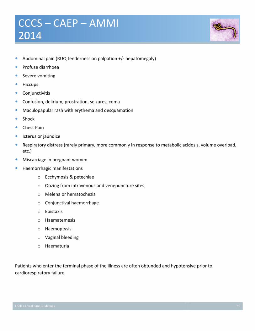
The following features tend to develop during the later phase of disease. It should be noted that often the features overlap and there is not a clear distinction between the phases. Additionally, despite the classification of Ebola as a “haemorrhagic fever” and the discussion of haemorrhagic symptoms below, bleeding is a predominant symptom in a minority of patients with Ebola.
Ebola Clinical Care Guidelines 19
CCCS – CAEP – AMMI 2014 2014
� Abdominal pain (RUQ tenderness on palpation +/-‐ hepatomegaly)
� Profuse diarrhoea
� Severe vomiting
� Hiccups
� Conjunctivitis
� Confusion, delirium, prostration, seizures, coma
� Maculopapular rash with erythema and desquamation
� Shock
� Chest Pain
� Icterus or jaundice
� Respiratory distress (rarely primary, more commonly in response to metabolic acidosis, volume overload, etc.)
� Miscarriage in pregnant women
� Haemorrhagic manifestations
o Ecchymosis & petechiae
o Oozing from intravenous and venepuncture sites
o Melena or hematochezia
o Conjunctival haemorrhage
o Epistaxis
o Haematemesis
o Haemoptysis
o Vaginal bleeding
o Haematuria
Patients who enter the terminal phase of the illness are often obtunded and hypotensive prior to cardiorespiratory failure.

Ebola Clinical Care Guidelines 20
CCCS – CAEP – AMMI 2014 2014
Should the patient enter the recovery phase, it can occur over days, weeks or months [5]. Many symptoms may persist during this phase including weakness, weight loss, headache, migratory arthralgias, desquamation, hair loss, and anaemia. Additionally, late occurrences of acute orchitis and uveitis have been reported[5].
Monitoring (Invasive/Non-‐invasive)
As previously mentioned, the care of patients with EVD is primarily supportive.[4] Patients with probable or confirmed EVD should be monitored in a setting that is capable of intensive and frequent monitoring of vital signs, fluid balance, and neurologic status. Ideally all patient monitors should be visible from outside of the patient’s room either via a window or satellite monitoring station and alarms must be audible from outside of the patient’s room with the doors closed. Video monitoring of the room can also enhance the effectiveness of patient monitoring. Finally, the patient should have a mechanism to signal for assistance and communicate with staff outside of the room. In addition to monitoring, nursing staff caring for the patient should have well prescribed parameters for alerting the patient’s attending physician if there is deterioration in the patient’s status.
Non-‐invasive cardiorespiratory monitoring Non-‐invasive cardiorespiratory monitoring including heart rate (EKG), respiratory rate, oxygen saturation (pulse oximetry) and non-‐invasive blood pressure (NIBP) should be available for all patients with Ebola, although the frequency of monitoring should be determined by the patient’s clinical condition. The use and frequency, in particular, of NIBP monitoring of patients will have to be assessed on an individual basis, depending upon the patient’s severity of illness, type of NIBP monitor and degree of capillary leak (ecchymosis & petechiae) as significant bruising may result particularly from older generation NIBP monitors that use repeat high cuff pressures. Newer generation of monitors and manual BP monitoring may be less traumatic.
Urine Output Hydration and volume status are particularly important factors in patients with Ebola given both their gastrointestinal losses and the potential for capillary leak or haemorrhage. Therefore, close monitoring of urine output is an essential tool for detecting volume depletion, particularly since accurate documentation of fluid balance (“in’s and out’s”) is often difficult in the setting of vomiting and diarrhoea. In less severely ill patients the measurement of voided urine is appropriate. However, if the patient is having significant
Ebola Clinical Care Guidelines 21
CCCS – CAEP – AMMI 2014 2014
diarrhoea or is seriously/critically ill the preferred method of monitoring urine output will be with a Foley catheter and urometer. This should be recorded on an hourly basis.
Sequential weight assessment may also be a useful index for fluid management particularly if the patient is exhibiting substantial unmeasured fluid losses due to diarrhea or insensible losses.
Invasive arterial blood pressure monitoring Invasive arterial blood pressure monitoring should be considered in select cases with hemodynamic instability requiring vasoactive agents and frequent blood-‐work monitoring. The potential benefits must be balanced against the risk of blood exposure and the potential for arterial spray of blood in the event of a circuit disconnect or leak. In most circumstances radial arterial access would be preferable over femoral access particularly given the frequency of soilage of the groin area, and for the possibility of coagulopathy among some patients. If the patient has central venous access in place and NIBP monitoring is effective, the addition benefit of arterial monitoring is minimal and may be outweighed by the associated risks to the patient, or of blood exposure to staff.
Central line access and central venous pressure (CVP) monitoring The current literature on the utility of CVP monitoring is mixed. Given the lack of strong evidence to support the utility of CVP monitoring we would not recommend the establishment of central venous access for the purpose of CVP monitoring alone. In such circumstances assessment of volume status based upon clinical examination of the jugular venous pressure (JVP) or other clinical indicators is most appropriate. However, if central venous access has been established for an alternative induction (difficult peripheral access, requirement for vasopressors, or electrolyte replacement) the use of CVP monitoring can be considered. Similarly, the use of central venous oxygen saturation as a measure of adequacy of forward flow may be useful if intravascular volume or cardiac function is uncertain and clinical examination is not adequately revealing.
End-‐tidal carbon dioxide (ETCO2) monitoring ETCO2 should be considered in patients who are receiving mechanical ventilation. The combination of ETCO2 monitoring and pulse oximetry may decrease the need for arterial blood gas analysis, potentially eliminating the need for an arterial catheter or for repeated arterial punctures and thus the risk of needle stick injuries to staff.

Ebola Clinical Care Guidelines 22
CCCS – CAEP – AMMI 2014 2014
Other invasive monitoring In general it is best to minimize any forms of invasive monitoring due to risks associated with coagulopathy, sharps exposure during insertion and body fluid exposure. It is unlikely that any forms of invasive monitoring aside from those listed above would be necessary for the management of a patient with EVD.
Body Fluid Control
Given that Ebola is primarily transmitted through body fluids (blood, urine, diarrhoea, emesis, saliva, and other fluids) control of these substances is vital in preventing transmission and protecting health care workers. If the patient is continent of faeces and urine, access to a private washroom should be ensured. However, if the patient is incontinent of urine or faeces the use of a Foley catheter and/or faecal collection system should be considered, especially in children. Nasogastric tubes with gastric suctioning or drainage may be useful in preventing or minimizing vomiting. If body fluids are spilled in the patient care environment the appropriate containment and environmental cleaning guidelines should be followed7. Consider the use of disposable products that absorb urine, vomit, stool and make them non-‐liquid – weighing can be used to assess quantity if necessary.
Airway Management & Ventilation
Pulmonary involvement of Ebola is not a common feature of the disease, however respiratory failure may occur in these patients requiring mechanical ventilation. Secondary causes of respiratory failure may include (but are not limited to) shock, fatigue from prolonged compensation of metabolic acidosis and iatrogenic complications (e.g. transfusion-‐related lung injury). Airway management may be required independent of respiratory failure for airway protection purposes, with situational examples including decreased level of consciousness or massive upper GI bleeding.
Non-‐invasive ventilation Non-‐invasive ventilation (NIV) may be considered for support of patients with EVD having rapidly reversible causes of respiratory failure; however, there are significant concerns that warrant caution and likely outweigh any potential benefits of NIV in this patient population. First, many patients with EVD have frequent vomiting
7 http://www.phac-aspc.gc.ca/id-mi/vhf-fvh/ebola-ipc-pci-eng.php

Ebola Clinical Care Guidelines 23
CCCS – CAEP – AMMI 2014 2014
which will increase the risk of aspiration. Second, NIV will cause prolonged risk of aerosolization, and therefore must be performed in a negative pressure isolation room. All staff managing a patient on NIV must wear the PPE required during aerosol generating procedures while in the patient room. Third, should the patient fail NIV and require immediate intubation, there is higher risk to staff by rushing to don PPE leading to a breach of infection control and possible transmission. Therefore, should a trial of NIV be performed, close and frequent monitoring must be performed to identify need for intubation as early as possible to allow sufficient time for staff to carefully prepare equipment and institute appropriate infection control practices. Fourth, the risk of oropharyngeal bleeding and hematemesis with an NIV mask in place may create significant risk to the patient for aspiration, with a significant delay in staff response to assist due to need to don PPE when entering the patient room. In general, it is safer to manage a patient with EVD and respiratory failure using a strategy including elective intubation, traditional mechanical ventilation including filtration of exhaled gases, and frequent monitoring of improvement in clinical status leading to a controlled attempt at extubation as the patient responds to therapy.
Endotracheal intubation The need for endotracheal intubation should ideally be recognized early enough to allow a non-‐emergent procedure, thus avoiding a potential rush leading to mistakes in use of personal protective equipment (PPE) and other infection control precautions. In addition to the usual clinical indicators suggesting the potential for a difficult intubation, patients with EVD may have nasopharyngeal or oropharyngeal bleeding impairing visualization of the vocal cords during intubation. An exhalation filter should be attached to the bag ventilation device.
Appropriate PPE must be worn when preparing for high-‐risk aerosol generating medical procedures, such as airway management. As per Infection Prevention and Control guidelines8, this must include protective fluid proof clothing that provides facial/eye protection including goggles or face shield, and a respirator mask with protection at least equivalent to meet N95 standards with appropriate fit testing for the specific respirator. For those properly trained in their use with meticulous attention paid to avoid self-‐contamination, clinicians may also consider using full hood powered air purifying respirators (PAPR) as a more comfortable option that provides better protection against sprayed body fluids while at the head of the bed, along with water impermeable clothing preventing any skin exposure such as a full body suit. Current infection control recommendations include having patients in negative pressure isolation rooms during performance of any
8 http://www.phac-aspc.gc.ca/id-mi/vhf-fvh/ebola-ipc-pci-eng.php

Ebola Clinical Care Guidelines 24
CCCS – CAEP – AMMI 2014 2014
aerosol generating medical procedures. Although the airborne spread of Ebola is not a usually recognized mechanism of transmission in humans, the potential for aerosol generation of other primarily droplet transmitted viruses has been demonstrated during some procedures such as intubation[6, 7], and the presence of an anteroom in typical negative pressure patient isolation rooms allows safer donning and doffing of PPE.
The patient should be intubated by a clinician highly experienced in airway management. Some adjunctive strategies beyond the use of direct laryngoscopy may be helpful. Video/optical laryngoscopy should be considered to allow better visualization of the vocal cords in anticipated or unanticipated difficult airway, and will increase the distance between the patient and the clinician during the intubation, which may reduce the likelihood of aerosolization exposure or inadvertent dislodgment of PPE.
Rapid sequence intubation, including the use of rapid-‐acting neuromuscular blockade, should be considered. Given the potential for hemodynamic instability in patients with EVD, the medication regimen for sedation should consider use of agents that are less likely to drop the blood pressure, such as ketamine, or use of agents to mitigate or prevent hypotension to due direct side-‐effects or sedatives, or loss of intrinsic sympathetic activation. Good intravenous access must be present to allow rapid fluid resuscitation in case the blood pressure drops during the intubation process, and vasopressors (e.g. phenylephrine or ephedrine) should be immediately available to administer as a bolus if required.
Mechanical ventilation Usual practices regarding invasive mechanical ventilation should be followed, including avoidance of excessive tidal volumes (keeping Vt < 6 ml/kg if possible), avoidance of excessive plateau pressures (keeping less than 30 cm H20), appropriate positive end expiratory pressure (PEEP) to avoid recurrent atelectrauma. Ventilators must have capability for HEPA filtration of exhaled gases. Given the potential risk for unexpected aerosol generating medical procedures (i.e., accidental extubation requiring immediate re-‐intubation), the patient should be maintained in a negative pressure isolation room. Ideally, an interface between the mechanical ventilator and the patient monitoring system will allow easier monitoring of ventilation parameters without frequent reentry into the patient room.
Fluid Resuscitation & Electrolytes
With critically ill EVD patients, hypovolemia is the most common and predictable anomaly. Accordingly, administration of fluids and electrolytes constitutes the first step in a series of supportive care interventions.

Ebola Clinical Care Guidelines 25
CCCS – CAEP – AMMI 2014 2014
Persistent fluid loss, potentially compounded by other causes for shock, often require ongoing fluid replacement. There are no studies specific to the treatment of patients with EVD to guide fluid management strategies in these patients. The suggestions below are largely extrapolated from other literature such as the management of Dengue Haemorrhagic Fever or septic shock.
Oral fluid and electrolyte replacement Oral fluid and electrolyte replacement is preferred in patients who are not critically ill, who are able to drink and not suffering from significant nausea and vomiting. Purpose designed oral rehydration solutions will provide the most effective volume replacement and electrolyte replacement in those with significant diarrhoea. Anecdotal observations made during the West African outbreak suggest, however, that severe cases may be associated with an inability to eat and drink adequately. Early NG tube insertion for fluid and electrolyte repletion should be considered, especially for children.
Intravenous access Intravenous (IV) access will be required for those patients who are unable to tolerate oral fluids or hemodynamically unstable. In the early stages of EVD and for those with milder manifestations of the illness peripheral IV access is suitable for fluid management. Large bore IVs (14-‐18 gauge, for adults, and age-‐appropriate for children) are preferred to allow large volume fluid resuscitation in the event the patient deteriorates. Central or peripherally inserted central venous access should be considered for patients who require intravenous electrolyte replacement (particularly potassium), vasopressors, or where vascular collapse limits peripheral IV access and increases the multiple attempts at IV insertion with the associated needle stick injuries and potential bleeding complications for the patient and blood exposure to staff. In the event that central venous access must be obtained, the risk of injury to either the patient or staff can be minimized by having an experienced physician conduct the procedure under direct ultrasound visualization with the patient calm or sedated. At all times, caregivers should adhere to recommended barrier precautions and personal protective equipment. Consideration should be given to using non-‐suture securing devices to minimize skin punctures as well as the risk of needle stick injuries. For both peripheral and central IV lines needle-‐less systems should be used to avoid sharps injuries.
Intravenous fluid replacement and resuscitation The amount of intravenous fluid administration required will depend upon the specific patient’s symptoms and should be guided by the degree of overt volume loss (diarrhoea, vomiting, and urination) as well as factors suggesting volume contraction: decreased skin turgor, dry mucous membranes, tachycardia, decreased urine output, and hypotension. We suggest that Ringer’s lactate (LR) should be the fluid of choice for volume replacement. This is based upon evidence extrapolated from the management of patients with Dengue Haemorrhagic Fever[8] and the management of septic shock[9-‐11]. There is evidence to suggest that

Ebola Clinical Care Guidelines 26
CCCS – CAEP – AMMI 2014 2014
compared with Normal Saline, Ringer’s Lactate may be associated with lower rates of mortality[11], renal failure[10], acidosis[12-‐14], and haemorrhage[15-‐17]. For patients that are hypotensive initial boluses of Ringer’s Lactate in the order of 20ml/kg should be considered and repeated as required until the heart rate, blood pressure and parameters of end-‐organ perfusion are within the desired range. In the event where large volumes of crystalloid solutions are being administered, consideration may be given to the use of albumin[18]. Artificial colloids (e.g. pentastarch or hydroxyethyl starch) should be avoided given their associated risks of renal injury[19], bleeding[20] and mortality[21]. In the event the patient is haemorrhaging and/or coagulopathic consideration should be given to the administration of packed red blood cells, platelets, fibrinogen and plasma as required based upon their hematologic laboratory values and clinical findings. A target haemoglobin of greater than 70 g/L is recommended. There is no evidence to support the transfusion of platelets and coagulation factors in patients with DIC who are not bleeding or who are not at high risk of bleeding. However, treatment is justified in patients who have serious bleeding, or are at high risk for bleeding, or require invasive procedures. Platelets should be administered to patients with a platelet count of less than 20 x 109/L of if less than 50 x 109/L and is associated with serious bleeding. If serious bleeding is occurring and the INR is greater then 2, fresh frozen plasma (FFP) should be administered. Similarly if the fibrinogen is low in the face of significant haemorrhage cryoprecipitate should be transfused. When managing a patient with EVD who is experiencing DIC and haemorrhage clinicians should consult a haematologist and/or their blood bank physician. In certain cases the potential efficacy of multiple transfusions must be weighed against the availability of resources.
Electrolyte replacement Clinicians should expect and monitor for significant electrolyte deficiencies (particularly hypokalemia and metabolic acidosis secondary to bicarbonate loss from vomiting and diarrhoea respectively) to occur in those patients suffering vomiting and/or high volume diarrhoea. As stated earlier, if tolerated, oral replacement of electrolytes is the preferred method. Consideration should be given to adding potassium and bicarbonate to appropriate maintenance IV fluids early for patients with diarrhoea to prevent severe hypokalaemia (assuming there is no evidence of renal failure) and metabolic acidosis. In the event that concentrated intravenous electrolyte replacements are necessary, central venous access will most likely be required in addition to ensuring appropriate cardiac monitoring is in place. Electrolyte replacement protocols that adhere to the ISMP9 or WHO10 best practices for concentrated electrolyte replacements should be used. Given the importance of electrolyte management in this patient population, central laboratory or point-‐of-‐care testing
9 https://www.ismp.org/tools/institutionalhighAlert.asp
10 http://www.who.int/patientsafety/solutions/patientsafety/PS-‐Solution5.pdf

Ebola Clinical Care Guidelines 27
CCCS – CAEP – AMMI 2014 2014
should include the ability to measure serum sodium, potassium, sodium, bicarbonate, creatinine, glucose, lactate, calcium, magnesium and phosphate.
Vasopressors
Hypotensive patients failing to respond to volume resuscitation, or while volume resuscitation is in progress, should be supported with vasopressor therapy in accordance with the guidelines for the management of septic shock[9]. Prescribing and monitoring of vasopressor agents should adhere to the recommendations for safe practice from the ISMP11. The guidelines for vasopressor use in septic shock are:
� A mean arterial blood pressure (MAP) target of 65-‐70 mmHg (or median for age in children) is a reasonable initial target in adults [22] but should be re-‐assessed based upon individual patient factors such as a history of hypertension and indicators or whether or not satisfactory end-‐organ perfusion is being achieved.
� Norepinephrine infusion (0-‐1.0 µg/kg/min) is the preferred first-‐line agent for managing hypotension.
o In the event norepinephrine is administered via a peripheral IV the maximum concentration used should be no more than 4mg/250 ml (‘single strength’), a large bore IV should be used preferably in the antecubital or other large vein with good flow and it should be closely monitored for signs of extravasation.
o Centrally administered norepinephrine can be mixed in concentrations of 8mg/250ml (‘double strength’) or 16mg/250ml (‘quad strength’).
� Vasopressin 0.03µg/min or 0.04µg/min may be used to minimize the dose of norepinephrine required
� Epinephrine can be added as a second line therapy
� Except in the instance of bradycardia and potentially paediatric patients, dopamine should generally be avoided given its association with increased rates of cardiac arrhythmias and mortality[9, 23].
� Dobutamine (0-‐20 µg/kg/min) can be considered if there is evidence of myocardial dysfunction or ongoing hypoperfusion following adequate volume resuscitation.
Antibiotics & Antivirals
The clinical manifestations of severe EVD may overlap symptoms and signs observed in septic shock of bacterial origin. The published literature is generally silent on the incidence of bacterial superinfection in the
11 http://www.ismp-‐canada.org/download/safetyBulletins/2014/ISMPCSB2014-‐1_ImprovingVasopressorSafety.pdf

Ebola Clinical Care Guidelines 28
CCCS – CAEP – AMMI 2014 2014
context of EVD. Almost all published data from previous case series were derived from settings where bacterial cultures of any type were unavailable. Post mortem pathology has been described in a limited number of cases. Extensive necrosis of a variety of organs, especially liver, spleen, kidneys, and gonads is associated with direct viral cellular invasion, although the gut appears to be relatively spared. Alveolar damage with interstitial edema is common on pathology of deceased patients but not a common clinical syndrome. A dysregulated highly pro-‐inflammatory and procoagulant state is typical.
It would appear that bacterial infection or sepsis is an uncommon manifestation of EVD. Nevertheless, early administration (within the first hour[24]) of broad-‐spectrum antibiotics to those with manifestations of possible sepsis-‐associated hypotension/shock is sensible. Consideration should be given to discontinuing antibiotics in those where cultures and other investigations do not reveal bacterial superinfection. There is no evidence-‐based guidance on the choice of specific antibiotics; however, patients frequently will be the midst of severe gastro-‐intestinal symptoms and antibiotics choosen to treat potential enteric pathogens, among others, may be reasonable.
Appropriate antimicrobial therapy for potential sepsis will be dependent upon where the patient is on the illness trajectory. Patients presenting acutely will likely have standard community pathogens if non-‐Ebola sepsis is a consideration. Drugs such as cefotaxime/ceftriaxone and ciprofloxacin/levofloxacin are appropriate in these cases absent a specific concern for S. aureus. Patients who are health care workers or develop potential bacterial sepsis after >48 hours in hospital are at greater risk for nosocomial pathogens including resistant gram-‐negatives and S. aureus (including MRSA). Β-‐lactam/β-‐lactamase inhibitors or carbapenems, potentially with vancomycin, may be appropriate in those cases.
Organ Support (IHD, CRRT, ECMO)
A small subset of patients with severe EVD will progress to organ failure requiring extracorporeal supports during the course of their illness. Any decision to use extracorporeal support must incorporate any baseline coagulopathy present.
Renal failure is common in severe cases, although reports of the use of dialysis are absent. In appropriate settings, dialysis should be considered in the patient with EVD and renal failure. The mode of dialysis, whether intermittent hemodialysis or continuous renal replacement therapy, should be individualized based upon the patients status and the treating clinician.
Liver dysfunction progressing to liver failure is a major consideration for severe EVD, with hepatocellular necrosis found in the few autopsies performed in severe EVD.[25] Extracorporeal liver support is not

Ebola Clinical Care Guidelines 29
CCCS – CAEP – AMMI 2014 2014
recommended in severe EVD, given the scant evidence base, and therapy for liver failure should be supportive, including correcting metabolic abnormalities, monitoring (clinical or non-‐invasive) for cerebral edema, and correcting coagulopathies.
Extracorporeal membrane oxygenation (ECMO) for cardiorespiratory failure has not been reported in severe EVD. The initial presentation of severe EVD very uncommonly includes severe respiratory or myocardial failure, with the nature of the shock typically being hypovolemic or distributive in nature.[5] The limited potential benefit of ECMO in this setting must be weighed against the potential for significant blood exposure to staff during cannulation and bleeding complications from the sites of cannulation due to the coagulopathy that can accompany EVD, particularly in severe and refractory cases. Further, the close monitoring and care demands associated with patients on ECMO would result in significant exposure time for staff. Therefore we do not recommend ECMO in patients with EVD.
Cardiopulmonary Resuscitation
Data, and clinical experience, with cardiopulmonary resuscitation (CPR) in patients suffering from EVD is completely lacking. However, the likelihood of survival with a good outcome from cardiac arrest with CPR in intensive care units is poor, approximately 3%[26, 27], and is not expected to be better than any adult12 patients with multi-organ failure which is even lower. Patients with late stage EVD who experience an un-witnessed cardiac arrest have minimal expectation of survival, and therefore not initiating resuscitation efforts is appropriate to avoid unnecessary risk to healthcare staff. Patients with multiorgan failure being supported with artificial life support in a critical care environment who deteriorate to cardiac arrest despite full support are also highly unlikely to survive. Chest compressions in a patient with end-stage EVD, in addition to being essentially futile, presents a potentially significant blood and body fluid exposure risk to the health care workers. In such patients an advance order to withhold CPR for cardiac arrest is appropriate. A very select group of patients with EVD who are experiencing clinical conditions that are potentially associated with better outcome from cardiac arrest, such as hypovolemia or electrolyte abnormalities, an acute coronary syndrome associated with ventricular arrhythmias, may warrant aggressive therapy including defibrillation and antidysrhythmic drugs for witnessed cardiac arrest. Despite the instinctive urge to rush into a patient isolation room to help, staff must not take shortcuts in donning appropriate PPE, which should meet the standards required for management of aerosol generating medical procedures. A child who suffers cardiac arrest due to severe EVD and multi-organ disease also has a very small likelihood of survival, and similar to adults, not initiating CPR is appropriate in this circumstance. If a clear reversible cause is present, however, this should be promptly treated. 12 Cardiac arrest in children is discussed separately at the end of this section.

Ebola Clinical Care Guidelines 30
CCCS – CAEP – AMMI 2014 2014
Medical management of cardiac arrest in a patient with Ebola, if attempted, should follow current BCLS and PALS/ACLS guidelines.
Symptom Management
Given that the overall management of EVD is supportive, effective symptom management composes a significant component of the therapies clinicians may offer their patients.
Symptom Aetiology & Implications Management
Seizure or Coma Aetiology unclear, potentially related to liver failure. Typically a ominous sign, seen typically shortly before death.
Airway management as required Benzodiazepines +/-‐ dilantin to control seizures Laboratory investigations (e.g. Na+, glucose) CT head if lateralizing features
Hypotension & Shock
Frequently an early manifestation of severe cases. Also a common manifestation of illness severity and plausible lethal pathway. Initially, hypovolemia from profound GI losses. A component of early distributive shock remains a possibility but has not been confirmed. Secondary sepsis remains a possibility but has not been confirmed.
Close monitoring of fluid balance Aggressive repletion of fluid and electrolyte losses (potassium, bicarbonate) Consider vasopressor therapy if hypotension continues despite adequate fluid resuscitation.
Dyspnea or Respiratory failure
Respiratory involvement not a cardinal feature. Polypnea seen at all stages of illness in the context of shock and profound metabolic acidosis.
Oxygen therapy +/-‐ mechanical ventilation
Severe diarrhoea and vomitting
Frequent early presentation, the aetiology of which remains unclear.
NG tube insertion & suction Haloperidol 1mg PO/IV/SC q8h standing. (0.25-‐0.5 mg in children) Metoclopramide 10mg PO/IV/SC q6h standing (0.1 mg/kg/dose in children)
Intolerant to oral intake
Common in severe cases. Associated with chest pains so esophagitis is a plausible mechanism but unproven.
Consider enteral nutrition if tolerated If enteral nutrition are poorly tolerated, consider TPN after 8 days of unsuccessful enteral nutrition
RUQ pain and hepatomegaly
Common in severe cases Unproven but hepatitis plausible
Monitoring of liver biochemistry, consideration of vitamin K for early signs of INR elevation, watch for hypoglycemia
Hemorrhage (GI & puncture sites)
Ominous sign, seen typically shortly before death Aetiology most likely DIC
Complete hematology and liver laboratory workup Consider platelet transfusions is low, FFP if INR elevated, cryoprecipitate if fibrinogen low
Fever, chills, headache and myalgias
Common Aetiology associated to viremia
Acetaminophen 650mg PO q4h PRN (maximum 4g in 24h) (Pediatrics 10-‐15 mg/kg/dose Q4H to in children, max of 5 doses per 75 mg/kg/day). Lower doses may need to be used in patients experiencing hepatic dysfunction. Non-‐steroidal anti-‐inflammatory medications should be strictly avoided due to their platelet-‐inhibiting effects, which could exacerbate hemorrhage
Pain Common, often involving abdomen, chest wall, headache and joint pain that are not adequately managed with acetaminophen alone.
Narcotics, morphine, fentanyl, hydromorphone if renal impairment
Prophylaxis and Preventative Measures for the Critically Ill
Patients with severe EVD have multi-‐organ disease and avoidance of secondary infections and complications is vital, given the associated immune dysregulation with acute EVD.[28] Standard protocols for the avoidance of nosocomial infections should include maintaining the head-‐of-‐bed at 30o for ventilated patients and limiting indwelling urinary catheter and central venous line days, where applicable. Specifically, strategies to avoid

Ebola Clinical Care Guidelines 31
CCCS – CAEP – AMMI 2014 2014
ventilator associated pneumonia, such as keeping the head of the bed elevated and chlorhexidine mouth care, should be followed. GI bleeds are frequent in severe EVD, and stress ulcer prophylaxis with either a histamine-‐2 receptor antagonist or a proton-‐pump inhibitor is recommended for patients who are mechanically ventilated. If patients develop coagulopathies they should not receive deep-‐venous thrombosis prophylaxis.
Nutrition
In less severely ill patients with Ebola, oral rehydration fluid should be provided to maintain intravascular volume and normal electrolytes. Smaller volume, more frequent eating may be better tolerated. Prophylactic antiemetic medication may be helpful to encourage oral intake. Oral nutritional supplements could be considered for those patients with suboptimal intake. In patients too unwell to safely swallow, placement of a nasogastric tube may be considered to allow provision of enteral feeds to meet nutritional and fluid needs. Similarly, intubated patients should have early placement of an orogastric tube and initiation of enteral feeds. Use extreme caution in placing a nasogastric tube in patients already experiencing mucosal bleeding due to haematological complications of Ebola infection. Patients already experiencing diarrhea may note worsening of their symptoms with enteral feeds, and therefore consideration of feeds with lower osmolality or semi-‐elemental feeds may help avoid worsening of diarrhea. Enteral feeds are highly preferred to parenteral nutrition, and there are no data describing the risks and benefits of using parenteral nutrition in patients with Ebola infection. Early consultation with a dietician is strongly recommended to assist with choosing appropriate enteral feeds, and ensuring that adequate caloric, protein and other nutrient needs are being met.
Experimental Antiviral Medications
The information regarding various experimental medications continues to evolve rapidly. Each institution should have in place a mechanism for rapid informed consent should utilization of such medications be contemplated. It is likely that clinical trials involving some of these medications will become available, and institutions should consider expedited review of these protocols as they are developed, so that they can be implemented in a timely fashion.
Palliative Care
Key Messages:
� Symptom management is important for patients, and should be provided early even for patients who are expected to survive.

Ebola Clinical Care Guidelines 32
CCCS – CAEP – AMMI 2014 2014
� Honest communication allows the patient and family to participate in good decision-‐making, and receive support during their grief and bereavement.
Patients may present in extremis, or they may deteriorate after receiving aggressive medical care for some time. Healthcare providers (HCP) have an obligation to provide appropriate symptom management for their patients.
There is a common belief that symptomatic therapies such as opioids or benzodiazepines hasten death, so these therapies are often avoided until death is imminent. In fact, studies have routinely shown that appropriate symptom management does not shorten life. Symptomatic treatments should not be delayed until the final moments, and should also be provided for patients who are expected to survive their illness.
The following are suggested medications and doses for common symptoms associated with EVD. For complex symptom management issues, consult a specialist Palliative Care physician.
Secretions
� Scopolamine hydrobromide 0.4mg SC/IV q4h PRN
Dyspnea (from pulmonary hemorrhage or other complication)
� Morphine 2.5-‐5mg IV/SC q30min PRN
� Hydromorphone 0.5-‐1mg IV/SC q30min PRN
o These are starting doses for opioid-‐naïve patients. Patients already receiving opioids would likely need higher doses. Give for reported dyspnea. In an unconscious patient, give for tachypnea (>25/min) or accessory muscle use.
� After 24h, add up total dose of morphine/hydromorphone given. If >15mg morphine or 3mg hydromorphone, start a standing dose. Take the total 24h dose from the previous day and divide by 6 to get the q4h dose. Continue PRN dose.
o SAMPLE ORDER:
o Day 1: Order 2.5mg morphine IV q30min PRN.
o Day 2: In first 24h, patient receives 12 doses.
o 12 doses x 2.5mg morphine = 30mg total morphine. Divide by 6 = 5mg.
o New order: 5mg IV morphine q4h standing, PLUS 2.5mg q30min PRN.
Ebola Clinical Care Guidelines 33
CCCS – CAEP – AMMI 2014 2014
Sudden Terminal Events (Massive hemoptysis, asphyxiation)
� Midazolam 5-‐10mg IV/SC q5min PRN
� Deliberate terminal sedation may be important to avoid severe suffering at the time of death. This would be standard practice for such events in palliative care units around the world, and is not considered euthanasia. It may be necessary to keep this medication near the bedside so that it can be given in a timely fashion.
Communication is another important component of palliative care. Patients dying of viral hemorrhagic fevers such as Ebola are different from our classical model of Palliative Care because the patients are often young and previously healthy. In such situations, physicians may hesitate to communicate dire prognoses. It is important to communicate honestly and openly with family members, so that they can participate effectively in care decisions, and be supported through their grief and bereavement.
Pregnancy & Obstetrics
Ebola virus disease may affect women of child-‐bearing age, and it is possible that pregnant women may present with this infection. Literature regarding EVD in pregnancy is limited but includes a single case-‐series of 15 women[29], which suggests an increased severity of illness, high incidence of spontaneous abortion and risk of severe genital bleeding.
Management should include consideration of alternative diagnoses, particularly those associated with fever and/or coagulopathy, such as puerperal sepsis, TTP, HELLP as well as benign gestational thrombocytopenia. Pregnancy normally is associated with a mild leukocytosis but platelet counts are usually unchanged. Pregnant women have friable upper airway mucosa, increasing the likelihood of bleeding related to tube insertions and manipulation. Clinical management would not be very different to the non-‐pregnant patient, with a few exceptions. Fever is likely harmful to the fetus and should be avoided. Left lateral positioning is important to prevent the supine hypotension syndrome, in the 2nd half of pregnancy. Usual infection control precautions should be supplemented with for planning for management of excessive blood loss, with higher blood loss anticipated post Cesarean section. Planning for spontaneous delivery should include adequate PPE and equipment for obstetric and neonatal teams, as well as an assessment of viability of the fetus (by Obstetrics and Neonatology), to avoid the risks associated with futile neonatal resuscitation.
Ebola Clinical Care Guidelines 34
CCCS – CAEP – AMMI 2014 2014
Paediatric Considerations
All of the above issues are applicable with the child with suspected or proven EVD. Children are especially susceptible to electrolyte abnormalities and hypovolemia; hence early recognition and aggressive treatment should be the standard of care. Given the expertise and infrastructure available, these children should be considered early for transport to the regional paediatric facility for on-‐going evaluation and care, especially given the challenges in obtaining intravenous and/or intra-‐arterial access and the availability of PICU beds.
Impact on Health Care Worker’s Caring for EVD Patients
It is understandable for HCWs to be concerned about the risk of becoming infected and transmitting infection to their family or friends. However, caring for EVD patients while following recommended infection prevention guidelines, including wearing appropriate PPE, presents a very low risk to HCWs of becoming infected. As such there are no restrictions (e.g. quarantine) placed upon HCWs who care for EVD patients with no infection control breaches. HCWs caring for EVD patients are recommended to self-‐monitor for fever, for the full details on current guidance please refer to13. Hospitals should have policies in place that provide necessary supports for HCWs providing care to EVD patients, including protocols for self-‐monitoring for fever, and for management of blood exposures, in the unlikely event that they occur.
Psychological Support (Patients, Families & Providers)
Rare and life threatening infectious diseases can be associated with significant psychological stress for both the patients who are infected with the diseases and their families as well as the providers who care for them.[30-‐33] The WHO Viral Haemorrhagic Fever handbook states:
“Psychological support for the patient and the family are very important in the management of VHF. Anxiety and fear are normal given the high mortality rate for confirmed VHF. It is important to communicate well with the patient and family, explaining the need for isolation and PPE, and to provide psychological
support from the beginning of care.”
Hospitals should plan to provide supports for both patients and their families. This includes ensuring that processes are in place to allow communication and visitation between patients and families. Given the
13 http://www.phac-aspc.gc.ca/id-mi/vhf-fvh/ebola-ipc-pci-eng.php

Ebola Clinical Care Guidelines 35
CCCS – CAEP – AMMI 2014 2014
concerns regarding transmission and desire to minimize patient exposure leveraging technologies such as internet video chat should be considered and the necessary IT infrastructure should be provided. Support services including Social Work, Chaplaincy and Psychiatrists should be available to support EVD patients and their families. Additionally, the stresses associated with coping with an EVD infection should be ‘normalized’ and acknowledged.
Support services for staff should also be provided. Hospitals should have a plan in place to support health care workers caring for EVD patients. If at all possible, resilience training should be provided in advance of staff providing care for EVD patients to potentially protect them from the stress of the situation.[34] Staff caring for EVD patients may face a variety of stressors including: fear of contracting the illness, concern for infecting family members, prolonged periods in PPE, social isolation, and fatigue from long hours of work.
Ebola Clinical Care Guidelines 36
CCCS – CAEP – AMMI 2014 2014
Inter-‐facility Transport of Patients with EVD
General Considerations for Aeromedical Evacuation
To date there is very little experience with aeromedical evacuation of patients with EVD, however all of the basic principles of aeromedical evacuation apply[35]. Transport of a high-‐risk patient should occur on an aircraft dedicated to single patient, without additional patients on board. Crew should be kept to the minimum required to carry out the flight and patient care duties in a safe manner. Those not directly involved in aircraft operation or patient care should not be present on board. A primary caregiver(s) should be assigned to the patient, based on the patient’s needs for care. The type and scope of practice should be determined based on the patient’s needs prior to transport and allow for medical management of any deterioration in clinical condition that could occur during transport. All patients and equipment must be secured within the aircraft in accordance with Transport Canada regulations. Additionally all equipment used on the aircraft must meet airworthiness testing standards. Finally, flight safety must remain a primary consideration for the aircrew, medical staff and the patient including response to in-‐flight emergencies and emergency egress.
Aircraft Selection Cabin airflow characteristics may reduce exposure of occupants to airborne infectious particles. Whenever possible, aircraft used to transport patients with diseases transmitted by airborne spread should have separate air-‐handling systems for the cockpit and cabin, with cockpit air at positive pressure relative to the cabin.
� Fixed-‐wing pressurized aircraft:
o Providers should consult the aircraft manufacturer to identify cabin airflow characteristics, including HEPA filtration and directional airflow capabilities, air outlet location, presence or absence of air mixing between cockpit and patient-‐care cabin during flight, and time and aircraft configuration required to perform a post-‐ mission airing-‐out of the aircraft.
o Aircraft with forward-‐to-‐aft cabin air flow and a separate cockpit cabin are preferred. Aft-‐to-‐forward cabin air flow may increase the risk of exposure of cabin and flight deck personnel if aerosol generating procedures are conducted. Aircraft that re-‐circulate cabin and flight-‐deck air without HEPA filtration ideally should be avoided. Aircraft ventilation should remain on at all times during transport, including during ground delays.
� Rotor-‐wing and non-‐pressurized aircraft
Ebola Clinical Care Guidelines 37
CCCS – CAEP – AMMI 2014 2014
o In aircraft with uncontrolled interior air flow, such as rotor-‐wing and small, non-‐ pressurized fixed-‐wing aircraft, all personnel should wear disposable N95 or higher-‐level masks during transport of patients where airborne spread of disease is possible. For cockpit crews, aircraft aviator tight-‐fitting face pieces capable of delivering air/oxygen that has not mixed with cabin air may be used in lieu of a disposable N95 respirator.
The type and size of aircraft will determine the extent to which optimal capabilities and practices can be carried out. The greatest separation possible between the aircrew and patient is desirable particularly for longer duration flights given that a lack of sufficient separation would require the aircrew to wear PPE which in turn could degrade their flying performance.
Logistical Pre-‐flight Planning and Post-‐flight Procedures
� Sufficient infection control supplies should be on board the aircraft to support the expected transport duration plus additional time in the event of delays or weather diversions.
� A Flight Surgeon or Air Ambulance Medical Director should validate the patient for medical suitability for aeromedical evacuation. This will involve balancing multiple factors including: the medical stability of the patient, potential for deterioration, benefit of the transfer to the patient’s clinical care, and ability to suitably mitigate the infectious risk posed by the patient to the medical staff, aircrew and the aircraft.
� Flight planning should identify locations for potential emergency diversion and coordinate logistical support in the event of an in-‐flight emergency.
� Upon termination of the transport, the air medical team should include in their patient care report the estimated duration of patient contact, description of PPE used, and any recognized breach(es) in infection control precautions encountered in flight.
� Appropriate cleaning and decontamination of the aircraft (or isolation unit) will be required and should be conducted by appropriately trained staff wearing the necessary PPE.14
Emergency procedures There should be a written plan addressing patient handling during in-‐flight or ground emergency situations related to the aircraft. All crew and passengers should be briefed on the emergency procedures prior to departure and crew should have been trained in and exercised the procedures. Activities such as donning life 14 http://www.phac-aspc.gc.ca/id-mi/vhf-fvh/ebola-ipc-pci-eng.php

Ebola Clinical Care Guidelines 38
CCCS – CAEP – AMMI 2014 2014
vests and emergency patient egress may create special exposure risks. Use of PPE must be weighed against time constraints and on-‐board emergency conditions such as cabin decompression or smoke in the cabin.
Waste disposal All waste should be considered hazardous, handled with the utmost care, and disposed of in accordance with local and regional requirements for regulated medical waste. Personnel handling waste should adhere to the same precautions and PPE use as those providing patient care.
Cleaning and disinfection
� Disinfectants should be available during transport to manage events that result in contamination.
� Compressed air should not be used for aircraft cleaning.
� Non-‐patient-‐care areas of the aircraft should be cleaned and maintained according to manufacturer’s recommendations.
� Patient care areas and equipment should be cleaned and disinfected with a disinfectant suitable for Ebola as outlined in the PHAC IPAC guidelines that has approved by the aircraft manufacturer.
� Personnel cleaning the aircraft and its equipment should wear PPE that is, at minimum, consistent with precautions for contact and droplet precautions. Additional PPE should be considered for cleaning of heavily contaminated environments or if aerosols could be generated.
� Contaminated seat cushions or webbed seats should be removed, placed in a biohazard bag, labeled, and sent for cleaning and disinfection or disposal.
� Contaminated reusable patient care equipment should be placed in biohazard bags, labeled, and sent for cleaning and disinfection, according to the manufacturer’s instructions.
Regional Aeromedical Evacuation
Regional aeromedical evacuation refers to short duration flights, typically less than 3 hours, conducted by provincial air ambulance resources within Canada.
Patient Isolation & PPE A designated isolation area should be established on the aircraft and patient movement restricted to this designated area. In general, patients should be placed as far away from the flight crew as possible. If available patients should be placed in a portable isolation unit to create the isolation area, however these are rarely available given the type and size of aircraft typically used for regional aeromedical evacuation. Medical staff
Ebola Clinical Care Guidelines 39
CCCS – CAEP – AMMI 2014 2014
should wear PPE in accordance with the same recommendations as for health care workers within the hospital setting. However, given the tight quarters, need for greater mobility, lack of ability to exit the patient care environment if there is not an isolation unit, and the variability of settings during the transport environment more durable, comfortable and modified PPE may be required in the transport setting. Patients should wear a surgical mask to reduce the risk of droplet production.
Patient transfers from bed to stretcher and movement in and out of the aircraft can be particularly high-‐risk exposure periods given the close contact and physical manipulation of the patient that is often required. Therefore, these transitions should be minimized as much as possible. If the patient cannot be separated from the aircrew by a sufficient distance and/or the use of an isolation unit, the aircrew will also have to wear PPE. Any potential aerosol generating procedures, such as intubation, that are likely to be required during transport should be anticipated and done electively prior to transport while in the sending hospital in a negative pressure isolation room. Alternatives to aerosol generating procedures (e.g. use of metered-‐dose inhalers instead of nebulizers for inhaled medications) should be sought for essential medical care while in transit.
If an isolation unit is not available, a designated isolation area should be established in which PPE appropriate for airborne and droplet precautions must be maintained. The minimum radius of 2 meters around the patient is recommended for such an area. Surfaces within the isolation area must be smooth, nonporous, and impermeable to permit thorough cleaning and disinfection. Waste receptacles should be placed inside the isolation area. Materials required for patient care must be organized outside the isolation area.
National & International Aeromedical Evacuation
The transportation of patients with EVD over long distances either within a country or between countries or transoceanic, involves many complex medical, logistical, and potentially diplomatic issues. Many of the same basic principles apply as discussed above, however, issues such as the long duration of the transport and potential transoceanic aspects of long distance aeromedical evacuation lead to additions challenges in transporting a patient with Ebola such as:
� Requirement for additional medical staff to allow crew rest out of PPE which must be balanced against the need to minimize the number of crew/staff exposed.
� Need for a containment unit for the patient or some other way to create a separate cold (clean) zone separate from the patient care area (hot zone) in order for medical staff to rest without PPE.
� Toilet facilities for the patient or a process for containing and disposing of patient body fluids.

Ebola Clinical Care Guidelines 40
CCCS – CAEP – AMMI 2014 2014
� Increased risk of patient deterioration to occur over the course of the transport.
� Potential requirement for refueling stops and therefore clearance to land in additional countries.
� Requirement of a process to occur, on short notice, for a health care facility at an alternate destination to assume care of the patient in the event of a diversion due to weather or mechanical issues, alternatively the ability to for the medical staff to continue to care for the patient for a prolonged period (days) on the aircraft or in a temporary location.
� Logistical issues associated with retrieving a patient from a developing country health care facility including transportation issues, security threats, and lodging for the aircrew and medical staff in a country with a major Ebola outbreak given the limitations of flight duty day and requirement for crew rest.
Patient Isolation & PPE Given the requirement to create separate environments (Figure 1) within the aircraft during prolonged transport, EVD patients should be transported in an isolation unit. The primary purposes of the isolation unit are to contain body fluids so that:
� Contamination of the aircraft is minimized
� Protection of the aircrew is enhanced and they are not required to wear PPE on the flight deck
� Medical staff are able to have a crew rest area where they do not require PPE
Isolation Unit(Hot Zone)
Isolation Unit(Hot Zone)
Anteroom(Warm Zone)
Anteroom(Warm Zone)
Crew Rest Area(Cold Zone)

Ebola Clinical Care Guidelines 41
CCCS – CAEP – AMMI 2014 2014
In addition to the medical and infection control requirements, the flight logistical requirements (range of the aircraft) will also factor into the decision as to which aircraft is most appropriate. The exact nature of the containment unit and the layout of the zones will vary based upon the type and size of the aircraft used. Features of the isolation unit should include:
� Ability to properly secure the isolation unit within the aircraft and the patient within the isolation unit during takeoff, landing and turbulence.
� Ability for staff to view and monitor the patient from outside the unit to minimize their exposure.
� Ability for the patient to communicate with the staff from inside the unit and vice versa.
� Sufficient space for staff to provide medical care, including procedures if necessary, storage of essential medical supplies, equipment and monitors properly secured. Most medical supplies and all PPE should be stored outside of the isolation unit.
� Toilet facilities (chemical) or a method to contain and dispose of body fluids as well as medical waste.
� Fluid impermeable to contain body fluids and prevent contamination of the aircraft and crew.
� Negative pressure or HEPA filters in the event that aerosol generating procedures are conducted.
� Capability to be disinfected and/or disposed.
The ‘warm zone’ includes the area, ideally a contained anteroom, where PPE is donned and doffed. The space must be large enough to allow PPE to be applied and removed without cross-‐contamination. There should also be a shelf or table upon which to place supplies while putting on PPE before entering the isolation unit. The ‘cold zone’ is reserved for crew rest and the storage of clean PPE and medical supplies.
In general, patients should be placed as far away from the flight crew as possible but also taking into consideration the pattern of airflow pattern within the aircraft. Medical staff should wear PPE in accordance with the same recommendations as for health care workers within the hospital setting when in the ‘hot zone’. As with regional aeromedical evacuation, the tight quarters, need for greater mobility, prolonged duration of the flight and time with patient contact, and the variability of settings during the transport may mandate more durable, comfortable and modified PPE.
Patient transfers from bed to stretcher and movement in and out of the aircraft can be particularly high-‐risk exposure periods given the close contact and physical manipulation of the patient that is often required.
Figure 1: Example of infection control zones for aeromedical evacuation

Ebola Clinical Care Guidelines 42
CCCS – CAEP – AMMI 2014 2014
Again, these transitions should be minimized as much as possible. Despite the isolation unit, any potential aerosol generating procedures, such as intubation, that are likely to be required during transport should be anticipated and done electively prior to transport while in the sending hospital in a negative pressure isolation room. Alternatives to aerosol generating procedures (e.g. use of metered-‐dose inhalers instead of nebulizers for inhaled medications) should be sought for essential medical care while in transit.
Ebola Clinical Care Guidelines 43
CCCS – CAEP – AMMI 2014 2014
Research
The current weighted case fatality rate of nearly 70% for all Ebola virus outbreaks is an unacceptable outcome. In addition to improving local, national and international response with personnel and supportive care, epidemiology, contact tracing and social mobilization in West Africa, we must also consider observational and experimental research as a core component of an EVD outbreak response. While improving the care of infected patients takes precedence, we must concurrently improve our research response by implementing observational studies, biological sampling protocols and interventional studies. Such studies should ideally be previously developed, vetted, funded and then approved in the jurisdictions likely to be affected. If we attempt to initiate clinical research only during the outbreak, it rarely occurs. Although the history of critical care therapeutics teaches us that the greatest benefit to patients is likely to emerge from consistent application of a system of critical care focused upon timely recognition, early resuscitation, supportive care and prevention of complications, promising interventions such as vaccination, convalescent plasma or monoclonal antibodies require investigation to determine their value. A lack of history of research acceptance in many jurisdictions is a common challenge; however, not engaging these challenges represents an irresponsible approach to improving medical care.
The World Health Organization has recently released a statement15 on the role of investigational therapies during this Ebola virus outbreak concluding:
In the particular context of the current Ebola outbreak in West Africa, it is ethically acceptable to offer unproven interventions that have shown promising results in the laboratory and in animal models but have not yet been evaluated for safety and efficacy in humans as potential treatment or prevention.
Ethical, scientific and pragmatic criteria must guide the provision of such interventions. The ethical criteria include transparency about all aspects of care, so that the maximum information is obtained about the effects of the interventions, fairness, promotion of cosmopolitan solidarity, informed consent, freedom of choice, confidentiality, respect for the person, preservation of dignity, involvement of the community and risk–benefit assessment.
If and when unproven interventions that have not yet been evaluated for safety and efficacy in humans but have shown promising results in the laboratory and in animal models are used to treat patients, those involved have a moral obligation to collect and share all the scientifically relevant data generated, including from treatments provided for “compassionate use”.
15 World Health Organization. Ethical considerations for use of unregistered interventions for Ebola virus disease (EVD). Available from:
http://who.int/csr/resources/publications/ebola/ethical-‐considerations/en/

Ebola Clinical Care Guidelines 44
CCCS – CAEP – AMMI 2014 2014
Researchers have a moral duty to evaluate these interventions (for treatment or prevention) in clinical trials that are of the best possible design in the current exceptional circumstances of the West African Ebola outbreak, in order to establish the safety and efficacy of the interventions or to provide evidence to stop their use. Continuous evaluation should guide future interventions.
A number of resources exist to assist with identification of pre-‐existing observational and experimental research protocols that may be assessed by appropriate research ethics boards, and offered to patients in an accelerated fashion, and may help to improve the understanding of this illness, and improve the care delivery during this and future outbreaks (e.g. ISARIC16).
16 ISARIC -‐ International Severe Acute Respiratory and Emerging Infection Consortium -‐ https://isaric.tghn.org/

Ebola Clinical Care Guidelines 45
CCCS – CAEP – AMMI 2014 2014
References
1. Christian MD, Kollek D, Schwartz B. Emergency preparedness: what every health care worker needs to know. CJEM 2005,7:330-‐337.
2. Stelfox HT, Bates DW, Redelmeier DA. Safety of patients isolated for infection control. JAMA : the journal of the American Medical Association 2003,290:1899-‐1905.
3. Organization WH. Clinical Management of Patients with Viral Haemorrhagic Fever: A Pocket Guide for the Front-‐line Health Worker. In. Geneva, Switzerland: World Health Organization; 2014.
4. Feldmann H, Geisbert TW. Ebola haemorrhagic fever. Lancet 2011,377:849-‐862. 5. Kortepeter MG, Bausch DG, Bray M. Basic clinical and laboratory features of filoviral hemorrhagic
fever. J Infect Dis 2011,204 Suppl 3:S810-‐816. 6. Scales DC, Green K, Chan AK, Poutanen SM, Foster D, Nowak K, et al. Illness in intensive care staff after
brief exposure to severe acute respiratory syndrome. Emerg. Infect. Dis. 2003,9:1205-‐1210. 7. Christian MD, Loutfy M, McDonald LC, Martinez KF, Ofner M, Wong T, et al. Possible SARS coronavirus
transmission during cardiopulmonary resuscitation. Emerging Infectious Diseases 2004,10:287-‐293. 8. Wills BA, Nguyen MD, Ha TL, Dong TH, Tran TN, Le TT, et al. Comparison of three fluid solutions for
resuscitation in dengue shock syndrome. N Engl J Med 2005,353:877-‐889. 9. Dellinger RP, Levy MM, Rhodes A, Annane D, Gerlach H, Opal SM, et al. Surviving sepsis campaign:
international guidelines for management of severe sepsis and septic shock: 2012. Crit Care Med 2013,41:580-‐637.
10. Yunos NM, Bellomo R, Hegarty C, Story D, Ho L, Bailey M. Association between a chloride-‐liberal vs chloride-‐restrictive intravenous fluid administration strategy and kidney injury in critically ill adults. JAMA 2012,308:1566-‐1572.
11. Raghunathan K, Shaw A, Nathanson B, Sturmer T, Brookhart A, Stefan MS, et al. Association between the choice of IV crystalloid and in-‐hospital mortality among critically ill adults with sepsis*. Crit Care Med 2014,42:1585-‐1591.
12. O'Malley CM, Frumento RJ, Hardy MA, Benvenisty AI, Brentjens TE, Mercer JS, et al. A randomized, double-‐blind comparison of lactated Ringer's solution and 0.9% NaCl during renal transplantation. Anesth Analg 2005,100:1518-‐1524, table of contents.
13. Zhou F, Peng ZY, Bishop JV, Cove ME, Singbartl K, Kellum JA. Effects of fluid resuscitation with 0.9% saline versus a balanced electrolyte solution on acute kidney injury in a rat model of sepsis*. Crit Care Med 2014,42:e270-‐278.
14. Reid F, Lobo DN, Williams RN, Rowlands BJ, Allison SP. (Ab)normal saline and physiological Hartmann's solution: a randomized double-‐blind crossover study. Clin Sci (Lond) 2003,104:17-‐24.
15. Kiraly LN, Differding JA, Enomoto TM, Sawai RS, Muller PJ, Diggs B, et al. Resuscitation with normal saline (NS) vs. lactated ringers (LR) modulates hypercoagulability and leads to increased blood loss in an uncontrolled hemorrhagic shock swine model. J Trauma 2006,61:57-‐64; discussion 64-‐55.

Ebola Clinical Care Guidelines 46
CCCS – CAEP – AMMI 2014 2014
16. Phillips CR, Vinecore K, Hagg DS, Sawai RS, Differding JA, Watters JM, et al. Resuscitation of haemorrhagic shock with normal saline vs. lactated Ringer's: effects on oxygenation, extravascular lung water and haemodynamics. Crit Care 2009,13:R30.
17. Waters JH, Gottlieb A, Schoenwald P, Popovich MJ, Sprung J, Nelson DR. Normal saline versus lactated Ringer's solution for intraoperative fluid management in patients undergoing abdominal aortic aneurysm repair: an outcome study. Anesthesia and analgesia 2001,93:817-‐822.
18. Delaney AP, Dan A, McCaffrey J, Finfer S. The role of albumin as a resuscitation fluid for patients with sepsis: a systematic review and meta-‐analysis. Crit Care Med 2011,39:386-‐391.
19. Zarychanski R, Abou-‐Setta AM, Turgeon AF, Houston BL, McIntyre L, Marshall JC, et al. Association of hydroxyethyl starch administration with mortality and acute kidney injury in critically ill patients requiring volume resuscitation: a systematic review and meta-‐analysis. JAMA 2013,309:678-‐688.
20. Haase N, Wetterslev J, Winkel P, Perner A. Bleeding and risk of death with hydroxyethyl starch in severe sepsis: post hoc analyses of a randomized clinical trial. Intensive Care Med 2013,39:2126-‐2134.
21. Perner A, Haase N, Guttormsen AB, Tenhunen J, Klemenzson G, Aneman A, et al. Hydroxyethyl starch 130/0.42 versus Ringer's acetate in severe sepsis. N Engl J Med 2012,367:124-‐134.
22. Asfar P, Meziani F, Hamel JF, Grelon F, Megarbane B, Anguel N, et al. High versus low blood-‐pressure target in patients with septic shock. N Engl J Med 2014,370:1583-‐1593.
23. De Backer D, Biston P, Devriendt J, Madl C, Chochrad D, Aldecoa C, et al. Comparison of dopamine and norepinephrine in the treatment of shock. N Engl J Med 2010,362:779-‐789.
24. Kumar A, Roberts D, Wood KE, Light B, Parrillo JE, Sharma S, et al. Duration of hypotension before initiation of effective antimicrobial therapy is the critical determinant of survival in human septic shock. Crit Care Med 2006,34:1589-‐1596.
25. Zaki SR, Goldsmith CS. Pathologic features of filovirus infections in humans. Curr Top Microbiol Immunol 1999,235:97-‐116.
26. Gershengorn HB, Li G, Kramer A, Wunsch H. Survival and functional outcomes after cardiopulmonary resuscitation in the intensive care unit. Journal of Critical Care 2012,27:421.e429-‐421.e417.
27. Tian J, Kaufman DA, Zarich S, Chan PS, Ong P, Amoateng-‐Adjepong Y, et al. Outcomes of Critically Ill Patients Who Received Cardiopulmonary Resuscitation. American Journal of Respiratory and Critical Care Medicine 2010,182:501-‐506.
28. Hensley LE, Young HA, Jahrling PB, Geisbert TW. Proinflammatory response during Ebola virus infection of primate models: possible involvement of the tumor necrosis factor receptor superfamily. Immunol Lett 2002,80:169-‐179.
29. Mupapa K, Mukundu W, Bwaka MA, Kipasa M, De Roo A, Kuvula K, et al. Ebola hemorrhagic fever and pregnancy. J Infect Dis 1999,179 Suppl 1:S11-‐12.
30. Maunder R, Hunter J, Vincent L, Bennett J, Peladeau N, Leszcz M, et al. The immediate psychological and occupational impact of the 2003 SARS outbreak in a teaching hospital. CMAJ 2003,168:1245-‐1251.
31. Maunder RG, Lancee WJ, Balderson KE, Bennett JP, Borgundvaag B, Evans S, et al. Long-‐term psychological and occupational effects of providing hospital healthcare during SARS outbreak. Emerging Infectious Diseases 2006,12:1924-‐1932.
Ebola Clinical Care Guidelines 47
CCCS – CAEP – AMMI 2014 2014
32. Rambaldini G, Wilson K, Rath D, Lin Y, Gold WL, Kapral MK, et al. The impact of severe acute respiratory syndrome on medical house staff: a qualitative study. Journal of general internal medicine 2005,20:381-‐385.
33. Styra R, Hawryluck L, Robinson S, Kasapinovic S, Fones C, Gold WL. Impact on health care workers employed in high-‐risk areas during the Toronto SARS outbreak. Journal of psychosomatic research 2008,64:177-‐183.
34. Aiello A, Khayeri MY, Raja S, Peladeau N, Romano D, Leszcz M, et al. Resilience training for hospital workers in anticipation of an influenza pandemic. J Contin Educ Health Prof 2011,31:15-‐20.
35. Hurd WW, Jernigan JG. Aeromedical evacuation : management of acute and stabilized patients. New York: Springer; 2003.

Ebola Clinical Care Guidelines 48
CCCS – CAEP – AMMI 2014 2014
Appendix 1 – ED Screening for & Response to Possible EVD Cases
The figure below provides an example of a general approach to screening and response to EVD patients.
A Detailed EVD Epi-‐link screen changes as the disease moves and this should be a downloadable from a site that continual updates this information. It should be similarly formatted in a check list manner AS ED will not do it if prolonged-‐they can’t-‐they are way to busy with sicker patients, and will over investigate and over-‐consult if this takes to much time. Language and illness may make it impossible to conduct a screen well= equivocal. If the suspicion is even remotely there…ED Should investigate.
Other examples:
UK: http://www.hpa.org.uk/webc/HPAwebFile/HPAweb_C/1317135155050

Ebola Clinical Care Guidelines 49
CCCS – CAEP – AMMI 2014 2014
APPENDIX 2 – Bedside Nursing Care
– pending, will be updated when available.