earliest record of megaphylls and leafy structures, and
TRANSCRIPT

© The Author(s) 2013. This article is published with open access at Springerlink.com csb.scichina.com www.springer.com/scp
*Corresponding author (email: [email protected])
Review
Geology August 2013 Vol.58 No.23: 27842793
doi: 10.1007/s11434-013-5799-x
Earliest record of megaphylls and leafy structures, and their initial diversification
HAO ShouGang* & XUE JinZhuang
Key Laboratory of Orogenic Belts and Crustal Evolution, School of Earth and Space Sciences, Peking University, Beijing 100871, China
Received January 14, 2013; accepted February 26, 2013; published online April 10, 2013
Evolutionary changes in the structure of leaves have had far-reaching effects on the anatomy and physiology of vascular plants, resulting in morphological diversity and species expansion. People have long been interested in the question of the nature of the morphology of early leaves and how they were attained. At least five lineages of euphyllophytes can be recognized among the Early Devonian fossil plants (Pragian age, ca. 410 Ma ago) of South China. Their different leaf precursors or “branch-leaf com-plexes” are believed to foreshadow true megaphylls with different venation patterns and configurations, indicating that multiple origins of megaphylls had occurred by the Early Devonian, much earlier than has previously been recognized. In addition to megaphylls in euphyllophytes, the laminate leaf-like appendages (sporophylls or bracts) occurred independently in several dis-tantly related Early Devonian plant lineages, probably as a response to ecological factors such as high atmospheric CO2 concen-trations. This is a typical example of convergent evolution in early plants.
Early Devonian, euphyllophyte, megaphyll, leaf-like appendage, branch-leaf complex
Citation: Hao S G, Xue J Z. Earliest record of megaphylls and leafy structures, and their initial diversification. Chin Sci Bull, 2013, 58: 27842793, doi: 10.1007/s11434- 013-5799-x
The origin and evolution of leaves in vascular plants was one of the most important evolutionary events affecting the atmospheric environment and all terrestrial life on Earth [1,2]. The concept of two types of leaves, viz. microphylls and megaphylls, has been widely used in studies of particu-larly Paleozoic compression fossils [3,4]. The origination and spread of megaphyll leaves in crown groups of euphyl-lophytes, such as horsetails, ferns and seed plants, have been discussed in recent reviews [5–8]. When tracing the origins of megaphylls, researchers suggested the trimero-phytes such as Psilophyton and Pertica of Early-Middle Devonian (mainly Emsian to early Eifelian; ca. 410–397 Ma ago) to be ancestral groups [5]. The three-dimensional (3-D) lateral branches of these plants were considered megaphyll precursors and the first widespread appearance of laminate megaphylls occurred in Late Devonian (Frasnian; 385 Ma ago) [9]. However, largely due to lack of fossil evidence from Upper Silurian-Lower Devonian deposits, the mor-
phology and evolutionary diversification of early leaves of basal euphyllophytes remain enigmatic.
During the past decades, many fairly large, complex megafossils from the Lower Devonian have been reported. These, particularly fossils from the Posongchong Formation (Pragian age) of Yunnan in southern China, suggest an early divergence of megaphylls, implying a need to re-evaluate the euphyllophyte diversification of the Early Devonian and the timing of the origin and evolution of various lineages [10]. This paper focuses on primitive leaves, leaf-like structures, and three-dimensional lateral branches of the morphologically well-preserved euphyllophytes Psilophyton, Pauthecophyton, Estinnophyton, Celatheca, and Eophyllo-phyton and on several plants with uncertain affinities, such as Adoketophyton, Stachyophyton, and Dibracophyton. All these genera have representative species reported from the Lower Devonian Posongchong Formation (Figure S1), which mainly outcrops in Wenshan, southeastern Yunnan, China [11,12]. The Posongchong Formation is Pragian, and most possibly, middle-late Pragian in age based on regional

Hao S G, et al. Chin Sci Bull August (2013) Vol.58 No.23 2785
stratigraphic correlation [11], dispersed spore assemblages [13], and fish assemblages [14]. These plants have been studied previously, but here we present a further examina-tion (including dégagement) and an updated interpretation. This paper is a part of the summary of the Posongchong Flora [11,12]. Evidence from this indicates that by the Early Devonian megaphylls had evolved in several different groups within the euphyllophyte clade and already showed wide variance of morphologies [15–18].
1 Earliest megaphylls and their parent plants
1.1 Psilophyton and Pauthecophyton
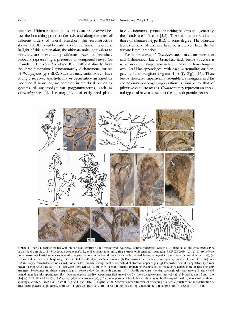
Psilophyton and Pauthecophyton regarded as trimerophytes, previously interpreted as leafless plants, have distal axes with lateral, three-dimensional, synchronously dichotomous trusses (Figure 1(a), (b)) [19–21]. These trusses have been called “incipient fronds”, “pinnules”, “dichotomous pin-nulelike appendages”, and “proto-leaves” [2,22]. Here, we prefer the term branch-leaf complexes (Psilophyton-type BLC) because these branches are basically axial in nature, non-planated, and unwebbed. These lateral dichotomous trusses, however, have a reduced morphology, implying a determinate growth, and most probably performed a photo-synthetic function similar to that performed by leaves. They lack apical meristems that have the capability of generating vegetative or reproductive organs. Distal divisions of the dichotomous trusses show a fundamentally leafy architec-ture, comprising a vascular bundle, thin-walled cells (cor-responding to parenchymous mesophylls), and epidermis and lacking peripheral sterome [3,19]. However, the main axes and branches of Psilophyton dawsonii have an outer cortex comprising a continuous layer of thick-walled sterome, a structure common in axes of many primitive tra-cheophytes but absent in Rhynie plants [19,22]. It is debata-ble whether the whole branching system or only the di-chotomous trusses were involved in the evolution of true leaves [20,23].
The three-dimensional lateral dichotomous trusses in Psilophyton might have been inherited by successive Mid-dle-Late Devonian plants [10,24]. Lateral dichotomous branching trusses of the Middle Devonian iridopteridaleans, such as Compsocradus and Ibyka, reduced to some degree and borne in a regular way (whorled or helical), are gener-ally called “ultimate appendages” or “ultimate branching systems” [25]. We consider these to be homologous to the BLC of Psilophyton. The pseudosporochnaleans including Lorophyton, Pseudosporochnus, and Wattieza, are a differ-ent case. Their axes bear whorled or irregularly arranged lateral branching systems (LBS) with ultimate dichotomous branching units [26]. The LBS of pseudosporochnaleans were described as “fronds” in early literature, and thus the ultimate branching units, were described as “pinnules”. Ul-timate appendages of more advanced cladoxylopsids and
Rhacophyton are similar in morphology to the BLC of Psilophyton, but show a more elaborate arrangement (subopposite or alternate) and some planation and lamina-tion into a single plane, indicating that further evolution had taken place [27].
1.2 Estinnophyton
Stems of Estinnophyton bear simple leaves, which are lat-eral dichotomous branching systems. They are planated but unwebbed and once- or twice-bifurcated at the middle part with two or four straight, tapering segments [18]. These leaves are arranged nearly in pseudowhorls, three to six per gyre (Figure 1(c), (d)). Fertile leaves are morphologically identical to sterile ones, clustered on axes to form loose, strobilus-like structure, and the sporangia are attached at the adaxial surfaces of the leaves by recurved stalks (Figures 1(e), 5(f)).
Estinnophyton has been considered a precursor of the sphenopsids in that its leaves have a pseudowhorled ar-rangement and the fertile leaves have recurved sporangia and sterile filiform extensions [18,28]. Typical sphenopsids with whorled simple leaves appeared in the Late Devonian [29,30]. The Middle Devonian iridopteridaleans (e.g. Ibyka) and pseudosporochnaleans (e.g. Calamophyton), particular-ly the former, have long been regarded as possible ancestral groups of the sphenopsids [22,25,31,32]. However, they have terminal sporangia, like Psilophyton. The characteris-tics of the fertile leaves of Estinnophyton, such as recurved sporangia, sterile filiform extensions, and pseudowhorled arrangement, are similar to those of Calamophyton [33]. These two taxa may have a close phylogenetic relationship, considering that reproductive characters should outweigh those from anatomy in early vascular plants [18]. The an-cient origin of the sphenopsids and simple leaves can be traced to the Early Devonian Estinnophyton.
1.3 Celatheca
Celatheca has main axes and three-dimensionally arranged lateral branches. The vegetative lateral branch has at least two to three orders of branching, finally forming a lateral branching system with ultimate dichotomous appendages. Through dégagement, and with reference to other Celatheca specimens, two reconstructions are shown in Figure 1(f), (g). Figure 1(f) shows that a lateral branch bifurcates once and the two unequal daughter branches bear ultimate dichoto-mous appendages with strongly recurved tips (cylindrical, ta-pering ends). The ultimate units are distributed distichously, at wide angles, showing a roughly pinnate arrangement. This is a characteristic morphology of Celatheca-type branch-leaf complexes (Celatheca-type BLC). The second reconstruc-tion shows a complex vegetative branching system (Figure 1(g)). There are many closely spaced laterals along part of the axis, some of which further divide to produce higher-order
2786 Hao S G, et al. Chin Sci Bull August (2013) Vol.58 No.23
branches. Ultimate dichotomous units can be observed be-low the branching point on the axis and along the axes of different orders of lateral branches. This reconstruction shows that BLC could constitute different branching orders. In light of this explanation, the ultimate units, equivalent to pinnules, are borne along different orders of branches, probably representing a precursor of compound leaves (or “fronds”). The Celatheca-type BLC differ distinctly from the three-dimensional synchronously dichotomous trusses of Psilophyton-type BLC. Such ultimate units, which have strongly recurved tips helically or decussately arranged on monopodial branches, are common in the distal branching systems of aneurophytalean progymnosperms, such as Tetraxylopteris [5]. The megaphylls of early seed plants
have dichotomous, pinnate branching patterns and, generally, the fronds are bifurcate [5,8]. These fronds are similar to those of Celatheca-type BLC to some degree. The bifurcate fronds of seed plants may have been derived from the bi-furcate lateral branches.
Fertile structures of Celatheca are located on main axes and dichotomous lateral branches. Each fertile structure is ovoid in overall shape, generally composed of four elongate- oval, leaf-like appendages, with each surrounding an elon-gate-ovoid sporangium (Figures 1(h)–(j), 5(g)) [16]. These fertile structures superficially resemble a synangium and the sporangium/appendage organization is similar to that of primitive cupulate ovules. Celatheca may represent an ances-tral type and have a close relationship with pteridosperms.
Figure 1 Early Devonian plants with branch-leaf complexes. (a) Psilophyton dawsonii. Lateral branching system [19], here called the Psilophyton-type branch-leaf complex. (b) Pauthecophyton gracile. Lateral dichotomous branching system with terminal sporangia. PKU-XH264b. (c)–(e) Estinnophyton yunnanense. (c) Partial reconstruction of a vegetative axis, with lateral, once or twice-bifurcated leaves arranged in low spirals or pseudowhorls. (d), (e) Lateral forked leaves, with sporangia in (e). BUH-Es.01. (f)–(j) Celatheca beckii. (f) Reconstruction of a branching system, based on Figure 3 of [16], as a Celatheca-type branch-leaf complex with more or less pinnate arrangement of ultimate dichotomous appendages. (g) Reconstruction of a vegetative specimen based on Figures 2 and 28 of [16], showing a branch-leaf complex with multi-ordered branching systems and ultimate appendages more or less pinnately arranged. Sometimes an ultimate appendage is borne below the branching point. (h)–(j) Fertile structures showing sporangia ((h) right arrow; (i) arrow) and, behind them, leaf-like appendages. (h) shows incomplete leaf-like appendages (left arrow) and (j) shows complete ones (arrows). (h), (i) From Figures 12 and 13 of [16]. (j) PUH.10-Cel. 01. (k)–(m) Polythecophyton demissum. (k), (l) Terminal portion of fertile branch showing umbrella-shaped fertile systems and pendulous sporangial clusters. From [34], Plate II, Figure 1, and Plate III, Figure 3. (m) Schematic reconstruction of branching of a fertile structure and reconstruction of attachment pattern of sporangia. From [34], Figure 2B. Bars: (a) 5 mm; (b) 2 mm; (c), (f), (h)–(j) 2 mm; (d), (e) 1 mm; (g) 4 mm; (k) (l) 5 mm; (m) 4 mm.

Hao S G, et al. Chin Sci Bull August (2013) Vol.58 No.23 2787
1.4 Polythecophyton
Polythecophyton exhibits a clear architecture of fertile structures but lacks anatomy and vegetative organs [34]. The axes branch dichotomously and helically in three di-mensions. The fertile branches are terminated by pendulous umbrella-shaped fertile structures (Figures 1(k)–(m), 5(h)). Each fertile structure may initially bifurcate, after which each component bears three to four alternately arranged, short axes, which divide into branchlets terminated by nu-merous slender fusiform sporangia in pairs or in groups of three or four.
This plant is similar to Pertica [35] and the aneurophyt-alean progymnosperms such as Aneurophyton and Rellimia [36,37] in branching pattern and particularly in fertile structures, sporangial morphology, and attachments. Some Pertica plants have been suggested to have a close relation-ship to aneurophytalean progymnosperms [22]. We suggest that Polythecophyton may represent basal aneurophytaleans as ordinal level.
1.5 Eophyllophyton
Eophyllophyton has both laminar leaves and branch-leaf complexes (Celatheca-type BLC) [15,17,38]. Generally the two laminated leaves curve inward towards each other to form a leaf pair, borne laterally or terminally on the axis (Figure 2(a),(b)). Leaf pairs along each axis appear an acropetal developmental series. That is, more pairs occur at the basal parts than at the distal parts, and those on the distal parts are younger and have curved tips (Figure 2(b)). The dichotomous pattern of the leaf pairs constitutes isotomous or anisotomous venation. Each leaf of the pair is served by a branching vein system, and each segment of the leaf is sup-ported by a single distal, tiny vein [17]. The leaves are very small (generally 2.2–6.0 mm long) and show great variabil-ity, from a three-dimensional branching pattern with weak lamination to more webbed, fan-shaped pattern with con-spicuous lamination and dissected segments (Figure 2(e), (f)). They can be suggested reduced compound leaves with highly dissected laminar divisions (corresponding to
Figure 2 Eophyllophyton bellum. (a) Lateral branch with small laterals terminating in leaf pairs, either vegetative or fertile. BUPb137. (b) Lateral branch showing an acropetally developmental transformation: the basal part has mature leaf pairs, and the distal part has younger ones (left arrow) and recurved sterile tips (right arrow). BUPb102’. (a), (b) From [38], Plate III, 1 and Plate I, 4. (c) A branch-leaf complex with ultimate appendages (dichotomy with re-curved tips) borne alternately along the axes, showing a roughly pinnate arrangement. PUH.10-Eop.01. (d) A leaf pair (two leaves, arrow indicates the faint appearance of bases of a leaf pair). The leaf margins of the laminate divisions are deeply incised and curved. A pinnately divided, laminar leaf reflects a branching system as shown in (c). PUH.10-Eop.02. (e) A fertile pair with two leaves shows a reduced, expanded branching system with weak lamination. The lower arrow points to common base of the two leaves, and upper arrow points to a sporangium. PUH.10-Eop.03. (f) A fan-shaped leaf with more con-spicuous lamination. Note that the margins of the laminar divisions are deeply incised (arrows). PUH.10-Eop.03, 04. (g) Fertile leaf cluster with numerous sporangia. BUPb127. Bars: (a)–(c) 3 mm; (d)–(g) 1 mm.

2788 Hao S G, et al. Chin Sci Bull August (2013) Vol.58 No.23
pinnules) which are borne alternately along a presumed mid-vein (Figure 2(d)). The divisions of the leaf are not strictly held in a single plane (Figure 3(a)). Most leaf pairs are fertile (Figures 2(g), 5(d)). Vegetative leaves are far less common, although the fertile and vegetative leaves are identical in morphology. Random sampling, observations and statistics from over 300 leaves within the same horizons show that fertile leaves are ca. six times more common than vegetative ones, indicating that most of the laminate leaves of Eophyllophyton are fertile (Figure S4, Table S1).
Aside from laminate webbed leaves, sterile branching axes with ultimate dichotomous appendages (Figure 2(c)), i.e. branch-leaf complexes (Celatheca-type BLC), occur at the lower parts or basal regions of Eophyllophyton. The ultimate dichotomies, which have markedly recurved tips, are alternately attached. In morphology and disposition of divisions, the BLC is fully comparable to a leaf (Figure 2(c), (d)). Sometimes, one of the recurved tips of an ultimate di-chotomy within a BLC terminates a leaf pair (vegetative or fertile) (Figure 2(b)). This implies that the tip of an ultimate dichotomy of the BLC corresponds to an arrested apex.
Anatomically, the leaf lamina is composed of veins, an outer single-layered epidermis, and a uniform mesophyll four to six cell layers thick. Mesophyll thin-walled cells, which appear to be randomly arranged, are polygonal or hexagonal in transverse view, elongated with oblique end walls in longitudinal view [15,17]. The mesophyll is struc-turally similar to that of a modern isobilateral leaf without differentiation into layers of palisade and spongy paren-chyma. It shows an absence of any conspicuous system of intercellular air channels. The petiole is relatively thick and contains several tracheids in a centrarch primary xylem strand like that of the axis. Small veins, containing one to a
few tracheids, occur midway between the upper and lower surfaces of the division segment (Figure 3(a)–(e)).
In Eophyllophyton, multiple, nearly spherical sporangia are arranged in adaxial rows on laminate leaves. Anatomi-cally, this plant has a columnar protostele, centrarch prima-ry xylem [15]. The character combination, which distin-guishes Eophyllophyton from zosterophylls and primitive lycopsids, trimerophytes, early ferns, fernlike plants, and progymnosperms, supports the assignment of Eophyllophy-ton to its own class and order.
The leaves of Eophyllophyton are distinct, but they ex-hibit more similarities to those of progymnosperms-seed plants than to those of early ferns or fernlike plants. The bifurcate morphology and broad laminar pinnules show strictly dichotomous venation, distinct characteristics of seed ferns, which differ from the fronds of ferns [5]. The early seed plants, like Elkinsia, exhibited highly dissected pinnules [8]. These were similar to the laminar divisions of Eophyllophyton’s leaves, although pinnule morphology can vary greatly. Anatomically, some axes of early seed plants have a small protostele and the departing leaf traces, like those of Eophyllophyton, have a single xylem bundle with centrarch or mesarch protoxylem abaxially situated [5,8].
2 Beyond megaphylls, plants with unusual leafy structures
Several Early Devonian plants such as Adoketophyton, Stachyophyton and Dibracophyton have fertile leaf-like ap-pendages (sporophylls or bracts) to constitute a strobilar structure with different sterile organs, but they are neither mi-crophyllous plants nor megaphyllous plants. The vegetative
Figure 3 Eophyllophyton bellum. (a) Reconstruction of a leaf showing that laminar divisions are not held in one plane. (b)–(e) Transverse sections at dif-ferent levels of the structurally preserved lamina, showing veins and mesophyll cells. (b) Through petiole of a leaf, note the main vein. Note main vein in (d) and observe that main vein in (c) and second-order veins in lateral divisions in (c) and (d) are missing. (e) Through distal region, note leaf vein. (b)–(e) From [17], Figure 3a–d. Bar: (a) 0.5 mm; (b)–(e) 250 µm.

Hao S G, et al. Chin Sci Bull August (2013) Vol.58 No.23 2789
axes of Adoketophyton dichotomously divide in three di-mensions with some laterals ending in terminal, circinately coiled tips (Figure S3(a)) [39]. This vegetative axis repre-sents another type of branch-leaf complex, Adoketophy-ton-type BLC. The strobilus is composed of fertile units, each of which consists of a fan-shaped sporophyll and a stalked sporangium (Figures 4(a), (b), 5(a)). The sporophyll is very regular in shape, with a parallel-sided stalk that ex-pands into a triangular lamina with straight side margins and rounded or undulate distal margin. In the transverse view, the sporophyllous laminae appear crescent-shaped, and the abaxial surface of one or two subjacent layers seems to consist of heavily carbonized and thick-walled cells. The internal tissues are a combination of thin-walled cells and tracheids. The tracheids appear scattered in the transverse
Figure 4 Adoketophyton subverticillatum. (a) Abaxial view of a sporo-phyll with a fan-shaped blade. (b) Lateral view of a fertile unit, with a sporangium adaxially attached on the sporophyll base. (a), (b) From [40], Plate I, Figures 3 and 2. (c) Transverse section of a sporophyllous lamina, showing variation in cells. Arrow points to probable tracheids. (d) Trans-verse section of distal part of a sporangium, showing structure of sporangi-al walls and marginal dehiscence grooves and thickenings (left arrow). Also note that the cells in the outermost layer are elliptical. Their long axes are perpendicular to the surface of the wall. Among them can be found prominent intercellular spaces (right arrow); dark material between two walls presumably represents a broken tapetal layer. (c), (d) From [40], Plate III, Figures 3 and 6. Bars: (a) 2 mm; (b) 1 mm; (c), (d) 60 µm.
section. The sporangial wall comprises six to nine rows of thin-walled, circular to polygonal cells, which form a single homogeneous layer, containing well-developed intercellular air channels (Figure 4(c), (d)).
In the original research, lacking detailed anatomical data, Adoketophyton was treated as Plantae Incertae Sedis [41], or had been placed in the barinophytopsids [42]. Consider-ing the positional similarity of sporophylls in Adoketophy-ton and in lycopsids, Kenrick and Crane [22] suggested that the sporophylls of Adoketophyton and lycopsids are homol-ogous, and Adoketophyton was placed in a basal position within the Lycophytina. Hao et al. [40] added anatomical data and described the vegetative terminal circinate tips of this plant. As to phylogenetic relationship, they suggested that Adoketophyton might represent an isolated group which evolved in parallel with lycopsids, and is far less related to the barinophytopsids. This plant is considered to be inde-pendent of the Zosterophyllopsida on the basis of the fertile unit composed of fan-shaped leaves and adaxially attached sporangia, and from Lycopsida in the absence of vegetative microphylls and presence of vegetative terminal circinate tips. Its centrarch primary xylem differs from the exarch order of maturation of both the zosterophyllopsids and ly-copsids. Now Adoketophyton is tentatively treated as class and order Incertae Sedis, with an uncertain phylogenetic position, though it is well known based on detailed mor-phology and anatomy.
Stachyophyton has lateral leaf-like branches arranged helically along main axes. The leaf-like branches generally show a fan-shaped form (Figures S2, S3(b)) [43], and are here considered a distinct type of branch-leaf complex, Stachyophyton-type BLC. These BLCs show morphological similarities to leaves with many divisions expanding distally in one plane, but they are rigid and unreduced, departing from the main axes. They show great variability, from the branching in a plane to more “webbed”, fan-shaped patterns with planation, and their distal segments are highly dissect-ed divisions with rounded or cuneate tips [44]. We speculate that these BLC’s retain an axial nature with a planate branching system but have distal foliar divisions and thus perform some photosynthetic functions. More intriguing, the strobili are located at the distal regions of the digitate branchlets of such fertile BLCs. The sporophylls are heli-cally arranged, elongated, and laminate with broad bases and bifurcated tips. Each elongated elliptical sporangium is adaxially attached on a sporophyll (Figure 5(b)).
In Stachyophyton, the digitate branching of leaf-like branches, strobili consisting of sporophylls and sporangia, and the exarch primary xylem distinguish it from other known groups of early vascular plants [43,44]. Li [42] placed Stachyophyton into barinophytopsids and suggested that its vegetative branches are slightly planate but not re-lated with megaphylls. In contrast, Wang and Cai [44] sug-gested that the vegetative branches as “megaphyll-like, leaf-like branches”. The presence of strobili borne on the
2790 Hao S G, et al. Chin Sci Bull August (2013) Vol.58 No.23
multiple branchlets of digitate “leaf-like branches” effec-tively distinguishes Stachyophyton from the contemporane-ous primitive lycopsids, sphenopsids and others. The Mid-dle-Upper Devonian lycopsids generally have terminal strobili, but they differ from Stachyophyton in the presence of microphylls. Stachyophyton probably represents another independent group, mainly considering lack of detailed morphology and anatomy, and particularly precence of dig-itate leaf-like branching structures and strobili.
Dibracophyton possesses terminal strobili. Each fertile unit comprises a stalked long-elliptical sporangium, with dehiscence into two equal valves, and two discrete long- ovate bracts covering the sporangium from above and below (Figure 5(c)) [45]. The sterile axes of Dibracophyton bear helically dichotomous appendages with curved/round tips. Some dichotomous appendages are alternately borne at the basal areas of the fertile axes. Dibracophyton resembles Barinophyton and Protobarinophyton [46–48]. Their spo-rangia have two valves and distal dehiscence (sporangial characters were demonstrated in B. norvegicum and Di-bracophyton), and possible unvascularized bracts. We sug-gest that these plants perhaps are close in affinity. Concern-ing other less well-known plants such as Bracteophyton variatum, Krithodeophyton croftii and Enigmophyton su-perbum [49,50], which were considered as putative mem-bers of barinophytopsids [41], their bract details and the exact relationship with sporangia need further examination. Moreover, the vegetative branches of most barinophytop-sids have not been determined. These plants were regarded as leafless, due to the limited material. It is interesting in Dibracophyton that, besides the independent vegetative axes, a few vegetative appendages are distributed along the basal area of the fertile axes, and that the long upper region of the fertile axes lacks any appendages (Figure S3(c)).
3 Multiple origins of megaphylls in the Early Devonian
The monophyly of euphyllophytes has been widely sup-ported [22,51]. However, as a foliar organ, the megaphylls of euphyllophytes have been shown to have evolved inde-pendently four, six, or even nine times in several different lineages [6,7,52]. Studies of leaf development and genetic expression have indicated that megaphylls are homoplastic across euphyllophytes [7,52]. The evolutionary scenario proposed by Zimmermann [53], in which three-dimensional lateral branches led to true leaves through overtopping, pla-nation and webbing (as the Telome Theory), has been ac-cepted by many neo- and paleobotanists [2]. Evidence from plant megafossils is anticipated to show intermediate exam-ples of these morphological transformations.
It has been suggested that early vascular plants of the Late Silurian through Middle Devonian, with few excep-tions, lack any true megaphyllous leaves. Lateral axes of a
plant from the late Early Devonian show an elliptical trace departing from a lobbed protostele. This plant has been ex-plained as the beginning of the evolution of megaphylls [54]. The branch-leaf complexes (three-dimensional lateral branch-ing systems) of trimerophytes of Emsian to early Eifelian age have been long considered the earliest megaphyll precursors [19,20]. However, the evidence from Posongchong Formation of the Pragian age, prior to the known other trimerophytes (e.g. Pertica and most Psilophyton species), indicates multiple ancient types of megaphylls and a much earlier appearance of laminate megaphylls (in Eophyllophyton).
Combined with fertile features and anatomy (where pre-sent) and comparisons with Middle-Late Devonian plants, several lineages can be traced and recognized (see above and Figure 5): (1) Psilophyton (and Pauthecophyton)- iridopteridaleans and pseudosporochnaleans (cladoxylop-sids), the latter groups being linked with early ferns or fern-like plants. They have paired elongate or fusiform sporangia borne at the ends of synchronous isotomies (Figure 5(e)) [19–23]; (2) Estinnophyton-sphenopsids. They share fertile structures with recurved sporangia and simple leaves in pseudowhorls (Figure 5(f)) [18,28]; (3) Polythecophyton- aneurophytaleans. Their fusiform sporangia are in clusters borne along the inner sides of the fertile structures (Figure 5(h)) [34]; (4) Celatheca-pteridosperms (seed ferns). Cela-theca bears elongate-oval fertile structures with sporangia and surrounding leaf-like appendages, structurally resembling a synangium or cupule (Figure 5(g)) [16]; and (5) Eophyllo-phyton, which shows a unique combination of characteristics, different from those of any known group (truly extinct) [15,17]. These five lineages, with different types of branch- leaf complexes (BLC) and initial leaves, clearly demonstrate the multiple origins of megaphylls (Figure 5), given that the differences among these BLC can be hardly explained by ecological factors and these BLC have a “modular construc-tion” that allows flexibility in organ production in response to changes in environmental conditions.
4 Implication of developmental mechanisms of leaves
The branch-leaf complexes of Psilophyton, Celatheca, and Eophyllophyton are all basically lateral organs with deter-minate growth and some degrees of reduction. The interac-tions between class 1 KNOX and ARP genes seem to be a shared mechanism responsible for determinacy of lateral appendages: KNOX genes are expressed in the shoot apical meristem (SAM) and ARP genes are expressed in leaf pri-mordial tissues [2,52]. The BLC and leaves of these plants exhibit features of an axial branching system, reflecting a ple-siomorphic state. The laminar leaves of Eophyllophyton ap-pear as miniatures of axial branching systems. The mesophyll cells are elongate, extending parallel to the vein strand, like cortical tissues of a lateral branch system. It seems reasonable

Hao S G, et al. Chin Sci Bull August (2013) Vol.58 No.23 2791
Figure 5 Phylogeny of basal tracheophytes (based on [12]) plotted against geological time (numerical ages from [55]), showing the appearance of major clades, including euphyllophytes in Late Silurian-Early Devonian. (a)–(h) Schematic reconstruction of fertile structures of selected plants from the Lower Devonian Posongchong Formation in Yunnan, southern China: (a) Adoketophyton; (b) Stachyophyton; (c) Dibracophyton; (d) Eophyllophyton; (e) Psilophy-ton (Pauthecophyton); (f) Estinnophyton; (g) Celatheca; (h) Polythecophyton. The phylogenetic framework is mainly based on the cladistic analyses of [12]. The dating of the oldest zosterophyllopsids and lycopsids is based on [56,57]. The sudden appearance of plants with unusual fertile features during the Early Devonian Pragian (ca. 410 Ma ago) is shown in (a)–(h).
to suggest that the genetic pathway controlling leaf devel-opment may have been built upon variation in the genetic mechanisms controlling the axial system.
As defined by Tomescu [52], a true leaf of vascular plants should have four features: vascularization, determi-nate growth, bilateral symmetry (adaxial-abaxial polarity), and definite arrangement (phyllotaxis). However, in the Early Devonian plants, these four features may have not evolved fully in any given type of ancient leaf (branch-leaf complexes, proto-leaves, or leaf precursors), and they show different acquisition sequences in different plant lineages. The branch-leaf complexes (BLC) of Psilophyton and Cela-theca show different branching patterns and, in our opinion, foreshadow different types of true leaves with different ve-nation and configuration. We use the term “branch-leaf complexes” because they are basically axial in nature, but their ultimate dichotomous appendages (or ultimate units) have undergone overtopping and reduction, showing erect
sterile tips (in Psilophyton) or recurved tips (in Celatheca). In this way, they seem to show a determinate growth pattern, indicating little or no possibility of further growth.
Megaphylls of Eophyllophyton bellum show a uniform, fundamentally leafy histology, consisting of a vascular bun-dle (vein), parenchymous mesophylls (thin-walled cells) and epidermis [15]. Similar structures occurred in at least ultimate units of the branch-leaf complexes (BLC) of Psilophyton dawsonii and Triloboxylon ashlandicum [19,58]. Although different in morphology, these early leaf- like structures have similar homogeneous thin-walled cells (mesophyll-like organization). That is, they lack adaxial- abaxial polarity and tissue differentiation between palisade and spongy cells. The subsequent acquisition of adaxi-al-abaxial polarity (and the associated genetic pathways) in euphyllophytes probably occurred independently. Various types of branch-leaf complexes and leaves in Early Devo-nian euphyllophytes (Psilophyton, Estinnophyton, Celatheca,

2792 Hao S G, et al. Chin Sci Bull August (2013) Vol.58 No.23
and Eophyllophyton) represent very different lineages, in-dicating a complex pattern. Class III HD-zip, KANADI, and YABBY genes, micro RNAs 165 and 166, and their interac-tions seem to contribute to the establishment of adaxi-al-abaxial polarity [2,8,59]. It is noteworthy that the leaves of phan mutants of the angiosperm plant Antirrhinum majus show radial symmetry [60]. They also show architecture similar to those ultimate units of the branch-leaf complexes (BLC) of Psilophyton dawsonii and Triloboxylon ashland-icum, and Eophyllophyton. Mutant analyses and gene ex-pression studies have revealed epigenetic phenomena that regulate leaf laminar development. In Eophyllophyton the tips of ultimate dichotomy of a BLC could be transformed into the laminar leaves demonstrating this phenomenon in the early stage of leaf evolution.
5 Appearance of fertile laminar leaf-like appendage (or leaves) as an adaptation for protection of sporangia
Plants such as Adoketophyton, Stachyophyton, Dibracophy-ton, and Celatheca have laminated leaf-like appendages (sporophylls or bracts) subtending sporangia [16,39-45]. The leaf-like appendages of Adoketophyton are vascularized and probably have heavily carbonized cuticles. The associ-ated sporangia have well-developed intercellular air chan-nels in their sporangial walls. Anatomical details of leaf-like appendages in Stachyophyton, Dibracophyton, and Cela-theca are unclear. These various leaf-like appendages have been considered a phenomenon of convergent evolution in that their parent plants show large differences in branching, sporangial features, and other characters. Based on biogeo-chemical models and studies of paleosol and plant stomatal indexes, the Early Devonian has been deduced to be a peri-od with high concentrations of atmospheric CO2 [1,9,61]. These laminar leaf-like structures, besides photosynthesis, may mainly have had a protective function, protecting the sporangia from solar heat in this harsh-atmosphere envi-ronment. Environmental factors and physiologic requisition, such as high atmospheric CO2 concentrations and high temperatures, may have driven selection and adaptation. As a result, fertile laminate leaf structures enclosing sporangia may have evolved independently in different plant lineages and have been laminated before the formation of vegetative leaf structures (e.g. Figure S4, Table S1). Unlike several lineages with megaphylls, the aforementioned plants, with unusual fertile leaf-like appendages, were short-lived and quickly became extinct. They have been replaced by plants with true leaves and sporophylls, i.e. lycopsids, ferns, hor- setails, and eventually by seed plants.
The authors thank Charles B. Beck and Dianne Edwards for revising the manuscript and providing helpful discussion, and thank William G. Chaloner for his valuable comments. This work was supported by the Na-tional Natural Science Foundation of China (41272018 and 40830211).
1 Beerling D J. Leaf evolution: Gases, genes and geochemistry. Ann Bot, 2005, 96: 345–352
2 Beerling D J, Fleming A J. Zimmermann’s telome theory of mega-phyll leaf evolution: A molecular and cellular critique. Curr Opin Plant Biol, 2007, 10: 4–12
3 Gifford E M, Foster A S. Morphology and Evolution of Vascular Plants. 3rd ed. New York: Freeman, 1989
4 Beck C B. An Introduction to Plant Structure and Development: Plant Anatomy for the Twenty-first Century. Cambridge: Cambridge Uni-versity Press, 2010
5 Galtier J. The origins and early evolution of the megaphyllous leaf. Int J Plant Sci, 2010, 171: 641–661
6 Cronk Q C B. Plant evolution and development in a postgenomic context. Nat Rev Genet, 2001, 2: 607–619
7 Boyce C K, Knoll A H. Evolution of developmental potential and the multiple independent origins of leaves in Paleozoic vascular plants. Paleobiology, 2002, 28: 70–100
8 Sanders H, Rothwell G W, Wyatt S E. Key morphological alterations in the evolution of leaves. Int J Plant Sci, 2009, 170: 860–868
9 Osborne C P, Beerling D J, Lomax B H, et al. Biophysical constraints on the origin of leaves inferred from the fossil record. Proc Natl Acad Sci USA, 2004, 101: 10360–10362
10 Crane P R, Herendeen P, Friis E M. Fossils and plant phylogeny. Am J Bot, 2004, 91: 1683–1699
11 Hao S G. A new zosterophyll from the Lower Devonian (Siegenian) of Yunnan, China. Rev Palaeobot Palynol, 1989, 57: 155–171
12 Hao S G, Xue J Z. The Early Devonian Posongchong flora of Yunnan —A Contribution to an Understanding of the Evolution and Early Diversification of Vascular Plants. Beijing: Science Press, 2013
13 Wang Y. Lower Devonian miospores from Gumu in the Wenshan District, Southeastern Yunnan (in Chinese). Acta Micropalaeontol Sin, 1994, 11: 319–332
14 Zhu M, Wang J Q, Fan J H. Early Devonian fishes from Guijiatun and Xujiachong Formations of Qujing, Yunnan, and related biostrati-graphic problems (in Chinese). Vertebr Palasiat, 1994, 32: 1–20
15 Hao S G, Beck C B. Further observations on Eophyllophyton bellum from the Lower Devonian (Siegenian) of Yunnan, China. Palaeontogr Abt B Palaeophytol, 1993, 230: 27–41
16 Hao S G, Gensel P G. A new genus and species, Celatheca beckii, from the Siegenian (Early Devonian) of southeastern Yunnan, China. Int J Plant Sci, 1995, 156: 896–909
17 Hao S G, Beck C B, Wang D M. Structure of the earliest leaves: Ad-aptations to high concentrations of atmospheric CO2. Int J Plant Sci, 2003, 164: 71–75
18 Hao S G, Wang D M, Wang Q. A new species of Estinnophyton from the Lower Devonian Posongchong formation, Yunnan, China; its phylogenetic and palaeophytogeograpical signficance. Bot J Linn Soc, 2004, 146: 201–216
19 Banks H P, Leclercq S, Hueber F M. Anatomy and morphology of Psilophyton dawsonii sp. n. from the late Lower Devonian of Quebec (Gaspé), and Ontario, Canada. Palaeontogr Am, 1975, 8: 77–127
20 Doran J B. A new species of Psilophyton from the Lower Devonian of northern New Brunswick, Canada. Can J Bot, 1980, 58: 2241–2262
21 Xue J Z, Hao S G, Zhu X, et al. A new basal euphyllophyte, Pau-thecophyton gen. nov., from the Lower Devonian (Pragian) of Yun-nan, China. Rev Palaeobot Palynol, 2012, 183: 9–20
22 Kenrick P, Crane P R. The Origin and Early Diversification of Land Plants, a Cladistic Study. Washington: Smithsonian Institution Press, 1997
23 Trant C A, Gensel P G. Branching in Psilophyton: A new species from the Lower Devonian of New Brunswick, Canada. Am J Bot, 1985, 72: 1256–1273
24 Rothwell G W. Fossils and ferns in the resolution of land plant phy-logeny. Bot Rev, 1999, 65: 188–218
25 Berry C M, Stein W E. A new iridopteridalean from the Devonian of Venezuela. Int J Plant Sci, 2000, 161: 807–827
26 Berry C M. A reconsideration of Wattieza Stockmans (here attributed to Cladoxylopsida) based on a new species from the Devonian of Venezuela. Rev Palaeobot Palynol, 2000, 112: 125–146

Hao S G, et al. Chin Sci Bull August (2013) Vol.58 No.23 2793
27 Xue J Z, Hao S G, Basinger J F. Anatomy of the Late Devonian Denglongia hubeiensis, with a discussion of the phylogeny of the Cladoxylopsida. Int J Plant Sci, 2010, 171: 107–120
28 Fairon-Demaret M. Estinnophyton gracile gen. et sp. nov., a new name for specimens previously determined Protolepidodendron wahnbachense Kräusel and Weyland from the Siegenian of Belgium. Bulletin de I'Academie royale de Belgique (Classe des sciences), 1978, 64: 597–609
29 Wang D M, Hao S G, Wang Q. Rotafolia songziensis gen. et comb. nov., a sphenopsid from the Late Devonian of Hubei, China. Bot J Linn Soc, 2005, 148: 21–37
30 Wang D M, Hao S G, Wang Q, et al. Anatomy of the Late Devonian sphenopsid Rotafolia songziensis, with a discussion of stelar archi-tecture of the Sphenophyllales. Int J Plant Sci, 2006, 167: 373–383
31 Stein W E, Wight D C, Beck C B. Possible alternatives for the origin of Sphenopsida. Syst Bot, 1984, 9: 102–118
32 Wang D M, Guo Y. Hamatophyton from the Late Devonian of Anhui Province, South China and evolution of Sphenophyllales. Acta Geol Sin-Engl, 2009, 83: 492–503
33 Leclercq S. Calamophyton primaevum: The complex morphology of its fertile appendage. Am J Bot, 1969, 56: 773–781
34 Hao S G, Gensel P G, Wang D M. Polythecophyton demissum, gen. et sp nov., a new plant from the Lower Devonian (Pragian) of Yun-nan, China and its phytogeographic significance. Rev Palaeobot Pal-ynol, 2001, 116: 55–71
35 Granoff J A, Gensel P G, Andrews H N. A new species of Pertica from the Devonian of eastern Canada. Palaeontogr Abt B Palaeophy-tol, 1976, 155: 119–128
36 Serlin B S, Banks H P. Morphology and anatomy of Aneurophyton, a progymnosperm from the Late Devonian of New York. Palaeontogr Am, 1978, 8: 343–359
37 Schweitzer H J, Matten L C. Aneurophyton germanicum and Pro-topteridium thomsonii from the Middle Devonian of Germany. Pal-aeontogr Abt B Palaeophytol, 1982, 184: 65–106
38 Hao S G. A new Lower Devonian genus from Yunnan, with notes on the origin of leaves (in Chinese). Acta Bot Sin, 1988, 30: 441–448
39 Zhu X, Xue J Z, Hao S G, et al. A new species of Adoketophyton from the Lower Devonian (Pragian) Posongchong Formation of Yunnan, China. Rev Palaeobot Palynol, 2011, 164: 238–246
40 Hao S G, Wang D M, Beck C B. Observations on anatomy of Ado-ketophyton subverticillatum from the Posongchong Formation (Pra-gian, Lower Devonian) of Yunnan, China. Rev Palaeobot Palynol, 2003, 127: 175–186
41 Li C S, Edwards D. A new genus of early land plants with novel strobilar construction from the Lower Devonian Posongchong For-mation, Yunnan Province, China. Paleontology, 1992, 35: 257–272
42 Li C S. Review on the origin and early evolution of lycopods. Yushania, 1992, 9: 185–194
43 Geng B Y. Stachyophyton gen. nov. discovered from Lower Devoni-
an of Yunnan and its significance (in Chinese). Acta Bot Sin, 1983, 25: 574–579
44 Wang Y, Cai C Y. Further observation on Stachyophyton yunnanense Geng from Posongchong Formation (Siegenian) of SE Yunnan, Chi-na (in Chinese). Acta Palaeontol Sin, 1996, 35: 99–108
45 Hao S G, Xue J Z, Zhu X, et al. A new genus of Early Devonian plants with novel strobilar structures and vegetative appendages from the Posongchong Formation of Yunnan, China. Rev Palaeobot Paly-nol, 2012, 171: 73–82
46 Brauer D F. Barinophyton citrulliforme (Barinophytales incertae sedis, Barinophytaceae) from the Upper Devonian of Pennsylvania. Am J Bot, 1980, 67: 1186–1206
47 Brauer D F. Heterosporous, barinophytacean plants from the Upper Devonian of North America and a discussion of the possible affinities of the Barinophytaceae. Rev Palaeobot Palynol, 1981, 33: 347– 362
48 Schweitzer H J, Giesen P. Further remains of Middle Devonian plants found at Wuppertal, Bergisches Land (Western Germany). Palaeon-togr Abt B Palaeophytol, 2008, 277: 101–140
49 Edwards D. A new plant from the Lower Old Red Sandstone of South Wales. Paleontology, 1968, 11: 683–690
50 Wang D M, Hao S G. Bracteophyton variatum gen. et sp. nov., an Early Devonian plant from the Xujiachong Formation of Yunnan, China. Int J Plant Sci, 2004, 165: 337–345
51 Bateman R M, Crane P R, Dimichele W A, et al. Early evolution of land plants: phylogeny, physiology, and ecology of the primary ter-restrial radiation. Ann Rev Ecol S, 1998, 29: 263–292
52 Tomescu A M F. Megaphylls, microphylls and the evolution of leaf development. Trends Plant Sci, 2009, 14: 5–12
53 Zimmermann W. Main results of the “Telome Theory”. The Palaeo-botanist, 1952, 1: 456–470
54 Gensel P G. A new Lower Devonian plant and the early evolution of leaves. Nature, 1984, 309: 785–787
55 Ogg J G, Ogg G, Gradstein F M. The Concise Geologic Time Scale. Cambridge: Cambridge University Press, 2008
56 Kotyk M E, Basinger J F, Gensel P G, et al. Morphologically com-plex plant macrofossils from the Late Silurian of Arctic Canada. Am J Bot, 2002, 89: 1004–1013
57 Rickards R B. The age of the earliest club mosses: the Silurian Barag-wanathia flora in Victoria, Australia. Geol Mag, 2000, 137: 207–209
58 Scheckler S E. Ontogeny of progymnosperms. I. shoots of Upper Devonian Aneurophytales. Can J Bot, 1976, 54: 202–219
59 Sanders H, Rothwell G W, Wyatt S. Paleontological context for the developmental mechanisms of evolution. Int J Plant Sci, 2007, 168: 719–728
60 Waites R, Hudson A. phantastica: A gene required for dorsoventrality of leaves in Antirrhinum majus. Development, 1995, 121: 2143–2154
61 Berner R A, Kothavala Z. GEOCARB III: A revised model of atmos-pheric CO2 over Phanerozoic time. Am J Sci, 2001, 301: 182–204
Open Access This article is distributed under the terms of the Creative Commons Attribution License which permits any use, distribution, and reproduction
in any medium, provided the original author(s) and source are credited.
Supporting Information
Figure S1 Stratigraphic column of the Posongchong Formation at the Zhichang section showing distribution of megafossil plants.
Figure S2 Stachyophyton yunnanense.
Figure S3 Partial restorations of Adoketophyton parvulum (a), Stachyophyton yunnanense (b), and Dibracophyton acrovatum (c).
Figure S4 Eophyllophyton bellum.
Table S1 Occurrences of fertile and sterile leaves in specimens of Eophyllophyton bellum
The supporting information is available online at csb.scichina.com and www.springerlink.com. The supporting materials
are published as submitted, without typesetting or editing. The responsibility for scientific accuracy and content remains en-tirely with the authors.