ear care workshop. aims for the practitioner to be able to confidently carry out ear examinations,...
TRANSCRIPT

Ear care workshop

AIMS
For the practitioner to be able to confidently carry out ear examinations, recognise
abnormalities and to carry out appropriate ear care.
Objectives• To enhance understanding of basic anatomy and
physiology of the ear.• Ear examination• To recognise abnormalities and associated care• Theory behind cerumen removal and associated
guidelines• To reflect on accountability, documentation • Practical session
Basic anatomy and physiologyof ear
Outer (External) ear
• Pinna and external acoustic canal.
• Canal lined with small hairs next to which are small ceruminous glands
• The Tympanic Membrane
MIDDLE EAR
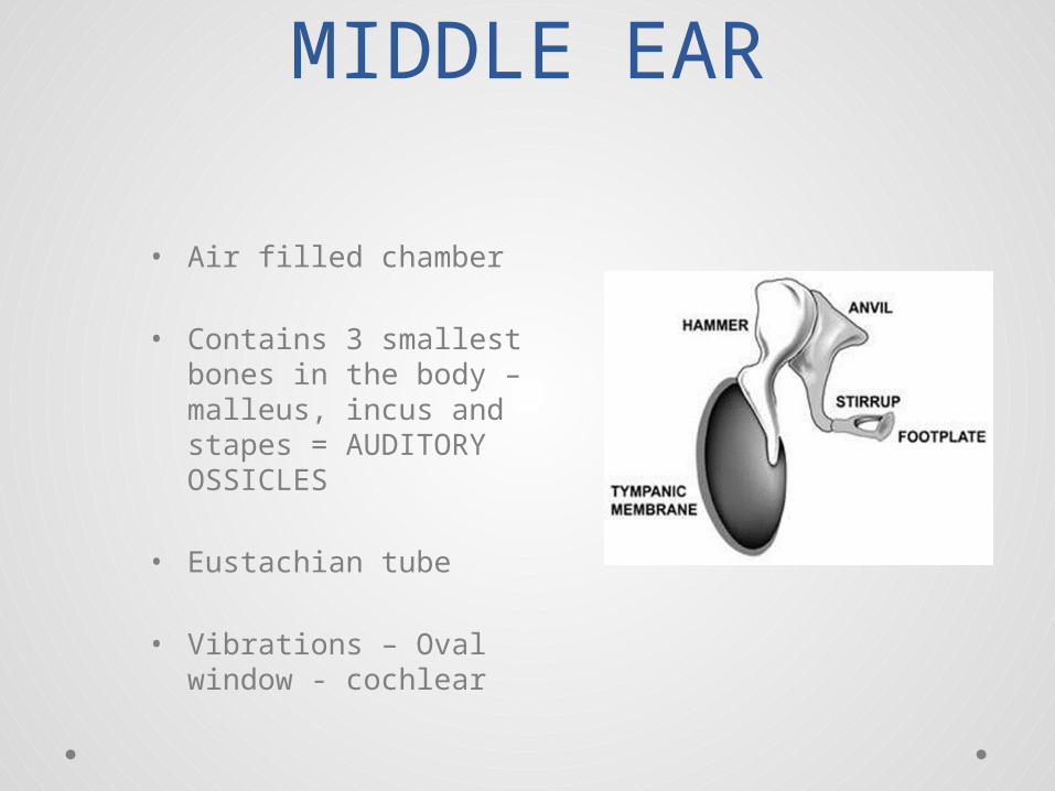
• Air filled chamber
• Contains 3 smallest bones in the body – malleus, incus and stapes = AUDITORY OSSICLES
• Eustachian tube
• Vibrations – Oval window - cochlear
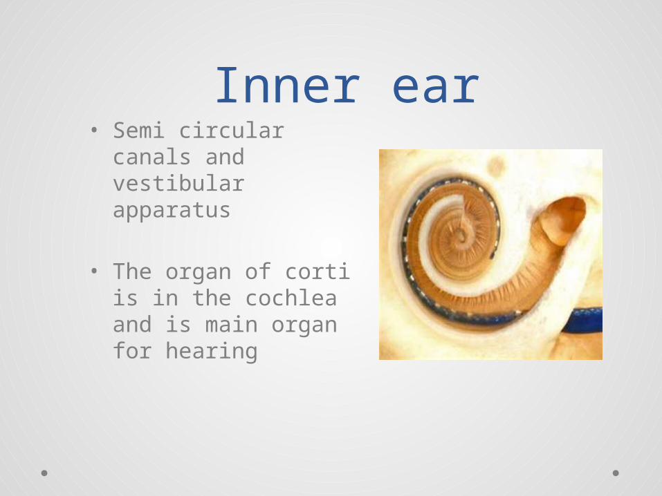
Inner ear• Semi circular canals
and vestibular apparatus
• The organ of corti is in the cochlea and is main organ for hearing

Nerves involved:
• Auditory nerve – inner ear ( CN VIII)• Facial nerve – middle ear ( CN VII)• Vagus Nerve - Outer ear (CN X)
Ear wax - Cerumen• Not Like paraffin! Made of a lot of different
chemical components
• Testerone control over production of sebum.
• What is its purpose?
People likely to produce excessive
wax • Learning disabilities• Anxious people• High lipid levels• Genetic tendency• Elderly

What issues can wax build up cause a patient?
Ear Examination
History of presenting condition:
What information would be required here?

History cont……
• PMH - ? Relevant
• Meds
• Allergies
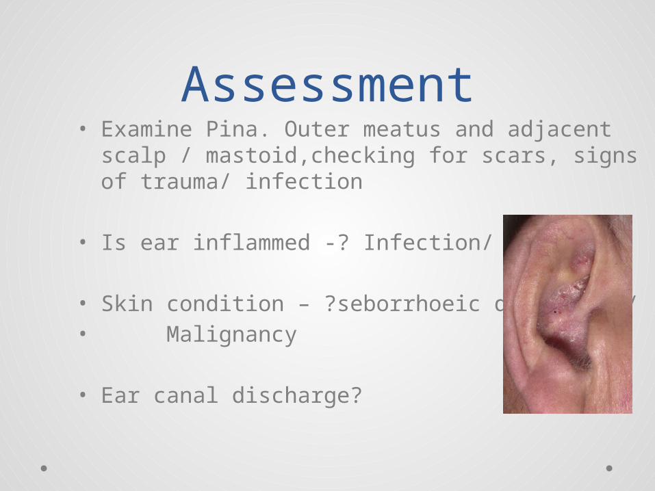
Assessment• Examine Pina. Outer meatus and adjacent scalp /
mastoid,checking for scars, signs of trauma/ infection
• Is ear inflammed -? Infection/ trauma
• Skin condition – ?seborrhoeic dermatitis/• Malignancy
• Ear canal discharge?

Identify wax……• In ear examination discern type of wax, position
in canal and % of occlusion
• Is wax healthy, or bacterial debris, or dry and crumbly

Assessment cont..Examine external auditory meatus.
Should be pain free.
What can cause pain?FuruncleTraumaOtitis externaFungal infection
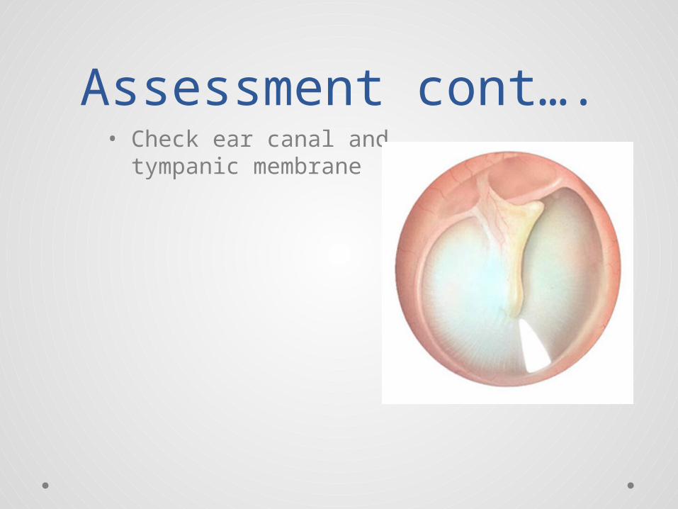
Assessment cont….• Check ear canal and
tympanic membrane
Assessment cont…• When withdrawing otoscope check external
auditory meatus carefully.
• Document……!• Document…..!• Document….!


Ear cleaning / irrigation
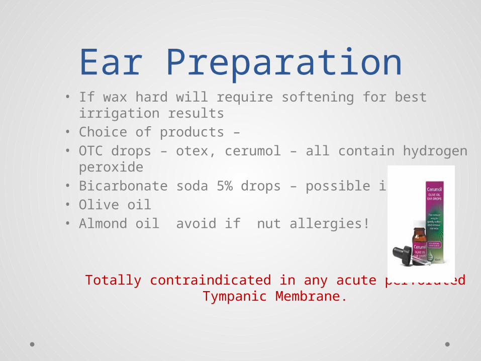
Ear Preparation• If wax hard will require softening for best irrigation
results• Choice of products – • OTC drops – otex, cerumol – all contain hydrogen
peroxide• Bicarbonate soda 5% drops – possible irritant• Olive oil• Almond oil avoid if nut allergies!
Totally contraindicated in any acute perforated Tympanic Membrane.
Application of drops• No formal length of time and number of times a
day
• Advise if using oil not to heat it up – room temp sufficient
• Not to over oil ears as becomes irritated

Manual removal• If dry and crusty possibly can be gently
manoeuvred out using Jobson Horne probe – head light and otoscope
• If too painful discontinue - skin becomes quickly traumatised
Ear Irrigation• Equipment required:• Auriscope• Head light / ? Eye protectors/ apron/ gloves
• Electronic ear irrigator• Warm water – approx room temp - NOT COLD!!• Noots receiver ( disposable or lined)• Jobson Horne probe/ cotton wool• Tissues, receivers• Waterproof cape / towel
Guidelines:……• ENSURE – device only used by trained clinician• ENSURE that warnings and cautions are
observed• ENSURE patient exhibits NO contra indications• ENSURE the unit been cleaned
Reasons for irrigation
• Remove and Improve!

Principles of ear irrigation
• Facilitate the removal of cerumen and foreign bodies
• Individual assessment of each patient by practitioner carrying out procedure.
Possible complications?
• Infection• Perforation• Tinnitus• Vagal nerve stimulation• Dizziness

Irrigation should not be carried out
when:• Informed consent not obtained and NSCP
consent form signed for procedure• Patient has had previous problems with
procedure in past• Hx of otitis media in past 6 weeks• Any form of ear surgery in past 18 months and
NOT been discharged from care of ENT• Perforation or Hx of mucus discharge in past 12
months• Cleft palate repaired or not• Presence of acute otitis externa
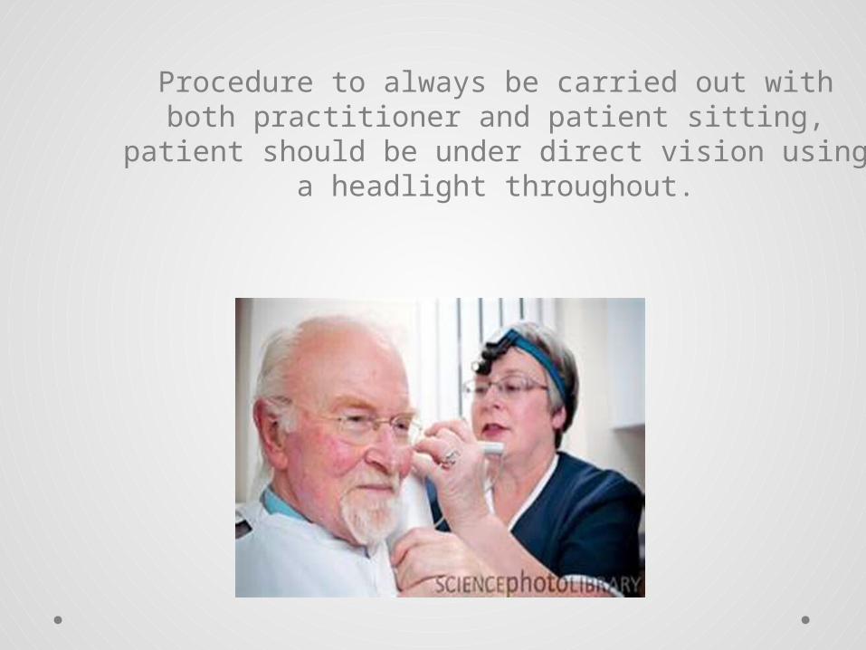
Procedure to always be carried out with both practitioner and patient sitting, patient should be under direct vision using a headlight throughout.

DOCUMENTATION…….
Record:• What the patient says• What you see in BOTH ears• What you do – including advice given• Why this treatment – rationale….
NMC record keeping guidelines 2010

Documentation tips…• Always compare both ears - L=R• Do not use word “Appears normal” - looks as
though not aware of what you are doing be definite. – “ Tympanic membrane normal” or “Tympanic membrane normal features not visible”
• Word “impacted” – to be used if evidence wax has been pushed down canal with implement such as cotton bud
• ‘Occluded’ should be used is canal full of wax – say whether dark and hard, softy and light wax.
• Document advice given to patient written/ verbal post procedure.

DEAFNESS3 types:
1. Conductive : Obstruction between external /middle ear
2. Sensori – neural: Obstruction between stapes footplate and the auditory centres of brain
3. Mixed Deafness: Combination of conductive and sensori – neural deafnessPractical Time


Hearing Aid Care..• General• Whistling• Hearing aid controls• Washing the ear mould• Retubing ear mould

Tinnitus• What is Tinnitis?
• How do you get it?
• Who is likely to get it?
• Hyperacusis

Patient Education• E – Educate - Why problem there?
• A – Advise – How to prevent recurring Regular check ups
• R – Resolve – Treat the Problem, applying
clinical judgement and ability

References and acknowledgements
• Rogers R, www.earcareservicesuk.com• www.tinnutis.org.uk• www.deafnessresearch.org.uk• Action on ENT Steering Board (2007). Guidance
Document in Ear Care. Primary Ear Care Centre. (http:// www.earcarecentre.com)
• Nursing and Midwifery Council (2006) Record Keeping Guidance for Nurses. NMC, London.
• Skills for Health :CHS20. Undertake examination of the external ear: -National Operation Standards: https://tools.skillsforhealth.org.uk/competence/show/html/id/350/ (last accessed 11th April 2012)