dttc electep - defense technical information center as one example, cell cycle control ... patients...
TRANSCRIPT

DTtC ELECTEP NOV 0 3 1995
F GRANT NO: DAMD17-94-J-4359
AD
•Original contains color plates: All DIIC reproduct- ions will be in black and white"
TITLE: Amplified Genes in Breast Cancer: Molecular Targets for Investigation and Therapy
PRINCIPAL INVESTIGATOR(S): Geoffrey M. Wahl, Ph.D.
CONTRACTING ORGANIZATION: The Salk Institute for Biological Studies
La Jolla, California 92037- 1099
REPORT DATE: September 1995
TYPE OF REPORT: Annual
PREPARED FOR: U.S. Army Medical Research and Materiel Command Fort Detrick, Maryland 21702-5012
DISTRIBUTION STATEMENT: Approved for public release; distribution unlimited
The views, opinions and/or findings contained in this report are those of the author(s) and should not be construed as an official Department of the Army position, policy or decision unless so designated by other documentation.
19951102 043 Dfl© QUALITY INSPECTED 8

m &0'Ci}krf-KTPJiOi; Form Approved
OMB No. 0704-0188
Public reporting burden for this collection of information is estimated to average 1 hour per response, including the time for reviewing instructions, searching existing data sources, gathering and maintaining the data needed, and completing and reviewing the collection of information. Send comments regarding this burden estimate or any other aspect of this collection of information, including sugaestions for reducing this burden, to Washington Headquarters Services, Directorate for Information Operations and Reports, 1215 Jefferson
-Davis Hiohway, Suite 1204, Arlington, VÄ 22202-4302, and to the Off ice of Management and Budget, Paperwork Reduction Project (0704-0188), Washington, DC 20503.
1. M m (Leave bias 2. REPORT DATü
September 1995
li Amplified Genes in Breast Cancer: Molecular Targets for i] Investigation and Therapy
. REPORT TYF'C Miff DATES COVERED
Annual _l_Sep 94 - 31 Aug 95
ÜGeoffrey M. Wahl, Ph.D.
i. pcr.roriOT:3 o*&f.xt?.fiWm FJA^E(E) M:S> mr>t:.?ss{vs.) The Salk Institute for Biological Studies La Jolla, California 92037-1099
U.S. Army Medical Research and Materiel Command Fort Detrick, Maryland 21702-5012
DAMD17-94-J-4359
C. PERFORR/ilKG ORGANIZATION REPORT H'EJ&üBER
10. SPONSORING / MONITORING AGENCY REPORT NUPJ1BER
]"(. mv?uiij u'ES
t2ä. D;rYi:r.i.!TiOf; / Al'AÜ.AEiüTY STATE^EivT'
Approved for public release; distribution unlimited 12b. DISTREUTIOK CODE
: IS. ARSIRACF (Maximum200 words)
Abstract The primary objective has been development of a rapid method for purifying pxtrachromosomaily amplified genes (DMs) from tumor cell lines without relying on eytogenetics. DMs were purified by isolating micronuclei induced by treatment with hydroxyurea. Micronuclei were dissociated from nuclei by lysis under conditions that disaggregated intermediate filaments. Rate zonal and density centrifugation steps completely separated micronuclei from nuclei, enabling preparation of probes for fluorescence in situ hybridization to metaphase chromosomes of normal peripheral blood lymphocytes to identify the genomic regions from which the DMs were generated. Four cell lines derived from different types of tumors with DM contents spanning the range typically reported in human biopsy material (e.g., 2-50 per nucleus) were analyzed. The procedures was effectively used for all cell lines, and showed that the DMs in each were derived from the c-myc locus at 8q.24. Exclusion of chromosomes from micronuclei was shown to be due to the presence of an element of chromosome function, such as a centromere, rather than to small size. These results are significant because they provide the first rapid, sensitive method for DM purification that should be applicable to human biopsy samples.
micronucleus purification; gene amplification; double minute chromosome purification; hydroxyurea
Jbr e a s t e anc e r__
Unclassified NSW"754u-01-2C.0-5500
or Ts:ir. PAGE
Unclassified
S. SECURtTY CLASSIFICATION 0" ABSTRACT
Unclassified
NUMBER OF PAGES
42 16. PRICE CODE
20. LIMITATION OF ABSTRACT
Unlimited Standard Form 298 (Rev. 2-89) Prescribed by ANSI Std. Z39-18 298-102

INSTRUCTIONS FOR COMPLETING SF 2§S
(RDP) is used in announcing and cataloging reports. It is important :V; with the rest of the report, particularly the cover and title page. ck of the form follow. It is important to stay within the lines to meet
p,"
to'.
-■■* A c'.:
r
• ' ■ ■ • •-).
Fii!! publication date --' v.-- ",. irf-iil?hle(e.g. 1
Date:. Covered. rim, final, etc. If apart dates (e.g. 10
A title ir. taken from ■rev;e'er-the most oOrrnation. When a hao one volume, '■ voAime number, and
On slfication
To i -[dude contract ine! IÖC: program «..., LUV. ~.er(s},task
' P [ f! ehe rfsl Use the
SO; TA
PlulCCt Tesl-
- Work Unit Accession Wo.
;.-/-> of parson(s} o repcrt. performing with the content of the iler, this should follow
" ">A?1A" n Namc(s) and
' nJoeJiQiOJi§i2^Il alphanumeric report
yaorzotion
LQ5. Acency Name(s) ,. \r. ?.,.,. i _ -
mtonno Aoencv
etes. Enter sewherc such as: tie..; Trans, of...; Tobe m;t is revised, include
■■:■: report supersedes
Block 12a. Distribution/Availability SfateniSQt. Denotes public availability or limitations. Cite any availability to the public. Enter additional limitations or special markings in a!! capitals (e.g. IMOFORN, REL, ITAR).
DOD - See DoDD 5230.24, "Distribution Statements on Technical Documents."
DOE - See authorities. I\SÄSÄ - See Handbook WHB 2/00./. WTIS - Leave blank.
Block 12b. Distribution Code.
DOB - Leave blank. DOE - Enter DOE distribution categories
from the Standard Distribution for Unclassified Scientific and Technical Reports.
WASA- Leave blank. MISS - Leave blank.
Block 13. Abstract. Include a brief (Maximum 200 words) factual summary of the most. significant information contained in the report.
Block 14. Subject Terms. Keywords or phrase- identifying major subjects in the report.
Block 15. Number of Pages. Enter the total number of pages.
Block 1S. Price Code. Enter appropriate price code (NTIS only).
Blocks 17.-19. Security Classifications. Self- explanatory. Enter U.S. Security Classification in accordance with U.S. Security Regulations (i.e., UNCLASSIFIED). If form contains classified information, stamp classification on the top and bottom of the page.
Block 20. Limitation of Abstract. This block must be completed to assign a limitation to the abstract. Enter either UL (unlimited} or SAR (same as report). An entry in this block is necessary if the abstract is to be limited. If blank, the abstract is assumed to be unlimited.
Standard Form 2SS Back (Rev. 2-89 *U.S.GPO:1993-0-358-779

FOREWORD
~ • ■ n«4-a.»-«-r-c»+-si4-Tr»Tie! conclusions and recommendations are SETS't£*SSS^SS tie not necessarily endorsed by the OS Army.
^ Where copyrighted material is quoted, permission has been obtained to use such material.
Where material from documents designated for limited distribution is quoted, permission has been obtained to use tne material.
Citations of commercial organizations and trade names in tEIS report do not constitute an official Department of Army endorsement or approval of the products or services of these organizations.
u^"m conducting research using animals, the investigator(s) idhered to the "Guide for the Care and Use of Laboratory ESS«," prepared by the Committee on Care and use °fQ^f
ratory
Animals of the Institute of Laboratory Resources, National Research Council (NIH Publication No. 86-23, Revised 1985).
(/'•-For the protection of human subjects, the investigator (s) Idhered to policies of applicable Federal Law 45 CFR 46.
^ in conducting research utilizing recombinant DNA technology, the-inveI?igator<l) adhered to current guidelines promulgated by the National institutes of Health.
- in the conduct of research utilizing recombinant DNA, the Investigator(s) adhered to the NIH Guidelines for Research Involving Recombinant DNA Molecules.
U in the conduct of research involving hazardous organisms, tne-investigator(s) adhered to the CDC-NIH Guide for Biosafety in Microbiological and Biomedical Laboratories.
P|I - /Signature Date

TABLE OF CONTENTS
Page
Front Cover 1
SF 298 Report Documentation Page 2
Foreword 3
Table of Contents 4
Introduction 5
Body 6
Conclusions 8
References 9

Introduction Loss of genetic stability during cancer progression is a recurrent theme in oncogenesis.
More than 50% of primary biopsy specimens and cell lines contain chromosomal aberrations that probably arose from defective recognition or repair of local DNA lesions, or from aberrant control of cell cycle transitions. As one example, cell cycle control mechanisms prevent gene amplification from occurring at measurable frequencies in normal cells, while it has been reported in a substantial fraction of cancer in vivo 1" 5. The overall goal of the experiments in this proposal was to develop a rapid method for identifying breast cancers containing amplified genes, and to develop effective means for eliminating amplified genes to attempt to reverse or moderate tumor phenotype.
Gene amplification can often, but not always, be detected cytogenetically. It is manifested microscopically as paired extrachromosomal acentric chromatin bodies called double minute chromosomes (DMs)3>65 dicentric chromosomes7^,^ or variously sized chromosomal expansions exhibiting abnormal banding patterns such as homogeneously staining regions (HSRs)">10'l 1. It is reasonable to assume that overexpressed amplified sequences contribute a selective or survival advantage since oncogene amplification correlates with a poor prognosis for patients with ovarian cancer (HER-2 /neu ), breast cancer (c-myc, HER-2 /neu), neuroblastoma (N-ravc), or small cell lung carcinoma (c-myc )12. In addition, elimination of extrachromosomally amplified drug resistance genes and oncogenes from rodent and human tumor cell lines restored drug sensitivity and decreased tumorigenicity, respectively^ - 17 if DMs in tumors encode genes that are rate limiting for cell growth in vivo, as they appear to be in vitro^' 15,185 (-fogn their elimination would provide a chemotherapeutic strategy targeted at a specific molecular defect uniquely found in cancer cells 19,20 This is an attractive possibility as DMs occur frequently in clinically important neoplasms including those of the breast21, lung22, ovary2-*, and colon2^.
Only a fraction of DMs or HSRs have been shown to contain known oncogenes. This is due to both the difficulty in obtaining adequate cytogenetic preparations and a lack of appropriate probes. The need for obtaining additional probes is demonstrated by recent comparative genomic hybridization (CGH)4>5>25 and microdissection26'27 analyses showing that a substantial fraction of breast cancers contain amplified sequences not detected by available molecular clones. While CGH and microdissection are yielding important insights into gene amplification and identifying new candidate oncogene loci, there is a growing need for a rapid, readily available strategy for isolating extrachromosomally amplified sequence.
We reported previously that DMs appear to be selectively entrapped in micronuclei during treatment with low concentrations of hydroxyurea (HU)13. This observation led us to attempt to develop a rapid and general method for micronucleus purification. The data in this Progress report show that we can use a combination of density and rate-zonal centrifugation steps to purify micronuclei that contain DNA highly enriched in DM sequences. This allowed us to generate probes for fluorescence in situ hybridization (FISH) to identify the normal chromosomal regions from which the DMs were generated. Furthermore, we show that selective inclusion of DMs in micronuclei is not related to their small size. Rather chromosomes are excluded from micronuclei because of the presence of a centromere or some other feature contributing to normal chromosome function. A manuscript containing this data has been written and submitted for publication, and is enclosed. To save space, the description of the data and methods are abbreviated in the following narrative, and all Figures are those in the attached manuscript. Experimental Methods and Results:
The Statement of Work listed four objectives to be explored concurrently in the first year. First was the analysis of metaphase spreads of breast cancer cell lines and biopsy specimens to detect DMs (from month 1).. Second was elimination of DMs from cell lines to determine effects on phenotype (months 6 -onwards). Third was the identification of new candidate regions amplified in DMs in breast cancer cell lines and biopsy specimens. The Fourth Task involving Clinical Trials was eliminated due to recommendations of the review panel.
The Review Panel provided useful guidance in urging us to abandon clinical trials and focus efforts on identification and elimination of DMs, and to analyze the consequences of DM elimination. The amount of money and personnel funded to pursue such studies caused us to severely modify our goals and methods of analysis. Financial limitations imposed by the budget have led to this Project being pursued by Drs. Noriaki Shimizu and Teru Kanda (the latter has a Ph.D. in molecular biology and an M.D. with a specialty in surgical oncology related to the breast). Part time assistance has been provided by a senior research technician, Ms. Kris Clarkin.
We first attempted to employ microdissection to isolate DM sequences as proposed in the Grant, but we quickly encountered problems relating to reproducible preparation of metaphase spreads and acquiring the specialized equipment required for this technique. We traveled to Texas to learn the technique from a long-time collaborator, Dr. John McGill. We realized that we needed a technique that would be far more rapid, readily applied by those who do not have the expertise demanded by this technique, and did not rely on preparation of high quality metaphase spreads. We therefore developed a new method based on purification of micronuclei as we had previously shown micronuclei to selectively entrap DMs.
Results Purification of hydroxyurea induced micronuclei in COLO 320DM cells Hydroxyurea (HU) inhibits ribonucleotide reductase, thereby halting cell cycle progression
in S phase when used at high concentration. Lower concentrations permit DNA replication but induce micronuclei^. HU effectively induces COLO 320DM cells to produce micronuclei that entrap the extrachromosomally amplified DNA at high efficiency (Fig. la). Therefore, we used COLO 320DM cells2°>29 f0 develop a DM purification strategy based on micronucleation.
The micronucleus purification method is described in Fig. 2a. We first determined the HU concentration producing maximal yield of micronuclei with minimal induction of apoptosis. This involved 3 day treatment of COLO 320DM cells with 100|lM HU. Efficient dissociation of micronuclei from nuclei was accomplished by treating cells with cytochalasin B to destroy actin filaments before and during homogenization, followed by increasing the pH of the lysis buffer to 8.5 to destroy intermediate filaments^. We purified micronuclei in the homogenate through the following 3 steps; 1) coarse separation by velocity sedimentation to remove most nuclei, 2) centrifugation through a 1.8 M sucrose layer to remove cytoplasmic components, and 3) fractionation by velocity sedimentation to completely remove nuclei. A high concentration of sucrose was included in each buffer to aid in the separation of micronuclei and to prevent their aggregation. Furthermore, we avoided wash steps to minimize loss of micronuclei.
The data in Fig. 1 and below show that the DNA in the final micronucleus preparation was highly enriched for DM sequences. We did not detect a single intact nucleus among thousands of micronuclei. More than 90% of the 4,6-diamino-2-phenylindole (DAPI)-positive particles were micronuclei based on their size and shape. Some debris was observed that was devoid of DNA as it failed to stain with DAPI. FISH using a c-myc cosmid probe showed that more than 80% of the RNase-treated, propidium iodide (Pl)-positive structures exhibited intense hybridization (Fig. lb and c), indicating the successful purification of micronuclei having DMs and/or submicroscopic DM precursors (i.e., episomes^^).
Estimating the purity of the micronucleated DM preparation The purity of the COLO 320DM micronuclei preparation was measured using competitive
PCR-^SS. The amount of DM sequences (c-myc) relative to a single-copy chromosomal control sequence ($-globin) was estimated using an internal control for each. A fixed amount of test DNA (e.g., derived from genomic DNA or micronuclei) was amplified along with serially diluted internal standard DNA in the same tube using a single primer pair for each. We then determined the dilution of standard DNA yielding equal amounts of product from test DNA. DNA standards were prepared using human c-myc or $-globin primers to amplify a fragment of the desired size

from a non-homologous DNA source (see Methodology). This generated standard DNA and test DNA products differing slightly in size that could be amplified using a single pair of primers.
Fig. 2b shows a typical result obtained from a competitive PCR experiment. The data show that DNA from human WS 1 diploid cells competed almost equally with the c-myc or ß- globin standard DNAs, indicating that these two genes exist in equal copy numbers in the WS 1 genome. On the other hand, DNA from COLO 320DM cells competed with the c-myc standard about 60-fold more efficiently than the fi-globin standard DNA with its cognate test DNA. This
result implies that c-myc is amplified approximately 60-fold relative to §-globin, which compares favorably to the reported value for c-myc amplification in this cell line determined by quantitative Southern blotting (30- to 60-fold)14'28. Purified micronuclear DNA from COLO 320DM cells was then subjected to competitive PCR amplification using either c-myc or $-globin primers. The DNA from micronuclei competed with the c-myc standard DNA about 8,000-fold more efficiently than with the $-globin standard DNA. This corresponds to a 128 -fold enrichment of c-myc sequences.
Generality of micronucleation procedure to obtain preparation enriched in DM DNA
We next examined the generality of the procedure by applying it to three additional tumor cell lines containing 4 to 16-fold amplification of c-myc genes. The average number of DMs per metaphase in all cell lines tested thus far ranged from a high of 44 + 28 in COLO 320DM to a low of 3.3 + 2.4 in the glioblastoma cell line D566 (ref. 38) (Table 1). This range encompasses that reported in many biopsy samples of human tumors^. Furthermore, the DMs in the medulloblastoma D425 (ref. 37) and in D566 were extremely small, and could barely be visualized using fluorescence microscopy of RNase treated and PI stained preparations. We treated these lines with 100 uM HU for 3 days and purified micronuclei as described for COLO 320DM cells. DNA from each cell line was analyzed by the competitive PCR method described above to assess the fold enrichment achieved by the micronucleation procedure. Table 1 shows that the micronucleus purification protocol produced c-myc enrichments ranging from 32 to 128-fold. It is important to emphasize that the micronucleation protocol employed was that derived for COLO 320DM, and was not optimized individually for each cell line. These data indicate that the protocol described in Fig. 2a can be applied to a broad diversity of cell lines derived from tumor types including promyelocytic leukemia, medulloblastoma, glioblastoma, and neuroendocrine.
Use of purified micronuclear DNA for FISH An important goal of DM purification is the isolation of DNA of sufficient quality and
quantity to prepare FISH probes to enable identification of the chromosomal location(s) that generated the DMs. To investigate whether the micronuclear DNA could be used for FISH, it was uniformly amplified by degenerate oligonucleotide-primed-PCR (DOP-PCR) as described by Telenius et al.39 to produce probes. The specificity of the probes generated from COLO 320DM and the other cell lines listed in Table 1 to localize the corresponding sequences in metaphase chromosomes isolated from normal human peripheral blood lymphocytes (Fig. Aa-d). The probe from each cell line hybridized solely to the terminus of the long arm of a medium size sub- metacentric chromosome. This position is consistent with the known location of c-myc (8q24)40, 41. Thus, the enrichment obtained from each cell line was sufficient to produce a highly specific FISH probe capable of detecting the single copy locus from which the DMs were generated. The data further reveal that the DMs in each cell line contain sequences derived from only a single chromosomal location. We have recently extended the procedure to a breast cell line BSMZ. Micronuclei were induced at high frequency in this cell line, but they failed to hybridize to a distinct locus (T. Kanda, data not shown). Metaphase analysis revealed that BSMZ does not contain DMs. We are currently searching for additional breast cancer cell lines which contain DMs to ascertain their chromosomal progenitor locus.

Minichromosomes containing a functional centromere were not trapped in HU-induced micronuclei The data presented above and elsewhere 13,16 sj10w that j)y[s ^Q preferentially captured by
micronuclei. This raises the question of the mechanism underlying their apparently selective inclusion. We have begun to explore this issue by determining whether a minichromosome that is the approximate size of a typical DM is incorporated efficiently into micronuclei. We employed the CHO hybrid XEW8.2.3 (refs 42, 43) which contains a 1000-2000 kb minichromosome derived from the centromeric region of human chromosome 1. This minichromosome has a functional centromere as it exhibits high mitotic stability and is detected by a human centromere-specific probe (e.g., see Fig 4a).
XEW8.2.3 cells were treated with several doses of HU for 3 or 7 days to assess micronucleation frequency. Many micronuclei were induced by HU in a dose dependent manner (Fig 4b and c). Hybridization of cell preparations showed that more than 95% of nuclei exhibited a single intense hybridization signal, attesting to the high efficiency of detection afforded by this probe (Fig 4b). By contrast, few if any micronuclei contained the minichromosome of human centromere origin (Fig. 4b and c). These data indicate that size is not an important determinant of whether a genetic element can be incorporated into micronuclei. Rather, the results indicate that centromeric and/or other sequences involved in chromosome segregation or subnuclear localization may exclude small DNA fragments from micronuclei..
Conclusions The work completed in the past year provides us with a rapid, efficient method for
assessing whether a tumor cell line contains extrachromosomally amplified genes, and it enables localization of the chromosomal region of origin. This will enable identification of cell lines with amplification of previously known regions, and elucidation of cell lines containing potentially new oncogenes. This work provides a valuable new strategy for molecular dissection of human tumors which we intend to apply to prognosis and treatment. It also raises several new questions that we will address concerning mechanisms of genetic instability and tumor progression.
The Study Section reviewing this proposal stated that HU may not be the drug of choice due to its rapid elimination and renal toxicity at high drug concentrations. We will investigate whether agents other than HU induce micronucleation, and whether conditions can be obtained to make them more efficient than HU. Another problem that will need to be investigated prior to extending the method described herein to human biopsy material is whether normal cells that contaminate the biopsy material will contribute to background. We have started to investigate these issues and have intriguing preliminary data indicating that agents that slow S-phase induce micronuclei in tumor, but not normal cells. We will expand this research in the first part of the next grant year. This represents a change in direction from the proposed Statement of Work, but it is being done based on recommendations implicit in the summary sheet.
The work completed thus far indicates the usefulness of the DM isolation method for cell lines representing diverse tumor types. An important goal for the next year will be to establish tumors in vivo from these cell lines and develop the conditions needed to detect DMs from tumor samples obtained by biopsying these tumors. We will also attempt to scale down the procedure to make it compatible for use with fine needle biopsies often performed on breast cancer patients. Dr. Kanda's surgical training and familiarity with the micronucleus preparation procedure will make him optimally suited for such demanding experiments. Once the method has been reduced to practice on tumor samples taken from animals, we will attempt to collaborate with physicians at UCSD and/or elsewhere to obtain human biopsy material for analysis.
We have noted that all tumor cell lines we have analyzed generate micronuclei. This raises the question of the biological role of such structures. It is possible that they are normally used to purge the cell of extrachromosally amplified material, or of viruses. On the other hand it is also possible that they could serve as vehicles for transfer of genetic information between tumor cells. This would provide an effective means of accelerating tumor evolution, similar to plasmid transfer between bacteria. We will investigate this possibility in the next year using specially constructed

model episomes driven by viral replication origins. These episomes will be built to express genes that can readily be detected upon introduction into suitable recipient cells. If this is successful, we will attempt to transfer DMs between cell lines. This will be aided by our substantial collection of cell lines with DMs encoding genes that can be readily selected using drug or phenotypic selections. This represents a change in focus from the statement of work, but the exciting implications of such studies readily justify the effort at this time.
A final goal for the upcoming year, if time and resources permit, will be to assess changes in tumor phenotype upon removal of DMs. We have published that HL60 promyelocytic leukemias differentiate upon reducing c-myc gene content, and COLO320DM become less tumorigneic in nude mice. We will investigate the phenotypic consequences of removing c-myc genes from COLO320DM, and we will obtain other cell lines with amplified genes other than c- myc. We will use micronucleation to reduce gene content, isolate the DMs to define their chromosmal regions of origin, and then assess whether the treated cells differentiate or apoptose. Our primary focus will be to do this in breast cancer cell lines. If they are unavailable, we will perform trials in cell lines established from other tumor types to begin to build a data base enabling predictions of likely consequences to be made for breast cancer cells.
References 1. Yin, Y., Tainsky, M.A., Bischoff, F.Z., Strong, L.C. & Wahl, G.M. Wild-type p53 restores cell cycle control and inhibits gene amplification in cells with mutant p53 alleles. Cell 70, 937-948 (1992) 2. Livingstone, L.R., White, A., Sprouse, J., Livanos, E., Jacks, T. & Tlsty, T.D. Altered cell cycle arrest and gene amplification potential accompany loss of wild-type p53. Cell 70, 923- 935(1992) 3. Benner, S.E., Wahl, G.M. & Von Hoff, D.D. Double minute chromosomes and homogeneously staining regions in tumors taken directly from patients versus in human tumor cell lines. Anti-Cancer Drugs 2, 11-25(1991) 4. Kallioniemi, A. et al. Comparative genomic hybridization for molecular cytogenetic analysis of solid tumors. Science 258, 818-821 (1992). 5. Kallioniemi, A. et al. Detection and mapping of amplified DNA sequences in breast cancer by comparative genomic hybridization. Proc. natn. Acad. Sei. U.S.A. 91, 2156-2160 (1994). 6. Cowell, J.K. Double minutes and homogeneously staining regions: gene amplification in mammalian cells. Annu. Rev. Genet. 16, 21-59 (1982) 7. Windle, B., Draper, B.W., Yin, Y., O'Gorman, S. & Wahl, G.M. A central role for chromosome breakage in gene amplification, deletion formation, and amplicon integration. Genes Dev. 5, 160-174 (1991) 8. Ma, C, Martin, S., Trask, B. & Hamlin, J.L. Sister chromatid fusion initiates amplification of the dihydrofolate reductase gene in Chinese hamster cells. Genes Dev. 7, 605- 620(1993) 9. Smith, K.A., Stark, M.B., Gorman, P.A. & Stark, G.R. Fusions near telomeres occur very early in the amplification of CAD genes in Syrian hamster cells. Proc. natn. Acad. Sei. U.S.A. 89, 5427-5431 (1992) 10. Stark, G.R. & Wahl, G.M. Gene amplification. Ann. Rev. Biochem.. 53, 447-491 (1984) 11. Biedler, J.L. & Spengler, B.A. A novel chromosome abnormality in human neuroblastoma and antifolate-resistant Chinese hamster cell lines in culture. /. Natl. Cancer Inst. 57, 683-695 (1976) 12. Brison, O. Gene amplification and tumor progression. Biochim. Biophys. Acta, 1155 25-41(1993) 13. Von Hoff, D.D. et al. Elimination of extrachromosomally amplified MYC genes from human tumor cells reduces their tumorigenicity. Proc. natn. Acad. Sei. U.S.A. 89, 8165-8169 (1992).

14. Shimizu, N. et al. Loss of amplified c-myc genes in spontaneously differentiated HL-60 cells. Cancer Res. 54,3561-3567(1994). 15. Eckhardt, S.G. et al. Induction of differentiation in HL-60 cells by the reduction of extrachromosomally amplified c-myc. Proc. natn. Acad. Sei. U.S.A. 91, 6674-6678 (1994). 16. Snapka, R.M. & Varshavsky, A. Loss of unstably amplified dihydrofolate reductase genes from mouse cells is greatly accelerated by hydroxyurea. Proc. natn. Acad. Sei. U.S.A. 80, 7533-7537 (1983) 17. Von Hoff, D.D., Waddelow, T., Forseth, B., Davidson, K., Scott, J. & Wahl, G.M. Hydroxyurea accelerates loss of extrachromosomally amplified genes from tumor cells. Cancer Res. 51, 6273-6279 (1991) 18. Holt, J.T., Redner, R.L. & Nienhuis, A.W. An oligomer complementary to c-myc mRNA inhibits proliferation of HL-60 promyelocytic cells and induces differentiation. Molec. cell. Biol. 8, 963-973 (1988) 19. Wright, J.A. et al. DNA amplification is rare in normal human cells. Proc. natn. Acad. Sei. U.S.A. 87, 1791-1795 (1990) 20. Tlsty, T.D. Normal diploid human and rodent cells lack a detectable frequency of gene amplification. Proc. natn. Acad. Sei. U.S.A. 87, 3132-3136 (1990) 21. Pathak, S. Cytogenetic analysis in human breast tumors. Cancer Genet. Cytogenet. 8, 125-138 (1986) 22. Nielsen, J.L. et al. Evidence of gene amplification in the form of double minute chromosomes is frequently observed in lung cancer. Cancer Genet. Cytogenet. 65, 120-124 (1993) 23. McGill, J.R. et al. Double Minutes are frequently found in ovarian carcinomas. Cancer Genet. Cytogenet. 71, 125-131 (1993) 24. McGill, J.R. et al. Mapping and characterization of a microdissected colon cancer double minute chromosome. Proc. Am. Assoc. Cancer Res. 36, 540 (1995) 25. Pinkel, D. Visualizing tumor amplification. Nature Genet. 8, 107-108 (1994). 26. Meltzer, P.S., Guan, X.-Y., Burgess, A. & Trent, J.M. Rapid generation of region specific probes by chromosome microdissection and their application. Nature Genet. 1, 24-28 (1992). 27. Guan, X.-Y., Meltzer, P.S., Dalton, W.S. & Trent, J.M. Identification of cryptic sites of DNA sequence amplification in human breast cancer by chromosome microdissection. Nature Genet. 8, 155-161 (1994). 28. Alitalo, K., Schwab, M., Lin, C.C., Varmus, H.E. & Bishop J.M. Homogeneously staining chromosomal regions contain amplified copies of an abundantly expressed cellular oncogene {c-myc) in malignant neuroendocrine cells from a human colon carcinoma. Proc. natn. Acad. Sei. U.S.A. 80, 1707-1711 (1983) 29. Quinn, L.A., Moore, G.E., Morgan, R.T. & Woods, L.K. Cell lines from human colon carcinoma with unusual cell products, double minutes, and homogeneously staining regions. Cancer Res. 39,4914-4924(1979) 30. Dhar, V., Searle, B.M., & Athwal, R.S. Transfer of Chinese hamster chromosome 1 to mouse cells and regional assignment of 7 genes: a combination of gene transfer and microcell fusion. Somat. Cell Mol. Genet. 10, 547-559 (1984) 31. Nusse, M. & Kramer, J. Flow cytometric analysis of micronuclei found in cells after irradiation. Cytometry 5, 20-25 (1984) 32. Hartzer, M.K., Pang, Y.-Y.S. & Robson, R.M. Assembly of vimentin in vitro and its implications concerning the structure of intermediate filaments. /. molec. Biol. 183, 365-375 (1985) 3 3. Wahl, G.M. The importance of circular DNA in mammalian gene amplification. Cancer Research 49, 1333-1340 (1989) 34. Siebert, P.D. & Larrick, W. PCR MIMICS: Competitive DNA fragments for use as internal standards in quantitative PCR. BioTechniques 14, 244-249 (1993). 35. Forster, E. Rapid generation of internal standards for competitive PCR by low-stringency primer annealing. BioTechniques 16,1006-1008(1994).
10

36. Collins, SJ. The HL-60 promyelocytic leukemia cell line: proliferation, differentiation, and cellular oncogene expression. Blood, 70, 1233-1244(1987) 37. Bigner, S.H., Friedman, H.S., Vogelstein, B., Oakes, WJ. & Bigner, D.D. Amplification of the c-myc gene in human medulloblastoma cell lines and xenografts. Cancer Res. 50, 2347-2350 (1990) 38. Ahmed Rasheed, B.K., et al. Alterations of TP53 gene in human gliomas. Cancer Res. 54, 1324-1330 (1994) 39. Telenius, H. et al. Cytogenetic analysis by chromosome painting using DOP-PCR amplified flow-sorted chromosomes. Genes Chrom. Cancer 4, 257-263 (1992). 40. Taub, R. et al. Translocation of the c-myc gene into the immunoglobulin heavy chain locus in human Burkitt lymphoma and murine plasmacytoma cells. Proc. natn. Acad. Sei. U.S.A. 79, 7837-7841 (1982) 41. Neel, B.G., Jhanwar, S.C., Chaganti, R.S.K. & Hayward, W.S. Two human c-onc genes are located on the long arm of chromosome 8. Proc. natn. Acad. Sei. U.S.A. 79, 7842- 7846 (1982) 42. Carine, K., Solu, J., Waltzer, E., Manch-Citron, J., Hamkalo, B.A. & Scheffler, I. E. Chinese hamster cells with a minichromosome containing the centromere region of human chromosome 1. Somat. Cell Mol. Genet. 12,479-491 (1986). 43. Carine, K., Jacquemin-Sablon, A., Waltzer, E., Mascarello, J. & Scheffler, I.E. Molecular characterization of human minichromosomes with centromere from chromosome 1 in human-hamster hybrid cells. Somat. Cell Mol. Genet. 15,445-460 (1989). 44. Fenech, M. & Morley, A.A. Measurement of micronuclei in lymphocytes. Mutat. Res. 147, 29-36 (1985) 45. Ford, J.H., Schultz, C.J. & Correll, A.T. Chromosome elimination in micronuclei: a common cause of hypoploidy. Am. J. Hum. Genet. 43, 733-740 (1988) 46. Heddle, J.A. & Carrano, A.V. The DNA content of micronuclei induced in mouse bone marrow by y-irradiation: evidence that micronuclei arise from acentric chromosomal fragments. Mutat. Res. 44, 63-69 (1977) 47. Heddle, J.A. et al. The induction of micronuclei as a measure of genotoxicity. Mutat. Res. 123, 61-118 (1983) 48. Becker, P., Scherthan, H. & Zankl, H. Use of a centromere-specific DNA probe (p82H) in nonisotopic in situ hybridization for classification of micronuclei. Gene. Chrom. Cancer 2, 59- 62(1990) 49. Li, J.C. & Kaminskas, E. Accumulation of DNA strand breaks and methotrexate cytotoxicity. Proc. natn. Acad. Sei. U.S.A. 81, 5694-5698 (1984) 50. Eki, T., Enomoto, T., Murakami, Y., Hanaoka, F. & Yamada, M. Characterization of chromosome aberrations induced by induction at a restrictive temperature in the mouse temperature-sensitive mutant tsFT20 strain containing heat-labile DNA polymerase a. Cancer Res. 47,5162-5170(1987) 51. Pinkel, D, Straume, T. & Gray, J.W. Cytogenetic analysis using quantitative, high- sensitivity, fluorescence hybridization. Proc. natn. Acad. Sei. U.S.A. 83, 2934-2938 (1986)
11

Selective capture of acentric fragments by micronuclei provides a rapid method for purifying
extrachromosomally amplified DNA
Noriaki Shimizul'2> Teru Kandal, and Geoffrey M. Wahl^
1 Gene Expression Laboratory, The Salk Institute for Biological Studies, 10010 N. Torrey
Pines Road, La Jolla, California 92037, USA.
2 Faculty of Integrated Arts and Sciences, Hiroshima University, Higashi-Hiroshima, 724,
JAPAN.
Correspondence should be addressed to G.M. W.

We present a strategy to purify amplified DNA on double minute chromosomes (DMs) to
enable analysis of their prevalence and contribution to tumorigenesis. Using cells from diverse
tumor types, we developed a general and rapid method to purify micronuclei that are known to
entrap extrachromosomal elements. The isolated DNA is highly enriched in DM sequences and
can be used to prepare probes to localize the progenitor single copy chromosomal regions. The
capture of DMs by micronuclei appeared dependent on their lack of a centromere rather than their
small size.
Loss of genetic stability during cancer progression is a recurrent theme in oncogenesis.
More than 50% of primary biopsy specimens and cell lines contain chromosomal aberrations that
probably arose from defective recognition or repair of local DNA lesions, or from aberrant control
of cell cycle transitions. As one example, cell cycle control mechanisms prevent gene
amplification from occurring at measurable frequencies in normal cells, while it has been reported
in a substantial fraction of cancer in vivo 1 ~ *.
Gene amplification can often, but not always, be detected cytogenetically. It is manifested
microscopically as paired extrachromosomal acentric chromatin bodies called double minute
chromosomes (DMs)3»°; dicentric chromosomes^'^, or variously sized chromosomal
expansions exhibiting abnormal banding patterns such as homogeneously staining regions
(HSRs)6'10'i l. it is reasonable to assume that overexpressed amplified sequences contribute a
selective or survival advantage since oncogene amplification correlates with a poor prognosis for
patients with ovarian cancer (HER-2 /neu ), breast cancer (c-myc, HER-2 /neu ), neuroblastoma
(N-myc), or small cell lung carcinoma (c-myc )^. In addition, elimination of
extrachromosomally amplified drug resistance genes and oncogenes from rodent and human tumor
cell lines restored drug sensitivity and decreased tumorigenicity, respectively^ - 17 if DMs in
tumors encode genes that are rate limiting for cell growth in vivo, as they appear to be in vitro^-
15,18; then their elimination would provide a chemotherapeutic strategy targeted at a specific
molecular defect uniquely found in cancer cells 19,20 This js m attractive possibility as DMs
occur frequently in clinically important neoplasms including those of the breast^1, lung22;
ovary23 5 and colon24.
Only a fraction of DMs or HSRs have been shown to contain known oncogenes. This is
due to both the difficulty in obtaining adequate cytogenetic preparations and a lack of appropriate
probes. The need for obtaining additional probes is demonstrated by recent comparative genomic
hybridization (CGH)4>5,25 ancj microdissection26,27 analyses showing that a substantial fraction
of breast cancers contain amplified sequenceTnot detected by available molecular clones. While
CGH and microdissection are yielding important insights into gene amplification and identifying
»=aaM«a—»■
new candidate oncogene loci, there is a growing need for a rapid, readily available strategy for
isolating extrachromosomally amplified sequence.
We reported previously that DMs appear to be selectively entrapped in micronuclei during
treatment with low concentrations of hydroxyurea (HU)13. This observation led us to develop
the rapid and general method for micronucleus purification described herein. We show that
purified micronuclei contain DNA highly enriched in DM sequences. This allows preparation of
probes for fluorescence in situ hybridization (FISH) to identify the normal chromosomal regions
from which the DMs were generated. Furthermore, we show that selective inclusion of DMs in
micronuclei is not related to their small size. Rather chromosomes are excluded from micronuclei
because of the presence of a centromere or some other feature contributing to normal chromosome
function.
Purification of hydroxyurea Induced micronuclei in COLO 320DM cells
HU inhibits ribonucleotide reductase, thereby halting cell cycle progression in S phase when used
at high concentration. Lower concentrations permit DNA replication but induce micronuclei13.
HU effectively induces COLO 320DM cells to produce micronuclei that entrap the
extrachromosomally amplified DNA at high efficiency (Fig. la). Therefore, we used COLO
320DM cells28'29 to develop a DM purification strategy based on micronucleation.
The micronucleus purification method is described in Fig. 2a. It reproducibly gives
micronuclei in higher yield and of greater purity than we could achieve using previously described
methods designed to isolate microcells for use in monochromosome transfer experiments3^ We
first determined the HU concentration producing maximal yield of micronuclei with minimal
induction of apoptosis. This involved 3 day treatment of COLO 320DM cells with lOOuM HU.
We then attempted to dissociate micronuclei from nuclei using citric acid treatment followed by
separation using flow cytometry as described previously31. However, this protocol caused severe
aggregation of micronuclei during subsequent purification steps. Efficient dissociation of
micronuclei from nuclei was accomplished by treating cells with cytochalasin B to destroy actin
filaments before and during homogenization, followed by increasing the pH of the lysis buffer to
8.5 to destroy intermediate filaments32. We purified micronuclei in the homogenate through the
following 3 steps; 1) coarse separation by velocity sedimentation to remove most nuclei, 2)
centrifugation through a 1.8 M sucrose layer to remove cytoplasmic components, and 3)
fractionation by velocity sedimentation to completely remove nuclei. A high concentration of
sucrose was included in each buffer to aid in the separation of micronuclei and to prevent their
aggregation. Furthermore, we avoided wash steps to minimize loss of micronuclei.
The data in Fig. 1 and below show that the DNA in the final micronucleus preparation was
highly enriched for DM sequences. We did not detect a single intact nucleus among thousands of
micronuclei. More than 90% of the 4,6-diamino-2-phenylindole (DAPI)-positive particles were
micronuclei based on their size and shape. Some debris was observed that was devoid of DNA as
it failed to stain with DAPI. FISH using a c-myc cosmid probe showed that more than 80% of the
RNase-treated, propidium iodide (Pl)-positive structures exhibited intense hybridization (Fig. lb
and c), indicating the successful purification of micronuclei having DMs and/or submicroscopic
DM precursors (i.e., episomes33).
Estimating the purity of the micronucleated DM preparation
The purity of the COLO 320DM micronuclei preparation was measured using competitive
PCR34'35. The amount of DM sequences (c-myc) relative to a single-copy chromosomal control
sequence ($-globin) was estimated using an internal control for each. A fixed amount of test DNA
(e.g., derived from genomic DNA or micronuclei) was amplified along with serially diluted
internal standard DNA in the same tube using a single primer pair for each. We then determined
the dilution of standard DNA yielding equal amounts of product from test DNA. DNA standards
were prepared using human c-myc or $-globin primers to amplify a fragment of the desired size
from a non-homologous DNA source (see Methodology). This generated standard DNA and test
DNA products differing slightly in size that could be amplified using a single pair of primers.

Fig. 2b shows a typical result obtained from a competitive PCR experiment. The data
show that DNA from human WS1 diploid cells competed almost equally with the c-myc or ß-
globin standard DNAs, indicating that these two genes exist in equal copy numbers in the WS1
genome. On the other hand, DNA from COLO 320DM cells competed with the c-myc standard
about 60-fold more efficiently than the $-globin standard DNA with its cognate test DNA. This
result implies that c-myc is amplified approximately 60-fold relative to $-globin, which compares
favorably to the reported value for c-myc amplification in this cell line determined by quantitative
Southern blotting (30- to 60-fold)14'28. Purified micronuclear DNA from COLO 320DM cells
was then subjected to competitive PCR amplification using either c-myc or $-globin primers. The
DNA from micronuclei competed with the c-myc standard DNA about 8,000-fold more efficiently
than with the $-globin standard DNA. This corresponds to a 128 -fold enrichment of c-myc
sequences.
Generality of microraucleatiom procedure to obtain preparation enriched in DM
We next examined the generality of the procedure by applying it to three additional tumor cell lines
containing 4 to 16-fold amplification of c-myc genes. The average number of DMs per metaphase
in all cell lines tested thus far ranged from a high of 44 ± 28 in COLO 320DM to a low of 3.3 ±
2.4 in the glioblastoma cell line D566 (ref. 38) (Table 1). This range encompasses that reported in
many biopsy samples of human tumors3. Furthermore, the DMs in the medulloblastoma D425
(ref. 37) and in D566 were extremely small, and could barely be visualized using fluorescence
microscopy of RNase treated and PI stained preparations. We treated these lines with 100 uM HU
for 3 days and purified micronuclei as described for COLO 320DM cells. DNA from each cell line
was analyzed by the competitive PCR method described above to assess the fold enrichment
achieved by the micronucleation procedure. Table 1 shows that the micronucleus purification
protocol produced c-myc enrichments ranging from 32 to 128-fold. It is important to emphasize
that the micronucleation protocol employed was that derived for COLO 320DM, and was not

optimized individually for each cell line. These data indicate that the protocol described in Fig. 2a
can be applied to a broad diversity of cell lines derived from tumor types including promyelocytic
leukemia, medulloblastoma, glioblastoma, and neuroendocrine.
Use of purified micronuclear DNA for FISH
An important goal of DM purification is the isolation of DNA of sufficient quality and quantity to
prepare FISH probes to enable identification of the chromosomal location(s) that generated the
DMs. To investigate whether the micronuclear DNA could be used for FISH, it was uniformly
amplified by degenerate oligonucleotide-primed-PCR (DOP-PCR) as described by Telenius et
al.39 to produce probes. The specificity of the probe generated from COLO 320DM micronuclei
was analyzed in two ways. It was first hybridized in situ to metaphase spreads of COLO 320DM
cells. Fig. Id shows significant hybridization to DMs, to chromosomal sites near the centromeric
region of a medium sized chromosome, and to opposite arms of a single metacentric chromosome
suggestive of a large chromosomal inversion. These three regions were identical to those stained
by the c-myc cosmid DNA probe (Fig. le). Other chromosomal regions were essentially devoid
of signal even if the signal was amplified using avidin-biotin sandwiches (data not shown). The
probe generated from purified micronuclei DNA also brightly stained micronuclei formed in
interphase COLO 320DM cells (data not shown).
Next, we used the PCR amplified probes generated from micronuclei derived from COLO
320DM and the other cell lines listed in Table 1 to localize the corresponding sequences in
metaphase chromosomes isolated from normal human peripheral blood lymphocytes (Fig. Aa-d).
The probe from each cell line hybridized solely to the terminus of the long arm of a medium size
sub-metacentric chromosome. This position is consistent with the known location of c-myc
(8q24)40,41 Thus, the enrichment obtained from each cell line was sufficient to produce a
highly specific FISH probe capable of detecting the single copy locus from which the DMs were
generated. The daia further reveal that the DMs in each cell line contain sequences derived from
only a single chromosomal location.
7

Minichromosomes containing a functional centromere were not trapped in HU-
The data presented above and elsewhere13'16 show that DMs are preferentially captured by
micronuclei. This raises the question of the mechanism underlying their apparently selective
inclusion. We have begun to explore this issue by determining whether a minichromosome that is
the approximate size of a typical DM is incorporated efficiently into micronuclei. We employed the
CHO hybrid XEW8.2.3 (refs 42, 43) which contains a 1000-2000 kb minichromosome derived
from the centromeric region of human chromosome 1. This minichromosome has a functional
centromere as it exhibits high mitotic stability and is detected by a human centromere-specific
probe (e.g., see Fig 4a).
XEW8.2.3 cells were treated with several doses of HU for 3 or 7 days to assess
micronucleation frequency. Many micronuclei were induced by HU in a dose dependent manner
(Fig Ab and c). Hybridization of cell preparations showed that more than 95% of nuclei exhibited
a single intense hybridization signal, attesting to the high efficiency of detection afforded by this
probe (Fig Ab). By contrast, few if any micronuclei contained the minichromosome of human
centromere origin (Fig. Ab and c). These data indicate that size is not an important determinant of
whether a genetic element can be incorporated into micronuclei. Rather, the results indicate that
centromeric and/or other sequences involved in chromosome segregation or subnuclear localization
may exclude small DNA fragments from micronuclei.
Discussion
Several useful methods have been developed in recent years to elucidate amplified sequences in
tumor biopsy samples^. CGH can reveal the chromosomal locations from which amplified
sequences in tumor cells arose4'5, but it does not differentiate sequences amplified in DMs from
those amplified within chromosomes. This is important for determining which tumors may benefit
from therapeutic strategies that eliminate extrachromosomally amplified sequences. Furthermore,
8
this method is compromised by the presence of normal cells in the biopsy specimen. Another
method requires mechanical isolation of DMs from metaphase preparations of tumor tissue26,27
This method requires specialized equipment and technical expertise, as well as good quality
metaphase spreads which are typically difficult to obtain. Microdissection is also restricted to
microscopically visible DMs, and cannot be used to obtain submicroscopic episomes known to be
present in many cell lines^. The isolation of only a few representative DMs among the many
present in a tumor cell population may also produce an underestimate of the heterogeneity in the
DM population. By contrast, the method reported here avoids the problems inherent in other
strategies by using micronuclei. This strategy bypasses the need for cytogenetics to obtain DNA
highly enriched for DMs or episomes that can then be used as a FISH probe. As the procedure
requires straightforward centrifugation protocols, it is accessible to most clinical and basic research
laboratories.
We examined the effectiveness of this strategy using four different human tumor lines.
The fold enrichment relative to a single copy chromosomal standard ranged from 32-128 fold.
The difference in apparent enrichment may be due to the use of a single protocol for
micronucleation which was not optimized for cell lines other than COLO 320DM. As the precise
amount of DNA which is amplified in each cell line is not known, it is difficult to provide an
accurate estimate of the absolute purity of each preparation.
The application of the DM enrichment procedure to human tumors requires several
conditions to be fulfilled. First, as biopsy specimens are often contaminated with normal cells, the
presence of the latter must not adversely affect the DM enrichment achieved by the micronucleation
procedure. Second, it must be possible to induce diverse cell types to produce micronuclei with an
acceptable frequency. Third, sufficient enrichment must be achievable to allow preparation of
FISH probe from cell populations in which a minority of cells contain DMs, or where the
amplification level is small. We have found that mixing COLO 320DM cells with a 100-fold
excess of normal cells still enables preparation of micronuclei that generate a probe that reacts
intensely with the amplified sequences (N. Shimizu, data not shown). This is expected as normal
9
B^ffl
cells typically produce micronuclei at very low efficiencies44'45 which is not increased by HU
treatment (N. Shimizu et al., manuscript in preparation). The data presented here show that the
micronucleation procedure optimized for COLO 320DM cells can be applied to a leukemia,
medulloblastoma and a glioblastoma cell line. The enrichments obtained were sufficient to
generate probes that hybridized intensely to the corresponding single copy c-myc locus. This
indicates the general utility of the procedure for human tumors derived from different cell lineages,
and for cells with a low DM incidence and amplification level. The data suggest that intense
hybridization of a micronucleus generated probe to single copy sequences in metaphase
chromosomes may be useful for both localizing the chromosomal progenitor locus, and for
identifying cell populations with extrachromosomally amplified sequences.
The mechanisms accounting for the selective entrapment of DMs by micronuclei remain to
be elucidated. Our data show that small size is not a determining factor. We consider two other
alternatives to be most likely. First, the micronuclei induced by HU treatment resemble those
induced by clastogenic agents in that they are highly enriched for acentric fragments 46 - 48 it js
possible that HU treatment generates DNA double-strand breaks at low efficiency by slowing
replication fork progression, as reported for other drugs or genetic manipulation49'50.
Micronuclei may be the result of reconstitution of the nuclear membrane around lagging
chromosomal fragments at mitosis. A second model is that acentric fragments such as DMs reside
in a different nuclear location than chromosomes. The presence of elements in ectopic nuclear
environments may promote "budding" of these sequences from interphase nuclei. Consistent with
this, we have observed nuclei in which DM sequences appear to bud out of the nuclear membrane
to form micronuclei (N. Shimizu and G.M. Wahl, unpublished observation). Our observation that
small chromosomes are effectively excluded from HU-induced micronuclei could provide the basis
for determining whether functional centromeres, telomeres, or both are required for exclusion
from HU-induced micronuclei. This, in turn, could provide the basis for a facile assay for
structure-function analyses of centromere or telomere function.
10

Many of the genetic changes resident in each cancer cell genome reflect the various
selections imposed on the cell during tumor progression. However, the contributions of specific
alterations to tumor cell growth or survival in vivo remain to be elucidated, and it is uncertain how
many changes detected in late stage tumors are of consequence. By contrast, the presence of
acentric extrachromosomal molecules implies that one or more of the genes they encode confers a
selective advantage or else they would be lost, as observed when drug selection is removed from
cells containing DM-encoded drug-resistance genes 16 The strategy described here can aid in both
identifying genes encoded by DMs, and in revealing how known and previously unknown
protooncogenes may contribute to particular stages of tumorigenesis.
Methodology
Cell lines. Human COLO 320DM neuroendocrine tumor cell lines^S, 29 were provided by D.
D. Von Hoff (University of Texas, San Antonio, Texas) and grown in RPMI1640 medium
supplemented with 10% fetal calf serum (FCS). The locations of amplified c-myc genes to DMs
was confirmed by FISH using c-myc cosmid DNA. Human HL-60 promyelocytic leukemia cell
line^ö was obtained from American Type Culture Collection (CCL240) and cultured in RPMI
1640 supplemented with 10% FCS. Human D425 medulloblastoma cell line37 and human D566
glioblastoma cell line^S were provided by S.H. Bigner (Duke University Medical Center, North
Carolina), and maintained in improved MEM Zinc Option Medium (Gibco) supplemented with 10
mM HEPES buffer and 10% FCS. The XEW8.2.3 cell line42' 43 was isolated by I. Scheffler
(University of California, San Diego, California) and maintained in DMEM supplemented with
10% FCS. Human WS1 diploid fibroblasts were obtained from American Type Culture
Collection (CRL1502) and maintained in DMEM supplemented with 10% heat-inactivated FCS
and 1 x MEM nonessential amino acids. All cells were grown at 37°C with 7% C02- HU
(Sigma) was added to the culture when passaged. Cells were grown in the presence of 100 fiM
HU for 3 days (unless otherwise noted).
11
i. Cells (1 x 10⁸ ~ 1 x 10⁹ cells) grown in the presence of lOOjiM
HU for 3days were harvested and washed twice with DMEM without serum. The cell pellet was
resuspended in 20 ml of prewarmed (37 °C) DMEM containing cytochalasin B (10 |Xg ml"1) and
incubated for 30 min at 37 °C. After collection by centrifugation at 200 x g for 5 min, the cells
were resuspended in 10 ml pre-chilled lysis buffer (10 mM Tris-HCl, 2 mM Mg-acetate, 3 mM
CaCl2, 0.32 M sucrose, 0.1 mM EDTA, 1 mM dithiothreitol, 0.1% (v / v) Nonidet P-40, 0.15
mM spermine, 0.75 mM spermidine and 10 [ig ml-1 cytochalasin B, pH 8.5, 4 °C), and Dounce-
homogenized (5 to 10 strokes using a loose-fitting pestle). The release of micronuclei from other
cellular components was confirmed by mixing a small portion of the homogenate with an equal
volume of phosphate buffered saline without divalent cations (PBS-) containing 2 jig ml"1 of
DAPI and examining under a fluorescence microscope. The homogenate was mixed with an equal
volume of 1.8 M Sucrose Buffer (10 mM Tris-HCl, 1.8 M sucrose, 5 mM Mg-acetate, 0.1 mM
EDTA, ImM dithiothreitol, 0.3% BSA, 0.15 mM spermine, 0.75 mM spermidine, pH 8.0, 4 °C),
and a 10 ml portion was layered on top of the layers of Sucrose Buffers (20 and 15 ml containing
1.8 M and 1.6 M of sucrose, respectively) in a 50 ml-tissue culture tube. This was centrifuged in a
JS-5.2 swinging bucket rotor (Beckman) at 2,000 rpm (gmax = 944) for 20 min at 4 °C. After
centrifugation, fractions were collected from the top of the tube and examined by DAPI staining as
described above. The upper 3 ml of the gradient usually did not contain micronuclei and was
discarded. Micronuclei were recovered from the next 12 ml. Although most nuclei were pelletted,
a significant amount of nuclear contamination was seen in this fraction. The fraction was layered
on a 5 ml cushion of 1.8 M Sucrose Buffer and centrifuged at 14,000 rpm (gmax = 34,700) for 90
min at 4 °C using the SW40 rotor (Beckman). The supernatant was removed completely, and the
pellet containing micronuclei and contaminating nuclei was collected and resuspended in 0.2 ml of
0.8 M Sucrose Buffer. The suspension was then layered on top of an 11 ml linear sucrose
gradient (1.0 to 1.8 M in Sucrose Buffer) made in a 15-ml tissue culture tube, and centrifuged at
1,500 rpm (gmax = 530) for 15 min at 4 °C using JS-5.2 rotor. After centrifugation, fractions (1
ml each collected from the top) were examined by DAPI staining. More than 90% of the DAPI-
12
stained particles seen in fractions 1 to 4 were micronuclei of various sizes. Most of the nuclei
pelleted under this condition, and absolutely no nuclear contamination was seen in the top four
fractions. The micronuclei fractions were diluted 5-fold by adding PBS- and then centrifuged at
2,000 rpm (gmax = 944) for 15 min at 4 °C using JS-5.2 rotor. The pellet was suspended in a
small amount of buffer (~ 0.1 ml). A portion of the purified micronuclei was fixed in methanol /
acetic acid (3/1), and examined by FISH using a c-myc cosmid probe. The remaining samples
were treated with proteinase K (60 [ig ml"1) and 0.05% Triton X-100 for 60 min at 50 °C
followed by inactivation of the enzyme at 94 °C for 12 min, and then used for gene quantification
by PCR or the generation of FISH probes.
Gene quantification by PCR. We quantified the levels of c-myc gene amplification and DM
enrichment relative to the single copy $-globin gene on chromosome 11 using competitive
PCR34'35. The primers used for the c-myc gene were myc-C, 5'd(CTG GGA TCT TCT CAG
CCT AT)3' and myc-D, 5'd(ACT CCT CTC ACC ATG AAG GT)3'. The sequences of primers
used for $-globin are IVS-I, 5'd(GTA TCA TGC CTC TTT GCA CC)3', and IVS-L, 5'd(AAG
GGC CTA GCT TGG ACT CA). The primer set myc-C and myc-D amplifies 400 bps of the
human c-myc gene intron 2, and the primer set IVS-I and IVS-L amplifies 214 bps of the human
$-globin gene intron 2. Internal standards for each gene were generated from salmon DNA or
Saccharomyces pombe DNA by PCR amplification using c-myc or $-globin primer pairs,
respectively, by lowering the annealing temperature to 42 °C or 47 °C, respectively. The products
were separated by agarose gel electrophoresis, and bands differing in size from the human DNA
PCR product were excised. The excised bands were further purified by three successive rounds
of PCR amplification at a higher annealing temperature (63 °C) and fractionation by agarose gel
electrophoresis. The final PCR products, each of which gave a single band in agarose gel
electrophoresis, were used as the internal standards in competitive PCR. Competitive PCR
reactions contained equal amounts of teit DNA and serial 2-fold dilutions of standard DNA. Each
tube (10 (0,1) contained 1 x Taq buffer (Invitrogen; N for c-myc , J for $-globin), 0.2 mM of each
13
m&
dNTP, 20 ng of each primer, 0.2 ml test DNA, serially-diluted known amounts of standard DNA,
and 0.4 u Taq DNA polymerase (Boehringer-Manheim). The tubes were heated to 95 °C for 3
min followed by 35 cycles at 94 °C for 1 min, 63 °C for 1 min, and 72 °C for 2 min. The
products were then separated by agarose gel electrophoresis, stained with ethidium bromide and
the intensities of the products from the test and standard DNA were compared.
FISBL DNA in the proteinase K-treated micronuclei preparation was uniformly amplified by
randomly primed PCR as reported by Telenius et al. 3^. Amplified products were labeled with
biotin using the "BioPrime DNA Labeling System" (BRL Life Technologies) as per the
manufacturer's protocol. The experiment in Fig. Id employed uniformly amplified products that
were FITC-labeled by the "Prime-It Fluor Fluorescence Labeling Kit" (Stratagene). c-Myc cosmid
DNA was obtained from Yuxin Yin (Salk Institute, La Jolla, CA)13 and labeled with biotin-14-
dCTP by random hexamer extension. Biotinylated human centromere probe cocktail was
purchased from Oncor. Metaphase spreads were treated with RNase (100 \ig ml-1 in 2 x SSC, 37
°C, 60 min), and hybridized using standard conditions^ 1. Hybridizations, except those to
centromeric sequences, used 50 - 100 ng of probe in a 15 jil of hybridization mixture containing
50% formamide, 10% dextran sulfate, 2 x SSC, 6 jig salmon sperm DNA, 3 fig shared human
placental DNA (only for the experiment of Fig. 3) and 3 (lg human COT I DNA (BRL). COT I
DNA was not used in the experiments of Fig. 4. The probe mixture was denatured at 75 °C for 5
min followed by 42 °C for 30 min. The slides with metaphase spreads were denatured in 70%
formamide, 2 x SSC at 72 °C for 2 min, dehydrated in ice cold 70%, 85% and 100% ethanol for 3
min each, and air dried. Hybridization was done at 37 °C in a moist chamber overnight. Slides
were then washed three times in 50% formamide, 2 x SSC at 45 °C for 3 min each, 3x3 min in 2
x SSC at 45 °C, and one time in 0.1 x SSC at 60 °C for 10 min. Slides were viewed at this point
when FITC labeled probes were used. Alternatively, the hybridization signal of biotin labeled
probe was deected with one layer of FFTC-conjugated avidin (Vector), amplified with biotinylated
anti-avidin (Vector) and a second layer of FITC-conjugated avidin. Slides were counterstained
14

with 0.5 jig mH of PI in Vectashield (Vector) and examined with a Zeiss fluorescence microscope
equipped with the appropriate epifiuorescence filters. All photographs were taken at a
magnification of 1000 x, except Fig. lb which was 400 x as indicated in the legend.
15

We thank Dr. Larry Gerace for his many helpful suggestions concerning fractionation of cellular
organelles, Dr. Sandra H. Bigner for her kind gift of D425 and D566 cells, and Dr. Immo
Scheffler for his kind gift of XEW8.2.3 cells. We also thank Drs Mint Aladjam, John Kolman,
Steven P. Linke and Thomas G. Paulson for their reading and suggestions on the manuscript.
We would like to acknowledge Dr. Hiroyasu Utiyama for generously providing a sabbatical leave
for N.S. at the Salk Institute. This work was supported by a grant from the Department of the
Army, Grant* DAMD17-94-J4359.
16

[References]
1. Yin, Y., Tainsky, M.A., Bischoff, F.Z., Strong, L.C. & Wahl, G.M. Wild-type p53
restores cell cycle control and inhibits gene amplification in cells with mutant p53 alleles.
Cell 70, 937-948(1992)
2. Livingstone, L.R., White, A., Sprouse, J., Livanos, E., Jacks, T. & Tlsty, T.D. Altered
cell cycle arrest and gene amplification potential accompany loss of wild-type p53. Cell
70, 923-935 (1992)
3. Benner, S.E., Wahl, G.M. & Von Hoff, D.D. Double minute chromosomes and
homogeneously staining regions in tumors taken directly from patients versus in human
tumor cell lines. Anti-Cancer Drugs 2, 11-25(1991)
4. Kallioniemi, A. et al. Comparative genomic hybridization for molecular cytogenetic analysis
of solid tumors. Science 258, 818-821 (1992).
5. Kallioniemi, A. et al. Detection and mapping of amplified DNA sequences in breast cancer
by comparative genomic hybridization. Proc. natn. Acad. Sei. U.S.A. 91, 2156-2160
(1994).
6. Cowell, J.K. Double minutes and homogeneously staining regions: gene amplification in
mammalian cells. Annu. Rev. Genet. 16, 21-59 (1982)
7. Windle, B., Draper, B.W., Yin, Y., O'Gorman, S. & Wahl, G.M. A central role for
chromosome breakage in gene amplification, deletion formation, and amplicon integration.
Genes Dev. 5, 160-174(1991)
8. Ma, C., Martin, S., Trask, B. & Hamlin, J.L. Sister chromatid fusion initiates
amplification of the dihydrofolate reductase gene in Chinese hamster cells. Genes Dev. 7,
605-620 (1993)
17

9. Smith, K.A., Stark, M.B., Gorman, P.A. & Stark, G.R. Fusions near telomeres occur
very early in the amplification of CAD genes in Syrian hamster cells. Proc. natn. Acad. Sei.
U.S.A. 89, 5427-5431 (1992)
10. Stark, G.R. & Wahl, G.M. Gene amplification. Ann. Rev. Biochem.. 53,
447-491 (1984)
11. Biedler, J.L. & Spengler, B.A. A novel chromosome abnormality in human neuroblastoma
and antifolate-resistant Chinese hamster cell lines in culture. J. Natl. Cancer Inst. 57, 683-
695(1976)
12. Brison, O. Gene amplification and tumor progression. Biochim. Biophys. Ada, 1155,
25-41 (1993)
13. Von Hoff, D.D. et al. Elimination of extrachromosomally amplified MYC genes from
human tumor cells reduces their tumorigenicity. Proc. natn. Acad. Sei. U.S.A. 89, 8165-
8169, (1992).
14. Shimizu, N. et al. Loss of amplified c-myc genes in spontaneously differentiated HL-60
cells. Cancer Res. 54,3561-3567(1994).
15. Eckhardt, S.G. et al. Induction of differentiation in HL-60 cells by the reduction of
extrachromosomally amplified c-myc. Proc. natn. Acad. Sei. U.S.A. 91, 6674-6678
(1994).
16. Snapka, R.M. & Varshavsky, A. Loss of unstably amplified dihydrofolate reductase
genes from mouse cells is greatly accelerated by hydroxyurea. Proc. natn. Acad. Sei.
U.S.A. 80, 7533-7537 (1983)
17. Von Hoff, D.D., Waddelow, T., Forseth, B., Davidson, K., Scott, J. & Wahl, G.M.
Hydroxyurea accelerates loss of extrachromosomally amplified genes from tumor cells.
Cancer Res. 51, 6273-6279 (1991)
18. Holt, J.T., Redner, R.L. & Nienhuis, AW. An oligomer complementary to c-myc mRNA
inhibits proliferation of HL-60 promyelocytic cells and induces differentiation. Molec. cell.
Biol. 8, 963-973 (1988)
18
19. Wright, J.A. et al DNA amplification is rare in normal human cells. Proc. natn. Acad. Sei.
U.S.A. 87, 1791-1795 (1990)
20. Tlsty, T.D. Normal diploid human and rodent cells lack a detectable frequency of gene
amplification. Proc. natn. Acad. Sei. U.S.A. 87, 3132-3136 (1990)
21. Pathak, S. Cytogenetic analysis in human breast tumors. Cancer Genet. Cytogenet. 8,
125-138 (1986)
22. Nielsen, J.L. et al. Evidence of gene amplification in the form of double minute
chromosomes is frequently observed in lung cancer. Cancer Genet. Cytogenet. 65, 120-
124 (1993)
23. McGill, J.R. et al. Double Minutes are frequently found in ovarian carcinomas. Cancer
Genet. Cytogenet. 71, 125-131 (1993)
24. McGill, J.R. et al. Mapping and characterization of a microdissected colon cancer double
minute chromosome. Proc. Am. Assoc. Cancer Res. 36, 540 (1995)
25. Pinkel, D. Visualizing tumor amplification. Nature Genet. 8,107-108(1994).
26. Meltzer, P.S., Guan, X.-Y., Burgess, A. & Trent, J.M. Rapid generation of region
specific probes by chromosome microdissection and their application. Nature Genet. 1, 24-
28 (1992).
27. Guan, X.-Y., Meltzer, P.S., Dalton, W.S. & Trent, J.M. Identification of cryptic sites of
DNA sequence amplification in human breast cancer by chromosome microdissection.
Nature Genet. 8, 155-161 (1994).
28. Alitalo, K., Schwab, M., Lin, C.C., Varmus, H.E. & Bishop J.M. Homogeneously
staining chromosomal regions contain amplified copies of an abundantly expressed cellular
oncogene (c-myc ) in malignant neuroendocrine cells from a human colon carcinoma. Proc.
natn. Acad. Sei. U.S.A. 80, 1707-1711 (1983)
29. Quinn, L.A., Moore, G.E., Morgan, R.T. & Woods, L.K. Cell lines from human colon
carcinoma with unusual cell products, double minutes, aid homogeneously staining regions.
Cancer Res. 39,4914-4924(1979)
19

30. Dhar, V., Searle, B.M., & Athwal, R.S. Transfer of Chinese hamster chromosome 1 to
mouse cells and regional assignment of 7 genes: a combination of gene transfer and
microcell fusion. Somat. Cell Mol. Genet. 10, 547-559 (1984)
31. Nusse, M. & Kramer, J. Flow cytometric analysis of micronuclei found in cells after
irradiation. Cytometry 5, 20-25 (1984)
32. Hartzer, M.K., Pang, Y.-Y.S. & Robson, R.M. Assembly of vimentin in vitro and its
implications concerning the structure of intermediate filaments. J. molec. Biol. 183, 365-
375 (1985)
3 3. Wahl, G.M. The importance of circular DNA in mammalian gene amplification. Cancer
Research 49, 1333-1340 (1989)
34. Siebert, P.D. & Larrick, W. PCR MIMICS: Competitive DNA fragments for use as internal
standards in quantitative PCR. BioTechniques 14,244-249(1993).
35. Forster, E. Rapid generation of internal standards for competitive PCR by low-stringency
primer annealing. BioTechniques 16,1006-1008(1994).
36. Collins, S.J. The HL-60 promyelocytic leukemia cell line: proliferation, differentiation, and
cellular oncogene expression. Blood, 70, 1233-1244(1987)
37. Bigner, S.H., Friedman, H.S., Vogelstein, B., Oakes, W.J. & Bigner, D.D. Amplification
of the c-myc gene in human medulloblastoma cell lines and xenografts. Cancer Res. 50,
2347-2350 (1990)
38. Ahmed Rasheed, B.K., et al. Alterations of TP53 gene in human gliomas. Cancer Res.
54, 1324-1330 (1994)
39. Telenius, H. et al. Cytogenetic analysis by chromosome painting using DOP-PCR amplified
flow-sorted chromosomes. Genes Chrom. Cancer 4, 257-263 (1992).
40. Taub, R. et al. Translocation of the c-myc gene into the immunoglobulin heavy chain locus
in human Burkitt lymphoma and murine plasmacytoma cells. Proc. natn. Acad. Sei. U.S.A.
79, 7837-7841 (1982)
20
41. Neel, B.G., Jhanwar, S.C., Chaganti, R.S.K. & Hayward, W.S. Two human c-onc genes
are located on the long arm of chromosome 8. Proc. natn. Acad. Sei. U.S.A. 79, 7842-
7846 (1982)
42. Carine, K., Solu, J., Waltzer, E., Manch-Citron, J., Hamkalo, B.A. & Scheffler, I. E.
Chinese hamster cells with a minichromosome containing the centromere region of human
chromosome 1. Somat. Cell Mol. Genet. 12,479-491 (1986).
43. Carine, K., Jacquemin-Sablon, A., Waltzer, E., Mascarello, J. & Scheffler, I.E. Molecular
characterization of human minichromosomes with centromere from chromosome 1 in
human-hamster hybrid cells. Somat. Cell Mol. Genet. 15,445-460 (1989).
44. Fenech, M. & Morley, A.A. Measurement of micronuclei in lymphocytes. Mutat. Res.
147, 29-36 (1985)
45. Ford, J.H., Schultz, C.J. & Correll, A.T. Chromosome elimination in micronuclei: a
common cause of hypoploidy. Am. J. Hum. Genet. 43, 733-740 (1988)
46. Heddle, J.A. & Carrano, A.V. The DNA content of micronuclei induced in mouse bone
marrow by y-irradiation: evidence that micronuclei arise from acentric chromosomal
fragments. Mutat. Res. 44, 63-69 (1977)
47. Heddle, J.A. et al. The induction of micronuclei as a measure of genotoxicity. Mutat. Res.
123, 61-118 (1983)
48. Becker, P., Scherthan, H. & Zankl, H. Use of a centromere-specific DNA probe (p82H) in
nonisotopic in situ hybridization for classification of micronuclei. Gene. Chrom. Cancer 2,
59-62(1990)
49. Li, J.C. & Kaminskas, E. Accumulation of DNA strand breaks and methotrexate
cytotoxicity. Proc. natn. Acad. Sei. U.S.A. 81, 5694-5698 (1984)
50. Eki, T., Enomoto, T., Murakami, Y., Hanaoka, F. & Yamada, M. Characterization of
chromosome aberrations induced by induction at a restrictive temperature in the mouse
temperature-sensitive mutant tsFT20 strain containing heat-labile DNA polymerase a.
Cancer Res. 47,5162-5170(1987)
21
51. Pinkel, D., Straume, T. & Gray, J.W. Cytogenetic analysis using quantitative, high-
sensitivity, fluorescence hybridization. Proc. natn. Acad. Sei. U.S.A. 83, 2934-2938
(1986)
22

[Legends to Figures]
Fig. 1 Analysis of micronuclei and micronuclei-DM probes from COLO 320DM cells, a,
Metaphase spread from COLO 320DM cells treated with 100 uM HU for 3 days hybridized to
biotinylated c-myc cosmid probe and detected by FTTC-avidin. The signal was amplified by a
second layer of biotinylated anti-avidin and FTTC-avidin. Micronucleus stained by the c-myc
probe is shown by the arrow, b, c, Purified micronuclei from COLO 320DM cells were fixed and
analyzed by FISH as in (a). (Magnification of b was 400 x, while all other photographs in this
paper are 1000 x.) d, DNA in the purified micronuclei was uniformly amplified, FTTC-labeled
and hybridized to metaphases from COLO 320DM cells. Some DMs stained by the probe are
indicated by small arrows. The three chromosomal regions stained by this probe are indicated by
large arrows and an arrow head, e, For comparison to (d), a COLO 320 DM metaphase
hybridized with c-myc cosmid probe. The signal was amplified as in (a). The same three
chromosomal regions as in (d) are indicated by arrows and arrow head.
Fig. 2 Purification of micronuclei from COLO 320DM cells, a, Outline of the micronucleus
purification procedure, b, Purity of DM sequences in the purified micronuclei. The amount of c-
myc and $-globin sequences in total genomic DNA from WS 1 or COLO 320DM, or from purified
COLO 320DM micronuclei was quantified by competitive PCR. A constant amount of test DNA
was amplified using c-myc or $-globin primer pairs in the presence of serially diluted standard
DNAs. PCR products were fractionated by agarose gel electrophoresis and stained with ethidium
bromide. The amount of standard DNA added to each reaction is noted above each lane. The
numbers are bolded and underlined when the test and standard DNA had roughly equivalent band
intensities.
23

Fig. 3 Metaphases from normal human peripheral blood lymphocytes hybridized to the
micronuclei probe from 4 different human tumor cell lines. Biotinylated probes were made from
purified micronuclei of a, COLO 320DM cells; b, HL-60 cells; c, D425 cells; and d, D566 cells,
and were hybridized to metaphases from normal human peripheral lymphocytes. The signal was
amplified as in Fig. la. All probes reproducibly detected one locus (arrows) consistent with the
known location of c-myc on 8q24.
Fig. 4 Minichromosomes containing centromere sequences were not entrapped in HU-
induced micronuclei. A human centromere probe cocktail was hybridized to metaphase
spreads from XEW8.2.3 cells, a, Minichromosome with human centromere sequence is
stained by the probe in the background of Chinese hamster chromosomes as indicated by the
arrow, b, Almost all of the hybridization signal (black arrows) seen in interphase cells was
located in nuclei but not in micronuclei (white arrows), c, Cells were treated with various
concentrations of HU for 3 or 7 days. Metaphase spreads were examined by FISH as above.
The numbers of total micronuclei (black bar) and the micronuclei stained by human
centromere probe (hatched bar) were scored and expressed as frequency of micronuclei (%)
relative to the number of interphase nuclei scored (more than 1000 for each point).
24

3 c o •- u
53 3
u C s
O
3
3
*s s a»
ex s
o Ä V3
s en u 0)
5*>
« E S s
_0j
CS
H
e
„I ÜH
o 3
o -°l o ü c
U "öS, T3 (+_, i <u O CQ, iß
o
OH o u
•c 3
&H
u "o X!
C3
^ rv Q S <H-H (U
o OH
OH
H
3 H
oo m
<N Tt VO ON CN >o "1 °„ ^ 00" <-T
m (N
00
vq i> (N
+1 vq en
en _; i> --<
+1 +1 ON ON
+1 en en
lit | u 8 " § ° o 5 ^ S 13 o 3 S^-5 o£ 2 §£ u6£^
cd
S o cd
I 5
Q o <u CN c en
K-l O o 1
(1) O HJ U U ffi
>n
Q
vo
Q
c o
>
T3
c cd
en
+1 C Cd
T3 <D «) GO <u S-H OH
<U T3 C cd
OJ C 3 O o GO Cd
Ü GO 03
<u bn s fc
■n GO <U cd Ö >> cd cd H-J «a £
■g cd
OH
GO e
<U *-'
g a «U g 1/3 .3 cd ö
.5 -o GO 0)
Q &
I § (U OH
H U cd £i


Cells
1 Culture in the presence of hydroxyurea
i Harvest
Dissociation of micronuclei from nuclei or other cellular components
a. Treatment with Cytochalasin B b. Dounce homogenization
(in 0.32 M sucurose, NP-40, pH 8.5)
Rate zonal separation (Coarse fractionation) I through 1.06, 1.4 and 1.8 M Sucrose Buffer
Peletting through 1.8 M Sucrose Buffer
Rate zonal seperation (Precise fractionation) through 1.0 to 1.8 M linear gradient of Sucrose Buffer
Micronuclear Fraction
Fix
Proteinase K Treatment and Inactivation
FISH analysis by c-myc cosmid probe
(Fig. 1b &c)
Random and uniform PCR amplification using Telenius primer
Biotin or FITC labeling
FISH probe (Fig. 1d&3)
Quantitation of gene dosage by PCR
(Fig. 2b & Table 1)
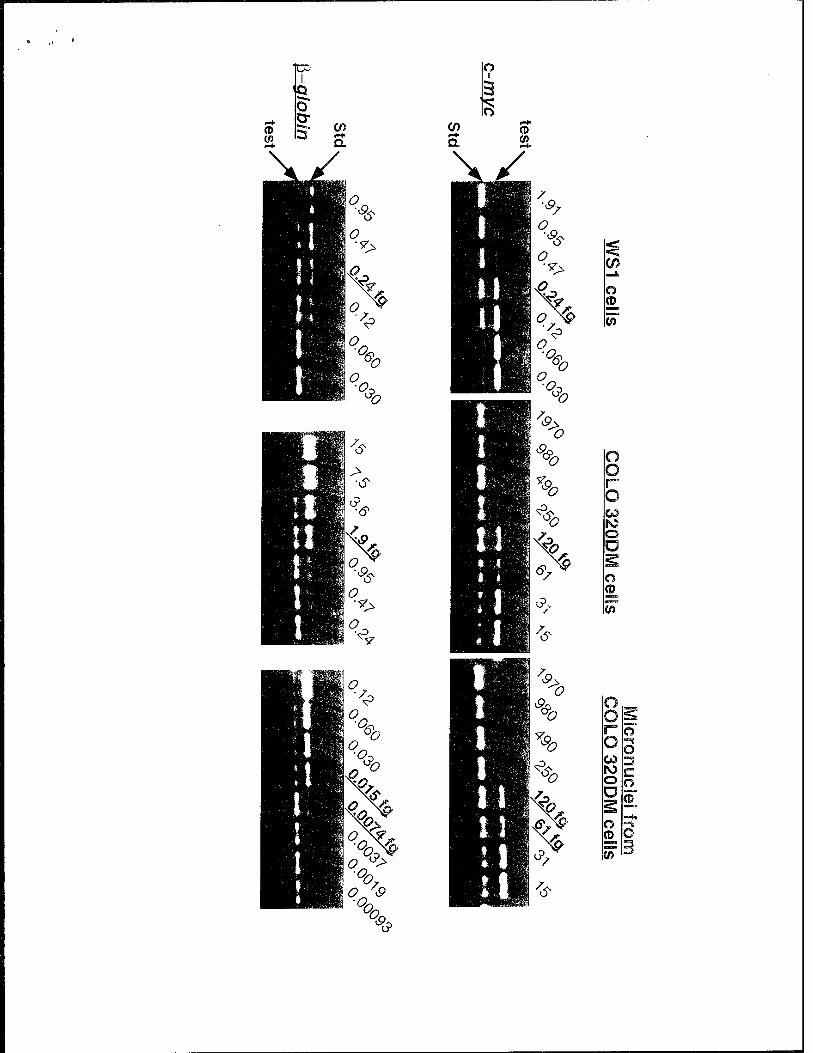
o
o.
O '*>
o.
V
c?>
-&
X£ V
o <D
O o o CD FO O O
o o
O CO
© Ö
o <D
O ™*
o =? c o
©


« •

50
40
30
u 3 C o u £ O 20.
ü c 0) 3 er
10 _
Concentration of
hydroxyurea (\iM)
Total micronuclei Micronuclei with human centromere
0 30 60 90 30 60 90
3 days 7 days