drug-eluting contact lens containing cyclosporine-loaded
TRANSCRIPT

RSC Advances
PAPER
Ope
n A
cces
s A
rtic
le. P
ublis
hed
on 2
8 M
ay 2
019.
Dow
nloa
ded
on 3
/15/
2022
10:
50:0
6 A
M.
Thi
s ar
ticle
is li
cens
ed u
nder
a C
reat
ive
Com
mon
s A
ttrib
utio
n 3.
0 U
npor
ted
Lic
ence
.
View Article OnlineView Journal | View Issue
Drug-eluting con
aDepartment of Materials Science and Engin
Technology (POSTECH), 77 Cheongam-ro
Korea. E-mail: [email protected]; Fax:bDepartment of Ophthalmology and Visual S
of Medicine, The Catholic University of Ko
06591, Korea. E-mail: [email protected]
1173
† Electronic supplementary informa10.1039/c9ra02858g
‡ These authors contributed equally to tco-rst authors.
Cite this: RSC Adv., 2019, 9, 16578
Received 16th April 2019Accepted 13th May 2019
DOI: 10.1039/c9ra02858g
rsc.li/rsc-advances
16578 | RSC Adv., 2019, 9, 16578–1658
tact lens containing cyclosporine-loaded cholesterol-hyaluronate micelles for dryeye syndrome†
Jonghwan Mun,‡a Jee won Mok,‡b Sanghoon Jeong,a Seonghwi Cho,a
Choun-Ki Joo*b and Sei Kwang Hahn *a
A contact lens is an attractive tool for the delivery of ophthalmic drugs, but it has several issues such as the
burst release of drugs and the limited drug loading capacity. To overcome these limitations, we developed
a cholesterol-hyaluronate (C-HA) micelle-embedded contact lens for efficient hydrophobic drug loading
and long-term controlled drug delivery. The contact lens was fabricated via photopolymerization of
hydroxyethyl methacrylate (HEMA) using ethylene glycol dimethacrylate (EGDMA) as a cross-linker. The
C-HA micelle-loaded contact lens showed statistically significant improvement in wettability and
mechanical strength, maintaining the optical transmittance. In vitro drug release tests revealed the
controlled delivery of cyclosporine for more than 12 days. Furthermore, the Schirmer tear test, corneal
fluorescein staining, and MMP9 fluorescein analysis confirmed its therapeutic effect on dry eye
syndrome in disease model rabbits.
1. Introduction
Eye diseases are commonly treated by periodic eye drops ofrelevant drugs due to patient compliance. In this case, however,the drug delivery efficiency is reported to be less than 1% withsignicant drug loss.1,2 In addition, the residence time of drugsdelivered by eye drops is in the range of 1–3 min in the tear lmwith low bioavailability.3–5 To overcome these problems, drugsare delivered at a high concentration or injected at the targetsite, which reduces patient compliance and causes other sideeffects.6 Contact lenses have been widely used for visioncorrection, eye protection, and aesthetic applications. Sincecontact lenses are directly placed on the cornea, they can be anideal system for delivering drugs to the anterior chamber. Forexample, glaucoma drug delivery via contact lenses showed 10times higher efficiency than that by eye drops.7 In addition,drugs can be continuously delivered into the eye througha contact lens rather than being temporarily delivered by eyedrops.8
eering, Pohang University of Science and
, Nam-gu, Pohang, Gyeongbuk 37673,
+82 54 279 2399; Tel: +82 54 279 2159
cience, Seoul St. Mary's Hospital, Collage
rea, 505, Banpo-dong, Seocho-gu, Seoul
; Fax: +82 2 533 3801; Tel: +82 2 2258
tion (ESI) available. See DOI:
his work and should be considered as
5
A variety of nanoparticles have been developed for long-termcontrolled drug delivery such as polymeric micelles, liposomes,and microemulsions.9–12 In particular, polymeric micelles haveattracted great attention due to their efficient hydrophobic drugloading and controlled drug delivery.13–15 Hydrophobic drugshave been encapsulated in amphiphilic block copolymers orconjugated to the hydrophilic polymers, forming a micellestructure.16–18 Hyaluronate (HA) is known to be hygroscopic andmaintains a high water content via chain–chain interactions.19
The superior biocompatibility of HA can provide comfort to theeye with a high water content.20 In addition, HA derivatives havebeen used to increase the bioavailability and the ocular resi-dence time of ophthalmic drugs.21,22
In this work, we developed a drug-eluting contact lens con-taining cyclosporine-loaded cholesterol-HA (C-HA) micelles forthe treatment of dry eye syndrome. Fig. 1a shows a schematicfor the preparation of the cyclosporine-loaded C-HA micellesand Fig. 1b shows the fabrication of a C-HA micelle-embeddedcontact lens. Cyclosporine is a hydrophobic drug for kerato-conjunctivitis sicca (dry eyes), and it was encapsulated in C-HAmicelles for the treatment of dry eye syndrome. Thecyclosporine-loaded C-HA micelles were characterized bydynamic light scattering (DLS), zeta potential analysis, trans-mission electron microscopy (TEM), and high-performanceliquid chromatography (HPLC). In addition, we assessed themechanical properties and wettability of the C-HA micelle-embedded contact lenses. Aer in vitro drug release tests, theSchirmer tear test, corneal uorescein staining, and uoresceinanalysis of metallopeptidase 9 (MMP9) were performed to
This journal is © The Royal Society of Chemistry 2019

Fig. 1 Schematic illustrations for (a) the preparation of cyclosporine-loaded C-HA micelles and (b) the fabrication of the micelle-embeddedcontact lens.
Paper RSC Advances
Ope
n A
cces
s A
rtic
le. P
ublis
hed
on 2
8 M
ay 2
019.
Dow
nloa
ded
on 3
/15/
2022
10:
50:0
6 A
M.
Thi
s ar
ticle
is li
cens
ed u
nder
a C
reat
ive
Com
mon
s A
ttrib
utio
n 3.
0 U
npor
ted
Lic
ence
.View Article Online
assess the therapeutic effect of the drug-eluting contact lens ondry eye disease (DED) model rabbits.
2. Experimental section2.1 Materials
Sodium hyaluronate (HA, MW 100 kDa) was purchased fromLifecore Biomedical (Chaska, MN). Tetrabutylammonium(TBA)-OH was obtained from Alfa Aesar (Ward Hill, MA). Dowexresin, ethylenediamine, cholesteryl chloroformate, hydroxyethylmethacrylate (HEMA), ethylene glycol dimethacrylate (EGDMA),cyclosporine, insulin, epithelial growth factor (EGF), hydrocor-tisone, and chleratoxin were obtained from Sigma-Aldrich (St.Louis, MO). Dulbecco's Modied Eagle's Medium/F-12 NutrientMixture Ham (DMEM/F-12) mixture (3/1), penicillin and strep-tomycin were obtained from WelGENE (Seoul, Korea).
2.2 Preparation methods
2.2.1 Synthesis of cholesterol-hyaluronate. HA was modi-ed with tetrabutylammonium salt to prepare HA-TBA, as re-ported elsewhere.23,24 Briey, a cation exchange resin of DOWEXwas mixed with TBA-OH to prepare Dowex-TBA resin. Then, itwas reacted with the sodium salt of HA for 3 h. The supernatantwas ltered to remove the Dowex resin, obtaining a clear HA-TBA solution, and lyophilized for 3 days. C-HA was synthe-sized as previously reported elsewhere.25 Cholesteryl-2-aminoethylcarbamate (CAEC) was synthesized by mixing cho-lesteryl chloroformate and ethylenediamine in anhydrousdichloromethane. The mixed solution was stirred on ice for 1 h.The resulting reaction solution was washed with DI water anddried over anhydrous magnesium sulfate. To synthesize C-HA,100 mg of HA-TBA was dissolved in DMSO (1 wt%) and reac-ted with 0.33 mL of CAEC dissolved in the mixture of DCM andmethanol (1 : 1, v/v). The reaction solution was stirred at room
This journal is © The Royal Society of Chemistry 2019
temperature for 30 min, which was mixed with 4.2 mg of 4-(4,6-dimethoxy-1,3,5-triazin-2-yl)-4-methylmorpholinium chloride.Aer stirring at room temperature for 24 h, the resultingproduct was dialyzed against a large excess amount of 0.5 MNaCl solution, 50% ethanol, and water and then lyophilized for3 days. The cholesterol content was determined by protonnuclear magnetic resonance (1H NMR) analysis (DPX500,Bruker, Germany) in deuterium oxide.
2.2.2 Cytotoxicity test of C-HA micelles. The cytotoxicity ofC-HA in human cornea epithelial cells (HCECs) was assessed bya CCK-8 assay. The cells were suspended in the mixture ofDMEM/F-12 (3/1) with 5% of fetal bovine serum (FBS), 5 mgmL�1 of insulin, 10 ng mL�1 of EGF, 500 ng mL�1 of hydro-cortisone, 30 ng mL�1 of choleratoxin, penicillin and strepto-mycin and seeded with 100 mL of the cell suspension containing1.3 � 105 cells per mL on each well of 96-well cell culture platesfor 24 h. C-HA with a cholesterol content of 3.5 mol% was dis-solved in SFM at the concentrations of 10, 20, 50, 100, 150, 200,and 500 mg mL�1 and placed in a 37 �C incubator for 24 h. Then,the samples were washed with PBS thrice and 10 mL of CCK-8assay solution was added to each well. Aer incubation for4 h, the optical density was measured at 480 nm with a micro-plate reader.
2.2.3 Preparation of cyclosporine/C-HA micelles. C-HA(0.1 wt%) was dissolved in water and cyclosporine was dis-solved in DMSO (0.25 wt%). The solutions weremixed and ultra-sonicated for 60 min. The resulting solution was poured intoa dialysis membrane (MWCO of 3500 Da) and dialyzed againstdistilled water for a day to remove the unloaded cyclosporineand DMSO.
2.2.4 Characterization of prepared cyclosporine/C-HAmicelles. The hydrodynamic diameter and zeta potential ofprepared C-HA micelles in an aqueous solution were measuredby DLS (Zetasizer Nano ZS, Malvern Instruments, Worcester-shire, UK). The successful formation of cyclosporine/C-HA
RSC Adv., 2019, 9, 16578–16585 | 16579
RSC Advances Paper
Ope
n A
cces
s A
rtic
le. P
ublis
hed
on 2
8 M
ay 2
019.
Dow
nloa
ded
on 3
/15/
2022
10:
50:0
6 A
M.
Thi
s ar
ticle
is li
cens
ed u
nder
a C
reat
ive
Com
mon
s A
ttrib
utio
n 3.
0 U
npor
ted
Lic
ence
.View Article Online
micelles was assessed by TEM (JEM-1011, JEOL). For TEManalysis, ca. 20 mL of the micelle solution was dropped on a TEMgrid and then air-dried.
2.2.5 Fabrication of cyclosporine/C-HA micelle-embeddedcontact lens. The cyclosporine/C-HA micelle solution (80 mL)was mixed with HEMA (320 mL). Then, EGDMA (10 mL) and TPOIrgacure (2.4 mg) were added into the above mixture. The nalmixed solution (80 mL) was injected into a contact lensmold andcured at 340 nm under a nitrogen environment for 15 min. Aerpolymerization, the contact lens was soaked in water forhydration. The encapsulation of cyclosporine/C-HA micellesinto the PHEMA contact lens was assessed by X-ray photoelec-tron spectroscopy (XPS).
2.3 Characterization methods
2.3.1 Optical transmittance analysis. The optical trans-mittance of the contact lens hydrogel was analyzed with a UV-Vis spectrometer (S-3100, Scinco). The fabricated contact len-ses were placed on the measurement holder and the trans-mittance was measured at the wavelength of 200–800 nm.
2.3.2 Tensile strength test. The tensile strength of C-HAmicelle-embedded contact lenses was measured using Instron3344 (Instron Corp). The loading rate was 5 mm min�1. Thespecimens had a width of 15 mm and a thickness of 10 mm. Thegauge length was 64 mm.
2.3.3 Equilibrium water content analysis. The equilibriumwater content (EWC) was calculated by measuring the weightsof contact lenses in the dried state (Wdry) and wet state (Wwet).The EWC value was determined by the increased weight ofcontact lenses during hydration using the following equation:
EWC ¼ (Wwet � Wdry)/Wdry � 100
2.3.4 Water evaporation test. The water evaporationthrough the contact lenses was assessed with 1.5 mL of e-tube.One mL of PBS was lled in 1.5 mL of e-tube and the contactlens was glued to the rim of the e-tube. The e-tube was kepttilted so that the PBS could contact the contact lens. Then, theweight of e-tubes was measured every 5 min for a total of60 min.
2.3.5 Water contact angle measurement. The contact angleof PHEMA contact lenses with and without C-HA micelles wasmeasured with Smartdrop (Femtofab) aer dropping 5 mL ofwater on the contact lenses.
2.3.6 In vitro drug loading and release test. The drugloading efficiency of cyclosporine in the C-HA micelles wasdetermined by HPLC analysis. Acetonitrile was added to themicelle solution at a volume ratio of 4/1 to break the micellestructure. HPLC analysis was performed using the followingsystems: a Waters 1525 binary HPLC pump, a Waters 2487dual k absorbance detector, a Waters 717 plus autosampler, anda Symmetry™ 300 C18 5 mm column (Waters, MA). The mobilephase was a mixture of acetonitrile and distilled water ata volume ratio of 70/30, and the ow rate was 0.8 mLmin�1. Thecolumn was placed in a column oven set at 80 �C and thedetection wavelength was 215 nm for cyclosporine. An in vitro
16580 | RSC Adv., 2019, 9, 16578–16585
drug release test of cyclosporine from the cyclosporine/C-HAmicelle-embedded contact lenses was carried out for 288 h.The cyclosporine/C-HA micelle-embedded contact lenses wereimmersed in 1 mL of PBS using a 24-well plate in an incubatorat 37 �C. At the predetermined time intervals, each PBS samplecontaining the contact lens was obtained and replaced withfresh PBS. The concentration of cyclosporine in the samples wasmeasured by HPLC, as described above.
2.4 In vivo therapeutic effect test
2.4.1 Preparation of dry eye syndrome model rabbits. Allanimal studies with New Zealand White rabbits (2–3 kg, OrientBio, Seoul, Korea) were approved by the Institutional ReviewBoard of Catholic University and performed in accordance withthe Association for Research in Vision and OphthalmologyStatement for the Use of Animals in Ophthalmic and VisionResearch. The rabbits received 3-concanavalin A injections (ConA, Sigma L7647), one each into the inferior lacrimal gland (ILG),the palpebral portion of the superior lacrimal gland (PSLG), andthe orbital portion of the superior lacrimal gland (OSLG). Usinga 26-gauge needle, 10 mg of Con A in 1 mL was injected, whichwas repeated aer 1 and 3 days.
2.4.2 Corneal uorescein staining and analysis. For uo-rescein staining, sodium uorescein was applied to the ocularsurface of the animals, typically without sedation. Five min aerapplication, corneal uorescein staining was scored undera microscope using a blue light. We observed that the irregu-larities in the eyes, such as abrasion and inammation, uo-resce with a greater intensity than that of the healthy cornealtissue.
2.4.3 Schirmer tear test. Schirmer strips (EagleVision,Denville, NJ) were inserted into the space between the corneaand the palpebral conjunctiva at the midpoint of the lower eyelid. The tear production was determined based on the length ofthe moisturized strip measured aer 5 min. The reading datawere obtained in triplicate and averaged for the analysis. Aerrecording the tear production at 5 min, the strips were le inplace for at least 20 mm until wetting.
3. Results and discussion3.1 Characteristics of cholesterol-hyaluronate micelles
C-HA was synthesized by the conjugation of HA-TBA with CAEC.The cholesterol content of C-HA was determined by 1H NMRanalysis (ESI Fig. S1†). The graed ratio of cholesteryl groupswas about 3.8 mol%, which was calculated from the integrationratio between the peaks of the N-acetyl group of HA (d ¼ 1.96,COCH3) and the methyl group of cholesterol (d ¼ 0.79, CH3). C-HA self-assembled into micelle-like nanoparticles in theaqueous solution due to the hydrophobic interaction betweencholesterols.26 The hydrophobic drug cyclosporine used for dryeye syndrome was loaded in the C-HAmicelles by the sonicationand dialysis method. The formation of cyclosporine/C-HAmicelles and the loading efficiency of cyclosporine within theC-HA micelles were analyzed by DLS and TEM. The diameterwas measured to be 290.0 � 35.95 nm (n ¼ 3) and the zeta
This journal is © The Royal Society of Chemistry 2019

Paper RSC Advances
Ope
n A
cces
s A
rtic
le. P
ublis
hed
on 2
8 M
ay 2
019.
Dow
nloa
ded
on 3
/15/
2022
10:
50:0
6 A
M.
Thi
s ar
ticle
is li
cens
ed u
nder
a C
reat
ive
Com
mon
s A
ttrib
utio
n 3.
0 U
npor
ted
Lic
ence
.View Article Online
potential of the particle was measured to be�17.4� 3 (n¼ 3) byDLS (ESI Fig. S2a and b†). The TEM image in Fig. S3† showsspherical particles with a diameter of ca. 300 nm. As shown inFig. S4,† there is negligible cytotoxicity in HCECs for the C-HAmicelle concentration from 10 to 500 mg mL�1.
3.2 Characterization of drug-eluting contact lens
3.2.1 Optical transmittance of C-HA micelle-embeddedcontact lens. The successful encapsulation of cyclosporine/C-HA micelles into the contact lens was conrmed by XPS anal-ysis (ESI Fig. S5†). The optical clarity of the micelle-embeddedcontact lenses was characterized by measuring the trans-mittance in the range from 200 nm to 800 nm (Fig. 2a). Thenormal contact lens exhibited ca. 95% of transmittance and theC-HA micelle-embedded contact lens showed ca. 90% of trans-mittance. The transparency decreased due to the C-HAmicelles,which might not signicantly affect visual clarity. The lettersunder the C-HA micelle-embedded contact lens can be clearlyseen without affecting visibility (inset, Fig. 2a).
Fig. 2 (a) The transmittance spectra of poly(2-hydroxyethyl methac-rylate) hydrogel contact lens (control, black) and C-HA micelle-embedded contact lens (red). (b) The tensile tests of normal contactlens (black) and C-HA micelle-embedded contact lens (red).
This journal is © The Royal Society of Chemistry 2019
3.2.2 Tensile strength of C-HA micelle-embedded contactlens. The mechanical properties of contact lens materials areimportant for long-term durability. Fig. 2b shows the stress–strain curves for the tensile property of the C-HA micelle-embedded contact lens by using Instron. The extension of theC-HA micelle-embedded contact lens was longer than that ofthe normal contact lens. In other words, the C-HA micelle-embedded PHEMA contact lens was more stretchable than thenormal contact lens, reecting that the C-HA micelle nano-particles in the contact lens improved the elasticity of thecontact lens. In addition, when the tensile specimen began tobreak, the C-HA micelle-embedded contact lens was slightlyextended, resisting the breakage of the hydrogel network.
3.2.3 Equilibrium water content of C-HA micelle-embedded contact lens. The swelling characteristics ofcontact lenses are very important parameters for ocular appli-cations. In this regard, the equilibrium water content (EWC)was measured and compared for the C-HA micelle-embeddedcontact lens and the control normal contact lens. EWC ofcontact lenses was measured by weighing the contact lensesbefore and aer swelling. Fig. 3a shows the EWC (%) values forthe C-HA micelle-embedded contact lens and the controlPHEMA contact lens. The water content of the 64 mg C-HAmicelle-embedded contact lens was ca. 43%, which was higherthan ca. 39% of the PHEMA contact lens (Fig. 3a). These resultsindicate that the wettability of the contact lens is drasticallyenhanced by the hydrophilic HA of the C-HA micelles in thecontact lens.
3.2.4 Hygroscopic characteristics of the C-HA micelle-embedded contact lens. The water evaporation test was per-formed to evaluate the hygroscopic property of contact lenses.The water evaporation rate was determined by measuring theweight change of a 1.5 mL e-tube aer sealing with the C-HAmicelle-embedded contact lens and the normal contact lensas a control. As shown in Fig. 3b, water evaporating from 1.5 mLof e-tube sealed with the C-HA micelle-embedded contact lenswas signicantly lower than that of the control contact lens. InFig. 3c, the water evaporation rate is calculated from the slope ofwater evaporated vs. time. The water evaporation rate wassignicantly reduced in the C-HA micelle-embedded contactlens (0.149 mg min�1) compared to that observed for the control(0.29 mg min�1). From the results, we could conrm theimproved hygroscopic property of the C-HA micelle-embeddedcontact lens.
3.2.5 Wettability of the C-HA micelle-embedded contactlens. Water contact angles were measured to determine thewettability of the micelle-embedded contact lens. The contactangles were measured immediately and aer 1 h aer droppinga 5 mL water droplet on the C-HA micelle-embedded contactlenses (Fig. 4a) and the normal control contact lenses (Fig. 4b).As a result, both groups showed a contact angle of about 77.55�
on average just aer dropping the 5 mL water droplet. However,aer 1 h, the average contact angle was reduced to 39.6� for theC-HA micelle-embedded contact lens and to 48.07� for thecontrol contact lens. The results show that C-HA micelle-embedded contact lenses have higher wettability than the
RSC Adv., 2019, 9, 16578–16585 | 16581

Fig. 3 (a) Equilibrium water content of C-HA micelle-embeddedcontact lens (32 mg of C-HA and 64 mg C-HA micelles) (n ¼ 3; **, P #
0.01; ***, P# 0.0001 vs. the control). (b) Water evaporation fromC-HAmicelle-embedded contact lens and normal contact lens over time (n¼ 3). (c) Water evaporation rate calculated from the slope of waterevaporated vs. time (n ¼ 3; **, P # 0.01; ***, P # 0.0001 vs. thecontrol).
Fig. 4 Photographs for the absorption of a water droplet on (a) normalcontact lens and (b) C-HA micelle-embedded contact lens. (c) Watercontact angles for C-HA micelle-embedded contact lenses andnormal contact lenses measured after 0 and 1 h (n¼ 3; **, P# 0.01; vs.the control).
16582 | RSC Adv., 2019, 9, 16578–16585
RSC Advances Paper
Ope
n A
cces
s A
rtic
le. P
ublis
hed
on 2
8 M
ay 2
019.
Dow
nloa
ded
on 3
/15/
2022
10:
50:0
6 A
M.
Thi
s ar
ticle
is li
cens
ed u
nder
a C
reat
ive
Com
mon
s A
ttrib
utio
n 3.
0 U
npor
ted
Lic
ence
.View Article Online
control contact lenses, implying the low adhesion to contami-nating lipids.
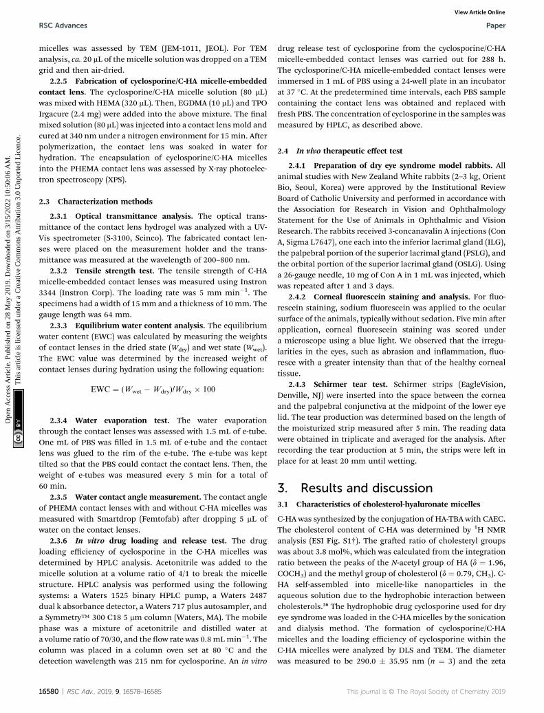
3.2.6 In vitro drug loading and release. The drug loadingefficiency of cyclosporine within the micelles was measured tobe ca. 64% by comparing the absorbance of cyclosporine at215 nm before and aer drug loading (Fig. 5a). The in vitro drugrelease from the cyclosporine/C-HA micelle-embedded contactlens was analyzed in PBS at 37 �C. The release rate of cyclo-sporine was determined by the calibration curve of cyclosporineat 215 nm using HPLC analysis with a C18 column (ESIFig. S6†). The recommended eye drop dosage for a 0.05%solution of cyclosporine is 2 drops per day. This means that thedaily dose of cyclosporine is ca. 2 mg. The amount of cyclo-sporine loaded in the contact lens was determined by the abovecyclosporine loading efficiency. Aer 12 days, approximately50% of the loaded cyclosporine was continuously released fromthe contact lenses in comparison to the burst drug release from
This journal is © The Royal Society of Chemistry 2019
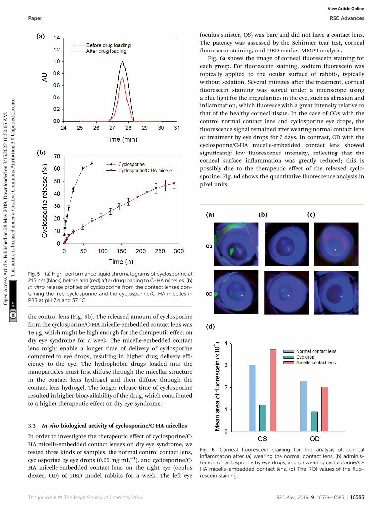
Fig. 5 (a) High-performance liquid chromatograms of cyclosporine at215 nm (black) before and (red) after drug loading to C-HAmicelles. (b)In vitro release profiles of cyclosporine from the contact lenses con-taining the free cyclosporine and the cyclosporine/C-HA micelles inPBS at pH 7.4 and 37 �C.
Paper RSC Advances
Ope
n A
cces
s A
rtic
le. P
ublis
hed
on 2
8 M
ay 2
019.
Dow
nloa
ded
on 3
/15/
2022
10:
50:0
6 A
M.
Thi
s ar
ticle
is li
cens
ed u
nder
a C
reat
ive
Com
mon
s A
ttrib
utio
n 3.
0 U
npor
ted
Lic
ence
.View Article Online
the control lens (Fig. 5b). The released amount of cyclosporinefrom the cyclosporine/C-HA micelle-embedded contact lens was16 mg, which might be high enough for the therapeutic effect ondry eye syndrome for a week. The micelle-embedded contactlens might enable a longer time of delivery of cyclosporinecompared to eye drops, resulting in higher drug delivery effi-ciency to the eye. The hydrophobic drugs loaded into thenanoparticles must rst diffuse through the micellar structurein the contact lens hydrogel and then diffuse through thecontact lens hydrogel. The longer release time of cyclosporineresulted in higher bioavailability of the drug, which contributedto a higher therapeutic effect on dry eye syndrome.
Fig. 6 Corneal fluorescein staining for the analysis of cornealinflammation after (a) wearing the normal contact lens, (b) adminis-tration of cyclosporine by eye drops, and (c) wearing cyclosporine/C-HA micelle-embedded contact lens. (d) The ROI values of the fluo-rescein staining.
3.3 In vivo biological activity of cyclosporine/C-HA micelles
In order to investigate the therapeutic effect of cyclosporine/C-HA micelle-embedded contact lenses on dry eye syndrome, wetested three kinds of samples: the normal control contact lens,cyclosporine by eye drops (0.05 mg mL�1), and cyclosporine/C-HA micelle-embedded contact lens on the right eye (oculusdexter, OD) of DED model rabbits for a week. The le eye
This journal is © The Royal Society of Chemistry 2019
(oculus sinister, OS) was bare and did not have a contact lens.The patency was assessed by the Schirmer tear test, cornealuorescein staining, and DED marker MMP9 analysis.
Fig. 6a shows the image of corneal uorescein staining foreach group. For uorescein staining, sodium uorescein wastopically applied to the ocular surface of rabbits, typicallywithout sedation. Several minutes aer the treatment, cornealuorescein staining was scored under a microscope usinga blue light for the irregularities in the eye, such as abrasion andinammation, which uoresce with a great intensity relative tothat of the healthy corneal tissue. In the case of ODs with thecontrol normal contact lens and cyclosporine eye drops, theuorescence signal remained aer wearing normal contact lensor treatment by eye drops for 7 days. In contrast, OD with thecyclosporine/C-HA micelle-embedded contact lens showedsignicantly low uorescence intensity, reecting that thecorneal surface inammation was greatly reduced; this ispossibly due to the therapeutic effect of the released cyclo-sporine. Fig. 6d shows the quantitative uorescence analysis inpixel units.
RSC Adv., 2019, 9, 16578–16585 | 16583

RSC Advances Paper
Ope
n A
cces
s A
rtic
le. P
ublis
hed
on 2
8 M
ay 2
019.
Dow
nloa
ded
on 3
/15/
2022
10:
50:0
6 A
M.
Thi
s ar
ticle
is li
cens
ed u
nder
a C
reat
ive
Com
mon
s A
ttrib
utio
n 3.
0 U
npor
ted
Lic
ence
.View Article Online
The immunouorescence staining analysis for a dry eyemarker of MMP9 conrmed the therapeutic effect of thecyclosporine/C-HA micelle-embedded contact lens treatment.Fig. 7a and b show the MMP9 staining in the cornea. Theintensity of MMP9 was slightly reduced in the eye treated bycyclosporine eye drops. In the case of rabbits wearing thecyclosporine/C-HA micelle-embedded contact lens, signicantreduction of MMP9 was observed in the cornea (Fig. 7c). Theresults indicated that cyclosporine was released from the C-HAmicelle-embedded contact lens and caused a signicant thera-peutic effect on dry eye syndrome.
Fig. 7d shows the tear production of eyes wearing thecyclosporine/C-HA micelle-embedded contact lens for 4 h a day
Fig. 7 Corneal inflammation analysis by the corneal fluoresceinstaining of DED marker of MMP9 after (a) cyclosporine eye drops and(b) wearing cyclosporine/C-HA micelle-embedded contact lens. (c)The ROI values for the MMP9 analysis. (d) The tear production of thecontrol eye (OS) and the eye wearing the contact lens (OD) at day 7:wearing the normal contact lens (sky blue), cyclosporine eye drops(green), and wearing the cyclosporine/C-HA micelle-embeddedcontact lens (pink, n ¼ 2).
16584 | RSC Adv., 2019, 9, 16578–16585
in comparison to that without wearing the contact lens. In thecase of cyclosporine eye drops, two drops were administered perday. The analysis of tear production aer 7 days showeddecrease in rabbits wearing the normal contact lens and slightincrease in rabbits treated by cyclosporine eye drops. Incontrast, the tear production was greatly enhanced for therabbits wearing the cyclosporine/C-HA micelle-embeddedcontact lens, suggesting that the drug was steadily releasedfrom the contact lens, which was sufficient for the therapeuticeffect.
4. Conclusions
C-HA was successfully synthesized by conjugating CAEC to HA-TBA in DMSO by amide bond formation between the aminegroups in CAEC and the carboxyl groups in HA. Cyclosporine asa dry eye therapeutic drug was loaded in the micellar structure ofthe synthesized C-HA. The formation of cyclosporine-loadedmicelles was conrmed by DLS, zeta potential, TEM, and HPLCanalyses. The transmittance analysis, EWC test, WE test, watercontact angle analysis, and tensile strength test conrmed theimproved physical and mechanical properties of C-HA micelle-embedded contact lenses compared to those of the normalPHEMA contact lens as a control. In vitro release tests showed thecontinuous release of cyclosporine from the cyclosporine/C-HAmicelle-embedded contact lens for more than 10 days. Finally,the therapeutic effect on dry eye syndrome was successfullyconrmed by the Schirmer tear test, corneal uorescein staining,and MMP9 uorescein analysis in the DEDmodel rabbits. Takentogether, we can conrm the feasibility of cyclosporine/C-HAmicelle-embedded contact lenses for further clinical develop-ment. These drug-eluting contact lenses would be greatly bene-cial for dry eye syndrome patients wearing contact lenses.
Conflicts of interest
There are no conicts to declare.
Acknowledgements
This research was supported by the Nano$Material TechnologyDevelopment Program (No. 2017M3A7B8065278) and the BasicScience Research Program (2017R1E1A1A03070458) of theNational Research Foundation (NRF) funded by the Ministry ofScience and ICT, Korea. This work was also supported byInterojo Co. and the World Class 300 Project (R&D) (S2482887)funded by the Ministry of SMEs and Startups of Korea.
References
1 S. Ding, Recent developments in ophthalmic drug delivery,Pharm. Sci. Technol. Today, 1998, 1(8), 328–335.
2 T. Losson and E. Stefansson, Cyclodextrins in eye dropformulations: enhanced topical delivery of corticosteroidsto the eye, Acta Ophthalmol. Scand., 2002, 80(2), 144–150.
This journal is © The Royal Society of Chemistry 2019
Paper RSC Advances
Ope
n A
cces
s A
rtic
le. P
ublis
hed
on 2
8 M
ay 2
019.
Dow
nloa
ded
on 3
/15/
2022
10:
50:0
6 A
M.
Thi
s ar
ticle
is li
cens
ed u
nder
a C
reat
ive
Com
mon
s A
ttrib
utio
n 3.
0 U
npor
ted
Lic
ence
.View Article Online
3 C. G. Wilson and Y. P. Zhu, Ocular contact time ofa carbonmer gel (GelTears) in humans, Br. J. Ophthalmol.,1998, 82(10), 1131–1134.
4 D. L. Meadows, J. R. Paugh, et al., A novel method to evaluateresidence time in humans using a nonpenetratinguorescent tracer, Invest. Ophthalmol. Visual Sci., 2002,43(4), 1032–1039.
5 G. R. Snibson, J. L. Greaves, et al., Precorneal residence timesof sodium hyaluronate solutions studied by quantitativegamma scintigraphy, Eye, 1990, 4, 594–602.
6 A. Farkouh, P. Frigo, et al., Systemic side effects of eye drops:A pharmacokinetic perspective, Clin. Ophthalmol., 2016, 10,2433–2441.
7 C. C. Peng, M. T. Burke, et al., Extended drug delivery bycontact lenses for glaucoma therapy, J. Controlled Release,2012, 162(1), 152–158.
8 F. H. Nasr, S. Khoee, et al., Preparation and evaluation ofcontact lenses embedded with polycaprolactone-basednanoparticles for ocular drug delivery, Biomacromolecules,2016, 17(2), 485–495.
9 S. K. Sahoo, F. Dilnawaz, et al., Nanotechnology in oculardrug delivery, Drug Discovery Today, 2008, 13, 144–151.
10 U. B. Kompella, A. C. Amrite, et al., Nanomedicines for backof the eye drug delivery, gene delivery, and imaging, Prog.Retinal Eye Res., 2013, 36, 172–198.
11 V. Delplace, S. Payne, et al., Delivery strategies for treatmentof age-related ocular diseases: From a biologicalunderstanding to biomaterial solutions, J. ControlledRelease, 2015, 219, 652–668.
12 A. Bochot, E. Fattal, et al., Liposomes for intravitreal drugdelivery: A state of the art, J. Controlled Release, 2012,161(2), 628–634.
13 K. Kataoka, A. Harada, et al., Block copolymer micelles fordrug delivery: Design, characterization and biologicalsignicance, Adv. Drug Delivery Rev., 2012, 64, 37–48.
14 K. Kazunori, G. S. Kwon, et al., Block copolymer micelles asvehicles for drug delivery, J. Controlled Release, 1993, 24,119–132.
This journal is © The Royal Society of Chemistry 2019
15 M. L. Adams, A. Lavasanifar, et al., Amphiphilic BlockCopolymer for Drug Delivery, J. Pharm. Sci., 2003, 92(7),1343–1355.
16 G. S. Kwon, M. Naito, et al., Block copolymer micelles asvehicles for hydrophobic drugs, Colloids Surf., B, 1994,2(4), 429–434.
17 A. Lavasanifar, J. Samuel, et al., Poly(ethylene oxide)-block-poly(L-amino acid) micelles for drug delivery, Adv. DrugDelivery Rev., 2002, 54(2), 169–190.
18 S. R. Croy and G. S. Kwon, Polymeric micelles for drugdelivery, Curr. Pharm. Des., 2006, 12(36), 4669–4684.
19 J. Necas, L. Bartosikova, et al., Hyaluronic acid (hyaluronan):a review, Vet. Med., 2008, 53(8), 397–411.
20 M. Ali and M. E. Byrne, Controlled release of high molecularweight hyalutonic acid frommolecularly imprinted hydrogelcontact lenses, Pharm. Res., 2009, 26(3), 714–726.
21 R. Gurny, H. Ibrahim, et al., Design and evaluation ofcontrolled release systems for the eye, J. Controlled Release,1987, 6(1), 367–373.
22 M. F. Saettone, P. Chetoni, et al., Evaluation of muco-adhesive properties and in vivo activity of ophthalmicvehicles based on hyaluronic acid, Int. J. Pharm., 1989,51(3), 203–212.
23 E. J. Oh, K. Park, et al., Synthesis, characterization, andpreliminary assessment of anti-Flt1 peptide-hyaluronateconjugate for the treatment of corneal neovascularization,Biomaterials, 2009, 30(30), 6026–6034.
24 E. J. Oh, J. W. Kim, et al., Signal Transduction of HyaluronicAcid-Peptide Conjugate for Formyl Peptide Receptor Like 1Receptor, Bioconjugate Chem., 2008, 19(12), 2401–2408.
25 W. Miao, G. Shim, et al., Cholesteryl hyaluronic acid-coated,reduced graphene oxide nanosheets for anti-cancer drugdelivery, Biomaterials, 2013, 34(37), 9638–9647.
26 X. Wei, T. H. Senanayake, et al., Hyaluronic acid-basednanogel-drug conjugates with enhanced anticancer acticitydesigned for the targeting of CD44-positive and drug-resistant tumors, Bioconjugate Chem., 2013, 24(4), 658–668.
RSC Adv., 2019, 9, 16578–16585 | 16585