
1
The analysis of Presenilin processing and activity with a focus on
its implications for Alzheimer’s disease pathogenesis using Danio
Rerio as a model organism
Lachlan Wilson
Supervised by Michael Lardelli and Joan Kelly
Submitted for partial fulfilment for the degree of Doctor of
Philosophy (PhD)
Discipline of Genetics
School of Molecular and Biomedical Science
The University of Adelaide University SA
AUSTRALIA

2
July 2013
Declaration
I certify that this work contains no material which has been accepted for the award of
any other degree or diploma in my name, in any university or other tertiary institution
and, to the best of my knowledge and belief, contains no material previously
published or written by another person, except where due reference has been made in
the text. In addition, I certify that no part of this work will, in the future, be used in a
submission in my name, for any other degree or diploma in any university or other
tertiary institution without the prior approval of the University of Adelaide and where
applicable, any partner institution responsible for the joint-award of this degree.
I give consent to this copy of my thesis when deposited in the University Library,
being made available for loan and photocopying, subject to the provisions of the
Copyright Act 1968.
The author acknowledges that copyright of published works contained within this
thesis resides with the copyright holder(s) of those works.
I also give permission for the digital version of my thesis to be made available on the
web, via the University’s digital research repository, the Library Search and also
through web search engines, unless permission has been granted by the University to
restrict access for a period of time.

3
Signed
………………………… ……………………….Date…………

4
Table of Contents
List of Publications contributed to during Ph.D candidature....................................5
Acknowledgements ..............................................................................................................6Abstract..................................................................................................................................8
CHAPTER I ....................................................................................................................... 101.1. Literature Review ...............................................................................................................101.2 Summary of Chapters II-IV and Links Between Them ..............................................53
CHAPTER II ..................................................................................................................... 56Research Paper I.........................................................................................................................56
CHAPTER III.................................................................................................................... 58Research Paper II .......................................................................................................................58Supplemental Data ..................................................................................................................122
CHAPTER IV..................................................................................................................126Research Paper III ..................................................................................................................126Supplemental Data ..................................................................................................................185
Discussion .........................................................................................................................188FurtherDirections ................................................................................................................193
References.........................................................................................................................197
APPENDIX I....................................................................................................................219Research Paper IV ..................................................................................................................219
APPENDIX II ..................................................................................................................220Research Paper V ....................................................................................................................220

5
List of Publications contributed to during Ph.D candidature
A zebrafish melanophore model of amyloid beta toxicity.
Morgan Newman, Lachlan Wilson, Giuseppe Verdile, Esther Camp
Ralph Martins and Michael Lardelli
Zebrafish, 2010, Vol7, No2 155-9.
The BACE1-PSEN-AβPP regulatory axis has an ancient role in response to low
oxygen/oxidative stress.
Seyyed Hani Moussavi Nik, Lachlan Wilson, Morgan Newman, Kevin Croft, Trevor
A Mori, Ian Musgrave and Michael Lardelli
Journal of Alzheimer’s Disease, 2011,
The Development of an in vivo γ-Secretase Assay using Zebrafish Embryos.
Lachlan Wilson and Michael Lardelli
Journal of Alzheimer’s Disease, 2013,

6
Acknowledgements
I would like to give my thanks and appreciation to:
Michael Lardelli, for providing me with this magnificent opportunity. Your
knowledge, wisdom and continual patience with me throughout my studies has been
so very appreciated. You have been a continuous inspiration for the scientific process
and the virtues of a critical thinking lifestyle.
Morgan Newman, for being a continuous support and always being ready to assist me
and my incessant questions. You have been there from the start to finish, from my
third year placement in the lab till now. Thank you.
Past and present members of the Genetics discipline for their support and advice over
the last few years. Particularly Simon Wells and Jai Denton for the advice, feedback
and for reminding me that no matter how long it takes you can get it done. It kept me
going in times of serious doubt.
Hani Mousavanik, you made everyday coming into the lab something to look forward
to. Always laughing, and seeing the funny side of life. Our conversations, both
intellectual and social have been enlightening and a treasure. You are a dear friend of
mine, I will miss not coming into the lab everyday to share in your company.

7
To Mum and Dad thank you for being there through thick and thin. I know I haven’t
always been the easiest person and I thank you for your constant support. I couldn’t
have made it in any capacity without the opportunities that you have given me. The
education, the love, understanding and continuous support. Thank you for letting me
be me.
Garth, I know you hate my sentimentality and romanticism, so I will keep it stark. I
would not be here, or the person I am without you. This, and all the things I have
done, would not have occurred without you in my life.
Finally, my friends for providing timely distractions and listening to me rabble on
about this and that.
Without these people (science related or not), none of this would have been possible.
This thesis is for Garth. My Brother. My Best Friend. My Constant.

8
Abstract
Aberrant proteolytic processing of AMYLOID BETA PRECURSOR PROTEIN
(AβPP) may result in an imbalance between production and clearance of the amyloid-
β (Aβ) peptide proteolytic product and promote neuronal dysfunction and death. β-site
amyloid-β A4 precursor protein-cleaving enzyme 1 (BACE1) with γ -secretase are
responsible for the cleavage of AβPP to produce Aβ peptide. Presenilin proteins form
the catalytic core of γ-secretase complexes. PRESENILIN1 (PSEN1) is the major
locus for mutations causing familial Alzheimer’s disease (FAD) and is also mutated in
Pick disease of brain, familial acne inversa and dilated cardiomyopathy. It is a critical
facilitator of Notch signalling. The zebrafish, Danio rerio, is a versatile vertebrate
model for investigating the molecular bases of Alzheimer’s disease (AD) pathology. It
possesses genes orthologous to human PSEN1 and PSEN2, and the genes appa and
appb that are duplicates of an ancestral AβPP orthologue).
This thesis primarily utilizes zebrafish as a system to investigate AD pathogenesis..
Chapter I describes an assay in which the level of a γ-secretase substrate (a modified
form of Appa protein) is observed in zebrafish embryos by western immunoblotting
relative to a co-expressed protein not subject to γ-secretase activity. Prior to the
development of this assay there existed no in vivo assay appropriate for directly
monitoring γ-secretase activity. The assay was subsequently used to analyse the
effects on γ-secretase activity of blocking translation of zebrafish psen1 and/or psen2.
Chapter II explores various truncations of human PSEN1 (or zebrafish Psen1)
protein that have differential effects on Notch signalling and cleavage of zebrafish
Appa (a paralogue of human AβPP). Different truncations can suppress or stimulate

9
Notch signalling but not Appa cleavage and vice versa. The results show that the
truncated protein potentially translated from these transcripts incorporates into stable
Psen1-dependent higher molecular weight complexes and suppresses cleavage of
Appa but not Notch signalling. In contrast, the truncated protein potentially produced
by the P242LfsX11 acne inversa mutation has no effect on Appa cleavage but,
unexpectedly, enhances Notch signalling. The results suggest novel hypotheses for the
pathological mechanisms underlying AD. Chapter III investigates truncated isoforms
of PRESENILIN known to form naturally. In particular a truncated PSEN2 isoform
“PS2V” has been previously identified. PS2V is formed by exclusion of exon 5 from
PSEN2 transcripts leading to a frameshift after exon 4 sequence and a premature stop
codon. This truncates the ORF/protein after PSEN2’s first transmembrane domain.
The K115Efx10 mutation in PSEN2 is the only completely truncating mutation of the
PRESENILIN genes that is thought to cause AD. K115Efx10 is especially interesting
since, if expressed, it would generate a truncated protein very similar to PS2V and
would be expected to boost Aβ production. Zebrafish possess an isoform of Psen1
that has a similar role to PS2V and zebrafish Psen1 truncated after exon 4 sequence
behaves in a similar manner to PS2V. We have modeled human and zebrafish PS2V
and K115Efx10-like mutations in zebrafish to investigate their effect on gene
expression profiles, γ-secretase activity and complex constitution.

10
CHAPTER I
1.1. Literature Review
It is estimated that there are over 35 million people in the world living with some
form of dementia [1, 2]. This number is expected to double within 20 years [1, 2]. Of
those diagnosed with dementia, Alzheimer’s disease (AD) accounts for over 50% of
patients [3]. AD is broken into two subtypes: Late onset AD (LOAD) and early onset
familial AD (FAD). LOAD occurs beyond the age of 65 and represents 95% of all AD
cases [3]. LOAD lacks a clear genetic etiology. However, the remaining 5% of cases
are due to the autosomally inherited FAD [4, 5]. Regardless of age of onset both
forms of AD share two pathological hallmarks - extracellular deposits of neuritic Aβ
plaques and intraneuronal aggregates of hyperphosphorlyated tau.
The fundamental pathology leading to these phenotypes is poorly understood. An
early form of what has become known as the ‘Amyloid Cascade Hypothesis’ was
postulated to explain AD neuronal death seen in patients [6-9]. It suggested that the
decreased clearance or increased production of Aβ peptides, derived from the amyloid
precursor protein (AβPP), resulted in the hydrophobic aggregation of toxic insoluble
plaques. This is supported by genetic analysis of FAD patients that has identified
causative mutations in AβPP along with mutations in the proteases responsible for its
cleavage, PRESENILIN 1 (PSEN1) and PRESENILIN 2 (PSEN2) [10, 11].
Proteolytic processing of AβPP by the PRESENILINs (PSEN) produces the Aβ

11
peptides. Of the Aβ plaques the longer Aβ42 peptide has been shown to be more
prone to aggregation and is toxic in vitro [12, 13]. FAD mutations in PSEN1 or AβPP
can lead to an increase in Aβ42 or decrease in Aβ40 production [14]. This has led
some to conclude that an increase in 42/40 ratio is pathogenic. This has been observed
in AD where higher levels of Aβ42 are inversely related to the age of onset of
dementia [15]. Furthermore in vitro and in vivo model systems have demonstrated that
higher order soluble aggregates of Aβ (oligomers) may contribute as toxic pathogenic
agents [16, 17] with distinct classes identified in both mouse and human AD brains
[18, 19]. However little is known about how they form in vivo or how they induce
toxicity.
With progressive developments and understanding of pathogenic AβPP activity the
nature of the original amyloid cascade hypothesis has changed over time for many
reasons [7]. Despite the volume of research investigating AβPP since the discovery of
Aβ [20] its non-pathological roles still remains largely unknown. Therefore it is not
surprising that Aβ’s involvement in AD progression has been questioned. This is
evident with the observation that disease severity does not correlate well with plaque
load [21-23]. Many AD patients with impaired memory have been shown to have no
plaques. Mouse studies support this with memory defects occurring prior to any
plaques being observed [24]. Likewise, in vivo neuroimaging has identified
cognitively normal people with severe plaque loads [25, 26]. This may be indicative
of people at high AD risk, but displays that the presence of plaques does not
necessitate cognitive defects. There is evidence to suggest that Aβ plaques may even
be protective [27]. Indeed under normal physiological conditions AβPP’s extracellular
fragments (released upon proteolytic processing) have roles in cell survival,

12
proliferation and adhesion [28]. Furthermore the small AβPP intracellular domain
(AICD), upon liberation by cleavage, is able to act as a transcriptional regulator [29].
Supported by the failures of Aβ-targeted therapies [30, 31], the idea that Aβ may be a
contributing factor, but not exclusively the cause of Alzheimer’s disease, appears to
be more consistent with the broad spectrum of accumulated data on AD. While the
concept of a single factor being the cause to this heterogonous disease seems unlikely,
there are candidates, particularly PRESENILIN that are coupled to a plethora of
pathological phenomena that have, quite rightly, not lead to this idea being entirely
dismissed. In this review I will discuss PRESENILIN and its γ-secretase related
activities in depth.
PRESENILIN
Mutations throughout the PRESENILIN genes are associated with four diseases in
particular – AD [32-37], Frontal Temporal Dementia (FTD) [38], dilated
cardiomyopathy [39-41], and acne inversa (AI) [42, 43]. The contribution of
PRESENILINs to disease pathology is not surprising given the crucial role it plays in
γ-secretase proteolytic activity (explored in great depth later in this review), in cell
signaling [44-48], tau phosphorylation [49-52], oxidative stress [49, 53, 54], calcium
homeostasis [55-68] and autophagy [69-72] – factors pivotal in neurodegeneration
and specifically AD pathogenesis. Of all known FAD mutations, 90% are within the
PRESENILIN genes, with the large majority (over 180, ∼ 94%) of them being
identified in PSEN1. Of these mutations, all are dominantly inherited, primarily as
missense mutations. Given that the bulk of all known FAD gene mutations exist in
PRESENILIN genes, and multiple molecular events considered to be critical in AD

13
pathology can be linked to PRESENILIN function, this implies that PRESENILIN
dysfunction may play a critical role in both familial and sporadic AD (Figure 1).
However, to date there is no convincing explanation as to how PRESENILIN function
may differ in the brains of normal, aged individuals and those who develop sporadic
AD.
Figure 1: PRESENILIN proteins interact directly with many molecular processes
thought to be involved in AD pathology. The PRESENILINs directly interact with
AβPP, Tau, GSK3alpha/β, and β-catenin. PRESENILIN also influences calcium ion
homeostasis. They regulate proteins controlling the cell cycle and are involved in
chromosome segregation during mitosis. They facilitate a great number of cell
signalling pathways through γ-secretase activity and also have highly conserved non-
γ-scretase functions involving microtubule/cytoskeletion function. Little is known
about these functions and so possible, non-γ-secretase effects of the numerous AD
mutations in the PRESENILINs are rarely considered.
Expression

14
Reverse transcription polymerase transcription reaction (RT-PCR) analysis has shown
ubiquitous expression of PSEN1 and PSEN2 in most adult human tissues [73-79].
This distribution of PSEN1 and PSEN2 in all brain regions and peripheral tissues has
been verified by immunoblot and immunohistochemistry experiments [79-81].
However, evidence in mice suggests that the levels of transcriptional expression of
PSEN1 and PSEN2 maybe be different over the course of development, particularly in
the developing brain [82-84]. Mouse studies have shown that PSEN1 is expressed
earlier than PSEN2, with as much as twice the amount of mRNA until day E12.5
when PSEN2 mRNA levels begin to match PSEN1[75]. Likewise in the cortex of
newborn mice there is as much as three times the amount of PSEN1 than PSEN2,
followed by mRNA levels equalising in mature mice [75]. As the two PRESENILINs
do not share the same pattern of altered expression, they may contribute
independently of each other to molecular events and not always share redundancy.
Subcellar localisation
Supporting the idea of multiple mutually exclusive roles of PSEN1 and PSEN2 is the
discordance between their individual localisation and abundance in those regions.
Recently there has been a dramatic revision regarding the subcellular localisation of
the PRESENILINs. This has significant implications on their function and
involvement in disease pathogenesis. Previously PSEN1 had been experimentally
localised to various compartment of the cell – the ER [85], Golgi [85], the trans Golgi
network [86], the ER-Golgi intermediate compartment [85], the nuclear envelope
[87], endosomes [88], lysosomes [89], mitochondria [90], kinetochores and
centrosomes [91], and plasma membrane. However it has been shown that PSEN1 and
PSEN2 are all predominately found within a specialised part of the ER that closely
associates with mitochondria, known as the mitochondrial associated membranes

15
(MAM) [92]. Indeed, in subcellular fractionation experiments PSEN2 appears to be
exclusively localised to the MAM. The same group further showed that the MAM is
the main site of γ -secretase activity within the cell as identified by a fluorescence
based energy transfer-based assay and the production of AICD.
Protein Structure, Functional Domains and Turnover
Currently there is no crystal structure for PRESENILIN. However we do know that
both PRESENILINs initially exist as a holoprotein of approximately 50kDa with nine
transmembrane domains [93, 94]. After endoproteolysis [95] [96] PRESENILIN
forms a 30kDa N-terminal fragment (NTF) (with a large intracellular loop between
transmembrane (TMD) 6 and TMD7) and a 20kDa C-terminal fragment (CTF) that
exist together as a heterodimer [97].
Despite numerous studies only the structure of the CTF has been elucidated [98]. The
CTF consists of soluble helix in the unstructured amino-terminal loop, a half-
membrane spanning helix and a severally kinked helical structure towards the
carboxyl terminus [98]. Though there is no NMR structure of the NTF the consensus
is that it has a classical transmembrane topology with six α-helices [98].
Full length PRESENILIN is degraded via the proteasomal pathway [99]. Both full
length PRESENILINs have a rapid turn over and a short half-life (approximately
1.5hrs) in contrast to their more stable endoproteolytic fragments that have half-lives
of up to 24 hours [100]. It is worth noting that upon the over expression of full length
PRESENILIN, it is able to accumulate when the amino terminal fragment (NTF) and
carboxyl terminal fragment (CTF) levels reach a saturation threshold [101].
PS2V and Hypoxia

16
A novel splice variant of PSEN2, PS2V, has been shown to exist in the brains of
patients with sporadic AD [102]. PS2V mRNA encodes the N-terminal portion of
PSEN2 from Met1- Leu119 and an additional 5 amino acids at its C-terminal [103].
The alternative splicing of PSEN2 is induced under hypoxic conditions [102].
Hypoxic conditions induce the expression of HMGA1a that causes the alternative
splicing of PSEN2 transcripts to create PS2V [104]. HMGA1a binds to a specific
binding site adjacent to the 5′splice site of exon 5 and interferes with the U1SNRP
splicing factor. This results in exclusion of exon 5 from PSEN2 pre-mRNA [105,
106]. PS2V proteins aggregate in intracellular inclusion bodies (PS2V bodies) that
exist in pyramidal cells of the cerebral cortex and the hippocampus at the early stages
of disease development [107]. PS2V is seen at raised levels in the brains of sporadic
Alzhimers disease (SAD) patients. PS2V is reported to increase Aβ levels and
influence tau protein conformation [104, 107]. In vitro studies showed that PS2V
diminishes the signaling pathway of the unfolded protein response (UPR) making
cells susceptible to various endoplasmic reticulum (ER) stresses, and also increases
Aβ production. PS2V is also known to change the conformation of tau protein, the
major component of neurofibillary tangles [104, 106-109]
γ-secretase independent activity
The vast majority of research on PRESENILIN has been focused on its role as the
catalytic core of the aspartyl protease γ-secretase. Although the mechanisms are
poorly defined, multiple studies have associated PRESENILIN with γ-secretase
independent functions [110-113].
There is a significant amount of data supporting an independent role of PRESENILIN
in Ca2+ homeostasis. AD mutations in PRESENILINs, as well as knockouts, have

17
resulted in an increase of Ca2+ concentration into the ER [58, 65, 114, 115]. Clinical
mutations in PRESENILINs have also been shown to affect capacitate calcium entry
(CCE) [58, 59], ryanodine-sensitive Ca2+ pools [116], and inositol 1,4,5-triphosphate
(InsP3)-mediated intracellular Ca2+ release [114, 117]. The fashion in which
PRESENILIN affects Ca2+ homesostasis is unknown, however it has been shown that
PRESENILINs can act as Ca2+ leak channels that allow passive movement of Ca2+
across the ER membranes [65, 115].
PRESENILIN is also known to interact with β-Catenin [118]. PRESENILIN 1 is able
to act as a scaffold to allow β-catenin to be phosphorylated by GSK-3β and PKA,
followed by rapid proteasomal degradation [119]. This mechanism works alongside
the Wnt-regulated axin-mediated pathway to regulate β-catenin turnover and
phosphorylation [120]. Alternatively PRESENILIN and β-catenin may interact at the
cell surface via E-cadherin interacting with β-catenin. However, this is γ-secretase
dependent where the cleavage of E-cadherin breaks down the complex and
redistributes β-catenin in the cell [121, 122].
γ-secretase
PRESENILIN is at the catalytic core of the γ-secretase complex, a proteolytic
complex critical in processing AβPP to Aβ peptides, as well as cleaving more than 90
different substrates [123-125]. The complex consists of four essential proteins,
PSEN1 or PSEN2, Nicastrin (NCT), anterior pharynx defective 1 (APH-1), and
PRESENILIN enhancer 2 (PSENEN or Pen-2). These four proteins are critical to the

18
catalysis, specificity and stability of the complex. A series of different studies have
shown that the stoichiometry of these respective proteins in the complex to be at a
ratio of 1:1:1:1[126, 127]. Given that in humans there are two homologues of PSEN,
and dual APH-1 genes such as APH-1a and APH-1b, there can be at least four
different forms of γ-secretase complex [128]. Confounding this is the existence of
alternately spliced forms of PRESENILIN and APH-1a, contributing to the myriad of
possible complex formations [129, 130].
Figure 2: The four members of the γ-secretase complex within the cellular membrane.
PRESENILIN has nine transmembrane domains which are cleaved into NTF and CTF
fragments, with two intramembrane aspartates (asterisks) which are the active sites of
the complex. The arrow indicates the site of endoprotelysis of PSEN. The remaining
proteins, NCT with its ectodomain in dark green, APH-1 and PSENEN (Pen-2) make
up the rest of the γ-secretase complex.
Members of the γ-secretase complex

19
In order to elucidate the activity of the complex a substantial amount of literature has
been published attempting to understand the structure and assembly that underpins the
complex’s proteolytic activity.
APH-1
In Homo sapiens there are two homologues of APH-1, APH-1a and APH-1b. Aph-1a
has two C-terminal splice variants known as long (APH-1aL) and short (APH-1aS).
APH-1a (being either splice variant APH-1aL or APH-1aS) and APH-1b appear to be
functionally redundant in their ability to form active γ-secretase complexes with the
other complex subunits [131], consistent with the fact that they are differentially
expressed in several tissues [132]. There also exists an additional variant in mice,
Aph-1c [133]. APH-1 has 7 transmembrane domains with the N-terminal in the
ER/lumen and the C-terminal in the cytosol [134]. Given the possibility for there to be
alternatively spliced forms of APH-1a and the existence of two APH-1 homologues, it
has been suggested that the inclusion of these different isoforms may induce different
catalytic activities and substrate specificity for γ-secretase [131, 135]. This is
supported by mouse knock out studies where the knock-out of Aph-1a leads to severe
embryonic phenotypes where as Aph-1b ablation does not cause any defects,
indicating that Aph-1a (and not Aph-1b) is critical for embryonic development [132,
136]. However Aph-1b knock-out mice exhibit acute behavioural phenotypes [137].
Although the exact role of APH-1 in γ -secretase is poorly defined APH-1 isoforms
have been shown to contribute to Aβ production [132]. Mutational analysis has
shown that the GXXG motif in transmembrane (TMD) 4 is critical for the assembly
and maturation of γ-secretase [138, 139]. Multiple polar residues in TMD5 and TMD6

20
of APH1 are known to influence the activity and assembly of γ-secretase complexes.
These residues may work to present the substrate to the protease [140, 141].
Nicastrin
NCT is a large type I transmembrane glycoprotein with a heavily glycosylated
ectodomain (ECD). It was discovered through co-immunoprecipitation studies with
PSEN that affected Notch and AβPP processing [142]. Early experiments showed the
interaction of NCT with membrane-tethered substrates that lead to the suggestion that
NCT could act as a receptor [143]. This suspicion was supported by the ectodomain of
NCT showing similarities to both aminopeptidases and the transferring receptor super
family [144]. Mutational analysis of the ectodomain has shown that the region from
residues 312-369 is able to modulate AβPP cleavage by γ-secretase while Notch S3-
cleavage is unaltered[145]. The appropriate processing of AβPP is dependent on the
availability of mature NCT, with studies showing that processing can be modulated by
directing NCT to various subcellular compartments [146]. Furthermore the glutamate
333 (E333) residue within the DAP domain of NCT is thought to be vital for substrate
delivery and binding for processing [143, 147]. This has been disputed however, with
other studies suggesting that E333 may instead have a significant role in the
maturation of NCT via the secretory pathway, and the overall assembly and
maturation of the γ-secretase complex [148]. The role of NCT in complex stability is
supported indirectly by an analysis of γ -secretase containing PSEN that has both
missense mutations F411Y and S438P, which is able to stabilise the complex whilst
dispensing of NCT [149]. Indeed, it has been shown that in NCT deficient fibroblasts
the Notch substrate can be processed, suggesting that it is not exclusively required for
γ-secretase processing [150]. In contrast γ-secretase has been inhibited by monoclonal
21
antibodies directed against the NCT ectodomain, masking the substrate-binding
region [151]. As is evident the exact role of NCT, be it substrate recruitment or
stabilisation of the γ -secretase complex, is poorly defined regardless of the verified
NCT-dependant difference in substrate processing [152].
PSENEN
In humans, PSENEN is a 101-amino acid protein with two putative transmembrane
domains with amino and carboxyl terminals that both face the lumen of the ER [153].
PSENEN is critical in stabilising the γ -secretase complex as well as promoting the
endoproteolytic cleavage of PRESENILIN into NTF and CTF fragments [154, 155].
The assembly of the γ-secretase complex
The formation of the γ-secretase complex is initiated when a sub-complex containing
the highly hydrophobic APH1 [134] and immature glycosylated forms of NCT
directly interact to form an intermediate scaffold for the assembling complex [130,
156-159]. The formation of this intermediate is governed by the ER retention factor,
retrieval to endoplasmic reticulum 1 protein (RER1p), which can inhibit APH1
binding to NCT [160, 161]. It has been suggested that PSEN (1 or 2) may directly
bind to the APH1-NCT scaffold followed by PSENEN [162]. It has also been
postulated that a sub-complex of PSEN-PSENEN may bind to the APH1-NCT
complex to form a stable γ-secretase complex [157, 163]. Experiments utilising
chimaeric PSEN1 have shown that the ‘NFGVVGM’ motif within TMD4 of PSEN1
is necessary for binding to PSENEN [164], while the proximal two-thirds of TMD1,
as well as the full sequence of PSENEN C-terminal domain [154, 165], is needed to
22
bind to PSEN1 [166]. Upon the binding of PSENEN, the PSEN TM6-TM7 loop
domain is positioned into the transmembrane channel to allow endoproteoylsis to
occur thereby activating the complex for cleavage of other substrates [167].
Transient and non-constitutive components of γ-secretase
Affiliated with the four key components of γ -secretase are several other transiently
associated factors known to regulate its activity [168]. The glycosylated type I
transmembrane protein CD147 has been identified as a subunit of the γ -secretase
complex in HeLa cell membrane studies [169]. Depletion of CD147 by RNA
interference results in increased Aβ production but does not alter the expression level
of γ-secretase components or AβPP substrates [169]. CD147 over-expression, on the
other hand, has no significant effect on Aβ production, γ -secretase components or
AβPP substrates. This suggests that the CD147 subunit within the γ -secretase
complex down-modulates the production of Aβ-peptides [169]. How CD147 interacts
with the other members of the γ-secretase complex, and the molecular mechanisms by
which it does, are unknown.
Glycogen synthase kinase-3 (GSK-3), involved in tau phosphorylation and associated
with neurofibrillary tangles, binds to PSEN1 [170]. It has been suggested that GSK-3β
aids PSEN endoproteolysis. Data also implies that PSEN1 may regulate the
interaction between tau and GSK-3 [170, 171].
Linked to γ-secretase regulation is phospholipase PLD1, a protein integral to a series
of diverse functions including signal transduction, membrane trafficking, cellular
pathways, mitosis regulation and the regulation of perinuclear intravesicular

23
membrane traffic [168, 172]. PSEN1 has been shown to interact and recruit PLD1 to
the golgi/trans-golgi network (TGN) via its hydrophilic loop region [173].
Interestingly over expression of PLD1 has been shown to decrease Aβ production
while down regulation of PLD1 increases Aβ production [173]. PLD1 has not been
shown to interact with any of the other γ -secretase components, and its regulative
activity is independent of it phospolipase catalytic activity [173].
Type I membrane protein TMP21 has been shown to be involved in regulating γ -
cleavage of AβPP, without disrupting ε-cleavage [174]. The protein is a member of
the p24 cargo protein family, involved in vesicular protein trafficking between the ER
and the Golgi complex [174]. Studies suggest that TMP21 acts to modulate γ -
secretase activity in a fashion that does not affect γ-secretase component expression
levels [174].
γ-secretase activity has also been shown to be stimulated by inflammatory cytokines.
Several studies have shown that INF-γ, IL-1 β, and TNF-α can stimulate γ-secretase
activity and its production of Aβ peptides [175-178].
The structure and function relationship of γ-secretase
Critical in analysing the function of γ-secretase has been the elucidation of the
membrane topology of its members. PSEN1 and PSEN2 have been shown via N-
linked glycoslation scanning to cross the membrane at least nine times with the
carboxyl and amino termini being located at the luminal and cytoplamsic sides,
respectively [93]. As stated earlier APH-1 is a 7 TMD protein with its carboxyl and
amino terminal on the cytoplasmic and luminal sides respectively [134]. NCT is a

24
type single-spaning membrane protein with a large extracellular domain that is tightly
folded and heavily glycolyslated upon maturation [179]. Finally, PSENEN spans the
membrane twice with the carboxyl and amino termini facing the ER lumen [153]
(Figure 2).
Cumulatively this 19 TMD complex is unstable making crystal purification
exceptionally difficult. However, a wide variety of different indirect approaches have
been utilised to elucidate its structure (See De Strooper et. al. for a review on these
studies) [97]. Rather than reflecting a similar type of γ -secretase structure, these
studies have yielded significant differences in shape and size of the catalytic complex
that reflects the monomeric or possible oligomeric states that can exist for the
complex. The discrepancy may also be due to the distinct experimental conditions
between the studies to extract, purify and reconstruct the complex, not to mention
resolution limitations at the atomic level when trying to divine the γ-secretase internal
structure.
One of the unique properties of γ-secretase is its ability to cleave membrane-spanning
substrates within a hydrophobic lipid bilayer. This is achieved by the section
bordering the two opposed catalytic aspartate residues in TMD6 and TMD7 of the
PSENs forming a catalytic pore structure by partly turning towards a hydrophilic
environment that allows for the intramembrane proteolysis [167, 180]. The formation
of a hydrophilic cavity within the membrane is supported by the GxGD catalytic motif
in TMD7 being water accessible, while the lumial section of TMD6 may provide a
site for substrate or inhibitor binding on the α-helix facing a hydrophilic environment.
The luminal end of TMD9 that extends to the carboxyl terminus may function as a
possible catalytic site. It also influences the recruitment of substrates due to it being

25
able to form an amphipathic α-helix-like structure that spreads out over the region
between the membrane and the extracellular environment. Likewise situated near the
catalytic center, are the residues around the proline-alanine-leucine (PAL) motif and
luminal side of the TMD9.
Cleavage specificity and efficiency of PSEN1 vs PSEN2 in γ-secretase
The γ-secretase complex is made up of four components of which two, PRESENILIN
and APH1, have multiple forms in humans. The PRESENILIN proteins, PSEN1 and
PSEN2, share ~67% identity. The two APH1 isoforms, APH1a (which has two C-
terminal splice variants, Aph-1aL and Aph-1aS) and APH-1b, also share a similar
degree of identity (~58% homology)[131]. These multiple PSEN and APH1 isoforms
are indicative of the heterogeneity of γ-secretase complexes and the potential for the
existence of different subtypes with variable specificities [131, 133, 135].
Several lines of evidence have specifically implicated both PSEN1 and PSEN2 in
AβPP and Notch processing [181-184]. Studies have shown that while both proteins
have overlapping substrate preferences [185], their selective affinity and catalytic
efficiencies differ [186]. This has been highlighted by the differential responses of
PSEN1- and PSEN2-mediated activities to γ-secretase inhibitors [187, 188]. Knockout
of PSEN1 and PSEN2 in mice produces differing phenotypes that have been attributed
to the differential actions of these ubiquitously-expressed proteins [189, 190].
Experiments using murine embryonic fibroblast and neuronal cells have also shown
that the depletion of PSEN2 has a minor affect on cleavage of AβPP, Notch and
ephrinB substrates in comparison to PSEN1 [182, 191, 192]. This is supported by an
in-vitro analysis using a flag tagged AβPP substrate and γ-secretase complexes

26
containing PSEN1 that showed that these are more active cleaving the AβPP substrate
than complexes containing PSEN2 [193]. However, a different study comparing Aβ
production from reconstituted human γ-secretase complexes containing either PSEN1
or PSEN2 suggested that those containing PSEN1 do not show significantly higher
activity that those containing PSEN2 [194]. Indeed it has been shown that γ-secretase
containing PSEN2 cleaves more AβPP than that containing PSEN1 in murine
microglia cells [195]. The discordance between these studies may be due to
differences in the affinity of PSEN1 and PSEN2 for other secretase components
and/or their localization and relative abundance within cells [92] rather than actual
differences in their catalytic activity.
Complex localisation and the location of γ-secretase cleavage
γ-secretase components (such as PRESENILINs) localization and substrate abundance
has the potential to have a significant impact on γ-secretase mediated cleavage. Prior
to γ-secretase cleavage the substrates require an N-terminal ectodomain stub for
catalytic processing. This predominately occurs either by α or β-secretase protease
cleavage. Lipoprotein-related protein (LRP)[196] and voltage-gated sodium channel
β-2 subunit (Navbeta2) [197] are also known to effectively cleave N-terminal stubs
for γ-secretase processing. α-secretase meditated ectodomain shedding is thought to
take place primarily at the cell surface [198-201]. It is worth noting that there is
evidence of AβPP being processed in the trans-golgi network by α-secretase. α-
secretase is able to constituently shed the ectodomain though it can potentially be
induced by an influx of Ca2+ or protein kinase C (PKC) activation by phorbol esters.

27
β-secretase cleavage occurs at the plasma membrane, in the endocytic compartments,
and in the trans-Golgi network or Golgi apparatus [202-206].
γ-secretase components localise to a variety of different cellular compartments,
leading to the possibility of a variety of different locations for substrate processing.
These include the plasma membrane, early and late endosomes, autophagic vacuoles,
lysosomes, mitochondria, Golgi, and the ER [88, 89, 207-211].
The location of the γ-secretase mediated cleavage of its substrates is poorly
understood. Most α-cleavage occurs at the cell surface, given that most of the
substrates ectodomain cleavage shedding is performed by the A Disintegrin And
Metalloproteinase (ADAM) family metalloproteases. The rest of the substrate stubs
can then be cleaved by γ-secretase at the plasma membrane or conversely after
endocytosis [212-214]. Endocytosis is thought to precede cleavage due to the
deposition of the CTF in the intracellular compartments and an overall decrease in
surface expression of the proteins. Furthermore AβPP endocytosis appears to be
critical for processing to Aβ, as the endocytic compartments are likely to provide the
optimal pH for β-site amyloid precursor protein cleavage enzyme (BACE) activity
[202, 203, 207, 215]. The AβPP C-terminal stub is then either processed in the
endocytic compartments by γ-secretase or recycled back to the plasma membrane
[210, 212, 216]. As AβPP traffics through the secretory and endocytic pathways it is
can be processed in the TGN, plasma membrane, and endocytic compartments.
However AβPP reaching the plasma membrane is preferentially cleaved by α-
secretase activity and undergoes non-amyloidogenic processing.
As yet an overall study to investigate the role of sub-cellular compartments in γ-
secretase cleavage has not been undertaken. The need for this has become even more

28
apparent given the recent discovery that PSEN1 protein appears to be primarily
localized to the MAM and that PSEN2 resides exclusively in this structure [92, 217].
Substrate requirements for γ-secretase cleavage
γ-secretase is known to cleave up to as many as 90 different substrates [123-125].
With varied function, localisation and structure these substrates nonetheless share
many common attributes [218-220]. Primarily the substrates consist of type-I
transmembrane proteins. They often feature an expansive ectodomain, likely
retaining cell adhesion molecule-like domains, a single spanning TMD, as well as a
cytoplamic C-terminal that is able to mediate and initiate intracellular signalling
events. It is worth noting that the breakdown of these cellular signalling events (such
as cell fate determination, neurite outgrowth and axon guidance) is often critical in the
pathology of AD [221, 222]. γ-secretase preferentially cleaves membrane-bound
protein stubs after ectodomain shedding of the full-length substrate [219]. Common to
most substrates is the γ-like cleavage at or close to the periphery of the cytoplasmic
and transmembrane domains. This ε-like cleavage site borders a region rich in lysine
and/or arginine resides. However it is important to note that evidence suggests that γ-
secretase cleavage is reliant on the conformation state of the transmembrane domain
rather than a recognition sequence at or adjoining the cleavage site [223].
The mechanism of γ-secretase cleavage on substrates: A case of progressive
proteolysis

29
As discussed previously the method and action of γ-secretase cleavage requires
specific conditions. γ-secretase initially undertakes an endopeptidase-like cleavage
succeeded by progressive carboxypeptidase-like catalysis (Figure 3) [224]. The first
cleavage events of the transmembrane substrate occur at what is called the ε-site,
which exist between the border of the membrane and cytosol [225, 226]. It has been
shown that to allow access to the catalytic site of γ-secretase the structure surrounding
the ε-site must be malleable in the membrane [227, 228]. Upon ε-cleavage the
substrates intracellular domain is then liberated from the membrane. γ-secretase then
proceeds to progressively shave off the rest of the hydrophobic substrate by every 3-4
residues from the cytosolic side [229]. This form of progressive proteolysis has been
clearly demonstrated with AβPP, where it is initially cleaved at the ε-site to produce
Aβ48 and Aβ49, which are then both progressively trimmed the variety of Aβ
peptides from 46 to 38 amino acids long [230, 231].

30
Figure 3: The model of processive proteolysis of the amyloid precursor protein
transmembrane domain by γ-secretase. PRESENILIN mutations may decrease
cleavage activity by lowering Aβ production while increasing the Aβ42 to Aβ40 ratio.
Longer Aβ, with a more hydrophobic transmembrane domain is more likely to be
retained in the plasma membrane, while the shorter peptides are more likely to be
released. Less catalytically efficient γ-secretase complexes would allow for more time
for the release of longer Aβ peptides. While FAD PRESENILIN mutations shift the
initial ε-cleavage site to produce more Aβ48 which leads to Aβ42 production.
γ-secretase assays
It is evident that many factors in AD pathogenesis are poorly understood. In order to
define AD pathogenesis, attempts to mimic in vivo activities by in vitro expression in
cell culture or abiotic assays have been undertaken. In particular in vitro γ-secretase
assays have proven vital in determining the mechanism of cleavage inhibitors [232-
235], the composition of the complex [236, 237] and action of its regulatory factors
[236, 238].

31
Aβ peptide production has been viewed as indicative of γ-secretase activity. Previously
enzyme-linked immunosorbent assays (ELISA) have been used to analyse conditioned
cell media for Aβ40 and Aβ42 levels [12, 239, 240]. However, low aggregate
concentration and complex heterogeneous cell lysate have made assaying intracellular
levels of Aβ difficult. It has been shown that Aβ40 and Aβ42 polypeptides non-
covalently interact with proteins to form complexes that reduce recovery efficiencies and
shield antibody epitopes [241, 242]. A lack of specific antibodies for other Aβ variants
has also limited the analysis of earlier cleavage events. However, in Aβ pathogenesis
many of the factors that exert an effect on Aβ accumulation have no direct affect on γ-
secretase activity [243]. These factors impact observations on Aβ and inappropriately
influence γ-secretase analysis. This is evident with the inherently high interassay
variation of the Aβ-antibody assays [244]. In contrast, cell based reporter assays have a
definitive advantage over traditional Aβ-antibody based assays by being able to directly
measure γ-secretase activity.
Cell based in vitro assays
Many cell-based reporter gene assays have been implemented to monitor γ-secretase
cleavage of AβPP and Notch. Predominately these assays have utilised a synthetic
version of AβPP (often the post-BACE cleaved C-terminal domain stub of AβPP - C100
or C99) or Notch (described in greater depth below). Transfection of cell lines with these
constructs either hybridised or attached to a reporter system provides direct indicators of

32
γ-secretase activity. As many of these assays utilise transfected cells, they are suitable for
large scale, high throughput, studies of γ-secretase inhibition.

33

34
Figure 4: A schematic representation of the γ-secretase cell-based assays.
A. Gal4 based luciferase reporter based assays. The membrane-tethered C-terminal
fragment of AβPP (C99) fused with a C-terminal Gal4/VP16 acts as the
immediate substrate for γ -secretase. The cleavage of C99-GVP by γ -secretase
releases the activator domain to the cytosol where it then traffics to the nucleus to
activate the expression of the luciferase reporter gene from the UAS promoter.
B. The γ-secretase-dependent GFP reporter assay by Sernee et al [243]. The cleavage
of C99-GVP by γ-secretase releases the activator domain to the cytosol where it
initiates the transcription of the green fluorescnence protein (GFP) gene by
binding to the UAS/ 5Gal-E1b domain of the GFP reporter construct.
C. The γ -secretase-dependent AβPP-GFP fusion assay developed by Florean et al
[250]. After γ-secretase cleavage of the C99-GFP fusion, the AICD-GFP is then
release to the cytosol. This enables the analysis of the fluorescence before and
after plasma membrane permeabilisation (PMP) in cells with functional or
inhibited γ-secretase. Only membranous fluorescence is conserved upon PMP; by
comparing residual membrane-bound fluorescence with the initial total
fluorescence measured before permeabilisation, the fluorescence retention ratio is
determined and γ-secretase activity can be calculated.
An early example of a γ-secretase cell based reporter assay, by Karlstrom et al [245],
utilises a C99 form of APP incorporated into a Gal4/VP16 transactivitation (GVP)
domain system (Figure 4A). These are transfected into carcinoma cells [245]. Upon
cleavage of the intermediate substrate by γ-secretase, the liberated C-terminal (which

35
includes the GVP moiety), translocates to the nuclease via localisation signals. The GVP
signals through a UAS-luciferase reporter gene via the transactiviation domain binding
specifically to a UAS promoter. With the direct correlation between AICD production
and γ -secretase activity having been previously established, the degree of luciferase
fluoresce correlates directly to the extent of γ-secretase activity [246]. Many γ-secretase
assays use similar reporter-based systems due to their relative ease and simplicity [247,
248]. The C99::GVP fusion protein is often only a few residues from the C-terminal of
the γ-secretase cleavage sites, minimising non-specific cleavage. The sensitive interaction
between the bait and prey of the reporter system also enables small changes in γ-secretase
activity to be observed. These assays exclusively measure the cleavage of the C99-GVP
protein, being insensitive to cellular endogenous γ-secretase substrates which could
otherwise confound results. Importantly, all γ-secretase activity can be recorded
regardless of the size of its cleavage product, either it be Aβ42, β 40, or another Aβ
fragment. This however highlights the assays limitations. Only overall cleavage activity
is measured, preventing the analysis of specific cleavage events when γ -secretase is
subjected to inhibitors and other treatments.
In order to optimise the responsiveness of the reporter system Liao et al [175] produced
an assay using a Gal4-luciferase reporter gene and C99-GVP [175]. In contrast to
Karlstrom et al [245], Liao et al [175] utilised tetracycline-regulated mammalian
expression to induce the expression of C99-GVP only when detection of γ-secretase was
required. This decreased the background luminescence from constitutive C99-GVP
expression. However, residual luminescence was still evident regardless of γ-secretase
activity being inhibited. It was found that some uncleaved chimeric C99-GVP was able to

36
localize to the nucleus, having escaped cleavage, to cause the residual luciferase
expression independent of γ-secretase activity.
Unlike the luciferase assays, Sernee et al [243] developed a cell-based γ-secretase assay
that utilised a modified AβPP substrate that would promote the expression of an
enhanced green fluorescent protein (EGFP) reporter (Figure 4B) [243]. The substrate
consisted of an AβPP signal peptide and an AβPP fragment with a Gal4-VP16
transcription factor. After AβPP cleavage the released AICD binds to the Gal4 binding
sites to promote the expression of EGFP. They successfully showed that γ-secretase
activity correlated with EGFP expression, with the degree of fluorescence parallel to the
extent of Aβ production. Unlike previous cell based systems this assay could isolate cells
with a stable difference in γ-secretase activity via a fluorescent activated cell sorter
(FACS). Additionally, the Sernee et al 2003 assay is unaffected by luciferase reporter
complications. Proteasome inhibitors have been shown to directly interfere with
luciferase reporter enzymes, like those used by Karlsstrom et al [245] and Liao et al
[175], by a post-transcriptional mechanism, disrupting reporter integrity [249]. It is
thought that they either inhibit the translation of the proteins, or by blocking proteasome
activity they enhance the proteolysis of the luciferase and β -galactosidase via an
independent pathway [249].
With the limitations of a reporter based system evident, Florean et al [250] expanded the
use of a GFP fragment to investigate γ-secretase by fusing the cleavage substrate to GFP
(Figure 4C) [250]. Upon transfection into carcinoma cells γ-secretase cleavage would
release the ICD-GFP fragment to the cytosol. By utilising plasma membrane

37
permeablisation (PMP), where only membrane florescence is conserved, they were able
to compare the total florescence, which is measured prior to PMP, to the residual
membrane-bound fluorescence. γ -secretase activity could be determined as the inverse
index of the fluorescence retention ratio. Furthermore, the construct’s stability, indicative
of the reduced protease degrading compared to unmodified AICD, enabled γ-secretase
cleavage to be evaluated by using antibodies against GFP and visualised on a western
blot. The Florean et al [250] GFP assay, unlike Sernee et al [243], provides a method that
allows comprehensive analysis of γ -secretase activity directly in cells without the need
for image acquisition or analysis. In contrast to cell-based reporter assays utilising the
Gal4/VP16, the Florean et al [250] assay does not depend upon the responsiveness of a
reporter.
These cell based assays provide a method that identifies cellular factors and inhibitors
that interact directly with γ-secretase as well as compounds that impact upstream of
enzyme activation. However, these assays cannot avoid indirect, whole cell-related
effects on substrate processing. Similarly the existence of possible confounding factors,
such as background reporter activity from uncleaved substrate and the aberrant
gene/protein expression patterns of carcinoma cell lines, still affect the integrity of these
assays.
Cell-free assays
Unlike cell based assays, cell-free assays enable the γ-secretase complex to be solubilised
with the intent that this will closely reflect the native enzymatic activity of the complex in

38
its normal environment, without being affected by background confounding factors. γ-
secretase cell-free assays often utilise the same reporter based systems. However, in cell-
free assays the AICD is not broken down as rapidly, unlike the rapid degradation seen
under cellular conditions. It has been demonstrated that AICD is degraded by
cytoplasmic metalloproteases, which are not present in cell-free assays, lending itself to a
more definitive correlation between AICD and γ-secretase over cell-based assays [251].
This is not unexpected since AICD has rapid turnover and a half-life that is potentially as
short as 5 min [252]. Indeed, one difficulty in determining a physiological role for AICD
has been its instability. The extreme instability of AICD has been linked to its rapid
degradation by a cytoplasmic thiol-dependent metalloprotease, the insulin-degrading
enzyme (IDE), which also works to clear extracellular Aβ and functions as an Aβ-
degrading enzyme [253-255]. The AICD may also be broken down by the proteasome
[256]. This however is controversial [257].
Cell-free assays are easy to manipulate as the solution conditions can be altered and
readily subjected to various treatments. Previous cell assays have utilised radiolabelled
CTF or in vitro translated radiolabelled AβPP CTF [95, 258, 259]. Unfortunately,
detection of cleavage by immunoprecipation against amyloid was limited due to a lack of
soluble γ-secretase to produce cleaved AβPP fragments.
To maintain its structural integrity it is essential to solubilise γ-secretase with a detergent
that is able to replicate the physical properties of its native bilayer. Cell free assays,
especially in regard to γ-secretase, require a stable complex in the detergent-solubilised
state. Hence the necessity to elucidate conditions where the physical environment of the
protein is least agitated by the annulment of its original membrane. At the same time a

39
detergent that stabilizes the protein, but is also compatible with the experimental
techniques, is required.
An early attempt at providing a specific lipid environment that could retain a stable
solubilised active γ-secretase was by Mclendon et al [260]. After isolating the membranes
from chinese hamster ovary CHO cells transfected with AβPP, Mclendon et al [260]
attempted to evaluate the effect of various detergents on γ-secretase activity by measuring
Aβ production. It was found that Tween® 80 and BRIJTM 35 were able to enhance Aβ
production by up to five times [260]. Since then several other studies investigated the
ability to recuperate catalytically adequate γ-secretase and have acknowledged the critical
nature of detergent choice. Li et al [261] were the first to use CHAPSO to yield an active
enzyme [261]. Pinnix et al [262] would go on to elucidate that TritonTM X-100, methyl β-
cyclodextrin, digitonin, NonidetTM P-40, CHAPS, and octyl β-glucoside detergent are
ineffective in recovering an active γ-secretase [262].
The majority of these studies used artificial systems with an overexpressed APP or
exogenous substrate, while neglecting to investigate the effect of detergent concentration
on endogenous AβPP processing. Franberg et al [263] decided to investigate the effect of
different detergents at a range of concentrations on γ-secretase processing of endogenous
AβPP CTFs in membranes from rat brain [263]. They aimed to determine the appropriate
detergent concentration in a sample that would not interfere with the membrane proteins
stability and crystallisation. At 0.25% they found that CHAPSO had a positive effect on
activity and determined the optimal CHAPSO concentration to be 0.4%, just below the
critical micelle concentration (CMC) of 0.5% [263]. The positive effect of CHAPSO on

40
γ-secretase activity could be a result of its ability to form mixed micelles with membrane
lipids below the CMC [264]. Indeed, γ-secretase is dependent on the lipid environment
for its activity, as shown by the favourable effects of sphingolipids and cholesterols on γ-
secretase activity [265-267]. The structure of CHAPSO resembles cholesterol and is
known to be effective at preserving interactions between cholesterol, sphingolipids, and
other proteins [268], and hence provide a suitable environment for γ-secretase.
Cellular conditions and lipid environment are known to have a dramatic effect on γ -
secretase activity [168]. Cellular cholesterol levels are able to regulate γ-secretase activity
[269]. This was shown when γ -secretase cleavage was inhibited by a decrease in
cholesterol levels [269]. A prevalent problem with cell free assays and the study of
membrane proteins is obtaining sufficient protein from a medium which typically has
membrane proteins in low abundance. Indeed Franburg et al [263] found that not only is
detergent concentration of importance but also protein concentration, and determined that
a 4:1 protein to detergent ratio is required for optimal γ-secretase activity [263].
pH has been show to have a pivotal role in analysing γ -secretase activity. This was
evident in a study by Yagishita et al [270] that investigated Aβ46 processing to Aβ40 and
Aβ43 in low density membrane domains (LDM) [270]. Their results suggested that Aβ40
and Aβ42 processing is functionally different, arising from different precursors, Aβ46
and Aβ45 respectively. However Zhao et al [271] reported that Aβ46 is a precursor to
both Aβ40 and Aβ42 [271]. A difference in the pH has been linked to the inconsistencies
between the two studies. Zhao et al [271] used a total membrane fraction at a pH of 6.5,
while Yagishita et al [270] utilised LDM extracts at a pH of 7.0. Yagishita et al [270]
determined that Aβ processing at a lower pH of 6.5 occurs in a γ-secretase-independent

41
manner by another protease, possibly involving cathepsin D. This highlights the
importance of a correct pH environment and brings into question many of the
observations on γ-secretase cleavage performed under alternate pH conditions.
The elucidation of the MAM as the main subcellular location of the PRESENILIN
proteins, γ-secretase activity and γ-secretase cleavage of AβPP in neural tissue [272] has
significant implications for assaying γ-secretase activity. The sub-cellular distribution of
the PRESENILINs was unknown until recently due to the MAM’s tight physical
association with mitochondria and its lipid raft-like characteristics that prevented its
permeabilisation by most immunohistochemistry detergents [272, 273]. Furthermore it
was recently shown that the site of γ-secretase cleavage within AβPP is affected by the
thickness of the lipid bilayer within which AβPP resides [274]. This raises the question of
how solubilisation of γ-secretase with detergents in cell-free assays affects observations
of AβPP cleavage and the different forms of Aβ formed by different PRESENILIN
mutants. Likewise changes in PRESENILIN activity have been shown to affect the
movement of cholesterol into and out of MAM membranes [275]. This raises the
possibility that changes in the profile of Aβ lengths observed in FAD PRESENILIN
mutants may be the secondary effect of changes in the lipid constitution of MAM
membranes rather than a direct effect of the mutations on the cleavage interaction
between a PRESENILIN molecule and an AβPP molecule within the γ-secretase complex
itself.
Regardless of the difficulties associated with working with membrane proteins, in
particular γ -secretase, their importance in the regulation of fundamental cellular

42
processes has lead, after much perseverance, to the development of effective assays that
provide useful information to their activity and control of fundamental biochemical
processes.
Animal models analysing γ-secretase and AβPP processing
Though in vitro analyses have proven effective, their ability to replicate the complexities
of an in vivo cellular system are limited. In order to thoroughly and properly investigate
cellular biology an in vivo system must be used. There is, and has been, a need to develop
appropriate in vivo systems to investigate the γ -secretase complex, and AD in general.
Importantly in vivo animal models provide readily available techniques and experimental
methods that are denied to human studies.
Mouse
Mouse models have been vital in broadening our knowledge of PSEN physiological role
in neurodegenerative disease. During the early stages of development PSEN proteins are
critical for the regulation of cell division. Psen1 and Psen2 mRNA is ubiquitous in the
neuro-epithelial cells during early development. In mature mice the expression of PSEN
is lowered but is found predominately in the neurons of the cortex, hippocampus and
cerebellum [276].
PRESENILIN 1 (PSEN1) knockout mice develop neurodegeneration of the cerebral
cortex and deterioration of memory and synaptic functions with increasing age [190].

43
These observed complications can be rescued by crossing these mice to a transgenic line
expressing human PSEN1. A similar phenotype is also seen in Notch1 knockout mice,
which show skeletal and somite defects [190]. The expression in transgenic mice of
human PSEN1 rescues the developmental phenotype of Psen1-/- mice [276, 277]. While
Psen1-/-mice show embryonic lethality, Psen2-/- mice are viable and fertile with only a
mild pulmonary phenotype and no central nervous system (CNS) abnormalities.
Abnormal expression of Notch ligand and downregulation of Notch target genes shows
that knockdown of both Psen1 and Psen2 results in the loss of Notch signaling [278].
To generate AD animal models exhibiting senile plaques and Aβ associated
neuropathology different types of transgenic mice that express human AβPP and other
genes involved in AD have been designed [279]. Games et al 1995 produced a line of
transgenic mice overexpressing FAD mutant human amyloid precursor protein V717F,
the PDAPP mouse [280]. These mice produce characteristic AD plaques with a loss of
synaptic density. The progressive nature of the plaque formation followed the regional
specificity of human AD, from the hippocampus to cortical and limbic areas. However no
apparent cell loss and no NFT pathology was observed [280]. Subsequently, more than
20 mouse models exist that replicate amyloid pathology via over-expressing AβPP
mutants [281-285].
In order to investigate the interaction between PRESENILIN and AβPP, FAD-associated
PRESENILIN mutations have been introduced into transgenic mice models. It was shown
that the over expression of M146L and M146V PSEN1 mutations selectively increased
brain Aβ42 while wildtype PSEN1 does not [286]. Similarly PSEN1 mutant mice crossed

44
with AβPP mutant mice have accelerated plaque deposition, suggesting PSEN mutants
increase Aβ plaque pathology [287, 288].
Transgenic mouse models that overexpress mutated human AβPP, with or without
mutated PRESENILIN, develop a diverse spectrum of vascular and parenchymal amyloid
deposits, cerebral amyloid angiopathy, dystrophic neurites and synapses, and amyloid-
associated neuroinflammation. However other characteristics of AD, such as
neurofibrillary tangles and nerve cell loss, are not effectively replicated in these models.
Though these lesions are similar to those in human AD brains, differences in
posttranslational modifications and other biochemical properties exist. Some models
predominantly develop neuritic plaques, while others exhibit diffuse plaques to a greater
extent [258, 283]. Transgenic mouse models lack widespread neuronal loss [289, 290] in
the hippocampus and neocortical regions commonly seen in AD brains [291, 292]. Only
region specific neuron loss that is coupled with amyloid accumulation has been observed
in mice [284].
A well-rounded, comprehensive AD mouse model has yet to be developed. Interestingly
Aβ disposition in transgenic AβPP and PSEN mice does not lead directly to neuronal cell
death. Rather this is provoked by a secondary factor, such as reactive oxygen species
(ROS) [289, 293]. Even though this is inconsistent with the amyloid cascade hypotheses,
it is consistent with human neuropathological studies that have observed no correlation
between the total amyloid accumulation and memory loss in AD [294].
Caenorhabditis elegans

45
In contrast to mouse models, the self-fertilizing nematode C.elegans is the preferred
model for investigating geronotology [295]. With a short lifespan, completely sequenced
genome, inexpensive maintenance and complete cell fate map, it provides an informative
investigative tool for human senile diseases [295].
C.elegans has been vital in identifying the components of γ -secretase. C.elegans
possesses an AβPP homolog, apl-1 [296]. APL-1 is expressed in neurons, hypodermal
cells and muscles [297]. The NTF of APL-1 has been shown to be critical for its function
as it is able to effectively rescue the loss of APL-1 [297]. C.elegans contains three
PRESENILIN orthologs; sel-12[298], hop-1[299], and spe-4[300]. Human PSEN1 and
PSEN2 activity is reminiscent of SEL-12 protein function, and this was utilised to
determine the transmembrane domains of human PSEN1[282]. C.elegans has been an
effective tool for investigating PRESENILIN function, as exemplified by the elucidation
of sel-12’s role in the conserved Notch pathway.
C.elegans provided the first invertebrate model for AD, involving the transgenic
expression of a human Aβ42 minigene from a constitutive muscle-specific promoter
[301, 302]. Human AβPP was utilised in preference to the invertebrate AβPP due the
absence of a native region encoding the neurotoxic Aβ42 [296, 303]. The lack of this
coding sequence meant that C.elegans was not initially thought to be an effective model
of AD, as mutated endogenous expression of AβPP would be irrelevant. However, the
human transgene model successfully produced a progressive paralysis phenotype, with
the deposition of intracellular Aβ deposits [301]. Though these models did not reflect AD
pathology, they successfully demonstrated that Aβ could form deposits in vivo, and could

46
be used effectively to investigate the correlation between Aβ sequence, amyloid
formation and oxidative damage [301, 302].
Transgenic nematodes have subsequently been used together with DNA microarrays to
derive global gene expression changes in Aβ-expressing strains [304]. It has provided a
method that allows gene expression profiles to be easily analysed after expression of Aβ
in vivo. Temperature-dependent induction of Aβ transgenic C.elegans has been produced
[304]. Inducible Aβ expression has enabled the identification of specific candidate Aβ
responsive genes, rather than a global description of responsive gene expression to Aβ
accumulation. It is hoped that these models will allow the recognition of core cellular
responses to Aβ accumulation.
The C.elegans system has also been used to give insight into initial Aβ metabolism and
chaperone association in early AD development [305]. Studies have previously indicated
that HSP-70 and alphaB-crystallin-related protein expression may be involved in AD
plaque formation [306, 307]. In C.elegans AβPP transgenic models, the chaperone
protein HSP-16 was found to co-localise with intracellular Aβ, suggesting it interacts
with monomeric Aβ or prefibrillar Aβ oligomers. Similarly they found that F26D10.3, a
cytoplasmic HSP70 chaperone, could specifically interact with Aβ42 [305]. The role of
these interactions is unknown.
The investigation of neurological damage from free-radical induced oxidative damage in
AD has utilised nematode transgenic models [303, 308]. Aβ expressing models have
shown higher levels of ROS when compared to wildtype [309, 310]. In an attempt to
associate Aβ expression and toxicity, Aβ expression in C.elegans induced oxidative stress

47
that produced a paralytic phenotype, supporting the idea that an Aβ soluble oligomer is
involved in mediating AD toxicity [310].
Drosophila melangaster
Drosophila, like C.elegans, has been used in human disease research, ranging from
investigating cell proliferation and differentiation, neuronal connectivity, cell cycle
control, and tissue patterning to apoptosis.
Drosophila homologs of AβPP and PSEN, Appl and Psen respectively, have been found.
Mutations in Drosophila Psen have been shown to give rise to Notch-like mutant
phenotypes. This has been invaluable in discerning the involvement of PRESENILIN in
Notch signalling. In conjunction with Psen, homologs of the three other constituents of
the γ -secretase complex have been found and are active within Drosophila [311, 312].
Appl expression is restricted to neurons, with APPL knock out still being viable [313].
Appl, shares approximately 30% amino identity with human AβPP but does not contain
the AβPP cleaved segment for producing pathogenic peptides [314]. However, deletions
of Appl result in a defective locomotor behaviour, which can be rescued by a human
AβPP [315]. Many groups have generated AβPP mediated AD-like pathologies in flies
by expressing transgenic wild type or FAD human AβPP. For example the co-expression
of human wild-type AβPP and BACE with Drosophila Psen leads to Aβ generation and
age dependent AD-like pathogenesis, and neurodegeneration and accumulation of Aβ.
The phenotype can be inhibited by loss of function mutations in Psen, whilst co-
expression with FAD-associated Psen transgenes enhances neurodegeneration.

48
Expression of FAD human AβPP with Drosophila FAD Psen in flies shows an increase
in Aβ42 production and enhanced amyloid deposition. Interestingly, when FAD AβPP
was expressed the flies showed axonal transport defects and neuronal apoptosis was
observed. The expression of Aβ42 and not Aβ40 in the fly brain is able to produce clear
amyloid deposits, age-dependent locomotor defects and neurodegeneration. Notably,
expression of Aβ42 and high levels of Aβ40 caused deficits in short-term memory,
indicating that excessive Aβ40 may also be toxic to synaptic plasticity. Unlike most
mouse models of Aβ, transgenic flies expressing Aβ42 exhibit extensive cell loss, a
difference that may be due to the fact that Aβ accumulates intracellularly within neurons
in flies, whereas they are extracellular in most mouse models. While the traditional
clinical pathology of AD is that of extracellular amyloid plaques, human AD brains show
significant accumulation of intraneuronal Aβ42.
Flies co-expressing AβPP and either β -secretase or a dominant–negative human
PRESENILIN have displayed neuronal-degeneration and amyloid plaque formation.
Particularly, AβPP involvement in the impairment of axonal transport in Drosophila has
been extensively studied [316, 317].
Though neurodegeneration is observed in fly models, the ability to recapitulate all of the
pathological features of AD is not possible with the limited complexity of flies. However
this does not dismiss them as an effective model for AD. They provide a sensitive genetic
system that enables easy, effective screening, and isolation of genes, which are not as
accessible in more complex models.

49
Danio rerio
Danio rerio (zebrafish) provide an effective vertebrate animal model for investigating
human cell and developmental biology. Zebrafish are easy to maintain, small, and are
sexually reproductive after 3 months, enabling the quick establishment of a transgenic
line. Their embryos develop rapidly, with gastrulation complete after 10 hours post
fertilization (hpf), while the organs are formed and functional within the first 5 days. This
rapid, external development of a predominantly transparent embryo highlights their
potential as a cell biology investigative model. Many non-invasive experiments can be
conducted, allowing dynamic cellular processes and early embryo development to be
observed. The injection of morpholino antisense oligonucleotides (MOs) into zebrafish
embryos has enabled the quick and easy assessment of gene function. These are typically
25 bases in length and designed to bind to a complementary sequence of RNA.
Morpholinos hybridized with mRNA can interfere with progression of
the ribosomal initiation complex from the 5' cap to the start codon. This prevents
translation of the coding region of the targeted transcript [318]. MOs efficiently block
translation initiation or mRNA splicing. Embryos injected with an MO at the one-cell
stage effectively show a loss of gene function [319]. Several hundred embryos can be
injected per hour, providing an effective method for genetic screens.
The zebrafish genes appa and appb are duplicates of an ancestral orthologue of human
AβPP [320]. Alignment of the sequence of the human APP695 protein isoform with the
sequences of the zebrafish Appa and Appb proteins reveal that Appa is 74% identical to
human AβPP, while Appb is 71% identical to human AβPP. The two zebrafish App
protein sequences share 77% identity with each other. The Appa protein sequence is

50
highly conserved, sharing 94% identity with human AβPP in the cytoplamsic region,
95% identity in the transmembrane domain and 80% identity in the Aβ42 region. Appb
shares 100% identity with human AβPP in the transmembrane domain, 94% in the
cytoplasmic region and is 71% identical to human Aβ. Appa is more similar than Appb to
the human App770 isoform of AβPP which is ubiquitously expressed and has the Kunitz
Protease Inhibitor (KPI) domain, whereas Appb has a greater similarity to human
APP695 which is expressed exclusively in the mammalian brain. Both appa and appb
have diffuse expression through the early stages of development. During segmentation
their expression patterns diverge, but by 25 hpf they share expression in the posterior
lateral line ganglia, the ventral diencepholon, in the telencephalon and the trigeminal
ganglia [321]. Indicative of conserved AβPP gene function in vertebrate evolution, the
expression pattern of appa and appb is similar to the mouse AβPP-695 isoform [322].
The injection of MOs blocking the translation of Appa and Appb proteins in zebrafish has
been used to analyze endogenous AβPP function in zebrafish [321]. Inhibition of Appa
during development does not appear to have a signficiant effect on viability in contrast to
Appb abolition, which has been showed to be critical in the correct convergent extension
cellular movements. The embryonic defects of blocked Appb can be rescued by the
coinjection of human AβPP, indicative of the conserved function between human and
zebrafish. It is worth noting that mRNA injection of the human AβPP extracellular
domain is able to effectively rescue the convergent extension defects, while a larger
protein containing the transmembrane domain (but lacking the EYNPTY motif, which
functions as a docking site for multiple adapter proteins) or AβPP with the Swedish
mutation (K595N/M596L) is not able to rescue the phenotype. The loss of Appb has also

51
been shown to cause defective neuronal development particularly axon outgrowth defects
that can be rescued only by full length human APP [323].
Zebrafish contain orthologues of human PSEN1 and PSEN2, identified as psen1 and
psen2 in Danio rerio respectively [324, 325]. The maternally inherited Psen1 shares 74%
identity to PSEN1 requiring the two critical aspartate residues for catalytic function
[325]. It has been shown that a decrease in psen1 expression results in a disruption of
somite formation indicative of disrupted Notch signaling in the presomitic mesoderm and
disruption of myoD expression [326, 327]. Similarly the inhibition of psen1 involvement
in reducing Notch signaling is supported by the loss of Notch target gene her6
(orthologue of human HES1) expression and increased expression of neural progenitor
cell marker neurogenin 1 (ngn1) [328]. In vitro, studies showed that zebrafish Psen1 is
able to replace human PSEN1 and is efficient in producing Aβ42 from AβPP containing
the Swedish FAD mutation (K670N/M671L) [325].
PSEN1 splice variants have been studied extensively utilizing MOs targeting splice
acceptor sites in zebrafish [329]. Experiments were initially aimed at producing the
human PSEN1 L271V [330] and mutations after PSEN1 exon 9 [331, 332], whereby the
MOs binding would exclude exons 8 and 9 respectively. However this failed, but rather
led to the splicing out of introns 7 and 8 that lead to truncated sequences after exon 6 and
7. These aberrant zebrafish psen1 transcripts resulted in dominant negative effects on
Psen1 activity [329]. The translation of such protein products is believed to be due to the
simultaneous inhibition of nonsense-mediated decay by MOs. Furthermore it was
observed that these dominant negative effects also suppressed Psen2 activity [329].

52
Zebrafish Psen2 shares 74% homology to human PSEN2, and 70% identity to Psen1
[333]. Sharing a highly conserved primary structure with human PSEN2, the two proteins
vary considerable in the amino and carboxyl terminal fragments of the cytoplasmic loop
domain [324]. Though expressed uniquely at the early stages of development the
expression of psen2 becomes more specific by 24 hours post fertilization (hpf). Psen2
protein expression begins from 6 hpf to 12 hpf, indicative of a tight translational
regulation of psen2. By 24 hpf psen2 transcript can be detected at high levels in cells
derived from the neural crest, in the anterior developing CNS and eye, and in the midline
of the developing spinal cord [334]. It has been shown in zebrafish that blocking of psen2
expression by MOs produces a reduction of pigmentation and hydrocephalus in similar
phenotypic fashion to the loss of psen1 [329, 334]. Reduced Psen2 translation causes an
expansion of brain ventricles at 48hpf and reduced trunk neural crest formation with a
greater amount of Dorsal Longitudinal Ascending (DoLA) neurons at 24 hpf [334].
Other components of γ-secretase, PSENEN (encoded by psenen) and APH-1 (encoded by
aph1b) have been identified in zebrafish. Aph1b share 62% identity to both of the human
APH-1 (APH-1a and APH-1b), while zebrafish psenen has 74% identity with human
PSENEN [335]. Like PSEN1, both genes encoding these proteins are maternally
expressed and are at their highest level of expression at 12hpf of development. Injection
of embryos with MOs blocking aph1b or psenen expression decreases PSEN1 CTF
levels, indicating that, as in mammals, Aph1b and Psenen are essential for stabilization of
PSEN1 fragments. Psenen knockdown results in a severe phenotype, including opaque
regions throughout the embryo indicating cell death. Being part of the γ -secretase
complex, Aph1b and Psenen are involved in Notch signaling. Injection of MOs blocking

53
aph1b and psenen translation decreases Notch target gene her6 expression and increases
neurog1 expression indicating a decrease in Notch signaling and γ -secretase activity
[336]. The orthologue of human NCT has been identified in zebrafish and shares 56%
identity [337]. The inhibition of γ -secretase complex activity by N-[N-(3,5-
Difluorophenacetyl-l-analyl)]-5-phenylglycine t-butyl ester (DAPT), has been shown to
reduce Aβ peptide levels in the brain [338] and impairs somitogenesis and neurogenesis
in developing embryos [339]. DAPT impairs Notch processing by disrupting Presenilin
activity to prevent Notch dependent cell fate decisions [339].
1.2 Summary of Chapters II-IV and Links Between Them
Zebrafish embryos have a unique combination of characteristics that allows genetic
manipulation and analysis. The embryos are numerous, macroscopic, external to the
mother and transparent making them easy to inject and observe. Changes in different
aspects of their rapid development can be used as bioassays to assess gene activity.
Currently there is no in vivo assay appropriate for directly monitoring γ-secretase activity.
Chapter II describes an assay in which the level of a γ-secretase substrate (a modified
form of Appa protein) is observed in zebrafish embryos by western immunoblotting
relative to a co-expressed protein not subject to γ-secretase activity. The assay was used
to analyze the effects on γ -secretase activity of blocking translation of zebrafish psen1
and/or psen2.

54
More than 180 PSEN1 and 23 PSEN2 mis-sense mutations have been isolated in families
with early onset Alzheimer’s disease (EOAD). No non-sense mutations have been found
in EOAD pedigrees which has led to the idea that truncated PRESENILIN proteins are
not functional in AD. One of the Lardelli labs most significant discoveries using
zebrafish has been to show that truncated PRESENILIN proteins can actually have potent
dominant negative effects on Notch signalling [340]. This may explain why mutations
causing truncation of PRESENILIN proteins are rarely ever found. The Lardelli lab has
continued to pioneer the use of zebrafish embryos to study AD, particularly PRESENILIN
gene and protein function ([340], [341], [282], [342], [343]). Chapter III furthers this
work to show that various truncations of human PSEN1 and zebrafish Psen1 protein have
clear differential effects on Notch signalling and cleavage of zebrafish Appa. Different
truncations can suppress or stimulate Notch signalling but not Appa cleavage and vice
versa. The G183V Picks disease mutation causes production of aberrant transcripts where
the open reading frame is truncated after exon 5 sequence. Paper IV shows that the
truncated protein potentially translated from these transcripts avidly incorporates into
stable Psen1-dependent higher molecular weight complexes and suppresses cleavage of
Appa but not Notch signalling. In contrast, the truncated protein potentially produced by
the P242LfsX11 acne inversa mutation has no effect on Appa cleavage but, unexpectedly,
enhances Notch signalling. The results suggest a novel hypothesis for the pathological
mechanisms underlying these diseases and illustrate the importance of investigating the
function of dominant mutations at physiologically-relevant expression levels and in the
normally heterozygous state in which they cause human disease rather than in isolation
from healthy alleles.

55
Two truncated isoforms of PRESENILIN are known to form naturally. Both are isoforms
of mammalian Psen2. “PS2β” is formed in brain tissue by the use of an alternative splice
donor site at the end of Psen2 exon 10. This causes a frameshift that terminates the
ORF/protein within the cytosolic loop domain. PS2β decreases γ-secretase activity by
inhibiting complex assembly [344]. A second, normal truncated PSEN2 isoform “PS2V”
has been identified [102]. PS2V is formed by exclusion of exon 5 from PSEN2 transcripts
leading to a frameshift after exon 4 sequence and a premature stop codon. This truncates
the ORF/protein after the first transmembrane domain of PSEN2. The K115Efx10
mutation in PSEN2 is the only completely truncating mutation of the PRESENILIN genes
that is thought to cause AD. K115Efx10 is especially interesting since, if expressed, it
would generate a truncated protein highly similar to PS2V and would be expected to
boost Aβ production. Zebrafish possess an isoform of Psen1 that has a similar role to
PS2V and zebrafish Psen1 truncated after exon 4 sequence behaves in a similar manner to
PS2V. Chapter III examines the evolutionary conservation of PS2V and models the
K115Efx10 mutation in zebrafish to investigate its effect on gene expression profiles, γ-
secretase activity and complex constitution.

56
CHAPTER II
Research Paper I
The Development of an in vivo γ-Secretase Assay using Zebrafish Embryos.
Lachlan Wilson and Michael Lardelli
Discipline of Genetics, School of Molecular and Biomedical Science, The University of
Adelaide, SA 5005, Australia
Journal of Alzheimer’s Disease, 2013 May 10

57
STATEMENT OF AUTHORSHIP
The Development of an in vivo ��-Secretase Assay using Zebrafish Embryos.
Journal of Alzheimer’s Disease, 2013 May 10
Lachlan Wilson (Candidate/Author)
Performed all experiments and wrote manuscript.
Certification that the statement of contribution is accurate
Signed
…………………………… …………………….Date…………
Michael Lardelli (co-author)
Planned the research and supervised development of work.
Certification that the statement of contribution is accurate
Signed
……………………………………… ………….Date…………

Journal of Alzheimer’s Disease 36 (2013) 521–534DOI 10.3233/JAD-130332IOS Press
521
The Development of an in vivo �-SecretaseAssay using Zebrafish Embryos
Lachlan Wilson∗ and Michael LardelliDiscipline of Genetics, School of Molecular and Biomedical Sciences, The University of Adelaide, SA, Australia
Accepted 4 April 2013
Abstract. Aberrant proteolytic processing of amyloid-� protein precursor by �-secretase complexes may result in an imbalancebetween production and clearance of the A� proteolytic product and promote neuronal dysfunction and death. Presenilin proteinsform the catalytic core of �-secretase complexes. The zebrafish, Danio rerio, is a versatile vertebrate model for investigatingthe molecular basis of Alzheimer’s disease pathology. It possesses genes orthologous to human PSEN1 and PSEN2 (psen1 andpsen2 respectively), and A�PP (appa and appb that are duplicates of an ancestral A�PP orthologue). Currently there is noin vivo assay appropriate for directly monitoring �-secretase activity. Here, we describe such an assay in which the level of a�-secretase substrate (a modified form of Appa protein) is observed in zebrafish embryos by western immunoblotting relative toa co-expressed protein not subject to �-secretase activity. We have used the assay to analyze the effects on �-secretase activityof blocking translation of transcripts of zebrafish psen1 and/or psen2.
Keywords: Alzheimer’s disease, amyloid-� protein precursor, assay, �-secretase, morpholinos, presenilin, truncated protein,zebrafish
INTRODUCTION
A detailed understanding of the molecular and cellu-lar basis of Alzheimer’s disease (AD) remains elusive.The most widely accepted possible mechanism fordevelopment of AD pathology is the ‘amyloid cas-cade hypothesis’ [1, 2]. This proposes that aberrantproteolytic processing of amyloid-� (A�) results in animbalance between A� production and A� clearance[2, 3]. This results in intraneuronal accumulation ofA� that promotes cell death.
A� is a cleavage fragment of the Amyloid-� A4 Pre-cursor Protein (A�PP). Full-length A�PP is composedof a large extracellular domain, a single transmembraneregion, and a short cytoplasmic tail. A� is generatedat the plasma membrane from A�PP via a series ofendoproteolytic cleavages by three membrane-bound
∗Correspondence to: Lachlan Wilson, Discipline of Genetics,School of Molecular and Biomedical Sciences, The University ofAdelaide, SA 5005, Australia. Tel.: +61 8 8313 4863; Fax: +61 88313 7534; E-mail: [email protected].
aspartyl proteases: �-, �-, and �-secretase [4, 5].Sequential cleavage of A�PP to form A� begins with�-secretase cleavage to produce a large N-terminalfragment (sA�PP�) and a lipid-membrane spanningC-terminal fragment of 99 amino acid (aa) residues inlength (C99) [6–8]. C99 is then cleaved sequentiallyby �-secretase in the transmembrane domain ([9–11]see below). �-secretase activity is attributed to a pro-tein complex comprised of nicastrin, anterior pharynaxdefective (APH), presenilin enhancer 2, and presenilin1 (PSEN1) or presenilin 2 (PSEN2) [12–19]. The �-secretase complex initially cleaves C99 at the �-sitenear the cytosolic surface of the lipid bilayer to releasethe cytosolic A�PP intracellular domain (AICD) thattranslocates to the nucleus and acts as a transcrip-tion factor. The N-terminus of AICD is either the Leu645 or Val 646 residue of the original A�PP protein(residue identities given are based on the 695 aa iso-form of A�PP), giving AICD lengths of 57–59 aadue to the heterogeneous �-secretase cleavage by �-secretase [20, 21]). AICDs of 48, 50, 51, and 53 aa
ISSN 1387-2877/13/$27.50 © 2013 – IOS Press and the authors. All rights reserved

522 L. Wilson and M. Lardelli / The Development of an in vivo �-Secretase Assay using Zebrafish Embryos
can also be generated by the ζ and �-cleavage activ-ities of the �-secretase complex [21–24]. �-secretasecleavage then mediates the release of A� peptides ofvariable lengths. The most abundant cleavage productis A�40, but the longer A�42 peptide is more evidentin AD senile plaques [25]. A�42 is more hydropho-bic and prone to aggregation than the A�40 fragment[26–29]. Evidence suggests that A�42 accelerates amy-loid plaque formation [26]. It is thought that the changein the ratio of A�40 to A�42 in the cell (increasedA�42 relative to A�40 present) affects neuron via-bility [26]. The size of the A� peptide liberated by�-secretase is dependent on the exact cleavage sitein the A�PP substrate remaining after �-secretasecleavage. The mechanism by which �-secretase pro-duces cleavage products of different length is not fullyunderstood, although several hypotheses exist [30–35].Furthermore the substrate specificity of the �-secretasecomplex and its catalytic core, presenilin, is poorlydefined [36].
�-secretase plays an important role in develop-ment and neurogenesis as highlighted by the diversityof its substrates [37]. �-secretase comes from aclass of intramembrane cleaving proteases (I-CliPs)that cleave peptide bonds within lipid bilayers. Ofall the I-CliPs, �-secretase has the greatest numberof known substrates, cleaving more than 90 type-I transmembrane proteins [36–38]. The activities ofthese substrates range from cell fate determination toneurite outgrowth and synapse formation. The cat-alytic core proteins of �-secretase complexes, thepresenilins, also have other crucial roles in cell signal-ing [39–43], tau phosphorylation [44–47], oxidativestress [44, 48, 49], calcium homeostasis [50–63],and autophagy [64–67]—factors pivotal in AD patho-genesis. The majority of familial AD cases are dueto mutations in the presenilin genes with over 180mutations known in PSEN1 alone [68] (http://www.molgen.ua.ac.be/ADMutations). These mutations maycause neuronal dysfunction by pathways independentof A�PP and A� [69–75]. Indeed, connections can befound between presenilin dysfunction and almost allaspects of AD pathology.
In order to investigate the role of presenilin/�-secretase in AD pathogenesis, many attempts to assess�-secretase activity by in vitro expression in cell cul-ture or cell-free assays have been undertaken. In vitro�-secretase assays have proven vital in elucidatingthe mechanisms of action of cleavage inhibitors, thecomposition of the complex, and the action of fac-tors regulating it [76–79]. These have mainly consistedof ELISA assays against A� peptide production, or
cell-based or cell-free reporter assays utilizing A�PP[78, 80–83] or Notch [81–83] �-secretase substrates. Inmany of these studies, �-secretase activity has provento be very sensitive to changes in lipids, pH, and tem-perature. It has been shown that, in a cell-free assay,A� processing at pH lower than 6.5 occurs in a �-secretase-independent manner via another protease,possibly involving cathepsin D [84]. This highlightsthe sensitivity of apparent �-secretase to correct cel-lular conditions and calls into question the validity ofsome observations on cleavage from in vitro systems.Previously our laboratory has examined �-secretaseactivity by observing relative transcript levels fromgenes controlled by Notch signaling. However, thereis currently no in vivo assay that has been used todirectly investigate the action of �-secretase activityat the molecular level.
In all non-human vertebrates investigated, singleorthologues of the two human presenilin genes havebeen found. The zebrafish orthologues of humanPSEN1 and PSEN2 are denoted psen1 and psen2,respectively [85, 86]. The paralogous zebrafish genesappa and appb (encoding proteins Appa and Appb)both show a high degree of identity to human A�PP andarose by duplication of an ancestral A�PP orthologue[86–90]. This paper describes the development of an invivo �-secretase assay utilizing a modified form of theprotein product of zebrafish appa to measure relativerates of �-secretase cleavage. Differences in levels ofthe �-secretase substrate between zebrafish embryossubjected to different treatments are quantified afternormalization against a co-expressed protein that isnot cleaved by �-secretase. We show that the proteinproducts of both the psen1 and psen2 genes contributeto the �-secretase activity required for cleavage of ourAppa-derived substrate.
MATERIALS AND METHODS
Ethics
This work was conducted under the auspices of TheAnimal Ethics Committee of The University of Ade-laide and in accordance with EC Directive 86/609/EECfor animal experiments and the Uniform Requirementsfor manuscripts submitted to Biomedical journals.
Isolation of appa cDNA and generation of C100
A positive cDNA clone of zebrafish appa695 wasobtained by RT-PCR and cloned using the ZeroBlunt TOPO PCR cloning Kit (Invitrogen, Carlsbad,

L. Wilson and M. Lardelli / The Development of an in vivo �-Secretase Assay using Zebrafish Embryos 523
CA, USA). The AppaC100 was amplified by poly-merase chain reaction (PCR) using the followingprimers C100F 5′-GCATGAATTCATGGAAGCTGAAGAGAGAC-3′ and C100R 5′-GCATGAATTCTCATTTGTCGTCGTCGTCTTTGTA GTCGTTGTGCATCTGCTCAAAG-3′. The amplified AppaC100was then cloned into the BamHI sites of the pEAPvector.
PCR amplification of intermediate fragments
Amplification of AppaC100 and dGFP were per-formed using primers listed in Table 1. A 50 �l PCRreaction with 1 unit of DyNAzyme DNA polymerase(Finnzymes, Espoo, Finland) and 400 ng of plasmidDNA template was performed. The thermal cyclingparameters were as follows: 94◦C for 2 min, followedby cycles of 94◦C for 30 s, 45◦C for 30 s, ramp up at0.5◦C/s to 72◦C, and 72◦C for 2 min. AppaC100 anddGFP were amplified through 20 and 10 PCR cycles,respectively.
PCR-mediated recombination and cloning intoexpression vectors
PCR-mediated recombination was modified fromFang et al. [91]. The thermal cycling parameters were:94◦C for 8 min followed by 10 cycles of 94◦C for 3 min,ramp up at 0.5◦C/s to 72◦C and 72◦C for 2 min. ThePCR reaction was performed in 50 �l total volume with1 unit of DyNAzyme and Pfu turbo polymerase (Strata-gene, La Jolla, CA, USA) mix (8 : 2 ratio) and 100 ngof AppaC100 and dGFP intermediate fragments as theDNA template and megaprimers.
Included 5′ to the C100 is a secretory signalsequence (ss) [92] (5-ATGTGGTGGCGCCTGTGGTGGCTGCTGCTGCTGCTGCTGCTGCTGTGGCCCATGGTGTGGGCC-3′) which serves to directthe protein substrate to the membrane and a con-sensus Kozak sequence (5′-GCCACCATG-3′).The AppaC100::dGFP, including the Kozak and sssequences, were ligated into the vector pXT7 betweenthe XhoI and SpeI sites. Using primers (5′-CGGGATCCACCATGTGGTGGCGCCTG-3′ and 5′-CCATGGATCTACACCATTGATCCTAGCAGA-3′), the
AppaC100::dGFP, including SS and Kozak sequences,were amplified and cloned into pT2AL200R150Gbetween BamHI and ClaI sites.
Removal of AICD
To remove the AICD, the ssAppaC100 frag-ment was amplified by PCR using the followingprimers: 5′-ATGGTGAGCAAGGGCGAGGAGCTG-3′ and 5′-GCCGTTCTGCTGCA TCTTAGATAG-3′.The PCR fragment was then purified using the Qua-gen PCR-purification kit, and transformed into DH5�electrocompetent cells.
Mutagenesis
Two separate primer extension reactions were con-ducted, one containing the forward primer and onecontaining the reverse primer: 5 �l of 10X Pfu Buffer,1 �l of 10 mM primer, 0.1–0.2 mg plasmid template,1 �l of 10 mM dNTP mix, and H2O to a final volumeof 50 �l. 1 �l of Pfu turbo polymerase (Stratagene,La Jolla, CA, USA) was then added. The reactionswere then subjected to PCR, with the following cyclingparameters: 94◦C for 30 s, 95◦C for 30 s, 55◦C for1 min, ramp up at 0.5◦C/s to 68◦C and then 68◦C for2 min. Steps 2–4 were repeated for 4 cycles. It was thenheld at 4◦C while 25 �l from each extension reactionwas combined. 1 �l Pfu polymerase was then added.The thermal cycling as above was repeated for a totalof 18 cycles. 25 �l was then removed of the reaction.10 units of DpnI enzyme was added. The reaction wasincubated at 37◦C for 1 h. 5 �l of the reaction was thentransformed into electrocompetent cells.
Automated sequencing of expression vectors
Automated sequencing of plasmid DNA was pre-pared for Cycle Sequencing using the Tol2 primer5′-GGCCCCTCTGCTAACCAT-3′ and Big Dye Ter-minator Mix (Applied Biosystems, Foster City, CA,USA) according to manufacturer’s instructions. Theproducts were analyzed on an ABI DNA sequencer(Applied Biosystems Foster City, CA, USA).
Table 1Primers utilized for the amplification of C100 and dGFP
Primer Primer Sequence (5′ – 3′)F Xho-C100 CCGCTCGAGCCACCATGTGGTGGCGCR C100::dGFP CTCGCCCTTGCTCACCATGTTGTGCATCGCTCAAAF dGFP-C100 TTTGAGCAGATGCACAACATGGTGAGCAAGGGCGAGR dGFP-Spe CCTGATCACTACACATTGTCCTAGCAGAAGC

524 L. Wilson and M. Lardelli / The Development of an in vivo �-Secretase Assay using Zebrafish Embryos
Table 2Morpholino sequences
Morpholino Morpholino Sequences (5′ - 3′)MoCont CCTCTTACCTCAGTTACAATTTATAMoPS1Tln ACTAAATCAGCCATCGGAACTGTGAMoPS2Tln GTGACTGAATTTACATGAAGGATGA
Morpholino, plasmid, and mRNA microinjection ofzebrafish embryos
Morpholinos were synthesized by Gene Tools LLC(Corvallis, OR, USA) and are listed in Table 2.
Fertilized zebrafish eggs were rinsed in embryomedium and injected before cleavage. Tol2 transposase(pCS-TP) mRNA was synthesized in vitro using themMESSAGE mMACHINE SP6 Kit (Ambion Inc.,Austin, TX, USA). All mRNA was precipitated in 5.3M LiCl, 35 mM EDTA and then redissolved in waterfor injection. Approximately 1 �l of a DNA/RNAsolution containing 50 ng/� l circular DNA ofpT2AL200R150G (as an expression standard) andpT2ssAppaC100::dGFP/ssAPPaC86::dGFP*/AppaC86::dGFP* were combined with 25 ng/� l transposasemRNA, and injected into fertilized eggs at the onecell stage. When an embryo was injected with acombination of morpholino(s), DNA and mRNA,the DNA/mRNA mix was injected first followed bymorpholino injection before the first cleavage. Toensure consistency of morpholino injection, eggswere always injected with solutions at 1 mM totalconcentration. Dilutions of experimental morpholinoswere carried out by mixing these with standardnegative control morpholino, MoCont. This ensuredthat assay variation observed was not due to changein the total injection concentration. The maximumconcentration of experimental morpholino injectedwas always 0.5 mM. Titration of morpholinos to findeffective concentrations has been shown previously[93].
Treatment of zebrafish embryos with DAPT
Zebrafish embryos were collected at fertilizationand groups of 20–25 embryos were placed in indi-vidual wells of a microtitre tray with 1 ml per groupof E3 medium (5 mM NaCl, 0.17 mM KCl, 0.33 mMCaCl2, 0.33 mM MgSO4). At 3 hpf, one group wasleft untreated, one group was treated with 1% DMSOin E3 medium, and a third group was treated with100 �M DAPT (�-secretase inhibitor IX, Calbiochem,San Diego, CA, USA) in 1% DMSO in E3 medium.Embryos were harvested for western blotting at 12 hpf.
The embryos were maintained in standard temperatureconditions in a humid incubator.
Western immunoblot analyses
Dechorionated and deyolked embryos were placedin sample buffer (2% sodium dodecyl sulfate (SDS),5% �-mercaptoethanol, 25% v/v glycerol, 0.0625 MTris–HCl (pH 6.8), and bromphenol blue), heatedimmediately at 100◦C for 5 min, and then storedat –20◦C prior to protein separation on 10%SDS–polyacrylamide gels. Proteins were transferredto nitrocellulose membranes using a semidry electro-transfer system. When immunoblotting with anti-GFPantibodies, the membranes were blocked with 5% w/vskim milk powder in TBST, incubated with a 1/5,000dilution of primary antibodies in TBST containing 1%w/v skim milk, washed in TBST, and incubated witha 1/20,000 dilution of donkey anti-Goat IgG (Rock-land Immunochemicals Inc., Gilbertsville, PA, USA).For incubation with anti-�-tubulin antibodies (Anti-body E7, Developmental Studies Hybridoma Bank,The University of Iowa, IA, USA), the conditions werethe same as for western blotting with anti-GFP anti-bodies except that the primary antibodies were diluted1/200 and donkey antimouse IgG secondary antibod-ies (Jackson ImmunoResearch Laboratories Inc., WestGrove, PA, USA) were diluted 1/3,000. After incu-bation with secondary antibodies, all the membraneswere washed four times for 10 min in TBST and visual-ized with luminol reagents (Amresco, Ohio, USA) byexposure to X-ray films (GE Healthcare LTD, Amer-sham HyperfilmTM ECL, UK) and the ChemiDoc™MP imaging system (Bio-Rad, Hercules, CA, USA).
RESULTS AND DISCUSSION
Construction of γ-secretase assay constructs
The zebrafish genes appa and appb are duplicates ofan ancestral orthologue of human A�PP. Alignment ofthe sequence of the human A�PP695 protein isoformwith the sequences of the zebrafish Appa and Appbproteins reveals that Appa is 74% identical to humanA�PP, while Appb is 71% identical to human A�PP.The two zebrafish App protein sequences share 77%identity (Fig. 1). The Appa protein sequence is highlyconserved in its C-terminal region that is subject to �-secretase-dependent cleavage (Fig. 1), whereas regionsknown to be subject to �- and �-secretase-dependentproteolysis are less conserved. To become a cleavagesubstrate for �-secretase, human A�PP must first be

L. Wilson and M. Lardelli / The Development of an in vivo �-Secretase Assay using Zebrafish Embryos 525
Fig. 1. Sequence alignment of A�PP proteins from human and zebrafish. Identical amino acids are shaded black. Dashes indicate gaps introducedfor optical alignment. Arrows indicate the cleavage sites of �-, �-, and �-secretase. The 40–42 aa A� peptide is boxed. Amino acid residuenumbering starts from the �-secretase cleavage site. The transmembrane domain is indicated by a dashed line.
proteolytically cleaved to remove most of its extra-cellular/luminal domain. This is performed by either�- or �-secretase [9, 94–97] to form the C-terminalfragments denoted C83 or C99 respectively. Whenexamining cleavage of A�PP in cells, the steady statelevels of these C-terminal products or the levels of A�have been regarded as indicating �-secretase activity.However, this is problematic, since cleavage of A�PPby �- or �-secretase might be rate-limiting and there isevidence that �-secretase expression may be dependenton �-secretase action in certain circumstances [98].Therefore, in order to study �-secretase activity inde-pendent of any rate limits imposed by �- or �-secretase,many groups have directly expressed A�PP’s C99 frag-ment itself in cells so that it is immediately availableto �-secretase for cleavage [94, 99].
Several groups have previously utilized truncatedand tagged substrates such as C99 or Notch�E (lackingmost of its extracellular domain) to monitor �-secretasecatalysis [78, 80–83, 100]. To construct a zebrafishassay for �-secretase cleavage of A�PP, we proposed touse a form of zebrafish Appa truncated to resemble theputative C-terminal fragment produced by �-secretasecleavage of Appa and fused to a highly active secre-tory signal sequence (ss, [92]) to ensure its insertioninto lipid bilayers. We describe this as ssAppaC100(see Fig. 2). The ssAppaC100 substrate was fused to adestabilized green fluorescent protein (dGFP) at its C-terminus to form ssAppaC100::dGFP. This constructwas generated as described in Materials and Methods.
To examine �-secretase cleavage of ssAp-paC100::dGFP, a set ratio of Tol2-based expressionconstructs producing either the ssAppaC100::dGFPsubstrate or free GFP (to act as an expression standard)was injected into embryos at the one-cell stage. At12 hpf, yolks are removed from the embryos andthe embryos were then lysed and subjected to SDSpolyacrylamide gel electrophoresis and westernimmunoblotting with anti-GFP antibody for detectionof GFP. The uncleaved ssAppaC100::dGFP substrateis weakly visible on western immunoblots as a bandof ∼43kDa. Its identity is confirmed by treatment
Fig. 2. The AppaC86:dGFP substate used to assay �-secretase cleav-age of Appa in zebrafish embryos. A secretory signal peptide (notshown) is fused at the N-terminus to the part of Appa predicted tobe downstream of the site of �-secretase cleavage. In the transmem-brane domain we have engineered the “Austrian mutation” (T714I)of human A�PP (*) to reduce �-secretase cleavage [143] and thecytosolic YENPTY motif and downstream aa residues of Appa havebeen deleted to reduce interference with normal Appa function andso increase embryo viability [144]. The resultant Appa fragmentis named “C86”. A destabilized form of green fluorescent protein(dGFP) is fused at the C-terminus of Appa to assist in detection in liv-ing injected embryos and on western immunoblots. The structure ofhuman A�PP and its cleavage products—secreted A�PP (sA�PP),A� and the A�PP Intracellular Domain (AICD)—are shown forcomparison (not to scale).
of the embryos with DAPT from 3 hpf until 12 hpfafter injection. This inhibits �-secretase cleavage ofssAppaC100::dGFP and increases the intensity of theband on the immunoblot (e.g., see Fig. 4). The smaller,cytosolic (dGFP-containing) product of �-secretasecleavage of ssAppaC100::dGFP proved highly unsta-ble and difficult to observe (data not shown, seediscussion below). However, as described later inthis paper, variations in the levels of the uncleavedderivatives of ssAppaC100::dGFP relative to free GFP(that is not subject to �-secretase cleavage) can be usedto measure relative differences in �-secretase activity.
Several missense mutations in A�PP have beenidentified in familial AD patients causing aminoacid residue (aa) changes that decrease the rate ofA�PP cleavage by �-secretase. Of these, the Austrian

526 L. Wilson and M. Lardelli / The Development of an in vivo �-Secretase Assay using Zebrafish Embryos
Fig. 3. The number of embryos surviving post injection ofthe series of Appa::dGFP constructs. Embryos were separatelyinjected at the one cell stage with pCS-TP transposase mRNAwith ssAppaC100::dGFP, ssAppaC86::dGFP* or AppaC86::dGFP*respectively. At 12 hpf, the percentage of embryos surviving wascalculated. Error bars show standard errors of the means. Thedenoted statistical significance is with regard to uninjected embryos.*p < 0.05, **p < 0.01, ***p < 0.001. ns, not significant. (Raw dataand statistical analysis is given in Supplementary Table 1; availablehere: http://dx.doi.org/10.3233/JAD-130332).
mutation (T639I) reduces cleavage efficiency due to itsclose proximity to the �-secretase cleavage sites [101].To increase the observable level of the �-secretase sub-strate in our assay, this mutation was introduced intothe subsequent derivatives of the ssAppaC100::dGFP
construct. The mutated substrates are denoted by anasterisk (e.g., see ssAppaC86::dGFP* below).
Modification of the γ-secretase substrate toincrease embryo viability
Approximately 10% of A�PP processing occursvia the amyloidogenic pathway in which the extra-cellular domain is cleaved by �-secretase [102–104].This releases an extracellular soluble form of A�PP,sA�PP�, and leaves C99 embedded in the lipidbilayer. Subsequent variable cleavage by �-secretasereleases a variety of possible A� peptides intolumenal/extracellular space with two possible AICDpeptides released into the cytosol. Although there areextensive studies on A�PP and the A� peptide, their invivo biological function and subcellular localizationsare not completely defined and their contributions tothe neural and vascular pathogenesis of AD are notcompletely understood [105–109].
To evaluate the viability of embryos expressing ourmodified Appa substrate in zebrafish, we recorded thenumber of embryos surviving to 12 hpf after injec-tion of a Tol2-based expression construct encodingAppaC100::dGFP (co-injected with mRNA coding forthe Tol2 transposase). Significantly fewer embryos sur-vive to 12 hpf after injection with AppaC100::dGFPrelative to embryos injected with the Tol2 vectorexpressing free GFP (p < 0.01) or relative to uninjectedembryos (p < 0.001) (Fig. 3). Thus, expression of theAppaC100::dGFP substrate appears to be somewhattoxic to embryos.
Forced expression of ssAppaC100::dGFP may betoxic due to either increased and inappropriate pro-
Fig. 4. Western blot analysis of substrate expression from embryos at 12 hpf injected with constructs for ssAppaC100::dGFP or ssAP-PaC86::dGFP* or AppaC86::dGFP*. The Western blot was probed with anti-GFP antibody. Labeled arrows indicate protein bands. Embryos wereinjected at the one cell with the respective constructs. ssAppaC100::dGFP runs at ∼43kDa, while ssAppaC86::dGFP* and AppaC86::dGFP* runat ∼42kDa. Denotes treatment with DAPT. At 3 hpf half of each injected batch of embryos was treated with 100 �M of DAPT until 12 hpf.

L. Wilson and M. Lardelli / The Development of an in vivo �-Secretase Assay using Zebrafish Embryos 527
duction of zebrafish A� (presumably produced whenour Appa-derived substrate construct is cleaved by �-secretase) and/or due to e.g. AICD acting as a positiveregulator of apoptosis [110]. Indeed, elevated levelsof AICD-like peptides have been identified in thebrains of both normal control and sporadic AD patients[111]. AICD contains at least three functionally impor-tant motifs able to interact with different intracellularadaptor proteins [112]. Of particular interest is theYENPTY motif that acts as an internalization signal viaclathrin-coated pits for membrane–associated receptorproteins [113–115]. Proteins containing phosphotyro-sine interaction domains (PTB domains) recognize thismotif. Mutation studies have suggested that YENPTYis the most important motif in AICD for inducingneurotoxicity [116, 117]. However, it has previouslybeen shown that the cytosolic sequences of A�PPcan be removed without affecting �-secretase cleav-age of the remaining protein [94]. Therefore, inan attempt to diminish the detrimental effects ofconstitutive C100::dGFP expression, the YENPTYmotif and the remaining eight C-terminal amino acidsresidues downstream of it were deleted from ssAp-paC100::dGFP to generate ssAppaC86::dGFP*.
The proportion of embryos surviving to 12 hpfafter injection of a Tol2-based construct drivingssAppaC86::dGFP* expression is significantly greaterthan for those injected with the construct express-ing the ssAppaC100::dGFP substrate that retains theYENPTY domain (18%, p < 0.05, Fig. 3). This sup-ports that overexpression of AICD contributes toincreased lethality.
Removal of the secretory signal sequence fromAppaC86::dGFP* still permits γ-secretasecleavage
A number of studies have suggested that A�PP’sC99 fragment can spontaneously insert into lipid bilay-ers. Indeed, the membrane insertion of an artificiallyexpressed A�PP C99 derivative (C100) lacking a sig-nal peptide was previously confirmed suggesting thatthe transmembrane domain could act as an internalsignal peptide [6, 118, 119]. To investigate the depen-dence of �-secretase cleavage of our Appa-derivedassay construct on secretory signal sequence inser-tion into the lipid bilayer, we deleted the secretorysignal sequence from ssAppaC86::dGFP* to generateAppaC86::dGFP* (see below).
The stability of the ssAppaC100::dGFP, ssAp-paC86::dGFP*, and AppaC86::dGFP* fusion proteinsand their ability to be cleaved by �-secretase activ-
ity was then examined. To do this, a set ratio of aTol2-based vector expressing an assay construct and asimilar vector expressing free GFP (plus mRNA encod-ing the Tol2 transposase) was injected into embryosat the one-cell stage of development. A subset ofeach injected batch of embryos was incubated from3 hpf onwards in the presence of the �-secretaseinhibitor DAPT (see Materials and Methods). West-ern immunoblotting to detect GFP in embryos lysed at12 hpf was performed and revealed that, as expected,the uncleaved protein product of ssAppaC100::dGFPappears (qualitatively) to be less stable (more read-ily cleaved) than that of ssAppaC86::dGFP* andAppaC86::dGFP* (Fig. 4). The fact that all these mod-ified substrates are subject to �-secretase cleavageis confirmed by their increased levels in the pres-ence of the �-secretase inhibitor DAPT (Fig. 4). Also,the removal of the YENPTY motif and downstreamamino acid residues evidently still permits �-secretasecleavage.
Intriguingly there was little difference in expres-sion level or �-secretase cleavage between ssAp-paC86::dGFP* and the AppaC86::dGFP* substratelacking the secretory signal sequence. The latter sub-strate is still available for cleavage by �-secretase. Thissupports that expression of A�PP C99-like fragmentslacking signal sequences is a reasonable way to assess�-secretase cleavage of A�PP.
The products of �-secretase cleavage of all of oursubstrates (i.e., in effect, derivatives of AICD fused todGFP) proved too unstable to detect by our westernblotting procedure. However, this is not unexpectedsince AICD has very rapid turnover and a half-life thatis potentially as short as 5 min [120]. Indeed, one diffi-culty in determining a physiological role for AICD hasbeen its instability. The extreme instability of AICDhas been linked to its rapid degradation by a cyto-plasmic thiol-dependent metalloprotease, the insulindegrading enzyme (IDE), that also works to clearextracellular A� and functions as an A�-degradingenzyme [121–123]. The AICD may also be brokendown by the proteasome [124]; however, this is debated[112].
γ-secretase cleavage of ssAppaC86::dGFP* isdependent upon both Psen1 and Psen2 proteins inzebrafish
The �-secretase complex is made up of four com-ponents of which two, presenilin and APH1, havemultiple forms in humans. The presenilin proteins,PSEN1 and PSEN2, share ∼67% identity. The two

528 L. Wilson and M. Lardelli / The Development of an in vivo �-Secretase Assay using Zebrafish Embryos
APH1 isoforms, APH1a (which has two C-terminalsplice variants, Aph-1aL and Aph-1aS) and APH-1b,also share a similar degree of identity (∼58% homol-ogy) [125]. These multiple PSEN and APH1 isoformsare indicative of the heterogeneity of �-secretase com-plexes and the potential for the existence of differentsubtypes with variable specificities [125–127].
Several lines of evidence have specifically impli-cated both PSEN1 and PSEN2 in A�PP and Notchprocessing [128–131]. Studies have shown that whileboth proteins have overlapping substrate preferences[132], their selective affinity and catalytic efficienciesdiffer [133]. This has been highlighted by the dif-ferential responses of PSEN1- and PSEN2-mediatedactivities to �-secretase inhibitors [134, 135]. Knock-out of Psen1 and Psen2 in mice produces differingphenotypes that have been attributed to the differentialactions of these ubiquitously-expressed proteins [136,137]. Experiments using murine embryonic fibroblastand neuronal cells have also shown that the depletionof PSEN2 has a minor effect on cleavage of A�PP,Notch, and ephrinB substrates in comparison to PSEN1[9, 129, 138]. This is supported by an in vitro analy-sis using flag tagged C100 substrates and �-secretasecomplexes containing PSEN1 that showed that theseare more active in cleavage of C100 than complexescontaining PSEN2 [139]. However, a different studycomparing A� production from reconstituted human�-secretase complexes containing either PSEN1 orPSEN2 suggested that those containing PSEN1 donot show significantly higher activity that those con-taining PSEN2 [140]. Indeed it has been shown that�-secretase containing PSEN2 cleaves more A�PPthan that containing PSEN1 in murine microglia cells[141]. The discordance between these studies may bedue to differences in the affinity of PSEN1 and PSEN2for other secretase components and/or their localiza-tion and relative abundance within cells [142] ratherthan actual differences in their catalytic activity.
To examine the dependence of the �-secretaseactivity detected by our Appa-based assay on com-plexes containing either zebrafish Psen1 or Psen2, weinjected embryos with the vectors expressing ssAp-paC86::dGFP* and free GFP, transposase mRNA aswell as morpholinos inhibiting translation of Psen1,Psen2, or both proteins simultaneously. All thesetreatments significantly increased the relative levelof uncleaved ssAppaC86::dGFP* relative to embryosinjected with a negative control (inactive) morpholino(Fig. 5). This indicates that both Psen1 and Psen2proteins contribute to �-secretase cleavage of theAppa-based substrate. Inhibition of Psen1 translation
Fig. 5. Assays of zebrafish ssAppaC86::dGFP* cleavage at 12 hpfafter injection of Mo’s blocking endogenous expression of zebrafishPsen1 and Psen2. The denoted statistical significance is with regardto untreated embryos. Blocking Psen1 (MoPS1Tln) and Psen2(MoPS2Tln) separately inhibits Appa Cleavage. Blocking bothPsen1 and Psen2 translation simultaneously (MoTln+MoPS2Tln)decreases Appa cleavage comparably to treatment with the �-secretase inhibitor DAPT. Injection of a control Mo (MoCont) hasno significant effect. Error bars show standard errors of the means.**p < 0.01, ***p < 0.001, ns, not significant. (Raw data and statisticalanalysis is given in Supplementary Table 2).
was not significantly different from inhibition of Psen2in reducing this �-secretase cleavage. This is consistentwith the observations of Yonemura et al. on the rolesof human PSEN1 and PSEN2 in the cleavage of A�PP[140]. However, simultaneous inhibition of translationof both zebrafish Psen1 and Psen2 apparently causeda significantly greater inhibition of �-secretase activ-ity than inhibition of Psen2 alone (p ≤ 0.01) but notinhibition of Psen1 alone suggesting that Psen1 mayactually play a slightly greater role that Psen2 in cleav-age of Appa in zebrafish. The simultaneous reductionof Psen1 and Psen2 translation inhibited �-secretaseactivity to a degree similar to exposure to DAPT, adirect chemical inhibitor of �-secretase (Fig. 5).
CONCLUSION
We have created a novel substrate, ssAp-paC86::dGFP*, for direct molecular analysis of

L. Wilson and M. Lardelli / The Development of an in vivo �-Secretase Assay using Zebrafish Embryos 529
�-secretase activity in vivo using zebrafish embryos.We have exploited the assay to analyze the effecton �-secretase activity of inhibiting translation of theendogenous zebrafish Psen1 and Psen2 proteins. Wenote that ectopic expression of the AICD of Appaappears to be deleterious to embryo survival, that asecretory signal sequence is not required for access ofa C100 fragment of Appa to �-secretase activity, andthat the YENPTY motif and downstream amino acidresidues are not required to permit �-secretase cleavageof Appa.
ACKNOWLEDGMENTS
The authors would like to thank Yanny Handoko forher initial work of the fusion of the AppaC100::dGFPconstruct and Svanhild Nornes for her contributionin the removal of the YENPTY motif. The Anti-body E7 was obtained from the Developmental StudiesHybridoma bank developed under the auspices of theNICHD and maintained by The University of Iowa,Department of Biology, Iowa City, IA 52242.
This work was supported by a project grant from theNational Health and Medical Research Council of Aus-tralia (453622), the ARC Special Research Centre forthe Molecular Genetics of Development (S00001541),and the School of Molecular and Biomedical Sciencesof The University of Adelaide.
Authors’ disclosures available online (http://www.j-alz.com/disclosures/view.php?id=1740).
SUPPLEMENTARY MATERIAL
Supplementary material can be found here: http://dx.doi.org/10.3233/JAD-130332
REFERENCES
[1] Tanzi RE, Bertram L (2005) Twenty years of the Alzheimer’sdisease amyloid hypothesis: A genetic perspective. Cell 120,545-555.
[2] Hardy JA, Higgins GA (1992) Alzheimer’s disease: Theamyloid cascade hypothesis. Science 256, 184-185.
[3] Hardy J, Selkoe DJ (2002) The amyloid hypothesis ofAlzheimer’s disease: Progress and problems on the road totherapeutics. Science 297, 353-356.
[4] Haass C, Selkoe DJ (1993) Cellular processing of beta-amyloid precursor protein and the genesis of amyloidbeta-peptide. Cell 75, 1039-1042.
[5] Kamenetz F, Tomita T, Hsieh H, Seabrook G, Borchelt D,Iwatsubo T, Sisodia S, Malinow R (2003) APP processingand synaptic function. Neuron 37, 925-937.
[6] Dyrks T, Weidemann A, Multhaup G, Salbaum JM, LemaireHG, Kang J, Muller-Hill B, Masters CL, Beyreuther K(1988) Identification, transmembrane orientation and bio-genesis of the amyloid A4 precursor of Alzheimer’s disease.EMBO J 7, 949-957.
[7] Busciglio J, Gabuzda DH, Matsudaira P, Yankner BA (1993)Generation of beta-amyloid in the secretory pathway in neu-ronal and nonneuronal cells. Proc Natl Acad Sci U S A 90,2092-2096.
[8] Higaki J, Quon D, Zhong Z, Cordell B (1995) Inhibitionof beta-amyloid formation identifies proteolytic precur-sors and subcellular site of catabolism. Neuron 14, 651-659.
[9] De Strooper B, Saftig P, Craessaerts K, Vanderstichele H,Guhde G, Annaert W, Von Figura K, Van Leuven F (1998)Deficiency of presenilin-1 inhibits the normal cleavage ofamyloid precursor protein. Nature 391, 387-390.
[10] Tarassishin L, Yin YI, Bassit B, Li YM (2004) Processing ofNotch and amyloid precursor protein by gamma-secretaseis spatially distinct. Proc Natl Acad Sci U S A 101, 17050-17055.
[11] Chyung JH, Raper DM, Selkoe DJ (2005) Gamma-secretaseexists on the plasma membrane as an intact complex thataccepts substrates and effects intramembrane cleavage. JBiol Chem 280, 4383-4392.
[12] Wolfe MS, De Los Angeles J, Miller DD, Xia W, Selkoe DJ(1999) Are presenilins intramembrane-cleaving proteases?Implications for the molecular mechanism of Alzheimer’sdisease. Biochemistry 38, 11223-11230.
[13] Yu G, Nishimura M, Arawaka S, Levitan D, Zhang L, Tan-don A, Song YQ, Rogaeva E, Chen F, Kawarai T, SupalaA, Levesque L, Yu H, Yang DS, Holmes E, Milman P,Liang Y, Zhang DM, Xu DH, Sato C, Rogaev E, Smith M,Janus C, Zhang Y, Aebersold R, Farrer LS, Sorbi S, BruniA, Fraser P, St George-Hyslop P (2000) Nicastrin mod-ulates presenilin-mediated notch/glp-1 signal transductionand betaAPP processing. Nature 407, 48-54.
[14] Goutte C, Tsunozaki M, Hale VA, Priess JR (2002) APH-1 is a multipass membrane protein essential for the Notchsignaling pathway in Caenorhabditis elegans embryos. ProcNatl Acad Sci U S A 99, 775-779.
[15] Lee SF, Shah S, Li H, Yu C, Han W, Yu G (2002) MammalianAPH-1 interacts with presenilin and nicastrin and is requiredfor intramembrane proteolysis of amyloid-beta precursorprotein and Notch. J Biol Chem 277, 45013-45019.
[16] Francis R, McGrath G, Zhang J, Ruddy DA, Sym M, ApfeldJ, Nicoll M, Maxwell M, Hai B, Ellis MC, Parks AL, Xu W,Li J, Gurney M, Myers RL, Himes CS, Hiebsch R, RubleC, Nye JS, Curtis D (2002) aph-1 and pen-2 are requiredfor Notch pathway signaling, gamma-secretase cleavage ofbetaAPP, and presenilin protein accumulation. Dev Cell 3,85-97.
[17] Steiner H, Winkler E, Edbauer D, Prokop S, Basset G,Yamasaki A, Kostka M, Haass C (2002) PEN-2 is an inte-gral component of the gamma-secretase complex requiredfor coordinated expression of presenilin and nicastrin. J BiolChem 277, 39062-39065.
[18] Haass C, Steiner H (2002) Alzheimer disease gamma-secretase: A complex story of GxGD-type presenilinproteases. Trends Cell Biol 12, 556-562.
[19] De Strooper B (2003) Aph-1, Pen-2, and Nicastrin withPresenilin generate an active gamma-Secretase complex.Neuron 38, 9-12.
[20] Gu Y, Misonou H, Sato T, Dohmae N, Takio K, Ihara Y(2001) Distinct intramembrane cleavage of the beta-amyloid

530 L. Wilson and M. Lardelli / The Development of an in vivo �-Secretase Assay using Zebrafish Embryos
precursor protein family resembling gamma-secretase-likecleavage of Notch. J Biol Chem 276, 35235-35238.
[21] Yu C, Kim SH, Ikeuchi T, Xu H, Gasparini L, Wang R,Sisodia SS (2001) Characterization of a presenilin-mediatedamyloid precursor protein carboxyl-terminal fragmentgamma. Evidence for distinct mechanisms involved ingamma -secretase processing of the APP and Notch1 trans-membrane domains. J Biol Chem 276, 43756-43760.
[22] Zhao G, Mao G, Tan J, Dong Y, Cui MZ, Kim SH, Xu X(2004) Identification of a new presenilin-dependent zeta-cleavage site within the transmembrane domain of amyloidprecursor protein. J Biol Chem 279, 50647-50650.
[23] Sastre M, Steiner H, Fuchs K, Capell A, Multhaup G,Condron MM, Teplow DB, Haass C (2001) Presenilin-dependent gamma-secretase processing of beta-amyloidprecursor protein at a site corresponding to the S3 cleavageof Notch. EMBO Reports 2, 835-841.
[24] Weidemann A, Eggert S, Reinhard FB, Vogel M, PaligaK, Baier G, Masters CL, Beyreuther K, Evin G (2002) Anovel epsilon-cleavage within the transmembrane domainof the Alzheimer amyloid precursor protein demonstrateshomology with Notch processing. Biochemistry 41, 2825-2835.
[25] Iwatsubo T, Odaka A, Suzuki N, Mizusawa H, Nukina N,Ihara Y (1994) Visualization of A beta 42(43) and A beta40 in senile plaques with end-specific A beta monoclonals:Evidence that an initially deposited species is A beta 42(43).Neuron 13, 45-53.
[26] Jarrett JT, Berger EP, Lansbury PT, Jr (1993) The car-boxy terminus of the beta amyloid protein is critical for theseeding of amyloid formation: Implications for the patho-genesis of Alzheimer’s disease. Biochemistry 32, 4693-4697.
[27] Jarrett JT, Lansbury PT, Jr (1993) Seeding “one-dimensionalcrystallization” of amyloid: A pathogenic mechanism inAlzheimer’s disease and scrapie? Cell 73, 1055-1058.
[28] Fraser PE, Nguyen JT, Inouye H, Surewicz WK, SelkoeDJ, Podlisny MB, Kirschner DA (1992) Fibril formationby primate, rodent, and Dutch-hemorrhagic analogues ofAlzheimer amyloid beta-protein. Biochemistry 31, 10716-10723.
[29] Pike CJ, Burdick D, Walencewicz AJ, Glabe CG, CotmanCW (1993) Neurodegeneration induced by beta-amyloidpeptides in vitro: The role of peptide assembly state. J Neu-rosci 13, 1676-1687.
[30] Qi-Takahara Y, Morishima-Kawashima M, Tanimura Y,Dolios G, Hirotani N, Horikoshi Y, Kametani F, MaedaM, Saido TC, Wang R, Ihara Y (2005) Longer forms ofamyloid beta protein: Implications for the mechanism ofintramembrane cleavage by gamma-secretase. J Neurosci25, 436-445.
[31] Rodriguez-Manotas M, Amorin-Diaz M, Cabezas-HerreraJ, Acedo-Martinez A, Llorca-Escuin I (2012) Are gamma-secretase and its associated Alzheimer’s disease gammaproblems? Med Hypotheses 78, 299-304.
[32] Xu X (2009) Gamma-secretase catalyzes sequential cleav-ages of the AbetaPP transmembrane domain. J AlzheimersDis 16, 211-224.
[33] Klafki H, Abramowski D, Swoboda R, Paganetti PA,Staufenbiel M (1996) The carboxyl termini of beta-amyloidpeptides 1-40 and 1-42 are generated by distinct gamma-secretase activities. J Biol Chem 271, 28655-28659.
[34] Citron M, Diehl TS, Gordon G, Biere AL, Seubert P, SelkoeDJ (1996) Evidence that the 42- and 40-amino acid forms ofamyloid beta protein are generated from the beta-amyloid
precursor protein by different protease activities. Proc NatlAcad Sci U S A 93, 13170-13175.
[35] Zhao G, Tan J, Mao G, Cui MZ, Xu X (2007) The samegamma-secretase accounts for the multiple intramembranecleavages of APP. J Neurochem 100, 1234-1246.
[36] McCarthy JV, Twomey C, Wujek P (2009) Presenilin-dependent regulated intramembrane proteolysis andgamma-secretase activity. Cell Mol Life Sci 66, 1534-1555.
[37] Haapasalo A, Kovacs DM (2011) The many substrates ofpresenilin/gamma-secretase. J Alzheimers Dis 25, 3-28.
[38] Hemming ML, Elias JE, Gygi SP, Selkoe DJ (2008)Proteomic profiling of gamma-secretase substrates and map-ping of substrate requirements. PLoS Biol 6, e257.
[39] Cowburn RF, Popescu BO, Ankarcrona M, Dehvari N,Cedazo-Minguez A (2007) Presenilin-mediated signaltransduction. Physiol Behav 92, 93-97.
[40] McCarthy JV (2005) Involvement of presenilins in cell-survival signalling pathways. Biochem Soc Trans 33,568-572.
[41] Annaert W, De Strooper B (1999) Presenilins: Molecu-lar switches between proteolysis and signal transduction.Trends Neurosci 22, 439-443.
[42] Koo EH, Kopan R (2004) Potential role of presenilin-regulated signaling pathways in sporadic neurodegenera-tion. Nat Med 10 Suppl, S26-33.
[43] Raurell I, Castano J, Franci C, Garcia de Herreros A,Dunach M (2006) Presenilin-1 interacts with plakoglobinand enhances plakoglobin-Tcf-4 association. Implicationsfor the regulation of beta-catenin/Tcf-4-dependent tran-scription. J Biol Chem 281, 1401-1411.
[44] Baki L, Shioi J, Wen P, Shao Z, Schwarzman A, Gama-Sosa M, Neve R, Robakis NK (2004) PS1 activates PI3Kthus inhibiting GSK-3 activity and tau overphosphorylation:Effects of FAD mutations. EMBO J 23, 2586-2596.
[45] Dewachter I, Ris L, Croes S, Borghgraef P, Devijver H, VoetsT, Nilius B, Godaux E, Van Leuven F (2008) Modulation ofsynaptic plasticity and Tau phosphorylation by wild-typeand mutant presenilin1. Neurobiol Aging 29, 639-652.
[46] Feng R, Wang H, Wang J, Shrom D, Zeng X, Tsien JZ (2004)Forebrain degeneration and ventricle enlargement caused bydouble knockout of Alzheimer’s presenilin-1 and presenilin-2. Proc Natl Acad Sci U S A 101, 8162-8167.
[47] Saura CA, Choi SY, Beglopoulos V, Malkani S, Zhang D,Shankaranarayana Rao BS, Chattarji S, Kelleher RJ, 3rd,Kandel ER, Duff K, Kirkwood A, Shen J (2004) Loss of pre-senilin function causes impairments of memory and synapticplasticity followed by age-dependent neurodegeneration.Neuron 42, 23-36.
[48] Zhu M, Gu F, Shi J, Hu J, Hu Y, Zhao Z (2008) Increasedoxidative stress and astrogliosis responses in conditionaldouble-knockout mice of Alzheimer-like presenilin-1 andpresenilin-2. Free Radic Biol Med 45, 1493-1499.
[49] Takahashi N, Kariya S, Hirano M, Ueno S (2003) Twonovel spliced presenilin 2 transcripts in human lymphocytewith oxidant stress and brain. Mol Cell Biochem 252, 279-283.
[50] LaFerla FM (2002) Calcium dyshomeostasis and intracel-lular signalling in Alzheimer’s disease. Nat Rev Neurosci 3,862-872.
[51] Mattson MP (2010) ER calcium and Alzheimer’s disease:In a state of flux. Sci Signal 3, pe10.
[52] Cook DG, Li X, Cherry SD, Cantrell AR (2005) Presenilin 1deficiency alters the activity of voltage-gated Ca2+channelsin cultured cortical neurons. J Neurophysiol 94, 4421-4429.

L. Wilson and M. Lardelli / The Development of an in vivo �-Secretase Assay using Zebrafish Embryos 531
[53] Leissring MA, Akbari Y, Fanger CM, Cahalan MD, MattsonMP, LaFerla FM (2000) Capacitative calcium entry deficitsand elevated luminal calcium content in mutant presenilin-1knockin mice. J Cell Biol 149, 793-798.
[54] Yoo AS, Cheng I, Chung S, Grenfell TZ, Lee H, Pack-ChungE, Handler M, Shen J, Xia W, Tesco G, Saunders AJ, Ding K,Frosch MP, Tanzi RE, Kim TW (2000) Presenilin-mediatedmodulation of capacitative calcium entry. Neuron 27, 561-572.
[55] Green KN, Demuro A, Akbari Y, Hitt BD, Smith IF, ParkerI, LaFerla FM (2008) SERCA pump activity is physiolog-ically regulated by presenilin and regulates amyloid betaproduction. J Cell Biol 181, 1107-1116.
[56] Cheung KH, Shineman D, Muller M, Cardenas C, Mei L,Yang J, Tomita T, Iwatsubo T, Lee VM, Foskett JK (2008)Mechanism of Ca2+ disruption in Alzheimer’s disease bypresenilin regulation of InsP3 receptor channel gating. Neu-ron 58, 871-883.
[57] Stutzmann GE, Smith I, Caccamo A, Oddo S, LaferlaFM, Parker I (2006) Enhanced ryanodine receptor recruit-ment contributes to Ca2+ disruptions in young, adult,and aged Alzheimer’s disease mice. J Neurosci 26, 5180-5189.
[58] Cheung KH, Mei L, Mak DO, Hayashi I, Iwatsubo T, KangDE, Foskett JK (2010) Gain-of-function enhancement ofIP3 receptor modal gating by familial Alzheimer’s disease-linked presenilin mutants in human cells and mouse neurons.Sci Signal 3, ra22.
[59] Zatti G, Burgo A, Giacomello M, Barbiero L, Ghidoni R,Sinigaglia G, Florean C, Bagnoli S, Binetti G, Sorbi S,Pizzo P, Fasolato C (2006) Presenilin mutations linked tofamilial Alzheimer’s disease reduce endoplasmic reticulumand Golgi apparatus calcium levels. Cell Calcium 39, 539-550.
[60] Nelson O, Tu H, Lei T, Bentahir M, de Strooper B, Bezproz-vanny I (2007) Familial Alzheimer disease-linked mutationsspecifically disrupt Ca2+ leak function of presenilin 1. J ClinInvestig 117, 1230-1239.
[61] Shen J, Kelleher RJ, 3rd (2007) The presenilin hypothe-sis of Alzheimer’s disease: Evidence for a loss-of-functionpathogenic mechanism. Proc Natl Acad Sci U S A 104,403-409.
[62] Michno K, Knight D, Campusano JM, van de Hoef D, Bou-lianne GL (2009) Intracellular calcium deficits in Drosophilacholinergic neurons expressing wild type or FAD-mutantpresenilin. PLoS One 4, e6904.
[63] Herms J, Schneider I, Dewachter I, Caluwaerts N, Kret-zschmar H, Van Leuven F (2003) Capacitive calcium entryis directly attenuated by mutant presenilin-1, independentof the expression of the amyloid precursor protein. J BiolChem 278, 2484-2489.
[64] Lee JH, Yu WH, Kumar A, Lee S, Mohan PS, PeterhoffCM, Wolfe DM, Martinez-Vicente M, Massey AC, Sovak G,Uchiyama Y, Westaway D, Cuervo AM, Nixon RA (2010)Lysosomal proteolysis and autophagy require presenilin 1and are disrupted by Alzheimer-related PS1 mutations. Cell141, 1146-1158.
[65] Williamson WR, Hiesinger PR (2010) On the role of v-ATPase V0a1-dependent degradation in Alzheimer disease.Commun Integr Biol 3, 604-607.
[66] Neely KM, Green KN, LaFerla FM (2011) Presenilin isnecessary for efficient proteolysis through the autophagy-lysosome system in a gamma-secretase-independent man-ner. J Neurosci 31, 2781-2791.
[67] Neely KM, Green KN (2011) Presenilins mediate effi-cient proteolysis via the autophagosome-lysosome system.Autophagy 7, 664-665.
[68] Sherrington R, Rogaev EI, Liang Y, Rogaeva EA, LevesqueG, Ikeda M, Chi H, Lin C, Li G, Holman K, Tsuda T, Mar L,Foncin JF, Bruni AC, Montesi MP, Sorbi S, Rainero I, PinessiL, Nee L, Chumakov I, Pollen D, Brookes A, Sanseau P,Polinsky RJ, Wasco W, Da Silva HA, Haines JL, Perkicak-Vance MA, Tanzi RE, Roses AD, Fraser PE, Rommens JM,St George-Hyslop PH (1995) Cloning of a gene bearing mis-sense mutations in early-onset familial Alzheimer’s disease.Nature 375, 754-760.
[69] Saura CA, Servian-Morilla E, Scholl FG (2011) Presenilin/gamma-secretase regulates neurexin processing at synapses.PLoS One 6, e19430.
[70] Kunimoto S, Nakamura S, Wada K, Inoue T (2010) Chronicstress-mutated presenilin 1 gene interaction perturbs neuro-genesis and accelerates neurodegeneration. Exp Neurol 221,175-185.
[71] Barthet G, Dunys J, Shao Z, Xuan Z, Ren Y, Xu J, Arbez N,Mauger G, Bruban J, Georgakopoulos A, Shioi J, RobakisNK (2013) Presenilin mediates neuroprotective functions ofephrinB and brain-derived neurotrophic factor and regulatesligand-induced internalization and metabolism of EphB2and TrkB receptors. Neurobiol Aging 34, 499-510.
[72] Shvartsman AL, Sarantseva SV, Vitek MP (2011) [Poten-tial role of presenilin 1 in regulation of synaptic function].Tsitologiia 53, 959-967.
[73] Ho A, Shen J (2011) Presenilins in synaptic function anddisease. Trends Mol Med 17, 617-624.
[74] Supnet C, Bezprozvanny I (2011) Presenilins function inER calcium leak and Alzheimer’s disease pathogenesis. CellCalcium 50, 303-309.
[75] Chan SL, Culmsee C, Haughey N, Klapper W, MattsonMP (2002) Presenilin-1 mutations sensitize neurons to DNAdamage-induced death by a mechanism involving perturbedcalcium homeostasis and activation of calpains and caspase-12. Neurobiol Dis 11, 2-19.
[76] McLendon C, Xin T, Ziani-Cherif C, Murphy MP, Find-lay KA, Lewis PA, Pinnix I, Sambamurti K, Wang R, FauqA, Golde TE (2000) Cell-free assays for gamma-secretaseactivity. FASEB J 14, 2383-2386.
[77] Czech C, Burns MP, Vardanian L, Augustin A, Jacobsen H,Baumann K, Rebeck GW (2007) Cholesterol independenteffect of LXR agonist TO-901317 on gamma-secretase. JNeurochem 101, 929-936.
[78] Hoke DE, Tan JL, Ilaya NT, Culvenor JG, Smith SJ, WhiteAR, Masters CL, Evin GM (2005) In vitro gamma-secretasecleavage of the Alzheimer’s amyloid precursor protein cor-relates to a subset of presenilin complexes and is inhibitedby zinc. FEBS J 272, 5544-5557.
[79] Kim SK, Park HJ, Hong HS, Baik EJ, Jung MW, Mook-JungI (2006) ERK1/2 is an endogenous negative regulator of thegamma-secretase activity. FASEB J 20, 157-159.
[80] Li YM, Lai MT, Xu M, Huang Q, DiMuzio-Mower J, Sar-dana MK, Shi XP, Yin KC, Shafer JA, Gardell SJ (2000)Presenilin 1 is linked with gamma-secretase activity in thedetergent solubilized state. Proc Natl Acad Sci U S A 97,6138-6143.
[81] Sernee MF, Evin G, Culvenor JG, Villadangos JA,Beyreuther K, Masters CL, Cappai R (2003) Selecting cellswith different Alzheimer’s disease gamma-secretase activityusing FACS. Differential effect on presenilin exon 9 gamma-and epsilon-cleavage. Eur J Biochem 270, 495-506.

532 L. Wilson and M. Lardelli / The Development of an in vivo �-Secretase Assay using Zebrafish Embryos
[82] Karlstrom H, Bergman A, Lendahl U, Naslund J, LundkvistJ (2002) A sensitive and quantitative assay for measuringcleavage of presenilin substrates. J Biol Chem 277, 6763-6766.
[83] Liao YF, Wang BJ, Cheng HT, Kuo LH, Wolfe MS(2004) Tumor necrosis factor-alpha, interleukin-1beta, andinterferon-gamma stimulate gamma-secretase-mediatedcleavage of amyloid precursor protein through a JNK-dependent MAPK pathway. J Biol Chem 279, 49523-49532.
[84] Yagishita S, Futai E, Ishiura S (2008) In vitro reconstitu-tion of gamma-secretase activity using yeast microsomes.Biochem Biophys Res Commun 377, 141-145.
[85] Leimer U, Lun K, Romig H, Walter J, Grunberg J, Brand M,Haass C (1999) Zebrafish (Danio rerio) presenilin promotesaberrant amyloid beta-peptide production and requires a crit-ical aspartate residue for its function in amyloidogenesis.Biochemistry 38, 13602-13609.
[86] Groth C, Nornes S, McCarty R, Tamme R, Lardelli M (2002)Identification of a second presenilin gene in zebrafish withsimilarity to the human Alzheimer’s disease gene prese-nilin2. Dev Genes Evol 212, 486-490.
[87] Newman M, Musgrave IF, Lardelli M (2007) Alzheimer dis-ease: Amyloidogenesis, the presenilins and animal models.Biochim Biophys Acta 1772, 285-297.
[88] Nornes S, Groth C, Camp E, Ey P, Lardelli M (2003)Developmental control of Presenilin1 expression, endopro-teolysis, and interaction in zebrafish embryos. Exp Cell Res289, 124-132.
[89] Musa A, Lehrach H, Russo VA (2001) Distinct expressionpatterns of two zebrafish homologues of the human APPgene during embryonic development. Dev Genes Evol 211,563-567.
[90] Joshi P, Liang JO, DiMonte K, Sullivan J, PimplikarSW (2009) Amyloid precursor protein is required forconvergent-extension movements during Zebrafish develop-ment. Dev Biol 335, 1-11.
[91] Fang G, Weiser B, Visosky A, Moran T, Burger H (1999)PCR-mediated recombination: A general method applied toconstruct chimeric infectious molecular clones of plasma-derived HIV-1 RNA. Nat Med 5, 239-242.
[92] Barash S, Wang W, Shi Y (2002) Human secretory signalpeptide description by hidden Markov model and genera-tion of a strong artificial signal peptide for secreted proteinexpression. Biochem Biophys Res Commun 294, 835-842.
[93] Nornes S, Newman M, Verdile G, Wells S, Stoick-CooperCL, Tucker B, Frederich-Sleptsova I, Martins R, Lardelli M(2008) Interference with splicing of Presenilin transcriptshas potent dominant negative effects on Presenilin activity.Hum Mol Genet 17, 402-412.
[94] Cupers P, Bentahir M, Craessaerts K, Orlans I, Vander-stichele H, Saftig P, De Strooper B, Annaert W (2001) Thediscrepancy between presenilin subcellular localization andgamma-secretase processing of amyloid precursor protein.J Cell Biol 154, 731-740.
[95] Brou C, Logeat F, Gupta N, Bessia C, LeBail O, DoedensJR, Cumano A, Roux P, Black RA, Israel A (2000) A novelproteolytic cleavage involved in Notch signaling: The roleof the disintegrin-metalloprotease TACE. Mol Cell 5, 207-216.
[96] Mumm JS, Schroeter EH, Saxena MT, Griesemer A, Tian X,Pan DJ, Ray WJ, Kopan R (2000) A ligand-induced extracel-lular cleavage regulates gamma-secretase-like proteolyticactivation of Notch1. Mol Cell 5, 197-206.
[97] Struhl G, Adachi A (2000) Requirements for presenilin-dependent cleavage of notch and other transmembraneproteins. Mol Cell 6, 625-636.
[98] Tamagno E, Guglielmotto M, Aragno M, Borghi R, AutelliR, Giliberto L, Muraca G, Danni O, Zhu X, Smith MA, PerryG, Jo DG, Mattson MP, Tabaton M (2008) Oxidative stressactivates a positive feedback between the gamma- and beta-secretase cleavages of the beta-amyloid precursor protein.J Neurochem 104, 683-695.
[99] Lichtenthaler SF, Multhaup G, Masters CL, Beyreuther K(1999) A novel substrate for analyzing Alzheimer’s diseasegamma-secretase. FEBS Lett 453, 288-292.
[100] Zhang C, Khandelwal PJ, Chakraborty R, Cuellar TL,Sarangi S, Patel SA, Cosentino CP, O’Connor M, Lee JC,Tanzi RE, Saunders AJ (2007) An AICD-based functionalscreen to identify APP metabolism regulators. Mol Neurode-gener 2, 15.
[101] Wiley JC, Hudson M, Kanning KC, Schecterson LC, Both-well M (2005) Familial Alzheimer’s disease mutationsinhibit gamma-secretase-mediated liberation of beta-amyloid precursor protein carboxy-terminal fragment. JNeurochem 94, 1189-1201.
[102] Marlow L, Cain M, Pappolla MA, Sambamurti K (2003)Beta-secretase processing of the Alzheimer’s amyloid pro-tein precursor (APP). J Mol Neurosci 20, 233-239.
[103] Almkvist O, Basun H, Wagner SL, Rowe BA, WahlundLO, Lannfelt L (1997) Cerebrospinal fluid levels of alpha-secretase-cleaved soluble amyloid precursor protein mirrorcognition in a Swedish family with Alzheimer disease anda gene mutation. Arch Neurol 54, 641-644.
[104] Kobayashi DT, Chen KS (2005) Behavioral phenotypesof amyloid-based genetically modified mouse models ofAlzheimer’s disease. Genes Brain Behav 4, 173-196.
[105] Puig KL, Combs CK (2012) Expression and function of APPand its metabolites outside the central nervous system. ExpGerontol, doi: 10.1016/j.exger.2012.07.009.
[106] Marchesi VT (2012) Alzheimer’s disease 2012: The greatamyloid gamble. Am J Pathol 180, 1762-1767.
[107] Reitz C (2012) Alzheimer’s disease and the amyloid cascadehypothesis: A critical review. Int J Alzheimers Dis 2012,369808.
[108] Castellani RJ, Lee HG, Siedlak SL, Nunomura A, HayashiT, Nakamura M, Zhu X, Perry G, Smith MA (2009) Reex-amining Alzheimer’s disease: Evidence for a protectiverole for amyloid-beta protein precursor and amyloid-beta.J Alzheimers Dis 18, 447-452.
[109] Hunter S, Brayne C (2012) Relationships between the amy-loid precursor protein and its various proteolytic fragmentsand neuronal systems. Alzheimers Res Therap 4, 10.
[110] Kogel D, Concannon CG, Muller T, Konig H, Bonner C,Poeschel S, Chang S, Egensperger R, Prehn JH (2012) TheAPP intracellular domain (AICD) potentiates ER stress-induced apoptosis. Neurobiol Aging 33, 2200-2209.
[111] Passer B, Pellegrini L, Russo C, Siegel RM, Lenardo MJ,Schettini G, Bachmann M, Tabaton M, D’Adamio L (2000)Generation of an apoptotic intracellular peptide by gamma-secretase cleavage of Alzheimer’s amyloid beta proteinprecursor. J Alzheimers Dis 2, 289-301.
[112] Buoso E, Lanni C, Schettini G, Govoni S, Racchi M (2010)beta-Amyloid precursor protein metabolism: Focus on thefunctions and degradation of its intracellular domain. Phar-macol Res 62, 308-317.
[113] Chen WJ, Goldstein JL, Brown MS (1990) NPXY, asequence often found in cytoplasmic tails, is required

L. Wilson and M. Lardelli / The Development of an in vivo �-Secretase Assay using Zebrafish Embryos 533
for coated pit-mediated internalization of the low densitylipoprotein receptor. J Biol Chem 265, 3116-3123.
[114] Koo EH, Squazzo SL (1994) Evidence that production andrelease of amyloid beta-protein involves the endocytic path-way. J Biol Chem 269, 17386-17389.
[115] Lai A, Sisodia SS, Trowbridge IS (1995) Characterization ofsorting signals in the beta-amyloid precursor protein cyto-plasmic domain. J Biol Chem 270, 3565-3573.
[116] Kim HS, Kim EM, Lee JP, Park CH, Kim S, Seo JH, ChangKA, Yu E, Jeong SJ, Chong YH, Suh YH (2003) C-terminalfragments of amyloid precursor protein exert neurotoxic-ity by inducing glycogen synthase kinase-3beta expression.FASEB J 17, 1951-1953.
[117] Shaked GM, Kummer MP, Lu DC, Galvan V, Bredesen DE,Koo EH (2006) Abeta induces cell death by direct interactionwith its cognate extracellular domain on APP (APP 597-624). FASEB J 20, 1254-1256.
[118] McPhie DL, Golde T, Eckman CB, Yager D, Brant JB, NeveRL (2001) Beta-Secretase cleavage of the amyloid precur-sor protein mediates neuronal apoptosis caused by familialAlzheimer’s disease mutations. Brain Res Mol Brain Res 97,103-113.
[119] Dyrks T, Dyrks E, Monning U, Urmoneit B, Turner J,Beyreuther K (1993) Generation of beta A4 from the amy-loid protein precursor and fragments thereof. FEBS Lett 335,89-93.
[120] Cupers P, Orlans I, Craessaerts K, Annaert W, De StrooperB (2001) The amyloid precursor protein (APP)-cytoplasmicfragment generated by gamma-secretase is rapidly degradedbut distributes partially in a nuclear fraction of neurones inculture. J Neurochem 78, 1168-1178.
[121] Edbauer D, Willem M, Lammich S, Steiner H, Haass C(2002) Insulin-degrading enzyme rapidly removes the beta-amyloid precursor protein intracellular domain (AICD). JBiol Chem 277, 13389-13393.
[122] Venugopal C, Pappolla MA, Sambamurti K (2007) Insulysincleaves the APP cytoplasmic fragment at multiple sites.Neurochem Res 32, 2225-2234.
[123] Farris W, Mansourian S, Chang Y, Lindsley L, Eckman EA,Frosch MP, Eckman CB, Tanzi RE, Selkoe DJ, GuenetteS (2003) Insulin-degrading enzyme regulates the levels ofinsulin, amyloid beta-protein, and the beta-amyloid precur-sor protein intracellular domain in vivo. Proc Natl Acad SciU S A 100, 4162-4167.
[124] Nunan J, Shearman MS, Checler F, Cappai R, Evin G,Beyreuther K, Masters CL, Small DH (2001) The C-terminalfragment of the Alzheimer’s disease amyloid protein pre-cursor is degraded by a proteasome-dependent mechanismdistinct from gamma-secretase. Eur J Biochem 268, 5329-5336.
[125] Shirotani K, Edbauer D, Prokop S, Haass C, Steiner H (2004)Identification of distinct gamma-secretase complexes withdifferent APH-1 variants. J Biol Chem 279, 41340-41345.
[126] Serneels L, Van Biervliet J, Craessaerts K, Dejaegere T,Horre K, Van Houtvin T, Esselmann H, Paul S, Schafer MK,Berezovska O, Hyman BT, Sprangers B, Sciot R, Moons L,Jucker M, Yang Z, May PC, Karran E, Wiltfang J, D’HoogeR, De Strooper B (2009) Gamma-Secretase heterogeneityin the Aph1 subunit: Relevance for Alzheimer’s disease.Science 324, 639-642.
[127] Hebert SS, Serneels L, Dejaegere T, Horre K, DabrowskiM, Baert V, Annaert W, Hartmann D, De StrooperB (2004) Coordinated and widespread expression ofgamma-secretase in vivo: Evidence for size and molecularheterogeneity. Neurobiol Dis 17, 260-272.
[128] Herreman A, Serneels L, Annaert W, Collen D, SchoonjansL, De Strooper B (2000) Total inactivation of gamma-secretase activity in presenilin-deficient embryonic stemcells. Nat Cell Biol 2, 461-462.
[129] Herreman A, Hartmann D, Annaert W, Saftig P, CraessaertsK, Serneels L, Umans L, Schrijvers V, Checler F, Vander-stichele H, Baekelandt V, Dressel R, Cupers P, HuylebroeckD, Zwijsen A, Van Leuven F, De Strooper B (1999) Pre-senilin 2 deficiency causes a mild pulmonary phenotypeand no changes in amyloid precursor protein processing butenhances the embryonic lethal phenotype of presenilin 1deficiency. Proc Natl Acad Sci U S A 96, 11872-11877.
[130] Zhang Z, Nadeau P, Song W, Donoviel D, Yuan M, Bern-stein A, Yankner BA (2000) Presenilins are required forgamma-secretase cleavage of beta-APP and transmembranecleavage of Notch-1. Nat Cell Biol 2, 463-465.
[131] Donoviel DB, Hadjantonakis AK, Ikeda M, Zheng H, Hys-lop PS, Bernstein A (1999) Mice lacking both presenilingenes exhibit early embryonic patterning defects. Genes Dev13, 2801-2810.
[132] Li YM, Xu M, Lai MT, Huang Q, Castro JL, DiMuzio-Mower J, Harrison T, Lellis C, Nadin A, Neduvelil JG,Register RB, Sardana MK, Shearman MS, Smith AL, ShiXP, Yin KC, Shafer JA, Gardell SJ (2000) Photoactivatedgamma-secretase inhibitors directed to the active site cova-lently label presenilin 1. Nature 405, 689-694.
[133] Jayadev S, Leverenz JB, Steinbart E, Stahl J, Klunk W, YuCE, Bird TD (2010) Alzheimer’s disease phenotypes andgenotypes associated with mutations in presenilin 2. Brain133, 1143-1154.
[134] Zhao B, Yu M, Neitzel M, Marugg J, Jagodzinski J, LeeM, Hu K, Schenk D, Yednock T, Basi G (2008) Identifica-tion of gamma-secretase inhibitor potency determinants onpresenilin. J Biol Chem 283, 2927-2938.
[135] Lee J, Song L, Terracina G, Bara T, Josien H, Asberom T,Sasikumar TK, Burnett DA, Clader J, Parker EM, ZhangL (2011) Identification of presenilin 1-selective gamma-secretase inhibitors with reconstituted gamma-secretasecomplexes. Biochemistry 50, 4973-4980.
[136] Shen J, Bronson RT, Chen DF, Xia W, Selkoe DJ, TonegawaS (1997) Skeletal and CNS defects in Presenilin-1-deficientmice. Cell 89, 629-639.
[137] Wong PC, Zheng H, Chen H, Becher MW, SirinathsinghjiDJ, Trumbauer ME, Chen HY, Price DL, Van der PloegLH, Sisodia SS (1997) Presenilin 1 is required for Notch1and DII1 expression in the paraxial mesoderm. Nature 387,288-292.
[138] Franberg J, Svensson AI, Winblad B, Karlstrom H, FrykmanS (2011) Minor contribution of presenilin 2 for gamma-secretase activity in mouse embryonic fibroblasts and adultmouse brain. Biochem Biophys Res Commun 404, 564-568.
[139] Lai MT, Chen E, Crouthamel MC, DiMuzio-Mower J, Xu M,Huang Q, Price E, Register RB, Shi XP, Donoviel DB, Bern-stein A, Hazuda D, Gardell SJ, Li YM (2003) Presenilin-1and presenilin-2 exhibit distinct yet overlapping gamma-secretase activities. J Biol Chem 278, 22475-22481.
[140] Yonemura Y, Futai E, Yagishita S, Suo S, Tomita T, Iwat-subo T, Ishiura S (2011) Comparison of presenilin 1 andpresenilin 2 gamma-secretase activities using a yeast recon-stitution system. J Biol Chem 286, 44569-44575.
[141] Jayadev S, Case A, Eastman AJ, Nguyen H, Pollak J, WileyJC, Moller T, Morrison RS, Garden GA (2010) Presenilin 2is the predominant gamma-secretase in microglia and mod-ulates cytokine release. PLoS One 5, e15743.

534 L. Wilson and M. Lardelli / The Development of an in vivo �-Secretase Assay using Zebrafish Embryos
[142] Area-Gomez E, de Groof AJ, Boldogh I, Bird TD, GibsonGE, Koehler CM, Yu WH, Duff KE, Yaffe MP, Pon LA,Schon EA (2009) Presenilins are enriched in endoplasmicreticulum membranes associated with mitochondria. Am JPathol 175, 1810-1816.
[143] Wiley JC, Hudson M, Kanning KC, Schecterson LC, Both-well M (2005) Familial Alzheimer’s disease mutations
inhibit gamma-secretase-mediated liberation of beta-amyloid precursor protein carboxy-terminal fragment.J Neurochem 94, 1189-1201.
[144] Barbagallo AP, Wang Z, Zheng H, D’Adamio L (2011)A single tyrosine residue in the amyloid precursor proteinintracellular domain is essential for developmental function.J Biol Chem 286, 8717-8721.

Unc
orre
cted
Aut
hor P
roof
Journal of Alzheimer’s Disease xx (20xx) x–xxDOI 10.3233/JAD-130332IOS Press
1
Supplementary Data1
The Development of an in vivo �-SecretaseAssay using Zebrafish Embryos
2
3
Lachlan Wilson and Michael Lardelli∗4
Discipline of Genetics, School of Molecular and Biomedical Sciences, The University of Adelaide, SA, Australia5
Accepted 4 April 2013
∗Correspondence to: Michael Lardelli, Discipline of Genetics,School of Molecular and Biomedical Sciences, The University ofAdelaide, SA 5005, Australia. Tel.: +61 8 8313 3212; Fax: +61 88313 7534; E-mail: [email protected].
ISSN 1387-2877/13/$27.50 © 2013 – IOS Press and the authors. All rights reserved

Unc
orre
cted
Aut
hor P
roof
2 L. Wilson and M. Lardelli / The Development of an in vivo �-Secretase Assay using Zebrafish Embryos
Supplementary Table 1Analysis of Appa lethality at 12 hpf after injection of DNA vectors encoding Appa zebrafish variants. To analyze the number of embryos survivingthe overexpression of Appa, embryos were separately injected at the one cell stage with pCS-TP transposase mRNA and ssAppaC100::dGFP,
ssAppaC86::dGFP*, AppaC86::dGFP* respectively. At 12 hpf, the percentage of embryos surviving was then calculated
Injected Viable at 12 hpf % Viable
Replicate 1C86ssAppaC100::dGFP 127 56 44.09ssAppaC86::dGFP* 84 59 70.23AppaC86::dGFP* 126 87 69.04GFP 75 54 72Uninjected 100 76 76
Replicate 2C86ssAppaC100::dGFP 140 72 51.43ssAppaC86::dGFP* 103 67 65.04AppaC86::dGFP* 95 72 75.79GFP 95 74 77.89Uninjected 125 112 89.6
Replicate 3C86ssAppaC100::dGFP 115 68 59.13ssAppaC86::dGFP* 87 65 74.71AppaC86::dGFP* 89 69 77.53GFP 112 88 78.57Uninjected 130 114 87.69
One-way analysis of varianceThe statistical significance of changes in relative intensity was assessed by one-way ANOVA with the Bonferroni Post Hoc test.
Significant?Bonferroni’s Multiple Comparison Test Mean Diff. t p < 0.05? Summary 95% CI of diff.
ssAppaC100::dGFP versus ssAppaC86::dGFP* −18.44 3.904 Yes * −35.36 to −1.526ssAppaC100::dGFP versus AppaC86::dGFP* −22.57 4.778 Yes ** −39.49 to −5.652ssAppaC100::dGFP versus Uninjected −32.88 6.961 Yes *** −49.80 to −15.96ssAppaC100::dGFP versus GFP −24.6 5.208 Yes ** −41.52 to −7.686ssAppaC86::dGFP* versus AppaC86::dGFP* −4.127 0.8736 No ns −21.04 to 12.79ssAppaC86::dGFP* versus Uninjected −14.44 3.056 No ns −31.35 to 2.481ssAppaC86::dGFP* versus GFP −6.16 1.304 No ns −23.08 to 10.76AppaC86::dGFP* versus Uninjected −10.31 2.183 No ns −27.23 to 6.608AppaC86::dGFP* versus GFP −2.033 0.4304 No ns −18.95 to 14.88Uninjected versus GFP 8.277 1.752 No ns −8.641 to 25.19

Unc
orre
cted
Aut
hor P
roof
L. Wilson and M. Lardelli / The Development of an in vivo �-Secretase Assay using Zebrafish Embryos 3
Supplementary Table 2Assays of zebrafish Appa cleavage at 12 hpf after injection of MO’s blocking zebrafish Psen1 and Psen2 expression. To assay �-secretase activity,we injected a mixture of DNA encoding AppaC86::GFP and free GFP (that is not subject to �-secretase cleavage). We then measured the relativeamounts* of the two proteins on western immunoblots. (Greater �-secretase activity results in a lower ratio of AppaC86::GFP to free GFP.)*The intensity of each western immunoblot band, either C86 or GFP, was quantified on a Typhoon trio imaging device (Amersham BioscienceCorp., Piscataway, NJ, USA). Variations in relative Appa cleavage due to various treatments are observed by comparing the AppaC86:dGFPbands on blots after normalization. To normalize the data, the intensities of the free GFP standards in each replicate set were normalized to themean of these intensities and then the C86 intensity values were adjusted accordingly. Fold changes for the C86 values were then calculatedagainst the “Untreated” sample and these values from the replicate sets were included in the statistical analysis. Appa assay cleavage at 12 hpf
after injection of Mo’s blocking endogenous expression of zebrafish Psen1 and Psen2
Intensity Intensity of Normalized Fold change relativeof C86 GFP standard to untreated C86
Replicate 1MoPS1Tln 16.84 12.78 15.7553581 1.5822977MoPS2Tln 12.37 11.34 13.0568406 1.3112878MoTln+MoPS2Tln 15.07 11.22 16.0573737 1.61262887MoCont 11.02 13.36 9.77787896 0.98198437Untreated 9.11 10.89 9.95726541 1DAPT 18.65 12.45 17.9613687 1.80384553Mean 12.0066667
Replicate 2MoPS1Tln 28.4 18.67 30.765892 1.49377378MoPS2Tln 27.31 19.34 28.6866743 1.3928217MoTln+MoPS2Tln 37.43 19.87 38.3427774 1.86165367MoCont 21.92 22.09 20.0652308 0.97422547Untreated 20.64 20.41 20.5960851 1DAPT 38.46 21.82 35.7155548 1.73409435Mean 20.3666667
Replicate 3MoPS1Tln 7.32 28.55 7.58045716 1.6925981MoPS2Tln 6.19 28.19 6.48552528 1.44811685MoTln+MoPS2Tln 7.16 26.53 7.90333071 1.76469074MoCont 5.15 30.82 4.93834028 1.10265452Untreated 4.78 31.47 4.4785925 1DAPT 9.73 32.06 8.92254476 1.99226537Mean 29.6033333
One-way analysis of varianceThe statistical significance of changes in relative intensity were assessed by one-way ANOVA with the Bonferroni Post Hoc test.
Significant?Bonferroni’s Multiple Comparison Test Mean Diff. t p < 0.01? Summary 99% CI of diff
MoTln versus MoPS2Tln 0.2033 2.622 No ns −0.1495 to 0.5562MoTln versus MoTln + MoPS2Tln −0.1567 2.02 No ns −0.5095 to 0.1962MoTln versus MoCont 0.57 7.35 Yes *** 0.2172 to 0.9228MoTln versus C86 0.5867 7.565 Yes **** 0.2338 to 0.9395MoTln versus DAPT −0.2533 3.266 No ns −0.6062 to 0.09951MoPS2Tln versus MoTln + MoPS2Tln −0.36 4.642 Yes ** −0.7128 to −0.007155MoPS2Tln versus MoCont 0.3667 4.728 Yes ** 0.01382 to 0.7195MoPS2Tln versus C86 0.3833 4.943 Yes ** 0.03049 to 0.7362MoPS2Tln versus DAPT −0.4567 5.888 Yes ** −0.8095 to −0.1038MoTln + MoPS2Tln versus MoCont 0.7267 9.37 Yes **** 0.3738 to 1.080MoTln + MoPS2Tln versus C86 0.7433 9.585 Yes **** 0.3905 to 1.096MoTln + MoPS2Tln versus DAPT −0.09667 1.246 No ns −0.4495 to 0.2562MoCont versus C86 0.01667 0.2149 No ns −0.3362 to 0.3695MoCont versus DAPT −0.8233 10.62 Yes **** −1.176 to −0.4705C86 versus DAPT −0.84 10.83 Yes **** −1.193 to −0.4872Further Statistical AnalysisPaired t-test p-valueMoPS1Tln 0.0096MoPS2Tln 0.011MoTln + MoPS2Tln 0.0094MoCont 0.7284DAPT 0.0084
ns, not significant, i.e., p ≥ 0.01; **p < 0.01, ***p < 0.001, **** p < 0.0001.

58
CHAPTER III
Research Paper II
Differential, dominant activation and inhibition of Notch signalling and APP
cleavage by truncations of PSEN1 in human disease.
Morgan Newman1, Lachlan Wilson1, Giuseppe Verdile2,3,4, Anne Lim1,
Imran Khan2,3,4, Sharon Pursglove5,6, Gavin Chapman5,6, Ralph Martins 2,3,4 and Michael
Lardelli1
1Discipline of Genetics, School of Molecular and Biomedical Science, The University of
Adelaide, SA 5005, Australia, 2Centre of Excellence for Alzheimer’s Disease Research
and Care, School of Exercise, Biomedical and Health, 3SirJames McCusker Alzheimer’s
Disease Sciences, Edith Cowan University, Joondalup, WA 6027, Australia,
4Neurosciences, University of Western Australia, Crawley, WA 6009, Australia,
Department of Pharmacology, Howard, 5 Development Biology Program, Victor Chang
Cardiac Research Institute, Sydney, NSW, Australia, 6St. Vincent's Clinical School,
University of New South Wales, Sydney, NSW, Australia.

59
STATEMENT OF AUTHORSHIP
Differential, dominant activation and inhibition of Notch signalling and APP
cleavage by truncations of PSEN1 in human disease.
Morgan Newman (Co-author)
• Editing of paper
• Performed injections into zebrafish embryos for Notch assays.
• Performed zebrafish Notch assays (all except where noted)
• Immunoprecipitation experiments (expect where noted)
• Injection of zebrafish embryos for QPCR analysis
Certification that the statement of contribution is accurate and permission is given for the
inclusion of the paper in the thesis
Signed………………………………………… ………….Date
Lachlan Wilson (Co-author)
• Performed all Appa assay experiments, statistical analysis and injections
• Performed all HMW western blot analysis and corresponding injections
• Performed all preliminary IP experiments, injections and preparations of IP samples.
• Performed zebrafish Notch assay on Acne inversa mutations.
Certification that the statement of contribution is accurate and permission is given for the
inclusion of the paper in the thesis

60
Signed
………………………… …………………….Date…………
Seyyed Hani Moussavi Nik (Co-author)
Performed all QPCR experiments.
Certification that the statement of contribution is accurate
Signed
……………………………… ………………….Date…………
Giuseppe Verdile (co-author)
• Human cell culture experiments on Notch and APP (preliminary and not in the
paper).
• Supervised development of work for Khan and intellectual contributions.
Certification that the statement of contribution is accurate and permission is given for the
inclusion of the paper in the thesis
Signed
………………………… ………………….Date…………
Anne Lim (co-author)
All experiments on the MAM and subcellular fractionation (except where noted).
Certification that the statement of contribution is accurate and permission is given for the
inclusion of the paper in the thesis

61
Signed
……………………………… …………………….Date…………
Imran Khan (co-author)
• Human cell culture experiments on Notch and APP (preliminary and not in the final
paper).
• Probing of MAM subcellular fraction blots.
Certification that the statement of contribution is accurate and permission is given for the
inclusion of the paper in the thesis
Signed
…………………………………… ……………….Date…………
Sharon Pursglove (co-author)
Performed mouse cell culture analysis of Notch (preliminary and not in the final paper).
Certification that the statement of contribution is accurate and permission is given for the
inclusion of the paper in the thesis
Signed on behalf by Gavin Chapman
………………………………………… ….Date…………
Gavin Chapman (co-author)
• Performed Human cell culture analysis of Notch (preliminary and not in the final
paper).

62
• Supervised development of work for Pursglove.
Certification that the statement of contribution is accurate and permission is given for the
inclusion of the paper in the thesis
Signed
………………………………… …………….Date…………
Ralph Martins (co-author)
Supervised development of work for Khan and Verdile.
Certification that the statement of contribution is accurate and permission is given for the
inclusion of the paper in the thesis
Signed
…………………………………… ……………….Date…………
Michael Lardelli (co-author)
Planned the research, wrote the manuscript, acted as corresponding author and supervised
development of work for Newman, Wilson, Moussavi Nik and Lim.
Certification that the statement of contribution is accurate and permission is given for the
inclusion of the paper in the thesis
Signed
……………………………… ………………….Date…………

63
Differential, dominant activation and inhibition of Notch signalling and APP
cleavage by truncations of PSEN1 in human disease
Morgan Newman*1, Lachlan Wilson*1, Giuseppe Verdile2,3,4, Anne Lim1, Imran
Khan2,3, Seyyed Hani Moussavi Nik1, Sharon Pursglove5, Gavin Chapman5, Ralph
Martins2,3, Michael Lardelli†1.
*Contributed equally to this work
† Author for correspondence
Affiliations:
1. Discipline of Genetics, School of Molecular and Biomedical Science, University of
Adelaide, SA 5005, Australia
2. Centre of Excellence for Alzheimer's Disease Research and Care, School of Medical
Sciences, Edith Cowan University, Joondalup, WA 6027, Australia
3. School of Psychiatry and Clinical Neurosciences, University of Western Australia,
Crawley, WA, 6009, Australia.
4. School of Public Health, Curtin University, Bentley, WA 6102, Australia

64
5. Developmental Biology Division, Victor Chang Cardiac Research Institute, NSW
2010, Australia
Full contact details for corresponding author:
Michael Lardelli
Discipline of Genetics, School of Molecular and Biomedical Sciences, The University of
Adelaide, SA 5005, Australia.
Tel. (+61 8) 83133212, Fax (+61 8) 83134362
email: [email protected]

65
Abstract
PRESENILIN1 (PSEN1) is the major locus for mutations causing familial Alzheimer’s
disease (FAD) and is also mutated in Pick’s disease of brain, familial acne inversa and
dilated cardiomyopathy. It is a critical facilitator of Notch signalling and many other
signalling pathways and protein cleavage events including production of the Amyloidβ
(Aβ) peptide from the AMYLOID BETA A4 PRECURSOR PROTEIN (AβPP). We
previously reported that interference with splicing of the zebrafish orthologue of PSEN1
creates dominant negative effects on Notch signalling. Here we extend this work to show
that various truncations of human PSEN1 (or zebrafish Psen1) protein have starkly
differential effects on Notch signalling and cleavage of zebrafish Appa (a paralogue of
human AβPP). Different truncations can suppress or stimulate Notch signalling but not
Appa cleavage and vice versa. The G183V Picks disease mutation causes production of
aberrant transcripts where the open reading frame is truncated after exon 5 sequence. We
show that the truncated protein potentially translated from these transcripts avidly
incorporates into very stable Psen1-dependent higher molecular weight complexes and
suppresses cleavage of Appa but not Notch signalling. In contrast, the truncated protein
potentially produced by the P242LfsX11 acne inversa mutation has no effect on Appa
cleavage but, unexpectedly, enhances Notch signalling. Our results suggest novel
hypotheses for the pathological mechanisms underlying these diseases and illustrate the
importance of investigating the function of dominant mutations at physiologically-
relevant expression levels and in the normally heterozygous state in which they cause
human disease rather than in isolation from healthy alleles.
66
Introduction
The human PRESENILIN proteins, PSEN1 and PSEN2, form the central components of
γ-secretase complexes that are required for the intra-membrane cleavage of over 70
different substrates including the AβPP protein from which the Aβ peptide is derived (1).
Aggregates of Aβ peptide are the major component of the neuritic plaques that are
diagnostic of Alzheimer’s disease (AD). Activation of γ-secretase activity appears to
require the autoproteolytic cleavage of PRESENILIN proteins followed by association of
the resulting N- and C-terminal fragments within γ-secretase complexes (2). These
complexes consist additionally of the proteins NICASTRIN, PSENEN and APH1A or
APH1B (reviewed by (3)). Alternatively, a variety of evidence supports that PSEN1 plays
an important role in macroautophagy (4). Lee and colleagues have shown data supporting
that an effect of PSEN1 on autophagy is via a γ-secretase-independent role of the PSEN1
holoprotein in acidification of lysosomes (4). However, other published data conflict with
this (5, 6). It is currently debated whether PSEN1’s primary role in inherited (familial)
AD (FAD) occurs via its function in γ-secretase activity or in autophagy (see (7) and
resulting correspondence).
Over a decade of research has focussed on the role of mutations in the PRESENILINS in
altering the γ-secretase cleavage of AβPP to support varieties of Aβ peptide. Much of this
work has been conducted in cultured cells such as HEK293 cells and using cell-free
assays where γ-secretase is solublised using the non-denaturing detergent CHAPS (3-[(3-
cholamidopropyl)dimethylammonio]-1-propanesulfonate, (8)). Currently, the dominant
idea is that FAD mutations promote the formation of more aggregation-prone forms of
67
Aβ such as the 42 amino acid residue (aa) form relative to the more common (and
possibly protective (9)) 40 aa form. This supposedly occurs in endosomes and at the
plasma membrane (10, 11). However, recent discoveries raise caveats over these ideas.
Winkler et al. (12) have shown that the thickness of the lipid bilayer (affected by its lipid
makeup) can influence the site of γ-secretase cleavage of AβPP and so alter Aβ size.
Since cell-free assays involve solubilisation of the γ-secretase complex, this must change
the lipid environment of γ-secretase and so probably affects Aβ product size. Also, Area-
Gomez et al (13) showed in an elegant paper in 2009 that, in mouse brain, the majority of
Psen1 protein, all detectable Psen2 protein, and most γ-secretase activity resides in a
detergent-impermeable lipid-raft like region of the endoplasmic reticulum named the
mitochondrial associated membranes (MAM). The implications of this revision of our
understanding of the subcellular distribution of PRESENILIN protein and γ-secretase
activity have not yet been examined.
The gene PSEN1 is the major locus for mutations causing FAD. Interestingly, all of the
more than 180 different mutations in PSEN1 known to cause FAD allow retention of the
C-terminal sequence of the protein. This – and the absence of FAD mutations in other
components of γ-secretase complexes – is consistent with an important role for an
autophagy function of the PSEN1 holoprotein in AD pathology. The only disease-causing
mutation of PSEN1 involving simple truncation of the open reading frame (rather than
partial interference with splicing resulting in multiple transcript isoforms) is the
P242LfsX11 mutation causing the inherited skin disease familial acne inversa (FAI) (14).
P242LfsX11 causes a frameshift in exon 7 resulting in a premature termination codon.
This mutation causes decreased PSEN1 transcript levels in lymphocytes (14) probably

68
due to nonsense mediated decay (NMD) of transcripts of the mutant allele. Nevertheless,
the mutant transcript can still be detected implying that low levels of truncated PSEN1
protein may be produced. Similarly, the G183V mutation causing Pick disease of brain
affects the splice donor site of exon 6 producing a transcript encoding a full-length
protein with a missense mutation (G183V, (15)) and also transcripts lacking either
sequence from exon 6 or exons 6 and 7 (16). These transcripts would have frameshifted
open reading frames putatively encoding proteins prematurely truncated after exon 5
sequence. Interestingly, in a mouse knock-in model of this mutation, Watanabe et al. (17)
observed aberrantly splicing of PSEN1 only in brain. The aberrant transcripts are subject
to NMD but can, nevertheless, be detected. When transfected into cultured HEK293 cells
or into MEF cells lacking endogenous PRESENILINS, the G183V full length transcript
itself gave only mild effects on Aβ production (15) or no effects on Notch cleavage (17)
respectively. This is consistent with the lack of AD pathology seen for G183V. Watanabe
et al. suggest that the Picks disease resulting from G183V may be due to decreased
overall γ-secretase activity but this contrasts with the observations of Wang et al. (14)
where the P242LfsX11 FAI mutation also reduced PSEN1 levels but was not reported to
produce Picks disease (and did not appear to cause FAD).
Another change in PRESENILIN gene splicing associated with human disease is
exclusion of exon 5 sequence from transcripts of PSEN2 in the brains of people with
sporadic, late onset AD (18, 19). This produces a truncated isoform of the PSEN2
protein, PS2V. Interestingly, this isoform can stimulate AβPP cleavage and Aβ
production when expressed in mouse Neuro 2a cells (20). We have also recently shown
the PS2V transcript to be up-regulated in guinea pig brain under conditions of hypoxia

69
and cholesterol loading (21). Up-regulation of this transcript was associated with
cholesterol-enhanced amyloidogenic processing of AβPP.
In mammals, PSEN1 protein is essential for Notch signalling since it is required for
cleavage of Notch receptors within their single transmembrane domains after their
extracellular domains are removed as a consequence of binding extracellular ligands.
PSEN1-controlled γ-secretase activity releases the Notch intracellular domain (NICD)
that subsequently enters the nucleus to regulate gene activity. Notch signalling is
essential for maintaining stem cells in an undifferentiated state (22), for controlling cell
fate decisions (reviewed by e.g. (23-26)) and is commonly dysregulated in cancer (27).
Changes in Notch signalling have been suggested to underlie FAI pathology (14) and this
is supported by the discovery of additional mutations causing FAI in other components of
γ-secretase complexes (in contrast to FAD) (14).
Rates of aberrant splicing have been seen to increase in ageing and cancerous cells
(reviewed in (28, 29)). Aberrant splicing of PSEN1 in particular was observed in sporadic
frontotemporal dementia ((30), of which Pick’s disease of brain is one type). While NMD
plays an essential role in the elimination of transcripts encoding aberrantly truncated
proteins it can be incompletely effective under certain circumstances, e.g. when a
premature termination codon occurs within 55 bp of a downstream exon-exon junction
(31). The modular nature of many proteins where separate domains have separate
activities means that it is common for truncated proteins to possess a subset of their
normal activities. This allows them to interfere dominantly with the function of normal
full-length proteins. Thus it is important to consider the activities of the potential protein

70
products of aberrant transcripts whenever detection of such transcripts suggests that
truncated proteins may exist.
Zebrafish embryos are an excellent system in which to analyse cellular responses to
changes in gene expression. Unlike cells grown in culture, the cells of zebrafish embryos
exist in an environment of normal substrates and cell-cell interactions. Simultaneous and
subtle up-regulation and down-regulation of gene expression is possible at levels
consistent with normal cell physiology. In previous work we attempted to use the unique
advantages of the zebrafish system to model particular rare FAD mutations in PSEN1
where protein coding exons are excluded but the reading frame is maintained. We did this
by blocking splice acceptor sites in the zebrafish orthologue of PSEN1 (named psen1)
using morpholino antisense oligonucleotides (“morpholinos”). Unexpectedly we
generated low levels of intron inclusion rather than exon exclusion but some of the
aberrant transcripts nevertheless had potently dominant negative effects on the activity of
both psen1 and the zebrafish orthologue of PSEN2 (named psen2) (32).
In the work described in this paper we confirm the predominant MAM-localisation of
PSEN1 protein in mammalian (mouse) brain. We then use our zebrafish embryo-based in
vivo assays of Notch signalling and AβPP cleavage to expand our previous study of the γ-
secretase activity of PSEN1 truncations including PSEN1 truncations relevant to human
diseases other than AD. We show that, at physiologically relevant levels, different
truncations of PSEN1 can have starkly contrasting effects on zebrafish Notch signalling
and Appa cleavage. Truncations resembling PS2V boost both Notch and Appa cleavage
in a manner dependent on the presence of endogenous zebrafish Psen1 protein but not
Psen2 protein. Truncations equivalent to some of the products of the G183V Pick disease

71
mutation reduce Appa cleavage but do not affect Notch. They also bind full-length
endogenous Psen1 and Psen2 and integrate into higher molecular weight complexes. In
contrast, the truncation produced by the P242LfsX11 acne inversa mutation unexpectedly
increases Notch signalling with no affect on Appa cleavage. Our discoveries imply that in
vivo analysis of γ-secretase activity may give more realistic insights into the role of γ-
secretase activity in human disease. Our observations also suggest transgenic approaches
to allow manipulation of Notch signalling without affecting AβPP cleavage and vice
versa.

72
Results
PSEN1 protein is concentrated in the MAM in brain
The majority of FAD is due to mutations in the genes encoding the human proteins
PSEN1 and PSEN2. In 2009 Area-Gomez et al. demonstrated that, in the mammalian
(mouse) brain, the greatest concentration of PSEN1 protein, γ-secretase activity and γ-
secretase cleavage of AβPP is localised to the MAM (13). They also showed that the
MAM is difficult to permeabilise for immunohistochemistry using standard detergents
since it has a lipid constitution similar to that of lipid rafts (13, 33). Also, Winkler et al
(12) recently showed that changes in the thickness of lipid bilayers can affect the
cleavage of AβPP proteins by γ-secretase to produce different forms of Aβ. However,
much of the debate on the role of γ-secretase activity in AD is based on data obtained
from cell-free assays where γ-secretase activity is solubilised using detergents. Such cell-
free assays may not entirely reflect the γ-secretase activity found in vivo. To confirm
independently the controversial MAM-localisation of PSEN1 protein we replicated the
sub-cellular fractionation of mouse brain by Area-Gomez et al. (13). As expected, this
showed that PSEN1 is predominantly localised to the MAM with lower concentrations in
the rest of the endoplasmic reticulum and in mitochondria (Supplemental Data Figure 1).
Delimiting the region of Psen1 in which truncation produces dominant negative
effects on Notch signalling
In addition to their role in γ-secretase, PRESENILIN proteins are thought to have
multiple cellular activities in e.g. phosphorylation of β-catenin (34, 35), Ca2+ transport
(36, 37) and macroautophagy (4). The involvement of the PRESENILINs in these other

73
activities has not been studied in depth and it is unclear to what extent these activities
interact with γ-secretase activity. Also, cell-free assays of γ-secretase activity have been
shown to be very sensitive to preparation and assay conditions (discussed in (38)).
Therefore, analysis of PRESENILIN activity in vivo may give a more realistic
understanding of their roles in cell biology and disease than analysis in cell-free systems.
γ-Secretase activity can be studied using cultured cells (such as HEK293 cells)
transfected with assay or other constructs (e.g. (39)). However, due to their unusual
environments, cultured cells commonly have unusual patterns of gene and protein
expression (40, 41) while high-level expression of genes/proteins through transfection
can produce physiologically irrelevant results such as the complete abolition of
endogenous Psen protein by exogenous Psen (42). Fertilized zebrafish eggs represent an
interesting alternative in vivo system in which to analyse gene activity since their large
size allows easy, subtle and multifactorial manipulation of gene activity at the 1-cell
stage. Also, their subsequent development through the various stages of embryogenesis
allows changes in phenotype to be used as bioassays of changes in gene activity.
γ-Secretase activity is required for the intra-membraneous cleavage of Notch receptors
that facilitates transcriptional regulation of genes by Notch intracellular domains
(reviewed by (43)). We (32, 44) and others (45-48) have previously observed changes in
Notch signalling in zebrafish embryos by examining changes in the expression of the
neurogenin1 gene that is a under transcriptional regulation by Notch (45). In previously
published work examining PRESENILIN’s γ-secretase activity we monitored spinal cord
neurogenin1 transcript levels in zebrafish embryos at 24 hours post fertilisation (hpf
@28.5 °C) to assess changes in Notch signalling. This led to discovery of dominant

74
negative effects on Notch signalling caused by interference with splicing of zebrafish
psen1 gene transcripts (32). These dominant negative effects occur upon interference
with splicing to zebrafish psen1 exons cognate with human PSEN1 exons 7 and 8 but not
6 or 9. (Hereafter the zebrafish exons are referred to by their human cognate numbers, see
Figure 1.) Injection into embryos of mRNA encoding zebrafish Psen1 protein truncated
after exon 6 (equivalent to the putative product of the aberrant transcript produced by
interference with splicing to exon 7 (32)) produced a similar dominant negative
phenotype supporting that this effect is due to the formation of truncated Psen1 protein
molecules. We have subsequently shown that our injection of synthetic mRNA into
zebrafish embryos gives total mRNA levels less than threefold higher than normal (see
Supplemental Data Figure 2). Thus, interference with the splicing of endogenous genes
and injection of synthetic mRNAs causes changes in gene activity in zebrafish embryos
at physiologically relevant levels.
Notch signalling is an important component of embryo development, stem cell
maintenance and human diseases including some cancers. Truncations of the N-terminal
fragment (NTF) of human PSEN1 feature in a number of human disease states (see the
Introduction and below). Therefore, we sought to use our zebrafish model to analyse how
truncations of the NTF in zebrafish Psen1 affect Notch signalling. Since the effects of
interference with splicing are not entirely predictable we focussed instead on injection of
synthetic mRNAs. We synthesised mRNAs encoding the entire NTF or forms of Psen1
truncated downstream of sequence encoded in exons 3, 4, 5, 6, 7, or 8 (Figure 1). These
were injected into zebrafish embryos and their effects on neurogenin1 expression in the

75
spinal cord (negatively regulated by Notch signalling) were analysed at 24 hpf.
Interestingly, we observed both enhancement and suppression of Notch signalling.
Injection of mRNA encoding a truncation of Psen1 after exon 3 sequence (Psen1Δ>3)
proved quite toxic to embryo survival unlike the other, longer truncations. Therefore, we
did not analyse this truncation further. Injection of mRNA coding for Psen1 truncated
after exon 4 sequence (Psen1Δ>4) caused significant change in neurogenin1 transcription
compared to embryos injected with a negative control mRNA (Figure 2A). Surprisingly,
Psen1Δ>4 acts in a “dominant positive” manner to decrease neurogenin1 transcription,
presumably by increasing Notch signalling. In contrast, mRNAs encoding Psen1 proteins
truncated after exon 5, 6, 7, or 8 sequence behaved as expected from our previously
published analysis using morpholinos (44). Truncation of Psen1 after exons 5 or 8
(Psen1Δ>5 and Psen1Δ>8) or the entire NTF caused no significant change in
neurogenin1 transcription relative to embryos injected with a negative control mRNA.
However, mRNAs encoding Psen1 truncated after exons 6 and 7 (Psen1Δ>6 and
Psen1Δ>7) caused an increase in neurogenin1 expression indicative of decreased Notch
signalling (Figure 2A).
Our results, and those of our previous study using morpholinos ((32)) initially led us to
believe that the region of psen1 flanked by exons 6 and 8 (i.e. exon 7) encoded a section
of the Psen1 protein within which any truncation would produce dominant negative
effects on Notch signalling. However, additional analysis showed this interpretation to be
incorrect (see later).

76
Truncations of Psen1 produce distinct and overlapping effects on Notch signalling
and cleavage of zebrafish Appa
γ-Secretase cleaves over 70 protein substrates in addition to Notch receptors (reviewed by
(1)). Among the most important of these for human disease is the AMYLOID
PRECURSOR PROTEIN, AβPP. AβPP is cleaved by γ-secretase after initial cleavage by
α–secretase or β–secretase in its N-terminal, luminal/extracellular domain close to the
lipid bilayer (49-52). Zebrafish possess two paralogous genes, appa and appb, derived
from an ancestral orthologue of human AβPP (53). We recently developed a zebrafish
assay for γ-secretase cleavage of Appa that monitors cleavage of an Appa substrate
truncated to resemble the putative 86 aa C-terminal product of β-secretase cleavage
(“AppaC86::GFP”, (38)).
Mutations (54) and drugs (55) affecting Presenilin activity have been reported to have
differential effects on Notch and AβPP cleavage. Therefore we exploited our Appa-based
assay to investigate whether our truncations of Psen1 had similar or different effects on
APP cleavage compared to Notch signalling.
Injection of mRNA encoding Psen1Δ>4 caused increased cleavage of Appa similar to the
effect of this truncation on Notch signalling. However, whereas Psen1Δ>5 had no effect
on Notch signalling, we found that it acted to inhibit γ-secretase cleavage of Appa (Figure
3A). Futhermore, in contrast to their dominant negative effect on Notch signalling,
Psen1Δ>6 and Psen1Δ>7 did not affect Appa cleavage. Psen1Δ>8 and the entire NTF did
not affect either Appa cleavage or Notch signalling in our zebrafish embryo system
(Figure 3A).

77
To support that our observations were the result of expression of truncated forms of
Psen1 at physiologically relevant levels we attempted to reproduce the above effects on
AppaC86::GFP substrate cleavage through directed interference with endogenous psen1
transcript splicing. Morpholinos that bind over the acceptor sites of exons 5, 7, 8, and 9
exist and can interfere with splicing of endogenous psen1 transcripts. These cause
retention of the respective upstream introns (see Supplemental Data Figure 3) and,
presumably, expression of transcripts with open reading frames prematurely terminated
after exons 4, 6, 7 and 8 respectively. The assay results from morpholino injection are
consistent with those seen for mRNA injection (Figure 3C).
The different effects of particular Psen1 truncations on Notch and Appa are consistent
with the idea that differently composed γ-secretase complexes are responsible for Notch
and AβPP cleavage (56, 57). Our observations also show that it should be possible to
construct transgenes that specifically inhibit Notch cleavage but not AβPP cleavage and
vice versa. Alternatively Psen1Δ>4 can be used to boost both Notch signalling and AβPP
cleavage in cells, tissues and transgenic animals.
Psen1Δ>4 requires endogenous, normal Psen1, but not Psen2 to increase γ-secretase
activity
Psen1Δ>4 is structurally equivalent to the “PS2V” isoform of human PSEN2 that is
induced in neurons by hypoxia and is found at increased levels in the brains of people
with sporadic, late onset AD. Consistent with our results, PS2V has been shown to
increase γ-secretase activity and Aβ levels when its expression is forced in the mouse
neuro2a neuroblastoma cell line (20). However, Psen1Δ>4 and PS2V do not possess the

78
NTF portion of the putative proteolytic site of γ-secretase that includes a conserved,
essential Asparagine residue (Asp 257 in human PSEN1 or Asp 263 in human PSEN2).
Truncations Psen1Δ>4, Δ>5, Δ>6, and Δ>7 also lack this residue and so would be
incapable themselves of contributing directly to γ-secretase cleavage. An explanation for
the ability of Psen1Δ>4 and PS2V to boost γ-secretase activity is that they are dependent
on endogenous, normal Presenilin proteins to do so.
Zebrafish embryos are a very versatile cellular system in which to dissect molecular
interactions since it is possible to block and force expression of multiple gene activities
simultaneously. To test the dependence of Psen1Δ>4 on endogenous Presenilin proteins
we injected mRNA encoding Psen1Δ>4 while simultaneously blocking translation of
either endogenous Psen1 or Psen2 using morpholinos (“MoPsen1Tln” and
“MoPsen2Tln” respectively). Analysis of Notch signalling using the neurogenin1
expression assay was then performed by comparing trunk spinal cord neurogenin1
expression in these embryos to that in control embryos that had been injected either with
negative control (inactive) morpholino “MoCont” or with the MoPsen1Tln or
MoPsen2Tln morpholinos as appropriate to block endogenous Psen1 or Psen2 translation
respectively (see Figure 4A). Psen1Δ>4 was unable to increase Notch signalling
(neurogenin1 expression) significantly in embryos lacking endogenous full-length Psen1
(Figure 4A). In contrast, blockage of Psen2 translation could not block the ability of
Psen1Δ>4 to boost Notch signalling (Figure 4A). This supports that Psen1Δ>4 acts
through normal Psen1 in order to boost γ-secretase activity while acting to a lesser extent
(if at all) through endogenous Psen2.

79
Psen1Δ>4 expression also boosts cleavage of Appa (see above). To support that
Psen1Δ>4’s dependence on endogenous Psen1 also applies for substrates other than
Notch we tested its ability to cleave AppaC86::GFP in the absence of Psen1 or Psen2
translation. Consistent with the results for Notch above, Psen1Δ>4 could only increase
AppaC86::GFP cleavage when Psen1 was present while blockage of Psen2 translation
had no effect on the ability of Psen1Δ>4 to increase AppaC86::GFP cleavage (Figure
4B).
Psen1 truncations incorporate into higher molecular weight complexes
It is commonly observed that Presenilin proteins are incorporated into higher molecular
weight (HMW) complexes that are sufficiently stable to withstand the highly denaturing
conditions involved in protein resolution by SDS-polyacrylamide gel electrophoresis
(PAGE, e.g. see (58-60)). Kim et al (59) previously showed that forced expression of the
NTF of human PSEN1 in mouse embryonic fibroblasts (MEFs) resulted in the formation
of such stable complexes. A simple model for the dominant effects of truncated Psen1 on
Notch signalling and Appa cleavage is that this occurs by interference with formation of
γ-secretase complexes. Our various truncated forms of zebrafish Psen1 have been tagged
at their N-termini using the FLAG antibody tag. To observe whether the Psen1
truncations could incorporate into HMW complexes we expressed them in zebrafish
embryos and then subjected embryo lysates to SDS-PAGE followed by western blotting
against the FLAG tag (Figure 5A). This revealed apparent complexes in the range of 50-
250 kDa while monomers of the Psen1 truncations in the range of ~15-30 kDa were
detected only weakly or not at all. In particular, truncations Psen1Δ>5, Δ>7, Δ>8 and
NTF avidly incorporated into SDS-resistant complexes of approximately 200 kDa or

80
greater (Figure 5A). However, it is known that forced over-expression of proteins in cells
can lead to the formation of non-specific SDS-resistant aggregates (e.g. (61)). Since our
analysis of Psen1Δ>4 has shown that its activity is dependent on the presence of
endogenous Psen1 (see above) we sought to demonstrate that the Psen1 truncations were
being incorporated into HMW complexes relevant to endogenous Psen1 activity. We
analysed the incorporation of the truncations into SDS-resistant complexes with and
without blockage of translation of endogenous Psen1 (Figure 5A). Distinct differences in
complex formation were observed for truncations Psen1Δ>5, Δ>7, Δ>8 and NTF when
these were expressed with or without endogenous Psen1. Apparently, the loss of
endogenous Psen1 enhances the incorporation of the truncated Psen1 molecules into
HMW complexes. This supports that the complex formation observed is specific and
related, in some way, to Psen1 activity (although not necessarily γ-secretase activity).
As an additional test that our truncated Psen1 proteins can interact with endogenous,
normal Psen1 protein we performed immuno-precipitation analysis under non-denaturing
conditions using ANTI-FLAG M2 Affinity Gel on embryos injected with mRNA coding
for either the most avid apparent interactor Psen1Δ>5 or (as a negative control) a form of
GFP tagged at its N-terminal with FLAG. Immunoblotting of the lysates and precipitates
clearly demonstrated a specific interaction of Psen1Δ>5 with Psen1 and, interestingly,
also with endogenous Psen2 while GFP failed to interact with either endogenous protein
(Figure 5B). This suggests that immuno-precipitation of our FLAG-tagged Psen1
truncations under non-denaturing conditions should co-precipitate interacting proteins
and permit their identification by proteomics methods.

81
Remarkably, loss of endogenous Psen1 apparently causes increased incorporation of
Psen1Δ>5 into the very largest HMW complexes but a dramatic decrease in the
incorporation of this truncation into an ~50 kDa complex with a concomitant increase in
levels of the monomeric form. A similar behaviour is shown by Psen1Δ>8 and these are
the two truncations that do not appear to influence Notch activity. The behaviour of
Psen1Δ>5 is particularly interesting since this is the only truncation that inhibited Appa
cleavage. This supports that Notch and AβPP are cleaved by γ-secretase activities in
distinct complexes. Also, Psen1Δ>5 resembles the putative protein products of the
abnormal transcripts caused by the G183V mutation of human PSEN1 that affects the
splice donor site of exon 6. This is the only PSEN1 mutation thought to cause FTD
without associated AD pathology. Our analysis suggests that the transcript putatively
encoding human PSEN1 truncated after exon 5 sequence may suppress APP cleavage and
Aβ peptide formation consistent with the lack of AD histopathology (Aβ plaques).
The ~50 kDa complex seen for all the injected truncations (but not Psen1NTF or the
negative control) does not appear to represent dimer formation since the Psen1Δ>5
truncation and larger truncated peptides have monomeric masses of 30 kDa or greater.
Also, the mass of the ~50 kDa complex does not appear to change with the size of the
truncation injected. Therefore, we suspect that the ~50 kDa complex represents the
protease-resistant core of a larger unstable entity that includes part or all of our FLAG-
tagged Psen1 truncations.
Truncations of human PSEN1 produce similar effects to their zebrafish equivalents
when expressed in zebrafish embryos

82
To confirm that our observations using zebrafish embryos are relevant to the human
system we sought to express truncations of human PSEN1 in the human kidney-derived
cell line HEK293. Truncations of human PSEN1 after exon 4, 5, 6, 7 and 8 sequence and
with N-terminal HA antibody tags were synthesised in the pcGlobin2 vector that can be
used for mRNA synthesis or to express genes downstream of a CMV promoter (62). The
truncations of human PSEN1 were named hPSEN1Δ>4, 5, 6, 7 or 8 to correspond to the
zebrafish Psen1 truncations. Unfortunately, repeated attempts to generate stably
transformed cell lines expressing the human PSEN1 truncations failed. Apparently, the
human PSEN1 truncations are too toxic to allow survival and growth of HEK293 cells,
possibly because transfection gives excessive, non-physiologically relevant levels of
expression. Therefore, we generated mRNAs encoding each of the human PSEN1
truncations and expressed these in zebrafish embryos by mRNA injection. Our assays for
Notch signalling (Figure 2B) and Appa cleavage (Figure 3B) showed that the human
PSEN1 truncations produce effects on these activities identical to their zebrafish Psen1
equivalents. This supports that our analysis of Presenilin activity in zebrafish is relevant
to our understanding of this activity in the human system.
The P242LfsX11 mutation of human PSEN1 causing acne inversa dominantly
increases Notch signalling but not Appa cleavage.
Recently Wang et al (2010) identified that mutations in three of the four protein
components of the γ-secretase complex, PSEN1, NICASTRIN and PSENEN, can cause
an inherited disease of hair follicles named acne inversa (AI). These researchers
discovered only one AI mutation in PSEN1 - a single base deletion, c.725delC, causing a
frameshift that results in the premature termination of the open reading frame. The

83
mutant protein structure is denoted as P242LfsX11. This is the first known inherited
mutation in PSEN1 exclusively causing truncation of the open reading frame. (The FAD
mutations of PSEN1 all still allow synthesis of mRNAs encoding entire open reading
frames.) Unexpectedly, none of the families with inherited AI show familial Alzheimer’s
disease and Wang et al. 2010 (14) suggested that AI may be caused by changes in Notch
signalling affecting follicle biology (14). PSEN1 transcript levels are reduced by varying
amounts in heterozygotes suggesting that nonsense mediated decay is destroying
transcripts possessing the premature termination codon generated by this mutation.
However, supplemental data published with the Wang et al. paper shows that low levels
of transcript containing the P242LfsX11 mutation are still observable in lymphocytes
from affected individuals. However, the P242LfsX11 mutation places a premature stop
codon within 20 bp upstream of the next downstream exon junction (Figure 6A). Thus,
the mutation potentially lies too close to the exon-exon junction for nonsense mediated
decay to be completely effective (according to the “50-55 bp rule” (31)).
We were surprised to see that P242LfsX11 lies in exon 7 since we believed that
truncating mutations in this region would have potently dominant negative effects on
Notch signalling and that even minor levels of the mutant transcript might interfere
fatally in embryo development – i.e. they would be dominant lethal (32). Therefore, we
tested the activity of the putative protein product of the P242LfsX11 mutation in our
zebrafish system. Construct hPSEN1-P242LfsX11 incorporating the P242LfsX11
mutation was generated in the pcGlobin2 vector using the human PSEN1 gene and then
mRNA was synthesised for injection into zebrafish embryos. No effect was observed on
Appa cleavage consistent with the fact that individuals with this AI mutation do not show

84
early onset FAD (Figure 6B). Also, Notch signalling was observed to increase rather than
decrease implying that Notch signalling in individuals heterozygous for P242LfsX11 may
be greater than that expected if P242LfsX11 was a simple, loss-of-function mutation
(Figure 6C). This discovery also means that it should be possible to construct transgenes
that boost Notch signalling without increasing AβPP cleavage.

85
Discussion
The PRESENILIN proteins are essential to facilitate many signalling pathways and
protein cleavage events. They modulate intracellular Ca2+ ion levels (36, 37), regulate β-
catenin turnover (34, 35) and influence autophagy (4). Therefore, changes in
PRESENILIN activity can have widespread effects on cell function and are associated
with numerous human diseases. In earlier work we showed that low levels of truncated
zebrafish Presenilin molecules can exert potent dominant negative effects on Notch
signalling (32). Truncated PRESENILIN proteins (or at least aberrantly spliced
transcripts) occur either as natural isoforms (e.g. PS2V (18)), result from mutation (e.g.
FAD (63), acne inversa (14)) or exist in particular disease states such as FTD (30). Thus
it is important to understand their effects on cell biology.
The discovery of the MAM as the primary subcellular location of the PRESENILIN
proteins, γ-secretase activity and γ-secretase cleavage of AβPP in neural tissue (13) is
revolutionising our understanding of the role of PRESENILIN mutations in FAD (33).
We have now independently confirmed the predominant localisation to the MAM of
mammalian Psen1 protein. The true sub-cellular distribution of the PRESENILINs
remained unknown until recently due to the MAM’s tight physical association with
mitochondria and its peculiar lipid raft-like characteristics that prevented its
permeabilisation by common immunohistochemistry detergents (13, 64). Also, Winkler
et al. recently observed that the site of γ-secretase cleavage within AβPP is affected by
the thickness of the lipid bilayer within which AβPP resides (12). This raises the question
of how solubilisation of γ-secretase with detergents in cell-free assays affects
observations of AβPP cleavage and the different forms of Aβ formed by different

86
PRESENILIN mutants. Interestingly, Area-Gomez et al recently showed that changes in
PRESENILIN activity affect movement of cholesterol into and out of MAM membranes
(33). This raises the possibility that changes in the profile of Aβ lengths observed in FAD
PRESENILIN mutants may be the secondary effect of changes in the lipid constitution of
MAM membranes rather than a direct effect of the mutations on the cleavage interaction
between a PRESENILIN molecule and an AβPP molecule within the γ-secretase complex
itself. Of course, many of the experimental caveats regarding cell-free and cell culture
approaches to analysis of γ-secretase would be obviated by the use of in vivo assays.
Indeed, our in vivo assay for γ-secretase cleavage of AβPP (Appa) is unique in this
regard.
In disease states, mutant genes encoding truncated PRESENILIN open reading frames are
always found concomitantly with wild-type PRESENILIN genes. However, previous
analyses of truncated PRESENILINs have examined their biochemical activities in
backgrounds lacking full-length proteins. Thus Laudon et al. 2004 (65) transfected
constructs expressing NTF truncations together with the C-terminal fragment (CTF) of
PSEN1 into mouse BD8 cells lacking endogenous Psen1 and Psen2 and saw Notch and
AβPP cleavage activity only for truncations with at least 288 amino acid residues (aa)
which is nearly the complete NTF. Similarly, Kim et al. 2005 (59) saw similar results for
Notch cleavage when examining truncations of the PSEN1 NTF together with full-length
CTF in mouse embryonic fibroblast cells (MEFs) lacking Psen1 and Psen2. The different
results we obtained for the activity of Presenilin truncations in APP cleavage and Notch
signalling compared to those above are likely because our analysis has examined the
expression of truncated PRESENILINs in the presence of endogenous gene expression.

87
This illustrates the importance of examining dominant mutations in their real,
heterozygous state - in the presence of a normal allele – rather than in isolation since the
activity of the dominant mutation can be dependent on the function of the normal allele.
Indeed, in our experimental system, when we blocked endogenous Psen1 expression in
the presence of Psen1Δ>4 we saw that this truncation could no longer affect γ-secretase
activity. This is expected since all the truncations except Psen1Δ>8 (which includes
almost the entire NTF) lack the Asp 257 residue that is thought to be essential to the γ-
secretase catalytic site (66). Thus, truncations Psen1Δ>4 and P242LfsX11 that both
boost Notch signalling must do this by stimulating, (by an unknown mechanism/s), the
activity of γ-secretase complexes comprised of normal Presenilin NTFs and CTFs.
Interaction of truncated Presenilin NTFs with functional γ-secretase complexes is also
supported by the data of Kim et al (59) who saw that incorporation of truncated NTFs
into higher molecular weight complexes was greatest in cells possessing both PSEN1 and
PSEN2 and was decreased in cells lacking one or both of these genes. For incorporation
of our truncated forms of Psen1 into higher molecular weight complexes we also
observed evidence for interactions with endogenous (non-mutant) Presenilin proteins.
Future work will aim to exploit the FLAG-tagged N-termini of our truncated Psen1
proteins to identify interacting proteins by co-immunoprecipitation and proteomics
analyses.
The stimulation of Appa cleavage and Notch signalling by Psen1Δ>4 is consistent with
the activity of the PS2V isoform that is an equivalent truncation of PSEN2. Forced
expression of PS2V in mouse neuro 2a cells was seen to increase the production of Aβ
peptide consistent with an increase in γ-secretase activity (20). Our observations suggest

88
that PS2V will be found to require full-length Presenilin expression for its ability to
increase γ-secretase activity. Since Psen1Δ>4 requires endogenous Psen1 but not Psen2
for its activity, it would be interesting to know whether, by analogy, PS2V requires
PSEN2 but not PSEN1 (or vice versa) to increase Aβ production.
The G183V mutation of human PSEN1 is remarkable as the only known mutation of this
gene thought to produce an exclusively Pick disease FTD phenotype rather than FTD
associated with AD. Dermaut et al. 2004 (15) showed that the full-length PSEN1 protein
incorporating the G183V missense mutation did not cause a greatly increased level of
Aβ42 to Aβ40 production when transfected into HEK293 cells although total levels of Aβ
production were not reported. This was consistent with brain histopathology showing
Pick bodies and western analysis showing no detectable Aβ. In a comment on
unpublished research regarding putative truncated PSEN1 from the aberrant transcript
splicing caused by G193V, Dermaut et al. 2005 (16) noted that, “cellular gamma-
secretase assays show that the truncated proteins behave as complete null alleles”.
However, no information was given on the cellular system used or whether endogenous
full-length PSEN1 was present. Watanabe et al. (17) engineered the G183V mutation of
human PSEN1 into the mouse Psen1 gene and observed aberrant splicing of Psen1
transcripts. However, the aberrant splicing was only observed in the brain and not other
tissues. In the mouse brain, homozygosity for the G183V mutation reduced the levels of
the normally spliced transcript by approximately half. (In humans, heterozygosity for
G183V rather than homozygosity presumably means that the reduction in transcript
levels would be less.) These researchers suggested that reduced γ-secretase activity in the
brain may be responsible for the Pick’s disease phenotype and noted that levels of Aβ are,

89
unexpectedly, reduced by more than half in cell-free assays conducted using mouse brain.
Their results supported that the reduction was due to the lower level of PSEN1 protein
rather than the G183V mis-sense mutation of the coding sequence since G183V mutant
PSEN1 protein had similar activity to wild type PSEN1 in transfected murine embryonic
fibroblasts (MEFs) lacking endogenous Presenilin activity. When the same cells were
transfected with a truncation after exon 5 sequence of the Psen1 open reading frame,
Watanabe et al. saw no γ-secretase activity. However, if we assume that our observations
of Psen1Δ>5 activity in the presence of endogenous, normal Presenilin are valid this
explains some apparently anomalous observations (below).
In our 2008 paper (32) we showed that a low levels of aberrantly splicing could,
nevertheless, have potent dominant phenotypes. Thus, we should not assume that the low
levels of Psen1Δ>5-like transcripts generated by aberrant splicing in the G183V mutant
carriers have no effect. Indeed, the analyses described in this paper show that Psen1Δ>5
truncated protein is notable for its avid incorporation into very stable higher molecular
weight complexes and its ability to bind full-length forms of Psen1 and Psen2. This may
explain why Watanabe et al. saw ~50% reductions in both Psen1 transcript levels and
Notch cleavage in their homozygous mutant mice but considerably more than a 50%
reduction in Aβ40 production (for Notch and Aβ synthesis measured by cell-free assays
on mouse cortex). Also, the idea that FTD might be due to a reduction of human PSEN1
transcript levels is not consistent with the absence of any reports of FAD in the FAI
mutant family bearing the P242LfsX11 mutation. These individuals show ~50%
reduction in wild type PSEN1 transcript levels. This reduction presumably occurs in all
cells of these individuals since it appears to be due to a frameshift mutation affecting all

90
the transcript products of the mutant allele. Also, aberrant PSEN1 transcript splicing has
been observed in sporadic FTD brains (30) and, apparently, for the L113P FAD mutation
of PSEN1 with features of FTD (67). Therefore, we suggest that truncated PSEN1
proteins, and in particular truncations similar to Psen1Δ>5, may play a decisive role in
the development of FTD pathology where PSEN1 gene activity is involved. Whether
Psen1Δ>5-like truncations perform this role via their effect on AβPP cleavage or by
effects on other PRESENILIN activities (such as autophagy, Ca2+ transport or β-catenin
phosphorylation) or via yet unknown activities not dependent on full-length
PRESENILINs remains to be seen. We can begin to investigate this by analysing the
higher molecular weight complexes into which Psen1Δ>5 becomes incorporated.
In zebrafish embryos, suppression of Appa cleavage by Psen1Δ>5 was accompanied by
an apparent dependence on endogenous Psen1 for complex formation but Psen1Δ>5 had
no effect on Notch signalling. This would be consistent with Notch and AβPP cleavage
being performed by different forms of γ-secretase complex and/or in different locations
within the cell. There is experimental data to support both of these ideas since γ-secretase
complexes comprised of different isoforms of Aph1 appear to show different proclivities
to cleave Notch and AβPP (57) and Notch is thought to be cleaved by γ-secretase in a
process involving endocytosis (reviewed by (68)) while cleavage of AβPP by γ-secretase
has been observed to be predominantly in the mitochondrial associated membranes
(MAM, (13)). Once again, it will be interesting to pursue the identity of the proteins
observed to bind so strongly to Psen1Δ>5 since these may reveal more about the
differential cleavage of γ-secretase substrates.

91
The Psen1Δ>6 and Psen1Δ>7 mutations both inhibited Notch signalling but did not affect
cleavage of Appa. However, the idea that these truncations might delimit a region of the
PSEN1 NTF within which any truncation could dominantly inhibit Notch signalling was
refuted by analysis of the truncation produced by the P242LfsX11 FAI mutation. We
observed that this mutation actually boosted Notch signalling rather than inhibiting it.
The discoverers of P242LfsX11 noted that levels of PSEN1 transcript are reduced in
lymphocytes taken from people bearing this mutation (14)). However, the mutant
transcript can still be detected at low levels (see the supplementary data published with
(14)). This is not unexpected since the P242LfsX11 mutation generates a frameshift
leading to a premature termination codon that lies within 20 nucleotides upstream of an
exon-exon junction that is well within the range defined by the “50-55-bp rule” for
inefficient nonsense mediated decay (31). Wang et al (14) suggest that the FAI phenotype
is due to haploinsufficiency for genes encoding components of the γ-secretase complex –
i.e. a loss of function phenotype. Our result suggests that the effects of these mutations,
or at least those of the P242LfsX11 mutation of PSEN1, may be more complex. The
putative loss of Notch signalling caused by P242LfsX11 may be less than expected or
(depending on the strength of the stimulation) the overall level of Notch signalling might
possibly be increased. Any other cleavage events/pathways mediated by γ-secretase may
be differentially affected compared to Notch. It will be informative to investigate the
differential effects (if they exist) on γ-secretase activity of FAI mutations in the other
components of the γ-secretase complex.
Despite the fact that γ-secretase activity is known to cleave over 70 different substrates
(1) it is common to study the effects of Notch signalling using chemical inhibitors of γ-

92
secretase (e.g. (69-71)). One justification for this simplistic approach is that the
phenotypes from loss of Notch1 or Psen1 in mouse embryos are similar (72, 73) and so
facilitation of Notch signalling may be the major role of γ-secretase during
embryogenesis. Of course, it would be preferable to modulate γ-secretase activity in a
more substrate-specific manner in order to dissect its role in development and disease.
Our discovery that particular truncations of Psen1 differentially affects the functions of
various γ-secretase substrates opens the door to a greater degree of substrate-specific
manipulation of γ-secretase activity in transgenic animals. It will be valuable to
investigate the differential effects of our truncations on γ-secretase substrates such as p75,
E-cadherin and others.

93
Materials and Methods
Zebrafish husbandry and animal ethics
Wild type zebrafish were maintained in a recirculated water system. All work with
zebrafish was conducted under the auspices of the Animal Ethics Committee of the
University of Adelaide.
Construction of DNAs for expression of truncated zebrafish Psen1 and human
PSEN1
cDNAs encoding Presenilin truncations (including truncations equivalent to the NTF)
were synthesised by PCR from zebrafish psen1 cDNA or human PSEN1 cDNA. All PCR
oligonucleotide primer combinations used for constructing cDNAs encoding Presenilin
truncations are described in Table 1. The zebrafish Psen1 truncations are fused at their N-
termini to a FLAG™ antibody tag, DYKDDDDK (74) that follows a start codon within a
consensus Kozak sequence (75). The human PSEN1 truncations are fused at their N-
termini to an HA tag (YPYDVPDYA (76)) that follows a start codon within a consensus
Kozak sequence. The cDNA encoding the putative protein product of the P242LfsX11
mutation is the human hPSEN1Δ>7 construct into which the P242LfsX11 mutation has
been introduced by QuikChange™ Site-Directed Mutagenesis (Stratagene Cloning
Systems, La Jolla, CA, USA). As such, it includes the novel codons found downstream of
this frameshift mutation before the premature termination codon. The cDNA constructs
for synthesis of the negative control zebrafish and human mRNAs denoted as “stop” are
full length cDNAs including N-terminal FLAG or HA tag fusions respectively but with
codon 4 of the Presenilin coding sequences mutated to the stop codon TAA. All

94
sequences were cloned into the pcGlobin2 vector (62) between the Bam HI and Eco RI
restriction sites (for zebrafish cDNAs) and Bam HI and Xho I restriction sites (for human
cDNAs) and their sequences confirmed by sequencing before their use for synthesis of
mRNA as previously described (32).
Injection of zebrafish embryos
mRNAs were injected as previously described (32). The morpholino antisense
oligonucleotides (morpholinos) used in this study and their method of use have been
described previously (32). In that paper, Morpholino MoPsen1Tln is named MoTln and
MoPsen2Tln is named MoPS2Tln in (32). The inactive, morpholino, MoCont, was used
as a negative control in embryo injections and to dilute the experimental morpholinos
above as described in (32).
Western immunoblot analyses of HMW complexes
At 6 hpf embryos were dechorionated, deyolked and placed in sample buffer (2% sodium
dodecyl sulfate [SDS], 5% β-mercaptoethanol, 25% v/v glycerol, 0.0625 M Tris–HCl
[pH 6.8], and bromphenol blue), heated immediately at 100°C for 5 min, and then stored
at −20°C prior to separation on 10% SDS–polyacrylamide gels. Proteins were transferred
to nitrocellulose membranes using a semidry electrotransfer system. When probed with
the anti-FLAG M2 monoclonal antibody (Sigma, Missouri, USA), the membranes were
blocked with 5% w/v skim milk powder in TBST and then incubated with a 1 in 5,000
dilution of the antibody in TBST containing 1% w/v skim milk powder. The membranes
were then washed four times in TBST and visualized with luminol reagents (Amresco,
Ohio, USA) by exposure to X-ray films (GE Healthcare LTD, Amersham HyperfilmTM

95
ECL, UK) and the ChemiDoc™ MP imaging system (Bio-Rad, Hercules, CA, USA). For
incubation with anti-β-tubulin antibodies (Antibody E7, Developmental Studies
Hybridoma Bank, The University of Iowa, IA, USA), primary antibodies were diluted to
1 in 200 in TBST containing 1% w/v skim milk. The membranes were then washed in
TBST and incubated with a donkey antimouse IgG secondary antibody (Jackson
ImmunoResearch Laboratories, Inc.) diluted to 1 in 3,000 in TBST containing 2% w/v
skim milk. After incubation with the secondary antibody the membranes were washed
and visualised in the same fashion as for immunoblotting with the anti-FLAG antibody.
Co-immunoprecipitation analyses
Embryos at the one-cell stage were injected with mRNA encoding Psen1Δ>5 or an N-
terminally FLAG-tagged GFP molecule. At 6 hpf embryos were dechorionated, deyolked
and lysed in cell lysis buffer (Sigma, Missouri, USA). For each FLAG-fusion protein
sample a 40µl suspension of ANTI-FLAG M2 affinity gel (Sigma, Missouri, USA) was
prepared and washed with TBS twice. The samples were incubated with the affinity gel
overnight at 4°C with gentle mixing. After incubation the affinity gel protein sample mix
was washed three times with TBS and was left in 10µl of sample buffer (2% sodium
dodecyl sulfate [SDS], 25% v/v glycerol, 0.0625 M Tris–HCl [pH 6.8], and bromphenol
blue) prior to separation on 12% SDS–polyacrylamide gels. Proteins were transferred to
nitrocellulose membranes using a semi-dry electrotransfer system. The membrane was
probed with the anti-FLAG M2 monoclonal antibody (Sigma, Missouri, USA) and the
polyclonal antibodies raised against Psen1 CTF and Psen2 NTF (previously described in
(77)).

96
qPCR
Embryos at the one-cell stage were injected with mRNAs encoding FLAG-tagged
Psen1Δ >6 or zebrafish Psen1 “stop” negative control. Total RNA was extracted from
whole embryos at 24 hpf using the QAIGEN RNeasy mini kit (QAIGEN, GmbH, Hilden,
Germany). Total RNA was used to synthesise first-strand cDNA by reverse transcription
(Superscript ΙΙΙ kit; Invitrogen, Camarillo, USA).
qPCR for psen1 and ef-1a expression was performed as described previously in Moussavi
Nik et al. 2012.
Assays of Notch signalling and Appa cleavage
Our assay for Notch signalling has previously been described (32) and involves
comparison of the relative transcription of the Notch target gene neurogenin1 in the trunk
region of negative control and treated embryos at 24 hpf embryos using in situ transcript
hybridisation followed by chi-square contingency tests. Increases or decreases in Notch
signalling are expressed values of p. Smaller values of p may indicate a stronger effect on
Notch signalling but cannot be interpreted as directly correlated with the magnitude of
the loss or gain of the signal. Raw data and statistical analysis is given in Supplemental
Data Table 1.
Our assay for γ-secretase cleavage of AβPP exploits a modified form of zebrafish Appa
(see Figure 2A) and will be described in detail elsewhere (Wilson et al. manuscript in
preparation). Briefly, mRNAs encoding a set ratio of “C86” fragment of zebrafish Appa
fused to destabilised green fluorescent protein (dGFP, see Figure 2A), and free GFP are
injected into embryos at the one-cell stage. At 12 hpf yolks are removed from embryos.

97
These are then lysed and subjected to SDS polyacrylamide gel electrophoresis and
western immunoblotting with anti-GFP antibody (Rockland Immunochemicals Inc.
Gilbertsville, PA, USA) for detection of GFP. Variations in relative APP cleavage due to
various treatments are observed by comparing the C86:dGFP bands on blots after
normalising against the free GFP bands. The C86:dGFP band intensity was quantified on
a Typhoon trio (Amersham Bioscience Corp., Piscataway, NJ, USA). Note that the γ-
secretase cleavage products of C86:dGFP are too unstable to be detected. Assays were
completely replicated three times and the statistical significance of changes in relative
intensity were assessed by one-way ANOVA with the Bonferroni Post Hoc test. Raw data
and statistical analysis is given in Supplemental Data Table 2.

98
Acknowledgements
The β-tubulin (E7) monoclonal antibody was obtained from the Developmental Studies
Hybridoma Bank developed under the auspices of the NICHD and maintained by The
University of Iowa, Department of Biology, Iowa City, IA, USA.

99
Funding
This work was supported by the Australian National Health and Medical Research
Council (NHMRC, Project Grant 453622 to ML and RM and Project Grant 1003209 to
GC), the Cancer Council of South Australia (research grant to ML), The Australian
Research Council (ARC) Centre for the Molecular Genetics of Development (research
funds to ML), the School of Molecular and Biomedical Sciences of the University of
Adelaide (support of ML and MN), the late Douglas Anders (funds to ML), grants from
the McCusker Alzheimer’s Disease Research Foundation, the Department of Veterans
Affairs, and Hollywood Private Hospital (to RM), funds from McCusker Alzheimer’s
Disease Research Foundation, particularly the generous support from Helen Sewell (to
GV), Edith Cowan University’s Strategic Research Grant (to GV) and the Australian
Research Council (Discovery Project Grant DP1094119 to GC). We are indebted to
Lindsay Carthew for his generous support of MN.

100
Conflict of Interest Statement
The authors declare no conflict of interest.

101
Figures
Figure 1
Structure of Presenilin1 protein and truncations. The upper part of the figure shows the
relationship of psen1 exons to protein structure. Alternating thick and thin areas on the
protein strand indicate the sequence coded by successive exons (numbered after their
cognate human exons). Horizontal grey lines indicate the region of the lipid bilayer. “D”
indicates the position of aspartate residues critical for the catalytic site (shown by a star).
The arrowhead indicates the site of endoproteolysis that creates the N-terminal fragment
(NTF) and C-terminal fragment (CTF) and the N- and C-termini are indicated. The
cytoplasmic loop domain and the approximate locations of the G183 Pick disease
mutation and P242LfsX11 acne inversa mutation are indicated. Zebrafish Psen1 and
human PSEN1 truncations were fused at their N-termini to FLAG or HA antibody tags
respectively. The lower part of the figure shows the putative structure of each truncation
of Psen1 and summarises their observed effects on Notch signalling and Appa cleavage.
↑indicates increase, ↓indicates decrease, 0 indicates no significant change.

102

103
Figure 2
Assays of relative Notch signalling activity in the presence of truncations of the NTF of
Psen1. For the Notch assay, comparisons of neurogenin1 expression in the trunk region
of the developing spinal cord at 24 hpf were made between embryos injected with non-
translatable Psen1 mRNA (i.e. as the negative control) and embryos injected with
mRNAs encoding truncations of the NTF of (A) zebrafish Psen1 and (B) human PSEN1
(see Materials and Methods and (32)). In A, Psen1Δ>4 stimulates Notch signalling while
Psen1Δ>6 and Δ>7 decrease it. Psen1Δ>5, Δ>8 and non-truncated NTF show no
significant effect (ns). In B, the same effects are observed for the human PSEN1
truncations.

104

105
Figure 3
Assays of zebrafish Appa cleavage at 12 hpf after injection of mRNAs encoding
truncations of the NTF of (A) zebrafish Psen1, (B) human PSEN1 or with morpholinos
putatively generating similar truncations by interference with splicing of endogenous
zebrafish psen1 transcripts (C). The assay measures levels of the Appa C86 fragment that
is a substrate for γ-secretase (see Materials and Methods). In A, Psen1Δ>4 increases
Appa cleavage while Psen1Δ>5 and the γ-secretase inhibitor DAPT decrease it. Other
truncations, the entire NTF (NTF) and a negative control mRNA in which codon 4 of the
full-length psen1 coding sequence is mutated to a stop codon (“stop”) have no significant
effect. Similar effects are observed using human PSEN1 truncations in B. In C,
morpholino antisense oligonucleotides blocking splice acceptor sites are used to interfere
with zebrafish psen1 transcript splicing as described in (32) putatively resulting in
truncated peptides similar to those indicated below within parentheses. Not all Psen1
truncations can be created in this way. “5Ac” hybridises to the acceptor site of exon 5
etc.. Error bars show standard errors of the means. “-ve*” represents embryos injected
with the inactive, control morpholino MoCont. **, p < 0.01; ***, p < 0.001; ****,
p<0.0001; ns, not significant (i.e. p ≥ 0.01).

106

107
Figure 4
Zebrafish Psen1Δ>4 acts through endogenous Psen1 to increase Notch signalling and
Appa cleavage. A, Assays of Notch signalling activity in zebrafish embryos at 24 hpf
after injection of mRNA encoding either zebrafish Psen1Δ>4 alone (Δ>4), Psen1Δ>4
mRNA with the MoPsen1Tln morpholino blocking endogenous Psen1 translation (Δ>4 –
Psen1), or Psen1Δ>4 mRNA with the MoPsen2Tln morpholino blocking endogenous
Psen2 translation (Δ>4 – Psen2). For the assay, comparisons of neurogenin1 expression
in the trunk region of the developing spinal cord at 24 hpf were made against control
embryos injected with either the MoPsen1Tln or MoPsen2Tln morpholino to block
translation of mRNA from the relevant endogenous presenilin gene (as indicated) or with
an inactive, negative control morpholino (all other results). All control embryos were also
injected with zebrafish “stop” negative control mRNA (see Materials and Methods). B,
Assays of Appa cleavage in embryos injected with the mRNA encoding the assay
substrate and with either zebrafish Psen1Δ>4 mRNA alone, Psen1Δ>4 mRNA with the
MoPsen1Tln morpholino blocking endogenous Psen1 translation, Psen1Δ>4 mRNA with
the MoPsen2Tln morpholino blocking endogenous Psen2 translation (Δ>4 – Psen2), with
inactive morpholino MoCont only (-ve cont.), or incubated in γ-secretase inhibitor
DAPT. All assays are conducted relative to embryos injected only with the substrate
mRNA.

108

109
Figure 5
Truncations of the Psen1 NTF are incorporated into higher molecular weight complexes.
A, Embryos were injected with mRNAs encoding the NTF truncations and with either
negative control morpholino MoCont or with a morpholino suppressing Psen1 translation
MoPsen1Tln (see lanes marked with *). Embryos were then lysed at 6 hpf and their
proteins separated by SDS PAGE before immunoblotting with anti-FLAG antibody.
Psen1Δ>5 (“Δ>5”) and Psen1Δ>8 (“Δ>8”) particularly show decreased formation of two
bands/complexes at ~50kDa (arrowheads) and increased apparent formation of
monomers (arrows) and higher molecular weight complexes when endogenous Psen1 is
reduced. The ~50 kDa complexes may include breakdown products of the truncations
since their size does not vary with the differences in truncation size. However, the
specificity of the anti-FLAG antibody detection of these complexes is illustrated by their
absence when endogenous Psen1 expression is blocked in embryos with Psen1Δ>5 or
Psen1Δ>8 and by the lack of detectable staining for injection of the zebrafish negative
control mRNA “stop” in which codon 4 of the full-length psen1 coding sequence is
mutated to a stop codon (“stop”, see Materials and Methods). Ponceau S staining of the
blots before antibody exposure confirmed similar amounts of total embryo proteins
present in each lane (not shown) and this is also illustrated by staining for β-Tubulin (β-
Tub). B, The constructs coding for zebrafish Psen1 truncations include N-terminal FLAG
antibody tags allowing co-immunoprecipitation of associated proteins. mRNA coding for
Psen1Δ>5 (FLAG- Psen1Δ>5) or for N-terminally FLAG-tagged GFP (FLAG-GFP) was
injected into zebrafish embryos before lysis under non-denaturing conditions at 6 hpf and

110
immunoprecipitation using ANTI-FLAG M2 Affinity Gel. Western blotting of the
immunoprecipitate revealed binding of endogenous Psen1 and Psen2 proteins only to
FLAG- Psen1Δ>5 and not to FLAG-GFP. As expected, this procedure conducted on
uninjected embryos as a negative control showed no co-immunoprecipitated proteins.

111
Figure 6
A, Diagram showing the single nucleotide change in exon 7 of human PSEN1 (light grey
shading) that causes the P242LfsX11 mutation and generates a premature stop codon
(dark grey shading) within 15 nucleotides of the downstream exon 7 / exon 8 boundary.
The novel theoretical C terminal of the truncated protein caused by P242LfsX11 is shown
in bold text. Nucleotide sequence in alternating exons is shown in alternating bold and
regular text. Codons are indicated using the single amino acid code. Nucleotide and
codon numbers are derived from ENSEMBL transcript PSEN1-001. B, Assay of
zebrafish Appa cleavage at 12 hpf after injection of mRNA encoding the truncated
protein putatively formed by the P242LfsX11 mutation. This showed no significant
change (ns) relative to a negative control in which embryos were injected with mRNA in
which codon 4 of the full-length PSEN1 coding sequence is mutated to a stop codon
(“stop”, see Materials and Methods). In contrast, the positive control, DAPT treatment,
decreased γ-secretase cleavage of Appa (see legend to Figure 3 for an explanation). The
stop control showed no significant change (ns) relative to untreated embryos. Bars show
standard errors of the means. C, Assay of relative Notch signalling activity (neurogenin1
expression) in the trunk region of the developing spinal cord of zebrafish embryos at 24
hpf after injection of mRNA encoding the truncated protein putatively formed by the
P242LfsX11 mutation. The mutant boosts Notch signalling relative to embryos injected
with the stop control (p = 0.0002).

112

113
Tables
Table 1
PCR primer and morpholino sequences
PCR primers Sequence (5′–3′) Amplifies with To form
zfEx4 GGAATTCTTACTGCTGTCCGTCCTTCTG zfForwardtag zebrafish Psen1Δ>4
zfEx5 GGAATTCTTACTTGTAGCATCTGTACTT zfForwardtag zebrafish Psen1Δ>5
zfEx6 GGAATTCTTACAAATAAATTAAGGAG zfForwardtag zebrafish Psen1Δ>6
zfEx7 GGAATTCTTAGTAGACTGAAATAGCAGCGAGGA
zfForwardtag zebrafish Psen1Δ>7
zfEx8 GGAATTCTTAGGAGTAGATGAGCGCTGGGA
zfForwardtag zebrafish Psen1Δ>8
zfNTF GGAATTCTTACATATTGAAGAGCCACACCAT
zfForwardtag zebrafish Psen1NTF
zfTAA CGGGATCCACCATGGACTACAAAGACGACGACGACAAAATGGCTGATTAAGTGCAG
zfFullLengthRev zebrafish “stop” negative control
zfForwardtag CGGGATCCACCATGGACTACAAAGACGACGACGACAAAATGGCTGATTTAGTGCAG
zfEx4, zfEx5, zfEx6, zfEx7, zfEx8, zfNTF
zebrafish Psen1Δ>4, Δ>5, Δ>6, Δ>7, Δ>8, NTF
zfFullLengthRev GGAATTCTTATATGTAGAACTGATGGACG
zfTAA zebrafish “stop” negative control
huEx4 CCGCTCGAGCTATAGCTGCCCATCCTTC huForwardtag human hPSEN1Δ>4
huEx5 CCGCTCGAGCTACTTATAGCACCTGTATTTATACA
huForwardtag human hPSEN1Δ>5
huEx6 CCGCTCGAGCTACACTTCCCCCAAGTAAATGAATG
huForwardtag human hPSEN1Δ>6
huEx7 CCGCTCGAGCTAATCATATACTGAAATCACAGCCAA
huForwardtag human hPSEN1Δ>7
huEx8 CCGCTCGAGCTATGAGGAGTAAATGAGAGCTGG
huForwardtag human hPSEN1Δ>8
huNTF GGGAGCTCTTACATATTCACCAACCACACCAT
huForwardtag human

114
hPSEN1NTF
huTAA CGGGATCCACCATGTACCCATACGATGTTCCAGATTACGCTATGACAGAGTAACCTGCACCG
huFullLengthRev human “stop” negative control
huForwardtag CGGGATCCACCATGTACCCATACGATGTTCCAGATTACGCTATGACAGAGTTACCTGCACCG
huEx4, huEx5, huEx6, huEx7, huEx8, huNTF.
human hPSEN1Δ>4, Δ>5, Δ>6, Δ>7, Δ>8, NTF
huFullLengthRev CCGCTCGAGCTAGATATAAAATTGATGGAATGC
huTAA human “stop” negative control
QuikChange™ Site-Directed Mutagenesis primers
Sequence (5′–3′) Used with primer
Produces
c725delcforward CAAGTACCTCCTGAATGGACTGCGTGGCTC
c725delcreverse P242LfsX11 acne inversa mutation
c725delcreverse GAGCCACGCAGTCCATTCAGGAGGTACTTG
c725delcforward P242LfsX11 acne inversa mutation
Morpholinos Sequence (5′–3′) Function
MoCont CCTCTTACCTCAGTTACAATTTATA Negative control morpholino
MoPsen1Tln ACTAAATCAGCCATCGGAACTGTGA Inhibits endogenous Psen1 translation
MoPsen2Tln GTGACTGAATTTACATGAAGGATGA Inhibits endogenous Psen2 translation
MoPsen15Ac GGTGTAGATCCTGCAGAAAAGAAGA Inhibits transcript splicing to the acceptor site of exon5
MoPsen16Ac GCTTGAATCACCTGCAAAAAACAAA Inhibits transcript splicing to the acceptor site of exon6

115
Abbreviations AD Alzheimer’s disease
APH1A ANTERIOR PHARYNX DEFECTIVE 1, C. ELEGANS, HOMOLOG OF, A
AβPP AMYLOID BETA A4 PRECURSOR PROTEIN ARC Australian Research Council CMV cytomegalovirus CTF C-terminal fragment (of a Presenilin protein) ER endoplasmic reticulum FAD familial Alzheimer’s disease FAI familial acne inversa FTD frontotemporal dementia GFP Green Fluorescent Protein HA Haemagglutinin HMW high molecular weight hpf hours post fertilisation kDa kiloDaltons MAM mitochondrial associated membranes morpholino morpholino antisense oligonucleotide NHMRC National Health and Medical Research Council (of Australia) NTF N-terminal fragment (of a Presenilin protein) PAGE polyacrylamide gel electrophoresis PSEN1,2 / PSEN1,2 human PRESENILIN1,2 gene / PRESENILIN1, 2 protein psen1,2 / Psen1,2 zebrafish presenilin1,2 gene / Presenilin1,2 protein PSENEN PRESENILIN ENHANCER 2, C. ELEGANS, HOMOLOG OF TBST Tris Buffered Saline with Tween buffer

116
References 1 Lleo, A. and Saura, C.A. (2011) gamma-secretase substrates and their implications for
drug development in Alzheimer's disease. Curr Top Med Chem, 11, 1513-1527. 2 Fukumori, A., Fluhrer, R., Steiner, H. and Haass, C. (2010) Three-amino acid spacing
of presenilin endoproteolysis suggests a general stepwise cleavage of gamma-secretase-mediated intramembrane proteolysis. J Neurosci, 30, 7853-7862.
3 St George-Hyslop, P. and Fraser, P.E. (2012) Assembly of the presenilin gamma-/epsilon-secretase complex. J Neurochem, 120 Suppl 1, 84-88.
4 Lee, J.H., Yu, W.H., Kumar, A., Lee, S., Mohan, P.S., Peterhoff, C.M., Wolfe, D.M., Martinez-Vicente, M., Massey, A.C., Sovak, G. et al. (2010) Lysosomal proteolysis and autophagy require presenilin 1 and are disrupted by Alzheimer-related PS1 mutations. Cell, 141, 1146-1158.
5 Zhang, X., Garbett, K., Veeraraghavalu, K., Wilburn, B., Gilmore, R., Mirnics, K. and Sisodia, S.S. (2012) A role for presenilins in autophagy revisited: normal acidification of lysosomes in cells lacking PSEN1 and PSEN2. J Neurosci, 32, 8633-8648.
6 Coen, K., Flannagan, R.S., Baron, S., Carraro-Lacroix, L.R., Wang, D., Vermeire, W., Michiels, C., Munck, S., Baert, V., Sugita, S. et al. (2012) Lysosomal calcium homeostasis defects, not proton pump defects, cause endo-lysosomal dysfunction in PSEN-deficient cells. J Cell Biol, 198, 23-35.
7 Kelleher, R.J., 3rd and Shen, J. (2010) Genetics. Gamma-secretase and human disease. Science, 330, 1055-1056.
8 Li, Y.M., Lai, M.T., Xu, M., Huang, Q., DiMuzio-Mower, J., Sardana, M.K., Shi, X.P., Yin, K.C., Shafer, J.A. and Gardell, S.J. (2000) Presenilin 1 is linked with gamma-secretase activity in the detergent solubilized state. Proc Natl Acad Sci U S A, 97, 6138-6143.
9 Giuffrida, M.L., Caraci, F., De Bona, P., Pappalardo, G., Nicoletti, F., Rizzarelli, E. and Copani, A. (2010) The monomer state of beta-amyloid: where the Alzheimer's disease protein meets physiology. Reviews in the neurosciences, 21, 83-93.
10 Koo, E.H., Squazzo, S.L., Selkoe, D.J. and Koo, C.H. (1996) Trafficking of cell-surface amyloid beta-protein precursor. I. Secretion, endocytosis and recycling as detected by labeled monoclonal antibody. Journal of cell science, 109 ( Pt 5), 991-998.
11 Yamazaki, T., Koo, E.H. and Selkoe, D.J. (1996) Trafficking of cell-surface amyloid beta-protein precursor. II. Endocytosis, recycling and lysosomal targeting detected by immunolocalization. Journal of cell science, 109 ( Pt 5), 999-1008.
12 Winkler, E., Kamp, F., Scheuring, J., Ebke, A., Fukumori, A. and Steiner, H. (2012) Generation of Alzheimer disease-associated amyloid beta42/43 peptide by gamma-secretase can be inhibited directly by modulation of membrane thickness. J Biol Chem, 287, 21326-21334.
13 Area-Gomez, E., de Groof, A.J., Boldogh, I., Bird, T.D., Gibson, G.E., Koehler, C.M., Yu, W.H., Duff, K.E., Yaffe, M.P., Pon, L.A. et al. (2009) Presenilins are enriched in endoplasmic reticulum membranes associated with mitochondria. Am J Pathol, 175, 1810-1816.

117
14 Wang, B., Yang, W., Wen, W., Sun, J., Su, B., Liu, B., Ma, D., Lv, D., Wen, Y., Qu, T. et al. (2010) Gamma-secretase gene mutations in familial acne inversa. Science, 330, 1065.
15 Dermaut, B., Kumar-Singh, S., Engelborghs, S., Theuns, J., Rademakers, R., Saerens, J., Pickut, B.A., Peeters, K., van den Broeck, M., Vennekens, K. et al. (2004) A novel presenilin 1 mutation associated with Pick's disease but not beta-amyloid plaques. Ann Neurol, 55, 617-626.
16 Dermaut, B., Kumar-Singh, S., Rademakers, R., Theuns, J., Cruts, M. and Van Broeckhoven, C. (2005) Tau is central in the genetic Alzheimer-frontotemporal dementia spectrum. Trends Genet, 21, 664-672.
17 Watanabe, H., Xia, D., Kanekiyo, T., Kelleher, R.J., 3rd and Shen, J. (2012) Familial frontotemporal dementia-associated presenilin-1 c.548G>T mutation causes decreased mRNA expression and reduced presenilin function in knock-in mice. J Neurosci, 32, 5085-5096.
18 Sato, N., Hori, O., Yamaguchi, A., Lambert, J.C., Chartier-Harlin, M.C., Robinson, P.A., Delacourte, A., Schmidt, A.M., Furuyama, T., Imaizumi, K. et al. (1999) A novel presenilin-2 splice variant in human Alzheimer's disease brain tissue. J Neurochem, 72, 2498-2505.
19 Smith, M.J., Sharples, R.A., Evin, G., McLean, C.A., Dean, B., Pavey, G., Fantino, E., Cotton, R.G., Imaizumi, K., Masters, C.L. et al. (2004) Expression of truncated presenilin 2 splice variant in Alzheimer's disease, bipolar disorder, and schizophrenia brain cortex. Brain Res Mol Brain Res, 127, 128-135.
20 Sato, N., Imaizumi, K., Manabe, T., Taniguchi, M., Hitomi, J., Katayama, T., Yoneda, T., Morihara, T., Yasuda, Y., Takagi, T. et al. (2001) Increased production of beta-amyloid and vulnerability to endoplasmic reticulum stress by an aberrant spliced form of presenilin 2. J Biol Chem, 276, 2108-2114.
21 Sharman, M., Moussavi Nik, S.H., Chen, M., Ong, D., Wijaya, L., Laws, S.M., Taddei, K., Newman, M., Lardelli, M., Martins, R.N., Verdile, G. (2013) The Guinea Pig as a Model for Sporadic Alzheimer’s Disease (AD): The Impact of Cholesterol Intake on Expression of AD-Related Genes. PLoS One, (in press).
22 Gustafsson, M.V., Zheng, X., Pereira, T., Gradin, K., Jin, S., Lundkvist, J., Ruas, J.L., Poellinger, L., Lendahl, U. and Bondesson, M. (2005) Hypoxia requires notch signaling to maintain the undifferentiated cell state. Dev Cell, 9, 617-628.
23 Tung, J.J., Tattersall, I.W. and Kitajewski, J. (2012) Tips, Stalks, Tubes: Notch-Mediated Cell Fate Determination and Mechanisms of Tubulogenesis during Angiogenesis. Cold Spring Harb Perspect Med, 2, a006601.
24 Thompson, P.K. and Zuniga-Pflucker, J.C. (2011) On becoming a T cell, a convergence of factors kick it up a Notch along the way. Semin Immunol, 23, 350-359.
25 Sato, C., Zhao, G. and Ilagan, M.X. (2011) An Overview of Notch Signaling in Adult Tissue Renewal and Maintenance. Curr Alzheimer Res.
26 Hester, S.D., Belmonte, J.M., Gens, J.S., Clendenon, S.G. and Glazier, J.A. (2011) A multi-cell, multi-scale model of vertebrate segmentation and somite formation. PLoS Comput Biol, 7, e1002155.
27 South, A.P., Cho, R.J. and Aster, J.C. (2012) The double-edged sword of Notch signaling in cancer. Semin Cell Dev Biol.

118
28 Meshorer, E. and Soreq, H. (2002) Pre-mRNA splicing modulations in senescence. Aging Cell, 1, 10-16.
29 Fackenthal, J.D. and Godley, L.A. (2008) Aberrant RNA splicing and its functional consequences in cancer cells. Dis Model Mech, 1, 37-42.
30 Evin, G., Smith, M.J., Tziotis, A., McLean, C., Canterford, L., Sharples, R.A., Cappai, R., Weidemann, A., Beyreuther, K., Cotton, R.G. et al. (2002) Alternative transcripts of presenilin-1 associated with frontotemporal dementia. Neuroreport, 13, 917-921.
31 Nagy, E. and Maquat, L.E. (1998) A rule for termination-codon position within intron-containing genes: when nonsense affects RNA abundance. Trends Biochem Sci, 23, 198-199.
32 Nornes, S., Newman, M., Verdile, G., Wells, S., Stoick-Cooper, C.L., Tucker, B., Frederich-Sleptsova, I., Martins, R. and Lardelli, M. (2008) Interference with splicing of Presenilin transcripts has potent dominant negative effects on Presenilin activity. Hum Mol Genet, 17, 402-412.
33 Area-Gomez, E., Del Carmen Lara Castillo, M., Tambini, M.D., Guardia-Laguarta, C., de Groof, A.J., Madra, M., Ikenouchi, J., Umeda, M., Bird, T.D., Sturley, S.L. et al. (2012) Upregulated function of mitochondria-associated ER membranes in Alzheimer disease. EMBO J, 31, 4106-4123.
34 Kang, D.E., Soriano, S., Xia, X., Eberhart, C.G., De Strooper, B., Zheng, H. and Koo, E.H. (2002) Presenilin couples the paired phosphorylation of beta-catenin independent of axin: implications for beta-catenin activation in tumorigenesis. Cell, 110, 751-762.
35 Soriano, S., Kang, D.E., Fu, M., Pestell, R., Chevallier, N., Zheng, H. and Koo, E.H. (2001) Presenilin 1 negatively regulates beta-catenin/T cell factor/lymphoid enhancer factor-1 signaling independently of beta-amyloid precursor protein and notch processing. J Cell Biol, 152, 785-794.
36 Nelson, O., Tu, H., Lei, T., Bentahir, M., de Strooper, B. and Bezprozvanny, I. (2007) Familial Alzheimer disease-linked mutations specifically disrupt Ca2+ leak function of presenilin 1. J Clin Invest, 117, 1230-1239.
37 Tu, H., Nelson, O., Bezprozvanny, A., Wang, Z., Lee, S.F., Hao, Y.H., Serneels, L., De Strooper, B., Yu, G. and Bezprozvanny, I. (2006) Presenilins form ER Ca2+ leak channels, a function disrupted by familial Alzheimer's disease-linked mutations. Cell, 126, 981-993.
38 Wilson, L. and Lardelli, M. (2013) The Development of an in vivo gamma-Secretase Assay using Zebrafish Embryos. Journal of Alzheimer's disease : JAD.
39 King, G.D., Cherian, K. and Turner, R.S. (2004) X11alpha impairs gamma- but not beta-cleavage of amyloid precursor protein. J Neurochem, 88, 971-982.
40 Montzka, K., Lassonczyk, N., Tschoke, B., Neuss, S., Fuhrmann, T., Franzen, R., Smeets, R., Brook, G.A. and Woltje, M. (2009) Neural differentiation potential of human bone marrow-derived mesenchymal stromal cells: misleading marker gene expression. BMC neuroscience, 10, 16.
41 Neumann, E., Riepl, B., Knedla, A., Lefevre, S., Tarner, I.H., Grifka, J., Steinmeyer, J., Scholmerich, J., Gay, S. and Muller-Ladner, U. (2010) Cell culture and passaging alters gene expression pattern and proliferation rate in rheumatoid arthritis synovial fibroblasts. Arthritis research & therapy, 12, R83.
42 Thinakaran, G., Harris, C.L., Ratovitski, T., Davenport, F., Slunt, H.H., Price, D.L., Borchelt, D.R. and Sisodia, S.S. (1997) Evidence that levels of presenilins (PS1 and

119
PS2) are coordinately regulated by competition for limiting cellular factors. J Biol Chem, 272, 28415-28422.
43 Fortini, M.E. (2009) Notch signaling: the core pathway and its posttranslational regulation. Dev Cell, 16, 633-647.
44 Nornes, S., Newman, M., Wells, S., Verdile, G., Martins, R.N. and Lardelli, M. (2009) Independent and cooperative action of Psen2 with Psen1 in zebrafish embryos. Exp Cell Res, 315, 2791-2801.
45 Geling, A., Steiner, H., Willem, M., Bally-Cuif, L. and Haass, C. (2002) A gamma-secretase inhibitor blocks Notch signaling in vivo and causes a severe neurogenic phenotype in zebrafish. EMBO Rep, 3, 688-694.
46 Cornell, R.A. and Eisen, J.S. (2002) Delta/Notch signaling promotes formation of zebrafish neural crest by repressing Neurogenin 1 function. Development, 129, 2639-2648.
47 Campbell, W.A., Yang, H., Zetterberg, H., Baulac, S., Sears, J.A., Liu, T., Wong, S.T., Zhong, T.P. and Xia, W. (2006) Zebrafish lacking Alzheimer presenilin enhancer 2 (Pen-2) demonstrate excessive p53-dependent apoptosis and neuronal loss. J Neurochem, 96, 1423-1440.
48 Itoh, M., Kim, C.H., Palardy, G., Oda, T., Jiang, Y.J., Maust, D., Yeo, S.Y., Lorick, K., Wright, G.J., Ariza-McNaughton, L. et al. (2003) Mind bomb is a ubiquitin ligase that is essential for efficient activation of Notch signaling by Delta. Dev Cell, 4, 67-82.
49 Lemere, C.A., Lopera, F., Kosik, K.S., Lendon, C.L., Ossa, J., Saido, T.C., Yamaguchi, H., Ruiz, A., Martinez, A., Madrigal, L. et al. (1996) The E280A presenilin 1 Alzheimer mutation produces increased A beta 42 deposition and severe cerebellar pathology. Nat Med, 2, 1146-1150.
50 Duff, K., Eckman, C., Zehr, C., Yu, X., Prada, C.M., Perez-tur, J., Hutton, M., Buee, L., Harigaya, Y., Yager, D. et al. (1996) Increased amyloid-beta42(43) in brains of mice expressing mutant presenilin 1. Nature, 383, 710-713.
51 Borchelt, D.R., Thinakaran, G., Eckman, C.B., Lee, M.K., Davenport, F., Ratovitsky, T., Prada, C.M., Kim, G., Seekins, S., Yager, D. et al. (1996) Familial Alzheimer's disease-linked presenilin 1 variants elevate Abeta1-42/1-40 ratio in vitro and in vivo. Neuron, 17, 1005-1013.
52 De Strooper, B., Saftig, P., Craessaerts, K., Vanderstichele, H., Guhde, G., Annaert, W., Von Figura, K. and Van Leuven, F. (1998) Deficiency of presenilin-1 inhibits the normal cleavage of amyloid precursor protein. Nature, 391, 387-390.
53 Musa, A., Lehrach, H. and Russo, V.A. (2001) Distinct expression patterns of two zebrafish homologues of the human APP gene during embryonic development. Dev Genes Evol, 211, 563-567.
54 Gong, P., Vetrivel, K.S., Nguyen, P.D., Meckler, X., Cheng, H., Kounnas, M.Z., Wagner, S.L., Parent, A.T. and Thinakaran, G. (2010) Mutation analysis of the presenilin 1 N-terminal domain reveals a broad spectrum of gamma-secretase activity toward amyloid precursor protein and other substrates. J Biol Chem, 285, 38042-38052.
55 Fraering, P.C., Ye, W., LaVoie, M.J., Ostaszewski, B.L., Selkoe, D.J. and Wolfe, M.S. (2005) gamma-Secretase substrate selectivity can be modulated directly via interaction with a nucleotide-binding site. J Biol Chem, 280, 41987-41996.

120
56 Mastrangelo, P., Mathews, P.M., Chishti, M.A., Schmidt, S.D., Gu, Y., Yang, J., Mazzella, M.J., Coomaraswamy, J., Horne, P., Strome, B. et al. (2005) Dissociated phenotypes in presenilin transgenic mice define functionally distinct gamma-secretases. Proc Natl Acad Sci U S A, 102, 8972-8977.
57 Serneels, L., Van Biervliet, J., Craessaerts, K., Dejaegere, T., Horre, K., Van Houtvin, T., Esselmann, H., Paul, S., Schafer, M.K., Berezovska, O. et al. (2009) gamma-Secretase heterogeneity in the Aph1 subunit: relevance for Alzheimer's disease. Science, 324, 639-642.
58 Podlisny, M.B., Citron, M., Amarante, P., Sherrington, R., Xia, W., Zhang, J., Diehl, T., Levesque, G., Fraser, P., Haass, C. et al. (1997) Presenilin proteins undergo heterogeneous endoproteolysis between Thr291 and Ala299 and occur as stable N- and C-terminal fragments in normal and Alzheimer brain tissue. Neurobiol Dis, 3, 325-337.
59 Kim, H., Ki, H., Park, H.S. and Kim, K. (2005) Presenilin-1 D257A and D385A mutants fail to cleave Notch in their endoproteolyzed forms, but only presenilin-1 D385A mutant can restore its gamma-secretase activity with the compensatory overexpression of normal C-terminal fragment. J Biol Chem, 280, 22462-22472.
60 Capell, A., Grunberg, J., Pesold, B., Diehlmann, A., Citron, M., Nixon, R., Beyreuther, K., Selkoe, D.J. and Haass, C. (1998) The proteolytic fragments of the Alzheimer's disease-associated presenilin-1 form heterodimers and occur as a 100-150-kDa molecular mass complex. J Biol Chem, 273, 3205-3211.
61 Zhang, Y.B., Howitt, J., McCorkle, S., Lawrence, P., Springer, K. and Freimuth, P. (2004) Protein aggregation during overexpression limited by peptide extensions with large net negative charge. Protein Expr Purif, 36, 207-216.
62 Ro, H., Soun, K., Kim, E.J. and Rhee, M. (2004) Novel vector systems optimized for injecting in vitro-synthesized mRNA into zebrafish embryos. Mol Cells, 17, 373-376.
63 De Jonghe, C., Cruts, M., Rogaeva, E.A., Tysoe, C., Singleton, A., Vanderstichele, H., Meschino, W., Dermaut, B., Vanderhoeven, I., Backhovens, H. et al. (1999) Aberrant splicing in the presenilin-1 intron 4 mutation causes presenile Alzheimer's disease by increased Abeta42 secretion. Hum Mol Genet, 8, 1529-1540.
64 Schon, E.A. and Area-Gomez, E. (2010) Is Alzheimer's disease a disorder of mitochondria-associated membranes? Journal of Alzheimer's disease : JAD, 20 Suppl 2, S281-292.
65 Laudon, H., Karlstrom, H., Mathews, P.M., Farmery, M.R., Gandy, S.E., Lundkvist, J., Lendahl, U. and Naslund, J. (2004) Functional domains in presenilin 1: the Tyr-288 residue controls gamma-secretase activity and endoproteolysis. J Biol Chem, 279, 23925-23932.
66 Wolfe, M.S., Xia, W., Ostaszewski, B.L., Diehl, T.S., Kimberly, W.T. and Selkoe, D.J. (1999) Two transmembrane aspartates in presenilin-1 required for presenilin endoproteolysis and gamma-secretase activity. Nature, 398, 513-517.
67 Raux, G., Gantier, R., Thomas-Anterion, C., Boulliat, J., Verpillat, P., Hannequin, D., Brice, A., Frebourg, T. and Campion, D. (2000) Dementia with prominent frontotemporal features associated with L113P presenilin 1 mutation. Neurology, 55, 1577-1578.
68 Pratt, E.B., Wentzell, J.S., Maxson, J.E., Courter, L., Hazelett, D. and Christian, J.L. (2011) The cell giveth and the cell taketh away: an overview of Notch pathway

121
activation by endocytic trafficking of ligands and receptors. Acta Histochem, 113, 248-255.
69 Cheng, H.T. and Kopan, R. (2005) The role of Notch signaling in specification of podocyte and proximal tubules within the developing mouse kidney. Kidney Int, 68, 1951-1952.
70 Li, S., Zhang, X., Wang, Y., Ji, H., Du, Y. and Liu, H. (2012) DAPT protects brain against cerebral ischemia by down-regulating the expression of Notch 1 and Nuclear factor kappa B in rats. Neurol Sci.
71 Kitzmann, M., Bonnieu, A., Duret, C., Vernus, B., Barro, M., Laoudj-Chenivesse, D., Verdi, J.M. and Carnac, G. (2006) Inhibition of Notch signaling induces myotube hypertrophy by recruiting a subpopulation of reserve cells. J Cell Physiol, 208, 538-548.
72 Shen, J., Bronson, R.T., Chen, D.F., Xia, W., Selkoe, D.J. and Tonegawa, S. (1997) Skeletal and CNS defects in Presenilin-1-deficient mice. Cell, 89, 629-639.
73 Wong, P.C., Zheng, H., Chen, H., Becher, M.W., Sirinathsinghji, D.J., Trumbauer, M.E., Chen, H.Y., Price, D.L., Van der Ploeg, L.H. and Sisodia, S.S. (1997) Presenilin 1 is required for Notch1 and DII1 expression in the paraxial mesoderm. Nature, 387, 288-292.
74 Einhauer, A. and Jungbauer, A. (2001) The FLAG peptide, a versatile fusion tag for the purification of recombinant proteins. J Biochem Biophys Methods, 49, 455-465.
75 Kozak, M. (1987) An analysis of 5'-noncoding sequences from 699 vertebrate messenger RNAs. Nucleic Acids Res, 15, 8125-8148.
76 Field, J., Nikawa, J., Broek, D., MacDonald, B., Rodgers, L., Wilson, I.A., Lerner, R.A. and Wigler, M. (1988) Purification of a RAS-responsive adenylyl cyclase complex from Saccharomyces cerevisiae by use of an epitope addition method. Mol Cell Biol, 8, 2159-2165.
77 Nornes, S., Groth, C., Camp, E., Ey, P., Lardelli, M., McCarty, R. and Tamme, R. (2003) Developmental control of Presenilin1 expression, endoproteolysis, and interaction in zebrafish embryos. Exp Cell Res, 289, 124-132.

122
Supplemental Data
Supplemental Data Figure 1
Fractionation of mouse brain cellular membranes into plasma membrane (PM),
mitochondrial membranes (Mito), mitochondrial associated membranes (MAM) and non-
MAM endoplasmic reticulum (ER). This was performed according to the procedure
described by Area Gomez et al. 2009 Am. J. Pathol.175: 1810-1816. Fractions were
identified by their physical behaviour during the fractionation procedure and confirmed

123
by western immunoblotting using antibodies recognising proteins MTCO1 (in
mitochondria, Abcam, Cambridge, UK, Cat. No. ab14705), Na+/K+-ATPase (in PM,
Abcam, Cat. No. ab7671), IP3R3 (in MAM, Millipore, Temecula, USA, Cat. No.
AB9076) and KDEL (predominantly in non-MAM ER, Abcam, ab12223). Secondary
antibodies were donkey anti-mouse Ig, (Rockland, Gilbertsville, USA, Cat. No. 610-703-
124) and donkey, anti-rabbit Ig, (Rockland, 611-703-127). Western immunoblotting used
the conditions recommended by the antibody providers.

124
Supplemental Data Figure 2
qPCR comparison of total psen1 transcript levels (spliced endogenous mRNA plus
injected synthetic mRNA encoding truncated Psen1 protein, Psen1D>6) in embryos at 24
hpf. Measurement of the psen1 qPCR signal was relative to the signal from qPCR of
eef1a1a. While comparisons between psen1- and eef1a1a-qPCR reactions cannot be
informative in terms of relative quantities of mRNA, the ratio of these relative qPCR
signals in uninjected versus injected embryos indicates less than a three-fold difference in
total psen1 mRNA levels between uninjected and injected embryos.

125
Supplemental Data Figure 3
Sequences showing predicted premature termination codons (PTCs) in psen1 transcripts with altered splicing due to injection of morpholinos MoPsen15Ac and MoPsen16Ac. Exonic sequence is shown in bold type. Intronic sequence is italicised. Premature stop codons are shown in red. Details of PTCs arising from other morpholinos affecting psen1 splicing can be found in the supplemental data accompanying Nornes et al. 2008 Hum. Mol. Genet. 17: 402-412.
Injection of MoPsen15Ac causes retention of intron 4 and a premature stop codon early in intron 4: Exon 4 CTA AAT GAT GGG ATG GAC ACC AGC CGG CAC ACA AGC AGC ACC GCG GCG CCG CCC AGC CGT AAT GAG GTG GAG CTG AAC GGG CAG CCG CCC ACC GCT CCG CCC CCG CAG GTG GTC ACA GAC AGT GAG GAA GAC GAG GAC GAG GAG CTC ACT CTC AAA TAT GGG GCG AAG CAC GTC ATC ATG CTG TTC ATT CCC GTC ACG CTC TGC ATG GTG GTG GTG GTG GCG ACC ATC AAA TCT GTC AGC TTC TAC ACA CAG Intron 4 AAG GAC GGA CAG CAG CTG TGA GGA GCT CCC TAA AAA Injection of MoPsen16Ac causes retention of intron 5 with the open reading frame continuing (but frameshifted) through exon 6 with a premature stop codon early in exon 7: Exon 5 ATC TAC ACC CCG TTT CGT GAG GAC ACG GAG ACG GTG GGT CAG CGA GCT CTG CAC TCC ATG CTC AAC GCC ATC ATC ATG ATC AGT GTG ATC GTG GTC ATG ACC CTG GTG CTG GTG GTG CTC TAC AAG Intron 5 TAC AGA TGC TAC AAG GTT TGC TCA CAT GAT GCT GAA TAC TCA TCA GCT ATT CTT TTT GCA CGC ATT ACA GAT TTG TTT TTT TTT Exon 6 TTT GCA GGT GAT TCA AGC CTG GCT GTT CTT CTC CAA CCT CCT Exon 7 CCT GCT CTT CTT TTT CTC CTT AAT TTA TTT GGG GGA AGT GTT CAA GAC GTA TAA CGT

Supplemental Table 1: Notch signaling assays Assays of Notch signaling by comparison of relative neurogenin1 expression in the trunk region of 24 hpf embryos. The procedure followed in this assay is described in Nornes et al. 2008 Hum. Mol. Genet. 17: 402-412. * human hPSEN1Δ>7 into which the P242LfsX11 mutation has been introduced.
Injection Embryos counted Change in neurog1 expression
Chi-square value
p-value
Same Changed
Effects of injection of mRNAs coding for truncated forms of zebrafish Psen1
mRNA Psen1Δ>4 mRNA stop (-ve control)
133 140
49 10 lighter 23.189 0.00000922
mRNA Psen1 Δ>5 mRNA stop (-ve control)
115 90
3 3 none 0.088 ns
mRNA Psen1 Δ>6 mRNA stop (-ve control)
84 83
39 10 darker 13.26
0.000027142
mRNA Psen1 Δ>7 mRNA stop (-ve control)
115 126
61 4 darker 44.579
0 (p is very small) very significant
mRNA Psen1 Δ>8 mRNA stop (-ve control)
73 47
6 5 none 0.166 ns
mRNA Psen1NTF mRNA stop (-ve control)
92 96
42 49 none 0.19 ns
Analysis of the dependence of Psen1 Δ>4’s ability to increase γ-secretase activity on the presence of endogenous Psen1 or Psen2
mRNA Psen1 Δ>4 and Psen1 blocked (MoPsen1Tln) mRNA stop (-ve control) and Psen1 not blocked
12 24
24 1
darker 23.955 9.9e-7
mRNA Psen1 Δ>4 and Psen1 blocked (MoPsen1Tln) mRNA stop (-ve control) and Psen1 blocked (MoPsen1Tln)
44 41
16 11
none 0.463 ns
mRNA Psen1 Δ>4 and Psen2 blocked (MoPsen2Tln) mRNA stop (-ve control) and Psen2 not blocked
4 19
20 4
darker 20.437 0.00000616
mRNA Psen1 Δ>4 and Psen2 blocked (MoPsen2Tln) mRNA stop (-ve control) and Psen2 blocked (MoPsen2Tln)
14 49
42 6
lighter 46.439
0 (p is very small) very significant
Effects of injection of mRNAs coding for truncated forms of human PSEN1
mRNA PSEN1 Δ>4 mRNA stop (-ve control)
93 138
34 14 lighter 16.17 0.00030813
mRNA PSEN1 Δ>5 mRNA stop (-ve control)
90 86
6 5 none 0.048 ns
mRNA PSEN1 Δ>6 mRNA stop (-ve control)
90 100
34 3 darker 25.039 5.60E-07
mRNA PSEN1 Δ>7 mRNA stop (-ve control)
80 79
48 4 darker 28.957 7.00E-08
mRNA PSEN1 Δ>8 mRNA stop (-ve control)
83 84
9 8 none 0.87 ns
mRNA PSEN1NTF mRNA stop (-ve control)
127 78
26 17 none 0.033 ns
mRNA PSEN1AcneInversa* mRNA stop (-ve control)
77 73
25 3 lighter 13.891 0.00019372

Intensity of AppaC86::dGFP
Intensity of free GFP standard
NormalisedFold change relative
to untreated AppaC86::dGFP
Replicate 1
Psen1Δ>4 0.94 18.59 1.012582869 0.470415235Psen1Δ>5 3.62 21.21 3.428727594 1.592882663Psen1Δ>6 2.16 20.73 2.097336054 0.974358606Psen1Δ>7 1.68 19.28 1.752181347 0.814010216Psen1Δ>8 1.46 19.11 1.535049363 0.713137294Psen1NTF 2.19 21.7 2.021018146 0.938903627Stop 2.55 23.64 2.107677458 0.979162909Untreated 2.62 23.74 2.152529921 1DAPT 2.73 13.31 3.656311842 1.69861139Mean 20.14555556
Replicate 2
Psen1Δ>4 1.03 17 1.122578074 0.383784994Psen1Δ>5 4.07 14.91 4.891214681 1.672199775Psen1Δ>6 3.86 22.2 3.132360954 1.070885992Psen1Δ>7 3.59 21.73 3.003589911 1.026861976Psen1Δ>8 3.44 21.37 2.944391172 1.006623217Psen1NTF 3.49 20.4 3.168424246 1.083154792Stop 3.31 20.48 2.990833383 1.022500797Untreated 3.92 23.42 2.925018143 1DAPT 3.32 6.6 5.466910951 1.869017792Mean 18.67888889
Replicate 3
Psen1Δ>4 1.04 24.92 1.125382664 0.531935965Psen1Δ>5 3.02 22.95 3.487077842 1.648241247Psen1Δ>6 2.92 29.32 2.686486044 1.269824566Psen1Δ>7 2.93 31.46 2.464730294 1.165007011Psen1Δ>8 2.13 25.17 2.2852562 1.080174777Psen1NTF 2.63 29.38 2.413865106 1.140964503Stop 2.04 23.26 2.332215765 1.10237121Untreated 2.36 29.96 2.11563559 1DAPT 4.29 27.92 4.168150937 1.97016488Mean 27.14888889
Assay of Appa cleavage at 12 hpf after injection of mRNAs encoding truncations of the NTF of zebrafish Psen1.
Supplementary Data Table 2: Assays of zebrafish Appa cleavage at 12 hpf after injection of mRNAs encoding truncations of the NTF of zebrafish Psen1To assay γ-secretase activity we injected a mixture of mRNAs encoding AppaC86::dGFP and free GFP (that is not subject to γ-secretase cleavage). We then measured the relative amounts* of the two proteins on western immunoblots. (Greater γ-secretase activity results in a lower ratio of AppaC86::GFP to free GFP.) Development of this assay is described in detail elsewhere (Wilson & Lardelli, Journal of Alzheimer's Disease, 2013, DOI 10.3233/JAD-130332, in press ).
* The intensity of each western immunoblot band, either AppaC86::dGFP or free GFP, was quantified on a Typhoon trio imaging device (Amersham Bioscience Corp., Piscataway, NJ, USA).
Variations in relative Appa cleavage due to various treatments are observed by comparing the AppaC86:dGFP bands on blots after normalisation. To normalise the data, the intensities of the free GFP standards in each replicate set were normalised to the mean of these intensities and then the AppaC86::dGFP intensity values were adjusted accordingly. Fold changes for the AppaC86::dGFP values were then calculated against the "untreated" sample and these values from the replicate sets were included in the statistical analysis.

Mean of Replicates 1,2, and 3In order to graph and compare each individual sample, the mean of their three replicates has been calculated with the standard error of the mean.
Replicate
Mean of Fold change relative to
untreated AppaC86::dGFP
Standard Error of the Mean
Psen1Δ>4 0.46 0.04359Psen1Δ>5 1.637 0.02404Psen1Δ>6 1.103 0.08819Psen1Δ>7 1 0.1044Psen1Δ>8 0.9333 0.1135Psen1NTF 1.033 0.03528Stop 1.053 0.05925DAPT 1.847 0.07881
Further Statistical Analysis
Paired t-test p-value
Psen1Δ>4 0.0065Psen1Δ>5 0.0014Psen1Δ>6 0.362Psen1Δ>7 1Psen1Δ>8 0.6164Psen1NTF 0.4631Stop 0.4774DAPT 0.0086
One-way analysis of variance
Mean Diff. t Significant?P < 0.01? Summary 99% CI of diff
Psen1Δ>4 vs Untreated 0.54 5.4 Yes ** 0.09013 to 0.9899Psen1Δ>5 vs Untreated 0.6367 6.367 Yes *** 0.1868 to 1.087Psen1Δ>6 vs Untreated 0.1033 1.033 No ns -0.3465 to 0.5532Psen1Δ>7 vs Untreated 0 0 No ns -0.4499 to 0.4499Psen1Δ>8 vs Untreated -0.06667 0.6667 No ns -0.5165 to 0.3832Psen1NTF vs Untreated -0.05333 0.5333 No ns -0.5032 to 0.3965Stop vs Untreated 0.03333 0.3333 No ns -0.4165 to 0.4832DAPT vs Untreated 0.8467 8.467 Yes **** 0.3968 to 1.297
Intensity of AppaC86::dGFP
Intensity of free GFP standard Normalised
Fold change relative to untreated
AppaC86::dGFP
Replicate 1
MoPsen15Ac 3.44 6.15 3.992027908 0.652647183MoPsen17Ac 7.32 7.91 6.735998787 1.101252479MoPsen18Ac 4.36 7.32 4.363306537 0.713346646MoPsen19Ac 5.17 7.17 5.279783103 0.863179228Untreated 6.31 7.55 6.116670711 1DAPT 11.29 8.23 9.896088275 1.617888022MoControl 5.68 7.05 5.893656909 0.963540002GFP control 1 6.79GFP control 2 7.76Mean 7.325555556
Replicate 2
MoPsen15Ac 4.28 28.67 4.418825559 0.293430552MoPsen17Ac 17.25 29.76 17.17496625 1.140497572MoPsen18Ac 10.74 27.43 11.53780786 0.766164058MoPsen19Ac 13.37 29.12 13.60062097 0.903144435Untreated 15.59 30.64 15.05918704 1DAPT 25.41 30.41 24.74206802 1.642988294MoControl 15.09 30.89 14.44889568 0.959473818GFP control 1 29.36GFP control 2 30.4Mean 29.63111111
The statistical significance of changes in relative intensity were assessed by one-way ANOVA with the Bonferroni Post Hoc test.
Assay of Appa cleavage at 12 hpf after injection of Mo’s blocking endogenous expression of zebrafish Psen1 and Psen2.

Replicate 3
MoPsen15Ac 8.87 11.61 9.357741702 0.460772675MoPsen17Ac 19.79 13.13 18.42973953 0.907475399MoPsen18Ac 17.68 11.89 18.2492394 0.898587621MoPsen19Ac 16.8 13.37 15.31706611 0.7542082Untreated 18.66 11.2 20.30880347 1DAPT 36.82 12.97 34.76870941 1.712001865MoControl 17.13 11.42 18.33686172 0.90290212GFP control 1 12.93GFP control 2 12.05Mean 12.28555556
Mean of Replicates 1,2, and 3In order to graph and compare each individual sample, the mean of their three replicates has been calculated with the standard error of the mean.
Replicates
Mean of Fold change relative to
untreated AppaC86::dGFP
Standard Error of the Mean
MoPsen15Ac 0.4667 0.104MoPsen17Ac 1.05 0.07095MoPsen18Ac 0.7933 0.05608MoPsen19Ac 0.8367 0.04485DAPT 1.657 0.02728MoControl 0.94 0.02
Further Statistical Analysis
Paired t-test p-value
MoPsen15Ac 0.036MoPsen17Ac 0.554MoPsen18Ac 0.0664MoPsen19Ac 0.0985DAPT 0.0017MoControl 0.0955
Bonferroni's Multiple Comparison Test Mean Diff. t Significant? P < 0.05? Summary 95% CI of diff
MoPsen15Ac vs Untreated -0.5333 5.639 Yes ** -0.8870 to -0.1796MoPsen17Ac vs Untreated 0.05 0.5287 No ns -0.3037 to 0.4037MoPsen18Ac vs Untreated -0.2067 2.185 No ns -0.5604 to 0.1470MoPsen19Ac vs Untreated -0.1633 1.727 No ns -0.5170 to 0.1904DAPT vs Untreated -0.6567 6.943 Yes **** -1.010 to -0.3030MoControl vs Untreated 0.06 0.6344 No ns -0.2937 to 0.4137
ns, not significant, i.e. p ≥ 0.01*, p<0.05**, p < 0.01***, p < 0.001****, p < 0.0001
Average Intensity of AppaC86::dGFP
Avergage intensity of free
GFP standard Normalised
Fold change relative to untreated
AppaC86::dGFP
Psen1Δ>4 7.29 26.41 7.851749083 0.457342911Psen1Δ>4 + MoPsen1Tln 25.11 30.57 23.39446444 1.362663574Psen1Δ>4 + MoPsen2Tln 12.53 30.99 11.4900297 0.669262807MoControl 17.47 28.49 17.54631487 1.022024855Untreated 16.83 28.04 17.16818801 1DAPT 32.08 27.19 33.67755373 1.961625403Mean 28.615 18.52138331
Replicate 2
Psen1Δ>4 1.73 23.94 1.789767442 0.511277687Psen1Δ>4 + MoPsen1Tln 4.7 23.05 5.031066003 1.43721007Psen1Δ>4 + MoPsen2Tln 1.17 24.23 1.196737465 0.341868529MoControl 3.64 27.71 3.212340368 0.91765998Untreated 3.44 24.36 3.500578035 1DAPT 5.92 25.49 5.754472375 1.643863476Mean 24.79666667 3.414160281
Replicate 3
Psen1Δ>4 2.42 10.5 2.478841567 0.330522993Psen1Δ>4 + MoPsen1Tln 11.23 9.79 12.2439523 1.632580238Psen1Δ>4 + MoPsen2Tln 3.95 12.85 3.183490785 0.424479287MoControl 8.21 10.43 8.463026173 1.128440306Untreated 6.56 9.22 7.499755304 1DAPT 15.19 11.78 13.75263125 1.833743995Mean 10.76166667
Assay of Appa cleavage at 12 hpf after injection of mRNA expressing zfPsen1 truncations after exon 4 and Mo’s blocking endogenous expression of zebrafish Psen1 and Psen2.

Mean of Replicates 1,2, and 3In order to graph and compare each individual sample, the mean of their three replicates has been calculated with the standard error of the mean.
Replicates
Mean of Fold change relative to
untreated AppaC86::dGFP
Standard Error of the Mean
Psen1Δ>4 0.4333 0.05364Psen1Δ>4 + MoPsen1Tln 1.477 0.08007Psen1Δ>4 + MoPsen2Tln 0.4767 0.09939MoControl 1.023 0.06064Untreated 1 0DAPT 1.81 0.09292
Further Statistical Analysis
Paired t-test p-valuePsen1Δ>4 0.0088Psen1Δ>4 + MoPsen1Tln 0.0271Psen1Δ>4 + MoPsen2Tln 0.0342MoControl 0.7375DAPT 0.0129
One-way analysis of variance
Bonferroni's Multiple Comparison Test Mean Diff. t Significant? P < 0.05? Summary 95% CI of diff
Psen1Δ>4 vs Psen1Δ>4 + MoPsen1Tln -1.043 10.19 Yes **** -1.417 to -0.6696Psen1Δ>4 vs Psen1Δ>4 + MoPsen2Tln -0.04333 0.423 No ns -0.4171 to 0.3304Psen1Δ>4 vs Untreated -0.5667 5.532 Yes ** -0.9404 to -0.1929Psen1Δ>4 vs DAPT -1.377 13.44 Yes **** -1.750 to -1.003Psen1Δ>4 vs MoControl -0.59 5.76 Yes ** -0.9638 to -0.2162Psen1Δ>4 + MoPsen1Tln vs Psen1Δ>4 + MoPsen2Tln 1 9.762 Yes **** 0.6262 to 1.374Psen1Δ>4 + MoPsen1Tln vs Untreated 0.4767 4.653 Yes ** 0.1029 to 0.8504Psen1Δ>4 + MoPsen1Tln vs DAPT -0.3333 3.254 No ns -0.7071 to 0.04044Psen1Δ>4 + MoPsen1Tln vs MoControl 0.4533 4.426 Yes * 0.07956 to 0.8271Psen1Δ>4 + MoPsen2Tln vs Untreated -0.5233 5.109 Yes ** -0.8971 to -0.1496Psen1Δ>4 + MoPsen2Tln vs DAPT -1.333 13.02 Yes **** -1.707 to -0.9596Psen1Δ>4 + MoPsen2Tln vs MoControl -0.5467 5.337 Yes ** -0.9204 to -0.1729Untreated vs DAPT -0.81 7.908 Yes **** -1.184 to -0.4362Untreated vs MoControl -0.02333 0.2278 No ns -0.3971 to 0.3504DAPT vs MoControl 0.7867 7.68 Yes **** 0.4129 to 1.160
ns, not significant, i.e. p ≥ 0.01*, p<0.05**, p < 0.01***, p < 0.001****, p < 0.0001
Average Intensity of AppaC86::dGFP
Average intensity of free
GFP standard Normalised
Fold change relative to untreated
AppaC86::dGFP
Replicate 1
hPSEN1Δ>4 0.92 12.12 0.932069122 0.403240907hPSEN1Δ>5 4.69 11.9 4.835541482 1.935204067hPSEN1Δ>6 2.42 12.27 2.422189451 0.96937042hPSEN1Δ>7 3.01 12.93 2.850962635 1.140967254hPSEN1Δ>8 3.07 13.03 2.882794716 1.153706587hPSEN1NTF 3.04 14.07 2.597188094 1.039405614Untreated 2.3 11.22 2.498724328 1Stop 2.37 11.33 2.553544739 1.021939359DAPT 4.46 11.66 4.685562291 1.875181763Mean 12.28111111
Replicate 2
hPSEN1Δ>4 0.76 11.4 0.847155061 0.524386531hPSEN1Δ>5 2.98 14.01 2.7177168 1.682258835hPSEN1Δ>6 1.3 8.6 1.731762879 1.07195621hPSEN1Δ>7 1.79 14.72 1.533756148 0.949390617hPSEN1Δ>8 1.22 14.69 1.048195703 0.648830107hPSEN1NTF 1.74 14.5 1.520641988 0.94127299Untreated 2.09 15.8 1.615516438 1DAPT 2.77 10.37 3.309228579 2.048402914stop 1.56 11.8 1.690437484 0.955679493Mean 12.87666667
Replicate 3
hPSEN1Δ>4 0.45 7.63 0.464951429 0.443247705hPSEN1Δ>5 2.24 8.97 1.934101084 1.843818113hPSEN1Δ>6 0.84 6.62 0.975407574 0.929875986hPSEN1Δ>7 1.04 7.67 1.069283401 1.019369732hPSEN1Δ>8 1.14 8.43 1.062320146 1.012731519hPSEN1NTF 1.27 8.21 1.21886386 1.161967842Untreated 1.05 7.9 1.048965226 1DAPT 1.64 6.91 1.844105308 1.7580233Stop 1.29 8.69 1.159601577 1.105471896Mean 7.892222222
Assay of Appa cleavage at 12 hpf after injection of mRNAs encoding truncations of the NTF of human PSEN1.
The statistical significance of changes in relative intensity were assessed by one-way ANOVA with the Bonferroni Post Hoc test.
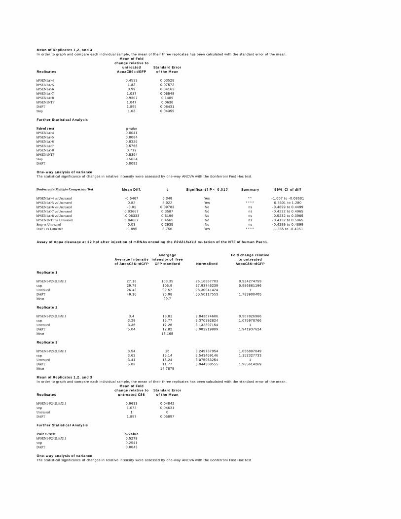
Mean of Replicates 1,2, and 3In order to graph and compare each individual sample, the mean of their three replicates has been calculated with the standard error of the mean.
Replicates
Mean of Fold change relative to
untreated AppaC86::dGFP
Standard Error of the Mean
hPSEN1Δ>4 0.4533 0.03528hPSEN1Δ>5 1.82 0.07572hPSEN1Δ>6 0.99 0.04163hPSEN1Δ>7 1.037 0.05548hPSEN1Δ>8 0.9367 0.1489hPSEN1NTF 1.047 0.0636DAPT 1.895 0.08431Stop 1.03 0.04359
Further Statistical Analysis
Paired t-test p-valuehPSEN1Δ>4 0.0041hPSEN1Δ>5 0.0084hPSEN1Δ>6 0.8326hPSEN1Δ>7 0.5766hPSEN1Δ>8 0.712hPSEN1NTF 0.5394Stop 0.5624DAPT 0.0092
One-way analysis of variance
Bonferroni's Multiple Comparison Test Mean Diff. t Significant? P < 0.01? Summary 99% CI of diff
hPSEN1Δ>4 vs Untreated -0.5467 5.348 Yes ** -1.007 to -0.08681hPSEN1Δ>5 vs Untreated 0.82 8.022 Yes **** 0.3601 to 1.280hPSEN1Δ>6 vs Untreated -0.01 0.09783 No ns -0.4699 to 0.4499hPSEN1Δ>7 vs Untreated 0.03667 0.3587 No ns -0.4232 to 0.4965hPSEN1Δ>8 vs Untreated -0.06333 0.6196 No ns -0.5232 to 0.3965hPSEN1NTF vs Untreated 0.04667 0.4565 No ns -0.4132 to 0.5065Stop vs Untreated 0.03 0.2935 No ns -0.4299 to 0.4899DAPT vs Untreated -0.895 8.756 Yes **** -1.355 to -0.4351
Average Intensity of AppaC86::dGFP
Avergage intensity of free
GFP standard Normalised
Fold change relative to untreated
AppaC86::dGFP
Replicate 1
hPSEN1-P242LfsX11 27.16 103.35 26.16567703 0.924274759stop 29.79 105.9 27.93746239 0.986861196Untreated 26.42 92.57 28.30941424 1DAPT 49.16 96.98 50.50117553 1.783900405Mean 99.7
Replicate 2
hPSEN1-P242LfsX11 3.4 18.81 2.843674606 0.907826966stop 3.29 15.77 3.370392824 1.075978766Untreated 3.36 17.26 3.132397154 1DAPT 5.04 12.82 6.082919889 1.941937624Mean 16.165
Replicate 3
hPSEN1-P242LfsX11 3.54 16 3.249737954 1.056807049stop 3.63 15.14 3.543469146 1.152327733Untreated 3.41 16.24 3.075053254 1DAPT 5.02 11.77 6.044368555 1.965614269Mean 14.7875
Mean of Replicates 1,2, and 3In order to graph and compare each individual sample, the mean of their three replicates has been calculated with the standard error of the mean.
Replicates
Mean of Fold change relative to
untreated C86Standard Error
of the Mean
hPSEN1-P242LfsX11 0.9633 0.04842stop 1.073 0.04631Untreated 1 0DAPT 1.897 0.05897
Further Statistical Analysis
Pair t-test p-valuehPSEN1-P242LfsX11 0.5279stop 0.2541DAPT 0.0043
One-way analysis of variance
The statistical significance of changes in relative intensity were assessed by one-way ANOVA with the Bonferroni Post Hoc test.
Assay of Appa cleavage at 12 hpf after injection of mRNAs encoding the P242LfsX11 mutation of the NTF of human Psen1.
The statistical significance of changes in relative intensity were assessed by one-way ANOVA with the Bonferroni Post Hoc test.

Bonferroni's Multiple Comparison Test Mean Diff. t Significant? P < 0.01? Summary 99% CI of diff
hPSEN1-P242LfsX11 vs Untreated -0.03667 0.581 No ns -0.3295 to 0.2562STOP vs Untreated 0.07333 1.162 No ns -0.2195 to 0.3662DAPT vs Untreated -0.8967 14.21 Yes **** -1.190 to -0.6038
ns, not significant, i.e. p ≥ 0.01**, p < 0.01***, p < 0.001 ****, p < 0.0001

126
CHAPTER IV
Research Paper III
HMGA1 controls an ancient alternative splicing mechanism that truncates
PRESENILIN proteins under hypoxia to boost γ-secretase activity and suppress the
unfolded protein response.
Hani Moussavi-Nik1, Morgan Newman1, Lachlan Wilson1, Esmaeil Ebrahimie1, Simon
Wells1, Mark Van Der Hoek7, Giuseppe Verdile2,3, Gavin Chapman5,6, Sharon
Pursglove,5,6, Louise O’Keefe1, Ralph Martins 2,3,4 and Michael Lardelli1
1Discipline of Genetics, School of Molecular and Biomedical Science, The University of
Adelaide, SA 5005, Australia, 2Centre of Excellence for Alzheimer’s Disease Research
and Care, School of Exercise, Biomedical and Health, 3Sir James McCusker Alzheimer’s
Disease Sciences, Edith Cowan University, Joondalup, WA 6027, Australia, 5
Development Biology Program, Victor Chang Cardiac Research Institute, Sydney, NSW,
Australia, 6St. Vincent's Clinical School, University of New South Wales, Sydney, NSW,
Australia, 7Adelaide Microarray Centre, IMVS Main Building, Frome Road, Adelaide,
SA 5000, Australia.
127
STATEMENT OF AUTHORSHIP
HMGA1 controls an ancient alternative splicing mechanism that truncates
PRESENILIN proteins under hypoxia to boost ��-secretase activity and suppress the
unfolded protein response.
Unsubmitted Manuscript.
Hani Moussavi-Nik (First Author)
• Development of PCR primers and RT- and Q- PCR analysis on:
o Hypoxia and chemical mimicry of hypoxia treated morpholino and mRNA
injected zebrafish embryos
o Hypoxia and chemical mimicry of hypoxia treated guinea pig cells
• Bioinfomatic analysis of PS2V binding site in different species.
• qRT-PCR of UPR related genes.
Certification that the statement of contribution is accurate and permission is given for the
inclusion of the paper in the thesis
Signed…………………………… …………………….Date
Morgan Newman (Co-author)
• Editing of paper
• Injection of HMGA1 morpholino and mRNA.
• Development and injection of UPR assay
• Zebrafish Notch assay (Except where noted)

128
Certification that the statement of contribution is accurate and permission is given for the
inclusion of the paper in the thesis
Signed…………………………… ……………………….Date
Lachlan Wilson (Candidate/Co-author)
• Performed all Appa assay experiments, statistical analysis and injections.
• Performed all HMW western blot analysis and corresponding injections.
• Performed all preliminary IP experiments, injections and preparations of IP samples.
• Performed zebrafish Notch assay on K115Efx10.
Certification that the statement of contribution is accurate and permission is given for the
inclusion of the paper in the thesis
Signed
……………………………… ………………….Date…………
Esmaeil Ebrahimie (Co-author)
• Microarray data analysis and gene expression network construction
Certification that the statement of contribution is accurate and permission is given for the
inclusion of the paper in the thesis
Signed………………………………………… …………….Date
Simon Wells (Co-author)
• Preliminary work on Notch assay analysis.

129
Certification that the statement of contribution is accurate and permission is given for the
inclusion of the paper in the thesis
Signed…………………………… …………………….Date
Mark Van Der Hoek (Co-author)
• Performed microarray and preliminary analysis
Certification that the statement of contribution is accurate and permission is given for the
inclusion of the paper in the thesis
Signed…………………………… ……………………….Date
Giuseppe Verdile (co-author)
• Human cell culture experiments on Notch and APP (preliminary and not in the
paper).
• Analysis of human PS2V in human cell culture.
Certification that the statement of contribution is accurate and permission is given for the
inclusion of the paper in the thesis
Signed
…………………………… ………………….Date…………
Gavin Chapman (co-author)
Analysis of Mouse cultured cells for PS2V.

130
Certification that the statement of contribution is accurate and permission is given for the
inclusion of the paper in the thesis
Signed
………………………… ………………….Date…………
Sharon Pursglove (co-author)
Performed mouse cell culture analysis of Notch
Certification that the statement of contribution is accurate and permission is given for the
inclusion of the paper in the thesis
Signed
………………………… ……………….Date…………
Louise O’Keefe (co-author)
Conducted hypoxia tests in flies for the alternative splicing of PS2V in Drosophila.
Certification that the statement of contribution is accurate and permission is given for the
inclusion of the paper in the thesis
Signed
………………………………… ……………….Date…………
Ralph Martins (co-author)
Supervised development of work for Verdile..

131
Certification that the statement of contribution is accurate and permission is given for the
inclusion of the paper in the thesis
Signed
……………………………………… ………….Date…………
Michael Lardelli (co-author)
Planned the research, wrote the manuscript, acted as corresponding author and supervised
development of work for Newman, Wilson, Moussavi-Nik and Wells.
Certification that the statement of contribution is accurate and permission is given for the
inclusion of the paper in the thesis
Signed
……………………………………… ………….Date…………

132
HMGA1 controls an ancient alternative splicing mechanism that
truncates PRESENILIN proteins under hypoxia to boost γ-secretase
activity and suppress the unfolded protein response.
Hani Moussavi-Nik*1, Morgan Newman*1, Lachlan Wilson*1, Esmaeil Ebrahimie2,
Simon Wells1, Mark Van Der Hoek3, Giuseppe Verdile4,5,6, Gavin Chapman7, Sharon
Pursglove7, Louise O’Keefe1, Ralph Martins4,5, Michael Lardelli†1.
*Joint first authors
† Author for correspondence
Affiliations:
1. Discipline of Genetics, School of Molecular and Biomedical Science, University of
Adelaide, SA 5005, Australia
2. Technology Enhanced Learning Group, University of South Australia, SA, 5000,
Australia
3. Adelaide Microarray Centre, IMVS Main Building, Frome Road, Adelaide, SA 5000,
Australia
4. Centre of Excellence for Alzheimer's Disease Research and Care, School of Medical
Sciences, Edith Cowan University, Joondalup, WA 6027, Australia
133
5. School of Psychiatry and Clinical Neurosciences, University of Western Australia,
Crawley, WA, 6009, Australia.
6. School of Public Health, Curtin University, Bentley, WA 6102, Australia
7. Developmental Biology Division, Victor Chang Cardiac Research Institute, NSW
2010, Australia
Full contact details for corresponding author:
Michael Lardelli
Discipline of Genetics, School of Molecular and Biomedical Sciences, The University of
Adelaide, SA 5005, Australia.
Tel. (+61 8) 83133212, Fax (+61 8) 83134362
email: [email protected]

134
Abstract
The PRESENILIN genes PSEN1 and PSEN2 encode two structurally related proteases
essential for γ-secretase cleavage of over 70 proteins within lipid bilayers. Together, over
200 mutations causing early onset, familial Alzheimer’s disease (FAD) occur in PSEN1
and PSEN2 but only one of these, the K115Efx10 mutation of PSEN2, causes truncation
of the open reading frame. If translated, the truncated product would resemble a naturally
occurring truncated isoform of PSEN2 named PS2V that is induced by hypoxia and found
at high levels in late onset Alzheimer’s disease (AD) brains. Here we use zebrafish
embryos to investigate the evolution of PS2V and its function compared to the
K115Efx10 truncation. We find that zebrafish possess a PS2V-like isoform, PS1IV,
produced from the fish’s PSEN1 orthologous gene rather than its PSEN2 orthologue. We
show that the molecular mechanism controlling formation of PS2V/PS1IV is conserved
since the divergence of teleosts and tetrapods and was probably present in the common
ancestor of the PSEN1 and PSEN2 genes. The structures of human PS2V and zebrafish
PS1V have diverged greatly but their ability to stimulate γ-secretase activity and suppress
the unfolded protein response is conserved. The K115Efx10 FAD mutation is similar to
PS2V in its ability to increase Notch signalling and γ-secretase cleavage of a zebrafish
orthologue of the AMYLOID BETA A4 PRECURSOR PROTEIN. This supports
increased Aβ peptide production as a molecular common link between K115Efx10 FAD
and sporadic late onset AD. Microarray analysis of PS1IV function reveals modulation of
immune/inflammatory responses and numerous other functions.

135
Introduction
The PRESENILIN genes PSEN1 and PSEN2 encode two structurally related proteins that
operate within γ-secretase complexes to cleave type 1 transmembrane proteins (Figure
1A, reviewed by (1)). For this reason they are essential to signalling by a wide variety of
proteins such as Notch receptors, p75 and Amyloidβ Precursor Protein (AβPP). The
PRESENILINs also have other functions including in autophagy (2), phosphorylation of
β-catenin (3) and control of chromosome segregation (4, 5). PSEN1 is the major locus for
autosomal dominant mutations causing familial Alzheimer’s disease (FAD) (6) the other
two known loci being PSEN2 and AβPP.
At least 95% of Alzheimer’s disease (AD) is sporadic. This disease shares similar
histopathology with FAD including deposition of plaques of Aβ peptide that is produced
from AβPP after cleavage by β-secretase and then γ-secretase (Figure 1A). However, the
causes and pathological mechanism behind sporadic AD are hotly debated. One
molecular change commonly observed in AD brains is an increase in alternative splicing
of PSEN2 transcripts to produce a truncated isoform lacking exon 5 sequence and thus
named “PS2V” (7, 8) (Figure 1 B,C). This isoform has been described as “aberrant”, (9)
probably because it had only been detected in human brain and not in mice or rats.
However, PS2V has the remarkable property that it can increase γ-secretase activity and
production of Aβ peptide from AβPP (8). Using human neuoblastoma SK-N-SH cells,
Sato et al. (7) showed that PS2V formation is induced by hypoxia but not other forms of
cellular stress. Under hypoxia expression of the HMGA1a protein is induced in neural

136
tissue and this binds to a sequence in exon 5 of PSEN2 transcripts. This interferes with
spliceosome complex function at the exon 5 donor site causing exon 4 to be ligated to
exon 6 and excluding exon 5 (10), (Figure 1C).
The HIGH MOBILITY GROUP AT-HOOK 1 (HMGA1) proteins are an ancient and
highly conserved family primarily known for their ability to bind chromatin (reviewed by
(11)). HMGA1 is expressed early in embryogenesis when embryos inhabit an hypoxic
environment and many relatively undifferentiated cells exist (12). Their expression can
be induced by hypoxia in adult neural tissues (13, 14) and is commonly reactivated in
cancer (reviewed by (15)). Hypoxia increases Notch signalling to maintain neural stem
cells in an undifferentiated state (16). This is known to involve hydroxylation of the
Notch receptor protein (17, 18) and Mukherjee et al. (19) recently showed that the HIFα
protein of Drosophila that controls many cellular hypoxic responses can interact directly
with Notch receptor to increase its signalling in the absence of ligand. Evidence is
accumulating that hypoxia (20, 21) and Notch (22) may play important roles in AD
pathogenesis but how these factors might be coupled to other factors such as Aβ
production is not obvious.
Over 180 mutations in PSEN1 and around 20 mutations in PSEN2 are thought to cause
FAD (6, 23). All are mis-sense mutations, deletions or small insertions that preserve the
C-terminal protein coding part of the open reading frame. Recently, a unique frameshift
mutation, K115Efx10, was discovered in PSEN2 and suggested to cause AD ((23), Figure

137
1B). If expressed this mutant allele would produce a truncated protein very similar to
PS2V.
We recently showed that PS2V production is not a unique human aberration but occurs in
the brains of guinea pigs and, presumably most other mammals (24). In this paper we
show that PS2V can also increase Notch signalling and that the alternative splicing
mechanism that produces PS2V in response to hypoxia is ancient and predates the gene
duplication event that formed PSEN1 and PSEN2 in vertebrates. In teleosts (bony fishes)
hypoxia induces expression of Hmga1a that acts on transcripts of the orthologue of
PSEN1 rather than PSEN2 to cause an alternative splicing outcome. The novel splice
product of zebrafish psen1, “PS1IV”, nevertheless codes for a protein capable of boosting
γ-secretase activity. Microarray analysis of PS1IV function demonstrates roles in
modulation of a wide variety of gene products. We also use our zebrafish assay system to
examine the activity of the putative truncated protein product of the K115Efx10 allele of
human PSEN2 and show that it can also boost γ-secretase activity. Our data support a
unified model of sporadic AD pathogenesis where PRESENILIN activity plays a central
role in responding to hypoxic conditions by boosting both Aβ production and Notch
signalling via PS2V formation. Chronic upregulation of Aβ production in brains with
ageing vasculature and insufficient oxygenation may eventually lead to oligomerisation
of Aβ with pathogenic consequences.

138
Materials and Methods
Ethics and permits
All experimentation with animals and work with genetically modified organisms was
conducted under the auspices of the University of Adelaide’s Animal Ethics Committee
and Institutional Biosafety Committee respectively.
Oligonucleotides
The sequences of all oligonucleotides (PCR primers and morpholino oligonucleotides)
used in this study are given in Supplementary Data File 1.
Construction and cloning of cDNAs for mRNA synthesis
cDNAs encoding PRESENILIN truncations (including truncations equivalent to the
NTF) were synthesised by PCR from zebrafish psen1 and psen2 cDNAs or human
PSEN1 and PSEN2 cDNAs. The zebrafish Presenilin truncations are fused at their N-
termini to the FLAG™ antibody tag, DYKDDDDK (25) that follows a start codon within
a consensus Kozak sequence (26). The human PSEN truncations are fused at their N-
termini to an HA tag (YPYDVPDYA, (27)) that follows a start codon within a consensus
Kozak sequence. The cDNA encoding the putative protein product of the K115Efx10
mutation is the human hPSEN2Δ>NTF construct into which the K115Efx10 mutation has
been introduced by QuikChange™ Site-Directed Mutagenesis (Stratagene Cloning

139
Systems, La Jolla, CA, USA). The cDNA constructs for synthesis of the negative control
zebrafish and human mRNAs denoted as “stop” are full length cDNAs including N-
terminal FLAG or HA tag fusions respectively but with codon 4 of the Presenilin coding
sequences mutated to the stop codon TAA. All sequences were cloned into the pcGlobin2
vector (28) between the Bam HI and Eco RI restriction sites (for zebrafish cDNAs) and
Bam HI and Xho I restriction sites (for human cDNAs) and their sequences confirmed by
sequencing before their use for synthesis of mRNA as previously described (29).
Injection of mRNAs and morpholinos
MoHmga1aTln and MoHmga1BindBlock (Psen1) were synthesized by GeneTools LLC
(Corvallis, OR, USA) and injected at 0.5mM (diluted to this concentration with MoCont
as previously (29)) at the one-cell stage. hPSEN2Δ>4 and hPS2V DNA constructs
including N-terminal FLAG tags DNA were synthesised by GenScript (Piscataway, NJ,
USA). cDNAs encoding FLAG-tagged zPsen1Δ>3 and zPS1IV were synthesised by PCR
from zebrafish psen1 cDNA. All sequences were cloned into the pcGlobin2 vector (30)
between the Bam HI and Eco RI restriction sites. Sequences were confirmed by
sequencing before use for synthesis of mRNA as previously described (29). mRNAs were
injected one-cell stage embryos at 100-200ng/µl (each mRNA concentration is indicated
in the table of neurogenin1-based Notch signalling assay results (Supplementary Data
File 2). As a control to show the ability of MoHmga1aTln to suppress hmga1a mRNA
translation, the first 135 bases of the hmga1a coding sequence was fused in frame to
EGFP in the N1-EGFP plasmid at the EcoRI/AgeI sites (hmga1a-135-EGFP) (BD
Biosciences, NJ, USA). The plasmid was linearised with NotI prior to injection. 50ng/ul
of the plasmid was co-injected with either 0.5 mM of MoHmga1aTln mixed with

140
MoCont or MoCont alone. Embryos were then observed for GFP fluorescence at ~10hpf
using fluorescence microscopy (see Supplementary Data File 3).
Exposure of embryos, explants and adult fish to hypoxia and NaN3
Danio rerio were bred and maintained at 28 °C on a 14 h light/10 h dark cycle. Embryos
were collected from natural mating, grown in embryo medium (E3, (31)) and staged. For
exposure of embryos with to sodium azide (NaN3, Sigma-Aldrich CHEMIE Gmbh,
Steinheim, Germany), this was performed at 100µM from 6 hpf until embryos reached
developmental stages equivalent to those attained under normoxia at 48 hpf at 28.5 °C
(30 embryos were used for each concentration). In the experiments for exposure of adult
brain tissue to sodium azide, this was performed at a concentration of 100µM in DMEM
medium after the tissue (from chicken and mouse brains) was cut into small fragments
(e.g. ~1mm diameter). This was not necessary for explanted zebrafish brains. In the
experiments conducted on adult zebrafish under low oxygen conditions, oxygen was
depleted by bubbling nitrogen gas through the medium. Oxygen concentrations were
measured using a dissolved oxygen meter (DO 6+, EUTECH instruments, Singapore).
The dissolved oxygen level in the hypoxia group was measured to be 1.20±0.6 mg/l for
adults; whereas the normal ambient oxygen levels were 6.6±0.45 mg/l. For exposure of
adult Drosophila melongaster to hypoxia, healthy flies were exposed to low oxygen
conditions (<1%, using AnaeroGen™, Oxoid, Basingstoke, UK) for 3 hours, and
immediately frozen for later RNA extraction as below.

141
Appa cleavage and Notch signalling assays
Assays for γ-secretase cleavage of Appa (32) and Notch signalling (29) were performed
as previously described (see Supplementary Data Files 4 and 2 respectively for raw data
and statistical analysis).
RT-PCR assays for detection of splice events homologous to formation of human
PS2V
Total RNA was extracted from samples mentioned above using the QIAGEN RNeasy
mini kit (QIAGEN, GmbH, Hilden, Germany). 700 ng of total RNA were used to
synthesize 25 µL of first-strand cDNA by reverse transcription (SuperScript ΙΙΙ kit;
Invitrogen, Camarillo, USA). For PCR amplification, 5 µL of the cDNA was used with
the following primer pairs as relevant: Chicken Psen2: Chic Psen2: Ex.4 F: (5'-
ACGGGCAGCTGATCTACACC-3') with Chic Psen2 Ex.6 R: (5'-
GACCTTTCCAGTGGATGCAA-3'); Chicken Psen1: Chic Psen1 Ex.4 F: (5'-
CTACACTCCCTTCACAGA-3'); Chic Psen1 Ex.6 R: (5'-
CAGGATAAGCCATGCAGT-3'); Mouse Psen2: Mm Psen2 Ex.4 F: (5'-
CATCGTAGTCATGACCATCTTCC-3'); MmPsen2 Ex.6 R: (5'-
ACACCATGCCCACTGCC3'); Mouse Psen1: Mm Psen1 Ex.4 F: (5'-
CTACACCCCATTCACAGA-3'); Mm Psen1 Ex.6 R: (5'-
CTGCTGCAGTCGAAGGGG-3'); and Drosophila melanogaster Psen: Dm Psen Ex.2
F: (5'-CTGCTGTCAAATCTCCAGGCTT-3'); Dm Psen Ex.2 R: (5'-
TCCGACCACTCCAAAGTTCC-3').
142
PCR was performed using GoTaq polymerase (Promega, Madison, USA) for 35 cycles
with an annealing temperature of 56 °C for 30 s, an extension temperature of 72 °C for
1 min and a denaturation temperature of 95 °C for 40 s. The PCR products were cloned
into the pGEM-T vector (Promega, Madison, USA) and sequenced.
Quantitative real-time PCR for detection of PS2V
The relative standard curve method for quantification was used to determine the
expression of experimental samples compared to a basis sample. For experimental
samples, target quantity was determined from the standard curve and then compared to
the basis sample to determine fold changes in expression. Gene specific primers were
designed for amplification of target cDNA and the cDNA from the ubiquitously
expressed control gene ef-1a. The reaction mixture consisted of 50ng/µl of cDNA. 18 µM
of forward and reverse primers and Power SYBR green master mix PCR solution
(Applied Biosystems).
To generate the standard curve cDNA was serially diluted (100 ng, 50 ng, 25 ng, 12.5
ng). Each sample and standard curve reaction was performed in triplicate for the control
gene and experimental genes. Amplification conditions were 2 min at 50 °C followed by
10 min at 95 °C and then 40–45 cycles of 15 s at 95 °C and 1 min at 60 °C. Amplification
was performed on an ABI 7000 Sequence Detection System (Applied Biosystems) using
96 well plates. Cycle thresholds obtained from each triplicate were averaged and
normalized against the expression of ef-1a, which has previously been demonstrated to be
suitable for normalization for zebrafish quantitative PCR (qPCR). Each experimental
sample was then compared to the basis sample to determine fold changes of expression.

143
The primers used for quantitative real-time PCR anaylsis of PS1IV mRNA levels are seen
in Supplementary Data File 1. See Supplementary Data File 5 for all qPCR raw data and
analysis.
Assay of the unfolded protein response (UPR)
Embryos were injected with mRNAs at the 1-cell stage. At 2hpf fertilized injected
embryos were split into 2 groups. One group was left untreated and the other group was
treated with 1.5µg/ml tunicamycin. At 24hpf total RNA was extracted from embryos and
reversed transcribed into cDNA for qPCR analysis. qPCR was performed to determine
the expression levels of unspliced xbp-1 and spliced xbp-1 in embryos that had been
injected with mRNAs encoding either zPsen1Δ>3, zPsen2Δ>4, hPSEN2Δ>4 or the
zebrafish “stop” negative control mRNA. Tunicamycin treatment causes ER stress and
induces the expression of the spliced form of xbp-1 (33). Any observed decrease in
spliced xbp-1 expression under tunicamycin treatment in the mRNA injected cDNA
samples can be interpreted as these mRNAs suppressing the ER stress response. See
Supplementary Data File 5 for qPCR raw data and analysis.
Microarray analysis
Embryos were collected from natural mating, grown in embryo medium (E3) and staged.
Morpholinos were synthesized by Gene Tools LLC (Corvallis, OR, USA) and are listed
in Supplementary Data File 1. Fertilized zebrafish eggs were injected prior to cleavage.
To ensure consistency of morpholino injection, eggs were always injected with solutions

144
at 1 mM total concentration. Dilutions of MoHMGA1 BindBlock morpholino was
carried out by mixing with standard negative control morpholino, MoCont. This ensured
that phenotypic variation observed was not due to change in the total injection
concentration. The maximum concentration of HMGA1 BindBlock injected was always
0.5 mM (i.e. a mixture of 0.5 mM MoCont + 0.5 mM MoHmga1BindBlock morpholino).
Titration of morpholinos to find effective concentrations has been shown previously (29).
For exposure of injected embryos to sodium azide this was performed at 100µM from
6 hpf until embryos reached developmental stages equivalent to those attained under
normoxia at 48 hpf at 28.5 °C (30 embryos were used for each injection batch).
Total RNA was extracted from different injection batches using the RNeasy Mini Kit
according to the manufacturer’s specifications. For each sample, the RNA concentration
was determined with a NanoVue™ UV–vis spectrophotometer (GE Healthcare Life
Sciences, Fairfield, USA). RNA integrity and quality were then estimated with an Agilent
2100 Bioanalyzer (Agilent Technologies, Palo Alto, CA), and the RNA integrity number
(RIN) index was calculated for each sample. Only RNAs with a RIN number>7.0 were
processed further.
To evaluate genome-wide changes in gene transcription related to the presence or
absence of PS1IV under mimicry of hypoxia, we performed hybridisation anaylsis using
the Zebrafish Genome Array (Affymetrix Inc. Santa Clara, CA). Briefly, mRNAs were
isolated from 300ng of total RNA derived from embryos injected with

145
MoHMGA1BindBlock morpholino at 0.5 mM (diluted to this concentration with MoCont
morpholino as previously described (29)) or with MoCont at 0.5 mM alone treated with
and without sodium azide using the Eukaryotic Poly-A RNA control Kit (Affymetrix) or
from uninjected embryos treated with sodium azide (see Figure 7). Subsequently,
mRNAs were subjected to double-strand cDNA synthesis using the GeneChip® One
Cycle cDNA Synthesis Kit (Affymetrix). The resulting double-stranded cDNAs were in
vitro transcribed into biotin-labelled cRNAs using the IVT Labelling Kit (Affymetrix).
After that, the biotin-labelled cRNAs were fragmented by heating and hybirdized with
the Affymetrix Zebrafish Genome Array. Finally, the signal intensity of the chip was
scanned using a GeneChip® Scanner 3000TG and analysed using the Partek Genomics
Suite software (www.partek.com). Cell files were imported and intensities adjusted for
GC content using Partek’s own method prior to RMA background correction and quantile
normalization (34-37). Mean gene probe summaries assessed for differential gene
expression by ANOVA with p-value adjusted using Step-up multiple test correction.
Each treatment had four biological replicates.
Statistical analysis of microarray data
Gene expression analysis was performed using the FlexArray package (McGill
University, Canada). MoHmga1BindBlock-injection and MoCont-injection were
assumed as treatments. Log2 ratio values were calculated as fold change by FlexArray.
Those genes having a Bayesian t-test (38) with p-value ≤ 0.05 and fold change ≥ +1 or
fold change ≤ -1 were accepted as significant differentially expressed genes. Bayesian t-

146
test is a powerful statistical test in determining differentially expressed gene s in two
different samples by decreasing Type I error rates (38).

147
Results
Note: The structure of the protein-coding exons in the PRESENILIN genes of zebrafish,
human and other species is largely conserved although exon numbers vary due to
differences in non-coding exons. For simplicity, below we will refer to the exons of
zebrafish and other species using the identity of their cognate exons in humans.
Hypoxia-inducible alternative splicing of psen1, not psen2 in zebrafish
We recently showed that PS2V is induced in the brains of guinea pigs and probably most
other mammals (except rats or mice) (24). We have also shown that hypoxia does not
induce formation of a PS2V-like alternative transcript from zebrafish psen2 (14).
However, the vertebrate PSEN1 and PSEN2 genes are paralogues formed by an ancient
gene duplication event that predates the divergence of the tetrapod and teleost lineages
(39, 40). After the gene duplication event each gene underwent a degree of sub-
functionalisation. To test whether hypoxia-inducible formation of “PS2V” might have
been retained in the PSEN1 gene lineage in bony fish rather than in the PSEN2 lineage
we tested for PS2V-equivalent exon loss in zebrafish psen1 by RT-PCR amplification of
cDNA spanning exons 3 to 6. We observed induction of alternative transcript formation
from psen1 using chemical mimicry of hypoxia (NaN3 treatment) in both embryos and
larvae (Figure 2A). The same alternative splicing event was detected under genuine
hypoxia and in the brains of adult fish (Figure 2A). Cloning and sequencing of cDNA
from the alternatively spliced transcript of psen1 showed that it lacks exon 4 sequence

148
rather than the loss of exon 5 as seen in human PS2V transcripts (Figure 2B, C and
Supplementary Data File 6). Therefore, we have named this alternative transcript of
zebrafish psen1 “zPS1IV”.
Manabe et al (9, 10) have clearly defined the mechanism that controls loss of exon 5
sequence from human PSEN2 transcripts to form PS2V. In humans, transcription of
HMGA1a is induced by hypoxia and then the HMGA1a protein binds to two sites at the
3’ end of exon 5 in PSEN2 transcripts (Figure 2B,D). This appears to block funcational
association of U1 snRNP with the exon 5 splice donor site causing alternative splicing of
exon 4 to exon 6 ((10) Figure 2B). We have previously shown that the DNA binding
region of zebrafish Hmga1a protein is highly similar to human HMGA1a but that the
nucleotide sequence at the 3’ end of exon 5 of zebrafish psen2 is not highly conserved
(14). This is consistent with the failure of zebrafish psen2 to produce a PS2V equivalent.
If the hypoxia-inducible alternative splicing of zebrafish psen1 represents an ancient
mechanism present in the common ancestor of the PSEN1 and PSEN2 genes then this
might still be controlled by induction of Hmga1a. We examined the sequence of psen1
for potential Hmga1 binding sites. Consistent with conservation of the alternative splicing
mechanism, we discovered that the best match to the HMGA1a binding site consensus
was at the 3’ end of the psen1 exon 5 (Figure 2D)
Since hypoxia-induced alternative splicing of psen1 transcripts causes loss of exon 4
rather than exon 5 we tested whether binding of Hmga1a at this site still controlled the
alternative splicing event. We designed a morpholino antisense oligonuclotide,

149
MoHmga1BindBlock, to bind over the upstream Hmga1 binding in exon 5 that,
hopefully, would not interfere with the function of this exon’s splice donor site (Figure
2D). Injection of this oligonucleotide into embryos followed by exposure to chemical
mimicry of hypoxia (100 µM NaN3) and RT-PCR analysis showed that
MoHmga1BindBlock greatly reduced formation of PS1IV under hypoxia while the
common, normoxic, splicing pattern still occurred (Figure 3A). Blockage of Hmga1
translation by injection of morpholino oligonucleotide MoHmga1aTlnBlock also greatly
reduced formation of zPS1IV under chemical mimicry of hypoxia (Figure 3B). We were
also able to force formation of PS1IV under normoxia by overexpression of Hmga1a in
zebrafish embryos through injection of mRNA containing the Hmga1 coding sequence
(Figure 3C1,D). The evolutionary conservation of this alternative splicing system was
demonstrated by the fact that forced expression of human HMGA1a in zebrafish embryos
by mRNA injection also caused formation of PS1IV (Figure 3C2). In both these cases, the
induction of PS1IV formation could be blocked by co-injection of MoHmga1aBindBlock
with the Hmga1a/HMGA1a mRNA while injection of a non-translatable control mRNA
was unable to induce PS1IV formation (Figure 3C1,C2,D). These experiments strongly
support that the mechanisms controlling hypoxia-inducible alternative splicing of
zebrafish psen1 and human PSEN2 have evolved from an ancient mechanism involving
HMGA1 control of splicing in the ancestral PRESENILIN gene which was duplicated to
form vertebrate PSEN1 and PSEN2.
Predictions of hypoxia-inducible alternative splicing in other vertebrate lineages

150
The correlation between hypoxia-inducible alternative splicing of PRESENILIN gene
transcripts and possession of a highly conserved HMGA1a-binding site in exon 5 can be
used to predict which gene, PSEN1 or PSEN2, might show this alternative splicing in any
vertebrate. Alignment of the 3’ region of exon 5 of PSEN1 and PSEN2 in various
vertebrates species supports that most mammals show hypoxia-inducible alternative
splicing in PSEN2 transcripts but not those of PSEN1 (Figure 2D). Interestingly rats and
mice (both species within the subfamily Murinae) lack the ability to produce PS2V (24)
and our tests have revealed no evidence for hypoxia-inducible splicing of Psen1
transcripts in these species (data not shown – see Materials and Methods). Gallus gallus
(chicken, a bird) and Xenopus laevis (African clawed frog, an amphibian) also do not
show high conservation of the HMGA1a-binding site in exon 5 of their PSEN1 or PSEN2
genes (Figure 2D). We have previously shown induction of PS2V in explanted guinea
pig brain tissue incubated in medium containing NaN3 (24). As expected from our
sequence analysis above, tests of mRNA extracted from chicken brains exposed to NaN3
failed to observe exclusion of exon 4 or exon 5 from transcripts of either PSEN1 or
PSEN2 (data not shown). Interestingly, the teleost Fugu rubripes (a puffer fish) does not
have a well-conserved Hmga1a-binding site in exon 5 of either its psen1 or psen2 gene
(Figure 2D) reinforcing how loss of hypoxia-controlled alternative splicing of
PRESENILIN transcripts need not be severely detrimental to survival.
To test whether this mechanism of alternative splicing exists in the single presenilin gene
of an insect we exposed Drosophila melanogaster to hypoxia before extraction of mRNA
and RT-PCR. However, no such alternative splicing was observed for the Drosophila

151
melanogaster PRESENILIN orthologue Psn (data not shown – see Materials and
Methods).
Both human PS2V and zebrafish PS1IV can boost γ-secretase activity and Notch
signalling
Human PS2V consists of 124 amino acid residues (aa) and is expected to possess one
transmembrane domain (Figure 2C). In contrast, deletion of zebrafish psen1 exon 4 to
form zPS1IV alters the open reading frame after exon 3 sequence to form a peptide
predicted to possess only 18 aa including the 6 novel residues at its C-terminal (Figure
2C). The preservation of this mechanism of alterative splicing of the PRESENILIN genes
for approximately 450 million years since the divergence of the teleost and tetrapod
vertebrate lineages (41) suggests that these peptides serve an important function(s) in
response to hypoxia despite their very different structures.
Sato et al. (8) showed that PS2V can boost γ-secretase activity and increase cleavage of
the Aβ peptide from the APP protein. In a recently submitted papers, we showed that
forced expression of a truncation of zebrafish Psen1 protein equivalent to human PS2V
(i.e. truncated after exon 4 sequence, “Psen1Δ>4”) dominantly increases cleavage of
zebrafish Appa (a paralogue of the human AβPP protein that is cleaved to produce the Aβ
peptide seen in Alzheimer’s disease brains) indicating increased γ-secretase activity.
Psen1Δ>4 expression also decreases neurogenin1 gene transcript levels in the trunk
region of zebrafish embryos at 24 hours of development indicating increased Notch

152
signalling. The equivalent truncation of human PSEN1, hPSEN1Δ>4, also possesses
these activities. The apparent increase in Notch activity caused by zebrafish Psen1Δ>4
requires the presence of non-truncated endogenous Psen1 (Newman et al, submitted). We
first asked whether, like Psen1Δ>4 and hPSEN1Δ>4, forced expression of human PS2V
in zebrafish embryos could boost γ-secretase activity. Consistent with our expectation,
injection of mRNA encoding human PS2V caused increased Appa cleavage (Figure 4A)
and decreased neurogenin1 activity (Figure 4B). This is evidence that PS2V not only
increases production of Aβ in human brain but also increases signalling by NOTCH
receptors. It also demonstrates that zebrafish can be used to analyse the function of
human PS2V.
Human PS2V, zebrafish Psen1Δ>4 and human hPSEN1Δ>4 can all increase cleavage of
Appa and Notch signalling so it remained to demonstrate this activity for a PS2V-
equivalent truncation of zebrafish Psen2, i.e. Psen2Δ>4. Surprisingly, injection of an
mRNA encoding this protein truncation had no measurable effect on Appa cleavage or
Notch signalling (Figure 4A, B). This is consistent with our failure to detect alternative
splicing of zebrafish psen2 transcripts under hypoxia or chemical hypoxia (14).
Our previous study analysing the effects of various truncations of the zebrafish Psen1
protein found that injection into embryos of mRNA encoding Psen1 truncated after exon
3 sequence (i.e. “zPsen1Δ>3”, similar to zPS1IV but lacking its novel C-terminal aas)
was very disruptive to embryo development. This precluded analysis of Appa cleavage or
Notch signalling for this Psen1 truncation (Newman et al. submitted). However, in an

153
attempt to overcome this limitation we diluted this mRNA until injected embryos were
able to develop to the 24 hpf stage (see Materials and Methods). Assessment of Appa
cleavage and Notch signalling then showed that mRNA encoding this small peptide can,
nevertheless, increase γ-secretase activity (Figure 5 A, B). Injection of mRNA encoding
zebrafish zPS1IV (i.e. like Psen1Δ>3 but including the novel aas caused by fusion of
exon3 to exon 5 sequence) also caused increased cleavage of Appa (Figure 5A).
However, the observed frequency of decreased neurogenin1 staining due to zPS1IV
indicating increased Notch signalling was not sufficient to achieve statistical significance
(a p value of >0.05 was obtained in our assay, Figure 5B). Differences in the ability of
zPsen1Δ>3 and zPS1IV to boost Notch signalling may be due to a lesser stability of
PS1IV and/or differing subcellular localisation of these two peptides due to differences in
their C-terminal amino acid residues.
To examine further the conservation of structure and function between zebrafish zPS1IV
and human PS2V we then examined the ability to boost Appa cleavage and Notch
signalling of exon 3-derived sequences from human PSEN2, PSEN1 and zebrafish psen2.
mRNAs encoding Presenilin peptides truncated after exon 3 sequence from these three
genes were unable to boost Appa cleavage or Notch signalling (Figure 5A,B). Thus,
while zebrafish PS1IV and human PS2V share the conserved ability to boost γ-secretase
activity, their structures have diverged sufficiently over time that exon 3 sequence from
human PS2V cannot functionally substitute for zebrafish PS1IV.
154
Suppression of the unfolded protein response (UPR) by human PS2V and orthologous
truncations is conserved
Sato et al. (8) previously showed that PS2V suppresses the unfolded protein response
when its expression is forced in human HEK293T cells (a kidney-derived cell line that
does not normally expressed PS2V, (13)). Induction of the UPR occurs via a change in
the splicing of transcripts of the gene XBP1 to form the isoform XBP-1S (42) and so
quantitative PCR (qPCR) can be used to assay the relative UPR response in zebrafish
embryos (42). To investigate further the conservation of function between zebrafish and
human PRESENILIN truncations we examined the effects on the UPR in zebrafish
embryos of forced expression of human PSEN2 truncated after exon 4 sequence (i.e.
hPSEN2Δ>4, equivalent to PS2V), zebrafish Psen1 truncated after exon 3 sequence
(zPsen1Δ>3, equivalent to PS1IV) and zebrafish Psen2 truncated after exon 4 sequence
to resemble human PS2V (zPsen2Δ>4). Zebrafish embryos injected with a non-
translatable control mRNA and then exposed to 1.5 µg/ml of the antibiotic tunicamycin at
2 hpf showed ~12-fold induction of the UPR at 24 hpg (Figure 6). In contrast, injection of
mRNA coding either for hPSEN2Δ>4 or zPsen1D>3 was able to suppress UPR induction
by tunicamycin to below 2-fold. Significantly, the truncation of zebrafish Psen2 to
resemble human PS2V (zPsen2Δ>4) showed little ability to suppress the UPR (Figure 6)
consistent with its inability to stimulate γ-secretase activity while the ability of
truncations PS2V and zPsen1D>3 to suppress the UPR parallels their effects on γ-
secretase activity. Thus the action on the UPR of the truncated PRESENILINs proteins

155
that are normally induced by hypoxia in humans and zebrafish has been conserved
despite their structural divergence.
The K115Efx10 mutation of human PSEN2 indicates that PS2V may be a molecular
common link between sporadic and familial Alzheimer’s disease.
Around two hundred dominant mutations in the human PRESENILIN genes are known to
cause familial Alzheimer’s disease (FAD). Most of these occur in PSEN1 but a number
of mutations have been described in PSEN2 (6, 23). Until recently all known FAD
mutations in the PRESENILINS were mis-sense mutations or internal deletions /
insertions that preserved C-terminal protein coding sequences. No FAD mutations have
been found in genes encoding the other components of a γ-secretase complexes.
Recently, Lee et al (2) showed that full-length (non-endoproteolysed) PSEN1 has a γ-
secretase-independent function in the acidification of lysosomes required for autophagy
and that FAD mutations in PSEN1 decrease a cell’s autophagy ability. Numerous studies
have shown decreased autophagy in AD brains (reviewed in (43)). The function of non-
endoproteolysed PSEN1 in autophagy and the recent discovery of families with
mutations in other γ-secretase complex components that do not have FAD (44) strongly
suggests that, contrary to some current hypotheses, decreased γ-secretase activity does
not cause Alzheimer’s disease. Instead, FAD-causing mutations in the PRESENILIN
genes may promote Aβ accumulation through a failure to clear aggregate forms of this
peptide.

156
Recently, a woman suffering early onset Alzheimer’s disease was reported to be
heterozygous for a nonsense mutation in PSEN2, K115Efx10 (23). This mutation is a
dinucleotide deletion causing a frameshift in the PSEN2 coding sequence only six codons
prior to the end of exon 4 (Figures 1B, 4AB). If transcripts of this gene are not degraded
through nonsense mediated decay then they should generate a peptide similar in structure
to PS2V and might act to increase Aβ production. To test whether the putative truncated
product of K115Efx10 increases γ-secretase activity we synthesised mRNA encoding it
and injected this into zebrafish embryos. This increased Appa cleavage and Notch
signalling (Figure 4A,B) consistent with K115Efx10 causing AD by increasing Aβ
production.
Gene expression network analysis suggests roles for zebrafish PS1IV in regulating
immune responses, transcription and ribosomal protein expression.
The cellular role of PS2V cannot be investigated in mice due to the apparent rapid
evolution of their Presenilin genes and the loss of this isoform (24). However, the
zebrafish is the next most genetically malleable vertebrate model and, as we have shown,
it does express the PS2V-orthologous isoform, PS1IV. The morpholino
MoHmga1BindBlock blocks specifically the formation of PS1IV under conditions where
it would normally be induced. Therefore, we exploited MoHmga1BindBlock to
investigate the effects on cellular function of loss of PS1IV under hypoxia using
microarray and subsequent gene network analysis.

157
Zebrafish embryos were injected with Hmga1BindBlock or the negative control
morpholino MoCont. They were then exposed to chemical mimicry of hypoxia from 36
hpf to 48 hpf before extraction of mRNA and analysis of gene expression on the
Affymetrix Zebrafish Gene 1.0 ST Array. A number of control microarray comparisons
were also made to identify genes for which expression is affected by the act of injection
or by the non-specific effects of the morpholino oligonucleotides (see Figure 7). Bayesian
t-tests produced lists of genes with significantly decreased or increased expression in the
comparisons. The final list of genes with significantly increased or decreased expression
due to decreased PS1IV formation under mimicry of hypoxia was generated after
removal of genes showing significantly altered expression in the controls (see Figure 7
and Tables 1 and 2). To validate the microarray results, we conducted quantitative real
time PCR (qPCR) analysis for a selection of the genes with significantly altered
expression (see Supplementary Data File 5) and that appear to represent nodes in a gene
network analysis to be described in detail elsewhere (Ebrahimie, Moussavi Nik, Newman
& Lardelli, manuscript in preparation).
Among the genes showing increased expression under hypoxia without PS1IV (i.e.
normally repressed by the formation of PS1IV) were numerous genes involved with
modulation of immune responses (HRH2, NLRP3, IL17RA, CD22, ACY1, IRF4, XCL2
etc., Table 1). However, without further analysis we are uncertain whether their
combined effect would be to, overall, activate or suppress immune responses and whether
these responses are restricted to any particular tissues (e.g. humoral or neural or both).
158
Genes involved in regulation of TOR signalling (RICTOR, PKD1) blood pressure and
formation of vascular structures (e.g. AGTR1 and ANGPTL2), and cell proliferation
(PIM3, ACY1, OLFM4) also show increased expression when PS1IV is suppressed. This
set of genes also included two known to be involved in neurological conditions, GAK
(associated with Parkinsons Disease (45)) and CAMTA1 (associated with cerebellar
ataxia, nonprogressive, with mental retardation (46)).
The number of genes showing significant decreases in expression due to PS1IV (i.e.
normally dependent on PS1IV for increased expression under mimicry of hypoxia) was
greater in number and included genes encoding transcription factors or proteins involved
in chromatin remodelling (e.g. NKX3-2, SRCAP, GBX2, TCF25, SMARCA4, POU4F2),
genes involved in ER function (CALR), intracellular signalling (OCRL, AIDA,
PLEKHA4, PLCB1, MAPKAPK5) and a considerable number of ribosomal proteins (see
Table 2). Once again, genes involved in immune response regulation were identified
(IL1B, NLRP12, GRAMD1B). A detailed gene network analysis from this microarray data
incorporating gene ontology information will be presented elsewhere ( Moussavi Nik,
Newman, Ebrahimie & Lardelli, manuscript in preparation).

159
Discussion
PRESENILIN genes are found in the plants, fungi and animals reflecting their important
roles in cell biology. However, despite considerable study (focussed mainly on their role
in γ-secretase activity) their functions are relatively poorly understood. Recently our
comprehension of PRESENILIN biology was radically altered by the analyses of Area-
Gomez et al. (47) who discovered that PRESENILIN proteins and the γ-secretase
cleavage of AβPP are concentrated in the mitochondrial associated membranes (MAM)
that exist in a perinuclear region of the ER closely juxtaposed to mitochondria. MAM are
responsible for regulating mitochondrial activity as well as disulphide bond formation
(reviewed in (48)). Both oxidative phosphorylation by mitochondria and disulphide bond
formation (oxidative protein folding) in the MAM are dependent on electron transport
chains that require oxygen for efficient function and that generate substantial quantities of
reactive oxygen species, especially if oxygen is limiting (49). In previous work (14, 21)
we have analysed the response to low oxygen levels of the genes encoding proteins
required for formation of the Aβ peptide that accumulates in the brains of people with
Alzheimer’s disease (7). We showed that, similar to humans, expression of these genes
appears to be upregulated in zebrafish brains under hypoxia. Therefore, increased
expression of Aβ under low oxygen is a selectively advantageous response that has been
conserved during almost half a billion years of divergent vertebrate evolution. This
supports that Aβ may be protective to neurons under stress (reviewed by (50)) and is
160
consistent with a number of analyses showing that Aβ can function as an antioxidant (51-
55) (although Aβ complexed with metal ions appears capable of generating reactive
oxygen species via the Fenton reaction (56, 57)).
In this paper we have shown further evidence for an important role of PRESENILIN
genes in cellular responses to low oxygen. The PS2V splicing isoform of human PSEN2
transcripts is generated under hypoxic conditions (7, 8) through induction of HMGA1a
that binds to exon 5 sequence in transcripts (9, 10). While PS2V appears absent from
mice and rats it is present in guinea pigs (and probably most other mammals) consistent
with their greater conservation of numerous genes implicated in Alzheimer’s disease
pathology (24). We have shown that an orthologue of the PS2V isoform, PS1IV, exists in
the zebrafish, a species of teleost (bony) fish. Like human PS2V, zebrafish PS1IV is
produced in response to hypoxia as a result of induction of zebrafish Hmga1a protein that
binds in transcribed psen1 exon sequence cognate with exon 5 of human PSEN2.
Unexpectedly however, this results in exclusion of exon 4 sequence rather than exon 5
sequence as in human PSEN2 transcripts. The resultant very short PS1IV peptide appears,
nevertheless, to be able to stimulate γ-secretase activity and suppress the unfolded protein
response in a similar way to PS2V (7, 8) despite loss of any apparent homology between
the PS2V and PS1IV sequences. Indeed, the two phenomena are probably related as
shown by Sato et al. (8) who were able to increase γ-secretase by suppression of the UPR
in mouse Neuro 2a cells using a dominant negative form of ER stress monitoring protein
ERN1 (IRE1) (8). We recently showed that the ability of PS2V-like molecules to
stimulate γ-secretase activity is reliant on the presence of full-length PRESENILIN

161
proteins, apparently by direct interaction with them (Newman et al. submitted). It will be
interesting to test both whether the short PS1IV protein retains this dependency and
whether the dominant negative form of ERN1 shows similar behaviour.
The conservation of PS2V function over nearly half a billion years is additional evidence
that stimulation of Aβ production under low oxygen is selectively advantageous. The
most parsimonious explanation for the conservation of this oxygen-sensitive alternative
splicing event in the PSEN2 gene of mammals and the psen1 gene of teleosts is that the
mechanism was present in the common ancestor of the PSEN1 and PSEN2 genes and was
lost from one of the duplicate pair due to sub-functionalisation after the divergence of the
teleost and tetrapod lineages. However, we found no evidence for formation of PS2V-
related splicing isoforms in those large vertebrate clades the amphibians and the birds and
even some mammals (mice, rats) and teleosts (Fugu rubripes) appear to have lost this
mechanism entirely. Deeper insight into the biological roles of PS2V and the existence of
any redundant mechanisms will be required to understand why this mechanism is not
absolutely conserved across all vertebrate species. Our microarray analysis of the gene
regulatory effects of PS1IV loss under chemical mimicry of hypoxia is the first
transcriptomic interrogation of the function of this conserved splicing event and is the
first step towards greater understanding of its role. It suggests involvement of PS1IV in
modulating immune, vascular and proliferative responses.
Why is PS2V expressed at high levels in the brains of people with late onset, sporadic
AD? Its presence is probably a marker of the high levels of oxidative stress known to

162
occur in brains with overt AD (58, 59) and suggests that this may be due to restricted
oxygen levels. (Although the hypoxic response regulator HIF1α protein is reported to be
downregulated in overt AD postmortem brains (60), HIF1α mRNA is observed to
increase during normal brain aging (61)). We suggest that PS2V and Aβ may be elements
of a homeostatic mechanism for regulating levels of reactive oxygen species in the brain.
Aβ production may be enhanced in the MAM to buffer increased ROS when oxygen
levels fluctuate below normal (possibly due to temporarily increased neuronal activity).
Aβ may also have a role in activating HIF1α expression in order to reduce oxidative
stress by increasing the flow of glucose through glycolysis and the hexose
monophosphate shunt (62). However, in an aging brain where vascular function is
deteriorating (such that both oxygen delivery and Aβ clearance is inefficient) both PS2V
and Aβ may become chronically upregulated and Aβ may begin to oligomerise and
aggregate. This would both reduce the levels of soluble Aβ antioxidant and possibly
damage neurons and vasculature further by generating additional oxidative stress to
further suppress oxidative phosphorylation. Such a model is consistent with numerous
observations suggesting a role for hypoxia in development of AD (reviewed by (63)) and
would also explain the close correlation between the risk factors for AD and
cardiovascular disease (64, 65). Our observation that the putative truncated protein
produced by the K115Efx10 FAD mutation of PSEN2 is similar to PS2V in its ability to
boost γ-secretase activity suggests that this mutation may be pathogenic via chronic
upregulation of Aβ production despite normal oxygen levels rather than due to decreased
γ-secretase activity as previously suggested (23). We are currently attempting to engineer

163
a homologous mutation into the psen1 gene of zebrafish so that its effects on the
transcriptome can be compared with those of PS1IV.

164
Acknowledgements
We thank Kapowie Poultry Services, Kapunda, South Australia for scavenged chicken
brains. This work was supported by grant 13033 from the Judith Jane Mason & Harold
Stannett Williams Memorial Foundation to ML and by the Australian National Health
and Medical Research Council (NHMRC, Project Grant 453622 to ML and RM and
Project Grant 1003209 to GC), the Cancer Council of South Australia (research grant to
ML), The Australian Research Council (ARC) Centre for the Molecular Genetics of
Development (research funds to ML), the School of Molecular and Biomedical Sciences
of the University of Adelaide (support of ML and MN), the late Douglas Anders (funds to
ML), grants from the McCusker Alzheimer’s Disease Research Foundation, the
Department of Veterans Affairs, and Hollywood Private Hospital (to RM), funds from
McCusker Alzheimer’s Disease Research Foundation, particularly the generous support
from Helen Sewell (to GV), Edith Cowan University’s Strategic Research Grant (to GV)
and the Australian Research Council (Discovery Project Grant DP1094119 to GC). We
are indebted to Lindsay Carthew for his generous support of MN.

165
Figures
Figure 1
A. Presenilins act within γ-secretase complexes to cleave Type 1 transmembrane domain
proteins such as Notch and AβPP within lipid bilayers. Before γ-secretase cleavage can
occur the proteins must be truncated in luminal spaces or extracellularly by other
proteases such as the α- or β-secretase activities that cleave AβPP. When AβPP is
cleaved by β-secretase, subsequent γ-secretase cleavage produces the Amyloidβ peptide
involved in Alzheimer’s disease. Many γ-secretase substrates have cytosolic domains that
migrate into the nucleus when released to modulate gene activity as part of transcription
regulatory complexes. B. Diagram illustrating the multiple transmembrane domain
structure of Presenilin proteins. Protein sequence derived from successive exons is
illustrated by alternating thick and thin lines. The coding exons are numbered according
to the human PSEN1 and PSEN2 genes. N- and C-termini are indicated as well as the site
of the endoproteolysis event (arrowhead) that is thought to activate γ-secretase activity.
“D” indicated the essential asparate residues at the catalytic site (star). The position of the
K115fx10 mutation in the PSEN2 coding sequence is indicated. This is thought to be the
only Alzheimer’s disease mutation known that truncates a PRESENILIN coding
sequence. (All other mutations are in-frame ands preserve C-terminal sequences). If
translated, K115Efx10 mutant transcripts would produce a truncated protein similar the
PS2V isoform of PSEN2 that is generated after an alternative splicing event that excludes
exon 5 sequences (dashed) from transcripts. C. Hypoxia causes mitochondria to generate
reactive oxygen species (ROS) that induce expression of HMGA1a in neurons. HMGA1a

166
binds to human PSEN2 transcripts at the 3’ end of exon 5 and excludes splicing factors.
Consequently, exon 4 is ligated to exon 6 resulting in a frameshift, early termination of
the coding sequence and translation of the truncated protein isoform PS2V.

167
168
Figure 2
RT-PCR amplification of sequence between exons 3 and 7 in zebrafish psen2 transcripts
does not indicate any alternative splicing induced by hypoxia (14) but this is seen for
zebrafish psen1 transcripts. A. NaN3 can be used to mimic hypoxia in zebrafish embryos
or explanted adult brains and results in exclusion of exon 4 sequence from transcripts to
generate the isoform PS1IV (filled arrowheads). This is also observed in the brains of
adult fish after exposure to actual hypoxia. cDNA amplified from full-length transcript is
indicated by unfilled arrowheads. B. In zebrafish, Hmga1a is induced by hypoxia (14)
and binds in the 3’ end of the exon 5 sequence of psen1 transcripts but causes exclusion
of exon 4 and ligation of exon 3 sequence to exon 5 resulting in a frameshift and
termination of the open reading in exon 5 (see also Supplementary Data File 6). This
generates the PS1IV isoform, the open reading frame of which would encode a protein
only 18 aa in length (see C) compared to the 124 aa of human PS2V (see C). C. PS2V
and putative PS1IV proteins. There is no recognisable sequence homology between these
protein sequences. D. The apparently bipartite binding site of HMGA1a protein on exon
5 of human PSEN2 transcripts is highly conserved in most mammals but not, apparently,
rodents of the family Muridae including Mus musculus and Rattus norvegicus or in the
chicken or the amphibian Xenopus laevis. In zebrafish the Hmga1-binding site is more
highly conserved in psen1 than psen2 transcripts implying that splicing of the ancestral
PRESENILIN gene was regulated by HMGA1-binding before the ancient duplication
event that formed PSEN1 and PSEN2. Yellow highlighting indicates the Hmga1a-binding

169
site in exon 5 of zebrafish psen1 transcripts. The sequence to which morpholino
MoHmga1BindBlock hybridises to inhibit the action of Hmga1 is shown. Arrowheads
indicate conserved HMGA1a-binding sites.

170

171
Figure 3
A. Mimicry of hypoxia by NaN3 exposure induces the alternative splicing event in psen1
transcripts that generates PS1IV as revealed by RT-PCR between exons 3 and 7. Injection
of an inactive, negative control morpholino MoCont has no apparent effect on PS1IV
formation (filled arrowhead) but injection of MoHmga1BindBlock that binds over part of
the putative Hmga1 binding site in exon 5 of psen1 transcripts inhibits PS1IV formation.
B. Injection into zebrafish embryos of a morpholino inhibiting translation of Hmga1a
mRNA, MoHmga1aTlnB, blocks formation of PS1IV under chemical mimicry of
hypoxia (filled arrowhead). C. Injection into zebrafish embryos of mRNA coding either
for zebrafish Hmga1a protein (C1) or human HMGA1a protein (C2) can induce formation
of PS1IV under normoxia (filled arrowheads). Co-injection of MoHmga1BindBlock
blocks this induction. D. qPCR detecting specifically PS1IV formation indicates at least a
3-fold induction of PS1IV by forced expression of zebrafish Hmga1a under normoxia.
All PCRs were conducted on embryos at 48 hpf. Unfilled arrowheads indicate cDNA
amplified from full-length trancripts.

172

173
Figure 4 A. Assay for γ-secretase cleavage of an Appa-derived substrate as described in (32).
Values significantly below 1.0 (horizontal line) indicate increased γ-secretase cleavage of
the substrate while values significantly above 1.0 indicate decreased γ-secretase activity.
**, p<0.01; ****, p<0.0001; ns, not significant (p≥0.01). Error bars are standard errors
of the means. DAPT indicates treatment with 100 µM of this γ-secretase inhibitor. All the
truncations except the PS2V-equivalent truncation of zebrafish Psen2 can stimulate γ-
secretase activity. B. Neurogenin1 expression-based assay of Notch signalling as
described in (29). All the truncations except the PS2V-equivalent truncation of zebrafish
Psen2 can significantly stimulate Notch signalling. + indicates p<0.000000001; ns, not
significant (p≥0.01).

174
175
Figure 5
A. Assay for γ-secretase cleavage of an Appa-derived substrate as described in (32).
Values significantly below 1.0 (horizontal line) indicate increased γ-secretase cleavage of
the substrate while values significantly above 1.0 indicate decreased γ-secretase activity.
Only zebrafish psen1 exon3-derived sequences, zPS1IV and zPsen1D>3 show an ability
to stimulate γ-secretase cleavage. The exon 3-derived protein sequences of zebrafish
Psen2 and human PSEN1 and PSEN2 lack this activity. (Truncations of human PSEN1
and PSEN2 proteins require exon 4-derived sequences in order to stimulate γ-secretase
cleavage of Appa, see Figure 4A). **, p<0.01; ****, p<0.0001; ns, not significant
(p≥0.01). Error bars are standard errors of the means. DAPT indicates treatment with
100 µM of this γ-secretase inhibitor. B. Neurogenin1 expression-based assay of Notch
signalling as described in (29). Only zebrafish psen1 exon3-derived sequence zPsen1D>3
shows an ability to stimulate Notch signalling. ns, not significant (p≥0.01).

176

177
Figure 6
A qPCR-based assay for induction of the unfolded protein response (UPR) by
tunicamycin (Tun) based on changes in zebrafish xbp1 splicing (42). Injection of negative
control untranslatable mRNA (Control) or mRNA encoding a truncation of human Psen2
protein equivalent to human PS2V (zPsen2D>4) is unable to prevent many fold induction
of the UPR. In contrast, an equivalent truncation of human PSEN2 (hPSEN2D>4) or
protein coded only by exon 3 of zebrafish psen1 (zPsen1D>3) can both severely inhibit
UPR induction (arrows). “-“ indicates no exposure to tunicamycin.

178
Figure 7
Microarray comparisons to detect statistically significant changes in gene expression
including the control comparisons used to refine the set of genes for which expression is
altered by loss of PS1V (under mimicry of hypoxia).

179
Tables
Table 1. Genes with altered transcript levels due to expression of PS1IV (p≤0.05)
Genes up-regulated by PS1IV Genes down-regulated by PS1IV GENE NAME
FOLD CHANGE*
p-VALUE qPCR ?
POSSIBLE ROLE
GENE NAME
FOLD CHANGE*
p-VALUE qPCR?
POSSIBLE ROLE
TUBB -4.01 2.33E-15 Cytoskelet. HSPA4 3.85 1.09E-11 Hsp70 prot8 IL1B -2.87 0.0075 ** Immun. PIM3 2.80 0.00020 Prolif.
POU4F2 -2.49 3.65E-12 Txn. factor CAMTA1 2.47 2.73E-07 ** EENPCA SRCAP -2.45 6.66E-07 Chromatin ANGPTL2 2.02 3.07E-07 Immun.
CKMT1A -2.36 8.44E-10 RICTOR 1.83 8.6E-05 ↑AKT RPL9 -2.26 2.09E-06 Ribo. prot. PKD1 1.77 0.00056 ** AIDA -2.24 1.02E-08 HRH2 1.72 9.85E-05 Immun. CALR -2.14 0.00021 UPR HLA-DPB1 1.67 0.0029 SYN2 -2.10 9.42E-07 IRF4 1.67 0.0017 ** Immun.
AMPD1 -2.08 0.00085 RBM5 1.64 0.0017 HSDL2 -2.08 4.92E-08 PTPRH 1.58 0.0035 GBX2 -2.02 1.28E-06 Brain dev. ECM2 1.49 0.00069 SSU72 -1.99 8.32E-05 IL17RA 1.47 0.0012 Immun. UBAC1 -1.92 3.98E-06 NLRP3 1.44 0.0088 Immun. LSMD1 -1.91 2.01E-08 ACY1 1.44 0.0033 Immun./Prolif. POP7 -1.85 1.68E-06 NOTCH2 1.44 0.039 TCF25 -1.83 4.22E-06 MSL1 1.40 0.018 RAB3C -1.80 5.40E-05 Prolif. OLFM4 1.40 0.00083 Prolif. RPS15A -1.79 0.048 Ribo. prot. NRN1L 1.39 0.051 SGCG -1.74 0.00065 Cytoskelet. EBP 1.37 0.040 Cholesterol RRM2 -1.70 0.00079 POLR3G 1.36 0.00065 CETN2 -1.69 1.93E-05 CD22 1.34 0.020 Immun. HSPA8 -1.67 0.016 FOXE3 1.31 0.0026 Txn. factor TRIB2 -1.65 4.84E-07 HTR3B 1.30 0.00088
MAPKAPK5 -1.60 7.23E-06 ** Immun. GAK 1.23 0.0065 TTC26 -1.58 0.0051 MRC2 1.23 0.048 RPL7A -1.58 1.18E-05 AGTR1 1.21 0.033
SLC25A5 -1.57 0.018 OCRL -1.53 0.0026 Abbreviations
SHISA7 -1.51 0.015 ↑AKT = Involved in activation of AKT RGS8 -1.48 0.0069 Apoptosis = Implicated in apoptosis or its regulation
TRIAP1 -1.46 9.17E-05 Apoptosis Brain dev. = Regulates brain development SMARCA4 -1.44 0.0127 ** Cholesterol = Implicated in cholesterol regulation
RPS20 -1.43 0.014 Chromatin = Implicated in regulation of chromatin structure RPL28 -1.39 0.0027 Ribo. prot. Cytoskelet. = Implicated in cytoskeleton or its regulation RPL26 -1.39 0.0027 Ribo. prot. EENPCA = Involved in Cellular Ataxia, Non-Progressive PLCB1 -1.36 0.0076 with Mental Retardation.
EEF1B2 -1.34 0.019 Immun. = Implicated in Immunity / Inflammation MRPS12 -1.30 0.0060 PIP3 = Binds to PIP3 NLRP12 -1.28 0.0017 Immun. Ribo. prot. = Ribosomal protein
GORASP1 -1.24 0.049 Txn. Factor = transcription factor PLEKHA4 -1.14 0.017 PIP3 signal UPR = Involved in unfolded protein response GRAMD1B -1.13 0.022 qPCR? = Direction of change in expression confirmed by qPCR
RPS19 -1.09 0.050 Ribo. prot. GZMM -1.08 0.048 Immun.
*This is the fold change in the presence of MoHmga1BindBlock, Hence genes normally upregulated by PS1IV show decreased expression and vice versa.

180
References 1 Smolarkiewicz, M., Skrzypczak, T. and Wojtaszek, P. (2013) The very many faces
of presenilins and the gamma-secretase complex. Protoplasma. 2 Lee, J.H., Yu, W.H., Kumar, A., Lee, S., Mohan, P.S., Peterhoff, C.M., Wolfe, D.M.,
Martinez-Vicente, M., Massey, A.C., Sovak, G. et al. (2010) Lysosomal proteolysis and autophagy require presenilin 1 and are disrupted by Alzheimer-related PS1 mutations. Cell, 141, 1146-1158.
3 Kang, D.E., Soriano, S., Xia, X., Eberhart, C.G., De Strooper, B., Zheng, H. and Koo, E.H. (2002) Presenilin couples the paired phosphorylation of beta-catenin independent of axin: implications for beta-catenin activation in tumorigenesis. Cell, 110, 751-762.
4 Li, J., Xu, M., Zhou, H., Ma, J. and Potter, H. (1997) Alzheimer presenilins in the nuclear membrane, interphase kinetochores, and centrosomes suggest a role in chromosome segregation. Cell, 90, 917-927.
5 Boeras, D.I., Granic, A., Padmanabhan, J., Crespo, N.C., Rojiani, A.M. and Potter, H. (2008) Alzheimer's presenilin 1 causes chromosome missegregation and aneuploidy. Neurobiol Aging, 29, 319-328.
6 Cruts, M., Theuns, J. and Van Broeckhoven, C. (2012) Locus-specific mutation databases for neurodegenerative brain diseases. Hum Mutat, 33, 1340-1344.
7 Sato, N., Hori, O., Yamaguchi, A., Lambert, J.C., Chartier-Harlin, M.C., Robinson, P.A., Delacourte, A., Schmidt, A.M., Furuyama, T., Imaizumi, K. et al. (1999) A novel presenilin-2 splice variant in human Alzheimer's disease brain tissue. Journal of Neurochemistry, 72, 2498-2505.
8 Sato, N., Imaizumi, K., Manabe, T., Taniguchi, M., Hitomi, J., Katayama, T., Yoneda, T., Morihara, T., Yasuda, Y., Takagi, T. et al. (2001) Increased production of beta-amyloid and vulnerability to endoplasmic reticulum stress by an aberrant spliced form of presenilin 2. Journal of Biological Chemistry, 276, 2108-2114.
9 Manabe, T., Katayama, T., Sato, N., Gomi, F., Hitomi, J., Yanagita, T., Kudo, T., Honda, A., Mori, Y., Matsuzaki, S. et al. (2003) Induced HMGA1a expression causes aberrant splicing of Presenilin-2 pre-mRNA in sporadic Alzheimer's disease. Cell Death and Differentiation, 10, 698-708.
10 Manabe, T., Ohe, K., Katayama, T., Matsuzaki, S., Yanagita, T., Okuda, H., Bando, Y., Imaizumi, K., Reeves, R., Tohyama, M. et al. (2007) HMGA1a: sequence-specific RNA-binding factor causing sporadic Alzheimer's disease-linked exon skipping of presenilin-2 pre-mRNA. Genes to Cells, 12, 1179-1191.
11 Cleynen, I. and Van de Ven, W.J. (2008) The HMGA proteins: a myriad of functions (Review). Int J Oncol, 32, 289-305.
12 Zhou, X., Benson, K.F., Ashar, H.R. and Chada, K. (1995) Mutation responsible for the mouse pygmy phenotype in the developmentally regulated factor HMGI-C. Nature, 376, 771-774.
13 Yanagita, T., Manabe, T., Okuda, H., Matsuzaki, S., Bando, Y., Katayama, T. and Tohyama, M. (2005) Possible involvement of the expression and phosphorylation of N-Myc in the induction of HMGA1a by hypoxia in the human neuroblastoma cell line. Neurosci Lett, 374, 47-52.

181
14 Moussavi Nik, S.H., Newman, M. and Lardelli, M. (2011) The response of HMGA1 to changes in oxygen availability is evolutionarily conserved. Exp Cell Res, 317, 1503-1512.
15 Shah, S.N. and Resar, L.M. (2012) High mobility group A1 and cancer: potential biomarker and therapeutic target. Histol Histopathol, 27, 567-579.
16 Gustafsson, M.V., Zheng, X., Pereira, T., Gradin, K., Jin, S., Lundkvist, J., Ruas, J.L., Poellinger, L., Lendahl, U. and Bondesson, M. (2005) Hypoxia requires notch signaling to maintain the undifferentiated cell state. Dev Cell, 9, 617-628.
17 Coleman, M.L., McDonough, M.A., Hewitson, K.S., Coles, C., Mecinovic, J., Edelmann, M., Cook, K.M., Cockman, M.E., Lancaster, D.E., Kessler, B.M. et al. (2007) Asparaginyl hydroxylation of the Notch ankyrin repeat domain by factor inhibiting hypoxia-inducible factor. J Biol Chem, 282, 24027-24038.
18 Zheng, X., Linke, S., Dias, J.M., Zheng, X., Gradin, K., Wallis, T.P., Hamilton, B.R., Gustafsson, M., Ruas, J.L., Wilkins, S. et al. (2008) Interaction with factor inhibiting HIF-1 defines an additional mode of cross-coupling between the Notch and hypoxia signaling pathways. Proc Natl Acad Sci U S A, 105, 3368-3373.
19 Mukherjee, T., Kim, W.S., Mandal, L. and Banerjee, U. (2011) Interaction between Notch and Hif-alpha in development and survival of Drosophila blood cells. Science, 332, 1210-1213.
20 Correia, S.C., Carvalho, C., Cardoso, S., Santos, R.X., Placido, A.I., Candeias, E., Duarte, A.I. and Moreira, P.I. (2013) Defective HIF Signaling Pathway And Brain Response To Hypoxia In Neurodegenerative Diseases: Not An "Iffy" Question! Current pharmaceutical design.
21 Moussavi Nik, S.H., Wilson, L., Newman, M., Croft, K., Mori, T.A., Musgrave, I. and Lardelli, M. (2012) The BACE1-PSEN-AbetaPP regulatory axis has an ancient role in response to low oxygen/oxidative stress. J Alzheimers Dis, 28, 515-530.
22 Ethell, D.W. (2010) An amyloid-notch hypothesis for Alzheimer's disease. The Neuroscientist : a review journal bringing neurobiology, neurology and psychiatry, 16, 614-617.
23 Jayadev, S., Leverenz, J.B., Steinbart, E., Stahl, J., Klunk, W., Yu, C.E. and Bird, T.D. (2010) Alzheimer's disease phenotypes and genotypes associated with mutations in presenilin 2. Brain, 133, 1143-1154.
24 Sharman, M.J., Moussavi Nik, S.H., Chen, M.M., Ong, D., Wijaya, L., Laws, S.M., Taddei, K., Newman, M., Lardelli, M., Martins, R.N. et al. (2013) The Guinea Pig as a Model for Sporadic Alzheimer's Disease (AD): The Impact of Cholesterol Intake on Expression of AD-Related Genes. PLoS One, 8, e66235.
25 Einhauer, A. and Jungbauer, A. (2001) The FLAG peptide, a versatile fusion tag for the purification of recombinant proteins. J Biochem Biophys Methods, 49, 455-465.
26 Kozak, M. (1987) An analysis of 5'-noncoding sequences from 699 vertebrate messenger RNAs. Nucleic Acids Res, 15, 8125-8148.
27 Field, J., Nikawa, J., Broek, D., MacDonald, B., Rodgers, L., Wilson, I.A., Lerner, R.A. and Wigler, M. (1988) Purification of a RAS-responsive adenylyl cyclase complex from Saccharomyces cerevisiae by use of an epitope addition method. Mol Cell Biol, 8, 2159-2165.

182
28 Ro, H., Soun, K., Kim, E.J. and Rhee, M. (2004) Novel vector systems optimized for injecting in vitro-synthesized mRNA into zebrafish embryos. Molecules and Cells, 17, 373-376.
29 Nornes, S., Newman, M., Verdile, G., Wells, S., Stoick-Cooper, C.L., Tucker, B., Frederich-Sleptsova, I., Martins, R. and Lardelli, M. (2008) Interference with splicing of Presenilin transcripts has potent dominant negative effects on Presenilin activity. Hum Mol Genet, 17, 402-412.
30 Ro, H., Soun, K., Kim, E.J. and Rhee, M. (2004) Novel vector systems optimized for injecting in vitro-synthesized mRNA into zebrafish embryos. Mol Cells, 17, 373-376.
31 Westerfield, M. (2007) The Zebrafish Book: A guide for the laboratory use of zebrafish (Danio rerio). University of Oregon Press, Eugene, OR, USA.
32 Wilson, L. and Lardelli, M. (2013) The Development of an in vivo gamma-Secretase Assay using Zebrafish Embryos. J Alzheimers Dis.
33 Gansner, J.M. and Gitlin, J.D. (2008) Essential role for the alpha 1 chain of type VIII collagen in zebrafish notochord formation. Dev Dyn, 237, 3715-3726.
34 Wu, Z.J., Irizarry, R.A., Gentleman, R., Martinez-Murillo, F. and Spencer, F. (2004) A model-based background adjustment for oligonucleotide expression arrays. Journal of the American Statistical Association, 99, 909-917.
35 Bolstad, B.M., Irizarry, R.A., Astrand, M. and Speed, T.P. (2003) A comparison of normalization methods for high density oligonucleotide array data based on variance and bias. Bioinformatics, 19, 185-193.
36 Irizarry, R.A., Bolstad, B.M., Collin, F., Cope, L.M., Hobbs, B. and Speed, T.P. (2003) Summaries of Affymetrix GeneChip probe level data. Nucleic Acids Res, 31, e15.
37 Irizarry, R.A., Hobbs, B., Collin, F., Beazer-Barclay, Y.D., Antonellis, K.J., Scherf, U. and Speed, T.P. (2003) Exploration, normalization, and summaries of high density oligonucleotide array probe level data. Biostatistics, 4, 249-264.
38 Fox, R.J. and Dimmic, M.W. (2006) A two-sample Bayesian t-test for microarray data. BMC bioinformatics, 7, 126.
39 Leimer, U., Lun, K., Romig, H., Walter, J., Grunberg, J., Brand, M. and Haass, C. (1999) Zebrafish (Danio rerio) presenilin promotes aberrant amyloid beta-peptide production and requires a critical aspartate residue for its function in amyloidogenesis. Biochemistry, 38, 13602-13609.
40 Groth, C., Nornes, S., McCarty, R., Tamme, R. and Lardelli, M. (2002) Identification of a second presenilin gene in zebrafish with similarity to the human Alzheimer's disease gene presenilin2. Dev Genes Evol, 212, 486-490.
41 Kumar, S. and Hedges, S.B. (1998) A molecular timescale for vertebrate evolution. Nature, 392, 917-920.
42 Hu, M.C., Gong, H.Y., Lin, G.H., Hu, S.Y., Chen, M.H., Huang, S.J., Liao, C.F. and Wu, J.L. (2007) XBP-1, a key regulator of unfolded protein response, activates transcription of IGF1 and Akt phosphorylation in zebrafish embryonic cell line. Biochem Biophys Res Commun, 359, 778-783.
43 Rubinsztein, D.C., DiFiglia, M., Heintz, N., Nixon, R.A., Qin, Z.H., Ravikumar, B., Stefanis, L. and Tolkovsky, A. (2005) Autophagy and its possible roles in nervous system diseases, damage and repair. Autophagy, 1, 11-22.

183
44 Wang, B., Yang, W., Wen, W., Sun, J., Su, B., Liu, B., Ma, D., Lv, D., Wen, Y., Qu, T. et al. (2010) Gamma-secretase gene mutations in familial acne inversa. Science, 330, 1065.
45 Tseng, W.E., Chen, C.M., Chen, Y.C., Yi, Z., Tan, E.K. and Wu, Y.R. (2013) Genetic Variations of GAK in Two Chinese Parkinson's Disease Populations: A Case-Control Study. PLoS One, 8, e67506.
46 Thevenon, J., Lopez, E., Keren, B., Heron, D., Mignot, C., Altuzarra, C., Beri-Dexheimer, M., Bonnet, C., Magnin, E., Burglen, L. et al. (2012) Intragenic CAMTA1 rearrangements cause non-progressive congenital ataxia with or without intellectual disability. Journal of medical genetics, 49, 400-408.
47 Area-Gomez, E., de Groof, A.J.C., Boldogh, I., Bird, T.D., Gibson, G.E., Koehler, C.M., Yu, W.H., Duff, K.E., Yaffe, M.P., Pon, L.A. et al. (2009) Presenilins Are Enriched in Endoplasmic Reticulum Membranes Associated with Mitochondria. American Journal of Pathology, 175, 1810-1816.
48 Simmen, T., Lynes, E.M., Gesson, K. and Thomas, G. (2010) Oxidative protein folding in the endoplasmic reticulum: tight links to the mitochondria-associated membrane (MAM). Biochim Biophys Acta, 1798, 1465-1473.
49 Bell, E.L., Klimova, T.A., Eisenbart, J., Moraes, C.T., Murphy, M.P., Budinger, G.R.S. and Chandel, N.S. (2007) The Q(o) site of the mitochondrial complex III is required for the transduction of hypoxic signaling via reactive oxygen species production. J Cell Biol, 177, 1029-1036.
50 Giuffrida, M.L., Caraci, F., De Bona, P., Pappalardo, G., Nicoletti, F., Rizzarelli, E. and Copani, A. (2010) The monomer state of beta-amyloid: where the Alzheimer's disease protein meets physiology. Reviews in the neurosciences, 21, 83-93.
51 Baruch-Suchodolsky, R. and Fischer, B. (2009) Abeta40, either soluble or aggregated, is a remarkably potent antioxidant in cell-free oxidative systems. Biochemistry, 48, 4354-4370.
52 Nadal, R.C., Rigby, S.E. and Viles, J.H. (2008) Amyloid beta-Cu2+ complexes in both monomeric and fibrillar forms do not generate H2O2 catalytically but quench hydroxyl radicals. Biochemistry, 47, 11653-11664.
53 Smith, M.A., Casadesus, G., Joseph, J.A. and Perry, G. (2002) Amyloid-beta and tau serve antioxidant functions in the aging and Alzheimer brain. Free Radic Biol Med, 33, 1194-1199.
54 Kontush, A. (2001) Amyloid-beta: an antioxidant that becomes a pro-oxidant and critically contributes to Alzheimer's disease. Free Radic Biol Med, 31, 1120-1131.
55 Kontush, A., Donarski, N. and Beisiegel, U. (2001) Resistance of human cerebrospinal fluid to in vitro oxidation is directly related to its amyloid-beta content. Free radical research, 35, 507-517.
56 Huang, X., Atwood, C.S., Hartshorn, M.A., Multhaup, G., Goldstein, L.E., Scarpa, R.C., Cuajungco, M.P., Gray, D.N., Lim, J., Moir, R.D. et al. (1999) The A beta peptide of Alzheimer's disease directly produces hydrogen peroxide through metal ion reduction. Biochemistry, 38, 7609-7616.
57 Rival, T., Page, R.M., Chandraratna, D.S., Sendall, T.J., Ryder, E., Liu, B., Lewis, H., Rosahl, T., Hider, R., Camargo, L.M. et al. (2009) Fenton chemistry and oxidative stress mediate the toxicity of the beta-amyloid peptide in a Drosophila model of Alzheimer's disease. Eur J Neurosci, 29, 1335-1347.

184
58 Smith, C.D., Carney, J.M., Starke-Reed, P.E., Oliver, C.N., Stadtman, E.R., Floyd, R.A. and Markesbery, W.R. (1991) Excess brain protein oxidation and enzyme dysfunction in normal aging and in Alzheimer disease. Proc Natl Acad Sci U S A, 88, 10540-10543.
59 Martins, R.N., Harper, C.G., Stokes, G.B. and Masters, C.L. (1986) Increased cerebral glucose-6-phosphate dehydrogenase activity in Alzheimer's disease may reflect oxidative stress. J Neurochem, 46, 1042-1045.
60 Liu, Y., Liu, F., Iqbal, K., Grundke-Iqbal, I. and Gong, C.X. (2008) Decreased glucose transporters correlate to abnormal hyperphosphorylation of tau in Alzheimer disease. FEBS Lett, 582, 359-364.
61 Lu, T., Pan, Y., Kao, S.Y., Li, C., Kohane, I., Chan, J. and Yankner, B.A. (2004) Gene regulation and DNA damage in the ageing human brain. Nature, 429, 883-891.
62 Soucek, T., Cumming, R., Dargusch, R., Maher, P. and Schubert, D. (2003) The regulation of glucose metabolism by HIF-1 mediates a neuroprotective response to amyloid beta peptide. Neuron, 39, 43-56.
63 Daulatzai, M.A. (2013) Death by a thousand cuts in Alzheimer's disease: hypoxia-the prodrome. Neurotoxicity research, 24, 216-243.
64 Launer, L.J. (2002) Demonstrating the case that AD is a vascular disease: epidemiologic evidence. Ageing research reviews, 1, 61-77.
65 Yarchoan, M., Xie, S.X., Kling, M.A., Toledo, J.B., Wolk, D.A., Lee, E.B., Van Deerlin, V., Lee, V.M., Trojanowski, J.Q. and Arnold, S.E. (2012) Cerebrovascular atherosclerosis correlates with Alzheimer pathology in neurodegenerative dementias. Brain, 135, 3749-3756.

185
Supplemental Data
Supplementary Data File 1
Table 1
PCR primer and morpholino sequences
PCR primers Sequence (5′–3′) Amplifies with To form
zfEx3 CGGAATTCTCACACATTATTGGCAGCATT
zfForwardtag zPsen1Δ>3
zfEx4 GGAATTCTTACTGCTGTCCGTCCTTCTG zfForwardtag zPsen1Δ>4
zfPS2Ex3 GGAATTCTTA CCGTTTGCTGTCTGTGTCCTGG
zfPS2Forwardtag zPsen2Δ>3
zfEx3++ CGGAATTCTCACGAAACGGGGTGTAGATCCACATTATTGGCAGCATT
zfForwardtag zPS1V
zfPS2Ex4 GGAATTCTTACCGCTGTCCGCTCTTC zfPS2Forwardtag zPsen2Δ>4
zfTAA CGGGATCCACCATGGACTACAAAGACGACGACGACAAAATGGCTGATTAAGTGCAG
zfFullLengthRev zebrafish “stop” negative control
zfForwardtag CGGGATCCACCATGGACTACAAAGACGACGACGACAAAATGGCTGATTTAGTGCAG
zfEx3, zfEx4. zPsen1Δ>3, zPsen1Δ>4,
zfFullLengthRev GGAATTCTTATATGTAGAACTGATGGACG
zfTAA zebrafish “stop” negative control
zfPS2Forwardtag CGGGATCCACCATGCAGTACAAAGACGACGACGACAAAATGAATACCTCAGACAGTGAAGAG
zfPS2Ex3, zfPS2Ex4
zPsen2Δ>3, zPsen2Δ>4,
huEx3 CCGCTCGAGCTATAGCTGCCCATCCTTCC huForwardtag hPSEN1Δ>3
huEx4 CCGCTCGAGCTATAGCTGCCCATCCTTC huForwardtag hPSEN1Δ>4
huPS2Ex3 CCGCTCGAGCTACCACTGGGCAGTGTTCTCTC
huPS2Forwardtag hPSEN2Δ>3
huPS2Ex4 CCGCTCGAGCTAGAGCTGTCCATTCTTCTCTG
huPS2Forwardtag hPSEN2Δ>4

186
huTAA CGGGATCCACCATGTACCCATACGATGTTCCAGATTACGCTATGACAGAGTAACCTGCACCG
huFullengthRev human “stop” negative control
huForwardtag CGGGATCCACCATGTACCCATACGATGTTCCAGATTACGCTATGACAGAGTTACCTGCACCG
huEx4, huEx3, hPSEN1Δ>4, Δ>3
huFullLengthRev CCGCTCGAGCTAGATATAAAATTGATGGAATGC
huTAA human “stop” negative control
huPS2Forwardtag CGGGATCCACCATGTACCCATACGATGTTCCAGATTACGCTATGCTCACATTCATGGCCTCTG
huPS2Ex3, huPS2Ex4
hPSEN2Δ>3, hPSEN2Δ>4
xbp-1 (u) sense GCCCAGCAGTCCCCAAAT xbp-1 (u/s) anti-sense
zebrafish full length xbp-1
xbp-1 (s) sense CTGAGTCCGCAGCAGGTG xbp-1 (u/s) anti-sense
zebrafish spliced xbp-1
xbp-1 (u/s) anti-sense
GGATGTCCAGAATACCAAGCAG xbp-1 (s) sense
xbp-1 (u) sense
zebrafish spliced xbp-1
zebrafish full length xbp-1
hmga1aEcoRI sense
CGGAATTCATGAGTGATTCTGGCAAGG hmga1aAgeI antisense
zebrafish Hmga1a
hmga1aAgeI antisense
ACCGGTCTTCCTCTGGGTCTCTTCG hmga1aEcoRI sense
zebrafish Hmga1a
QuikChange™ Site-Directed Mutagenesis primers
Sequence (5′–3′) Used with primer
Produces
K115Efx10forward
GCGCTTCTACACAGAGAAGAATGGACAGCT
K115Efx10reverse
K115Efx10
K115Efx10reverse
AGCTGTCCATTCTTCTCTGTGTAGAAGCGC
K115Efx10forward
K115Efx10
Morpholinos Sequence (5′–3′) Function
MoCont CCTCTTACCTCAGTTACAATTTATA Negative control morpholino
MoHmga1aTln
CTGTGTCCTTGCCAGAATCACTCAT MoHmga1a blocking morpholino
MoHmga1aBind CTTGTAGAGCACCACCAGCACCAGG Psen1 Hmga1a binding site blocker morpholino

187
Block(Psen1)
zf
SMARCA4 (F)
CCGCATGAAAAATCACCACTG zf
SMARCA4 (R)
Full length zebrafish SMARCA4
zf
SMARCA4 (R)
GGGCAGCAGGAAGTTGAGC zf
SMARCA4 (F)
Full length zebrafish SMARCA4
zfIL1B (F) GTCATCCAAGAGCGTGAAGTGA zfIL1B (R) Full length zebrafish IL1B
zfIL1B (R) GCTCTCAGTGTGACGGCCT zfIL1B (F) Full length zebrafish IL1B
zf MAPKAPK5(F)
CTGGTGTGGATCAACGAGGAA zf MAPKAPK5(R)
Full length zebrafish MAPKAPK
zf MAPKAPK5(R)
GTAACCCAGACGCTCATTCCAC zf MAPKAPK5(F)
Full length zebrafish MAPKAPK
zfIRF4 (F) GCCATCATCAGCGAGTCATCT zfIRF4(R) Full length zebrafish IRF4
zfIRF4(R) TGCCCCTCGTCCTCCAAATAGA zfIRF4 (F) Full length zebrafish IRF4
zfCAMTA1(F) TGAGAAGCCGACTTTGCCTGCTCC zfCAMTA1(R) Full length zebrafish CAMTA1
zfCAMTA1(R) AGGCAAAGTCGGCTTCTCAA zfCAMTA1(F) Full length zebrafish CAMTA1
zf PKD1(F) CGCAGGTTGGAGATTACAGTTTAG zf PKD1(R) Full length zebrafish PKD1
zf PKD1(R) GGCAGGAAGTACACAGTGTTGTTG zf PKD1(F) Full length zebrafish PKD1
zfPS1Ex4.dl (F) TGTGGATCTACACCCCGTTTC zfPS1Ex4.dl (R) Zebrafish psen1 IV (zPSIV)
zfPS1Ex4.dl (R) AGCAGGAGGAGGTTGGAGAAG zfPS1Ex4.dl (F) Zebrafish psen1 IV (zPSIV)

Supplementary Data 2
Injection Embryos counted Change in neurogenin-1
expression
Chi-square value
p-value
Same Changed Zebrafish (FLAG TAG mRNA) zPsen1Δ>3 100ng/ul Stop
87 93
67 34
lighter 8.47 0.0036
zPsen1ΔIV100ng/ul Stop
139 103
37 32
none 0.32 0.57281206 (ns)
zPsen1Δ>4 200ng/ul Stop
133 140
49 10
lighter 23.189 0.00000922
zPsen2Δ>3 100ng/ul Stop
83 90
33 32
none 0.15 0.70141891 (ns)
zPsen2Δ>4 200ng/ul Stop
93 78
61 45
none 2.69 0.60670571 (ns)
Human (HA TAG mRNA except where noted*) hPSEN1Δ>3 100ng/ul Stop
134 103
31 30
none 0.64 0.42298749 (ns)
hPSEN1Δ>4 200ng/ul Stop
93 138
34 14
lighter 16.17 0.00030813
hPSEN2Δ>3 200ng/ul Stop
131 147
27 35
none 0.26 ns
hPSEN2Δ>4 200ng/ul Stop
66 104
60 11
lighter 41.9 0.00000000 (p is very small)
hPSEN2Δ>4 FLAG* 200ng/ul Stop
81 100
28 15
lighter 5.768 0.0163
hPS2V 200ng/ul Stop
40 51
20 5
lighter 10.1 0.0014
K115Efx10 200ng/ul Stop
89 101
37 7
lighter 19.95 0.000008

Supplementary Data File 3
MoHmga1aTln Control
As a control to show the ability of MoHmga1aTln to suppress hmga1a mRNA
translation, the first 135 bases of the hmga1a coding sequence was fused in frame to
EGFP in the N1-EGFP plasmid at the EcoRI/AgeI sites (hmga1a-135-EGFP) (BD
Biosciences, NJ, USA) (Primers listed in Table 1). The plasmid was linearised with
NotI prior to injection. 50ng/ul of the plasmid was co-injected with either 0.5 mM of
MoHmga1aTln mixed with MoCont or MoCont alone. Embryos were then observed
for GFP fluorescence at ~10hpf using fluorescence microscopy.
Supplementary Figure.
A1,A2, injection of hmga1a-135-EGFP and MoCont (13 out of 30 injected embryos
had fluorescent spots of GFP expression). B1,B2, injection of hmga1a-135-EGFP and
MoHmga1aTln (none out of 33 injected embryos had GFP expression).

Intensity of AppaC86::d
GFP
Intensity of free GFP standard
Normalised
Fold change relative to untreated
AppaC86::dGFP
Replicate 1
hPSEN1Δ>4 10.03 11.42 5.483028846 0.40053639hPS2V 6.96 5.08 9.42040724 0.688162695K115Efx10 13.12 11.91 6.354072398 0.464166302hPSEN2Δ>4 9.25 9.98 6.751767534 0.493218013
Assay of Appa cleavage at 12 hpf after injection of mRNAs encoding truncations of the NTF (primarily Δ>4) of human and zebrafish PRESENILIN1 and 2.
Supplementary Data Files 4: Assays of zebrafish Appa cleavage at 12 hpf after injection of mRNAs encoding truncations of the NTF of human and zebrafish PRESENILIN1 and PRESENILIN2To assay γ-secretase activity we injected a mixture of mRNAs encoding AppaC86::dGFP and free GFP (that is not subject to γ-secretase cleavage). We then measured the relative amounts* of the two proteins on western immunoblots. (Greater γ-secretase activity results in a lower ratio of AppaC86::GFP to free GFP.) Development of this assay is described in detail elsewhere (Wilson & Lardelli, Journal of Alzheimer's Disease, 2013, DOI 10.3233/JAD-130332, in press ).
* The intensity of each western immunoblot band, either AppaC86::dGFP or free GFP, was quantified on a Typhoon trio imaging device (Amersham Bioscience Corp., Piscataway, NJ, USA). Variations in relative Appa cleavage due to various treatments are observed by comparing the AppaC86:dGFP bands on blots after normalisation. To normalise the data, the intensities of the free GFP standards in each replicate set were normalised to the mean of these intensities and then the AppaC86::dGFP intensity values were adjusted accordingly. Fold changes for the AppaC86::dGFP values were then calculated against the "untreated" sample and these values from the replicate sets were included in the statistical analysis.

zPsen1Δ>4 6.59 8.01 6.462337387 0.472075081zPsen2Δ>4 18.77 9.53 14.77553592 1.079355952stop 11.91 6.38 14.14986143 1.033650301Untreated 8.93 3.67 13.68921521 1DAPT 16.49 4.74 23.03283654 1.682553468
7.857777778
Replicate 2
hPSEN1Δ>4 9.56 25.21 8.519528883 0.501568637hPS2V 9.64 22.93 9.557554491 0.562680126K115Efx10 8.29 23.4 8.04772554 0.473792247hPSEN2Δ>4 7.85 25.12 7.026712931 0.413682362zPsen1Δ>4 6.83 23.9 6.480188642 0.381506939zPsen2Δ>4 13.46 18.49 15.9734718 0.940403231stop 14.82 20.16 16.49885642 0.971334101Untreated 15.02 19.76 16.98576874 1DAPT 37.58 25.65 32.76266347 1.928830186
22.73555556
Replicate 3
hPSEN1Δ>4 1.23 13.16 1.139945632 0.437164493hPS2V 1.24 8.79 1.591123596 0.610189399K115Efx10 0.68 10.28 0.789923885 0.302932583hPSEN2Δ>4 2.16 15.87 1.524487133 0.584634588zPsen1Δ>4 1.25 10.94 1.384786154 0.531059833zPsen2Δ>4 3.62 14.05 3.092220007 1.185853741stop 3.2 13.73 2.816962668 1.080293675Untreated 2.44 11.42 2.607589706 1DAPT 4.87 12.12 4.926484233 1.889286578
12.26222222

Mean of Replicates 1,2, and 3
Replicate
Mean of Fold change
relative to untreated
AppaC86::dGFP
Standard Error of the
Mean
hPSEN1Δ>4 0.4533 0.03528hPS2V 0.62 0.03786K115Efx10 0.41 0.05508hPSEN2Δ>4 0.4933 0.0491zPsen1Δ>4 0.46 0.04359zPsen2Δ>4 1.07 0.07234stop 1.027 0.0318DAPT 1.833 0.07753
Further Statistical Analysis
Paired t-test p-value
hPSEN1Δ>4 0.0041hPS2V 0.0098K115Efx10 0.0086hPSEN2Δ>4 0.0093zPsen1Δ>4 0.0065zPsen2Δ>4 0.4353stop 0.4899DAPT 0.0085
In order to graph and compare each individual sample, the mean of their three replicates has been calculated with the standard error of the mean.

One-way analysis of variance
Mean Diff. t Significant? p< 0.01? Summary 99% CI of diff
hPSEN1Δ>4 vs Untreated -0.5467 7.769 Yes **** -0.8632 to -0.2301hPS2V vs Untreated 0.38 5.401 Yes ** 0.06347 to 0.6965K115Efx10 vs Untreated 0.59 8.385 Yes **** 0.2735 to 0.9065hPSEN2Δ>4 vs Untreated -0.5067 7.201 Yes **** -0.8232 to -0.1901zPsen1Δ>4 vs Untreated -0.54 7.675 Yes **** -0.8565 to -0.2235zPsen2Δ>4 vs Untreated 0.07 0.9949 No ns -0.2465 to 0.3865stop vs Untreated 0.02667 0.379 No ns -0.2899 to 0.3432DAPT vs Untreated -0.8333 11.84 Yes **** -1.150 to -0.5168
Intensity of AppaC86::d
GFP
Intensity of free GFP standard Normalised
Fold change relative to untreated
AppaC86::dGFP
Replicate 1
hPSEN1Δ>3 3.43 33.17 3.600492766 0.613967413hPSEN2Δ>3 7.04 40.62 5.887339923 1.003927822
The statistical significance of changes in relative intensity were assessed by one-way ANOVA with the Bonferroni Post Hoc test.
Assay of Appa cleavage at 12 hpf after injection of mRNAs encoding truncations of the NTF (primarily Δ>3) of human and zebrafish PRESENILIN 1 and 2.

zPS1IV 2.82 34.77 2.830906747 0.482735171zPsen1Δ>3 2.76 36.02 2.671834981 0.45560975zPsen2Δ>3 6.42 40.81 5.333906317 0.909554573stop 6.15 34.49 6.223119897 1.061186085Untreated 5.19 30.37 5.864305973 1DAPT 9.23 28.99 10.79411546 1.840646703Mean 34.905
Replicate 2
hPSEN1Δ>3 5.87 8.47 7.796011899 0.886538176hPSEN2Δ>3 12.78 16.03 9.309060982 1.058597402zPS1IV 4.83 12.96 4.694463064 0.533839709zPsen1Δ>3 10.64 19.88 4.500767476 0.51181325zPsen2Δ>3 6.61 11.45 7.216271691 0.820611927stop 8.93 12.51 8.998181458 1.023245152Untreated 8.02 11.39 8.793768964 1DAPT 12.96 8.16 17.53101834 1.993572769Mean 12.60625
Replicate 3
hPSEN1Δ>3 12.55 38.25 12.64120166 0.860441851hPSEN2Δ>3 15.95 33.48 18.04051389 1.227953922zPS1IV 7.43 46.24 5.943228653 0.404534537zPsen1Δ>3 5.22 41.09 4.87317415 0.331699715zPsen2Δ>3 13.49 32.47 15.61170776 1.062633686stop 18.74 35.1 20.40826369 1.38911827Untreated 13.97 36.54 14.69152349 1DAPT 30.28 45.07 25.1403374 1.711213777Mean 38.53

Mean of Replicates 1,2, and 3
Replicates
Mean of Fold change
relative to untreated
AppaC86::dGFP
Standard Error of the
MeanhPSEN1Δ>3 0.7867 0.08876hPSEN2Δ>3 1.097 0.06888zPS1IV 0.47 0.03786zPsen1Δ>3 0.4333 0.05364zPsen2Δ>3 0.93 0.07stop 1.157 0.1172DAPT 1.847 0.0809
Further Statistical Analysis
Paired t-test p-value
hPSEN1Δ>3 0.1381hPSEN2Δ>3 0.2956zPS1IV 0.0051zPsen1Δ>3 0.0088zPsen2Δ>3 0.4226stop 0.3132DAPT 0.009
One-way analysis of variance
Bonferroni's Multiple Comparison Test Mean Diff. t Significant? p< 0.01? Summary 95% CI of diff
The statistical significance of changes in relative intensity were assessed by one-way ANOVA with the Bonferroni Post Hoc test.
In order to graph and compare each individual sample, the mean of their three replicates has been calculated with the standard error of the mean.

hPSEN1Δ>3 vs Untreated -0.2133 2.079 No ns -0.6760 to 0.2493hPsen2Δ>3 vs Untreated -0.09667 0.942 No ns -0.5593 to 0.3660zPS1IV vs Untreated -0.53 5.165 Yes ** -0.9927 to -0.06734zPsen1Δ>3 vs Untreated -0.5667 5.522 Yes ** -1.029 to -0.1040zPsen2Δ>3 vs Untreated -0.07 0.6821 No ns -0.5327 to 0.3927stop vs Untreated 0.1567 1.527 No ns -0.3060 to 0.6193DAPT vs Untreated -0.8467 8.251 Yes **** -1.309 to -0.3840
ns, not significant, i.e. p ≥ 0.01*, p<0.05**, p < 0.01***, p < 0.001****, p < 0.0001
Nomenclature
DAPT
Stop
Untreated AppaC86::dGFP Untreated.
Treatment with dipeptide γ-secretase inhibitor N-[N-(3,5-difluorophenacetyl)-L- alanyl]-S-phenylglycine t-butyl ester (DAPT)AppaC86::dGFP co-injected with the negative control encoding zebrafish full length cDNA including N-terminal FLAG with codon 4 of the Presenilin coding sequences mutated to the stop codon TAA.

Supplementary Data File 5
Observed decrease in spliced xbp-1 expression under tunicamycin treatment in the mRNA injected cDNA samples. ‘FC’ denotes Fold Change. P values are from 1-way ANOVA statistical analysis.
Replicate 1 Replicate 2 Replicate 3
Control Stop (FC) 1 1 1 Control Stop + Tunicamycin (FC) 12.65 11.85 12.23
zPsen2Δ>4 (FC) 1 1 1 zPsen2Δ>4 + Tunicmycin (FC) 9.62 8.53 9.52
P value (zPsen2Δ>4 + Tunicamycin vs zPsen2Δ>4)
0.00065 0.00021 0.00078
Replicate 1 Replicate 2 Replicate 3 Control TAA 1 1 1
Control Stop+ Tunicamycin (FC) 12.65 11.85 12.23 zPsen1Δ>3 (FC) 1 1 1
zPsen1Δ>3 + Tunicmycin (FC) 1.37 1.72 2.23 P value (zPsen2Δ>4 + Tunicamycin vs
zPsen2Δ>4) 0.00042 0.0053 0.0005
Replicate 1 Replicate 2 Replicate 3 Control Stop (FC) 1 1 1
Control Stop+ Tunicamycin (FC) 12.65 11.85 12.23 hPsen2Δ>4 (FC) 1 1 1
hPsen2Δ>4 + Tunicamycin (FC) 1.37 1.54 1.65 P value 0.0023 0.0056 0.0012
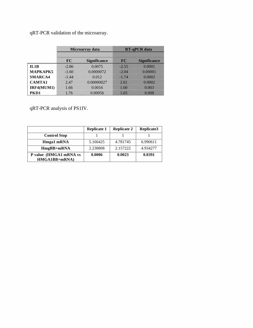
qRT-PCR validation of the microarray. Microarray data RT-qPCR data FC Significance FC Significance IL1B -2.86 0.0075 -2.55 0.0001 MAPKAPK5 -1.60 0.0000072 -2.04 0.00001 SMARCA4 -1.44 0.012 -1.74 0.0002 CAMTA1 2.47 0.00000027 2.61 0.0002 IRF4(MUM1) 1.66 0.0016 1.60 0.003 PKD1 1.76 0.00056 1.65 0.008 qRT-PCR analysis of PS1IV.
Replicate 1 Replicate 2 Replicate3 Control Stop 1 1 1
Hmga1 mRNA 5.166425 4.781745 6.990611 HmgBB+mRNA 2.238808 2.157222 4.934277
P value (HMGA1 mRNA vs HMGA1BB+mRNA)
0.0006 0.0023 0.0391

Supplementary Data File 6
The sequence of cDNA from the alternatively spliced transcript of zebrafish psen1.
GTTCCGATGGCTGATTTAGTGCAGAATGCTGCCAATAATGTG|GATCTACACCCCGTTTCGTGAGGACACGGAGACGGTGGGTCAGCGAGCTCTGCACTCCATGCTCAACGCCATCATCATGATCAGTGTGATCGTGGTCATGACCCTGGTGCTGGTGGTGCTCTACAAGTACAGATGCTACAAG|GTGATTCAAGCCTGGCTGTTCTTCTCCAACCTCCTCCTGCTCTTCTTTTTCTCCTTAATTTATTTGGG
Exon 3 Exon 5
Exon 6
Stop codon Start codon

188
Discussion
In chapter II of this thesis, a novel assay has been developed that allows for the in vivo
analysis of γ-secretase activity. This is the first assay of its kind in zebrafish. This assay
has the ability to be used to investigate a wide range of γ-secretase modulating events,
including measuring the inhibition of AβPP cleavage by newly developed drugs and
analyzing the effect that PSEN mutations have on γ-secretase processing. We have
effectively used this assay to show that both Psen1 and Psen2 proteins contribute to γ-
secretase cleavage of the Appa (a paralogue of human AβPP) substrate in zebrafish. This
is consistent with the observations of Yonemura et al on the roles of human PSEN1 and
PSEN2 in the cleavage of AβPP [194]. This assay was utilized in Chapter III to show
that various truncations of human PSEN1 (or zebrafish Psen1) protein have starkly
differential effects on Notch signalling and cleavage of Appa. Different truncations can
suppress or stimulate Notch signalling but not Appa cleavage and vice versa. This is
significant as an aberrant transcript resulting from the G183V PSEN mutation in Picks
disease suppresses cleavage of Appa but not Notch signalling. However, in contrast, the
truncated protein potentially produced by the P242LfsX11 acne inversa mutation has no
effect on Appa cleavage and unexpectedly enhances Notch signalling. Wang et al [42]
suggest that the FAI phenotype is due to haploinsufficiency for genes encoding
components of the γ -secretase complex – i.e. a loss of function phenotype. Our result
suggests that the effects of these mutations, or at least those of the P242LfsX11 mutation
of PSEN1, may be more complex. This hypothesis has been explored further in chapter

189
IV where we investigated the evolution of PS2V and its function compared to the
K115Efx10 truncation. We have found that zebrafish possess a PS2V-like isoform,
PS1IV, produced from the Danio rerio PSEN1 orthologous gene rather than its PSEN2
orthologue. We showed that the molecular mechanism controlling formation of
PS2V/PS1IV and the ability to stimulate γ-secretase activity and suppress the unfolded
protein response is conserved. The K115Efx10 FAD mutation is similar to PS2V in its
ability to increase Notch signalling and γ-secretase cleavage of zebrafish Appa. This
supports an increase in Aβ peptide production as a molecular common link between
K115Efx10 FAD and sporadic late onset AD.
Each chapter has its own discussion and conclusion that goes into the individual findings
in detail. Those discussions will not be reiterated here. I will however, describe some
concerns and address some of the issues that have been highlighted during the peer
review of the papers, in particular chapter III.
A common criticism of research utilizing zebrafish is its appropriateness as a model of
human AD. In disease states such as AD, genes encoding truncated PRESENILINs are
found concomitantly with wild-type PRESENILIN genes. However, previous analyses of
truncated PRESENILINs have examined their biochemical activities in backgrounds
lacking full-length proteins. This is explained in greater detail on pg 85 in chapter III.
However it is worth reiterating that differences between the results obtained in chapters
III and IV for the activity of Presenilin truncations in AβPP cleavage and Notch
signalling compared to previous work by others in cell free systems are probably because

190
our analysis has examined the expression of truncated PRESENILINs in the presence of
endogenous gene expression. This highlights the importance of examining dominant
mutations in their real, heterozygous state - in the presence of a normal allele – rather
than in isolation since the activity of the dominant mutation can be dependent on the
function of the normal allele.
It has been suggested (by one reviewer) that the injection of synthetic mRNA into
zebrafish embryos is in many ways a less controlled experiment than transient or stable
overexpression of PSEN in human cell lines. However, experiments involving genes
transfected into cultured cells involve much higher levels of expression than are
physiologically normal. The cells themselves can display aberrant patterns of gene
expression due to the fact that they are not in their normal three-dimensional environment
of cell-cell contacts and signalling. If the cells are from an immortalised line then further
abnormalities of gene expression must exist. One of the great advantages of work with
zebrafish embryos is the ability to, in a very controlled fashion, simultaneously force and
suppress (or otherwise manipulate) the expression of multiple genes. The experiments in
chapters III and IV are based on this. The internal consistency of the data supports the
validity of this approach. The advantages of our approach relative to cell-free assays and
cell transfection are described in the text of the manuscripts.
To further support the physiological relevance of our data we made an effort to monitor
and measure non-endogenous gene expression. In chapters II-IV of this thesis,
morpholino antisense oligonucleotides have been used that interfere with the splicing of
the endogenous psen to generate truncated proteins. It has been previously shown that the

191
levels of interference with the endogenous transcripts are, in most cases, minor but can
still cause strong dominant phenotypes [329]. Since it is the endogenous transcript that is
being manipulated the levels of expression of the truncated protein must be
physiologically relevant. Quantitative PCR has also been performed to compare the levels
of psen1 mRNA with and without injection of one of the deletion constructs. The results
show that levels of total mRNA are ~3 fold higher in the mRNA-injected embryos. This
demonstrates that injected mRNAs are at levels comparable to the endogenous mRNA.
In chapter III and IV we have used different techniques to measure AβPP cleavage and
Notch signalling in zebrafish. The assay for Notch signalling relies on the measurement
of neurogenin 1 transcripts by in situ hybridization, which are negatively regulated by
Notch signaling. In contrast, AβPP cleavage is measured after overexpression of a
truncated and GFP-tagged AβPP construct by Western blotting as developed in Chapter
II. It was suggested by one reviewer that to accurately compare the effects of an
intervention/treatment on AβPP and Notch signalling it is absolutely critical to use assays
that rely on the same methodology, e.g., measurement of proteolytic release of
endogenous AICD/NICD domains by Western blotting. The reviewer citeed that this has
been illustrated by the misleading marketing of so-called “NOTCH-sparing” γ-secretase
inhibitors like BMS-708163. This inhibitor was initially described to offer a 200-fold
window between the inhibition of NOTCH and AβPP cleavage. These results were
obtained with a reporter-based transcriptional assay to measure NOTCH activity and
measurement of AβPP cleavage by quantification of an AβPP proteolytic product.
However, the reviewer cited recent studies using proteolytic assays for both AβPP and

192
NOTCH cleavage that show that this window does not exist and that this inhibitor might
even be more selective for Notch [345, 346]. The reviewer suggested that the claims in
chapters III and IV that some PSEN splice variants differentially affected AβPP and
NOTCH cleavage are not valid and could be entirely due to the different methodologies
of the assays. However these comments above are based on views founded in, and framed
by, cell-free assays of γ-secretase activity. As discussed in Chapter III (pg. 84) as well as
the introduction to this thesis (pg. 36), there are serious questions over the relevance of
these assays for Alzheimer’s disease. One should not judge the relevance of an in vivo
assay by the standards of an in vitro assay for which γ-secretase activity is examined
under conditions that are far from normal. While it is true that the AβPP and Notch
assays utilised in this thesis have different methodological bases, it is highly unlikely that
the observations of differences in the effects on AβPP cleavage and Notch signalling of
different truncations are artefacts of the procedures themselves. If this were true, one
would not expect that some truncations produce increases in both AβPP cleavage and
Notch signalling while others produce differences. To support the validity of our assays
we subsequently performed the experiments showing that the Psen1Δ>4 truncation
requires the presence of endogenous Psen1 in order to stimulate both the γ-secretase
cleavage of Appa and Notch signalling.

193
Further Directions
Development of the Appa γ-secretase assay
Chapter II describes the development of an assay for the cleavage of a modified Appa
protein in zebrafish to monitor of γ-secretase activity. This assay is effective and forms a
prototype for in vivo assays of γ-secretase cleavage of the many other substrates of this
enzyme activity. However, it also has scope for improvement. As noted in Chapter II the
products of γ-secretase cleavage of all of the variant Appa substrates (in effect derivatives
of C100 fused to dGFP) proved too unstable to detect by our Western blotting procedure.
This is not unexpected since AICD has very rapid turnover and a halflife that is
potentially as short as 5 min [252]. Indeed, one difficulty in determining a physiological
role for AICD has been its instability. Furthermore, the Appa::C86* substrate was
coupled to an exceptionally destablised GFP tag. It would be ideal to have an assay where
one could measure the ratio of cleaved to uncleaved substrate. This might be achieved by
having a more stable form of GFP tagged to the Appa substrate. This form of GFP would
have greater perdurance, possibly allowing visualisation of the cleavage fragment. The
current assay requires the careful co-injection of equal concentrations and volumes of a
standard together with the assay construct. It would be beneficial to include an expression
standard that is automatically stoichiometrically co-expressed with the assay construct.
This could be achieved by having a mCherry tag linked to the assay substrate via a viral
2a sequence (that causes translation of two separate peptides from one open reading
frame in a mRNA). This would allow for the co-expression of mCherry with the

194
Appa::dGFP fusion protein in a 1:1 ratio and would make comparison of the results of
multiple injections simpler and more accurate. The ability to perform in vivo, high
throughput, simple analyses requiring only a moderate degree of technical proficiency
would increase our ability to investigate PRESENILIN activity and γ-secretase cleavage
events.
The development of γ-secretase assays utilizing other substrates
While the study of AβPP proteolytic cleavage is important for AD research and a well-
understood mechanism for the analysis γ-secretase activity, it is not provide a complete
picture of γ -secretase. The substrate specificity and regulation of γ -secretase cleavage
events, as outline in the introduction of this thesis, remain poorly understood. By
investigating the processing of other substrates we could garner insight into what
substrate characteristics are critical for recruitment to γ-secretase processing.
The p75NTR and NRH1 transmembrane proteins share high sequence similarity in their
transmembrane and intracellular (ICD) domains. However, Kanning et al (2003)
observed that while p75NTR is a substrate of γ-secretase, NRH1 is not, despite the two
proteins sharing the same sequence at the location of γ-secretase cleavage sites [347]. As
yet the p75NTR and NRH1 orthologs have not both been defined in Danio rerio. this
could be done by utilising protein database searches, BLAST analyses and genomic
alignments of homologous sequences. Then, assay constructs for these proteins could be
developed (in a fashion similar to development of the Appa::dGFP substrate described in

195
Chapter II) If assays could be developed in which p75NTR cleavage via γ-secretase could
be observed and NRH1 cleavage (not mediated by γ-secretase) could be demonstrated, it
would allow experiments such as amino acid residue swap or domain swap experiments
to be undertaken. This might allow one to determine the essential structural substrate
requirements for cleavage by γ-secretase.
The localisation of PRESENILIN truncations to the Mitochondrial Associated
Membranes
The PSEN1 protein is largely localised to the MAM and PSEN2 resides exclusively in
this structure [17]. In Chapters III and IV of this thesis the diverse effects that different
PSEN truncations have on Notch signaling and AβPP cleavage are shown, but where do
truncations of the PRESENILINs localise in order to exert their dominant effects? Does
differential localisation of the truncations explain differences in their activities? Fusing
GFP to the N-termini of PRESENILIN truncations could enable observation of their sub-
cellular localisation by confocal microscopy. This would show whether PRESENILIN
truncations with different activities are localised to different positions within cells and
may help to explain why different truncations of PRESENILIN proteins can result in very
different diseases. For example, the PS2V truncation seen in sporadic AD may show a
different localisation to the PSEN P242LfsX11 mutation associated with familial acne
inversa (FAI). Alternatively, if they show similar localisations then their different disease
effects would more likely be due to differences in their protein structures and their

196
interactions with binding partners.
To identify the binding partners of truncated PRSENILIN proteins that integrate into
higher molecular weight (HMW) complexes.
As seen in Chapter III it has been shown that truncated forms of PRESENILIN can
incorporate into HMW complexes (some of which are sufficiently stable to survive
SDS-PAGE). Therefore, these complexes could be analyzed in detail using 2-
dimensional blue native PAGE (BN-PAGE). Western blotting to detect their N-
terminal FLAG tags would identify the positions of spots containing truncated forms
of Psen. The protein components of the same spots on identical gels could then be
analysed by proteomic analysis such as MALDI-TOF. Anti-FLAG antibodies could
also be used to immunoprecipitate complexes containing FLAG-tagged, truncated
PRESENILIN followed by 2-dimensional SDS-PAGE and analysis of the protein
composition of spots identified by silver staining.
The failure to detect PRESENILIN truncations in γ-secretase complexes would imply
that these truncations exert their effects on Notch signalling and Appa cleavage by
means other than incorporation. Psen1 truncated after exon 5 sequence appears to
integrate avidly into HMW complexes but does not affect γ-secretase function. The
detection of differences in the components of proteins binding to Psen1 truncated after
exon 5 sequence and forms of Psen1 truncated after exon 3,4,6 and exon 7 sequences
could reveal which interactions are important for modulating γ-secretase activity.

197
References
1. Prince, M., et al., The global prevalence of dementia: a systematic review and metaanalysis. Alzheimer's and dementia : the journal of the Alzheimer's Association, 2013. 9(1): p. 63-75 e2.
2. Wimo, A., et al., The worldwide economic impact of dementia 2010. Alzheimer's and dementia : the journal of the Alzheimer's Association, 2013. 9(1): p. 1-11 e3.
3. Bekris, L.M., et al., Genetics of Alzheimer disease. Journal of geriatric psychiatry and neurology, 2010. 23(4): p. 213-27.
4. Campion, D., et al., Early-onset autosomal dominant Alzheimer disease: prevalence, genetic heterogeneity, and mutation spectrum. American journal of human genetics, 1999. 65(3): p. 664-70.
5. Brickell, K.L., et al., Early-onset Alzheimer disease in families with late-onset Alzheimer disease: a potential important subtype of familial Alzheimer disease. Archives of neurology, 2006. 63(9): p. 1307-11.
6. Hardy, J. and D. Allsop, Amyloid deposition as the central event in the aetiology of Alzheimer's disease. Trends in pharmacological sciences, 1991. 12(10): p. 383-8.
7. Hardy, J. and D.J. Selkoe, The amyloid hypothesis of Alzheimer's disease: progress and problems on the road to therapeutics. Science, 2002. 297(5580): p. 353-6.
8. Hardy, J.A. and G.A. Higgins, Alzheimer's disease: the amyloid cascade hypothesis. Science, 1992. 256(5054): p. 184-5.
9. Selkoe, D.J., The molecular pathology of Alzheimer's disease. Neuron, 1991. 6(4): p. 487-98.
10. Price, D.L. and S.S. Sisodia, Mutant genes in familial Alzheimer's disease and transgenic models. Annual review of neuroscience, 1998. 21: p. 479-505.
11. Alonso Vilatela, M.E., M. Lopez-Lopez, and P. Yescas-Gomez, Genetics of Alzheimer's disease. Archives of medical research, 2012. 43(8): p. 622-31.
12. Suzuki, N., et al., An increased percentage of long amyloid beta protein secreted by familial amyloid beta protein precursor (beta APP717) mutants. Science, 1994. 264(5163): p. 1336-40.
13. Younkin, S.G., Evidence that A beta 42 is the real culprit in Alzheimer's disease. Annals of neurology, 1995. 37(3): p. 287-8.
14. Scheuner, D., et al., Secreted amyloid beta-protein similar to that in the senile plaques of Alzheimer's disease is increased in vivo by the presenilin 1 and 2 and APP mutations linked to familial Alzheimer's disease. Nature medicine, 1996. 2(8): p. 864-70.
15. Bentahir, M., et al., Presenilin clinical mutations can affect gamma-secretase activity by different mechanisms. Journal of neurochemistry, 2006. 96(3): p. 732-42.

198
16. Walsh, D.M. and D.J. Selkoe, A beta oligomers - a decade of discovery. Journal of neurochemistry, 2007. 101(5): p. 1172-84.
17. Glabe, C.C., Amyloid accumulation and pathogensis of Alzheimer's disease: significance of monomeric, oligomeric and fibrillar Abeta. Sub-cellular biochemistry, 2005. 38: p. 167-77.
18. Lesne, S., et al., A specific amyloid-beta protein assembly in the brain impairs memory. Nature, 2006. 440(7082): p. 352-7.
19. Shankar, G.M., et al., Amyloid-beta protein dimers isolated directly from Alzheimer's brains impair synaptic plasticity and memory. Nature medicine, 2008. 14(8): p. 837-42.
20. Ethell, D.W., An amyloid-notch hypothesis for Alzheimer's disease. The Neuroscientist : a review journal bringing neurobiology, neurology and psychiatry, 2010. 16(6): p. 614-7.
21. Hyman, B.T., K. Marzloff, and P.V. Arriagada, The lack of accumulation of senile plaques or amyloid burden in Alzheimer's disease suggests a dynamic balance between amyloid deposition and resolution. Journal of neuropathology and experimental neurology, 1993. 52(6): p. 594-600.
22. McLean, C.A., et al., Soluble pool of Abeta amyloid as a determinant of severity of neurodegeneration in Alzheimer's disease. Annals of neurology, 1999. 46(6): p. 860-6.
23. Terry, R.D., et al., Physical basis of cognitive alterations in Alzheimer's disease: synapse loss is the major correlate of cognitive impairment. Annals of neurology, 1991. 30(4): p. 572-80.
24. Lesne, S., L. Kotilinek, and K.H. Ashe, Plaque-bearing mice with reduced levels of oligomeric amyloid-beta assemblies have intact memory function. Neuroscience, 2008. 151(3): p. 745-9.
25. Nordberg, A., Amyloid plaque imaging in vivo: current achievement and future prospects. European journal of nuclear medicine and molecular imaging, 2008. 35 Suppl 1: p. S46-50.
26. Villemagne, V.L., et al., The ART of loss: Abeta imaging in the evaluation of Alzheimer's disease and other dementias. Molecular neurobiology, 2008. 38(1): p. 1-15.
27. Caughey, B. and P.T. Lansbury, Protofibrils, pores, fibrils, and neurodegeneration: separating the responsible protein aggregates from the innocent bystanders. Annual review of neuroscience, 2003. 26: p. 267-98.
28. Turner, P.R., et al., Roles of amyloid precursor protein and its fragments in regulating neural activity, plasticity and memory. Progress in neurobiology, 2003. 70(1): p. 1-32.
29. Beckett, C., et al., Nuclear signalling by membrane protein intracellular domains: the AICD enigma. Cellular signalling, 2012. 24(2): p. 402-9.
30. Gerald, Z. and W. Ockert, Alzheimer's disease market: hope deferred. Nature reviews. Drug discovery, 2013. 12(1): p. 19-20.
31. Assal, F. and M. van der Meulen, Pharmacological interventions in primary care: hopes and illusions. Frontiers of neurology and neuroscience, 2009. 24: p. 54-65.
32. Supnet, C. and I. Bezprozvanny, Presenilins function in ER calcium leak and Alzheimer's disease pathogenesis. Cell Calcium, 2011. 50(3): p. 303-9.

199
33. Matsubara-Tsutsui, M., et al., Molecular evidence of presenilin 1 mutation in familial early onset dementia. American journal of medical genetics, 2002. 114(3): p. 292-8.
34. Marjaux, E., D. Hartmann, and B. De Strooper, Presenilins in memory, Alzheimer's disease, and therapy. Neuron, 2004. 42(2): p. 189-92.
35. Korovaitseva, G.I., et al., Presenilin polymorphisms in Alzheimer's disease. Lancet, 1997. 350(9082): p. 959.
36. Newman, M., I.F. Musgrave, and M. Lardelli, Alzheimer disease: amyloidogenesis, the presenilins and animal models. Biochimica et biophysica acta, 2007. 1772(3): p. 285-97.
37. Wolfe, M.S., Processive proteolysis by gamma-secretase and the mechanism of Alzheimer's disease. Biological chemistry, 2012. 393(9): p. 899-905.
38. Sjogren, M. and C. Andersen, Frontotemporal dementia--a brief review. Mechanisms of ageing and development, 2006. 127(2): p. 180-7.
39. Li, D., et al., Mutations of presenilin genes in dilated cardiomyopathy and heart failure. American journal of human genetics, 2006. 79(6): p. 1030-9.
40. Gianni, D., et al., Protein aggregates and novel presenilin gene variants in idiopathic dilated cardiomyopathy. Circulation, 2010. 121(10): p. 1216-26.
41. Li, A., et al., Changes in the expression of the Alzheimer's disease-associated presenilin gene in drosophila heart leads to cardiac dysfunction. Current Alzheimer research, 2011. 8(3): p. 313-22.
42. Wang, B., et al., Gamma-secretase gene mutations in familial acne inversa. Science, 2010. 330(6007): p. 1065.
43. Pink, A.E., et al., Mutations in the gamma-secretase genes NCSTN, PSENEN, and PSEN1 underlie rare forms of hidradenitis suppurativa (acne inversa). The Journal of investigative dermatology, 2012. 132(10): p. 2459-61.
44. Cowburn, R.F., et al., Presenilin-mediated signal transduction. Physiology & behavior, 2007. 92(1-2): p. 93-7.
45. McCarthy, J.V., Involvement of presenilins in cell-survival signalling pathways. Biochemical Society transactions, 2005. 33(Pt 4): p. 568-72.
46. Annaert, W. and B. De Strooper, Presenilins: molecular switches between proteolysis and signal transduction. Trends in neurosciences, 1999. 22(10): p. 439-43.
47. Koo, E.H. and R. Kopan, Potential role of presenilin-regulated signaling pathways in sporadic neurodegeneration. Nature medicine, 2004. 10 Suppl: p. S26-33.
48. Raurell, I., et al., Presenilin-1 interacts with plakoglobin and enhances plakoglobin-Tcf-4 association. Implications for the regulation of beta-catenin/Tcf-4-dependent transcription. The Journal of biological chemistry, 2006. 281(3): p. 1401-11.
49. Baki, L., et al., PS1 activates PI3K thus inhibiting GSK-3 activity and tau overphosphorylation: effects of FAD mutations. The EMBO journal, 2004. 23(13): p. 2586-96.
50. Dewachter, I., et al., Modulation of synaptic plasticity and Tau phosphorylation by wild-type and mutant presenilin1. Neurobiology of aging, 2008. 29(5): p. 639-52.

200
51. Feng, R., et al., Forebrain degeneration and ventricle enlargement caused by double knockout of Alzheimer's presenilin-1 and presenilin-2. Proceedings of the National Academy of Sciences of the United States of America, 2004. 101(21): p. 8162-7.
52. Saura, C.A., et al., Loss of presenilin function causes impairments of memory and synaptic plasticity followed by age-dependent neurodegeneration. Neuron, 2004. 42(1): p. 23-36.
53. Zhu, M., et al., Increased oxidative stress and astrogliosis responses in conditional double-knockout mice of Alzheimer-like presenilin-1 and presenilin-2. Free radical biology and medicine, 2008. 45(10): p. 1493-9.
54. Takahashi, N., et al., Two novel spliced presenilin 2 transcripts in human lymphocyte with oxidant stress and brain. Molecular and cellular biochemistry, 2003. 252(1-2): p. 279-83.
55. LaFerla, F.M., Calcium dyshomeostasis and intracellular signalling in Alzheimer's disease. Nature reviews. Neuroscience, 2002. 3(11): p. 862-72.
56. Mattson, M.P., ER calcium and Alzheimer's disease: in a state of flux. Science signaling, 2010. 3(114): p. pe10.
57. Cook, D.G., et al., Presenilin 1 deficiency alters the activity of voltage-gated Ca2+ channels in cultured cortical neurons. Journal of neurophysiology, 2005. 94(6): p. 4421-9.
58. Leissring, M.A., et al., Capacitative calcium entry deficits and elevated luminal calcium content in mutant presenilin-1 knockin mice. The Journal of cell biology, 2000. 149(4): p. 793-8.
59. Yoo, A.S., et al., Presenilin-mediated modulation of capacitative calcium entry. Neuron, 2000. 27(3): p. 561-72.
60. Green, K.N., et al., SERCA pump activity is physiologically regulated by presenilin and regulates amyloid beta production. The Journal of cell biology, 2008. 181(7): p. 1107-16.
61. Cheung, K.H., et al., Mechanism of Ca2+ disruption in Alzheimer's disease by presenilin regulation of InsP3 receptor channel gating. Neuron, 2008. 58(6): p. 871-83.
62. Stutzmann, G.E., et al., Enhanced ryanodine receptor recruitment contributes to Ca2+ disruptions in young, adult, and aged Alzheimer's disease mice. The Journal of neuroscience : the official journal of the Society for Neuroscience, 2006. 26(19): p. 5180-9.
63. Cheung, K.H., et al., Gain-of-function enhancement of IP3 receptor modal gating by familial Alzheimer's disease-linked presenilin mutants in human cells and mouse neurons. Science signaling, 2010. 3(114): p. ra22.
64. Zatti, G., et al., Presenilin mutations linked to familial Alzheimer's disease reduce endoplasmic reticulum and Golgi apparatus calcium levels. Cell Calcium, 2006. 39(6): p. 539-50.
65. Nelson, O., et al., Familial Alzheimer disease-linked mutations specifically disrupt Ca2+ leak function of presenilin 1. The Journal of clinical investigation, 2007. 117(5): p. 1230-9.
66. Shen, J. and R.J. Kelleher, 3rd, The presenilin hypothesis of Alzheimer's disease: evidence for a loss-of-function pathogenic mechanism. Proceedings of the

201
National Academy of Sciences of the United States of America, 2007. 104(2): p. 403-9.
67. Michno, K., et al., Intracellular calcium deficits in Drosophila cholinergic neurons expressing wild type or FAD-mutant presenilin. PLoS One, 2009. 4(9): p. e6904.
68. Herms, J., et al., Capacitive calcium entry is directly attenuated by mutant presenilin-1, independent of the expression of the amyloid precursor protein. The Journal of biological chemistry, 2003. 278(4): p. 2484-9.
69. Lee, J.H., et al., Lysosomal proteolysis and autophagy require presenilin 1 and are disrupted by Alzheimer-related PS1 mutations. Cell, 2010. 141(7): p. 1146-58.
70. Williamson, W.R. and P.R. Hiesinger, On the role of v-ATPase V0a1-dependent degradation in Alzheimer disease. Communicative & integrative biology, 2010. 3(6): p. 604-7.
71. Neely, K.M., K.N. Green, and F.M. LaFerla, Presenilin is necessary for efficient proteolysis through the autophagy-lysosome system in a gamma-secretase-independent manner. The Journal of neuroscience : the official journal of the Society for Neuroscience, 2011. 31(8): p. 2781-91.
72. Neely, K.M. and K.N. Green, Presenilins mediate efficient proteolysis via the autophagosome-lysosome system. Autophagy, 2011. 7(6): p. 664-5.
73. Sherrington, R., et al., Cloning of a gene bearing missense mutations in early-onset familial Alzheimer's disease. Nature, 1995. 375(6534): p. 754-60.
74. Rogaev, E.I., et al., Familial Alzheimer's disease in kindreds with missense mutations in a gene on chromosome 1 related to the Alzheimer's disease type 3 gene. Nature, 1995. 376(6543): p. 775-8.
75. Lee, M.K., et al., Expression of presenilin 1 and 2 (PS1 and PS2) in human and murine tissues. The Journal of neuroscience : the official journal of the Society for Neuroscience, 1996. 16(23): p. 7513-25.
76. Benkovic, S.A., et al., Regional and cellular localization of presenilin-2 RNA in rat and human brain. Experimental neurology, 1997. 145(2 Pt 1): p. 555-64.
77. Berezovska, O., et al., Developmental regulation of presenilin mRNA expression parallels notch expression. Journal of neuropathology and experimental neurology, 1997. 56(1): p. 40-4.
78. Berezovska, O., M.Q. Xia, and B.T. Hyman, Notch is expressed in adult brain, is coexpressed with presenilin-1, and is altered in Alzheimer disease. Journal of neuropathology and experimental neurology, 1998. 57(8): p. 738-45.
79. Lah, J.J., et al., Light and electron microscopic localization of presenilin-1 in primate brain. The Journal of neuroscience : the official journal of the Society for Neuroscience, 1997. 17(6): p. 1971-80.
80. Moussaoui, S., et al., Immunohistochemical analysis of presenilin-1 expression in the mouse brain. FEBS letters, 1996. 383(3): p. 219-22.
81. Blanchard, V., et al., Immunohistochemical analysis of presenilin 2 expression in the mouse brain: distribution pattern and co-localization with presenilin 1 protein. Brain research, 1997. 758(1-2): p. 209-17.

202
82. Kumar, A. and M.K. Thakur, Presenilin 1 and 2 are expressed differentially in the cerebral cortex of mice during development. Neurochemistry international, 2012. 61(5): p. 778-82.
83. Culvenor, J.G., et al., Presenilin 2 expression in neuronal cells: induction during differentiation of embryonic carcinoma cells. Experimental cell research, 2000. 255(2): p. 192-206.
84. Thakur, M.K. and S. Ghosh, Age and sex dependent alteration in presenilin expression in mouse cerebral cortex. Cellular and molecular neurobiology, 2007. 27(8): p. 1059-67.
85. Annaert, W.G., et al., Presenilin 1 controls gamma-secretase processing of amyloid precursor protein in pre-golgi compartments of hippocampal neurons. The Journal of cell biology, 1999. 147(2): p. 277-94.
86. Siman, R. and J. Velji, Localization of presenilin-nicastrin complexes and gamma-secretase activity to the trans-Golgi network. Journal of neurochemistry, 2003. 84(5): p. 1143-53.
87. Kimura, N., et al., Age-related changes in the localization of presenilin-1 in cynomolgus monkey brain. Brain research, 2001. 922(1): p. 30-41.
88. Vetrivel, K.S., et al., Association of gamma-secretase with lipid rafts in post-Golgi and endosome membranes. The Journal of biological chemistry, 2004. 279(43): p. 44945-54.
89. Pasternak, S.H., et al., Presenilin-1, nicastrin, amyloid precursor protein, and gamma-secretase activity are co-localized in the lysosomal membrane. The Journal of biological chemistry, 2003. 278(29): p. 26687-94.
90. Ankarcrona, M. and K. Hultenby, Presenilin-1 is located in rat mitochondria. Biochemical and biophysical research communications, 2002. 295(3): p. 766-70.
91. Li, J., et al., Alzheimer presenilins in the nuclear membrane, interphase kinetochores, and centrosomes suggest a role in chromosome segregation. Cell, 1997. 90(5): p. 917-27.
92. Area-Gomez, E., et al., Presenilins are enriched in endoplasmic reticulum membranes associated with mitochondria. The American journal of pathology, 2009. 175(5): p. 1810-6.
93. Laudon, H., et al., A nine-transmembrane domain topology for presenilin 1. The Journal of biological chemistry, 2005. 280(42): p. 35352-60.
94. Spasic, D., et al., Presenilin-1 maintains a nine-transmembrane topology throughout the secretory pathway. The Journal of biological chemistry, 2006. 281(36): p. 26569-77.
95. Wolfe, M.S., et al., Two transmembrane aspartates in presenilin-1 required for presenilin endoproteolysis and gamma-secretase activity. Nature, 1999. 398(6727): p. 513-7.
96. Fukumori, A., et al., Three-amino acid spacing of presenilin endoproteolysis suggests a general stepwise cleavage of gamma-secretase-mediated intramembrane proteolysis. The Journal of neuroscience : the official journal of the Society for Neuroscience, 2010. 30(23): p. 7853-62.
97. De Strooper, B., T. Iwatsubo, and M.S. Wolfe, Presenilins and gamma-Secretase: Structure, Function, and Role in Alzheimer Disease. Cold Spring Harbor perspectives in medicine, 2012. 2(1): p. a006304.

203
98. Sobhanifar, S., et al., Structural investigation of the C-terminal catalytic fragment of presenilin 1. Proceedings of the National Academy of Sciences of the United States of America, 2010. 107(21): p. 9644-9.
99. Kim, T.W., et al., Endoproteolytic cleavage and proteasomal degradation of presenilin 2 in transfected cells. The Journal of biological chemistry, 1997. 272(17): p. 11006-10.
100. Ratovitski, T., et al., Endoproteolytic processing and stabilization of wild-type and mutant presenilin. The Journal of biological chemistry, 1997. 272(39): p. 24536-41.
101. Brunkan, A.L. and A.M. Goate, Presenilin function and gamma-secretase activity. Journal of neurochemistry, 2005. 93(4): p. 769-92.
102. Sato, N., et al., A novel presenilin-2 splice variant in human Alzheimer's disease brain tissue. Journal of Neurochemistry, 1999. 72(6): p. 2498-505.
103. Sato, N., et al., Increased production of beta-amyloid and vulnerability to endoplasmic reticulum stress by an aberrant spliced form of presenilin 2. The Journal of Biological Chemistry, 2001. 276(3): p. 2108-14.
104. Manabe, T., et al., Induced HMGA1a expression causes aberrant splicing of Presenilin-2 pre-mRNA in sporadic Alzheimer's disease. Cell Death Differ, 2003. 10(6): p. 698-708.
105. Ohe, K. and A. Mayeda, HMGA1a trapping of U1 snRNP at an authentic 5' splice site induces aberrant exon skipping in sporadic Alzheimer's disease. Molecular Cell Biology, 2010. 30(9): p. 2220-8.
106. Manabe, T., et al., HMGA1a: sequence-specific RNA-binding factor causing sporadic Alzheimer's disease-linked exon skipping of presenilin-2 pre-mRNA. Genes to Cells, 2007. 12(10): p. 1179-91.
107. Smith, M.J., et al., Expression of truncated presenilin 2 splice variant in Alzheimer's disease, bipolar disorder, and schizophrenia brain cortex. Brain Research Molecular Brain Research, 2004. 127(1-2): p. 128-35.
108. Katayama, T., et al., Induction of neuronal death by ER stress in Alzheimer's disease. The Journal of Chemical Neuroanatomy, 2004. 28(1-2): p. 67-78.
109. Nishikawa, A., et al., Novel function of PS2V: change in conformation of tau proteins. Biochem Biophys Res Commun, 2004. 318(2): p. 435-8.
110. Parks, A.L. and D. Curtis, Presenilin diversifies its portfolio. Trends in genetics : TIG, 2007. 23(3): p. 140-50.
111. Huppert, S.S., et al., Analysis of Notch function in presomitic mesoderm suggests a gamma-secretase-independent role for presenilins in somite differentiation. Developmental cell, 2005. 8(5): p. 677-88.
112. Thinakaran, G. and A.T. Parent, Identification of the role of presenilins beyond Alzheimer's disease. Pharmacological research : the official journal of the Italian Pharmacological Society, 2004. 50(4): p. 411-8.
113. Hass, M.R., et al., Presenilin: RIP and beyond. Seminars in Cellular and Developmental Biology, 2009. 20(2): p. 201-10.
114. Leissring, M.A., I. Parker, and F.M. LaFerla, Presenilin-2 mutations modulate amplitude and kinetics of inositol 1, 4,5-trisphosphate-mediated calcium signals. The Journal of biological chemistry, 1999. 274(46): p. 32535-8.

204
115. Tu, H., et al., Presenilins form ER Ca2+ leak channels, a function disrupted by familial Alzheimer's disease-linked mutations. Cell, 2006. 126(5): p. 981-93.
116. Chan, S.L., et al., Presenilin-1 mutations increase levels of ryanodine receptors and calcium release in PC12 cells and cortical neurons. The Journal of biological chemistry, 2000. 275(24): p. 18195-200.
117. Leissring, M.A., et al., Alzheimer's presenilin-1 mutation potentiates inositol 1,4,5-trisphosphate-mediated calcium signaling in Xenopus oocytes. Journal of neurochemistry, 1999. 72(3): p. 1061-8.
118. Zhang, Z., et al., Destabilization of beta-catenin by mutations in presenilin-1 potentiates neuronal apoptosis. Nature, 1998. 395(6703): p. 698-702.
119. Kang, D.E., et al., Presenilin couples the paired phosphorylation of beta-catenin independent of axin: implications for beta-catenin activation in tumorigenesis. Cell, 2002. 110(6): p. 751-62.
120. De Strooper, B. and W. Annaert, Where Notch and Wnt signaling meet. The presenilin hub. The Journal of cell biology, 2001. 152(4): p. F17-20.
121. Marambaud, P., et al., A presenilin-1/gamma-secretase cleavage releases the E-cadherin intracellular domain and regulates disassembly of adherens junctions. The EMBO journal, 2002. 21(8): p. 1948-56.
122. Serban, G., et al., Cadherins mediate both the association between PS1 and beta-catenin and the effects of PS1 on beta-catenin stability. The Journal of biological chemistry, 2005. 280(43): p. 36007-12.
123. McCarthy, J.V., C. Twomey, and P. Wujek, Presenilin-dependent regulated intramembrane proteolysis and gamma-secretase activity. Cellular and molecular life sciences : CMLS, 2009. 66(9): p. 1534-55.
124. Haapasalo, A. and D.M. Kovacs, The many substrates of presenilin/gamma-secretase. Journal of Alzheimer's disease : JAD, 2011. 25(1): p. 3-28.
125. Hemming, M.L., et al., Proteomic profiling of gamma-secretase substrates and mapping of substrate requirements. PLoS biology, 2008. 6(10): p. e257.
126. Sato, T., et al., Active gamma-secretase complexes contain only one of each component. The Journal of biological chemistry, 2007. 282(47): p. 33985-93.
127. Edbauer, D., et al., Reconstitution of gamma-secretase activity. Nature cell biology, 2003. 5(5): p. 486-8.
128. De Strooper, B., Aph-1, Pen-2, and Nicastrin with Presenilin generate an active gamma-Secretase complex. Neuron, 2003. 38(1): p. 9-12.
129. The structure of the presenilin 1 (S182) gene and identification of six novel mutations in early onset AD families. Alzheimer's Disease Collaborative Group. Nature genetics, 1995. 11(2): p. 219-22.
130. Gu, Y., et al., APH-1 interacts with mature and immature forms of presenilins and nicastrin and may play a role in maturation of presenilin.nicastrin complexes. The Journal of biological chemistry, 2003. 278(9): p. 7374-80.
131. Shirotani, K., et al., Identification of distinct gamma-secretase complexes with different APH-1 variants. The Journal of biological chemistry, 2004. 279(40): p. 41340-5.
132. Serneels, L., et al., Differential contribution of the three Aph1 genes to gamma-secretase activity in vivo. Proceedings of the National Academy of Sciences of the United States of America, 2005. 102(5): p. 1719-24.

205
133. Serneels, L., et al., gamma-Secretase heterogeneity in the Aph1 subunit: relevance for Alzheimer's disease. Science, 2009. 324(5927): p. 639-42.
134. Fortna, R.R., et al., Membrane topology and nicastrin-enhanced endoproteolysis of APH-1, a component of the gamma-secretase complex. The Journal of biological chemistry, 2004. 279(5): p. 3685-93.
135. Hebert, S.S., et al., Coordinated and widespread expression of gamma-secretase in vivo: evidence for size and molecular heterogeneity. Neurobiology of disease, 2004. 17(2): p. 260-72.
136. Ma, G., et al., APH-1a is the principal mammalian APH-1 isoform present in gamma-secretase complexes during embryonic development. The Journal of neuroscience : the official journal of the Society for Neuroscience, 2005. 25(1): p. 192-8.
137. Dejaegere, T., et al., Deficiency of Aph1B/C-gamma-secretase disturbs Nrg1 cleavage and sensorimotor gating that can be reversed with antipsychotic treatment. Proceedings of the National Academy of Sciences of the United States of America, 2008. 105(28): p. 9775-80.
138. Kleiger, G., et al., GXXXG and AXXXA: common alpha-helical interaction motifs in proteins, particularly in extremophiles. Biochemistry, 2002. 41(19): p. 5990-7.
139. Lee, S.F., et al., A conserved GXXXG motif in APH-1 is critical for assembly and activity of the gamma-secretase complex. The Journal of biological chemistry, 2004. 279(6): p. 4144-52.
140. Pardossi-Piquard, R., et al., APH1 polar transmembrane residues regulate the assembly and activity of presenilin complexes. The Journal of biological chemistry, 2009. 284(24): p. 16298-307.
141. Chen, A.C., et al., Aph-1 associates directly with full-length and C-terminal fragments of gamma-secretase substrates. The Journal of biological chemistry, 2010. 285(15): p. 11378-91.
142. Yu, G., et al., Nicastrin modulates presenilin-mediated notch/glp-1 signal transduction and betaAPP processing. Nature, 2000. 407(6800): p. 48-54.
143. Shah, S., et al., Nicastrin functions as a gamma-secretase-substrate receptor. Cell, 2005. 122(3): p. 435-47.
144. Fagan, R., et al., Nicastrin, a presenilin-interacting protein, contains an aminopeptidase/transferrin receptor superfamily domain. Trends in biochemical sciences, 2001. 26(4): p. 213-4.
145. Chen, F., et al., Nicastrin binds to membrane-tethered Notch. Nature cell biology, 2001. 3(8): p. 751-4.
146. Morais, V.A., et al., Cellular localization of Nicastrin affects amyloid beta species production. FEBS letters, 2008. 582(3): p. 427-33.
147. Dries, D.R., et al., Glu-333 of nicastrin directly participates in gamma-secretase activity. The Journal of biological chemistry, 2009. 284(43): p. 29714-24.
148. Chavez-Gutierrez, L., et al., Glu(332) in the Nicastrin ectodomain is essential for gamma-secretase complex maturation but not for its activity. J Biol Chem, 2008. 283(29): p. 20096-105.
149. Futai, E., S. Yagishita, and S. Ishiura, Nicastrin is dispensable for gamma-secretase protease activity in the presence of specific presenilin mutations. The Journal of biological chemistry, 2009. 284(19): p. 13013-22.

206
150. Zhao, G., et al., Gamma-secretase composed of PS1/Pen2/Aph1a can cleave notch and amyloid precursor protein in the absence of nicastrin. The Journal of neuroscience : the official journal of the Society for Neuroscience, 2010. 30(5): p. 1648-56.
151. Hayashi, I., et al., Neutralization of the gamma-secretase activity by monoclonal antibody against extracellular domain of nicastrin. Oncogene, 2012. 31(6): p. 787-98.
152. Pamren, A., et al., Mutations in nicastrin protein differentially affect amyloid beta-peptide production and Notch protein processing. The Journal of biological chemistry, 2011. 286(36): p. 31153-8.
153. Crystal, A.S., et al., Membrane topology of gamma-secretase component PEN-2. The Journal of biological chemistry, 2003. 278(22): p. 20117-23.
154. Steiner, H., et al., PEN-2 is an integral component of the gamma-secretase complex required for coordinated expression of presenilin and nicastrin. The Journal of biological chemistry, 2002. 277(42): p. 39062-5.
155. Prokop, S., et al., Requirement of PEN-2 for stabilization of the presenilin N-/C-terminal fragment heterodimer within the gamma-secretase complex. The Journal of biological chemistry, 2004. 279(22): p. 23255-61.
156. Chiang, P.M., et al., Specific domains in anterior pharynx-defective 1 determine its intramembrane interactions with nicastrin and presenilin. Neurobiology of aging, 2012. 33(2): p. 277-85.
157. Shirotani, K., et al., Immature nicastrin stabilizes APH-1 independent of PEN-2 and presenilin: identification of nicastrin mutants that selectively interact with APH-1. Journal of neurochemistry, 2004. 89(6): p. 1520-7.
158. Arawaka, S., et al., The levels of mature glycosylated nicastrin are regulated and correlate with gamma-secretase processing of amyloid beta-precursor protein. Journal of neurochemistry, 2002. 83(5): p. 1065-71.
159. Yang, D.S., et al., Mature glycosylation and trafficking of nicastrin modulate its binding to presenilins. The Journal of biological chemistry, 2002. 277(31): p. 28135-42.
160. Kaether, C., et al., Endoplasmic reticulum retention of the gamma-secretase complex component Pen2 by Rer1. EMBO reports, 2007. 8(8): p. 743-8.
161. Spasic, D., et al., Rer1p competes with APH-1 for binding to nicastrin and regulates gamma-secretase complex assembly in the early secretory pathway. The Journal of cell biology, 2007. 176(5): p. 629-40.
162. Kaether, C., et al., The presenilin C-terminus is required for ER-retention, nicastrin-binding and gamma-secretase activity. The EMBO journal, 2004. 23(24): p. 4738-48.
163. Capell, A., et al., Gamma-secretase complex assembly within the early secretory pathway. The Journal of biological chemistry, 2005. 280(8): p. 6471-8.
164. Kim, S.H. and S.S. Sisodia, Evidence that the "NF" motif in transmembrane domain 4 of presenilin 1 is critical for binding with PEN-2. The Journal of biological chemistry, 2005. 280(51): p. 41953-66.
165. Prokop, S., C. Haass, and H. Steiner, Length and overall sequence of the PEN-2 C-terminal domain determines its function in the stabilization of presenilin fragments. Journal of neurochemistry, 2005. 94(1): p. 57-62.

207
166. Kim, S.H. and S.S. Sisodia, A sequence within the first transmembrane domain of PEN-2 is critical for PEN-2-mediated endoproteolysis of presenilin 1. The Journal of biological chemistry, 2005. 280(3): p. 1992-2001.
167. Tolia, A., L. Chavez-Gutierrez, and B. De Strooper, Contribution of presenilin transmembrane domains 6 and 7 to a water-containing cavity in the gamma-secretase complex. The Journal of biological chemistry, 2006. 281(37): p. 27633-42.
168. Zhou, S., et al., Regulation of gamma-secretase activity in Alzheimer's disease. Biochemistry, 2007. 46(10): p. 2553-63.
169. Zhou, S., et al., CD147 is a regulatory subunit of the gamma-secretase complex in Alzheimer's disease amyloid beta-peptide production. Proceedings of the National Academy of Sciences U S A, 2005. 102(21): p. 7499-504.
170. Takashima, A., et al., Presenilin 1 associates with glycogen synthase kinase-3beta and its substrate tau. Proceedings of the National Academy of Sciences U S A, 1998. 95(16): p. 9637-41.
171. Kirschenbaum, F., et al., Glycogen synthase kinase-3beta regulates presenilin 1 C-terminal fragment levels. Journal of Biological Chemistry, 2001. 276(33): p. 30701-7.
172. Klein, J., Functions and pathophysiological roles of phospholipase D in the brain. Journal of Neurochemistry, 2005. 94(6): p. 1473-87.
173. Cai, D., et al., Presenilin-1 regulates intracellular trafficking and cell surface delivery of beta-amyloid precursor protein. Journal of Biological Chemistry, 2003. 278(5): p. 3446-54.
174. Dolcini, V., et al., TMP21 regulates Abeta production but does not affect caspase-3, p53, and neprilysin. Biochemical and Biophysical Research Communications, 2008. 371(1): p. 69-74.
175. Liao, Y.F., et al., Tumor necrosis factor-alpha, interleukin-1beta, and interferon-gamma stimulate gamma-secretase-mediated cleavage of amyloid precursor protein through a JNK-dependent MAPK pathway. Journal of Biological Chemistry, 2004. 279(47): p. 49523-32.
176. Blasko, I., et al., TNFalpha plus IFNgamma induce the production of Alzheimer beta-amyloid peptides and decrease the secretion of APPs. Faseb Journal, 1999. 13(1): p. 63-8.
177. Blasko, I., et al., Costimulatory effects of interferon-gamma and interleukin-1beta or tumor necrosis factor alpha on the synthesis of Abeta1-40 and Abeta1-42 by human astrocytes. Neurobiology of Disease, 2000. 7(6 Pt B): p. 682-9.
178. Gianni, D., et al., Platelet-derived growth factor induces the beta-gamma-secretase-mediated cleavage of Alzheimer's amyloid precursor protein through a Src-Rac-dependent pathway. The Journal of biological chemistry, 2003. 278(11): p. 9290-7.
179. Shirotani, K., et al., Gamma-secretase activity is associated with a conformational change of nicastrin. The Journal of biological chemistry, 2003. 278(19): p. 16474-7.
180. Sato, C., et al., Structure of the catalytic pore of gamma-secretase probed by the accessibility of substituted cysteines. The Journal of neuroscience : the official journal of the Society for Neuroscience, 2006. 26(46): p. 12081-8.

208
181. Herreman, A., et al., Total inactivation of gamma-secretase activity in presenilin-deficient embryonic stem cells. Nature cell biology, 2000. 2(7): p. 461-2.
182. Herreman, A., et al., Presenilin 2 deficiency causes a mild pulmonary phenotype and no changes in amyloid precursor protein processing but enhances the embryonic lethal phenotype of presenilin 1 deficiency. Proceedings of the National Academy of Sciences of the United States of America, 1999. 96(21): p. 11872-7.
183. Zhang, Z., et al., Presenilins are required for gamma-secretase cleavage of beta-APP and transmembrane cleavage of Notch-1. Nature cell biology, 2000. 2(7): p. 463-5.
184. Donoviel, D.B., et al., Mice lacking both presenilin genes exhibit early embryonic patterning defects. Genes & development, 1999. 13(21): p. 2801-10.
185. Li, Y.M., et al., Photoactivated gamma-secretase inhibitors directed to the active site covalently label presenilin 1. Nature, 2000. 405(6787): p. 689-94.
186. Jayadev, S., et al., Alzheimer's disease phenotypes and genotypes associated with mutations in presenilin 2. Brain : a journal of neurology, 2010. 133(Pt 4): p. 1143-54.
187. Zhao, B., et al., Identification of gamma-secretase inhibitor potency determinants on presenilin. The Journal of biological chemistry, 2008. 283(5): p. 2927-38.
188. Lee, J., et al., Identification of presenilin 1-selective gamma-secretase inhibitors with reconstituted gamma-secretase complexes. Biochemistry, 2011. 50(22): p. 4973-80.
189. Shen, J., et al., Skeletal and CNS defects in Presenilin-1-deficient mice. Cell, 1997. 89(4): p. 629-39.
190. Wong, P.C., et al., Presenilin 1 is required for Notch1 and DII1 expression in the paraxial mesoderm. Nature, 1997. 387(6630): p. 288-92.
191. De Strooper, B., et al., Deficiency of presenilin-1 inhibits the normal cleavage of amyloid precursor protein. Nature, 1998. 391(6665): p. 387-90.
192. Franberg, J., et al., Minor contribution of presenilin 2 for gamma-secretase activity in mouse embryonic fibroblasts and adult mouse brain. Biochemical and biophysical research communications, 2011. 404(1): p. 564-8.
193. Lai, M.T., et al., Presenilin-1 and presenilin-2 exhibit distinct yet overlapping gamma-secretase activities. The Journal of biological chemistry, 2003. 278(25): p. 22475-81.
194. Yonemura, Y., et al., Comparison of presenilin 1 and presenilin 2 gamma-secretase activities using a yeast reconstitution system. The Journal of biological chemistry, 2011. 286(52): p. 44569-75.
195. Jayadev, S., et al., Presenilin 2 is the predominant gamma-secretase in microglia and modulates cytokine release. PLoS One, 2010. 5(12): p. e15743.
196. Cam, J.A., et al., The low density lipoprotein receptor-related protein 1B retains beta-amyloid precursor protein at the cell surface and reduces amyloid-beta peptide production. The Journal of biological chemistry, 2004. 279(28): p. 29639-46.
197. Kim, D.Y., et al., BACE1 regulates voltage-gated sodium channels and neuronal activity. Nature cell biology, 2007. 9(7): p. 755-64.

209
198. Huovila, A.P., et al., Shedding light on ADAM metalloproteinases. Trends in biochemical sciences, 2005. 30(7): p. 413-22.
199. Schlondorff, J., J.D. Becherer, and C.P. Blobel, Intracellular maturation and localization of the tumour necrosis factor alpha convertase (TACE). The Biochemical journal, 2000. 347 Pt 1: p. 131-8.
200. Jolly-Tornetta, C. and B.A. Wolf, Protein kinase C regulation of intracellular and cell surface amyloid precursor protein (APP) cleavage in CHO695 cells. Biochemistry, 2000. 39(49): p. 15282-90.
201. Skovronsky, D.M., et al., Protein kinase C-dependent alpha-secretase competes with beta-secretase for cleavage of amyloid-beta precursor protein in the trans-golgi network. The Journal of biological chemistry, 2000. 275(4): p. 2568-75.
202. Daugherty, B.L. and S.A. Green, Endosomal sorting of amyloid precursor protein-P-selectin chimeras influences secretase processing. Traffic, 2001. 2(12): p. 908-16.
203. Kinoshita, A., et al., Demonstration by FRET of BACE interaction with the amyloid precursor protein at the cell surface and in early endosomes. Journal of cell science, 2003. 116(Pt 16): p. 3339-46.
204. Stephens, D.J. and B.M. Austen, Metabolites of the beta-amyloid precursor protein generated by beta-secretase localise to the trans-Golgi network and late endosome in 293 cells. Journal of neuroscience research, 1996. 46(2): p. 211-25.
205. Walter, J., et al., Phosphorylation regulates intracellular trafficking of beta-secretase. The Journal of biological chemistry, 2001. 276(18): p. 14634-41.
206. Chyung, J.H. and D.J. Selkoe, Inhibition of receptor-mediated endocytosis demonstrates generation of amyloid beta-protein at the cell surface. The Journal of biological chemistry, 2003. 278(51): p. 51035-43.
207. Kim, S.H., et al., Evidence that assembly of an active gamma-secretase complex occurs in the early compartments of the secretory pathway. The Journal of biological chemistry, 2004. 279(47): p. 48615-9.
208. Hansson, C.A., et al., Nicastrin, presenilin, APH-1, and PEN-2 form active gamma-secretase complexes in mitochondria. The Journal of biological chemistry, 2004. 279(49): p. 51654-60.
209. Jutras, I., et al., Gamma-secretase is a functional component of phagosomes. The Journal of biological chemistry, 2005. 280(43): p. 36310-7.
210. Fukumori, A., et al., Presenilin-dependent gamma-secretase on plasma membrane and endosomes is functionally distinct. Biochemistry, 2006. 45(15): p. 4907-14.
211. Chyung, J.H., D.M. Raper, and D.J. Selkoe, Gamma-secretase exists on the plasma membrane as an intact complex that accepts substrates and effects intramembrane cleavage. The Journal of biological chemistry, 2005. 280(6): p. 4383-92.
212. Kaether, C., et al., Amyloid precursor protein and Notch intracellular domains are generated after transport of their precursors to the cell surface. Traffic, 2006. 7(4): p. 408-15.
213. Vaccari, T., et al., Endosomal entry regulates Notch receptor activation in Drosophila melanogaster. The Journal of cell biology, 2008. 180(4): p. 755-62.

210
214. Gupta-Rossi, N., et al., Monoubiquitination and endocytosis direct gamma-secretase cleavage of activated Notch receptor. The Journal of cell biology, 2004. 166(1): p. 73-83.
215. Vassar, R., et al., Beta-secretase cleavage of Alzheimer's amyloid precursor protein by the transmembrane aspartic protease BACE. Science, 1999. 286(5440): p. 735-41.
216. Zhang, M., et al., Presenilin/gamma-secretase activity regulates protein clearance from the endocytic recycling compartment. FASEB journal : official publication of the Federation of American Societies for Experimental Biology, 2006. 20(8): p. 1176-8.
217. Schon, E.A. and E. Area-Gomez, Is Alzheimer's disease a disorder of mitochondria-associated membranes? Journal of Alzheimer's disease : JAD, 2010. 20 Suppl 2: p. S281-92.
218. Beel, A.J. and C.R. Sanders, Substrate specificity of gamma-secretase and other intramembrane proteases. Cellular and molecular life sciences : CMLS, 2008. 65(9): p. 1311-34.
219. Struhl, G. and A. Adachi, Requirements for presenilin-dependent cleavage of notch and other transmembrane proteins. Molecular cell, 2000. 6(3): p. 625-36.
220. Kopan, R. and M.X. Ilagan, Gamma-secretase: proteasome of the membrane? Nature reviews. Molecular cell biology, 2004. 5(6): p. 499-504.
221. Bossy-Wetzel, E., R. Schwarzenbacher, and S.A. Lipton, Molecular pathways to neurodegeneration. Nature medicine, 2004. 10 Suppl: p. S2-9.
222. Querfurth, H.W. and F.M. LaFerla, Alzheimer's disease. The New England journal of medicine, 2010. 362(4): p. 329-44.
223. Weihofen, A. and B. Martoglio, Intramembrane-cleaving proteases: controlled liberation of proteins and bioactive peptides. Trends in cell biology, 2003. 13(2): p. 71-8.
224. Takami, M. and S. Funamoto, gamma-Secretase-Dependent Proteolysis of Transmembrane Domain of Amyloid Precursor Protein: Successive Tri- and Tetrapeptide Release in Amyloid beta-Protein Production. International journal of Alzheimer's disease, 2012. 2012: p. 591392.
225. Yu, C., et al., Characterization of a presenilin-mediated amyloid precursor protein carboxyl-terminal fragment gamma. Evidence for distinct mechanisms involved in gamma -secretase processing of the APP and Notch1 transmembrane domains. The Journal of biological chemistry, 2001. 276(47): p. 43756-60.
226. Gu, Y., et al., Distinct intramembrane cleavage of the beta-amyloid precursor protein family resembling gamma-secretase-like cleavage of Notch. The Journal of biological chemistry, 2001. 276(38): p. 35235-8.
227. Sato, T., et al., A helix-to-coil transition at the epsilon-cut site in the transmembrane dimer of the amyloid precursor protein is required for proteolysis. Proceedings of the National Academy of Sciences of the United States of America, 2009. 106(5): p. 1421-6.
228. Pester, O., et al., The backbone dynamics of the amyloid precursor protein transmembrane helix provides a rationale for the sequential cleavage mechanism of gamma-secretase. Journal of the American Chemical Society, 2013. 135(4): p. 1317-29.
211
229. Tomita, T. and T. Iwatsubo, Structural biology of presenilins and signal Peptide peptidases. The Journal of biological chemistry, 2013. 288(21): p. 14673-80.
230. Qi-Takahara, Y., et al., Longer forms of amyloid beta protein: implications for the mechanism of intramembrane cleavage by gamma-secretase. The Journal of neuroscience : the official journal of the Society for Neuroscience, 2005. 25(2): p. 436-45.
231. Takami, M., et al., gamma-Secretase: successive tripeptide and tetrapeptide release from the transmembrane domain of beta-carboxyl terminal fragment. The Journal of neuroscience : the official journal of the Society for Neuroscience, 2009. 29(41): p. 13042-52.
232. Kornilova, A.Y., C. Das, and M.S. Wolfe, Differential effects of inhibitors on the gamma-secretase complex. Mechanistic implications. The Journal of biological chemistry, 2003. 278(19): p. 16470-3.
233. Beher, D., et al., Selected non-steroidal anti-inflammatory drugs and their derivatives target gamma-secretase at a novel site. Evidence for an allosteric mechanism. The Journal of biological chemistry, 2004. 279(42): p. 43419-26.
234. Tian, G., et al., The mechanism of gamma-secretase: multiple inhibitor binding sites for transition state analogs and small molecule inhibitors. The Journal of biological chemistry, 2003. 278(31): p. 28968-75.
235. Hoke, D.E., et al., In vitro gamma-secretase cleavage of the Alzheimer's amyloid precursor protein correlates to a subset of presenilin complexes and is inhibited by zinc. Febs Journal, 2005. 272(21): p. 5544-57.
236. Takasugi, N., et al., The role of presenilin cofactors in the gamma-secretase complex. Nature, 2003. 422(6930): p. 438-41.
237. Kimberly, W.T., et al., Gamma-secretase is a membrane protein complex comprised of presenilin, nicastrin, Aph-1, and Pen-2. Proceedings of the National Academy of Sciences of the United States of America, 2003. 100(11): p. 6382-7.
238. Fraering, P.C., et al., Purification and characterization of the human gamma-secretase complex. Biochemistry, 2004. 43(30): p. 9774-89.
239. Scheuner, D., et al., Secreted amyloid beta-protein similar to that in the senile plaques of Alzheimer's disease is increased in vivo by the presenilin 1 and 2 and APP mutations linked to familial Alzheimer's disease. Nature Medicine, 1996. 2(8): p. 864-70.
240. Gravina, S.A., et al., Amyloid beta protein (A beta) in Alzheimer's disease brain. Biochemical and immunocytochemical analysis with antibodies specific for forms ending at A beta 40 or A beta 42(43). The Journal of biological chemistry, 1995. 270(13): p. 7013-6.
241. Elliott, P.R., et al., Inhibitory conformation of the reactive loop of alpha 1-antitrypsin. Nature Structural and Molecular Biology, 1996. 3(8): p. 676-81.
242. Poduslo, J.F., et al., Permeability and residual plasma volume of human, Dutch variant, and rat amyloid beta-protein 1-40 at the blood-brain barrier. Neurobiology of Disease, 1997. 4(1): p. 27-34.
243. Sernee, M.F., et al., Selecting cells with different Alzheimer's disease gamma-secretase activity using FACS. Differential effect on presenilin exon 9 gamma- and epsilon-cleavage. European Journal of Biochemistry, 2003. 270(3): p. 495-506.

212
244. Bouwman, F.H., et al., Usefulness of longitudinal measurements of beta-amyloid1-42 in cerebrospinal fluid of patients with various cognitive and neurologic disorders. Clinical Chemistry, 2006. 52(8): p. 1604-6.
245. Karlstrom, H., et al., A sensitive and quantitative assay for measuring cleavage of presenilin substrates. The Journal of biological chemistry, 2002. 277(9): p. 6763-6.
246. Kakuda, N., et al., Equimolar production of amyloid beta-protein and amyloid precursor protein intracellular domain from beta-carboxyl-terminal fragment by gamma-secretase. The Journal of biological chemistry, 2006. 281(21): p. 14776-86.
247. Wiley, J.C., et al., Familial Alzheimer's disease mutations inhibit gamma-secretase-mediated liberation of beta-amyloid precursor protein carboxy-terminal fragment. Journal of Neurochemistry, 2005. 94(5): p. 1189-201.
248. Zhang, L., et al., Biochemical characterization of the gamma-secretase activity that produces beta-amyloid peptides. Biochemistry, 2001. 40(16): p. 5049-55.
249. Deroo, B.J. and T.K. Archer, Proteasome inhibitors reduce luciferase and beta-galactosidase activity in tissue culture cells. The Journal of biological chemistry, 2002. 277(23): p. 20120-3.
250. Florean, C., et al., High content analysis of gamma-secretase activity reveals variable dominance of presenilin mutations linked to familial Alzheimer's disease. Biochimica Biophysica Acta, 2008. 1783(8): p. 1551-60.
251. Edbauer, D., et al., Insulin-degrading enzyme rapidly removes the beta-amyloid precursor protein intracellular domain (AICD). The Journal of biological chemistry, 2002. 277(16): p. 13389-93.
252. Cupers, P., et al., The amyloid precursor protein (APP)-cytoplasmic fragment generated by gamma-secretase is rapidly degraded but distributes partially in a nuclear fraction of neurones in culture. Journal of neurochemistry, 2001. 78(5): p. 1168-78.
253. Edbauer, D., et al., Insulin-degrading enzyme rapidly removes the beta-amyloid precursor protein intracellular domain (AICD). The Journal of biological chemistry, 2002. 277(16): p. 13389-93.
254. Venugopal, C., M.A. Pappolla, and K. Sambamurti, Insulysin cleaves the APP cytoplasmic fragment at multiple sites. Neurochemical research, 2007. 32(12): p. 2225-34.
255. Farris, W., et al., Insulin-degrading enzyme regulates the levels of insulin, amyloid beta-protein, and the beta-amyloid precursor protein intracellular domain in vivo. Proceedings of the National Academy of Sciences of the United States of America, 2003. 100(7): p. 4162-7.
256. Nunan, J., et al., The C-terminal fragment of the Alzheimer's disease amyloid protein precursor is degraded by a proteasome-dependent mechanism distinct from gamma-secretase. European journal of biochemistry / FEBS, 2001. 268(20): p. 5329-36.
257. Buoso, E., et al., beta-Amyloid precursor protein metabolism: focus on the functions and degradation of its intracellular domain. Pharmacological research : the official journal of the Italian Pharmacological Society, 2010. 62(4): p. 308-17.

213
258. McGowan, E., et al., Amyloid phenotype characterization of transgenic mice overexpressing both mutant amyloid precursor protein and mutant presenilin 1 transgenes. Neurobiology of Disease, 1999. 6(4): p. 231-44.
259. Grilli, M., et al., Alzheimer's disease linking neurodegeneration with neurodevelopment. Functional Neurology, 2003. 18(3): p. 145-8.
260. McLendon, C., et al., Cell-free assays for gamma-secretase activity. FASEB Journal, 2000. 14(15): p. 2383-6.
261. Li, Y.M., et al., Presenilin 1 is linked with gamma-secretase activity in the detergent solubilized state. Proceedings of the National Academy of Sciences of the United States of America, 2000. 97(11): p. 6138-43.
262. Pinnix, I., et al., A novel gamma -secretase assay based on detection of the putative C-terminal fragment-gamma of amyloid beta protein precursor. The Journal of biological chemistry, 2001. 276(1): p. 481-7.
263. Franberg, J., et al., Rat brain gamma-secretase activity is highly influenced by detergents. Biochemistry, 2007. 46(25): p. 7647-54.
264. Womack, M.D., D.A. Kendall, and R.C. MacDonald, Detergent effects on enzyme activity and solubilization of lipid bilayer membranes. Biochimica Biophysica Acta, 1983. 733(2): p. 210-5.
265. Wahrle, S., et al., Cholesterol-dependent gamma-secretase activity in buoyant cholesterol-rich membrane microdomains. Neurobiology of Disease, 2002. 9(1): p. 11-23.
266. Sawamura, N., et al., Modulation of amyloid precursor protein cleavage by cellular sphingolipids. The Journal of biological chemistry, 2004. 279(12): p. 11984-91.
267. Vetrivel, K.S., et al., Spatial segregation of gamma-secretase and substrates in distinct membrane domains. The Journal of biological chemistry, 2005. 280(27): p. 25892-900.
268. Banerjee, P., et al., Differential solubilization of lipids along with membrane proteins by different classes of detergents. Chemistry and Physics of Lipids, 1995. 77(1): p. 65-78.
269. Grimm, M.O., et al., Independent inhibition of Alzheimer disease beta- and gamma-secretase cleavage by lowered cholesterol levels. The Journal of biological chemistry, 2008. 283(17): p. 11302-11.
270. Yagishita, S., et al., Abeta46 is processed to Abeta40 and Abeta43, but not to Abeta42, in the low density membrane domains. The Journal of biological chemistry, 2008. 283(2): p. 733-8.
271. Zhao, G., et al., The same gamma-secretase accounts for the multiple intramembrane cleavages of APP. Journal of Neurochemistry, 2007. 100(5): p. 1234-46.
272. Area-Gomez, E., et al., Presenilins are enriched in endoplasmic reticulum membranes associated with mitochondria. American Journal of Pathology, 2009. 175(5): p. 1810-6.
273. Schon, E.A. and E. Area-Gomez, Is Alzheimer's disease a disorder of mitochondria-associated membranes? Journal of Alzheimers Disease, 2010. 20 Suppl 2: p. S281-92.

214
274. Winkler, E., et al., Generation of Alzheimer disease-associated amyloid beta42/43 peptide by gamma-secretase can be inhibited directly by modulation of membrane thickness. The Journal of Biological Chemistry, 2012. 287(25): p. 21326-34.
275. Area-Gomez, E., et al., Upregulated function of mitochondria-associated ER membranes in Alzheimer disease. EMBO Journal, 2012. 31(21): p. 4106-23.
276. van Tijn, P., et al., Presenilin mouse and zebrafish models for dementia: Focus on neurogenesis. Progress in Neurobiology, 2011. 93(2): p. 149-64.
277. Sarasa, M. and P. Pesini, Natural non-trasgenic animal models for research in Alzheimer's disease. Current Alzheimer Research, 2009. 6(2): p. 171-8.
278. Donoviel, D.B., et al., Mice lacking both presenilin genes exhibit early embryonic patterning defects. Genes and Development, 1999. 13(21): p. 2801-10.
279. Yamada, K. and T. Nabeshima, Animal models of Alzheimer's disease and evaluation of anti-dementia drugs. Pharmacology and Therapeutics, 2000. 88(2): p. 93-113.
280. Games, D., et al., Alzheimer-type neuropathology in transgenic mice overexpressing V717F beta-amyloid precursor protein. Nature, 1995. 373(6514): p. 523-7.
281. Hsiao, K., et al., Correlative memory deficits, Abeta elevation, and amyloid plaques in transgenic mice. Science, 1996. 274(5284): p. 99-102.
282. Newman, M., I.F. Musgrave, and M. Lardelli, Alzheimer disease: amyloidogenesis, the presenilins and animal models. Biochimica et Biophysica Acta, 2007. 1772(3): p. 285-97.
283. Sturchler-Pierrat, C., et al., Two amyloid precursor protein transgenic mouse models with Alzheimer disease-like pathology. Proceedings of the National Academy of Sciences of the United States of America, 1997. 94(24): p. 13287-92.
284. Calhoun, M.E., et al., Neuron loss in APP transgenic mice. Nature, 1998. 395(6704): p. 755-6.
285. Calhoun, M.E., et al., Neuronal overexpression of mutant amyloid precursor protein results in prominent deposition of cerebrovascular amyloid. Proceedings of the National Academy of Sciences of the United States of America, 1999. 96(24): p. 14088-93.
286. Duff, K., et al., Increased amyloid-beta42(43) in brains of mice expressing mutant presenilin 1. Nature, 1996. 383(6602): p. 710-3.
287. Borchelt, D.R., et al., Accelerated amyloid deposition in the brains of transgenic mice coexpressing mutant presenilin 1 and amyloid precursor proteins. Neuron, 1997. 19(4): p. 939-45.
288. Holcomb, L., et al., Accelerated Alzheimer-type phenotype in transgenic mice carrying both mutant amyloid precursor protein and presenilin 1 transgenes. Nature Medicine, 1998. 4(1): p. 97-100.
289. Irizarry, M.C., et al., Abeta deposition is associated with neuropil changes, but not with overt neuronal loss in the human amyloid precursor protein V717F (PDAPP) transgenic mouse. Journal of Neuroscience, 1997. 17(18): p. 7053-9.
290. Takeuchi, A., et al., Age-related amyloid beta deposition in transgenic mice overexpressing both Alzheimer mutant presenilin 1 and amyloid beta precursor protein Swedish mutant is not associated with global neuronal loss. American Journal of Pathology, 2000. 157(1): p. 331-9.

215
291. West, M.J., et al., Differences in the pattern of hippocampal neuronal loss in normal ageing and Alzheimer's disease. Lancet, 1994. 344(8925): p. 769-72.
292. Gomez-Isla, T., et al., Profound loss of layer II entorhinal cortex neurons occurs in very mild Alzheimer's disease. Journal of Neuroscience, 1996. 16(14): p. 4491-500.
293. Irizarry, M.C., et al., APPSw transgenic mice develop age-related A beta deposits and neuropil abnormalities, but no neuronal loss in CA1. Journal of Neuropathology and Experimental Neurology, 1997. 56(9): p. 965-73.
294. Braak, H. and E. Braak, Neuropathological stageing of Alzheimer-related changes. Acta Neuropathologica, 1991. 82(4): p. 239-59.
295. Link, C.D., C. elegans models of age-associated neurodegenerative diseases: lessons from transgenic worm models of Alzheimer's disease. Experimental Gerontology, 2006. 41(10): p. 1007-13.
296. Daigle, I. and C. Li, apl-1, a Caenorhabditis elegans gene encoding a protein related to the human beta-amyloid protein precursor. Proceedings of the National Academy of Sciences of the United States of America, 1993. 90(24): p. 12045-9.
297. Hornsten, A., et al., APL-1, a Caenorhabditis elegans protein related to the human beta-amyloid precursor protein, is essential for viability. Proceedings of the National Academy of Sciences of the United States of America, 2007. 104(6): p. 1971-6.
298. Levitan, D., et al., Assessment of normal and mutant human presenilin function in Caenorhabditis elegans. Proceedings of the National Academy of Sciences of the United States of America, 1996. 93(25): p. 14940-4.
299. Li, X. and I. Greenwald, HOP-1, a Caenorhabditis elegans presenilin, appears to be functionally redundant with SEL-12 presenilin and to facilitate LIN-12 and GLP-1 signaling. Proceedings of the National Academy of Sciences of the United States of America, 1997. 94(22): p. 12204-9.
300. Arduengo, P.M., et al., The presenilin protein family member SPE-4 localizes to an ER/Golgi derived organelle and is required for proper cytoplasmic partitioning during Caenorhabditis elegans spermatogenesis. Journal of Cell Science, 1998. 111 ( Pt 24): p. 3645-54.
301. Link, C.D., Expression of human beta-amyloid peptide in transgenic Caenorhabditis elegans. Proceedings of the National Academy of Sciences of the United States of America, 1995. 92(20): p. 9368-72.
302. Link, C.D., et al., Visualization of fibrillar amyloid deposits in living, transgenic Caenorhabditis elegans animals using the sensitive amyloid dye, X-34. Neurobiology of Aging, 2001. 22(2): p. 217-26.
303. Wu, Y. and Y. Luo, Transgenic C. elegans as a model in Alzheimer's research. Current Alzheimer Research, 2005. 2(1): p. 37-45.
304. Link, C.D., et al., Gene expression analysis in a transgenic Caenorhabditis elegans Alzheimer's disease model. Neurobiol Aging, 2003. 24(3): p. 397-413.
305. Fonte, V., et al., Interaction of intracellular beta amyloid peptide with chaperone proteins. Proceedings of the National Academy of Sciences of the United States of America, 2002. 99(14): p. 9439-44.
306. Sonnhammer, E.L. and R. Durbin, Analysis of protein domain families in Caenorhabditis elegans. Genomics, 1997. 46(2): p. 200-16.

216
307. Butterfield, D.A., et al., Elevated oxidative stress in models of normal brain aging and Alzheimer's disease. Life Sciences, 1999. 65(18-19): p. 1883-92.
308. Yatin, S.M., et al., In vitro and in vivo oxidative stress associated with Alzheimer's amyloid beta-peptide (1-42). Neurobiology of Aging, 1999. 20(3): p. 325-30; discussion 339-42.
309. Yang, A.J., et al., Intracellular A beta 1-42 aggregates stimulate the accumulation of stable, insoluble amyloidogenic fragments of the amyloid precursor protein in transfected cells. The Journal of biological chemistry, 1995. 270(24): p. 14786-92.
310. Drake, J., C.D. Link, and D.A. Butterfield, Oxidative stress precedes fibrillar deposition of Alzheimer's disease amyloid beta-peptide (1-42) in a transgenic Caenorhabditis elegans model. Neurobiology of Aging, 2003. 24(3): p. 415-20.
311. Niimura, M., et al., Aph-1 contributes to the stabilization and trafficking of the gamma-secretase complex through mechanisms involving intermolecular and intramolecular interactions. The Journal of biological chemistry, 2005. 280(13): p. 12967-75.
312. Guo, M., et al., A reporter for amyloid precursor protein gamma-secretase activity in Drosophila. Human Molecular Genetics, 2003. 12(20): p. 2669-78.
313. Luo, L.Q., L.E. Martin-Morris, and K. White, Identification, secretion, and neural expression of APPL, a Drosophila protein similar to human amyloid protein precursor. The Journal of neuroscience : the official journal of the Society for Neuroscience, 1990. 10(12): p. 3849-61.
314. Torroja, L., et al., Neuronal overexpression of APPL, the Drosophila homologue of the amyloid precursor protein (APP), disrupts axonal transport. Current Biology, 1999. 9(9): p. 489-92.
315. Luo, L., T. Tully, and K. White, Human amyloid precursor protein ameliorates behavioral deficit of flies deleted for Appl gene. Neuron, 1992. 9(4): p. 595-605.
316. Gunawardena, S. and L.S. Goldstein, Disruption of axonal transport and neuronal viability by amyloid precursor protein mutations in Drosophila. Neuron, 2001. 32(3): p. 389-401.
317. Stokin, G.B., et al., Axonopathy and transport deficits early in the pathogenesis of Alzheimer's disease. Science, 2005. 307(5713): p. 1282-8.
318. Summerton, J. and D. Weller, Morpholino antisense oligomers: design, preparation, and properties. Antisense Nucleic Acid Drug Development, 1997. 7(3): p. 187-95.
319. Nasevicius, A. and S.C. Ekker, Effective targeted gene 'knockdown' in zebrafish. Nature Genetics, 2000. 26(2): p. 216-20.
320. Musa, A., H. Lehrach, and V.A. Russo, Distinct expression patterns of two zebrafish homologues of the human APP gene during embryonic development. Development genes and evolution, 2001. 211(11): p. 563-7.
321. Joshi, P., et al., Amyloid precursor protein is required for convergent-extension movements during Zebrafish development. Developmental biology, 2009. 335(1): p. 1-11.
322. Sarasa, M., et al., Alzheimer beta-amyloid precursor proteins display specific patterns of expression during embryogenesis. Mech Dev, 2000. 94(1-2): p. 233-6.

217
323. Song, P. and S.W. Pimplikar, Knockdown of amyloid precursor protein in zebrafish causes defects in motor axon outgrowth. PLoS One, 2012. 7(4): p. e34209.
324. Groth, C., et al., Identification of a second presenilin gene in zebrafish with similarity to the human Alzheimer's disease gene presenilin2. Development genes and evolution, 2002. 212(10): p. 486-90.
325. Leimer, U., et al., Zebrafish (Danio rerio) presenilin promotes aberrant amyloid beta-peptide production and requires a critical aspartate residue for its function in amyloidogenesis. Biochemistry, 1999. 38(41): p. 13602-9.
326. Nornes, S., et al., Developmental control of Presenilin1 expression, endoproteolysis, and interaction in zebrafish embryos. Experimental Cell Research, 2003. 289(1): p. 124-32.
327. Nornes, S., et al., Developmental control of Presenilin1 expression, endoproteolysis, and interaction in zebrafish embryos. Experimental cell research, 2003. 289(1): p. 124-32.
328. Campbell, W.A., et al., Zebrafish lacking Alzheimer presenilin enhancer 2 (Pen-2) demonstrate excessive p53-dependent apoptosis and neuronal loss. Journal of neurochemistry, 2006. 96(5): p. 1423-40.
329. Nornes, S., et al., Interference with splicing of Presenilin transcripts has potent dominant negative effects on Presenilin activity. Human molecular genetics, 2008. 17(3): p. 402-12.
330. Kwok, J.B., et al., Presenilin-1 mutation L271V results in altered exon 8 splicing and Alzheimer's disease with non-cored plaques and no neuritic dystrophy. The Journal of biological chemistry, 2003. 278(9): p. 6748-54.
331. Perez-Tur, J., et al., A mutation in Alzheimer's disease destroying a splice acceptor site in the presenilin-1 gene. Neuroreport, 1995. 7(1): p. 297-301.
332. Prihar, G., et al., Structure and alternative splicing of the presenilin-2 gene. Neuroreport, 1996. 7(10): p. 1680-4.
333. van Tijn, P., et al., Presenilin mouse and zebrafish models for dementia: focus on neurogenesis. Progress in neurobiology, 2011. 93(2): p. 149-64.
334. Nornes, S., et al., Independent and cooperative action of Psen2 with Psen1 in zebrafish embryos. Experimental cell research, 2009. 315(16): p. 2791-801.
335. Francis, R., et al., aph-1 and pen-2 are required for Notch pathway signaling, gamma-secretase cleavage of betaAPP, and presenilin protein accumulation. Developmental cell, 2002. 3(1): p. 85-97.
336. Campbell, W.A., et al., Zebrafish lacking Alzheimer presenilin enhancer 2 (Pen-2) demonstrate excessive p53-dependent apoptosis and neuronal loss. The Journal of Neurochemistry, 2006. 96(5): p. 1423-40.
337. Strausberg, R.L., et al., Generation and initial analysis of more than 15,000 full-length human and mouse cDNA sequences. Proceedings of the National Academy of Sciences of the United States of America, 2002. 99(26): p. 16899-903.
338. Dovey, H.F., et al., Functional gamma-secretase inhibitors reduce beta-amyloid peptide levels in brain. The Journal of Neurochemistry, 2001. 76(1): p. 173-81.
339. Geling, A., et al., A gamma-secretase inhibitor blocks Notch signaling in vivo and causes a severe neurogenic phenotype in zebrafish. EMBO Reports, 2002. 3(7): p. 688-94.

218
340. Nornes, S., et al., Interference with splicing of Presenilin transcripts has potent dominant negative effects on Presenilin activity. Human Molecular Genetics, 2008. 17(3): p. 402-12.
341. Groth, C., et al., Identification of a second presenilin gene in zebrafish with similarity to the human Alzheimer's disease gene presenilin2. Development Genes and Evolution, 2002. 212(10): p. 486-90.
342. Nornes, S., et al., Developmental control of Presenilin1 expression, endoproteolysis, and interaction in zebrafish embryos. Experimental Cell Research, 2003. 289(1): p. 124-32.
343. Newman, M., et al., Altering presenilin gene activity in zebrafish embryos causes changes in expression of genes with potential involvement in Alzheimer's disease pathogenesis. The Journal of Alzheimers Disease, 2009. 16(1): p. 133-47.
344. Suzuki, Y., et al., An alternative spliced mouse presenilin-2 mRNA encodes a novel gamma-secretase inhibitor. FEBS Letters, 2009. 583(9): p. 1403-8.
345. Crump, C.J., et al., BMS-708,163 targets presenilin and lacks notch-sparing activity. Biochemistry, 2012. 51(37): p. 7209-11.
346. Chavez-Gutierrez, L., et al., The mechanism of gamma-Secretase dysfunction in familial Alzheimer disease. The EMBO journal, 2012. 31(10): p. 2261-74.
347. Kanning, K.C., et al., Proteolytic processing of the p75 neurotrophin receptor and two homologs generates C-terminal fragments with signaling capability. The Journal of neuroscience : the official journal of the Society for Neuroscience, 2003. 23(13): p. 5425-36.

219
APPENDIX I
Research Paper IV
A zebrafish melanophore model of amyloid beta toxicity.
Morgan Newman1, Lachlan Wilson1, Giuseppe Verdile2,3,4, Esther Camp1
Ralph Martins 2,3,4 and Michael Lardelli1
1Discipline of Genetics, School of Molecular and Biomedical Science, The University of
Adelaide, SA 5005, Australia, 2Centre of Excellence for Alzheimer’s Disease Research
and Care, School of Exercise, Biomedical and Health, 3SirJames McCusker Alzheimer’s
Disease Sciences, Edith Cowan University, Joondalup, WA 6027, Australia,
4Neurosciences, University of Western Australia, Crawley, WA 6009, Australia,
Department of Pharmacology, Howard.
Zebrafish, 2010, Vol7, No2 155-9.

A Zebrafish Melanophore Model of Amyloidb Toxicity
Morgan Newman,1 Lachlan Wilson,1 Esther Camp,1,* Giuseppe Verdile,2–4
Ralph Martins,2–4 and Michael Lardelli1
Abstract
Reliable animal models are required to facilitate the understanding of neurodegenerative pathways in Alzhei-mer’s disease. Animal models can also be employed to search for disease-modifying drugs. The embryos andlarvae of zebrafish are particularly advantageous for this purpose. For Alzheimer’s disease, drugs that canameliorate amyloidb (Ab) toxicity have therapeutic and=or prophylactic potential. We attempted to generate azebrafish model of Ab toxicity that would be viable and fertile but have a highly visible pigmentation phenotypein larvae. The larvae could then be arrayed in microtiter plates to screen compound libraries for drugs acting toreduce Ab toxicity. We used the promoter of the zebrafish mitfa (nacre) gene to drive expression of the patho-logical 42 amino acid species of human Ab, Ab42, specifically in the highly visible melanophores (melanocytes) oftransgenic zebrafish. However, the transgenic fish only showed an aberrant pigment phenotype in adults at theadvanced age of 16 months. Nevertheless, our results show that alteration of zebrafish pigment pattern may beuseful for analysis of toxic peptide action.
Introduction
There is considerable evidence supporting the idea thatAlzheimer’s disease is caused primarily by the accumu-
lation of amyloidb (Ab) peptides in the brain. The Ab42 iso-form is more hydrophobic than shorter forms. Consequently,it is less soluble and more prone to aggregation to formoligomers and, ultimately, amyloid plaques. Oligomerizationand accumulation of Ab42 is thought to result in neuronalcytotoxicity that induces neuropathological events leading toneurodegeneration.1 The exact mechanism by which Abpeptide accumulation induces neurotoxicity is unclear.
Reliable animal models are required to facilitate study ofthe neurodegenerative pathways in Alzheimer’s disease(AD). They aid in elucidation of the molecular, cellular, andpathological changes that trigger the onset of cognitive de-cline. Further, models are needed that can facilitate testing forcompounds that affect various points of the pathogenic cas-cade in the hope of finding disease-modifying drugs.
Most transgenic models of Ab action have been generatedin mice.2,3 However, the utility of these mice in screening druglibraries for compounds that may combat Ab toxicity is lim-ited. Two invertebrate models better suited to drug screening
exist. In 1995, Link4 generated a model of Ab molecular pa-thology in Caenorhabditis elegans by engineering these animalsto express human Ab42 specifically in muscle cells. Thesenematodes produced muscle-specific Ab deposits that wereimmunoreactive with anti-Ab antibodies. The transgenic lar-vae displayed progressive muscle paralysis and vacuoles thatwere attributed to Ab toxicity. A Drosophila model of Ab42
toxicity was developed by Crowther and colleagues5 in 2005.They expressed the Arctic mutant form of human Ab in thecentral nervous system (CNS) and retina of transgenic flies.This resulted in intracellular accumulation of Ab associatedwith progressive locomotor deficits and premature death.
The zebrafish is an advantageous model organism forstudies of developmental gene function and disease processesin the nervous system. As vertebrates, they possess a brainstructure similar to that found in mammals. However, theiruse in drug screening is facilitated by their ready availabilityin large numbers, their small size as embryos and newlyhatched larvae, and their development outside the mother inan aqueous medium. Transgenic zebrafish can be generatedthrough use of efficient vectors such as the Sleeping Beautytransposase system.6 We employed this system to gener-ate transgenic zebrafish possessing human Ab42 under the
1Discipline of Genetics, School of Molecular and Biomedical Science, The University of Adelaide, Adelaide, Australia.2Centre of Excellence for Alzheimer’s Disease Research and Care, School of Exercise, Biomedical, and Health Sciences, Edith Cowan
University, Joondalup, Australia.3Sir James McCusker Alzheimer’s Disease Research Unit, Hollywood Private Hospital, Nedlands, Australia.4School of Psychiatry and Clinical Neurosciences, University of Western Australia, Crawley, Australia.*Present address: School of Biological Sciences and Cell and Developmental Biology, University of East Anglia, Norwich, Norfolk,
United Kingdom.
ZEBRAFISHVolume 7, Number 2, 2010ª Mary Ann Liebert, Inc.DOI: 10.1089=zeb.2009.0628
155

control of the mitfa promoter that drives expression specifi-cally in their highly visible melanophores.7 We aimed toproduce zebrafish in which Ab42 toxicity caused aberrantpigmentation patterns on embryos or larvae without affectingtheir viability or fertility. These animals could then be used toscreen for drugs that suppress the transgenic phenotype. Suchdrugs would be candidate suppressors of Ab42 toxicity.
Materials and Methods
Transgene construction and transgenesis
Linearized pT2-mitfa-Ab-EGFP DNA (see Results section)was coinjected into embryos at the one-cell stage with trans-posase mRNA generated from the pSBRNAX plasmid.6 Thetransposase recognizes inverted repeats flanking the insert inpT2 and excises the insert for integration into the genome.Adult fish were raised from injected embryos and were thenoutbred individually to wild-type fish. Progeny from eachmating were pooled, and genomic DNA was purified fromeach pool for testing by polymerase chain reaction (PCR) forthe presence of the pT2-mitfa-Ab-EGFP transgene (Fig. 1A andsee PCR1 below). An improved PCR test with less nonspecificamplification (Fig. 1A and see PCR2 below) was developedto test for the transgene in tail-clips from individual agedadult fish.
Nucleic acid purification=preparation
Genomic DNA preparation from zebrafish tail-clips wasperformed essentially as described by Rehbein and Bogerd,8
with adult fish anesthetized in 168mg=mL tricaine solutionbefore tail-clipping. Proteases were removed by phenol=chloroform extraction and then ethanol precipitation. RNAwas extracted from adult zebrafish skin using the QiagenRNeasy Mini kit (Qiagen, Hilden, Germany). RNA sampleswere precipitated with lithium chloride (Ambion, Austen, TX)and re-dissolved in water before treatment with RQ1Dnaseto remove any traces of contaminating DNA. These werethen reverse transcribed into cDNA as previously describedin Newman et al.9
Polymerase chain reactions
PCR1: 30 cycles of 948C 30 s at 948C, 1 min at 588C, and 1 minat 728C using primer pair 1: 50CCATGGTGTGGGCCGA30
and 50TCACGCTATGACAACCACCG30.PCR2: 30 cycles of 30 s at 948C, 1 min at 608C, and 1 min at
728C using primer pair 2: 50GGCCCATCAGACAACAAAGT30
and 50TTCGCTCACGCTATGACAAC30.Nested PCR for detection of expression of mitfa: The initial
PCR used primers 50GGTTCATGGATGCAGGACTT30 and50GCTGGAAGAAGCTACAACGG30 with 30 cycles of 20 s at988C, 20 s at 638C, and 30 s at 728C (25mL total volume). Two mi-croliters of the initial PCR was then placed into a second, nestedPCR using the primers 50CACGATTCCAGTTTCAGCAA30 and50AATACGGAGCAGGAGATGTC30 with 30 cycles of 20 s at988C, 20 s at 658C, and 30 s at 728C (25mL total volume).
Whole-mount in situ hybridization
Embryos were fixed in 4% formaldehyde in the embryomedium before manual de-chorionation. Whole-mount in situtranscript hybridization (WISH) was then performed essen-
tially as described by Jowett10 using a digoxigenin-labeledantisense EGFP RNA probe.
Results
To construct a transgene for expression of human Ab42
peptide specifically in zebrafish melanophores, we fused anoptimized secretory signal peptide sequence (‘‘HMMþ38,’’see Barash et al.11) to DNA coding for human Ab42 followedby a stop codon. This coding sequence was then inserted intoa derivative of the Sleeping Beauty vector pT26 containing a*1 kb fragment of the zebrafish mitfa promoter (GenBankaccession AF211890, and see Dorsky et al.12). The insertion ofthe HMMþ38::Ab42 fusion into the pT2 derivative was per-formed such that it separated the vector’s EGFP coding se-quences from the mitfa promoter. The resultant transgene wasnamed pT2-mitfa-Ab-EGFP (Fig. 1A). Transcripts from themitfa promoter of pT2-mitfa-Ab-EGFP should include thevector’s EGFP coding sequences, but these would not betranslated.
To generate transgenic fish, linearized pT2-mitfa-Ab-EGFPDNA was coinjected into embryos with transposase mRNA atthe one-cell stage.6 At 24 hpf a subset of the injected embryoswas fixed for WISH against EGFP coding sequence. Thisshould detect transcripts from the transgene that include thenontranslated EGFP coding sequence. As expected, this re-vealed cells expressing the transgene in positions consistentwith migrating melanophores (e.g., see Fig. 1B). Extendedstaining was required to reveal these cells (32 h at 48C and 8 hat room temperature), implying that the cloned mitfa pro-moter is not highly active and=or that the transcripts arerelatively unstable.
Ten adult fish were raised from injected embryos and werethen outbred individually to wild-type fish. Progeny fromeach mating were pooled before extraction of genomic DNAto test for the presence of the transgene (see PCR1 in Materialsand Methods section). This revealed that two of the originalinjected adult fish were transmitting the transgene to theirprogeny. Out-crossed progeny from these fish were thengrown to adulthood. Transgenic individuals were detectedfrom among the progeny using a PCR (PCR1) on genomicDNA purified from the fish after tail-clipping. Ultimately, twoindependent lines of transgenic fish, line 1 and line 2, wereestablished.
We could not discern any abnormalities in embryos andlarvae from lines 1 or 2, when visible pigmentation developedafter 24 h of development. Therefore, these transgenic lineswere not useful for screening of compound libraries to finddrugs that ameliorate Ab toxicity. However, by 16 months ofage some adult fish in both lines began to show loss of stripesof melanin pigmentation but retention of normal reflectivepigment (in iridophores) and yellow pigmentation (in xan-thophores). In many cases, small foci of melanin pigmentationcould be observed, especially in the fins of the stripelessfish (Fig. 1D lower panel). We do not know whether thisrepresents cells with abnormal melanosome distribution orcells dying in a manner consistent with a progressive, age-dependent phenomenon (or both).
The populations of each transgenic line of fish were theoutbred progeny of single individuals heterozygous for thetransgene, so one would expect approximately half the fish inthese populations to possess the transgene (unless multiple
156 NEWMAN ET AL.

transgene inserts were present in a line). At 16 months the line1 population had 7 of 30 fish and the line 2 population had 3 of22 fish that lacked normal dark stripes. At 19 months line 1had 10 of 30 fish and line 2 had 6 of 22 fish that lacked normaldark stripes. The final observations were made at 24 months,where 11 of 30 fish (33%) of the line 1 population and 6 of 22fish (27%) of the line 2 population lacked normal dark stripes.In contrast, the progeny of a nontransgenic sibling of thefounders of transgenic lines 1 and 2 all showed normal pig-mentation at 24 months of age (21 fish observed).
The late onset of the apparent pigment loss phenotype ofthe mitfa-Ab-EGFP transgene implies that the transgene isactive in melanophores (or their precursors) in adult skin.(However, it is possible that early transgene expression mightpredispose aged melanophores to premature loss, e.g., bysensitizing these cells to accumulated stress.) Since it has notyet been demonstrated formally that the endogenous mitfapromoter is active in the skin of adult wild-type zebrafish, we
performed a nested reverse transcription (RT)-PCR test13 todetect mitfa transcripts. No amplified product was evidentafter agarose gel electrophoresis of the reaction products.However, when 2mL of the initial PCR was then placed into asecond, nested PCR a signal indicating the presence of mitfacDNA was evident (Fig. 1C). Hence, mitfa is transcribed inadult zebrafish skin.
A similar nested RT-PCR was used to detect expression ofthe mitfa-Ab-EGFP transgene in the skin of affected transgenicfish. Total cellular RNA was extracted from the excised dorsalskin of individual transgenic fish from line 1. However, de-spite performing RT-PCR with three separate sets of initialand nested PCR primer pairs, we were unable to detect thepresence of transgene transcripts (data not shown). This maybe due to a combination of factors (see Discussion section).
To support that the age-dependent stripe loss was due tothe presence of the transgene, we tested the transgenic statusof eight stripeless fish and eight fish with stripes in the line 1population as well as three fish from a wild-type population.An improved PCR test with less nonspecific amplification,PCR2, was performed on DNA samples prepared from theclipped tails of these fish. All stripeless fish and six of eightstriped fish were shown to be transgenic (Fig. 1E, F). As ex-pected, the PCR test was negative for the wild-type fish (datanot shown). Thus, presumed expression of Ab42 in the mela-nophores of zebrafish results in noticeable loss of melaninpigmentation by 16 months. If this represents melanophore
FIG. 1. (A) The pT2-mitfa-Ab-EGFP transgene. The trans-poson construct in the Sleeping Beauty vector. The bindingpositions of the primer sets used for polymerase chain re-action (PCR) tests are indicated above the transgene. Arrowsat the end of the transgene are the inserted repeats recog-nized by the transposase mRNA. (B) Whole-mount in situtranscript hybridization for transcripts containing EGFPcoding sequences in a 24 hpf embryo. A lateral view (slightlyoblique, sagittal optical section) at the midregion of the yolkextension (ye). Dorsal is to the top. Arrowheads indicateputative immature melanophores lacking pigmentation. Theupper indicated cell was observed to lie near the level of thehorizontal myoseptum in the region between the notochord(nc) and the myotome (my), consistent with the medialpathway of melanocyte migration in zebrafish ( Jesuthasan,1996). The indicated cells appeared somewhat dendritic. (C)Nested reverse transcription (RT)-PCR to detect mitfa tran-scription in the skin of wild-type adult zebrafish. Lane 1 is thePCR product from wild-type skin; lane 2 is the negative (noRNA=cDNA) control. M indicates the DNA marker (‘‘2-LogDNA Ladder’’). (D) Adult zebrafish at 16 months. Upperpanel: a representative normal adult zebrafish. Lower panel: atransgenic fish that had specific loss of melanophores. (1) and(2) are enlarged images of corresponding areas of the caudalfin (see boxes). (E, F) PCR on genomic DNA from zebrafishtail clips to detect the presence of the transgene. Eight stripedfish (E, lanes 1–8) and eight stripeless fish (F, lanes 1–8) froman outcross of line 1 were tail clipped, and genomic DNA wastested for the presence of the transgene. ‘þ’ indicates detectionof transgene and ‘�’ indicates no detection. (F) Lane 9 isblank, lane 10 is a negative control (no DNA) reaction, andlane 11 is from a reaction containing pT2-mitfa-Ab-EGFPplasmid DNA as a template as a positive control. M indicatesthe DNA marker, which is ‘‘2-Log DNA Ladder’’ suppliedby New England BioLabs (Ipswich, MA). Color imagesavailable online at www.liebertonline.com=zeb.
ZEBRAFISH MELANOPHORE MODEL OF AMYLOIDb TOXICITY 157

loss, it may occur by their premature death and=or failure tomaintain a precursor cell population (if these also expressmitfa). Unfortunately, by the time the pigmentation pheno-type was observed at 16 months, these fish were no longerfertile, thus precluding propagation of the transgenic lines forfurther observation.
Discussion
Three types of pigment cells differentiate from the neuralcrest during zebrafish embryo development: the starkly visi-ble black melanophores, the less obvious yellow xantho-phores, and the silvery reflective iridophores.14 Zebrafishnacre mutants lack all melanophores and have marginallyreduced xanthophores and increased iridophore numbers.7
The zebrafish mitfa=nacre gene is a duplicate of an ancestralortholog of the human MITF gene.7 MITF is a key regulator ofthe melanocyte=melanophore lineage and is involved in thedifferentiation, growth, and survival of pigment cells.15 Im-portantly, Mitf is expressed in adult mouse hair follicles16 andMITF is expressed in adult human skin since melanocytes aredistributed throughout the epidermis.15 This study providesRT-PCR evidence for expression of endogenous mitfa inadult zebrafish skin. We presume that this occurs primarily orexclusively in melanophores since, in humans, the TYR-OSINASE gene (required for melanin pigment formation inmelanocytes) is activated by binding of the MITF transcrip-tion factor.17 This idea is supported by the apparent latemanifestation of the toxic effect of Ab42 expression in zebra-fish; that is, aberrant pigmentation was not observed until16 months of age (although the possibility exists that expres-sion of Ab42 from the mitfa promoter in melanophores ormelanophore precursors may predispose melanophores ortheir precursors for cell death at a later time point).
Transcripts from the mitfa-Ab-EGFP transgene could bedetected in injected embryos at 24 hpf (Fig. 1B). The positionsof the cells expressing the transgene were consistent withwhat we know of melanophore differentiation and migra-tion,18 supporting that the transgene is active in these cells, atleast at this early time point. Our failure to detect transcriptsof the transgene by RT-PCR in the skin of affected adults maybe due to one or a combination of factors. For example, thetransgene may be transcribed at relatively low levels com-pared to the endogenous mitfa gene due to possible lack ofenhancer elements in the cloned mitfa promoter. (Indeed,detection of the transgene by WISH in the as yet pigmentlessmelanophores of the 24 hpf embryo required an extendedstaining reaction.) Also, the insertion sites of the transgene inlines 1 and 2 may have been suboptimal for promoter ac-tivity, and=or the transgene transcript may be relativelyunstable. These factors, combined with the small proportionof all adult skin cells that melanophores represent and theongoing toxic effects of the transgene on the melanophoresin these aged fish, may have reduced the concentrationof transgene mRNA to levels below which our RT-PCR couldnot amplify it. Nevertheless, we think that it is highly im-probable that the observed pigment loss phenotypes areartefactual since only fish possessing the transgene wereaffected and the presence of the transgene specificallyaffected melanophores rather than the other two pigmentcell types (xanthophores and iridophores) in which mitfa isnot expressed. (Note, mitfa expression has been observed in
some cells that may be xanthophore precursors,19 but xan-thophores do not require mitfa activity to differentiate.7)Unfortunately, the age and infertility of the transgenic fishprevented us from propagating them for future analysis andlimited the number of individuals we had available to study.We note that in the only previously published work to haveused the mitfa promoter in germline transgenics, Pattonet al.20 only reported expression of their oncogenic transgenewithin tumor tissue and not in embryos (see Fig. 3C of Pattonet al.20), and detection of the action of the transgene in adultfish was by observation of changes in pigmentation patternalone.
Melanocyte stem cells (MSC) exist in humans and mice, asevident through the mammalian hair follicle cycle.21 Age-associated graying is a result of loss of MSCs from the hairfollicle.22 The existence of MSCs in zebrafish adults has beenproposed since melanophores can be observed throughoutthe regenerating caudal fin 4 days after its amputation.23
Thus, the delayed phenotype observed in the transgenic fishexpressing human Ab42 could be due to the presence of MSCsduring development and in adults producing new melano-phores as the transgenic ones die. It is possible that, as the fishage, the ability of MSCs to generate melanophores and newMSCs declines, such that the toxicity of human Ab42 eventu-ally reduces MSC and melanophore numbers. Therefore, theage-related loss of melanophores due to human Ab42 toxicitymay be similar to age-associated graying in humans, which isdue to loss of MSCs.
The mitfa promoter has previously been used to analyze thefunction of the human BRAF gene activity in melanoma for-mation using zebrafish,20 but we believe that our study is thefirst example of using this promoter to examine the function ofa protein not commonly associated with melanophore bio-logy. Future work will examine the effects of increased Abtoxicity and whether this can overcome melanophore regen-eration to produce an early phenotype more amenable toscreening of libraries of chemical compounds.
Acknowledgments
This work was carried out under the auspices of the AnimalEthics Committee of The University of Adelaide. Researchwas supported by funds from the School of Molecular andBiomedical Research of the University of Adelaide and byNational Health and Medical Research Council (NHMRC)Project Grant 453622 awarded to Michael Lardelli and RalphMartins. Ralph Martins is supported by grants from theMcCusker Foundation for Alzheimer’s Disease Research.Giuseppe Verdile is supported by the National Health andMedical Research Council and is generously supported byGrants Mr. Warren Milner (Milner English College—Perth,Western Australia) and Ms. Helen Sewell through theMcCusker Foundation for Alzheimer’s Disease Research.
Disclosure Statement
No competing financial interests exist.
References
1. Kirkitadze MD, Kowalska A. Molecular mechanisms initi-ating amyloid beta-fibril formation in Alzheimer’s disease.Acta Biochim Pol 2005;52:417–423.
158 NEWMAN ET AL.

2. Woodruff-Pak DS. Animal models of Alzheimer’s disease:therapeutic implications. J Alzheimers Dis 2008;15:507–521.
3. Morrissette DA, Parachikova A, Green KN, Laferla FM.Relevance of transgenic mouse models to human Alzheimerdisease. J Biol Chem 2009;284:6033–6037.
4. Link CD. Expression of human beta-amyloid peptide intransgenic Caenorhabditis elegans. Proc Natl Acad Sci U S A1995;92:9368–9372.
5. Crowther DC, Kinghorn KJ, Miranda E, Page R, Curry JA,Duthie FA, et al. Intraneuronal Abeta, non-amyloid aggre-gates and neurodegeneration in a Drosophila model ofAlzheimer’s disease. Neuroscience 2005;132:123–135.
6. Davidson AE, Balciunas D, Mohn D, Shaffer J, Hermanson S,Sivasubbu S, et al. Efficient gene delivery and gene expres-sion in zebrafish using the Sleeping Beauty transposon. DevBiol 2003;263:191–202.
7. Lister JA, Robertson CP, Lepage T, Johnson SL, Raible DW.nacre encodes a zebrafish microphthalmia-related proteinthat regulates neural-crest-derived pigment cell fate. Devel-opment 1999;126:3757–3767.
8. Rehbein H, Bogerd J. Identification of genetically modifiedzebrafish (Danio rerio) by protein- and DNA-analysis.J Consumer Prot Food Safety 2007;2:122–125.
9. Newman M, Tucker B, Nornes S, Ward A, Lardelli M. Alteringpresenilin gene activity in zebrafish embryos causes changes inexpression of genes with potential involvement in Alzheimer’sdisease pathogenesis. J Alzheimers Dis 2009;16:133–147.
10. Jowett T. Tissue In Situ Hybridization. New York: JohnWiley and Sons, 1997.
11. Barash S, Wang W, Shi Y. Human secretory signal peptidedescription by hidden Markov model and generation of astrong artificial signal peptide for secreted protein expres-sion. Biochem Biophys Res Commun 2002;294:835–842.
12. Dorsky RI, Raible DW, Moon RT. Direct regulation of nacre,a zebrafish MITF homolog required for pigment cell for-mation, by the Wnt pathway. Genes Dev 2000;14:158–162.
13. Porter-Jordan K, Rosenberg EI, Keiser JF, Gross JD, RossAM, Nasim S, Garrett CT. Nested polymerase chain reactionassay for the detection of cytomegalovirus overcomes falsepositives caused by contamination with fragmented DNA.J Med Virol 1990;30:85–91.
14. Lister JA. Development of pigment cells in the zebrafishembryo. Microsc Res Tech 2002;58:435–441.
15. Tachibana M. MITF: a stream flowing for pigment cells.Pigment Cell Res 2000;13:230–240.
16. Nakayama A, Nguyen MT, Chen CC, Opdecamp K, Hodg-kinson CA, Arnheiter H. Mutations in microphthalmia, themouse homolog of the human deafness gene MITF, affectneuroepithelial and neural crest-derived melanocytes differ-ently. Mech Dev 1998;70:155–166.
17. Yasumoto K, Yokoyama K, Takahashi K, Tomita Y, Shiba-hara S. Functional analysis of microphthalmia-associatedtranscription factor in pigment cell-specific transcription ofthe human tyrosinase family genes. J Biol Chem 1997;272:503–509.
18. Camp E, Lardelli M. Tyrosinase gene expression in zebrafishembryos. Dev Genes Evol 2001;211:150–153.
19. Parichy DM, Ransom DG, Paw B, Zon LI, Johnson SL. Anorthologue of the kit-related gene fms is required for de-velopment of neural crest-derived xanthophores and a sub-population of adult melanocytes in the zebrafish, Danio rerio.Development 2000;127:3031–3044.
20. Patton EE, Widlund HR, Kutok JL, Kopani KR, AmatrudaJF, Murphey RD, et al. BRAF mutations are sufficient topromote nevi formation and cooperate with p53 in thegenesis of melanoma. Curr Biol 2005;15:249–254.
21. Oshima H, Rochat A, Kedzia C, Kobayashi K, Barrandon Y.Morphogenesis and renewal of hair follicles from adultmultipotent stem cells. Cell 2001;104:233–245.
22. Nishimura EK, Granter SR, Fisher DE. Mechanisms of hairgraying: incomplete melanocyte stem cell maintenance inthe niche. Science 2005;307:720–724.
23. Rawls JF, Johnson SL. Zebrafish kit mutation reveals pri-mary and secondary regulation of melanocyte developmentduring fin stripe regeneration. Development 2000;127:3715–3724.
Address correspondence to:Morgan Newman, Ph.D.
Discipline of GeneticsSchool of Molecular and Biomedical Science
The University of AdelaideAdelaide 5005
Australia
E-mail: [email protected]
ZEBRAFISH MELANOPHORE MODEL OF AMYLOIDb TOXICITY 159


220
APPENDIX II
Research Paper V
The BACE1-PSEN-AβPP regulatory axis has an ancient role in response to low
oxygen/oxidative stress.
Seyyed Hani Moussavi Nik1, Lachlan Wilson1, Morgan Newman1, Kevin Croft2, Trevor
A Mori2, Ian Musgrave3 and Michael Lardelli1
1Discipline of Genetics, School of Molecular and Biomedical Sciences, The University of
Adelaide, SA, Australia 2School of Medicine and Pharmacology, University of Western
Australia, Level 4, Medical Research Foundation Building Rear, Perth, WA,
Australia 3Discipline of Pharmacology, School of Medical Sciences, The University of
Adelaide, SA, Australia.
Journal of Alzheimer’s Disease, 2012;28(3):515-30.

Journal of Alzheimer’s Disease 28 (2012) 515–530DOI 10.3233/JAD-2011-110533IOS Press
515
The BACE1-PSEN-AβPP Regulatory Axishas an Ancient Role in Response to LowOxygen/Oxidative Stress
Seyyed Hani Moussavi Nika,∗, Lachlan Wilsona, Morgan Newmana, Kevin Croftb, Trevor A. Morib,Ian Musgravec and Michael LardelliaaDiscipline of Genetics, School of Molecular and Biomedical Sciences, The University of Adelaide, SA, AustraliabSchool of Medicine and Pharmacology, University of Western Australia, Level 4, Medical Research FoundationBuilding Rear, Perth, WA, AustraliacDiscipline of Pharmacology, School of Medical Sciences, The University of Adelaide, SA, Australia
Accepted 20 September 2011
Abstract. Oxygen homeostasis is essential for the development and normal physiology of an organism. Hypoxia causes themitochondrial electron transport chain to generate higher levels of reactive oxygen species resulting in oxidative stress. Hypoxiacan be a direct consequence of hypoperfusion, a common vascular component among Alzheimer’s disease (AD) risk factors,and may play an important role in AD pathogenesis. Beta-site amyloid-� A4 precursor protein-cleaving enzyme 1 (BACE1)is responsible, with �-secretase, for cleavage of the amyloid-� protein precursor (A�PP) to produce amyloid-� (A�) peptide.A recent study observed that oxidative stress increases BACE1 expression via a regulatory pathway dependent on �-secretasecleavage of A�PP and this increases A� peptide production. Zebrafish embryos represent normal cells in which complex andsubtle manipulations of gene activity can be performed to facilitate analysis of genes involved in human disease. Here we identifyand describe the expression of bace1, the zebrafish ortholog of human BACE1. We observe that the zebrafish AD-related genesbace1, psen1, psen2, appa, and appb all show increased mRNA levels under hypoxia. A dominant negative form of psen1putatively blocking �-secretase activity blocks bace1 upregulation under hypoxia. Hypoxia increases catalase gene mRNAindicating increased oxidative stress but we did not observe increased levels of F2-isoprostanes that indicate peroxidation ofarachidonic acid, possibly due to relatively low levels of arachidonic acid in zebrafish. Our results demonstrate that upregulationof PSEN1 & 2, A�PP and the �-secretase-dependent upregulation of BACE1 is an ancient, conserved, and thus selectivelyadvantageous response to hypoxia/oxidative stress.
Keywords: Alzheimer’s disease, amyloid-� protein precursor, BACE1, hypoxia, oxidative stress, presenilins, �-secretase,zebrafish
Supplementary data available online: http://www.j-alz.com/issues/28/vol28-3.html#supplementarydata02
∗Correspondence to: Seyyed Hani Moussavi Nik, Discipline ofGenetics, School of Molecular and Biomedical Sciences, The Uni-versity of Adelaide, SA 5005, Australia. Tel.: +61 8 8303 4863; Fax:+61 8 8303 4362; E-mail: [email protected].
INTRODUCTION
The human brain consumes approximately 20% ofthe body’s energy budget [1]. Thus, supply of oxy-gen to brain tissue is critical for maintaining energyproduction and neural function. Disruption of energymetabolism and especially mitochondrial function maybe fundamental to many neurodegenerative diseases
ISSN 1387-2877/12/$27.50 © 2012 – IOS Press and the authors. All rights reserved

516 S.H. Moussavi Nik et al. / The BACE1-PSEN-AβPP Regulatory Axis
including Alzheimer’s disease (AD) [2, 3]. Correctfunctioning of neural vasculature is critical for supplyof oxygen to the brain, and there are many disparatelines of evidence indicating a vascular component indevelopment of AD. For example, the risk factorsfor AD and cardiovascular disease are almost iden-tical [4, 5] and intervention to improve cardiovascularhealth is the only known effective intervention to pre-vent AD [6]. Polymorphisms in genes associated withvascular function appear to raise the risk of devel-oping AD [7], and changes in cortical blood flowhave been observed in AD patients [8]. Several studieshave shown that cerebrovascular disease such as strokeincreases the risk of dementia (particularly AD) by 2-fold among elderly patients [9–11]. Neural hypoxiacan be a direct consequence of hypoperfusion, a com-mon vascular component among the AD risk factors[12]. Hypoxia causes the mitochondrial electron trans-port chain (ETC) to emit increased amounts of reactiveoxygen species (ROS) at the Qo site of complexIII [13], which is consistent with increased oxidativestress as one of the earliest markers of AD pathol-ogy. Many of the arguments supporting an importantvascular component for AD are summarized by Stone[14].
Oxygen homeostasis is essential for the devel-opment and physiology of an organism. Hypoxiainducible factor 1 (HIF-1) is the principal factor in reg-ulating oxygen homeostasis [15]. HIF-1 is a memberof the basic helix-loop-helix transcription factor familywhich specifically binds to 5′-RCGTG-3′ hypoxia-responsive elements (HRE) in a gene’s promoter region[16]. HIF-1 consists of two subunits: The � sub-unit (HIF-1�), the expression of which is regulatedby oxygen, and the � subunit (HIF-1�, Arnt), whichis ubiquitously expressed [16]. The stability of HIF-1� protein is regulated through its oxygen-dependentdegradation domain [17]. It is degraded through theubiquitition-proteasome pathway under normoxic con-ditions [17, 18]. When oxygen is in short supply, HIF-1binds to HREs in promoter regions, thereby activating awide range of genes involved in angiogenesis, erythro-poiesis, cell death, and energy metabolism (reviewedin Sharp et al. [19]). Levels of HIF-1� transcripts areincreased in the human frontal cortex as it ages [20]but HIF-1 expression appears to be suppressed in thebrains of people with diagnosed AD [21]. This suggeststhat increased HIF-1 expression may be a protective,compensatory response to inefficient oxygen deliveryby aging vasculature, and it illustrates the fact thatAD is not a normal progression of aging but, rather,a pathological state.
There is also a large body of evidence suggest-ing that accumulation and deposition of amyloid-�peptide (A�) contributes to the development of ADneuropathology. A� is generated by sequential cleav-ages of amyloid-� protein precursor (A�PP), by �-and�-secretases dependent primarily on the activity of theBACE1 and PSEN1 genes respectively (reviewed in[22, 23]). Sun and colleagues [24] showed that hypoxicconditions can increase A� production while Souecket al. [25] showed that HIF-1 can be upregulated by A�suggesting regulatory relationships between HIF-1 andA�.
Two homologous �-site A�PP cleaving enzymes,BACE1 and BACE2, have been identified [26]. Bothenzymes are capable of carrying out �-cleavage ofA�PP in vitro and in cell culture [27]. BACE1 isa member of the pepsin-like family of aspartyl pro-teases. It is a type I transmembrane protein, whichcontains the characteristic dual active site motif (D-T/S-G-T/S) of an aspartic protease. BACE1 has a tissuespecific expression pattern and it is highly expressedin the pancreas and brain. BACE2 is expressed at verylow levels in brain and is mostly expressed in periph-eral tissues (reviewed in [28, 29]). The majority ofBACE1 is located in the Golgi and endosomal com-partments [30]. BACE1 undergoes a series of complexposttranslational modifications during its maturation.ProBACE1 is cleaved by FURIN or other membersof the convertase family to remove the 24 aminoacid N-terminal region of the propeptide within thetrans-Golgi network (TGN) [31]. Mature BACE1 hasfour N-glycosylation sites (Asn153, −172, −223 and−354) that are necessary for stability and that facili-tate the interaction of �-secretase with A�PP resultingin increased cleavage of A�PP [32]. Phosphorylationof the cytoplasmic domain of BACE1 is necessaryfor correct maturation and its intracellular traffick-ing through the TGN and endosomal system [33].BACE1 gene expression is tightly controlled at bothtranscriptional and translational levels [30, 31]. Inter-estingly, Tamagno and coworkers [34] showed thatoxidative stress boosts expression of BACE1, PSEN1,�-secretase activity and, consequently A� produc-tion but that BACE1 cannot be upregulated withoutPSEN/�-secretase activity.
In recent studies it has been reported that hypoxiacan alter A�PP processing by increasing BACE1 aswell as �-secretase expression and activity, resulting inA� overproduction [35, 36]. It also has been shown thatoverexpression of HIF-1� in neuronal cells increasesBACE1 mRNA and protein levels, whereas down-regulation of HIF-1� reduces the levels of BACE1

S.H. Moussavi Nik et al. / The BACE1-PSEN-AβPP Regulatory Axis 517
[36]. The orthology between human HIF-1 and -2 andzebrafish hif-1 and hif-2 has been established. Hypoxicconditions increase zebrafish hif-1and hif-2 transcriptsand protein levels [37].
In this study we report the identification by phyloge-netic and conserved synteny analysis of the zebrafishorthologue of human BACE1, zebrafish bace1. Weshow that zebrafish bace1 is expressed in a man-ner similar to that seen in mammals including itsinduction by low oxygen levels during embryogen-esis and in adult brain. We also report that hypoxiaor chemical blockage of the mitochondrial ETC coor-dinately increases mRNA levels of the zebrafishorthologues/paralogues of human BACE1, PSEN1,PSEN2, and AβPP and that the upregulation ofzebrafish bace1 activity is dependent on �-secretaseactivity. Thus, the coordinate regulation of these genesin response to hypoxia/oxidative stress is conservedbetween zebrafish and mammals.
MATERIALS AND METHODS
Ethics
This work was conducted under the auspices of TheAnimal Ethics Committee of The University of Ade-laide and in accordance with EC Directive 86/609/EECfor animal experiments and the Uniform Requirementsfor Manuscripts Submitted to Biomedical Journals.
Zebrafish husbandry and experimental procedures
Danio rerio were bred and maintained at 28◦Con a 14 h light/10 h dark cycle. Embryos were col-lected from natural mating, grown in embryo medium(E3) [38] and staged. In the experiments conductedunder low oxygen conditions, oxygen was depletedby bubbling nitrogen gas through the medium. Oxy-gen concentrations were measured using a dissolvedoxygen meter (DO 6+, EUTECH instruments, Sin-gapore). The dissolved oxygen level in the hypoxiagroup was set at 0.67 ± 0.06 mg/l for the embryos and1.15 ± 0.6 mg/l for adults; whereas the normal ambi-ent oxygen levels were 6.6 ± 0.45 mg/l. Embryos wereexposed to hypoxia from 6 h post fertilization (hpf)until they reached a developmental stage equivalent tothat of embryos under normal oxygen at 24 hpf or 48hpf at 28.5◦C. Adults were exposed to hypoxia for 3 hbefore immediate sacrifice and, if necessary, storageof brain tissue at –70◦C before analysis. For expo-sure of embryos with intact chorions to sodium azide(NaN3, Sigma-Aldrich CHEMIE Gmbh, Steinhiem,
Germany), this was performed at different concentra-tions from 6 hpf until embryos reached developmentalstages equivalent to those attained under normoxia at24 hpf or 48 hpf at 28.5◦C (30 embryos were used foreach concentration).
Phylogenetic and synteny analyses
The conserved regions of BACE1-related proteinsequences were aligned using ClustalW with a gapopening penalty of 10.0 and gap extension penalty of3.0 for the pairwise alignment stage and a gap openingpenalty of 10.0 and gap extension penalty of 5.0 forthe multiple alignment stage [39]. MrBayes was usedto generate a phylogenetic tree. The program was rununder the JC69 (Juke-Cantor) model, with estimatedproportion, estimated gamma distribution parameter,and optimized tree topology [40]. Tree reliability wasestimated by the Maximum Likelihood Test Method,using the GTR model as support [41].
For synteny analysis, the following loci wereinvestigated: In humans, region 117.07–117.48 Mof chromosome 11; in mouse, region 45.4–45.75 M ofchromosome 9; in bovine, region 26.41–27.05 Mof chromosome 15; in zebrafish, region 0.05–9.5 Mof chromosome 15. The genes contained in the aboveregions were compared using the Sanger Ensembl(http://www.ensembl.org/Danio rerio/Info/Index) andNCBI Entrez Gene databases (http://www.ncbi.nlm.nih.gov/gene) to identify homologues.
RT-PCR assay and cloning of bace1 mRNAexpression
Total RNA was extracted from whole embryos atdifferent stages of development and from variousadult zebrafish tissues using the QAIGEN RNeasymini kit (QAIGEN, GmbH, Hiden, Germany). Equalconcentrations of total RNA from adult tissuesand embryos were used to synthesise first-strandcDNA by reverse transcription (Superscript III kit;Invitrogen, Camarillo, USA). For PCR amplification,5 �L of cDNA was used with the primers: Bace1 F.(5′-CGCTCACTCTCCTCCTCCTACA-3′) andBace1 R. (5′-CGCCATTACTGTCCCCGT-3′). PCRwas performed using Taq polymerase for 35 cycleswith an annealing temperature of 58◦C for 30 s, exten-sion temperature of 72◦C for 1 min and denaturationtemperature of 95◦C for 40 s. The PCR products werecloned into the pGEM-T vector (Promega Corp.,Madison, WI, USA) and sequenced.

518 S.H. Moussavi Nik et al. / The BACE1-PSEN-AβPP Regulatory Axis
Table 1Gene-specific primers used for qPCR
Zebrafish gene Forward primer Reverse primer Product size (bp)
psen1 5′-CTCTGCACTCCATGCTCAACG-3′ 5′-CAGCCAGGCTTGAATCACCTT-3′ 113psen2 5′-GCGGCTCATCTATACCCCATT-3′ 5′-CTTGTAGAGCAGCACCAGGATG-3′ 133appa 5′-GGTGGAGGTGCCGTCAGA-3′ 5′-CGCTCTGGATGTTGATGTGC-3′ 110appb 5′-TAATGCTGACGACTTTGTCCCTC-3′ 5′-TGCCCTCTTTGGTGCTGATC-3′ 98bace1 5′-TTACATAGAGATGGCGGTGGG-3′ 5′-GAGGAGAGTGAGCGGTGGTAATA-3′ 115cat 5′-TAAAGGAGCAGGAGCGTTTGGCTA-3′ 5′-TTCACTGCGAAACCACGAGGATCT-3′ 120ef-1� 5′-CCAACTTCAACGCTCAGGTCA-3′ 5′-CAAACTTGCAGGCGATGTGA-3′ 100�-actin 5′-TTCTGGTCGGTACTGGTATTGTG-3′ 5′-ATCTTCATCAGGTAGTCTGTCAGGTT-3′ 110
Whole-mount in situ hybridization (WISH)
bace1 cDNA was amplified by PCR from wild typeuntreated zebrafish cDNA from adult brain tissues.PCR primers were designed to amplify a target regionof 700 bp. The primers were: Bace1.F: (5′-ATCCGCCGAGAGTGGTACTAT-3′) and Bace1.R: (5′-CCATCACACACAGCGGCA-3′). A 25 �L PCR reac-tion included 1 mM of each cDNA gene-specificprimer, 10 mM dNTPs, and 2 U of Dynazyme DNApolymerase (Finnzymes Oy, Espoo, Finland). PCRwas performed for 35 cycles with temperature at94◦C for 30 s, at 58◦C for 1 min, and at 72◦C for1 min. The PCR product was purified from a 1-2%agarose/Tris–Acetate–EDTA gel prior to cloning intothe pGEMT vector system (Promega Corp., Madison,WI, USA). A Digoxigenin 11-uridine 5-triphosphate(DIG) antisense labeled RNA probe was then gener-ated using SP6 or T7 RNA polymerase from a cDNAamplified clone using M13 primers. Embryos werefixed in 4% formaldehyde in embryo medium beforemanual dechorionation. WISH was then performed aspreviously described [42, 43].
Quantitative real-time RT-PCR (qPCR)
The relative standard curve method for quantifi-cation was used to determine the expression ofexperimental samples compared to a basis sample. Forexperimental samples, target quantity was determinedfrom the standard curve and then compared to thebasis sample to determine fold changes in expression.Gene specific primers were designed for amplifica-tion of target cDNA (Table 1) and the cDNA from theubiquitously expressed control gene ef-1a. The reac-tion mixture consisted of 50 ng/� l of cDNA, 18 �M offorward and reverse primers and Power SYBR greenmaster mix PCR solution (Applied Biosystems, FosterCity, USA).
To generate the standard curve cDNA was seriallydiluted (100 ng, 50 ng, 25 ng, 12.5 ng). Each sample
and standard curve reaction was performed in triplicatefor the ef-1� gene and experimental genes. Amplifica-tion conditions were 2 min at 50◦C followed by 10 minat 95◦C and then 40–45 cycles of 15 s at 95◦C and1 min at 60◦C. Amplification was performed on an ABI7000 Sequence Detection System (Applied Biosys-tems, Foster city, USA) using 96 well plates. Cyclethresholds obtained from each triplicate were aver-aged and normalized against the expression of ef-1a,which has previously been demonstrated to be suitablefor normalization for zebrafish qPCR in experimentsinvolving hypoxia [44]. Each experimental samplewas then compared to the basis sample to determinefold changes of expression. Each experiment was con-ducted three times and triplicate PCRs were performedfor each sample. The statistical significance of dif-ferences between experimental samples and normoxiccontrols was calculated using a two-tailed T -test. Notethat, to check the suitability of ef-1a as a standardagainst which to measure changes in the expressionof other genes under hypoxia/chemical hypoxia, wealso performed qPCR to assay another common qPCRstandard, �-actin, under our experimental conditionsusing ef-1a as a standard. No significant changesin �-actin mRNA levels were observed thus sup-porting that changes in AD-related genes describedin this paper are real and not due to changes inef-1a expression (See Supplementary Figure 1. avail-able online: (http://www.j-alz.com/issues/28/vol28-3.html#supplementarydata02).
Injection of embryos
Morpholinos were synthesized by Gene ToolsLLC (Corvallis, OR, USA) and are: Mo7Ac;(5′-ACGTCTTGAACACTTCCCTGGAGGG-3′) andMoCont; (5′- CCTCTTACCTCAGTTACAATTTATA-3′). Fertilized zebrafish eggs were injected prior tocleavage. To ensure consistency of morpholino injec-tion, eggs were always injected with solutions at 1 mMtotal concentration. Dilution of Mo7Ac morpholino

S.H. Moussavi Nik et al. / The BACE1-PSEN-AβPP Regulatory Axis 519
was carried out by mixing with standard negativecontrol morpholino, MoCon as previously described[45]. The concentration of Mo7Ac injected was0.5 mM (i.e., a mixture containing 0.5 mM MoContand 0.5 mM Mo7Ac morpholino).
Lipid peroxidation assay
Adult zebrafish were exposed to hypoxic condi-tions and brain tissue was then immediately removedand frozen at −70◦C until measurement of F2-isoprostanes, a biomarker for lipid peroxidation, bystable isotope dilution gas chromatography mass spec-trometry as previously described [46].
RESULTS
Identification of the zebrafish bace1 gene by aphylogenetic approach
The BACE1 gene is a member of the pepsin-likefamily of aspartyl proteases, which are capable of car-rying out �-site cleavage of A�PP in vitro and in cellculture (reviewed in [26]). This gene has been puta-tively recognized in mammals (e.g., human, mouse,and bovine), chickens, and frogs. To identify geneswith possible homology to BACE1 in zebrafish, NCBIand Ensembl Genome databases were searched usingtBLASTn with the human BACE1 protein sequence asa probe.
We found one candidate previously identified aszgc: 77409 in the NCBI genome database and locatedon chromosome 15 (NM 205704.1). The tBLASTnE-value for alignment between human BACE1 andthis candidate was 0.0001 and was far smaller thanfor any other alignments indicating it as a homologueof human BACE1. The amino acid residue identitybetween human BACE1 and the candidate is 76%.To examine the phylogenetic relationship between thecandidate and human BACE1, a Bayesian analysis wasperformed comparing the zebrafish gene with pro-tein sequences of the BACE1 orthologues of human,mouse, bovine, and chicken, as well as the most closelyrelated BACE gene family, the BACE2 genes of allthese species (see Supplementary Table 1 for a listof the sequence database entries used in this analy-sis). The gene identified putatively as zebrafish bace1clusters with other vertebrate BACE1 genes while aputative zebrafish bace2 orthologue clusters with othervertebrate BACE2 genes (Fig. 1A).
To validate further the identified candidate gene asthe zebrafish orthologue of human BACE1, we ana-
lyzed different motifs of human BACE1 protein and thezebrafish protein. Human BACE1 protein possesses acatalytic site with two active site domains located atamino acid (aa) residues 92–95 (DTGS) and 289–292(DSGT) and a single transmembrane domain locatednear the C-terminus, which regulates BACE1 trans-port and promotes interaction with substrates [47].The cytoplasmic domain is involved in BACE1 cel-lular trafficking and compartmentalization [48]. Asseen in Fig. 1C, the active catalytic site of humanBACE1 and the zebrafish candidate protein show ahigh level of conservation. Also in the cytoplasmicdomain, the endosome targeting motif (DXXLL) at aaresidues 496–500 [49] and the ubiquitination site at aaresidue 501 of human BACE1 [50] are conserved in thezebrafish candidate protein. The results support that thegene identified as bace1 is the zebrafish orthologue ofhuman BACE1 (Fig. 1A, C).
Analysis of synteny conservation supports theconclusion of the phylogenetic analysis
To support the putative orthology between the can-didate gene and human BACE1, neighboring genes oneach side of the zebrafish candidate gene were inves-tigated using the NCBI Entrez Gene database. Thisled to identification of four zebrafish genes, dscml1,tagln, Loc 100002762, and pcsk7 that are apparentorthologues of human genes and syntenic with, anddownstream and upstream of, human BACE1 on chro-mosome 11. The human orthology relationships of thezebrafish genes tagln and pcsk7 have been establishedpreviously [51, 52]. tBLASTn searches confirmed thatthese neighboring genes of human BACE1 share high-est sequence identity with their nominal orthologues inzebrafish and not other homologous genes elsewhere inthe genome (data not shown). The locations of ortho-logues of these syntenic genes in the mouse, bovine andfrog genomes were also identified (Fig. 1B). Compar-isons of closely linked genes in the human, zebrafish,mouse and bovine genomes identified significant con-servation of synteny at this locus. Therefore, analysis ofsynteny conservation supports the proposed orthologybetween zebrafish bace1 and human BACE1.
Expression of zebrafish bace1
Human BACE1 has a tissue specific expression pat-tern. It is expressed at its highest levels in the pancreasand also at relatively high levels in the brain [53].The spatial and temporal expression of zebrafish bace1was determined during early development and in adult

520 S.H. Moussavi Nik et al. / The BACE1-PSEN-AβPP Regulatory Axis
Bt chromosome 15
Dr chromosome15
A
B
Hschromosome 11
Mm chromosome 9
Fig. 1. A) Phylogenetic tree of Bace1 protein family generated using MrBayes. Numbers represent the aLRT branch-support values. B) Schematicshowing genes syntenic with the proposed zebrafish bace1 orthologue and the human (Hs), mouse (Mm), and bovine (Bt) BACE1 genes. Thechromosomal positions of these genes are shown on the numbered horizontal baselines. Numbers on the baselines indicate gene positions on thechromosomes (M = megabases). Arrows indicate the direction of gene transcription. C) Amino acid residue sequence alignment of the zebrafishBace1 candidate and human BACE1 performed using ClustalW with default parameters. Dark shading indicates complete residue identity acrossall species. (Yellow Box: Active sites. Green box: Lipid raft sorting motif. Red box: Endosome targeting motif. Blue box: Ubiquitination motif).

S.H. Moussavi Nik et al. / The BACE1-PSEN-AβPP Regulatory Axis 521
C
Fig. 1. (Continued)
tissue. Zebrafish bace1 transcripts are easily detectableby reverse transcriptase PCR (RT-PCR) from 48 hpfand become restricted to adult brain tissue (Fig. 2A).However, bace1 was undetectable by whole mountin situ transcript hybridization even after extendedstaining of 48hpf embryos. Indeed, the ZFIN databasecontains no record of any expression pattern for thebace1/zgc: 77409 locus in the survey by Thisse et al.[54]. To look for alternative splicing of bace1, we
analyzed zebrafish ESTs, but no splice variant forbace1 was detected (data not shown).
Hypoxia and ETC inhibition induce bace1expression after embryogenesis
Recently it was shown that, under hypoxic con-ditions [24] or oxidative stress [34] human BACE1gene expression is induced resulting in increased

522 S.H. Moussavi Nik et al. / The BACE1-PSEN-AβPP Regulatory Axis
�-secretase activity [24, 34]. In SK-N-BE neuroblas-toma cells [55] and N2 mouse neuroblastoma cellsexpressing human A�PP [36], overexpression of HIF-1� increases the levels of BACE1 transcript and proteinwhereas, down regulation of HIF-1� reduces the levelsof BACE1.
To test how bace1 and other zebrafish genesrelated to those genes involved in AD respond tohypoxia/oxidative stress, we first needed to demon-strate that hypoxia produces oxidative stress in fish.
Initially, we examined lipid peroxidation in the brainsof adult fish exposed to hypoxia for 3 h by examin-ing the level of F2-isoprostanes [46, 56]. However,we found that F2-isoprostane levels were generallylow in fish brain compared to the brains of mam-mals and no observable difference was measured forbrains under hypoxia relative to normoxia (Fig. 3A).F2-isoprostanes are derived from peroxidation ofarachidonic acid. The levels of arachidonic acid in fishbrain tissue are very low (∼1% of total fatty acids) [57]
A
B
0
0.5
1
1.5
2
2.5
3
3.5
4
4.5
Bac
e1 r
elat
ive
fold
ch
ang
e
P=
8.3
2x10
- 3
P=
2.6
7x10
-5
P=
4.8
3x10
-3
P=
5.67
x10-3
P=
1.36
x10-2
Nor
mox
ia
Nor
mox
ia
NaN
3
Hyp
oxia
Hyp
oxia
Nor
mox
ia
NaN
3
Hyp
oxia
24 h
pf
48 h
pf
Adu
lt
Bra
in
Fig. 2. A) RT-PCR of bace1 at different developmental stages. B) Quantitative RT-PCR analysis of bace1 expression under normoxia relativeto that under hypoxia or mitochondrial ETC-inhibition by sodium azide. Expression of ef-1a was used as a standard. Each experiment wasconducted three times and triplicate PCRs were performed for each sample. Representative experiments are shown. Fold changes in geneexpression are shown relative to normoxia. Statistical significance values are for comparisons to normoxia, (two-tailed T -tests). Error bars showthe standard errors of the means.

S.H. Moussavi Nik et al. / The BACE1-PSEN-AβPP Regulatory Axis 523
Fig. 3. A) Assessment of lipid peroxidation in zebrafish brain tis-sue under hypoxic conditions by measurement of F2-isoprostanes.B) qPCR analysis of cat expression under normoxia relative to thatunder hypoxia or mitochondrial ETC-inhibition by sodium azide.Representative experiments are shown. Fold changes in gene expres-sion are shown relative to normoxia. Significance values are forcomparisons to normoxia (two-tailed T -tests). Error bars show thestandard errors of the means
compared to the levels in human brain (∼11% of totalfatty acids) [58], which contributes to the low levelsof F2-isoprostanes observed and may mean that F2-isoprostanes are not a suitable biomarker for oxidativestress in fish.
Increased expression of the gene encoding Cata-lase, cat, has previously been used as a marker ofoxidative stress in zebrafish [59, 60]. We analyzed catmRNA levels in our hypoxic and normoxic brain sam-ples by qPCR. Under hypoxia, cat mRNA levels areincreased 2.8 fold which supports that hypoxia pro-duces increased oxidative stress in fish (Fig. 3B).
To test whether hypoxia increases levels of bace1in zebrafish as observed for its human orthologue,comparisons of relative levels of bace1 mRNA undernormoxia and hypoxia were performed by qPCR. Thisshowed that embryos exposed to hypoxia, when theyreached a developmental stage equivalent to 24 h ofdevelopment under normoxia, had decreased levels ofbace1 relative to normoxic controls (Fig. 2B). How-ever, in hypoxic zebrafish at a developmental stageequivalent to the normoxic 48 hpf hatching stage ofdevelopment (that marks the end of embryogenesis)and in brain tissue from adults exposed to hypoxia,relatively increased levels of bace1 were observed(Fig. 2A, B). The different responses of genes tohypoxia at 24 hpf and 48 hpf are entirely consis-tent with the expression of other hypoxia-induciblegenes in zebrafish [61, 62]. Since hypoxia raisesROS production and oxidative stress by inhibiting themitochondrial ETC, we also tested whether chemicalinhibition of the ETC using sodium azide could pro-duce similar gene regulatory effects. We found thatETC inhibition using sodium azide caused similarresponses to those seen for hypoxia at both 24 and 48hpf (Fig. 2B). These data support that zebrafish bace1is induced by hypoxia/oxidative stress in a manner sim-ilar to its human orthologue by 48 hpf of development.
Hypoxic conditions and ETC inhibition alsoinduce expression of psen1, psen2, appa, and appb
Recently studies in rat and human cardiac cell mod-els showed that PSEN2 mRNA levels were increasedduring exposure to hypoxic conditions. This increaseof PSEN2 mRNA levels was the result of regulationof PSEN2 transcription by HIF-1 [63, 64]. To testwhether hypoxia increases levels of psen2 mRNA inzebrafish as seen for its human othologue, relativelevels of psen2 mRNA under normoxia and hypoxiawere measured by qPCR. We observed that psen2mRNA levels are increased under hypoxic conditionsin embryos at a developmental stage equivalent tothat attained by embryos under normoxia at 48 hpfand in brain tissue from adults exposed to hypoxia(Fig. 3A). We also tested whether hypoxic conditionselevate transcript levels of the psen2 homologue psen1,since PSEN1 levels are increased in human neuroblas-toma cells under conditions of oxidative stress [34].The results demonstrated that psen1 mRNA levels areincreased in embryos under hypoxia at a develop-mental stage equivalent to that attained after 48 hpfunder normoxia and in brain tissue from adults exposedto hypoxia (Fig. 3B). This is consistent with recent

524 S.H. Moussavi Nik et al. / The BACE1-PSEN-AβPP Regulatory Axis
0
0.5
1
1.5
2
2.5
3
pse
n2
rela
tive
fo
ld c
han
ge
Nor
mox
ia
NaN
3 H
ypox
ia 24
hpf
Nor
mox
ia
Nor
mox
ia
Hyp
oxia
Hyp
oxia
NaN
3 48
hpf
Adu
lt B
rain
P=
2.1
2x10
-2 P
= 3
.54x
10 -3
P=
2.3
4x10
-3
P=
1.8
9x10
-4
P=
1.3
4x10
-3
0
0.5
1
1.5
2
2.5
3
3.5
4
pse
n1
rela
tive
fo
ld c
han
ge
Nor
mox
ia
Nor
mox
ia
Nor
mox
ia
NaN
3
NaN
3
Hyp
oxia
Hyp
oxia
Hyp
oxia
P=
5.2
3x10
-2
P=
5.0
4x10
-2
P=
7.3
4x10
-3
P=
6.3
4x10
-3
P=
3.2
4x10
-3
24 h
pf
48 h
pf
Adu
lt B
rain
A
B
Fig. 4. qPCR analysis of psen2 and psen1 expression under normoxia relative to that under hypoxia or mitochondrial ETC-inhibition by sodiumazide. Representative experiments are shown. Fold changes in gene expression are shown relative to normoxia. Significance values are forcomparisons to normoxia (two-tailed T -tests). Error bars show the standard errors of the means.
observations from mammalian animal models exposedto hypoxia/oxidative stress conditions [65].
Recent reports showed that A�PP levels are inducedunder hypoxic conditions [36, 66]. This induction ofA�PP leads to increased �- and �-secretase cleav-age and increased A� production (reviewed in [67]).Zebrafish possess two paralogues of the human AβPPgene, appa, and appb [68]. To test whether hypoxicconditions increase zebrafish appa and appb expres-sion as for the human AβPP gene, we used qPCR to
measure the relative levels of appa and appb mRNAunder normoxia and hypoxia. Our results showed that,in hypoxic embryos equivalent to the normoxic 48hpfdevelopmental stage and in brain tissue from adultsexposed to hypoxia, both appa and appb transcriptlevels are increased (Fig. 4).
Expression of the genes psen1, psen2, appa, andappb also paralleled that seen for bace1 at 24 hpf bynot showing an increase under either hypoxia or chem-ical inhibition of the mitochondrial ETC. In contrast,

S.H. Moussavi Nik et al. / The BACE1-PSEN-AβPP Regulatory Axis 525
at the developmental stage of 48 hpf, both hypoxiaand chemical inhibition of the mitochondrial ETCshowed increased expression of all these genes. Thesegenes were also all upregulated under hypoxia in adultzebrafish brain. If under hypoxic conditions bace1,psen1, psen2, appa, and appb are all upregulated, thenwe can presume that A� production would also beupregulated although this is not yet possible to testin zebrafish due to the lack of a suitable assay.
Inhibition of γ-secretase activity prevents bace1upregulation under ETC-inhibition
Interestingly, recent studies have demonstrated thatoxidative stress [34] and hypoxia [24, 69] can regu-late both �-secretase and �-secretase activities. Indeed,increased BACE1 expression is dependent on the pres-ence of both A�PP and �-secretase activity [34]. To testwhether �-secretase activity mediates the upregula-tion of bace1 transcript levels under hypoxia/oxidativestress in zebrafish embryos, we sought to block�-secretase simultaneously with ETC inhibition. Thelarge volumes of support medium required in hypoxiaexperiments made chemical inhibition of �-secretasein embryos or adults under hypoxia impractical, and wefound that sodium azide for ETC inhibition appears toreact chemically with the �-secretase inhibitor DAPT.Therefore, we injected embryos with a morpholinoantisense oligonucleotide (“Mo7Ac”) that gives par-tial interference with psen1 splicing and putativelygenerates truncated, dominant negative Psen1 proteinspecies that dominantly interfere with the �-secretaseactivity of both Psen1 and Psen2 [45]. When Mo7Ac-injected embryos are subjected to ETC-inhibition withsodium azide, bace1 transcript levels do not increase.However, embryos injected with a negative controlmorpholino (“MoCont”) do show increased bace1transcript levels in the presence of sodium azide(for zebrafish at the developmental equivalent of 48hpf) (Fig. 5). Thus, �-secretase activity appears tobe required for upregulation of bace1 under hypoxiawhich is consistent with the results seen in mammalianand human cell models.
DISCUSSION
The human BACE1 gene has been extensively stud-ied in relation to AD. To gain better insight into thefunction and dysfunction of BACE1, various animalmodels have been used. The zebrafish model systemhas been demonstrated to be a highly manipulable ver-tebrate model for genetic studies due to a combination
of features including easy embryo accessibility, largeembryo size, rapid embryonic development, short gen-eration time, and a well-characterized genome.
In this study we sought to test whether zebrafishmight be a suitable model in which to examine theresponses of AD-related genes to hypoxia/oxidativestress. First we confirmed the identity of a potentialBACE1 orthologue in zebrafish, bace1, using phylo-genetic and conserved synteny analysis. Alignmentbetween human BACE1 and zebrafish Bace1 putativeproteins shows high conservation, and there is a highlevel of conservation of gene synteny between humanchromosome 11 and zebrafish chromosome 15 in thisregion.
BACE1 is primarily responsible for the �-secretasecleavage of A�PP. BACE1/�-secretase is an asparticprotease highly expressed in neuronal cells (reviewedin [26]). BACE1-knockout mice initially show normaldevelopment, behavior, and physiology [29], but sev-eral studies have shown that loss of BACE1 resultsin a variety of behavioral and physiological deficits,e.g., memory loss [70], emotional deficits [71], andmyelination deficits in peripheral nerves [72]. Remark-ably, hypoxia elevates BACE1 expression and activity,which results in increased A� production, as shownin vitro and in transgenic mouse models of AD [24].One proposed mechanism for upregulation of BACE1expression under hypoxia is that this occurs via acti-vation of HIF-1, a transcription factor that regulatesoxygen homeostasis. HIF-1 has been shown to bind tothe BACE1 promoter to regulate activity of the BACE1gene [36, 55].
Kajimura et al. showed that hypoxia significantlyincreases Hif-1� and HIF-1� mRNA expression dur-ing zebrafish embryogenesis [37]. Our results indicatethat exposing zebrafish embryos to hypoxia or chem-ical inhibition of the mitochondrial ETC leads to areduction of bace1 transcript levels by a developmentalstage equivalent to 24 hpf under normoxia. However,at a developmental stage equivalent to 48 hpf (whenembryos begin to hatch) or in adult brain, low oxy-gen increases bace1 expression. This is consistent withthe results seen in mammalian models and cells underhypoxic conditions. We have previously shown that theregulation of the zebrafish gene hmga1 under hypoxiais dramatically different at the 24 hpf developmentalstage compared to what is seen at the 48 hpf devel-opmental stage (when hatching can occur) and ourobservations in this study are consistent with that. Wehave also previously analyzed apoptosis in, and thesurvival of, zebrafish embryos under ETC-inhibitionresembling hypoxia [62]. An increase in cell death

526 S.H. Moussavi Nik et al. / The BACE1-PSEN-AβPP Regulatory Axis
0
0.5
1
1.5
2
2.5
3
3.5
4
4.5
Nor
mox
ia
Nor
mox
ia
Nor
mox
ia
NaN
3
Hyp
oxia
NaN
3
Hyp
oxia
Hyp
oxia
24 h
pf
48 h
pf
Adu
lt Br
ain
P= 1
.65x
10 -3
P= 1
.23x
10 -5
P= 1
.87x
10 -4
P= 3
.67x
10 -4
P= 7
.24x
10 -5
0
0.5
1
1.5
2
2.5
3
app
a re
lati
ve f
old
ch
ang
e
Nor
mox
ia
Nor
mox
ia
Nor
mox
ia
NaN
3
Hyp
oxia
NaN
3
Hyp
oxia
Hyp
oxia
24 h
pf
48 h
pf
Adu
lt Br
ain
P= 1
.56x
10 -3
P= 3
.54x
10 -4
P= 1
.78x
10 -5
P= 1
.56x
10 -4
P= 1
.34x
10 -3
app
a re
lati
ve f
old
ch
ang
e
A
B
Fig. 5. qPCR analysis of appa and appb expression under normoxia relative to that under hypoxia or mitochondrial ETC-inhibition by sodiumazide. Fold changes in gene expression are shown relative to normoxia. Significance values are for comparisons to normoxia (two-tailed T -tests).Error bars show the standard errors of the means.
was observed in the tail tip, rostral brain, and hatch-ing glands of 48 hpf embryos after 24 h of exposureto 900 �M sodium azide. However, this cell death didnot prevent resumption of development after normoxiawas restored [62], and this does not support that apresumed increase in A� formation under hypoxia inzebrafish is toxic. Indeed, our previous work on trans-genic expression of the 42 aa form of human A� inzebrafish melanophores is not consistent with a potenttoxicity for this peptide [73].
Psen1 and Psen2 influence multiple molecular path-ways, with well documented roles as components of
�-secretase complexes [65]. Recent reports showedthat hypoxia induces PSEN1, PSEN2, and A�PPexpression in brain tissue resulting in increased �-secretase activity and cleavage of A�PP therebyincreasing levels of A� species [63, 65–67]. Our datashowed that exposure of zebrafish embryos to hypoxiaor ETC-inhibition led to reduced transcript levels ofpsen1, psen2, appa, and appb by a developmentalstage equivalent to 24 hpf under normoxia. However,at a developmental stage equivalent to 48 hpf or inadult brain, low oxygen levels increased psen1, psen2,appa, and appb expression. ETC-blockage produced

S.H. Moussavi Nik et al. / The BACE1-PSEN-AβPP Regulatory Axis 527
Mo Cont.
Mo 7AC
Normoxia
Fig. 6. Inhibition of �-secretase activity inhibits upregulation ofbace1 mRNA levels. In mitochondrial ETC-inhibited embryos ata developmental stage equivalent to 48 hpf, �-secretase activity wasinhibited by the presence of morpholino Mo7Ac that acts on psen1transcript splicing to create a dominant negative effect. This pre-vented the increase of bace1 mRNA levels. Embryos containing anegative control morpholino. (MOCont) still showed bace1 upregu-lation under ETC-inhibition. Brackets indicate the comparisons forwhich significance values are given (two-tailed T -tests). Error barsshow the standard errors of the means.
identical effects to hypoxia at developmental stagesequivalent to both 24 and 48 hpf.
In a recent article, Herrup (2010) proposes thatAD begins with an initiating insult/injury that trig-gers a chronic, stressful inflammatory process and thateventually causes a “change of state” of brain cellphysiology leading to degeneration and death [74].He envisages an “amyloid deposition cycle” whereA� oligomerization results in microglial activationand other inflammatory responses which drive furtheraccumulation of A�. If we consider the brain’s vas-culature and its role in supplying oxygen to neuralcells, another form of amyloid deposition cycle caneasily be conceived centered around hypoxia, oxidativestress, and progressive damage to brain microvascula-ture (below).
The work in this study shows that the coordi-nated upregulation of BACE1, PSEN1 & 2, andA�PP (and, therefore, presumably A� synthesis) inresponse to low oxygen/oxidative stress has been con-served in vertebrates during more than 400 millionyears since the mammalian and teleost fish lineagesdiverged. Presumably therefore, this response is selec-tively advantageous and is a protective response tooxygen stress. The idea that A� production might
be a protective response to hypoxia/oxidative stressis supported by a number of papers pointing out anantioxidant role for A� [75–77], and there is evidencefor a neuroprotective role for the A�40 peptide [78, 79].A human brain with damaged/aged vasculature wouldbe expected to show insufficient oxygenation of neu-ral tissues and increased oxidative stress. Under theseconditions we would expect to see upregulation of theabove genes and protective A� production. However,if the oxygen deficiency is chronic and clearance ofthe raised A� levels is deficient due to vascular dam-age, A� may form substrates that actually promoteROS formation (reviewed in [80]) and inflammatoryresponses might also be initiated. Cells of the vas-culature, particularly endothelial cells, appear to beespecially sensitive to ROS/oxidative stress (reviewedin [81]), and increased damage to the brain’s vas-culature will only enhance hypoxia/oxidative stressresulting in a positive feedback loop. Eventually, thepositive feedback loop of low oxygen/oxidative stressleading to vascular and neural damage that furtherexacerbates hypoxia/oxidative stress might result in apathological change-of-state leading to greatly acceler-ated neurodegeneration and frank AD where the earlierregulatory relationships (e.g., between HIF-1� and A�production [20]) are obscured.
To conclude, our examination of bace1 and othergenes in zebrafish has shown that the gene regula-tory axis consisting of BACE1, PSEN1 & 2, and AβPPresponds strongly to hypoxia/oxidative stress and thatthis regulatory response is highly evolutionarily con-served. The regulatory responses of these genes tohypoxia/oxidative stress may represent a core elementin the development of AD. The zebrafish has provenitself to be a very versatile system for analyzing therelationships between the gene/proteins of AD andfuture work should focus on the responses of thesegenes to changes in oxygen availability.
ACKNOWLEDGMENTS
This work was supported by funds from the CancerCouncil of South Australia (Research Project GrantsS 24/04 and 627124) and the Discipline of Geneticsat the School of Molecular and Biomedical Sciencesof The University of Adelaide and by generous dona-tions from Mr Leslie Darling and Dr Mojgan Vatani.The sources of funding listed above played no role inthe study design; in the collection, analysis and inter-pretation of data; in the writing of the report; or in thedecision to submit the paper for publication.
528 S.H. Moussavi Nik et al. / The BACE1-PSEN-AβPP Regulatory Axis
Authors’ disclosures available online (http://www.j-alz.com/disclosures/view.php?id=1013).
REFERENCES
[1] Herculano-Houzel S (2011) Scaling of brain metabolism witha fixed energy budget per neuron: Implications for neuronalactivity, plasticity and evolution. PLoS One 6, e17514.
[2] Beal MF (1995) Aging, energy, and oxidative stress in neu-rodegenerative diseases. Ann Neurol 38, 357-366.
[3] Bowling AC, Beal MF (1995) Bioenergetic and oxidativestress in neurodegenerative diseases. Life Sci 56, 1151-1171.
[4] Schwartz E, Wicinski B, Schmeidler J, Haroutunian V, HofPR (2010) Cardiovascular risk factors affect hippocampalmicrovasculature in early AD. Transl Neurosci 1, 292-299.
[5] Nation DA, Hong S, Jak AJ, Delano-Wood L, Mills PJ, BondiMW, Dimsdale JE (2011) Stress, exercise, and Alzheimer’sdisease: A neurovascular pathway. Med Hypotheses 76, 847-854.
[6] Qiu C, Xu W, Fratiglioni L (2010) Vascular and psychoso-cial factors in Alzheimer’s disease: Epidemiological evidencetoward intervention. J Alzheimers Dis 20, 689-697.
[7] Yuan Q, Zuo X, Jia J (2009) Association between promoterpolymorphisms of vascular endothelial growth factor geneand sporadic Alzheimer’s disease among Northern ChineseHan. Neurosci Lett 457, 133-136.
[8] van Beek AH, Lagro J, Olde-Rikkert MG, Zhang R, ClaassenJA (2011) Oscillations in cerebral blood flow and corti-cal oxygenation in Alzheimer’s disease. Neurobiol Aging.doi:10.1016/j.neurobiolaging.2010.11.016.
[9] Ukraintseva S, Sloan F, Arbeev K, Yashin A (2006) Increasingrates of dementia at time of declining mortality from stroke.Stroke 37, 1155-1159.
[10] Kalaria RN (2000) The role of cerebral ischemia inAlzheimer’s disease. Neurobiol Aging 21, 321-330.
[11] White L, Petrovitch H, Hardman J, Nelson J, DavisDG, Ross GW, Masaki K, Launer L, Markesbery WR(2002) Cerebrovascular pathology and dementia in autopsiedHonolulu-Asia Aging Study participants. Ann N Y Acad Sci977, 9-23.
[12] Atkinson L, Boyle JP, Pearson HA, Peers C (2006) Chronichypoxia inhibits Na+/Ca2+ exchanger expression in corticalastrocytes. Neuroreport 17, 649-652.
[13] Bell EL, Klimova TA, Eisenbart J, Moraes CT, Murphy MP,Budinger GR, Chandel NS (2007) The Qo site of the mito-chondrial complex III is required for the transduction ofhypoxic signaling via reactive oxygen species production. JCell Biol 177, 1029-1036.
[14] Stone J (2008) What initiates the formation of senile plaques?The origin of Alzheimer-like dementias in capillary haemor-rhages. Med Hypotheses 71, 347-359.
[15] Huang LE, Willmore WG, Gu J, Goldberg MA, Bunn HF(1999) Inhibition of hypoxia-inducible factor 1 activation bycarbon monoxide and nitric oxide. Implications for oxygensensing and signaling. J Biol Chem 274, 9038-9044.
[16] Wang GL, Jiang BH, Rue EA, Semenza GL (1995)Hypoxia-inducible factor 1 is a basic-helix-loop-helix-PASheterodimer regulated by cellular O2 tension. Proc Natl AcadSci U S A 92, 5510-5514.
[17] Ivan M, Kondo K, Yang H, Kim W, Valiando J, Ohh M,Salic A, Asara JM, Lane WS, Kaelin WG Jr (2001) HIFalphatargeted for VHL-mediated destruction by proline hydroxy-lation: Implications for O2 sensing. Science 292, 464-468.
[18] Jaakkola P, Mole DR, Tian YM, Wilson MI, Gielbert J,Gaskell SJ, Kriegsheim A, Hebestreit HF, Mukherji M,Schofield CJ, Maxwell PH, Pugh CW, Ratcliffe PJ (2001)Targeting of HIF-alpha to the von Hippel-Lindau ubiquityla-tion complex by O2-regulated prolyl hydroxylation. Science292, 468-472.
[19] Sharp FR, Bernaudin M (2004) HIF1 and oxygen sensing inthe brain. Nat Rev Neurosci 5, 437-448.
[20] Lu T, Pan Y, Kao SY, Li C, Kohane I, Chan J, Yankner BA(2004) Gene regulation and DNA damage in the ageing humanbrain. Nature 429, 883-891.
[21] Liu Y, Liu F, Iqbal K, Grundke-Iqbal I, Gong CX (2008)Decreased glucose transporters correlate to abnormal hyper-phosphorylation of tau in Alzheimer disease. FEBS Lett 582,359-364.
[22] Maltsev AV, Bystryak S, Galzitskaya OV (2011) The role ofbeta-amyloid peptide in neurodegenerative diseases. AgeingRes Rev 10, 440-452.
[23] Verdile G, Fuller S, Atwood CS, Laws SM, Gandy SE, MartinsRN (2004) The role of beta amyloid in Alzheimer’s disease:Still a cause of everything or the only one who got caught?Pharmacol Res 50, 397-409.
[24] Sun X, He G, Qing H, Zhou W, Dobie F, Cai F, Staufenbiel M,Huang LE, Song W (2006) Hypoxia facilitates Alzheimer’sdisease pathogenesis by up-regulating BACE1 gene expres-sion. Proc Natl Acad Sci U S A 103, 18727-18732.
[25] Soucek T, Cumming R, Dargusch R, Maher P, SchubertD (2003) The regulation of glucose metabolism by HIF-1mediates a neuroprotective response to amyloid beta peptide.Neuron 39, 43-56.
[26] Vassar R, Bennett BD, Babu-Khan S, Kahn S, Mendiaz EA,Denis P, Teplow DB, Ross S, Amarante P, Loeloff R, Luo Y,Fisher S, Fuller J, Edenson S, Lile J, Jarosinski MA, Biere AL,Curran E, Burgess T, Louis JC, Collins F, Treanor J, Rogers G,Citron M (1999) Beta-secretase cleavage of Alzheimer’s amy-loid precursor protein by the transmembrane aspartic proteaseBACE. Science 286, 735-741.
[27] Hussain I, Powell DJ, Howlett DR, Chapman GA, GilmourL, Murdock PR, Tew DG, Meek TD, Chapman C, SchneiderK, Ratcliffe SJ, Tattersall D, Testa TT, Southan C, Ryan DM,Simmons DL, Walsh FS, Dingwall C, Christie G (2000) ASP1(BACE2) cleaves the amyloid precursor protein at the beta-secretase site. Mol Cell Neurosci 16, 609-619.
[28] Zacchetti D, Chieregatti E, Bettegazzi B, Mihailovich M,Sousa VL, Grohovaz F, Meldolesi J (2007) BACE1 expressionand activity: Relevance in Alzheimer’s disease. NeurodegenerDis 4, 117-126.
[29] Vassar R (2002) Beta-secretase (BACE) as a drug target forAlzheimer’s disease. Adv Drug Deliv Rev 54, 1589-1602.
[30] Christensen MA, Zhou W, Qing H, Lehman A, Philipsen S,Song W (2004) Transcriptional regulation of BACE1, thebeta-amyloid precursor protein beta-secretase, by Sp1. MolCell Biol 24, 865-874.
[31] Benjannet S, Elagoz A, Wickham L, Mamarbachi M, MunzerJS, Basak A, Lazure C, Cromlish JA, Sisodia S, Checler F,Chretien M, Seidah NG (2001) Post-translational processingof beta-secretase (beta-amyloid-converting enzyme) and itsectodomain shedding. The pro- and transmembrane/cytosolicdomains affect its cellular activity and amyloid-beta produc-tion. J Biol Chem 276, 10879-10887.
[32] Capell A, Steiner H, Willem M, Kaiser H, Meyer C, Walter J,Lammich S, Multhaup G, Haass C (2000) Maturation and pro-peptide cleavage of beta-secretase. J Biol Chem 275, 30849-30854.
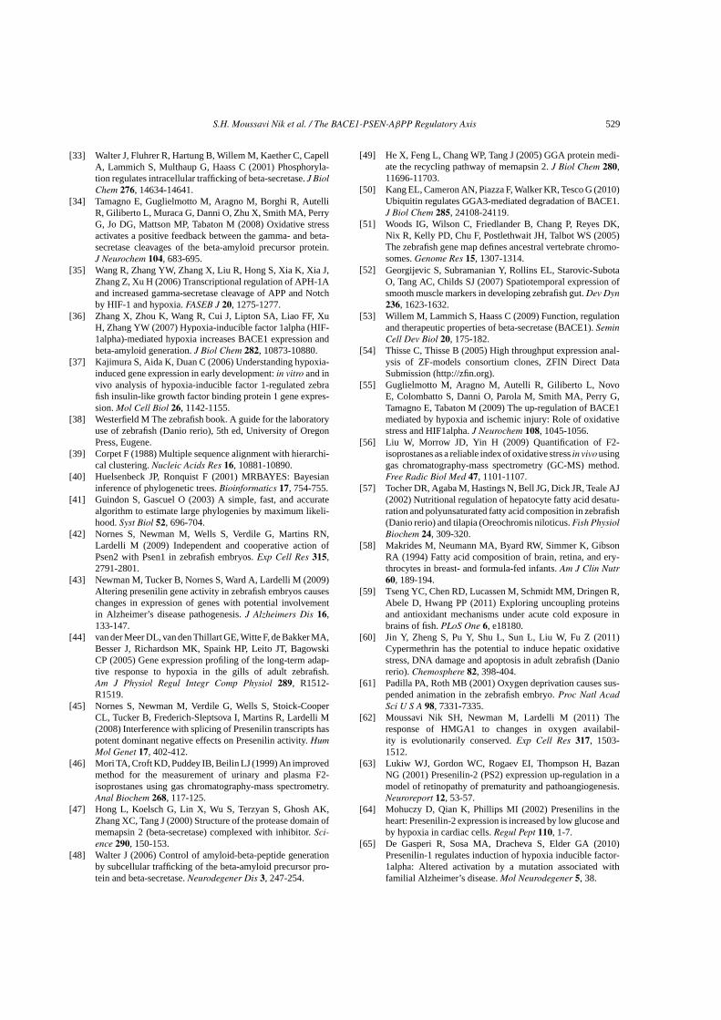
S.H. Moussavi Nik et al. / The BACE1-PSEN-AβPP Regulatory Axis 529
[33] Walter J, Fluhrer R, Hartung B, Willem M, Kaether C, CapellA, Lammich S, Multhaup G, Haass C (2001) Phosphoryla-tion regulates intracellular trafficking of beta-secretase. J BiolChem 276, 14634-14641.
[34] Tamagno E, Guglielmotto M, Aragno M, Borghi R, AutelliR, Giliberto L, Muraca G, Danni O, Zhu X, Smith MA, PerryG, Jo DG, Mattson MP, Tabaton M (2008) Oxidative stressactivates a positive feedback between the gamma- and beta-secretase cleavages of the beta-amyloid precursor protein.J Neurochem 104, 683-695.
[35] Wang R, Zhang YW, Zhang X, Liu R, Hong S, Xia K, Xia J,Zhang Z, Xu H (2006) Transcriptional regulation of APH-1Aand increased gamma-secretase cleavage of APP and Notchby HIF-1 and hypoxia. FASEB J 20, 1275-1277.
[36] Zhang X, Zhou K, Wang R, Cui J, Lipton SA, Liao FF, XuH, Zhang YW (2007) Hypoxia-inducible factor 1alpha (HIF-1alpha)-mediated hypoxia increases BACE1 expression andbeta-amyloid generation. J Biol Chem 282, 10873-10880.
[37] Kajimura S, Aida K, Duan C (2006) Understanding hypoxia-induced gene expression in early development: in vitro and invivo analysis of hypoxia-inducible factor 1-regulated zebrafish insulin-like growth factor binding protein 1 gene expres-sion. Mol Cell Biol 26, 1142-1155.
[38] Westerfield M The zebrafish book. A guide for the laboratoryuse of zebrafish (Danio rerio), 5th ed, University of OregonPress, Eugene.
[39] Corpet F (1988) Multiple sequence alignment with hierarchi-cal clustering. Nucleic Acids Res 16, 10881-10890.
[40] Huelsenbeck JP, Ronquist F (2001) MRBAYES: Bayesianinference of phylogenetic trees. Bioinformatics 17, 754-755.
[41] Guindon S, Gascuel O (2003) A simple, fast, and accuratealgorithm to estimate large phylogenies by maximum likeli-hood. Syst Biol 52, 696-704.
[42] Nornes S, Newman M, Wells S, Verdile G, Martins RN,Lardelli M (2009) Independent and cooperative action ofPsen2 with Psen1 in zebrafish embryos. Exp Cell Res 315,2791-2801.
[43] Newman M, Tucker B, Nornes S, Ward A, Lardelli M (2009)Altering presenilin gene activity in zebrafish embryos causeschanges in expression of genes with potential involvementin Alzheimer’s disease pathogenesis. J Alzheimers Dis 16,133-147.
[44] van der Meer DL, van den Thillart GE, Witte F, de Bakker MA,Besser J, Richardson MK, Spaink HP, Leito JT, BagowskiCP (2005) Gene expression profiling of the long-term adap-tive response to hypoxia in the gills of adult zebrafish.Am J Physiol Regul Integr Comp Physiol 289, R1512-R1519.
[45] Nornes S, Newman M, Verdile G, Wells S, Stoick-CooperCL, Tucker B, Frederich-Sleptsova I, Martins R, Lardelli M(2008) Interference with splicing of Presenilin transcripts haspotent dominant negative effects on Presenilin activity. HumMol Genet 17, 402-412.
[46] Mori TA, Croft KD, Puddey IB, Beilin LJ (1999) An improvedmethod for the measurement of urinary and plasma F2-isoprostanes using gas chromatography-mass spectrometry.Anal Biochem 268, 117-125.
[47] Hong L, Koelsch G, Lin X, Wu S, Terzyan S, Ghosh AK,Zhang XC, Tang J (2000) Structure of the protease domain ofmemapsin 2 (beta-secretase) complexed with inhibitor. Sci-ence 290, 150-153.
[48] Walter J (2006) Control of amyloid-beta-peptide generationby subcellular trafficking of the beta-amyloid precursor pro-tein and beta-secretase. Neurodegener Dis 3, 247-254.
[49] He X, Feng L, Chang WP, Tang J (2005) GGA protein medi-ate the recycling pathway of memapsin 2. J Biol Chem 280,11696-11703.
[50] Kang EL, Cameron AN, Piazza F, Walker KR, Tesco G (2010)Ubiquitin regulates GGA3-mediated degradation of BACE1.J Biol Chem 285, 24108-24119.
[51] Woods IG, Wilson C, Friedlander B, Chang P, Reyes DK,Nix R, Kelly PD, Chu F, Postlethwait JH, Talbot WS (2005)The zebrafish gene map defines ancestral vertebrate chromo-somes. Genome Res 15, 1307-1314.
[52] Georgijevic S, Subramanian Y, Rollins EL, Starovic-SubotaO, Tang AC, Childs SJ (2007) Spatiotemporal expression ofsmooth muscle markers in developing zebrafish gut. Dev Dyn236, 1623-1632.
[53] Willem M, Lammich S, Haass C (2009) Function, regulationand therapeutic properties of beta-secretase (BACE1). SeminCell Dev Biol 20, 175-182.
[54] Thisse C, Thisse B (2005) High throughput expression anal-ysis of ZF-models consortium clones, ZFIN Direct DataSubmission (http://zfin.org).
[55] Guglielmotto M, Aragno M, Autelli R, Giliberto L, NovoE, Colombatto S, Danni O, Parola M, Smith MA, Perry G,Tamagno E, Tabaton M (2009) The up-regulation of BACE1mediated by hypoxia and ischemic injury: Role of oxidativestress and HIF1alpha. J Neurochem 108, 1045-1056.
[56] Liu W, Morrow JD, Yin H (2009) Quantification of F2-isoprostanes as a reliable index of oxidative stress in vivo usinggas chromatography-mass spectrometry (GC-MS) method.Free Radic Biol Med 47, 1101-1107.
[57] Tocher DR, Agaba M, Hastings N, Bell JG, Dick JR, Teale AJ(2002) Nutritional regulation of hepatocyte fatty acid desatu-ration and polyunsaturated fatty acid composition in zebrafish(Danio rerio) and tilapia (Oreochromis niloticus. Fish PhysiolBiochem 24, 309-320.
[58] Makrides M, Neumann MA, Byard RW, Simmer K, GibsonRA (1994) Fatty acid composition of brain, retina, and ery-throcytes in breast- and formula-fed infants. Am J Clin Nutr60, 189-194.
[59] Tseng YC, Chen RD, Lucassen M, Schmidt MM, Dringen R,Abele D, Hwang PP (2011) Exploring uncoupling proteinsand antioxidant mechanisms under acute cold exposure inbrains of fish. PLoS One 6, e18180.
[60] Jin Y, Zheng S, Pu Y, Shu L, Sun L, Liu W, Fu Z (2011)Cypermethrin has the potential to induce hepatic oxidativestress, DNA damage and apoptosis in adult zebrafish (Daniorerio). Chemosphere 82, 398-404.
[61] Padilla PA, Roth MB (2001) Oxygen deprivation causes sus-pended animation in the zebrafish embryo. Proc Natl AcadSci U S A 98, 7331-7335.
[62] Moussavi Nik SH, Newman M, Lardelli M (2011) Theresponse of HMGA1 to changes in oxygen availabil-ity is evolutionarily conserved. Exp Cell Res 317, 1503-1512.
[63] Lukiw WJ, Gordon WC, Rogaev EI, Thompson H, BazanNG (2001) Presenilin-2 (PS2) expression up-regulation in amodel of retinopathy of prematurity and pathoangiogenesis.Neuroreport 12, 53-57.
[64] Mohuczy D, Qian K, Phillips MI (2002) Presenilins in theheart: Presenilin-2 expression is increased by low glucose andby hypoxia in cardiac cells. Regul Pept 110, 1-7.
[65] De Gasperi R, Sosa MA, Dracheva S, Elder GA (2010)Presenilin-1 regulates induction of hypoxia inducible factor-1alpha: Altered activation by a mutation associated withfamilial Alzheimer’s disease. Mol Neurodegener 5, 38.
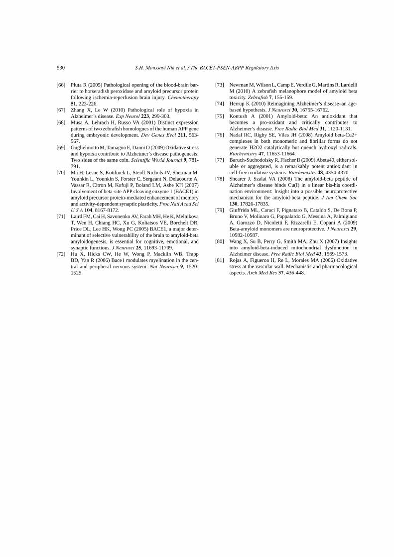
530 S.H. Moussavi Nik et al. / The BACE1-PSEN-AβPP Regulatory Axis
[66] Pluta R (2005) Pathological opening of the blood-brain bar-rier to horseradish peroxidase and amyloid precursor proteinfollowing ischemia-reperfusion brain injury. Chemotherapy51, 223-226.
[67] Zhang X, Le W (2010) Pathological role of hypoxia inAlzheimer’s disease. Exp Neurol 223, 299-303.
[68] Musa A, Lehrach H, Russo VA (2001) Distinct expressionpatterns of two zebrafish homologues of the human APP geneduring embryonic development. Dev Genes Evol 211, 563-567.
[69] Guglielmotto M, Tamagno E, Danni O (2009) Oxidative stressand hypoixa contribute to Alzheimer’s disease pathogenesis:Two sides of the same coin. Scientific World Journal 9, 781-791.
[70] Ma H, Lesne S, Kotilinek L, Steidl-Nichols JV, Sherman M,Younkin L, Younkin S, Forster C, Sergeant N, Delacourte A,Vassar R, Citron M, Kofuji P, Boland LM, Ashe KH (2007)Involvement of beta-site APP cleaving enzyme 1 (BACE1) inamyloid precursor protein-mediated enhancement of memoryand activity-dependent synaptic plasticity. Proc Natl Acad SciU S A 104, 8167-8172.
[71] Laird FM, Cai H, Savonenko AV, Farah MH, He K, MelnikovaT, Wen H, Chiang HC, Xu G, Koliatsos VE, Borchelt DR,Price DL, Lee HK, Wong PC (2005) BACE1, a major deter-minant of selective vulnerability of the brain to amyloid-betaamyloidogenesis, is essential for cognitive, emotional, andsynaptic functions. J Neurosci 25, 11693-11709.
[72] Hu X, Hicks CW, He W, Wong P, Macklin WB, TrappBD, Yan R (2006) Bace1 modulates myelination in the cen-tral and peripheral nervous system. Nat Neurosci 9, 1520-1525.
[73] Newman M, Wilson L, Camp E, Verdile G, Martins R, LardelliM (2010) A zebrafish melanophore model of amyloid betatoxicity. Zebrafish 7, 155-159.
[74] Herrup K (2010) Reimagining Alzheimer’s disease–an age-based hypothesis. J Neurosci 30, 16755-16762.
[75] Kontush A (2001) Amyloid-beta: An antioxidant thatbecomes a pro-oxidant and critically contributes toAlzheimer’s disease. Free Radic Biol Med 31, 1120-1131.
[76] Nadal RC, Rigby SE, Viles JH (2008) Amyloid beta-Cu2+complexes in both monomeric and fibrillar forms do notgenerate H2O2 catalytically but quench hydroxyl radicals.Biochemistry 47, 11653-11664.
[77] Baruch-Suchodolsky R, Fischer B (2009) Abeta40, either sol-uble or aggregated, is a remarkably potent antioxidant incell-free oxidative systems. Biochemistry 48, 4354-4370.
[78] Shearer J, Szalai VA (2008) The amyloid-beta peptide ofAlzheimer’s disease binds Cu(I) in a linear bis-his coordi-nation environment: Insight into a possible neuroprotectivemechanism for the amyloid-beta peptide. J Am Chem Soc130, 17826-17835.
[79] Giuffrida ML, Caraci F, Pignataro B, Cataldo S, De Bona P,Bruno V, Molinaro G, Pappalardo G, Messina A, PalmigianoA, Garozzo D, Nicoletti F, Rizzarelli E, Copani A (2009)Beta-amyloid monomers are neuroprotective. J Neurosci 29,10582-10587.
[80] Wang X, Su B, Perry G, Smith MA, Zhu X (2007) Insightsinto amyloid-beta-induced mitochondrial dysfunction inAlzheimer disease. Free Radic Biol Med 43, 1569-1573.
[81] Rojas A, Figueroa H, Re L, Morales MA (2006) Oxidativestress at the vascular wall. Mechanistic and pharmacologicalaspects. Arch Med Res 37, 436-448.


![Amyloid Precursor Protein (APP) processing in Alzheimer’s ...Presenilin 1 7297 P ELISA, IHC-Fr, IHC-P, WB Presenilin 1 [APS 11] 15456 M ELISA, IF, IHC-P, WB Presenilin 1 [APS 18]](https://cdn.vdocuments.us/doc/165x107/5f41361f607f43742c6d85e0/amyloid-precursor-protein-app-processing-in-alzheimeras-presenilin-1-7297.jpg)


