E U R O P E A N U R O L O G Y 6 9 ( 2 0 1 6 ) 4 1 – 4 9
ava i lable at www.sciencedirect .com
journal homepage: www.europeanurology.com
Platinum Priority – EditorialReferring to the article published on pp. 16–40 of this issue
Synopsis of the PI-RADS v2 Guidelines for Multiparametric
Prostate Magnetic Resonance Imaging and Recommendations
for Use
Jelle O. Barentsz a,*,y, Jeffrey C. Weinreb b,y, Sadhna Verma c, Harriet C. Thoeny d,Clare M. Tempany e, Faina Shtern f, Anwar R. Padhani g, Daniel Margolis h, Katarzyna J. Macura i,Masoom A. Haider j, Francois Cornud k, Peter L. Choyke l
a Department of Radiology and Nuclear Medicine Radboudumc, Nijmegen, The Netherlands; b Yale School of Medicine, New Haven, CT, USA; c University of
Cincinnati, Cincinnati, OH, USA; d Harvard University, Boston, MA, USA; e University Hospital of Bern, Bern, Switzerland; f AdMeTech Foundation, Boston, MA,
USA; g Paul Strickland Scanner Centre, Mount Vernon Hospital, Northwood, Middlesex, UK; h University of California, Los Angeles, CA, USA; i Johns Hopkins
University, Baltimore, MD, USA; j University of Toronto, Sunnybrook Health Sciences Centre, Toronto, Canada; k Rene Descartes University, Paris, France;l National Institutes of Health, Bethesda, MD, USA
Rapid technical advances have enabled multiparametric
magnetic resonance imaging (mpMRI) combined with
magnetic resonance (MR)–targeted biopsy to become
valuable tools for early detection of clinically significant
prostate cancer (PCa) while reducing overdiagnosis of
indolent PCa [1–6]. There has been concern, however, that
the widespread implementation and acceptance of mpMRI
could be impaired by a lack of standardisation of image
acquisition, interpretation and reporting guidance, and
inter- and intraobserver variability that could result in poor
clinical test performance in daily practise [7].
To expedite clinical evaluation and large-scale imple-
mentation of mpMRI, in May 2010 AdMeTech Foundation’s
International Prostate MRI Working Group recommended
development of standards of clinical performance by
establishing a prostate imaging reporting and assessment
system using BI-RADS (Breast Imaging and Reporting
Archiving Data System) as a model. Dickinson et al [8]
attempted to develop criteria for standardised acquisition
and interpretation of mpMRI, but they noted that it was
extremely difficult to define such criteria, even among
experts in the field, and that reliable implementation into
daily clinical practice remained problematic. To overcome
these limitations, the European Society of Urogenital
DOI of original article: http://dx.doi.org/10.1016/j.eururo.2015.08.052.* Corresponding author. Department of Radiology, Radboud University NijmE-mail address: [email protected] (J. Barentsz).y These authors are co-first authors.
http://dx.doi.org/10.1016/j.eururo.2015.08.0380302-2838/# 2015 European Association of Urology. Published by Elsevier
Radiology (ESUR) developed consensus-based guidelines
for prostate mpMRI, including clinical indications, minimal
and optimal imaging acquisition protocols, and a structured
category assessment system known as the Prostate Imaging
and Reporting and Data System (PI-RADS) version 1 (PI-
RADS v1) [9].
Since its publication in 2012, the PI-RADS v1 system has
achieved some acceptance, especially in Europe, and has
been validated in prospective studies, randomised trials,
and systematic analyses. A recent systematic review and
meta-analysis [10] evaluating 14 published studies using
PI-RADS v1 showed pooled sensitivity and specificity of 78%
(95% confidence interval [CI], 72–89%) and 79% (95% CI: 68–
86%), respectively, for detecting significant PCa, demon-
strating that mpMRI significantly changes the risk distribu-
tion of men with newly diagnosed PCa towards an increased
prevalence of high-risk disease. Improved risk management
with better identification of significant versus insignificant
cancers may lead to more specific and individualised
treatment options and less overtreatment of indolent
disease.
For PI-RADS v1, it was not specified exactly how to
combine the scores from each MRI sequence to derive an
overall category assessment. This led to confusion in its
egen Medical Centre, PO Box 9101, Nijmegen, 6500 HB, The Netherlands.
B.V. All rights reserved.
E U R O P E A N U R O L O G Y 6 9 ( 2 0 1 6 ) 4 1 – 4 942
application, and variable approaches were used. This
contributed to the variability of PI-RADS v1 performance
[10]. To improve this performance, Vache et al [11] suggested
refinement of the weighting given to each individual mpMRI
parameter.
In early 2012, a joint steering committee of the American
College of Radiology, ESUR, and AdMeTech Foundation
agreed to collaborate on the development of an improved
PI-RADS version 2 (PI-RADS v2). The PI-RADS v2 document
was released online in December 2014 [12]. The specific
aims were to establish guidelines for minimum acceptable
technical parameters for prostate mpMRI, to simplify and
standardise the terminology and content of mpMRI reports,
to develop assessment categories that summarise the levels
of suspicion or risk of having significant PCa, to reduce
variability in imaging interpretations, to educate and
enhance communication with referring clinicians, to enable
standardised data collection for outcomes monitoring, and
to facilitate quality assurance and research with the overall
aims of improving patient outcomes. PI-RADS v2 is intended
to be a ‘‘living’’ document that evolves as clinical experience
and scientific validation data accrue.
The complete PI-RADS v2 document includes informa-
tion regarding clinical considerations and technical speci-
fications for mpMRI, normal anatomy and benign findings,
guidelines and caveats for assessment and reporting of
prostate mpMRI examinations, figures illustrating relevant
findings on MR images, a diagram for mapping of findings,
report templates, and a lexicon of terminology. An online
atlas of findings and cases is also being developed as a
learning and reference tool (http://www.acr.org/
Quality-Safety/Resources/PIRADS). This paper provides a
short description of PI-RADS v2. It provides discussion of
some of the key differences and improvements compared
with PI-RADS v1 (Table 1) and is focussed on the assessment
criteria for detection and diagnosis of significant PCa on
mpMRI examinations and clinical uses and limitations.
PI-RADS v2 is not intended to be a comprehensive PCa
diagnosis manual; it should be used in conjunction with
other resources. Its intended clinical application is for the
diagnostic evaluation and risk assessment of patients with
suspected PCa prior to or after transrectal ultrasound
(TRUS) biopsy. It has not been developed for detecting
suspected recurrent PCa following therapy.
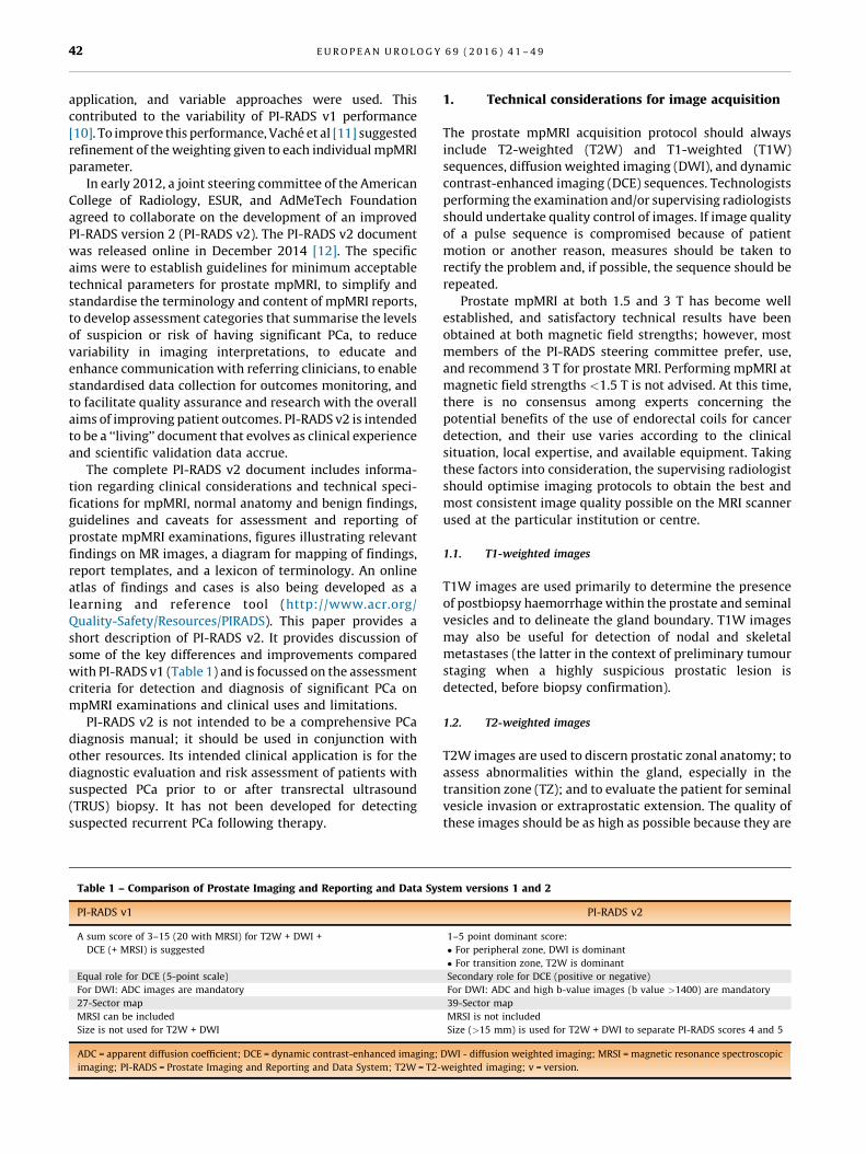
Table 1 – Comparison of Prostate Imaging and Reporting and Data Sys
PI-RADS v1
A sum score of 3–15 (20 with MRSI) for T2W + DWI +
DCE (+ MRSI) is suggested
Equal role for DCE (5-point scale)
For DWI: ADC images are mandatory
27-Sector map
MRSI can be included
Size is not used for T2W + DWI
ADC = apparent diffusion coefficient; DCE = dynamic contrast-enhanced imaging;
imaging; PI-RADS = Prostate Imaging and Reporting and Data System; T2W = T2-
1. Technical considerations for image acquisition
The prostate mpMRI acquisition protocol should always
include T2-weighted (T2W) and T1-weighted (T1W)
sequences, diffusion weighted imaging (DWI), and dynamic
contrast-enhanced imaging (DCE) sequences. Technologists
performing the examination and/or supervising radiologists
should undertake quality control of images. If image quality
of a pulse sequence is compromised because of patient
motion or another reason, measures should be taken to
rectify the problem and, if possible, the sequence should be
repeated.
Prostate mpMRI at both 1.5 and 3 T has become well
established, and satisfactory technical results have been
obtained at both magnetic field strengths; however, most
members of the PI-RADS steering committee prefer, use,
and recommend 3 T for prostate MRI. Performing mpMRI at
magnetic field strengths <1.5 T is not advised. At this time,
there is no consensus among experts concerning the
potential benefits of the use of endorectal coils for cancer
detection, and their use varies according to the clinical
situation, local expertise, and available equipment. Taking
these factors into consideration, the supervising radiologist
should optimise imaging protocols to obtain the best and
most consistent image quality possible on the MRI scanner
used at the particular institution or centre.
1.1. T1-weighted images
T1W images are used primarily to determine the presence
of postbiopsy haemorrhage within the prostate and seminal
vesicles and to delineate the gland boundary. T1W images
may also be useful for detection of nodal and skeletal
metastases (the latter in the context of preliminary tumour
staging when a highly suspicious prostatic lesion is
detected, before biopsy confirmation).
1.2. T2-weighted images
T2W images are used to discern prostatic zonal anatomy; to
assess abnormalities within the gland, especially in the
transition zone (TZ); and to evaluate the patient for seminal
vesicle invasion or extraprostatic extension. The quality of
these images should be as high as possible because they are
tem versions 1 and 2
PI-RADS v2
1–5 point dominant score:
� For peripheral zone, DWI is dominant
� For transition zone, T2W is dominant
Secondary role for DCE (positive or negative)
For DWI: ADC and high b-value images (b value >1400) are mandatory
39-Sector map
MRSI is not included
Size (>15 mm) is used for T2W + DWI to separate PI-RADS scores 4 and 5
DWI - diffusion weighted imaging; MRSI = magnetic resonance spectroscopic
weighted imaging; v = version.
E U R O P E A N U R O L O G Y 6 9 ( 2 0 1 6 ) 4 1 – 4 9 43
the key images for detecting significant cancers, especially
in the TZ.
1.3. Diffusion weighted imaging
DWI reflects and measures the random motion of water
molecules, the so-called Brownian motion, which becomes
impeded focally when cancer is present. DWI is a key
component of prostate mpMRI examinations, especially for
detection of significant cancers in the peripheral zone (PZ).
Diffusion weighted (DW) images are used to calculate
apparent diffusion coefficient (ADC) maps (with monoexpo-
nential fitting of DW images acquired at b values
�1000 s/mm2). High b-value images (�1400 s/mm2) should
be obtained (by direct acquisition or computed from the
source DWI images) to facilitate detection of clinically
significant PCa. It is now well established that the ADC value
of a focal tumour is inversely correlated with the Gleason
pattern: The lower the ADC value, the higher the Gleason
pattern.
1.4. Dynamic contrast-enhanced imaging
DCE is the acquisition of rapidly obtained T1W images
before, during, and after the intravenous bolus administra-
tion of a gadolinium-based contrast agent. Currently, the
added value of DCE is not firmly established regarding
tumour detection, with most published data showing that
the added value of DCE over the combination of T2W and
DWI is modest. Nevertheless, it is recommended that DCE
should be included in all prostate mpMRI examinations to
assist in the identification of some small, significant
cancers; to assist in the diagnosis of nonmalignant causes
of raised serum prostate-specific antigen (PSA), such as
inflammation; and to provide additional information if DWI
is technically limited. Although important, its role in
determining PI-RADS v2 assessment categories is secondary
to T2W and DWI. DCE serves primarily to help detect
significant PCa and not to characterise it.
2. PI-RADS assessment
PI-RADS v2 uses a 5-point assessment scale indicating the
likelihood that mpMRI findings correlate with the presence
of clinically significant PCa at a particular anatomic
location. Based on the current capabilities of mpMRI,
clinically significant disease is defined as Gleason score >7
(including 3 + 4 with prominent but not predominant
Gleason grade 4), and tumour volume >0.5 ml, and/or
extraprostatic extension. PI-RADS assessment categories
derived from mpMRI examinations relate to likely histo-
pathologic findings only and do not incorporate other
patient or cancer characteristics, such as PSA or clinical
cancer risk categories, and they do not directly inform the
choice of treatments available if PCa is diagnosed.
The PI-RADS v2 assessment categories are defined with
the following scores:
� 1
: Very low (clinically significant PCa is highly unlikely tobe present)
� 2
: Low (clinically significant PCa is unlikely to be present)� 3
: Intermediate (the presence of clinically PCa disease isequivocal)
� 4
: High (clinically significant PCa is likely to be present)� 5
: Very high (clinically significant PCa is highly likely tobe present)
Important differences between PI-RADS v1 and PI-RADS
v2 are presented in Table 1. Assignment of a PI-RADS
assessment category for each lesion is based on the scoring
of T2W, DWI, and DCE sequences performed sequentially
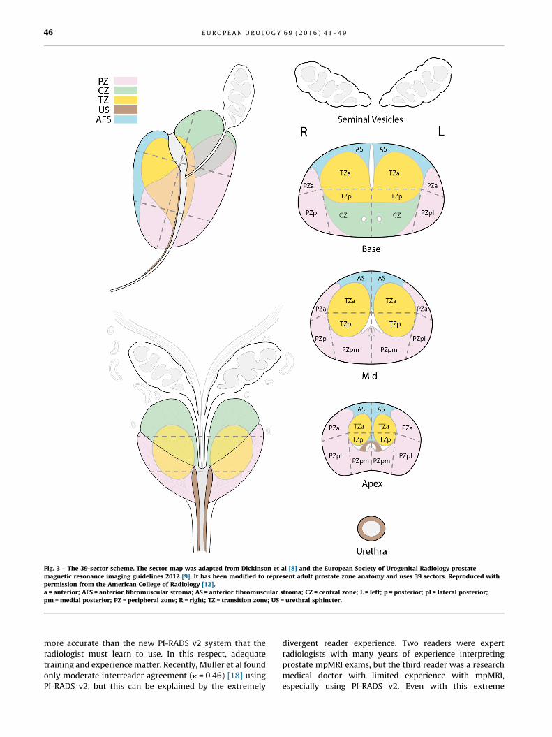
according to zonal anatomy, as described in Figures 1 and 2.
To localise each lesion, a 39-sector scheme was developed
(Fig. 3). The scoring for T2W and DWI uses a 5-point scale;
for DCE, a 2-point scale (positive or negative) is used. The
latter is one of the major differences: In PI-RADS v2, contrast
enhancement is either present or absent.
The most important difference between PI-RADS v1 and
PI-RADS v2 is that to assign an overall PI-RADS v2 lesion
assessment category, the scores from the T2W, DWI, and
DCE sequences are not summated but rather are applied
sequentially. The dominance of certain sequences (param-
eters) is used according to zonal anatomy. For the PZ, DWI is
the primary determining sequence; therefore, for a detected
PZ lesion, if the DWI score is 4 and the T2W score is 3, the PI-
RADS assessment category should be 4. For the TZ, T2W is
the primary determining sequence; if a detected TZ lesion
has a T2W score of 4 and its DWI score is 2, the PI-RADS
assessment category should be 4 (Figs. 1 and 2).
When T2W and DWI are of adequate diagnostic quality,
DCE plays a minor role in determining PI-RADS assessment
category; however, DCE has a supporting role in the
indeterminate category 3 PZ lesions. Absence of early
enhancement within a lesion usually adds little informa-
tion, and diffuse enhancement not localised to a specific
T2W or DWI abnormality can be seen in the setting of
inflammation, high-grade prostatic intraepithelial neopla-
sia, atypical small acinar proliferation, after biopsy, or with
a sparse Gleason 3 + 3 tumour with an inflammatory focus.
Moreover, DCE does not contribute to the overall assess-
ment category when the findings suggest a low (PI-RADS
1 or 2) or high (PI-RADS 4 or 5) likelihood of clinically
significant PCa in the PZ. When a PZ lesion has a DWI score
of 3, a positive DCE increases the likelihood that the finding
corresponds to a clinically significant PCa and thus
upgrades the assessment category to PI-RADS 4 (Fig. 1).
Likewise, when a TZ lesion has a T2W score of 3, a DWI
score of 5 upgrades the assessment category to PI-RADS 4
(Fig. 2).
Finally, because larger tumours have an increased
chance of being significant, for PI-RADS v2, a size criterion
for T2WI and DWI was introduced. Based on the findings of
Wolters at al [13], a cut-off of 1.5 cm was proposed to
separate a score of 4 from 5 in both the PZ and the TZ.
3. Reporting mpMRI
The following clinical information should be available to
radiologists at the time of MRI reporting: recent serum PSA

[(Fig._1)TD$FIG]
No abnormality seen on DWIand ADC
Indis�nct hypointensity on ADC
Focal mild/moderatehypointensity on ADC andisointense/mild hyperintensityon high b-value DWI.
Focal markedly hypointense onADC and markedly hyperintenseon high b-value DWI; <1.5 cm
Similar findings to above but>1.5 cm in maximal dimensionor definite extraprosta�cextension/invasive findings
Nega�vefocal and earlyenhancement
Posi�vefocal and earlyenhancement
1
2
3
4
5
Final PIRADSCategory
Dynamic ContrastEnhancement
DWI Score
1
2
3
4
5
Peripheral Zone Lesion
Fig. 1 – PI-RADS v2 assessment for the peripheral zone.ADC = apparent diffusion coefficient; DWI = diffusion weighted imaging.
E U R O P E A N U R O L O G Y 6 9 ( 2 0 1 6 ) 4 1 – 4 944
level and PSA history; date and results of prostate biopsy,
including number of cores, locations, and Gleason scores
of positive biopsies (with percentage of core involvement
and/or core length); and other relevant clinical history,
including ethnicity, family history, digital rectal examina-
tion (DRE) findings, and prior prostate therapy.
The report should include a measurement of prostate
gland volume. It can be combined with PSA to calculate PSA
density.
3.1. Mapping
All suspicious intraprostatic lesions seen on mpMRI should
be assigned to their zonal location, either PZ (including the
central zone [CZ]) or TZ on the sector map, and assigned a
PI-RADS overall assessment category. Because the CZ, like
the TZ, often shows hypointensity on T2W and ADC and
high signal on the high b-value images, it can mimic
significant PCa; therefore, in PI-RADS v2, this zone is
separately indicated in the 39-sector scheme (Fig. 3).
Findings with a PI-RADS assessment category of 3, 4, or
5 should be assigned on the sector map (Fig. 3), and the
index (dominant) lesion should be identified. The index
lesion is the one with the highest PI-RADS assessment
category or, alternatively, the largest lesion if there is more
than one with the same category. If there are more than four
suspicious findings, then only the four with the highest PI-
RADS assessment categories should be reported. Reporting
of additional or definitely benign findings is optional but
may be helpful as landmarks to guide biopsy or to track
lesions on subsequent examinations. If a suspicious finding
extends beyond the boundaries of one sector, all neighbour-
ing involved sectors should be indicated on the sector map
as a single lesion.
3.2. Measurement of lesions
With current techniques, mpMRI has been shown to
underestimate tumour size, volume, and extent, especially
for Gleason grade 3 disease. Nonetheless, the following
measurement rules are recommended. The minimum
requirement is to report the single largest dimension of a
suspicious lesion on an axial image. If the largest dimension
of a suspicious lesion is on sagittal and/or coronal images,
this measurement and imaging plane should be reported.
PZ lesions should be measured on ADC maps. TZ lesions
should be measured on T2W images. If lesion measure-
ments are difficult or compromised on ADC maps (for PZ) or
T2W (for TZ), then measurement should be made on
sequences that show the lesion outline the best. The image
number or series and sequence used for measurement
should be indicated.
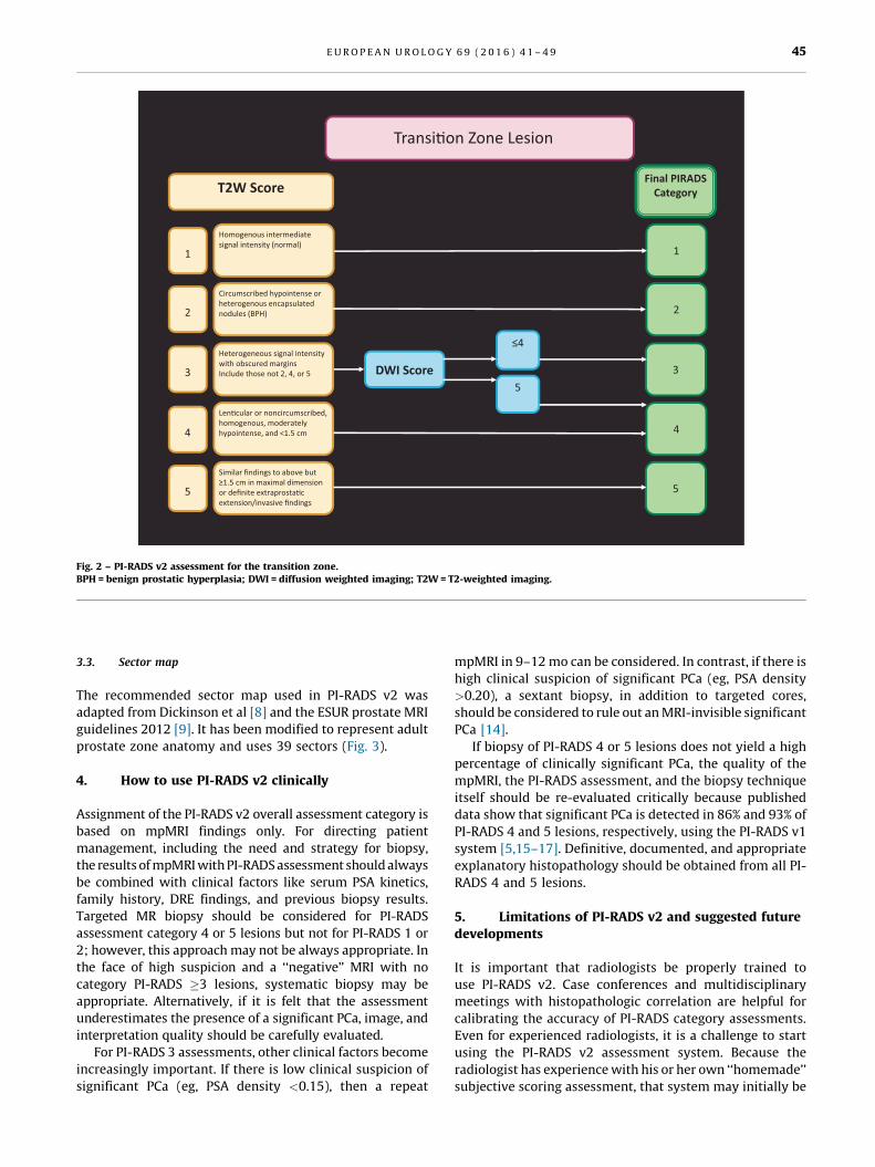
[(Fig._2)TD$FIG]
Final PIRADSCategory
Homogenous intermediatesignal intensity (normal)
Circumscribed hypointense orheterogenous encapsulatednodules (BPH)
Heterogeneous signal intensitywith obscured marginsInclude those not 2, 4, or 5
Len�cular or noncircumscribed,homogenous, moderatelyhypointense, and <1.5 cm
Similar findings to above but≥1.5 cm in maximal dimensionor definite extraprosta�cextension/invasive findings
≤4
5
1
2
3
4
5
DWI Score
T2W Score
1
2
3
4
5
Transi�on Zone Lesion
Fig. 2 – PI-RADS v2 assessment for the transition zone.BPH = benign prostatic hyperplasia; DWI = diffusion weighted imaging; T2W = T2-weighted imaging.
E U R O P E A N U R O L O G Y 6 9 ( 2 0 1 6 ) 4 1 – 4 9 45
3.3. Sector map
The recommended sector map used in PI-RADS v2 was
adapted from Dickinson et al [8] and the ESUR prostate MRI
guidelines 2012 [9]. It has been modified to represent adult
prostate zone anatomy and uses 39 sectors (Fig. 3).
4. How to use PI-RADS v2 clinically
Assignment of the PI-RADS v2 overall assessment category is
based on mpMRI findings only. For directing patient
management, including the need and strategy for biopsy,
the results of mpMRI with PI-RADS assessment should always
be combined with clinical factors like serum PSA kinetics,
family history, DRE findings, and previous biopsy results.
Targeted MR biopsy should be considered for PI-RADS
assessment category 4 or 5 lesions but not for PI-RADS 1 or
2; however, this approach may not be always appropriate. In
the face of high suspicion and a ‘‘negative’’ MRI with no
category PI-RADS �3 lesions, systematic biopsy may be
appropriate. Alternatively, if it is felt that the assessment
underestimates the presence of a significant PCa, image, and
interpretation quality should be carefully evaluated.
For PI-RADS 3 assessments, other clinical factors become
increasingly important. If there is low clinical suspicion of
significant PCa (eg, PSA density <0.15), then a repeat
mpMRI in 9–12 mo can be considered. In contrast, if there is
high clinical suspicion of significant PCa (eg, PSA density
>0.20), a sextant biopsy, in addition to targeted cores,
should be considered to rule out an MRI-invisible significant
PCa [14].
If biopsy of PI-RADS 4 or 5 lesions does not yield a high
percentage of clinically significant PCa, the quality of the
mpMRI, the PI-RADS assessment, and the biopsy technique
itself should be re-evaluated critically because published
data show that significant PCa is detected in 86% and 93% of
PI-RADS 4 and 5 lesions, respectively, using the PI-RADS v1
system [5,15–17]. Definitive, documented, and appropriate
explanatory histopathology should be obtained from all PI-
RADS 4 and 5 lesions.
5. Limitations of PI-RADS v2 and suggested future
developments
It is important that radiologists be properly trained to
use PI-RADS v2. Case conferences and multidisciplinary
meetings with histopathologic correlation are helpful for
calibrating the accuracy of PI-RADS category assessments.
Even for experienced radiologists, it is a challenge to start
using the PI-RADS v2 assessment system. Because the
radiologist has experience with his or her own ‘‘homemade’’
subjective scoring assessment, that system may initially be
[(Fig._3)TD$FIG]
Fig. 3 – The 39-sector scheme. The sector map was adapted from Dickinson et al [8] and the European Society of Urogenital Radiology prostatemagnetic resonance imaging guidelines 2012 [9]. It has been modified to represent adult prostate zone anatomy and uses 39 sectors. Reproduced withpermission from the American College of Radiology [12].a = anterior; AFS = anterior fibromuscular stroma; AS = anterior fibromuscular stroma; CZ = central zone; L = left; p = posterior; pl = lateral posterior;pm = medial posterior; PZ = peripheral zone; R = right; TZ = transition zone; US = urethral sphincter.
E U R O P E A N U R O L O G Y 6 9 ( 2 0 1 6 ) 4 1 – 4 946
more accurate than the new PI-RADS v2 system that the
radiologist must learn to use. In this respect, adequate
training and experience matter. Recently, Muller et al found
only moderate interreader agreement (k = 0.46) [18] using
PI-RADS v2, but this can be explained by the extremely
divergent reader experience. Two readers were expert
radiologists with many years of experience interpreting
prostate mpMRI exams, but the third reader was a research
medical doctor with limited experience with mpMRI,
especially using PI-RADS v2. Even with this extreme
E U R O P E A N U R O L O G Y 6 9 ( 2 0 1 6 ) 4 1 – 4 9 47
difference in reader experience, PI-RADS v2 showed moder-
ate interreader agreement [18]. Furthermore, the overall
PI-RADS v2 assessment resulted in better estimation of the
risk of significant PCa, and PI-RADS v2 scores were
concordant with pathology results in both PZ and TZ (area
under the curve of 0.86 and 0.87, respectively). This
underscores the intrinsic robustness of the PI-RADS v2
assessment technique but also emphasises the needs for
specific PI-RADS v2 training, documentation of observer
variability according to reader experience, and further data
on intra- and interobserver variability. In another study in
which adequate training was performed—consisting of
>2 wk of intensive personalised teaching of both technicians
and expert readers followed by PI-RADS supervised reading
with >300 mpMRI scans—k statistics showed substantial
agreement (0.77), with 92% agreement for PI-RADS v1
categories 1–3 versus 4 and 5 [3].
Because the dominant factors for PI-RADS v2 assess-
ment are T2W for the TZ and DWI for the PZ, identification
of the zonal location of the lesion is vital. Areas in which
this may be especially problematic include the interface of
the CZ, the intraprostatic seminal vesicles and PZ at the
base of the gland, and the interface of the anterior horn
of the PZ with TZ and the anterior fibromuscular stroma.
The anterior-apical region is another problematic region
requiring special attention.
The ability to reliably detect and characterise clinically
significant PCa in the TZ depends on the more subjective
T2W anatomic criteria. Benign prostatic hyperplasia (BPH)
is intrinsically heterogeneous, including ill-defined struc-
tures and those that are highly cellular and vascular. This is
why tumour detection in the TZ is less accurate compared
with the PZ; the normal PZ has a more homogenous
appearance. Although T2W images are dominant in the TZ,
any lesion with low ADC and high signal intensity on high b-
value images should be regarded with caution and should
be carefully evaluated and biopsied if necessary. In clinical
practice, visually bright foci in the TZ on high b-value DWI
help draw the radiologist’s attention to a potential lesion
and trigger more detailed analysis of this area using the rest
of the MR data set. In this sense, initial localisation of the
suspicious region using the b-value >1400 images can be
useful in raising one’s confidence regarding the presence of
a lesion, even if the final PI-RADS category is largely
determined by T2W imaging.
Compared with PI-RADS v1, the role of DCE in PI-RADS v2
is limited, and future studies will show whether this
sequence can be omitted. Although it is advised that a PZ
lesion with a DWI score of 3 should be upgraded when DCE is
positive (Fig. 1), one should be aware that focal prostatitis
also frequently enhances and thus may create a false-positive
result.
In the same vein, it should be noted for TZ lesions with a
score of 3 on T2W images that a DWI score of 5 upgrades the
final PI-RADS assessment (Fig. 2). The cut-off was placed at
5 for this upgrade because BPH nodules can resemble
tumours (being hypercellular or proliferative on histology).
When such lesions are <1.5 cm, there is less confidence
about their nature and perhaps less need for immediate
sampling, but follow-up of these smaller lesions is
recommended nevertheless. Validation studies using tar-
geted systematic biopsy and whole-mount prostate histo-
pathology for documenting these specific phenomena are
needed to refine future versions of PI-RADS.
PI-RADS v2 uses size criteria with a threshold of 1.5 cm
to separate PI-RADS 4 and 5 lesions on T2W and DW images.
In practise, a focal lesion on DWI<1.5 cm that is most likely
to represent significant PCa by virtue of extraprostatic
extension or invasive behaviour should be assigned to the
PI-RADS 5 category. Regardless, because biopsy is recom-
mended for both PI-RADS 4 and 5 lesions, the management
of such lesions is unlikely to be altered. Again, further
validation of this 1.5-cm cut-off is required.
Another limitation of PI-RADS v2 is that the evaluation of
ADC maps and high b-value images is subjective despite the
numerical nature of ADC (unit: �10�3 mm2/s). The
definitions of markedly hypointense signals on ADC maps
and markedly hyperintense signals on high b-value images
remain subjective but understood nevertheless by experi-
enced radiologists. It would be helpful if threshold values
could be assigned for ADC values for insignificant and
significant PCa and for benign pathologies including
prostatitis. The problem of choosing cut-off values relates
to the fact that ADC values depend on the choice of b values
for DW images used for calculations (hence the recommen-
dation to use only b values <1000 s/mm2), but it also
depends on the diffusion time achieved on diffusion
sequences (which is highly dependent on scanner specifica-
tions) and on a variety of other technical factors. A solution
would be for each institution to determine its own ADC cut-
off value based on biopsy and prostatectomy results. To
enable comparisons between imaging systems, ADC mea-
surements can also be calibrated using biological tissues
with low variance in diffusion properties (eg, brain) or test
objects made of bioequivalent materials or ice-water
phantoms [19,20].
Although mpMRI is an accurate technique for detecting
significant PCa, it misses significant PCa at a low percentage
(6–25%). Lesions missed are usually invasive PCa inter-
mixed with normal tissue or low-grade or mucinous PCa;
however, tumour volume of missed PCa is usually low
(<0.5 ml), and only 7–14% have >20% Gleason grade �4
components [6,16,21–23]. The rate at which this occurs is
low for PI-RADS 1 and 2 lesions and higher for PI-RADS
3 lesions. The exact prevalence of all PCa and significant PCa
for each PI-RADS v2 category has yet to be determined, and
investigators are encouraged to re-evaluate their historical
archives and to perform prospective studies to document
the prevalence of missed significant disease according to PI-
RADS v2 assessment categories.
As already noted, PI-RADS v2 needs to be tested and
validated in different clinical scenarios. Initially, data are
required on test performance (rates of detection of clinically
significant and insignificant PCa) in first- or repeat-biopsy
patient subpopulations. Validation should be undertaken
using MR-targeted biopsy, systematic biopsy (saturation or
template techniques), and whole-mount histopathology.
Comparison with the current standard of systematic TRUS

E U R O P E A N U R O L O G Y 6 9 ( 2 0 1 6 ) 4 1 – 4 948
biopsy should also be undertaken. The need for targeted
biopsy alone or in combination with systematic TRUS
biopsy should be assessed. Long-term follow-up data are
also needed before we can conclude that PI-RADS v2 is
effective in directing patient management and for improv-
ing outcomes of patients with suspected PCa. The role of PI-
RADS v2 assessments in directing the selection and
monitoring of patients undergoing active surveillance
would be interesting. It is anticipated that as evidence
continues to accrue in the field of mpMRI and for MRI-
targeted in-bore or out-of-bore biopsies and image-guided
focal therapy interventions, specific recommendations
and/or algorithms regarding need for biopsy and manage-
ment will be included in future versions of PI-RADS. PI-
RADS v2 is the next step in prostate mpMRI standardisa-
tion, helping to objectively improve the detection and
localisation of significant PCa. Consequently, its use in
clinical practise is highly recommended.
The PI-RADS steering committee strongly supports the
continued development of promising MRI methodologies
for assessment of PCa and local staging using novel and/or
advanced research tools not included in PI-RADS v2.
Consideration will be given to incorporating them into
future versions of PI-RADS as relevant data and experience
become available.
Conflicts of interest: The authors have nothing to disclose.
Funding support: The research and development of AdMeTech Founda-
tion’s International Prostate MRI Working Group were made possible by
a grant awarded and managed by the U.S. Army Medical Research and
Materiel Command (USAMRMC) and Telemedicine and Advanced
Technologies Research Center (TATRC) at Fort Detrick, Maryland, under
contract numbers W81XWH-09-0552 and W81XWH-11-1-0077.
Acknowledgment statement: The authors are grateful to the other
members of the ACR Joint PI-RADS Steering Committee of the American
College of Radiology, AdMeTech Foundation’s International Prostate MRI
Working Group, and the Prostate MRI subcommittee of the European
Society of Urogenital Radiology for developing PI-RADS v2, in particular,
Mythreyi Chatfield of the American College of Radiology.
References
[1] Futterer JJ, Briganti A, De Visschere P, et al. Can clinically significant
prostate cancer be detected with multiparametric magnetic reso-
nance imaging?. A systematic review of the literature. Eur Urol
2015;68:1045–53.
[2] Schoots IG, Roobol MJ, Nieboer D, Bangma CH, Steyerberg EW,
Hunink MG. Magnetic resonance imaging-targeted biopsy may
enhance the diagnostic accuracy of significant prostate cancer
detection compared to standard transrectal ultrasound-guided bi-
opsy: a systematic review and meta-analysis. Eur Urol 2015;68:
438–50.
[3] Pokorny MR, de Rooij M, Duncan E, et al. Prospective study of
diagnostic accuracy comparing prostate cancer detection by trans-
rectal ultrasound-guided biopsy versus magnetic resonance (MR)
imaging with subsequent MR-guided biopsy in men without pre-
vious prostate biopsies. Eur Urol 2014;66:22–9.
[4] Panebianco V, Barchetti F, Sciarra A, et al. Multiparametric mag-
netic resonance imaging vs. standard care in men being evaluated
for prostate cancer: a randomized study. Urol Oncol 2015;33:
17.e1–7.
[5] Delongchamps NB, Peyromaure M, Schull, et al. Prebiopsy magnetic
resonance imaging and prostate cancer detection: comparison of
random and targeted biopsies. J Urol 2013;189:493–9.
[6] Siddiqui MM, Rais-Bahrami S, Turkbey B, et al. Comparison of
MR/ultrasound fusion-guided biopsy with ultrasound-guided
biopsy for the diagnosis of prostate cancer. JAMA 2015;313:
390–7.
[7] Heidenreich A. Consensus criteria for the use of magnetic resonance
imaging in the diagnosis and staging of prostate cancer: not ready
for routine use. Eur Urol 2011;59:495–7.
[8] Dickinson L, Ahmed HU, Allen C, et al. Magnetic resonance imaging
for the detection, localisation, and characterisation of prostate
cancer: recommendations from a European consensus meeting.
Eur Urol 2011;59:477–94. http://dx.doi.org/10.1016/j.eururo.
2010.12.009, Epub 2010 Dec 21. PMID: 21195536.
[9] Barentsz JO, Richenberg J, Clements R, et al. ESUR prostate MR
guidelines 2012. Eur Radiol 2012;22:746–57.
[10] Hamoen EH, de Rooij M, Witjes JA, Barentsz JO, Rovers MM. Use of
the Prostate Imaging Reporting and Data System (PI-RADS) for
prostate cancer detection with multiparametric magnetic reso-
nance imaging: a diagnostic meta-analysis. Eur Urol 2015;67:
1112–21.
[11] Vache T1, Bratan F, Mege-Lechevallier F, Roche S, Rabilloud M,
Rouviere O. Characterization of prostate lesions as benign or malig-
nant at multiparametric MR imaging: comparison of three scoring
systems in patients treated with radical prostatectomy. Radiology
2014;272:446–55.
[12] PI-RADSTM Prostate Imaging and Reporting and Data System: 2015,
version 2. American College of Radiology Web site. http://www.acr.
org/�/media/ACR/Documents/PDF/QualitySafety/Resources/
PIRADS/PIRADS%20V2.pdf.
[13] Wolters T, Roobol MJ, van Leeuwen PJ, et al. A critical analysis of the
tumor volume threshold for clinically insignificant prostate cancer
using a data set of a randomized screening trial. J Urol 2011;185:
121–5.
[14] Radtke JP, Kuru TH, Boxler S, et al. Comparative analysis of trans-
perineal template saturation prostate biopsy versus magnetic
resonance imaging targeted biopsy with magnetic resonance
imaging-ultrasound fusion guidance. J Urol 2015;193:87–94.
[15] Renard-Penna R, Mozer P, Cornud F, et al. Prostate Imaging Report-
ing and Data System and Likert scoring system: multiparametric
MR imaging validation study to screen patients for initial biopsy.
Radiology 2015;275:458–68.
[16] Portalez D, Mozer P, Cornud F, et al. Validation of the European
Society of Urogenital Radiology scoring system for prostate
cancer diagnosis on multiparametric magnetic resonance imag-
ing in a cohort of repeat biopsy patients. Eur Urol 2012;62:
986–96.
[17] Delongchamps NB, Lefevre A, Bouazza N, Beuvon F, Legman P,
Cornud F. Detection of significant prostate cancer with magnetic
resonance targeted biopsies–should transrectal ultrasound-mag-
netic resonance imaging fusion guided biopsies alone be a standard
of care? J Urol 2015;193:1198–204.
[18] Muller BG, Shih JH, Sankineni S, et al. Prostate cancer: interobserver
agreement and accuracy with the revised Prostate Imaging Report-
ing and Data System at multiparametric MR imaging. Radiology
2015:142818.
[19] Malyarenko DI, Newitt D, J Wilmes L, et al. Demonstration of
nonlinearity bias in the measurement of the apparent diffusion
coefficient in multicenter trials. Magn Reson Med. In press. http://
dx.doi.org/10.1002/mrm.25754
[20] Padhani AR, Liu G, Koh DM, et al. Diffusion-weighted magnetic
resonance imaging as a cancer biomarker: consensus and recom-
mendations. Neoplasia 2009;11:102–25.

E U R O P E A N U R O L O G Y 6 9 ( 2 0 1 6 ) 4 1 – 4 9 49
[21] Abd-Alazeez M, Ahmed HU, Arya M, et al. The accuracy of multi-
parametric MRI in men with negative biopsy and elevated PSA
level–can it rule out clinically significant prostate cancer? Urol
Oncol 2014;32:45.e17–22.
[22] Abd-Alazeez M, Kirkham A, Ahmed HU, et al. Performance of multi-
parametric MRI in men at risk of prostate cancer before the first
biopsy: a paired validating cohort study using template prostate
mapping biopsies as the reference standard. Prostate Cancer Prostatic
Dis 2014;17:40–6.
[23] Arumainayagam N1, Ahmed HU, Moore CM, et al. Multipara-
metric MR imaging for detection of clinically significant prostate
cancer: a validation cohort study with transperineal template
prostate mapping as the reference standard. Radiology 2013;268:
761–9.


