REGULACIÓN DEL CITOESQUELETO DE ACTINA
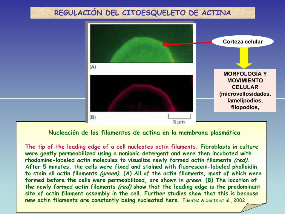
Nucleación de los filamentos de actina en la membrana plasmática
The tip of the leading edge of a cell nucleates actin filaments. Fibroblasts in culture were gently permeabilized using a nonionic detergent and were then incubated with rhodamine-labeled actin molecules to visualize newly formed actin filaments (red).After 5 minutes, the cells were fixed and stained with fluorescein-labeled phalloidinto stain all actin filaments (green). (A) All of the actin filaments, most of which were formed before the cells were permeabilized, are shown in green. (B) The location of the newly formed actin filaments (red) show that the leading edge is the predominant site of actin filament assembly in the cell. Further studies show that this is because new actin filaments are constantly being nucleated here. Fuente: Alberts et al., 2002
Corteza celular
MORFOLOGÍA Y MOVIMIENTO
CELULAR(microvellosidades,
lamelipodios, filopodios,
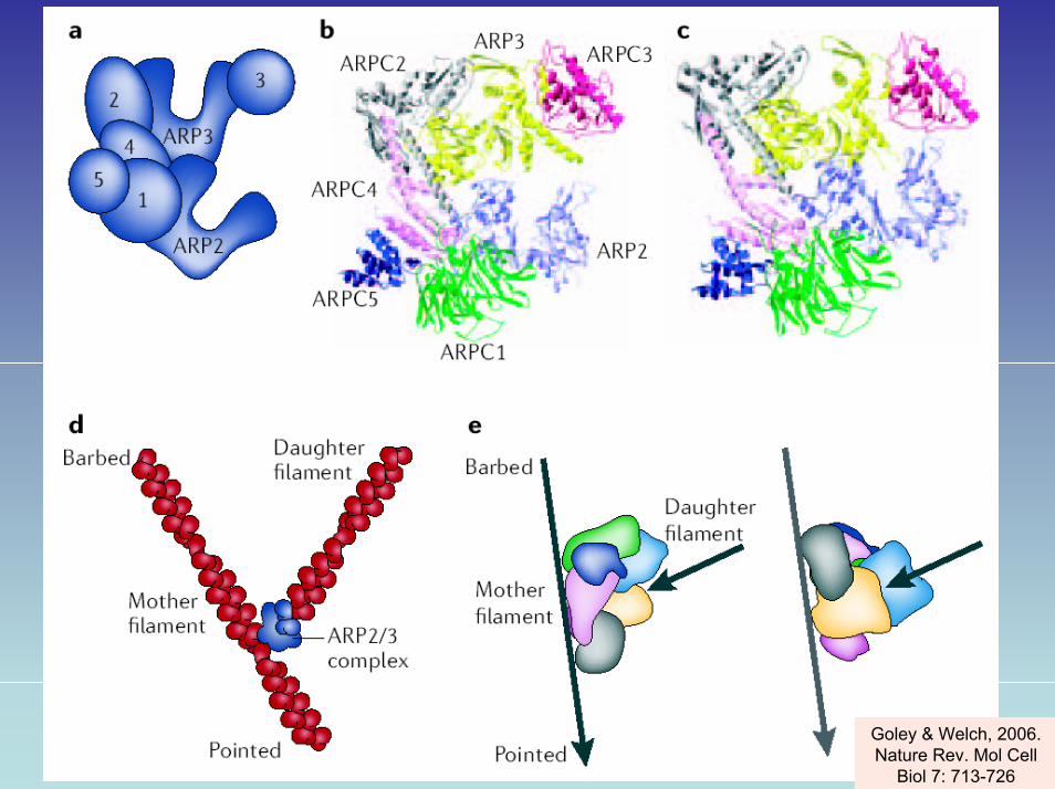
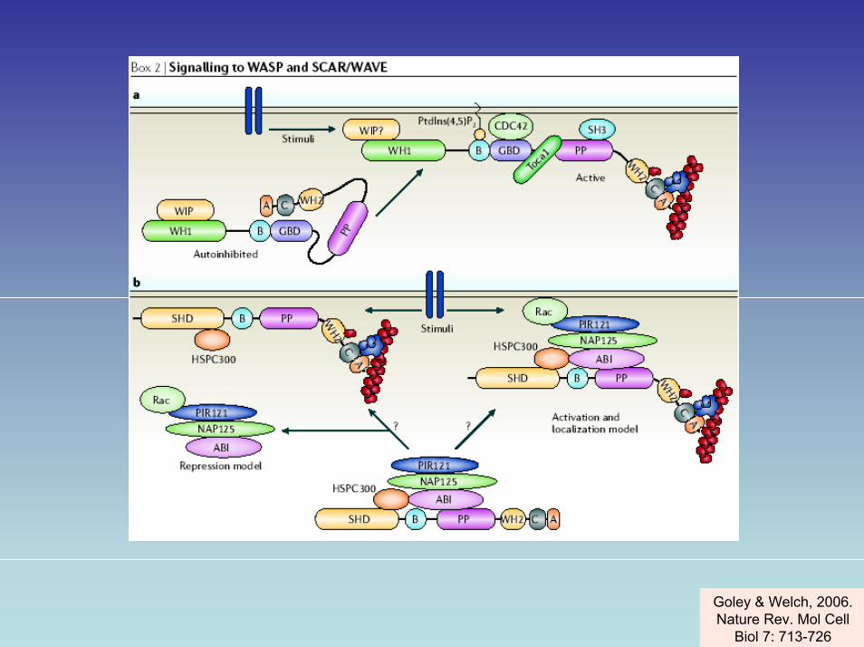
Goley & Welch, 2006. Nature Rev. Mol Cell
Biol 7: 713-726
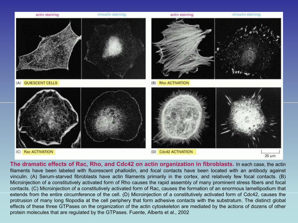
The dramatic effects of Rac, Rho, and Cdc42 on actin organization in fibroblasts. In each case, the actinfilaments have been labeled with fluorescent phalloidin, and focal contacts have been located with an antibody againstvinculin. (A) Serum-starved fibroblasts have actin filaments primarily in the cortex, and relatively few focal contacts. (B) Microinjection of a constitutively activated form of Rho causes the rapid assembly of many prominent stress fibers and focal contacts. (C) Microinjection of a constitutively activated form of Rac, causes the formation of an enormous lamellipodium that extends from the entire circumference of the cell. (D) Microinjection of a constitutively activated form of Cdc42, causes the protrusion of many long filopodia at the cell periphery that form adhesive contacts with the substratum. The distinct global effects of these three GTPases on the organization of the actin cytoskeleton are mediated by the actions of dozens of other protein molecules that are regulated by the GTPases. Fuente, Alberts et al., 2002
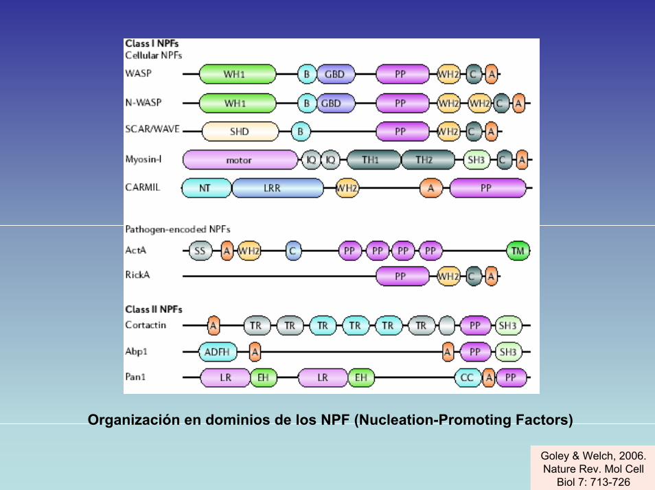
Organización en dominios de los NPF (Nucleation-Promoting Factors)
Goley & Welch, 2006. Nature Rev. Mol Cell
Biol 7: 713-726
Goley & Welch, 2006. Nature Rev. Mol Cell
Biol 7: 713-726

Goley & Welch, 2006. Nature Rev. Mol Cell
Biol 7: 713-726
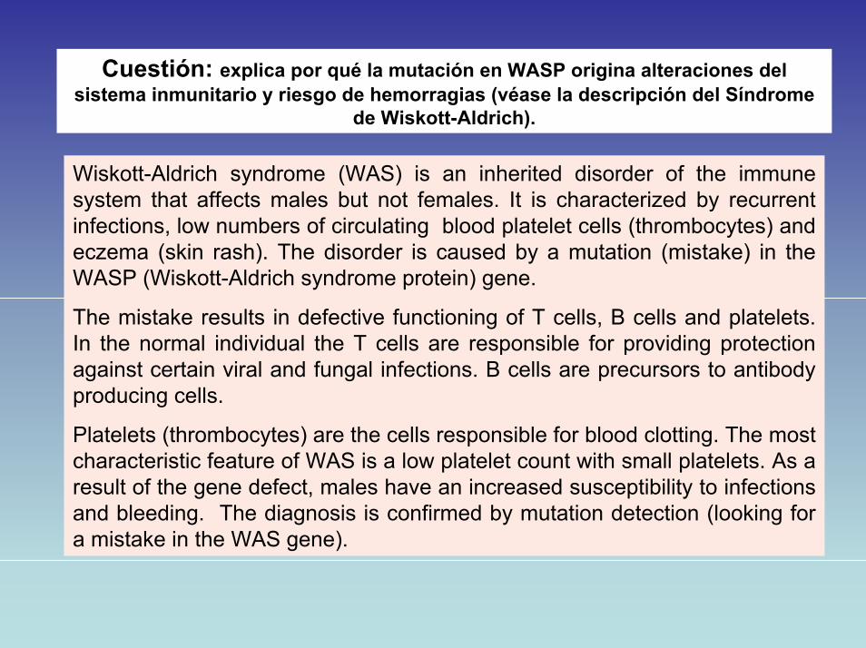
Cuestión: explica por qué la mutación en WASP origina alteraciones del sistema inmunitario y riesgo de hemorragias (véase la descripción del Síndrome
de Wiskott-Aldrich).
Wiskott-Aldrich syndrome (WAS) is an inherited disorder of the immune system that affects males but not females. It is characterized by recurrent infections, low numbers of circulating blood platelet cells (thrombocytes) and eczema (skin rash). The disorder is caused by a mutation (mistake) in theWASP (Wiskott-Aldrich syndrome protein) gene.
The mistake results in defective functioning of T cells, B cells and platelets. In the normal individual the T cells are responsible for providing protection against certain viral and fungal infections. B cells are precursors to antibody producing cells.
Platelets (thrombocytes) are the cells responsible for blood clotting. The most characteristic feature of WAS is a low platelet count with small platelets. As a result of the gene defect, males have an increased susceptibility to infections and bleeding. The diagnosis is confirmed by mutation detection (looking fora mistake in the WAS gene).


