Proteomic and biochemical studies of lysine malonylation suggests its malonic
aciduria-associated regulatory role in mitochondrial function and fatty acid
oxidation
Gozde Colak1¶, Olga Pougovkina2¶, Lunzhi Dai1¶, Minjia Tan3, Heleen te Brinke2, He
Huang1, Zhongyi Cheng4, Jeongsoon Park5, Xuelian Wan3, Xiaojing Liu6, Wyatt W. Yue7,
Ronald J. A. Wanders2,8, Jason W. Locasale6, David B. Lombard5, Vincent C. J. de
Boer2,8*, and Yingming Zhao1,3*
1Ben May Department of Cancer Research, University of Chicago, Chicago, IL, 60637,
USA
2 Laboratory Genetic Metabolic Diseases, Department of Clinical Chemistry, Academic
Medical Center, University of Amsterdam, Meibergdreef 9, 1105 AZ, Amsterdam, The
Netherlands
3State Key Laboratory of Drug Research, Shanghai Institute of Materia Medica, Chinese
Academy of Sciences, Shanghai, 201203, P.R. China
4PTM Biolabs, Chicago, IL 60612, USA
5Department of Pathology and Institute of Gerontology, University of Michigan, Ann
Arbor, MI 48109, USA
6Division of Nutritional Sciences, Cornell University, Ithaca, NY 14853
7Structural Genomics Consortium, University of Oxford, Oxford OX3 7DQ, UK
8Department of Pediatrics, Emma’s Children Hospital, Academic Medical Center,
University of Amsterdam, Meibergdreef 9, 1105 AZ Amsterdam, The Netherlands
1
MCP Papers in Press. Published on August 28, 2015 as Manuscript M115.048850
Copyright 2015 by The American Society for Biochemistry and Molecular Biology, Inc.

¶ Equal contribution.
*Correspondence:
[email protected], ph: (773) 834-1561
[email protected], ph: (31) 20 566 3927
Running title: Proteomic analysis of mammalian lysine malonylome
2

ABBREVIATIONS: Acetyl-CoA acetyltransferase (ACAT), ATP citrate lyase (ACLY),
Acetyl-CoA carboxylase 1 (ACC1), Acyl-CoA binding protein (ACBP), carbamoyl
phosphatase synthase 1 (CPS1), Carnitine Palmitoyl Transferase 1 (CPT1), fatty acid
synthase (FAS), gene ontology (GO), high-performance liquid chromatography (HPLC),
histone deacetylase (HDAC), Hydroxymethylglutaryl-CoA lyase (HMGCL), 3-hydroxy-3-
methylglutaryl-CoA synthase 2 (HMGCS2), Kyoto Encyclopedia of Genes and
Genomes (KEGG), lysine acetylation (Kac), lysine malonylation (Kmal), lysine
succinylation (Ksucc), mass spectrometry (MS), long chain-fatty acid transporter (LC-
FATP), malonyl-CoA decarboxylase (MCD), MCD wild type human fibroblasts (MCD+/+),
MCD deficient fibroblasts (MCD-/-), nicotine adenine dinucleotide (NAD+), post-
translational modification (PTM), Search Tool for the Retrieval of Interacting
Genes/Proteins (STRING), SIRT5 knock out (SIRT5 KO), pyruvate dehydrogenase
complex (PDC), stable isotope labeling by amino acids in cell culture (SILAC), sirtuin
(Sirt).
3

Summary
The protein substrates of SIRT5-regulated lysine malonylation (Kmal) remain
unknown, hindering its functional analysis. In this report, we carried out proteomic
screening, identifying 4042 Kmal sites on 1426 proteins in mouse liver, and 4943 Kmal
sites on 1822 proteins in human fibroblasts. Elevated malonyl-CoA in Malonyl-CoA
decarboxylase (MCD) deficient cells induces Kmal levels in substrate proteins. We
identified 461 Kmal sites showing more than 2-fold increase in response to MCD
deficiency, as well as 1452 Kmal sites detected only in MCD-/- fibroblast but not
MCD+/+ cells, suggesting a pathogenic role of Kmal in MCD deficiency. Cells with
increased lysine malonylation displayed impaired mitochondrial function and fatty acid
oxidation, suggesting that lysine malonylation plays a role in pathophysiology of malonic
aciduria. Our study establishes an association between Kmal and a genetic disease,
and offers a rich resource for elucidating the contribution of the Kmal pathway and
malonyl-CoA to cellular physiology and human diseases.
4
INTRODUCTION
Reversible acetylation at lysine residues in proteins has been extensively studied
over the past few decades (1,2). This modification is now known to have important
regulatory roles in diverse cellular processes and physiological conditions, such as
transcription, metabolism, and aging (3-5). Dysregulation of the lysine acetylation
pathway is associated with various diseases, such as cardiovascular disease and
cancer (6,7). In addition to acetylation, recent studies show that lysine residues in
proteins can be modified by a family of short-chain acylations: Propionylation,
butyrylation, crotonylation, malonylation, succinylation, glutarylation, and 2-
hydroxyisobutyrylation (8-13). Notable among the seven types of new lysine acylation
pathways are lysine malonylation (Kmal), succinylation (Ksucc), and glutarylation (Kglu).
Each of the three types of modifications have an acidic carboxylic group that changes
the charge status from +1 to -1 charge at physiological pH, which is similar to that
caused by protein phosphorylation, but more significant than lysine acetylation (Figure
1A). Accordingly, these acidic lysine acylations likely have a more substantial impact on
the substrate protein’s structure and function than lysine acetylation, when modified at
the same lysine residue(s). Recent studies demonstrate that pyruvate dehydrogenase
complex (PDC), succinate dehydrogenase, and carbamoyl phosphatase synthase 1
(CPS1) can be regulated by Ksucc and Kglu, respectively, suggesting that acidic lysine
acylation pathways can have unique functions distinct from the widely studied lysine
acetylation pathway (11,14).
5
Kmal was initially identified in both E. coli and mammalian cells by using HPLC-
MS/MS, co-elution of synthetic peptides, isotopic labeling, and Western blotting analysis
with pan anti-Kmal antibodies (8,15). The Lin group and our group have previously
demonstrated robust enzymatic activities of SIRT5, both in vitro and in vivo, in
demalonylation, desuccinylation, and deglutarylation(9,11,14,15). The demalonylation
and desuccinylation activities of SIRT5 require NAD+, but can be inhibited by
nicotinamide, a class III HDAC inhibitor (15). Given the fact that isotopic malonate can
label lysine malonylation and that acyl-CoAs are the precursor for other lysine
acylations (e. g., acetyl-CoA for lysine acetylation), malonyl-CoA is likely the precursor
for the lysine malonylation reaction (8,15). Despite this progress, the substrates for this
new modification pathway remain largely unknown, representing a major bottleneck for
studying its biological functions.
Malonyl-CoA is a tightly regulated metabolic intermediate in mammalian cells
(16). Malonyl-CoA is produced by acetyl-CoA carboxylase and consumed by malonyl-
CoA decarboxylase (MCD, E.C. 4.1.1.9), fatty acid synthase (FAS), and fatty acid
elongases (16) (Figure 1B). In addition to being a key intermediate for fatty acid
biosynthesis and fatty acid elongation, malonyl-CoA has diverse regulatory functions.
Malonyl-CoA was shown to be a potent inhibitor of carnitine palmitoyl transferase 1
(CPT1) and thereby regulates hepatic fatty acid synthesis, β-oxidation, and ketogenesis
(16) (Figure 1B). It was reported that malonyl-CoA can function as a key intermediate in
the hypothalamus as an energy sensor (17). Higher malonyl-CoA levels are observed in
skeletal muscle biopsies of type 2 diabetic patients (18). Elevated fatty acid oxidation
6

observed during cardiac ischemia/reperfusion has been attributed to the reduction of
malonyl-CoA levels in the heart. Accordingly, increasing malonyl-CoA levels has been
proposed as a strategy to improve cardiac function (19). Acetyl-CoA carboxylases,
enzymes that are known to be important for biosynthesis of malonyl-CoA, are
associated with physiology and diseases. Mice with genetic knockout of acetyl-CoA
carboxylase 2 gene are resistant to obesity and diabetes, when fed with high calorie
diets (20). Accordingly, acetyl-CoA carboxylases have been proposed as drug targets
for diverse human diseases, including diabetes, obesity, and cancer (21). Nevertheless,
potential roles for malonyl-CoA in human pathology are not well understood.
MCD is a 55 kDa enzyme that catalyzes conversion of malonyl-CoA to acetyl-
CoA, thus maintaining homeostatic levels of these metabolites in mitochondria and
peroxisomes. In the cytosol, malonyl-CoA is controlled by two enzymes with opposite
activities, MCD and acetyl-CoA carboxylase. MCD deficiency, or malonic aciduria, is an
inborn metabolic disorder caused by MCD mutations that reduce or eliminate activity of
this enzyme and therefore compromise conversion of malonyl-CoA to acetyl-CoA (22).
These patients have high levels of malonylcarnitine (C3DC) in blood and high level of
organic acids, such as malonic acid, in the urine (23). Diverse symptoms are observed
among the malonic aciduria patients, including delayed development, seizures, diarrhea,
vomiting, low blood sugar (hypoglycemia), and cardiomyopathy (22). It appears that
inhibition of fatty acid catabolism caused by high level of malonyl-CoA is at least
partially responsible for the manifestations of disease. We recently showed that MCD
deficient patient cells (MCD-/-) show increased Kmal levels (24). Therefore it is
7

therefore possible that Kmal could be an important mechanism mediating the
pathophysiology of MCD deficiency. Nevertheless, how the increased Kmal levels,
caused by high level of malonic acid in malonic aciduria patients and other diseases,
impacts cellular function and regulates physiology remains unknown.
In this study, we used a proteomic approach to identify Kmal substrates and map
their modification sites, by affinity enrichment of malonylated peptides and
HPLC/MS/MS analysis. We identified 4016 Kmal sites on 1395 proteins in SIRT5-
knockout mouse liver, and 4943 Kmal peptides on 1831 proteins in MCD+/+ and MCD-/-
human fibroblasts. Four hundred sixty-one Kmal sites on 339 proteins showed a 2-fold
increase or more in MCD-/- cells relative to MCD+/+ cells, and 1452 Kmal sites on 822
proteins were only detected in MCD-/- cells, suggests that MCD activity has a profound
impact on Kmal levels and distribution. The malonylated proteins induced in MCD-/-
cells are associated with diverse pathways, including fatty acid metabolism and
neurological diseases. We further showed that MCD -/- cells with increased lysine
malonylation have impaired mitochondrial respiration and fatty acid oxidation. Our
proteomics data illuminates the landscape of the Kmal modification in mammalian cells,
offer a valuable resource for studying its biology, and proposes possible roles of Kmal in
diseases associated with dysregulation of malonyl-CoA homeostasis.
MATERIALS AND METHODS
8

Materials — Chemicals were purchased as analytical grade from Sigma-Aldrich,
Inc. (St. Louis, MO). Modified sequencing-grade trypsin was purchased from Promega
Corporation (Madison, WI). Pan anti-malonyllysine antibody and pan anti-malonyllysine
agarose beads were from PTM Biolabs, Inc (Chicago, IL). MS grade water and
acetonitrile were from Thermo Fisher Scientific (Waltham, MA). C18 ZipTips were
purchased from Millipore Corporation (Billerica, MA). SILAC DMEM media (CCFDA003-
132J01) was purchased from UCSF Cell Culture Facility (San Francisco, CA).
XerumFree reagent (XF205) was purchased from MayFlower Bioscience (St. Louis,
MO). Dialyzed serum (Gibco-26400) was purchased from Life Technologies, Thermo
Fisher Scientific (Grand Island, NY).
Preparation of mouse liver lysate —Four two-month old male Sirt5 KO mice
(25,26) were anesthetized with isoflurane overdose, and the blood in the liver was
removed by perfusion with ice-cold PBS for 5 min. Liver was homogenized in a glass
dounce homogenizer in SDS lysis buffer (20 mM Tris HCl pH 6.8, 1% SDS, 5% β-
Mercaptoethanol, 10% glycerol, 25 mM nicotinamide). The lysates from four livers were
pooled together and the sample was clarified by centrifugation at 16,000g. The protein
in the supernatant was precipitated with 10% (v/v) trichloroacetic acid. Then the
precipitated proteins were in-solution digested with trypsin as previously described (27).
Preparation of SILAC samples — Human dermal fibroblast cells lines: MCD+/+
(control cells) and MCD-/- (malonyl-CoA decarboxylase deficient cells) were obtained
from Gaslini BioBank, Italy. The cells were grown in SILAC DMEM, supplemented with
9

L-Glutamine (584 mg/L), 10% (v/v) dialyzed serum, and 2% (v/v) Serum Free reagent.
Regular L-Lysine (12C614N2) and L-Arginine (12C6
14N2) were added to the “Light” media
(final concentration: 100mg/L) used for culturing MCD-/- cells. “Heavy” isotopic L-Lysine
(13C615N2) and “light” L-Arginine (12C6
14N2) was added to the “Heavy” media (final
concentration: 100mg/L) used for culturing MCD+/+ control cells. Both cells were grown
in parallel, until MCD+/+ cells were sufficiently labeled by the isotopic lysine.
Both MCD+/+ and MCD-/- cells were lysed in SDS buffer (20 mM Tris HCl pH 6.8,
1% SDS, 5% β-Mercaptoethanol, 10% glycerol, 25 mM nicotinamide). Twelve milligram
of each cell lysate were mixed and precipitated overnight by 10% TCA for tryptic
digestion.
HPLC Fractionation — The tryptic peptides were fractionated by using a
reversed-phase column (Luna C18 10 mm x 250 mm, 5 µm particle, 100 Å pore size,
Phenomenex Inc., Torrance, CA) in Discovery VP preparative HPLC system (Shimadzu
Corp., Kyoto, Japan). The peptides were fractionated into 75 fractions using a gradient
from 2% to 90% buffer B (10 mM ammonium formate in 90% acetonitrile and 10% water,
pH 7.8) in buffer A (10 mM ammonium formate in water, pH 7.8) at a flow rate of 4
ml/min in 60 min. The 75 fractions were finally combined equally into 5 final fractions for
mouse liver samples, and 10 final fractions for MCD SILAC sample, respectively. Each
fraction was condensed by using SpeedVac (ThermoSavant SPD111V). The peptide
solution was used for immunoaffinity enrichment.
10

Affinity Enrichment of the Peptides Containing Kmal — The peptides containing
Kmal were enriched using a procedure described previously (27). The tryptic peptides
from each fraction were resolubilized in 100 mM NH4HCO3 (pH 8.0). Samples were
centrifuged at 20,000g for 10 min to remove insoluble particles. The peptides were
incubated with 15 µL of agarose beads conjugated with anti-malonyl lysine antibody at
room temperature for 4 h with gentle rotation. The beads were washed three times with
NETN buffer, twice with ETN buffer (50 mM Tris HCl, pH 8.0, 100 mM NaCl, 1 mM
EDTA) and once with water. Enriched Kmal peptides were eluted from the beads by
washing three times with 0.1% trifluoroacetic acid. The eluted Kmal peptides were dried
in a SpeedVac.
Nano-HPLC-MS/MS Analysis — The enriched Kmal samples were first desalted
using OMIX C18 tips (Agilent Technologies Inc., Santa Clara, CA) and then dissolved in
solvent A (0.1% formic acid in water). Samples were injected onto a manually packed
reversed-phase C18 column (100 mm × 75 µm, 3-µm particle size, Dr. Maisch GmbH,
Ammerbuch, Germany ) connected to an Easy-nLC 1000 HPLC system (Thermo Fisher
scientific Inc., Waltham, MA). Peptides were eluted from 5% to 90% solvent B (0.1%
formic acid and 1% water in acetonitrile) in solvent A with a 1 h gradient at a flow rate of
200 nl/min. The analytes were directly ionized and sprayed into a Q Exactive mass
spectrometer (Thermo Fisher scientific Inc., Waltham, MA) by a Nanospray Flex™ Ion
Sources. Full MS scans were acquired in the Orbitrap mass analyzer over the range
m/z 300-1400 with a mass resolution of 70,000 at m/z 200. The 15 most intense peaks
of the precursor ions were fragmented in the HCD collision cell with normalized collision
11

energy of 27, and tandem mass spectra were acquired with a mass resolution of 17,500
at m/z 200. Lock mass at m/z 445.120024 was enabled for internal calibration of full MS
spectrum. Ions with either a single charge or more than 4 charges were excluded from
MS/MS fragmentation and the dynamic exclusion duration was set to 25s.
Data Processing and Analysis — MaxQuant software (v 1.3.0.5) was used for
identifying and quantifying protein and malonylated peptides. Peaklist generation and
precursor mass recalibration of the raw MS data were carried out by MaxQuant
software. Trypsin was specified as the cleavage enzyme and the maximum number of
missed cleavage was set at 3. Methionine oxidation, protein N-terminal acetylation,
lysine acetylation (Kac), Kmal (specified for neutral loss of CO2 in MS/MS
fragmentation), and Ksucc were specified as variable modifications, and cysteine
alkylation by iodoacetamide was specified as a fixed modification for all database
searching. Database searching was performed against the UniProt mouse (50,807
sequences,release date:May, 2013) or human (88,817 sequences, release date:
February, 2014) reference protein sequence database concatenated with reversed
decoy database with initial precursor mass tolerance of 7 ppm. Mass tolerance for
fragment ions was set at 20 ppm. False discovery rate (FDR) thresholds for protein,
peptide and modification site were fixed at 0.01. The identified peptides with MaxQuant
Andromeda score below 50 and localization probability below 0.75 were removed prior
to bioinformatics analysis.
12

Malonyl-CoA Measurement —The cells were treated with 15 µM Orlistat or
vehicle only for 24 h at 80% confluence. The media were quickly removed and the dish
was placed on top of dry ice. One ml of extraction solvent (80% methanol/water) was
immediately added, and the dishes were transferred to the -80 °C freezer. The dishes
were left for 15 min and then cells were scraped into extraction solvent on dry ice. The
whole solution was centrifuged with the speed of 20,000 g at 4 °C for 10 min. Here, cell
extracts were prepared from three wells to make biological triplicates. The supernatant
from tissue extract was transferred to a new tube for LC/MS/MS analysis. All samples
were dried in a vacuum concentrator (Speed Vac).
Ultimate 3000 UHPLC (Dionex) was coupled to Q Exactive-Mass spectrometer
(QE-MS, Thermo Scientific) for metabolite separation and detection. For Acyl-
Coenzyme A (Acyl-coA) analysis, a reversed phase liquid chromatography (RPLC)
method was used. A Luna C18 column (100 x 2.0 mm i.d., 3 µm; Phenomenex) was
employed with mobile phase A: water with 5 mM ammonium acetate (pH = 6.8), and
mobile phase B: methanol, at a flow rate of 0.2ml/min. The linear gradient was: 0 min, 2%
B; 1.5 min, 2% B; 3 min, 15% B; 5.5 min, 95%B; 14.5 min, 95%B; 15 min, 2%B, 20 min,
2% B. The column was at room temperature.
The Q Exactive mass spectrometer (QE-MS) was equipped with a HESI probe, and
the relevant parameters were as follows: heater temperature, 120 °C; sheath gas, 30;
auxiliary gas, 10; sweep gas, 3; spray voltage, 3.6 kV for positive mode. The capillary
temperature was set at 320 °C, and S-lens was 55. A full scan range was set at 300 to
13

1000 (m/z). The resolution was set at 70 000 (at m/z 200). The maximum injection time
(max IT) was 200 ms. Automated gain control (AGC) was targeted at 3 × 10⁶ ions. For
CoA analysis, cell extract was dissolved into 30 µl of water with 50 mM ammonium
acetate, pH6.8. Samples were centrifuged at 20,000 g at 4 °C for 3 min and the
supernatant was transferred to LC vials. The injection volume was 8 µl for CoA analysis.
Raw data collected from LC/MS/MS were processed on Thermo Scientific software
Sieve 2.0. Peak alignment and detection were performed according to manufacturer’s
protocols. For a targeted metabolomics analysis, a frameseed including Acyl-CoA
metabolites that has been previously validated was used for targeted metabolite
analysis with data collected in positive mode, with the m/z width set at 8 ppm. Statistical
significance was calculated based on student’s t test (unpaired, two tailed).
Motif Analysis for Lysine Malonylation Substrates — The standalone version of
IceLogo (version 1.2) software was used to analyze the preference of flanking Kmal site
sequence from mouse liver or human MCD cells (28). The embedded Swiss-Prot “Mus
musculus” or "Homo sapiens" was used as the negative set. Six flanking amino acid
residues on each side of a lysine malonylated site were selected as the positive set.
Functional Enrichment Analysis — Functional enrichment analysis of lysine
malonylated proteins was carried out using DAVID (Functional Annotation
Bioinformatics Microarray Analysis) Bioinformatics Resources v 6.7 with the total mouse
or human genome information as the background (29). All identified lysine malonylated
14

proteins were subjected to database analyses using Gene Ontology (GO) (30) and
Kyoto Encyclopedia of Genes and Genomes (KEGG) metabolic pathways (31). GO FAT
database from DAVID was selected in this analysis. The family-wide false discovery
rate was corrected by Benjamini-Hochberg method using adjusted P value cutoff 0.05.
Protein-Protein Interaction Network Analysis — Protein-protein interaction
networks of lysine malonylome were analyzed using STRING (Search Tool for the
Retrieval of Interacting Genes/Proteins) database (version 9.1, confidence score 0.7)
visualized by Cytoscape software (version 3.1.0) with MCODE App toolkit (32). The
confidence score is the approximate probability that a predicted link exists between two
enzymes in the same metabolic map in the KEGG database. Confidence limits are as
follows: low confidence 0.2 (or better), medium confidence 0.5, high confidence 0.75,
the highest confidence 0.95.
Protein complex enrichment analysis — Manually-curated core complexes
indexed by CORUM (the comprehensive resource of mammalian protein complexes)
database were used for the analysis of lysine malonylated substrates
(http://mips.helmholtz-muenchen.de/genre/proj/corum). Mouse or human complexes
indexed in the database were used for enrichment analysis of mouse liver or MCD
human cells by Fisher’s exact test. Complexes with adjusted p-value< 0.01 were
considered as significant.
15

Kmal stoichiometry calculation — Absolute stoichiometry calculation of
malonylated site in SILAC samples was based on the previously reported algorithm (33)
with slight modification (34). The calculation was based on the MS quantification data
(SILAC ratio) of the Kmal peptides (x), the corresponding protein (z), and the
corresponding unmodified peptide (y), with the assumption that only one type of PTM
occurs at a given site. The SILAC ratios of unmodified peptides (y) and proteins (z)
were calculated from the global protein expression analysis using the whole cell lysate
mixture of SILAC labeled MCD+/+ and MCD-/- cells without antibody affinity enrichment.
The calculation was assumed that only one type of PTM occurred at the given site of
interest. The unmodified peptide was defined as the longest completely digested part of
the peptide sequence derived from the malonylated peptide, which contains no other
PTM. The absolute stoichiometry was calculated based on the SILAC ratios of x, y, and
z using the same formula as previously reported (33).
Mitochondrial respiratory flux analysis — Measurements of cellular oxygen
consumption were performed using an extracellular flux analyzer (Seahorse BioScience,
Billerica, U.S.A.). Fao hepatoma cells were incubated for 24 hours in culture medium
(DMEM supplemented with 2mM HEPES, 2% Pen/Strep and 10% FBS) containing 50
mM malonate. Next, cells were plated at 20,000 cells/well in Seahorse 96 well culture
plates followed by overnight incubation in malonate-free medium. Human fibroblasts
were maintained and plated in DMEM supplemented with 2mM HEPES, 2% Pen/Strep
and 10% FBS at 30.000 cells/well. Seahorse mitochondrial function analysis was
performed using the digitonin cell permeabilization protocol (35). Prior to measurements
16

of respiration, culture medium was replaced with MAS buffer (pH, 7.4, 220 mM mannitol,
70 mM sucrose, 10mM KH2PO4, 5mM MgCl2, 2 mM HEPES, 1 mM EGTA and 0.6%
BSA-fatty acid free). Oxygen consumption rate (OCR) was analyzed following a single
injection of either pyruvate/malate/ADP/digitonin, succinate/rotenone/ADP/digitonin or
octanoylcarnitine/malate/ADP/digitonin, dissolved in MAS buffer without BSA at pH 7.4.
Final digitonin concentration was 30 ug/ml for Fao hepatoma cells and 100 ug/ml for
fibroblasts. Final substrate concentrations were: pyruvate (5 mM), malate (2.5 mM),
succinate (10 mM), octanoylcarnitine (100uM), ADP (1 mM). After injection of substrate,
oligomycin was injected at 1.5 uM final concentration followed by injection of antimycin
(2.5 uM) and rotenone (1.25 uM).
Very-long chain acyl-CoA dehydrogenase (VLCAD) activity analysis — VLCAD
activity was analyzed by monitoring the specific conversion of palmitoyl-CoA (C16:0-
CoA) into palmitenoyl-CoA (C16:1-CoA) in cell lysates (36). Cell lysates (0.1 mg/ml)
were incubated in 0.125 mM Tris pH 8.0 with 0.4 mM ferrocenium and 0.25 mM
palmitoyl-CoA for 10 minutes at 37 degrees and reaction was stopped by addition of 10
ul 2N HCl followed by neutralization with 10 ul 2M KOH / 0.6M MES. Samples were
deproteinated with acetonitrile followed by separation of substrate and products on a
reversed-phase C18 HPLC and UV detection.
Long-chain 3-hydroxy-acyl-CoA dehydrogenase (LCHAD) activity analysis —
LCHAD activity was analyzed by incubating cell lysates (0.1 mg/ml) with 3-
ketopalmitoyl-CoA (0.26 mM, synthesized in house) and NADH 0.4 mM in MES/Kpi
17

(100mM/200mM) buffer with 0.1% Triton (pH 6.2) for 5 minutes at 37 degrees using a
procedure previously described (37). To control for the conversion of 3-ketopalmitoyl-
CoA by short-chain 3-hydroxy-acyl-CoA dehydrogenase (SCHAD), samples were
incubated with and without N-Ethyl-Maleimide (NEM), because NEM inhibits only
LCHAD and not SCHAD. After incubation, reactions were stopped with 10 ul 2N HCL
followed by neutralization with 10 ul 2M KOH / 0.6M MES. Samples were deproteinated
with acetonitrile followed by separation of substrate and products on a reversed-phase
C18 HPLC and UV detection.
Immunocytochemistry — MCD +/+ and MCD-/- cells were grown on coverslips
and treated with 15µM orlistat for 48 hr. Mito-tracker red was added to the culture
medium at 0.1µM final concentration and incubated for 30 min. The cells were washed
with PBS twice and fixed with 4% (v/v) paraformaldehyde and permeabilized with 0.2%
(v/v) Triton-X. The cells were blocked with 2% bovine serum albumin for 2 hrs and
incubated with the corresponding primary antibodies at 1.5ug/ml final concentration
overnight. The cells were washed with PBS twice and incubated with secondary
antibody Alexa Flour 488 (Invitrogen, Grand Island, NY) for 2 hrs, and washed with PBS
twice. Hoechst (BD Biosciences, San Jose, CA) is added at 2µg/ml final concentration
and incubated for 15 min. The coverslips are washed with PBS twice and mounted.
The imaging was performed by using Leica SP2 DMIRE2 confocal microscope, with
HCX PL APO Ibd.BL 63X 1.4 oil objective.
18

RESULTS
Kmal is affected by SIRT5 and MCD
Our previous studies showed that SIRT5 can catalyze removal of malonyl groups
from malonylated lysine residues, both in vitro and in vivo (15). In addition, exogenous
malonate can boost lysine malonylation, possibly by increasing intracellular
concentrations of malonyl-CoA catalyzed by a short-chain acyl-CoA synthase (15).
Consistent with this result, Sirt5 KO mice showed increased Kmal and Ksucc levels
compared to their wild-type counterparts, but not Kac (Figure 2A).
We previously showed, by western blotting analysis, that Kmal levels are higher
in MCD-/- cells than MCD+/+ cells (24). This result, in combination with our earlier
observation that malonate can enhance Kmal (15), supports a hypothesis that MCD-/-
induces malonyl-CoA concentration that in turn boost Kmal. If this is true, a reduction of
lipid biosynthesis by reduced activity of fatty acid synthase (FAS) may also increase
malonyl-CoA and Kmal levels. To test this, we treated both control MCD+/+ and MCD-/-
cells with orlistat, an inhibitor of fatty acid synthase (38). Consistent with our hypothesis,
we observed an increase of Kmal levels in response to orlistat in MCD+/+ cells (Figure
2B). In addition, orlistat further increased Kmal levels in MCD-/- cells compared to
MCD+/+ cells while Kac and Ksucc levels remained largely unchanged (Figure 2C).
To test whether the enhanced Kmal levels are correlated with higher amounts of
malonyl-CoA, we measured intracellular malonyl-CoA levels in MCD+/+ and MCD-/-
cells using HPLC/MS-based metabolomics method. Our data showed that orlistat
19

significantly increased intracellular malonyl-CoA levels in both cell lines (Figure 2D),
suggesting that increased lysine malonylation induced via orlistat treatment might be
due to enhanced concentration of malonyl-CoA.
Taken together, three different strategies for enhancing malonyl-CoA levels lead
to increased levels of lysine malonylation. This result is consistent with our previous
work showing that increasing crotonyl-CoA, succinyl-CoA and glutaryl-CoA levels all
result in increases of their respective lysine acylations (11,13,39).
Proteomic identification of Kmal peptides
Identifying protein substrates is critical to studying the biology of a PTM pathway,
as was demonstrated in characterization of the lysine acetylation pathway (40-43). To
identify Kmal substrate proteins and their modification sites, we used a proteomic
approach involving affinity enrichment and subsequent HPLC/MS/MS analysis (Figure
3). Two experimental models were used, Sirt5 KO mice and MCD deficient fibroblasts
from malonic aciduria patients. Analysis of Kmal substrates in mouse liver allows us to
identify Kmal substrates in an organ important for cellular metabolism (Figure 3A). The
liver also has the highest lysine malonylation levels among the mouse tissues that we
screened (Figure S1). Quantification of Kmal substrates in MCD-deficient cells versus
wild-type controls can reveal key Kmal substrates whose modification status is changed
in response to malonic aciduria, and whose increased malonylation may play a
pathogenic role in this disorder (Figure 3B).
20

Protein extracts from liver tissues of Sirt5 KO mice were prepared, tryptically
digested, and resolved into 5 fractions by high-pH reversed phase (RP) HPLC. Kmal
peptides were enriched using pan anti-malonyllysine antibody. The enriched Kmal
peptides were analyzed by HPLC/MS/MS (Figure 3A). The acquired raw MS data were
analyzed by MaxQuant software with a false discovery rate (FDR) of 0.01 at protein and
peptide level for the identification of Kmal peptides. To ensure high confidence of the
identifications, we removed Kmal peptides with Andromeda scores between 40 and 50,
and localization probability below 0.75, prior to bioinformatic analysis (Table S1A). The
Andromeda score is used for ranking the confidence of peptide identification for the
MS/MS spectrum by the Andromeda search engine integrated in Maxquant software. A
higher score indicates a more confident peptide identification. This analysis led to
identification of 4016 Kmal sites in 1395 proteins in Sirt5 KO mouse liver (Figure 3C,
top). A significant portion of the 427 malonylated peptides (9.6% of the total) with
Andromeda scores between 40 and 50 may represent true positives, and these proteins
were listed as Kmal candidates (Table S1B).
In a parallel experiment, we identified and quantified Kmal peptides in human
dermal fibroblasts isolated from normal individuals (MCD+/+, labeled with “Heavy” lysine
isotope) and from malonic aciduria patients that are deficient in MCD (MCD-/-, labeled
with “Light” lysine isotope). Equal amount of protein lysates from both MCD+/+ and
MCD-/- cells were combined in a 1:1 ratio and processed using the same procedure as
described above for analysis of Kmal peptides. The study identified 4943 Kmal sites,
with Andromeda scores >50, on 1831 proteins in human fibroblasts (MCD+/+ and MCD-
21

/- combined, Figure 3C, Table S1C). We considered 732 Kmal sites with Andromeda
scores between 40 and 50 as the true positive Kmal candidate peptides, and these are
listed in Table S1D.
Among the Kmal substrates, we identified 21 histone marks in mouse liver and
19 histone marks in human fibroblasts (Table S2). Interestingly, most of them were not
located at N-terminal tails of histones (Figure 3D). Similarly, 35 non-redundant histone
lysine succinylation sites have been reported in mouse liver, which mostly localizes to
C-terminal globular domains as well (14). These results suggest that both Kmal and
Ksucc histone marks are likely to have differential regulatory function from the widely
studied histone acetylation marks (Table S2).
We previously reported that Ala and Gly were over-represented in the flanking
sequences of Ksucc sites, whereas Arg was largely depleted at both -1 and +1 positions
(14). Similarly, we evaluated the flanking sequences of Kmal sites to identify if there
was a structural preference for the location of this modification on the peptides. Motif
analyses of Kmal sites in mouse liver (Figure S2A, left) and human fibroblasts (Figure
S2B, left) showed significant similarity. Aliphatic amino acids, including Ala, Val, Ile and
Gly, were over-represented at the flanking sequence of Kmal sites, similar to the
situation with Ksucc sites, whereas Ser, Pro and Leu were under-represented. In
contrast to the similarity of Kmal and Ksucc flanking sequences, positively charged
residues such as Lys and Arg predominate in Kac motifs in mouse liver (44).
22

Quantification of changes in Kmal modification levels from MCD deficient cell
versus its wild type
Using SILAC-based quantitative proteomics approach, we quantified the
difference of Kmal substrate levels between MCD+/+ and MCD-/- cells, based on the
levels of Kmal peptides and those of protein expression. In parallel, we also quantified
changes of protein expression using whole cell lysates derived from a mixture of SILAC
labeled MCD+/+ and MCD-/- cells. The changes of Kmal peptides were normalized to
the change of their corresponding proteins’ levels in MCD cells. Normalized changes of
Kmal peptides were used for the subsequent analysis.
Among 4943 Kmal sites on 1822 proteins identified in MCD human fibroblasts,
3181 Kmal sites on 1257 proteins could be quantified (Table S1C-D). Among the 1762
unquantified Kmal sites, 1452 are present only in MCD-/- cells (“Light” only); these are
the Kmal peptides that have no detected signal in "Heavy-labeled peptide" (Intensity H)
from MCD+/+ cells, but significant intensity for the corresponding "Light-labeled peptide
(Intensity L)" from MCD-/- cells, by MaxQuant analysis (Table S1C). The median
MCD+/+:MCD-/- ratio of the quantifiable Kmal sites was 0.8284 (Figure 4A). These
results clearly suggest that MCD deficiency has an impact in elevating Kmal levels in
MCD-/- cells. 461 Kmal sites on 339 proteins increased in abundance by 2 fold or more
(normalized log2 ratio (MCD+/+:MCD-/-) ≤-1), while 1452 Kmal sites on 822 substrate
proteins were present in “Light”-only MCD-/- cells (Figure 4B, Table S1C) Forty-eight
Kmal sites on 38 Kmal proteins showed more than a 10-fold increase in MCD-/- cells
(Table S1C). We considered these Kmal substrates to represent the core group of
23

MCD-/- stimulated Kmal substrates. KEGG pathway analysis indicated that these
substrates are associated with TCA cycle, oxidative phosphorylation, amino acid
degradation (valine, leucine, isoleucine, and lysine), fatty acid metabolism and
propanoate metabolism pathways (Table S5E).
To calculate the stoichiometry of Kmal in MCD+/+ and MCD-/- cells, we modified
a reported algorithm (45) as we described previously (14,46). The calculation was
based on the successful quantification of a Kmal site, its corresponding protein, and the
unmodified peptide form in the SILAC experiment (for details, see (34)). To achieve a
more accurate calculation, we removed those Kmal sites that were previously reported
to be acetylated and succinylated (47,48), to minimize errors caused by the two
modifications at the same residues. This analysis enabled us to calculate the
stoichiometry of 325 Kmal sites on 222 proteins in MCD-/- cells, with calculated
stoichiometries ranging from 0.07% to 50.0% and in MCD+/+ cells, with a range from
0.01% to 48.6%, respectively (Figure 4C, Table S3). The two highest Kmal
stoichiometry sites were K376 of adenylyl cyclase-associated protein 1 (50.0% in MCD-
/- cells and 48.6% in MCD+/+ cells) and K41 of phosphoglycerate kinase (49.4% in
MCD-/- cells and 48.2% in MCD+/+ cells). Very long chain acyl-CoA dehydrogenase
(ACADVL, also known as VLCAD) catalyzes the first step of mitochondrial fatty acid
oxidation. Nine Kmal sites were identified in VLCAD, among which five sites were
detected in MCD-/- cells only, while the other four were up-regulated in MCD -/- cells,
suggesting a dramatic increase of Kmal on this protein. Dynamic increase of two Kmal
sites in VLCAD was 390- and 137-fold, respectively, in MCD-/- cells. Among 324 sites
24
whose stoichiometries were determined, 179 sites (55%) have more than a 2-fold
increase of Kmal stoichiometry in MCD-/- cells (Table S3). For example, malonylation at
K295 of mitochondrial 10-formyltetrahydrofolate dehydrogenase, responsible for
formate oxidation, is increased from 0.7% in MCD+/+ cells to 31% in MCD-/- cells.
Malonylation at K126 of prohibitin-2, a mediator of transcriptional repression by nuclear
hormone receptors, increased from 12.8% to 42.3% in MCD-/- cells.
Overlap among Kmal, Ksucc and Kac sites
To understand the similarities and differences among Kmal, Ksucc, and Kac sites,
we compared our lysine malonylome data with previously published data (14,41,43,47).
We found that, of all the identified Kmal sites in mouse liver, 640 (16%) sites (Figure 5A,
right) and 595 (42%) proteins (Figure 5A, left) overlapped with Kac sites in mouse
embryonic fibroblasts (MEF) (43). Five hundred and ten (36.5%) Kmal sites (Figure 5A,
right) and 262 (6.5%) proteins (Figure 5A, left) overlapped with Ksucc sites in SIRT5 KO
mouse liver (14). When we pooled the Ksucc sites reported in Sirt5 KO mouse liver and
MEFs, and carried out the same analysis, 706 (17.6%) sites (Figure S2C, right) and 406
(29%) proteins (Figure S2C, left) overlapped with Kmal sites identified in Sirt5 KO
mouse liver. Interestingly, we found that a significant portion of the malonylated proteins
(46.2%) and sites (71.1%) identified in our mouse liver data do not overlap with the
previously reported Ksucc and Kac data.
In a parallel experiment, we carried out similar analysis for the human
malonylome. In this experiment, we obtained the Kac and Ksucc data from previous
25

publications (41,47). Among the Kmal sites identified in human fibroblasts (combination
of MCD+/+ and MCD-/-), 776 Kmal sites (Figure 5B, right) and Kmal 827 proteins
(Figure 5B, left) overlapped with the human Kac proteome (41), and 671 sites (Figure
5B, right) and 550 proteins (Figure 5B, left) overlapped with the human Ksucc proteome
(47). Similar to the mouse malonylome data, a significant portion of the human
malonylated proteins (46.7%) and sites (75.7%) did not overlap with previously reported
Ksucc and Kac data. Overall, the spectrum of lysine sites and protein targets subject to
malonylation shows substantial non-overlap with Kac and Ksucc, suggesting that this
modification likely plays roles in modulating biological processes distinct from other
lysine PTMs.
Cellular localization of lysine malonylomes
SIRT5, a regulatory enzyme of Ksucc, Kglu, as well as Kmal, localizes
predominantly to mitochondria, but is also present in the cytosol and nucleus (14,49).
Previously, we reported that 17.8% of Ksucc substrates (351) are localized in the
mitochondria in mouse liver (Figure S2F, left) (14). Among the Ksucc substrates
identified in human cervical cancer cells (Hela) (47), 17% Ksucc substrates exclusively
localizes to mitochondria (Figure S2F, right).
To understand the cellular localization of Kmal substrates in mouse liver, we
performed the same analysis for the Kmal dataset generated from mouse liver. Here,
we compared our Kmal dataset with the mitochondria genes annotated in GO database
(50). Of all the identified Kmal substrates, 316 (58%) of them are present in the
26
mitochondria and 274 (50%) of them are exclusively mitochondrial proteins (Figure 5C,
left). Therefore, a comparable fraction of Kmal and Ksucc proteins from mouse liver
localizes to mitochondria.
In parallel, we carried out similar analysis for Kmal proteins derived from human
fibroblasts. Our result shows a striking difference of subcellular localization among Kmal
substrates. Among the 1024 Kmal substrates identified in human fibroblasts, 338 (33%)
of them localize mitochondria, of which 265 (26%) of them are exclusively mitochondrial
(Figure 5C, right). The number of mitochondrial Kmal substrates from either mouse liver
or human fibroblasts is comparable. However, in human fibroblasts, we identified a
significantly higher number of nuclear and cytosolic substrates, with 262 (30%) and 342
(39%) proteins, respectively (Figure 5C, right). The cellular enzymes that catalyze lysine
malonylation in mammalian cells are still unknown.
Additionally, we compared the Kmal proteins and sites in mitochondria to our
previously reported Ksucc data (14). We found 198 mitochondrial Kmal proteins (13.9%
of all Kmal proteins) (Figure S2D, left) and 432 mitochondrial Kmal sites (10.7% all
Kmal sites) (Figure S2D, right) overlapped with mitochondrial Ksucc in mouse liver,
whereas 37% mitochondrial Kmal proteins and 62% Kmal sites did not overlap. In
human fibroblasts, 59% mitochondrial Kmal proteins (199) (Figure S2E, left) and 31%
mitochondrial Kmal sites (344) (Figure S2E, right) overlapped with mitochondrial Ksucc
data (47).
27
We also performed immunostaining of MCD+/+ and MCD -/- human fibroblasts
with anti-malonyllysine, anti-acetyllysine and anti-succinyllysine antibodies along with
Hoechst nuclear stain and MitoTracker Red (Figure 6A and 6B). Our staining results
suggest that the strongest signals for Kac and Ksucc are confined in nucleus in both
MCD+/+ and MCD-/- human fibroblasts (Figure 6A and 6B, 2nd and 3th rows). However,
Kmal signals are distributed among cytosol and nucleus in MCD+/+ cells (Figure 6A, top
row). Interestingly, most of the Kmal signal overlaps with MitoTracker Red in MCD-/-
cells (Figure 6B, top row) suggesting that Kmal levels increase specifically in the
mitochondria of MCD-/- cells.
Functional annotation of lysine malonylomes
To understand the biological functions of Kmal proteins, we performed
enrichment analysis by using the Gene Ontology (GO) database (30), and Kyoto
Encyclopedia of Genes and Genomes (KEGG) (51) for Kmal substrates identified in
mouse liver and human fibroblasts. The GO biological process analysis of mouse liver
Kmal substrates (Table S4) showed enrichment in oxidation/reduction (adj P= 5.45x 10-
51), protein translation (adj P = 5.71x 10-49), cofactor metabolism (adj P =4.18x 10⁻²⁶),
and fatty acid metabolism (adj P = 5.09x 10⁻¹²) (Figure S3A, left). The GO analysis of
human fibroblast malonylome (Figure S3B, left, Table S5) showed enrichment in protein
expression processes such as translation (adj P = 2.22x 10-55), translation elongation
(adj P = 7.43x 10⁻²⁶), tRNA aminoacylation (adj P =1.01x 10⁻²⁰), and intracellular
transport (adj P =5.30x 10⁻³¹). Proteins associated with fatty acid β-oxidation were also
enriched in the human fibroblast malonylome (adj P =2.34x 10⁻⁷, Table S5).
28
The molecular function analysis of mouse liver Kmal substrates showed
enrichment in nucleotide binding (adj P = 9.35x 10⁻³⁵), cofactor binding (adj P = 1.04x
10-27), and ATP binding (adj P = 1.86x 10⁻¹⁵) (Figure S3A, right). Kmal substrates in
human fibroblasts were associated with nucleotide binding (adj P = 3.22x 10-45),
nucleoside binding (adj P = 2.77x 10⁻²⁹), ATP binding (adj P = 4.73x 10⁻²⁶), as well as
aminoacyl-tRNA ligase activity (adj P= 2.74x 10⁻²⁰) (Figure S3B, right) supporting the
idea that Kmal may be involved in regulating protein translation.
There was no significant difference between GO (Figure S4A) and KEGG
pathway enrichments (Figure S4B) of all the proteins identified in human fibroblasts
versus Light-only protein substrates derived from MCD-/- cells (Table S6). In addition,
there was a significant overlap between the KEGG pathway analysis of mouse liver
(Figure S3C) and human fibroblasts (Figure S3D). The top enriched categories of
KEGG pathways for lysine-malonylated substrates were ribosome,
valine/leucine/isoleucine degradation, proteasome and fatty acid metabolism (Figure
S3C and S3D). Twenty nine of 45 key enzymes in mouse and 22 of 45 key enzymes in
humans involved in regulation of fatty acid metabolism were lysine malonylated (Figure
S5A and S5B, Table S5F). Among these, five enzymes (FAS, ACC1, ACLY, AMPK and
CPT1) are closely associated with malonyl-CoA metabolism (Figure S5C).
Of particular note are a few proteins involved in fatty acid metabolism. We found
that Acetyl-CoA acetyl transferase 1 (ACAT1), an enzyme participating in multiple
29

metabolic pathways including fatty acid metabolism, was malonylated at seven sites:
K174, K190, K243, K251, K263, K268, and K273 (Figure 4D). The Kmal level of
(MCD+/+: MCD-/- SILAC ratio of 0.0044) K263 of ACAT1, was increased more than
200-fold in MCD-/- cells. K263 is in close proximity with Coenzyme A binding site and
possibly makes two hydrogen bonds with Coenzyme A, suggesting a possibility that this
residue is important in regulating the protein’s function. K263 was previously reported to
be acetylated and succinylated as well (41,47). In addition, among all the Kmal sites of
ACAT1, K174 is acetylated, and K251 is succinylated (Choudhary et al., 2009).
Therefore, these Kmal sites might also contribute to regulation of protein function,
depending on the type of modification. Hydroxymethylglutaryl-CoA lyase (HMGCL) is
malonylated at three lysine sites (K48, K93, and K137), of which K48 malonylation is
increased roughly 39-fold in MCD-/- cells. HMGCL exclusively localizes to mitochondria
and is specifically responsible for leucine degradation, as well as ketone production
during fat breakdown. HMGCL deficiency is a rare genetic disease that causes
metabolic acidosis and hypoglycemia (52). A K-to-N mutation at K48 of HMGCL ablates
enzymatic activity, which suggests that K48 is a critical position for enzymatic function
(53). Therefore, lysine malonylation of K48 may lead to changes in enzymatic activity of
this protein. ATP citrate lyase (ACLY) catalyzes conversion of citrate to acetyl-CoA
(Figure S5C), which can be converted further to malonyl-CoA by ACC1. Among the 14
Kmal sites in ACLY, K68, located next to ATP binding site (K66-K67), is malonylated,
and therefore might alter the ATP binding ability of the protein. Enrichment of fatty acid
metabolism proteins in the malonylomes in both mouse liver and human fibroblasts
30

suggests a possible feedback regulation of fatty acid biosynthesis by malonyl-CoA-
mediated lysine malonylation.
Mitochondrial function and fatty acid oxidation is impaired in MCD-/- cells
Integration of our bioinformatic analyses of lysine malonylated proteins identified
in mouse liver and human fibroblast demonstrates that among metabolic pathways,
proteins involved in fatty acid metabolism were preferentially heavily malonylated. Fatty
acid synthesis, which utilizes malonyl-CoA as substrate for synthesis and chain
elongation, primarily occurs in the cytosol, whereas fatty acid oxidation occurs in
mitochondria and peroxisomes. Because MCD-/- cells showed greatly increased Kmal
immunostaining in mitochondria compared to MCD+/+ cells (Figure 6), and MCD
deficient patients have been reported to present with pathologies similar to patients with
fatty acid oxidation defects, we wanted to understand whether mitochondrial function
and fatty acid oxidation is affected in MCD -/- cells. Long-chain fatty acids are broken
down to medium and short-chain fatty acids in mitochondria by very-long chain acyl-
CoA dehydrogenase (VLCAD) and medium-chain acyl-CoA dehydrogenase (MCAD)
together with the mitochondrial trifunctional protein (MTP) complex encoded by the
HADHA and HADHB genes. MTP complex consists of hydroxyl-acyl-CoA
dehydrogenase (LCHAD), long-chain enoyl-CoA hydratase (LCEH), and long-chain
keto-acyl-CoA thiolase (LCKAT) enzymatic activities. In MCD-/- cells multiple
mitochondrial fatty acid oxidation proteins were heavily malonylated, and both VLCAD
and HADHA were substantially more malonylated than in WT cells (Figure 7A and
31
Figure S7B). Many of the detectable malonylated lysine sites were only present in MCD-
/- cells. Examination of the crystal structure of VLCAD (PDB code 2uxw, 3b96) reveals
that sites of lysine malonylation are scattered across the polypeptide (Figure 7B). Three
of the Kmal sites (K278, K331, K480) occupy highly conserved amino acid positions
among VLCAD orthologues. A majority of the lysine sites are surface-exposed, and their
malonylation may impact different properties of the protein: three (K480, K482, K55) are
located at the putative surface of membrane attachment (54); two (K635, K639) are
found in proximity to the dimerization interface, and three (K276, K278, K331) are
positioned near the active site where FAD and acyl-CoA molecules bind (Figure 7B).
We next analyzed whether VLCAD enzymatic activity was affected by malonylation in
MCD-/- cells. Indeed, VLCAD activity was decreased 45% in MCD-/- cells as compared
to MCD+/+ cells (Figure 7C). Basal expression levels of VLCAD protein were similar in
MCD+/+ and MCD-/- cells (Figure S7A). Furthermore, the LCHAD activity of MTP was
significantly decreased in MCD-/- cells as well (Figure 7D).
Accumulation of cytosolic malonyl-CoA is known to inhibit CPT1, which is located
on the outer membrane of the mitochondria. CPT1, together with CACT and CPT2,
imports acyl-CoAs into the mitochondria for beta oxidation. Our data now suggest that
increase of lysine malonylation on proteins within the mitochondrial matrix can also
inhibit fatty acid oxidation. To test if mitochondrial function and fatty acid oxidation are
indeed affected by lysine malonylation, we studied impact of malonate on mitochondrial
function in Fao liver cells. Previously, we have shown that malonate treatment induces
significant lysine malonylation (15). To eliminate any confounding effects from direct
32

interference of malonate itself on mitochondrial function, we treated cells with malonate
for one day, followed by a malonate-free overnight incubation, prior to analysis of
mitochondrial function. Malonate treatment of cells significantly reduced the oxygen
consumption rate (OCR) in the context of pyruvate, succinate and octanoylcarnitine
mitochondrial oxidation (Figure 7E, F and G). CPT1 is not required for oxidation of
octanoylcarnitine; hence inhibition of CPT1 by malonyl-CoA cannot explain the
observed decrease in OCR in the presence of octanoylcarnitine. Instead, this finding
likely indicates that either oxidative phosphorylation or fatty acid oxidation activity is
decreased by lysine malonylation.
Finally, we analyzed both succinate and octanoylcarnitine-driven OCR in MCD -/-
and MCD+/+ cells. Interestingly, succinate-driven OCR was only mildly reduced in
MCD-/- cells (Figure 7H and 7J), whereas octanoylcarnitine-driven OCR was 40%
decreased in MCD-/- cells as compared to MCD+/+ cells (Figure 7I, 7J). Together,
these findings suggest that malonyl-CoA can inhibit mitochondrial fatty acid oxidation in
MCD-/- cells, possibly through elevated lysine malonylation, independently of effects on
CPT1.
DISCUSSION
In this study, we performed the first global proteomic analysis of the lysine
malonylome by using Sirt5 KO mouse liver and human dermal fibroblasts. Overall, we
identified 4042 lysine malonylated peptides in 1426 proteins in Sirt5 KO mouse liver,
33
and 4943 malonylated peptides in 1822 proteins in human fibroblasts. Four hundred
sixty-one Kmal sites on 339 proteins showed a 2-fold increase or more in MCD-/- cells
relative to MCD+/+ cells, and 1452 Kmal sites on 822 proteins were only detected in
MCD-/- cells, suggests that MCD activity has a profound impact on Kmal levels.
Our analysis revealed intriguing differences between Kmal substrates versus
other lysine PTMs (10,41). First, Kmal substrates show divergent cellular localization
patterns between liver and fibroblast cells (Figure 5C). In mouse liver, Kmal and Ksucc
predominantly localized to mitochondria, with a small number of substrate proteins in
the cytosol and nucleus. In contrast, in the case of MCD+/+ human fibroblasts, Kmal
proteins were distributed among cytosol and nucleus (Figure 5C, right), whereas in
MCD-/- cells, increased localization of Kmal substrates in the mitochondria was
observed. Malonyl CoA is reported to localize to extracellular, membrane, mitochondrial
and peroxisomal spaces of the cell according to the Human Metabolome Database
(http://www.hmdb.ca/metabolites/hmdb01175). The concentration of malonyl CoA in
mitochondria is not known. It is likely that mitochondrial malonyl CoA is the cofactor for
lysine malonylation reaction.
Second, the identification of a large number of Kmal substrates in the cytosol and
nucleus of human fibroblasts suggests the potential existence of enzyme(s) catalyzing
transfer of malonyl groups from malonyl-CoA to lysine residues. It has been proposed
that this process occurs non-enzymatically in the high-pH chemical environment of
mitochondria (4,47,55). However, this in vitro spontaneous protein acylation cannot
34
exclude the possibility of an enzyme-catalyzed PTM reaction, as in the case of lysine
acetylation, which can occur via both non-enzymatic and enzyme-catalyzed reactions.
Given the fact that the pH is lower in the cytosol and nucleus than in mitochondria, and
that the subcellular localization of Kmal substrates is very different in liver versus
fibroblasts, it is possible that there is significant enzyme-catalyzed lysine malonylation
outside mitochondria in human fibroblasts.
Third, as much as 2693 Kmal sites remain at similar levels (with less than a 2-
fold change) in human fibroblasts, with or without the expression of MCD enzyme.
Cellular localization analysis showed that these Kmal substrates were not enriched in
mitochondria. In stark contrast, the proteins showing increased Kmal in MCD deficiency
(more than 2 fold change) were enriched in mitochondrion (Table S6F). This suggests
that the increased Kmal occurring in the context of MCD deficiency primarily impacts
mitochondrial functions, including respiration. Indeed, we showed that lysine
malonylation inhibited mitochondrial function and impaired octanoylcarnitine oxidation in
MCD -/- cells. Because mitochondrial octanoylcarnitine oxidation does not require CPT1,
our studies demonstrate that malonyl-CoA can also impact fatty acid oxidation and
mitochondrial function via malonylation of proteins located in the mitochondrial matrix,
independently of CPT1. This implies that malonyl-CoA can play a major role in
controlling mitochondrial function by lysine malonylation of mitochondrial matrix proteins.
Diverse pathological symptoms have been observed in patients with inborn MCD
deficiency, several of which are also common in fatty acid oxidation disorders, such as
35
cardiomyopathy, muscle weakness and hypoglycemia (56)(57). This observation has
led to the hypothesis that CPT1 inhibition by elevated malonyl-CoA levels could play a
role in the pathophysiology of MCD deficiency. Indeed, palmitate and myristate
oxidation was severely reduced in MCD deficient patient fibroblasts, implying a possible
role of malonyl-CoA in inhibition of fatty acid oxidation in pathogenesis of this disorder
(58). In light of our result that malonyl-CoA accumulation can impact metabolic
pathways via CPT1-independent lysine malonylation, it seems likely that accumulation
of mitochondrial lysine malonylation also plays a pathogenic role in MCD deficiency.
Moreover, KEGG pathway analysis of Kmal substrates showed enrichment of the
modification in pathways besides those associated with fatty acid metabolism. MCD
deficient patients can suffer from delayed neurological development (59). Although the
pathogenic mechanism of this effect is still not well understood, it has been suggested
that disruption of the interaction between malonyl-CoA and CPT1 might be a cause (60).
Our data suggest that elevated Kmal on many mitochondrial proteins may represent
another mechanism of the pathology associated with malonic aciduria. Since Kmal
levels are regulated by SIRT5, this raises the possibility that pharmacologic strategies to
increase SIRT5 activity may represent a rational treatment strategy in MCD deficiency.
Identification, characterization and proteomic screening of three acidic lysine
acylation pathways, malonylation, succinylation, and glutarylation, suggest association
of these pathways with multiple inborn metabolic diseases. In this study, our results
suggest that elevated malonic acid in MCD deficient cells can induce Kmal levels in
substrate proteins that in turn might impair the activities of key cellular metabolic
36
enzymes, such as VLCAD and LCHAD. Glutaric Acidemia I (GA, OMIM: 231670) is
caused by homozygous or compound heterozygous mutations in the gene encoding
glutaryl-CoA dehydrogenase (GCDH). A previous study demonstrated that GA patients
as well as GCDHKO mice display increased levels of glutaryl-CoA (61). We showed
that glutarylation suppresses CPS1 enzymatic activity in cell lines, mice, and a model of
glutaric academia type I disease. This result suggests that up-regulation of glutaric acid
and glutaryl-CoA can lead to elevated levels of Kglu that in turn modulate activities of at
least some substrate proteins (11). Additionally, we previously demonstrated that lysine
propionylation and lysine butyrylation also accumulate in propionyl-CoA carboxylase
(PCC) deficiency and short-chain acyl-CoA dehydrogenase (SCAD) deficiency,
respectively (24). Furthermore , mutations in the genes that are involved in succinyl-
CoA metabolism, such as ketoglutarate dehydrogenase, succinyl-CoA–3-ketoacid-
Aoenzyme A transferase and succinyl-CoA ligase, lead to metabolic diseases (62).
Weinert et al. demonstrated that loss of succinyl-CoA ligase in yeast results in
increased lysine succinylation, suggesting that accumulation of mitochondrial succinyl-
CoA can increase mitochondrial succinylation. Taken together, a new hypothesis has
emerged from studies of these acidic lysine acylations: Elevated levels of acyl-CoA can
induce lysine acylation in substrate proteins that may modulate their functions and
possibly contribute to disease (Graphical abstract).
Mechanistic understanding of Kmal, Ksucc, and Kglu pathway dysregulation in
inborn metabolic diseases may be relevant for developing novel therapeutic strategies
for these diseases. For example, it may be possible to activate SIRT5 and alleviate the
symptomatology in these conditions. Moreover, this mechanistic understanding can be
37

instrumental for the analysis of the role of lysine acylation in other diseases, like
diabetes and cancer, where disturbance of metabolic homeostasis plays a critical role.
REFERENCES
1. Roth, S. Y., Denu, J. M., and Allis, C. D. (2001) Histone acetyltransferases. Annual review of biochemistry 70, 81-120
2. Yang, X. J., and Seto, E. (2007) HATs and HDACs: from structure, function and regulation to novel strategies for therapy and prevention. Oncogene 26, 5310-5318
3. Chang, H. C., and Guarente, L. (2014) SIRT1 and other sirtuins in metabolism. Trends in endocrinology and metabolism: TEM 25, 138-145
4. Wagner, G. R., and Hirschey, M. D. (2014) Nonenzymatic protein acylation as a carbon stress regulated by sirtuin deacylases. Molecular cell 54, 5-16
5. Giblin, W., Skinner, M. E., and Lombard, D. B. (2014) Sirtuins: guardians of mammalian healthspan. Trends in genetics : TIG 30, 271-286
6. Haberland, M., Montgomery, R. L., and Olson, E. N. (2009) The many roles of histone deacetylases in development and physiology: implications for disease and therapy. Nature reviews. Genetics 10, 32-42
7. Lee, J. H., Choy, M. L., and Marks, P. A. (2012) Mechanisms of resistance to histone deacetylase inhibitors. Advances in cancer research 116, 39-86
8. Xie, Z., Dai, J., Dai, L., Tan, M., Cheng, Z., Wu, Y., Boeke, J. D., and Zhao, Y. (2012) Lysine succinylation and lysine malonylation in histones. Molecular & cellular proteomics : MCP 11, 100-107
9. Du, J., Zhou, Y., Su, X., Yu, J. J., Khan, S., Jiang, H., Kim, J., Woo, J., Kim, J. H., Choi, B. H., He, B., Chen, W., Zhang, S., Cerione, R. A., Auwerx, J., Hao, Q., and Lin, H. (2011) Sirt5 is a NAD-dependent protein lysine demalonylase and desuccinylase. Science 334, 806-809
10. Park, J., Chen, Y., Tishkoff, D. X., Peng, C., Tan, M., Dai, J., Xie, Z., Zhang, Y., Zwaans, B. M., Skinner, M. E., Lombard, D., and Zhao, Y. (2013) SIRT5-mediated lysine desuccinylation impacts diverse metabolic pathways. Molecular cell 50(6), 919-30.
11. Tan, M., Peng, C., Anderson, K. A., Chhoy, P., Xie, Z., Dai, L., Park, J., Chen, Y., Huang, H., Zhang, Y., Ro, J., Wagner, G. R., Green, M. F., Madsen, A. S., Schmiesing, J., Peterson, B. S., Xu, G., Ilkayeva, O. R., Muehlbauer, M. J., Braulke, T., Muhlhausen, C., Backos, D. S., Olsen, C. A., McGuire, P. J., Pletcher, S. D., Lombard, D. B., Hirschey, M. D., and Zhao, Y. (2014) Lysine glutarylation is a protein posttranslational modification regulated by SIRT5. Cell metabolism 19, 605-617
12. Dai, L., Peng, C., Montellier, E., Lu, Z., Chen, Y., Ishii, H., Debernardi, A., Buchou, T., Rousseaux, S., Jin, F., Sabari, B. R., Deng, Z., Allis, C. D., Ren, B., Khochbin, S., and Zhao, Y. (2014) Lysine 2-hydroxyisobutyrylation is a widely distributed active histone mark. Nature chemical biology 10, 365-370
13. Tan, M., Luo, H., Lee, S., Jin, F., Yang, J. S., Montellier, E., Buchou, T., Cheng, Z., Rousseaux, S., Rajagopal, N., Lu, Z., Ye, Z., Zhu, Q., Wysocka, J., Ye, Y., Khochbin, S., Ren, B., and Zhao, Y. (2011) Identification of 67 histone marks and histone lysine crotonylation as a new type of histone modification. Cell 146, 1016-1028
38

14. Park, J., Chen, Y., Tishkoff, D. X., Peng, C., Tan, M., Dai, L., Xie, Z., Zhang, Y., Zwaans, B. M., Skinner, M. E., Lombard, D. B., and Zhao, Y. (2013) SIRT5-mediated lysine desuccinylation impacts diverse metabolic pathways. Molecular cell 50, 919-930
15. Peng, C., Lu, Z., Xie, Z., Cheng, Z., Chen, Y., Tan, M., Luo, H., Zhang, Y., He, W., Yang, K., Zwaans, B. M., Tishkoff, D., Ho, L., Lombard, D., He, T. C., Dai, J., Verdin, E., Ye, Y., and Zhao, Y. (2011) The first identification of lysine malonylation substrates and its regulatory enzyme. Molecular & cellular proteomics : MCP 10, M111 012658
16. Saggerson, D. (2008) Malonyl-CoA, a key signaling molecule in mammalian cells. Annual review of nutrition 28, 253-272
17. Wolfgang, M. J., and Lane, M. D. (2008) Hypothalamic malonyl-coenzyme A and the control of energy balance. Molecular endocrinology 22, 2012-2020
18. Bandyopadhyay, G. K., Yu, J. G., Ofrecio, J., and Olefsky, J. M. (2006) Increased malonyl-CoA levels in muscle from obese and type 2 diabetic subjects lead to decreased fatty acid oxidation and increased lipogenesis; thiazolidinedione treatment reverses these defects. Diabetes 55, 2277-2285
19. Fillmore, N., and Lopaschuk, G. D. (2014) Malonyl CoA: A promising target for the treatment of cardiac disease. IUBMB life 66, 139-146
20. Abu-Elheiga, L., Oh, W., Kordari, P., and Wakil, S. J. (2003) Acetyl-CoA carboxylase 2 mutant mice are protected against obesity and diabetes induced by high-fat/high-carbohydrate diets. Proceedings of the National Academy of Sciences of the United States of America 100, 10207-10212
21. Tong, L., and Harwood, H. J., Jr. (2006) Acetyl-coenzyme A carboxylases: versatile targets for drug discovery. Journal of cellular biochemistry 99, 1476-1488
22. FitzPatrick, D. R., Hill, A., Tolmie, J. L., Thorburn, D. R., and Christodoulou, J. (1999) The molecular basis of malonyl-CoA decarboxylase deficiency. American journal of human genetics 65, 318-326
23. Santer, R., Fingerhut, R., Lassker, U., Wightman, P. J., Fitzpatrick, D. R., Olgemoller, B., and Roscher, A. A. (2003) Tandem mass spectrometric determination of malonylcarnitine: diagnosis and neonatal screening of malonyl-CoA decarboxylase deficiency. Clinical chemistry 49, 660-662
24. Pougovkina, O., Te Brinke, H., Wanders, R. J., Houten, S. M., and de Boer, V. C. (2014) Aberrant protein acylation is a common observation in inborn errors of acyl-CoA metabolism. Journal of inherited metabolic disease 37, 709-714
25. Lombard, D. B., Alt, F. W., Cheng, H. L., Bunkenborg, J., Streeper, R. S., Mostoslavsky, R., Kim, J., Yancopoulos, G., Valenzuela, D., Murphy, A., Yang, Y., Chen, Y., Hirschey, M. D., Bronson, R. T., Haigis, M., Guarente, L. P., Farese, R. V., Jr., Weissman, S., Verdin, E., and Schwer, B. (2007) Mammalian Sir2 homolog SIRT3 regulates global mitochondrial lysine acetylation. Molecular and cellular biology 27, 8807-8814
26. Nakagawa, T., Lomb, D. J., Haigis, M. C., and Guarente, L. (2009) SIRT5 Deacetylates carbamoyl phosphate synthetase 1 and regulates the urea cycle. Cell 137, 560-570
27. Kim, S. C., Chen, Y., Mirza, S., Xu, Y., Lee, J., Liu, P., and Zhao, Y. (2006) A clean, more efficient method for in-solution digestion of protein mixtures without detergent or urea. J Proteome Res 5, 3446-3452
28. Colaert, N., Helsens, K., Martens, L., Vandekerckhove, J., and Gevaert, K. (2009) Improved visualization of protein consensus sequences by iceLogo. Nat Methods 6, 786-787
29. Huang da, W., Sherman, B. T., and Lempicki, R. A. (2009) Bioinformatics enrichment tools: paths toward the comprehensive functional analysis of large gene lists. Nucleic acids research 37, 1-13
30. Ashburner, M., Ball, C. A., Blake, J. A., Botstein, D., Butler, H., Cherry, J. M., Davis, A. P., Dolinski, K., Dwight, S. S., Eppig, J. T., Harris, M. A., Hill, D. P., Issel-Tarver, L.,
39

Kasarskis, A., Lewis, S., Matese, J. C., Richardson, J. E., Ringwald, M., Rubin, G. M., and Sherlock, G. (2000) Gene ontology: tool for the unification of biology. The Gene Ontology Consortium. Nature genetics 25, 25-29
31. Kanehisa, M., and Goto, S. (2000) KEGG: kyoto encyclopedia of genes and genomes. Nucleic Acids Res 28, 27-30
32. Jensen, L. J., Kuhn, M., Stark, M., Chaffron, S., Creevey, C., Muller, J., Doerks, T., Julien, P., Roth, A., Simonovic, M., Bork, P., and von Mering, C. (2009) STRING 8--a global view on proteins and their functional interactions in 630 organisms. Nucleic Acids Res 37, D412-416
33. Olsen, J. V., Vermeulen, M., Santamaria, A., Kumar, C., Miller, M. L., Jensen, L. J., Gnad, F., Cox, J., Jensen, T. S., Nigg, E. A., Brunak, S., and Mann, M. (2010) Quantitative phosphoproteomics reveals widespread full phosphorylation site occupancy during mitosis. . Sci Signaling Jan 12, ra3. doi: 10.1126/scisignal.2000475
34. Colak, G., Xie, Z., Zhu, A. Y., Dai, L., Lu, Z., Zhang, Y., Wan, X., Chen, Y., Cha, Y. H., Lin, H., Zhao, Y., and Tan, M. (2013) Identification of lysine succinylation substrates and the succinylation regulatory enzyme CobB in Escherichia coli. Mol Cell Proteomics 12, 3509-3520
35. Salabei, J. K., Gibb, A. A., and Hill, B. G. (2014) Comprehensive measurement of respiratory activity in permeabilized cells using extracellular flux analysis. Nature protocols 9, 421-438
36. Nouws, J., Nijtmans, L., Houten, S. M., van den Brand, M., Huynen, M., Venselaar, H., Hoefs, S., Gloerich, J., Kronick, J., Hutchin, T., Willems, P., Rodenburg, R., Wanders, R., van den Heuvel, L., Smeitink, J., and Vogel, R. O. (2010) Acyl-CoA dehydrogenase 9 is required for the biogenesis of oxidative phosphorylation complex I. Cell metabolism 12, 283-294
37. Wanders, R. J., L, I. J., van Gennip, A. H., Jakobs, C., de Jager, J. P., Dorland, L., van Sprang, F. J., and Duran, M. (1990) Long-chain 3-hydroxyacyl-CoA dehydrogenase deficiency: identification of a new inborn error of mitochondrial fatty acid beta-oxidation. Journal of inherited metabolic disease 13, 311-314
38. Kridel, S. J., Axelrod, F., Rozenkrantz, N., and Smith, J. W. (2004) Orlistat is a novel inhibitor of fatty acid synthase with antitumor activity. Cancer research 64, 2070-2075
39. Zhang, Z., Tan, M., Xie, Z., Dai, L., Chen, Y., and Zhao, Y. (2011) Identification of lysine succinylation as a new post-translational modification. Nat Chem Biol 7, 58-63
40. Kim, S. C., Sprung, R., Chen, Y., Xu, Y., Ball, H., Pei, J., Cheng, T., Kho, Y., Xiao, H., Xiao, L., Grishin, N. V., White, M., Yang, X. J., and Zhao, Y. (2006) Substrate and functional diversity of lysine acetylation revealed by a proteomics survey. Molecular cell 23, 607-618
41. Choudhary, C., Kumar, C., Gnad, F., Nielsen, M. L., Rehman, M., Walther, T. C., Olsen, J. V., and Mann, M. (2009) Lysine acetylation targets protein complexes and co-regulates major cellular functions. Science 325, 834-840
42. Zhao, S., Xu, W., Jiang, W., Yu, W., Lin, Y., Zhang, T., Yao, J., Zhou, L., Zeng, Y., Li, H., Li, Y., Shi, J., An, W., Hancock, S. M., He, F., Qin, L., Chin, J., Yang, P., Chen, X., Lei, Q., Xiong, Y., and Guan, K. L. (2010) Regulation of cellular metabolism by protein lysine acetylation. Science 327, 1000-1004
43. Chen, Y., Zhao, W., Yang, J. S., Cheng, Z., Luo, H., Lu, Z., Tan, M., Gu, W., and Zhao, Y. (2012) Quantitative acetylome analysis reveals the roles of SIRT1 in regulating diverse substrates and cellular pathways. Mol Cell Proteomics 11, 1048-1062
44. Hebert, A. S., Dittenhafer-Reed, K. E., Yu, W., Bailey, D. J., Selen, E. S., Boersma, M. D., Carson, J. J., Tonelli, M., Balloon, A. J., Higbee, A. J., Westphall, M. S., Pagliarini, D. J., Prolla, T. A., Assadi-Porter, F., Roy, S., Denu, J. M., and Coon, J. J. (2013) Calorie
40

restriction and SIRT3 trigger global reprogramming of the mitochondrial protein acetylome. Molecular cell 49, 186-199
45. Olsen, J. V., Vermeulen, M., Santamaria, A., Kumar, C., Miller, M. L., Jensen, L. J., Gnad, F., Cox, J., Jensen, T. S., Nigg, E. A., Brunak, S., and Mann, M. (2010) Quantitative phosphoproteomics reveals widespread full phosphorylation site occupancy during mitosis. Science signaling 3, ra3
46. Colak, G., Xie, Z. Y., Zhu, A. Y., Dai, L. Z., Lu, Z. K., Zhang, Y., Wan, X. L., Chen, Y., Cha, Y. H., Lin, H. N., Zhao, Y. M., and Tan, M. J. (2013) Identification of Lysine Succinylation Substrates and the Succinylation Regulatory Enzyme CobB in Escherichia coli. Molecular & Cellular Proteomics 12, 3509-3520
47. Weinert, B. T., Scholz, C., Wagner, S. A., Iesmantavicius, V., Su, D., Daniel, J. A., and Choudhary, C. (2013) Lysine succinylation is a frequently occurring modification in prokaryotes and eukaryotes and extensively overlaps with acetylation. Cell reports 4, 842-851
48. Wild, P., Farhan, H., McEwan, D. G., Wagner, S., Rogov, V. V., Brady, N. R., Richter, B., Korac, J., Waidmann, O., Choudhary, C., Dotsch, V., Bumann, D., and Dikic, I. (2011) Phosphorylation of the autophagy receptor optineurin restricts Salmonella growth. Science 333, 228-233
49. Michishita, E., Park, J. Y., Burneskis, J. M., Barrett, J. C., and Horikawa, I. (2005) Evolutionarily conserved and nonconserved cellular localizations and functions of human SIRT proteins. Molecular biology of the cell 16, 4623-4635
50. Pagliarini, D. J., Calvo, S. E., Chang, B., Sheth, S. A., Vafai, S. B., Ong, S. E., Walford, G. A., Sugiana, C., Boneh, A., Chen, W. K., Hill, D. E., Vidal, M., Evans, J. G., Thorburn, D. R., Carr, S. A., and Mootha, V. K. (2008) A mitochondrial protein compendium elucidates complex I disease biology. Cell 134, 112-123
51. Mertins, P., Qiao, J. W., Patel, J., Udeshi, N. D., Clauser, K. R., Mani, D. R., Burgess, M. W., Gillette, M. A., Jaffe, J. D., and Carr, S. A. (2013) Integrated proteomic analysis of post-translational modifications by serial enrichment. Nat Methods 10, 634-637
52. Montgomery, C., Pei, Z., Watkins, P. A., and Miziorko, H. M. (2012) Identification and characterization of an extramitochondrial human 3-hydroxy-3-methylglutaryl-CoA lyase. The Journal of biological chemistry 287, 33227-33236
53. Carrasco, P., Menao, S., Lopez-Vinas, E., Santpere, G., Clotet, J., Sierra, A. Y., Gratacos, E., Puisac, B., Gomez-Puertas, P., Hegardt, F. G., Pie, J., and Casals, N. (2007) C-terminal end and aminoacid Lys48 in HMG-CoA lyase are involved in substrate binding and enzyme activity. Molecular genetics and metabolism 91, 120-127
54. McAndrew, R. P., Wang, Y., Mohsen, A. W., He, M., Vockley, J., and Kim, J. J. (2008) Structural basis for substrate fatty acyl chain specificity: crystal structure of human very-long-chain acyl-CoA dehydrogenase. The Journal of biological chemistry 283, 9435-9443
55. Wagner, G. R., and Payne, R. M. (2013) Widespread and enzyme-independent Nepsilon-acetylation and Nepsilon-succinylation of proteins in the chemical conditions of the mitochondrial matrix. The Journal of biological chemistry 288, 29036-29045
56. Salomons, G. S., Jakobs, C., Pope, L. L., Errami, A., Potter, M., Nowaczyk, M., Olpin, S., Manning, N., Raiman, J. A., Slade, T., Champion, M. P., Peck, D., Gavrilov, D., Hillman, R., Hoganson, G. E., Donaldson, K., Shield, J. P., Ketteridge, D., Wasserstein, M., and Gibson, K. M. (2007) Clinical, enzymatic and molecular characterization of nine new patients with malonyl-coenzyme A decarboxylase deficiency. Journal of inherited metabolic disease 30, 23-28
57. Houten, S. M., and Wanders, R. J. (2010) A general introduction to the biochemistry of mitochondrial fatty acid beta-oxidation. Journal of inherited metabolic disease 33, 469-477
41
58. Bennett, M. J., Harthcock, P. A., Boriack, R. L., and Cohen, J. C. (2001) Impaired mitochondrial fatty acid oxidative flux in fibroblasts from a patient with malonyl-CoA decarboxylase deficiency. Molecular genetics and metabolism 73, 276-279
59. de Wit, M. C., de Coo, I. F., Verbeek, E., Schot, R., Schoonderwoerd, G. C., Duran, M., de Klerk, J. B., Huijmans, J. G., Lequin, M. H., Verheijen, F. W., and Mancini, G. M. (2006) Brain abnormalities in a case of malonyl-CoA decarboxylase deficiency. Molecular genetics and metabolism 87, 102-106
60. Malvagia, S., Papi, L., Morrone, A., Donati, M. A., Ciani, F., Pasquini, E., la Marca, G., Scholte, H. R., Genuardi, M., and Zammarchi, E. (2007) Fatal malonyl CoA decarboxylase deficiency due to maternal uniparental isodisomy of the telomeric end of chromosome 16. Annals of human genetics 71, 705-712
61. Koeller, D. M., Woontner, M., Crnic, L. S., Kleinschmidt-DeMasters, B., Stephens, J., Hunt, E. L., and Goodman, S. I. (2002) Biochemical, pathologic and behavioral analysis of a mouse model of glutaric acidemia type I. Human molecular genetics 11, 347-357
62. Ostergaard, E. (2008) Disorders caused by deficiency of succinate-CoA ligase. Journal of inherited metabolic disease 31, 226-229
ACKNOWLEDGEMENTS
This work was supported by the National Institute of Health (NIH) (GM105933
and CA160036 to Y.Z., R01GM101171 and R21CA177925 to D.B.L., T32-AG000114 to
J. P., and R00CA168997 to J.W.L.). V.C.J.d.B. was supported by grants from the
Netherlands Organization for Scientific Research (NWO: VENI grant 916.10.065) and
the Academic Medical Center, Amsterdam, The Netherlands. M.T. was supported by
the National Science and Technology Major Project of the Ministry of Science and
Technology of China (No.2012ZX09301001-007), the National Basic Research Program
of China (973 Program, No. 2014CBA02004), the Natural Science Foundation of China
(No. 31370814), and the Shanghai Pujiang Program (No. 13PJ1410300). We thank the
Cell line and DNA Biobank from Patients affected by Genetic Diseases (Instituto G.
Gaslini) (project no. GTB12001), funded by Telethon Italy for providing cell lines.
42

During the course of this submission, a paper describing proteomic analysis of lysine
malonylation was published in Molecular Cell (2015 Jun 9. pii: S1097-2765(15)00358-5).
FIGURE LEGENDS
Graphical Abstract: Schematic illustration of hypothetical mechanisms of how
Kmal and Kglu may contribute to disease phenotype. MCD and GCDH deficiencies
leads to elevated malonate/malonyl-CoA (top) and glutarate/glutaryl CoA (bottom) levels,
respectively. This can cause dynamic changes in Kmal (top) and Kglu (bottom) which
may contribute to disease phenotypes of patients with MCD deficiency and GCDH
deficiency.
Figure 1. Lysine malonylation and biosynthesis of malonyl-CoA. (A) Structures of
malonyl-lysine (Kmal), succinyl-lysine (Ksucc) and glutaryl-lysine (Kglu). SIRT5 is an
enzyme with demalonylation, desuccinylation and deglutarylation activities. (B)
Illustration of malonyl-CoA metabolism. FAS, fatty acid synthase; ACC1, ACC2, acetyl-
CoA carboxylase1 and 2; MCD, malonyl-CoA decarboxylase; CPT1, carnitine palmitoyl
transferase 1.
Figure 2. Dynamic changes of lysine malonylation vs other lysine acylations. (A)
Lysine acylation levels in hepatocytes from (SIRT5+/+) and Sirt5 knockout (SIRT5-/-)
mouse. Four pairs of mice were used. From top panel to bottom: anti-acetyllysine blot,
anti-succinyllysine blot, anti-malonyllysine blot, and Coomassie blue loading control. B)
43
MCD+/+ cells treated with 5µM and 15µM Orlistat for 48hr. Top: anti-malonyllysine blot,
bottom: ponceau loading control. (C) Dynamics of lysine acylation in response to orlistat,
a FAS inhibitor. The MCD +/+ and MCD-/- cells were both treated with 15µM orlistat for
48 hr. From left to right: anti-malonyllysine blot, anti-acetyllysine blot, anti-succinyllysine
blot, and Coomassie blue loading control. (D) Relative malonyl-CoA levels of MCD+/+
and MCD-/- cells, with and without 24h orlistat treatment. See also Figure S1.
Figure 3. Schematic representation of experimental workflow. (A) Profiling of lysine
malonylation substrates in SIRT5 KO mouse liver. (B) Identification and quantification of
lysine malonylation substrates, using SILAC and mass spectrometry, in MCD+/+
(“Heavy”) and MCD-/- (“Light”) cell lines. (C) Pie charts showing the total numbers of the
identified lysine malonylated sites in mouse liver (top), MCD+/+ and MCD-/- human cells
(bottom). The number of Kmal sites with their corresponding MaxQuant Andromeda
score ranges and percentiles are indicated. (D) Representation of lysine malonylated
histone sites in mouse and human histones. A1 and S1 represent the first alanine and
serine residues of the protein, respectively.
Figure 4. Stoichiometry analysis of lysine malonylome. (A) Scatter plot showing the
peptide intensities (i.e., the summed precursor-ion intensities of each peptide, derived
from MaxQuant software) of the quantifiable lysine malonylated peptides in relation to
their dynamic change in response to MCD knockout. Kmal Ratio (MCD+/+: MCD-/-): MS
signal intensity from MCD+/+ divided by that from MCD-/-.Red: log2 Ratio
(MCD+/+:MCD-/-) ≤ -1, Green: -1 ≤ log2 Ratio (MCD+/+:MCD-/-) ≤ 1; Purple: log2 Ratio
44
(MCD+/+:MCD-/-) ≥1. (B) Histogram showing the distribution of the log2 ratio
(MCD+/+:MCD-/-) SILAC ratios of Kmal sites in MCD+/+ cells over MCD-/- cells. The y-
axis represents the number of Kmal peptides in each category before (green), and after
(red) normalization to the protein amount. (C) Stoichiometry analysis of lysine
malonylation sites in MCD+/+ and MCD-/- human fibroblasts. X-axis represents
individual Kmal sites while Y-axis represents the stoichiometry percentage. (D) Three
dimensional protein structure of acetyl CoA acetyltransferase 1 (ACAT1) shown with
lysine malonylation sites (K190, K243, K251, K263, K268, K273) and the coenzyme A
binding site. Dash black lines represent hydrogen bonds.
Figure 5. Analysis of Kmal substrates vs Kac and Ksucc substrates. (A) Venn
diagrams showing the numbers of overlapping and non-overlapping Kmal, Ksucc and
Kac proteins (left) and modification sites (right) in the mouse proteome. The mouse Kac
and Ksucc datasets were obtained from two previous publications (10,43). (B) Venn
diagrams showing the numbers of overlapping and non-overlapping Kmal, Ksucc and
Kac proteins (left) and modification sites (right ) in human proteome. All identified Kmal
sites in MCD+/+ and MCD-/- cells were combined for this comparison. The human Kac
and Ksucc datasets were from two previous works (41,47). (C) Graphical representation
of subcellular localization of lysine malonylated proteins. In each panel, bar diagrams
show the numbers of modified proteins that are exclusively located in cytosol, nuclei
and mitochondria in mouse liver (left) and human cells (right). See also Figure S2 and
S3.
45
Figure 6, Immunocytochemistry imaging of (A) MCD+/+ and (B) MCD-/- cells. Top to
bottom: Kmal, Kac and Ksucc staining, and 2nd antibody only (negative) control. Left to
right: Hoechst nuclear stain, corresponding PTM, mito-tracker red, and overlapped
channels.
Figure 7. Lysine malonylation impacts mitochondrial function and fatty acid
oxidation. (A) The SILAC ratios of lysine malonylation sites of VLCAD determined by
quantitative proteomics between MCD+/+ and MCD -/- fibroblast cells, respectively. A
number of malonylated lysines were only detected in MCD -/- cells. (B) VLCAD protein
structure with mapped lysine malonylation sites (RCSB protein databank number 2UXW
and 3B96). (C) Palmitoyl-CoA dehydrogenase activity of the VLCAD enzyme in cell
lysates from MCD +/+ and MCD -/- cells. Bars represent means +/- standard error of the
mean (n=3-6). (D) 3-keto-palmitoyl-CoA dehydrogenase activity of the LCHAD enzyme
in cell lysates from MCD +/+ and MCD -/- cells. Bars represent means +/- standard error
of the mean (n=3). (E-G) Respiration analysis of digitonin permeabilized Fao hepatoma
cells that were exposed to 50 mM malonate for one day followed by overnight
incubation in malonate-free medium (see text for details). Respiration analysis was
performed with pyruvate/malate (E), succinate/rotenone (F) or octanoylcarnitine/malate
(G). (H-J) Respiration analysis of digitonin permeabilized MCD +/+ and MCD -/-
fibroblasts similar to F and G. OCR = oxygen consumption rate, OM = oligomycin,
AM/rot = antimycin/rotenone. * = p-value <0.05, ** = p-value < 0.01, *** p-value < 0.001.
See also Figure S7.
46

47
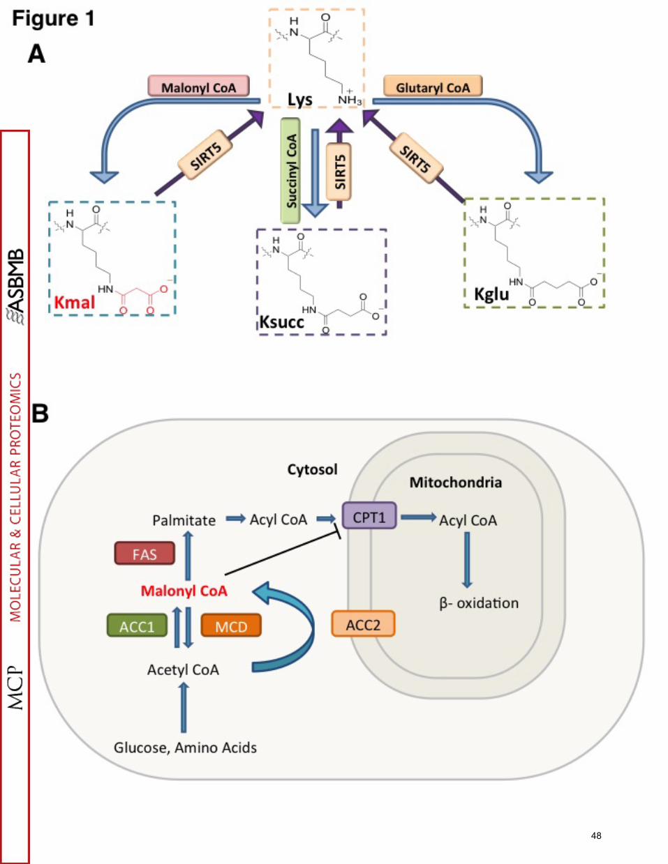
48

49

50
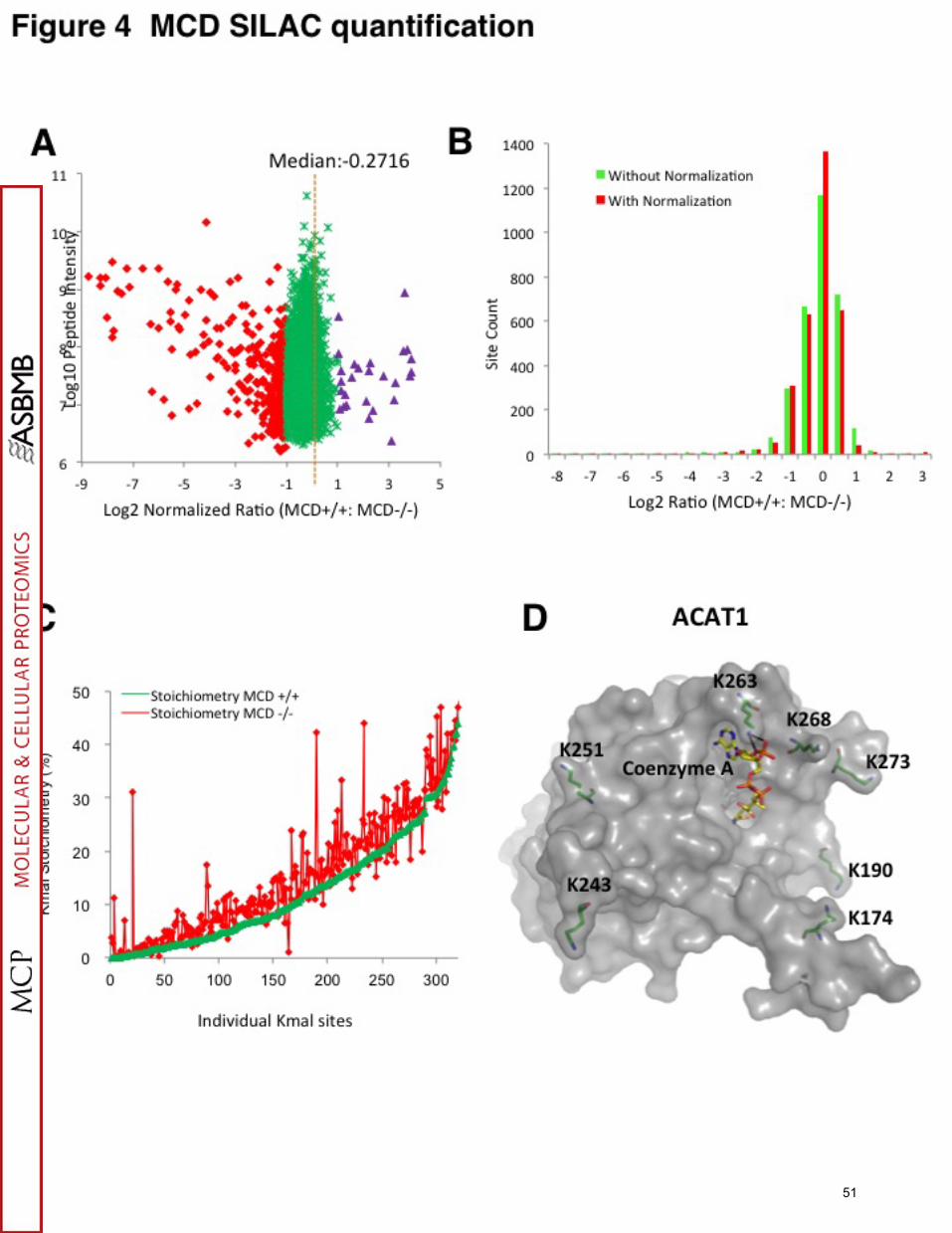
51

52
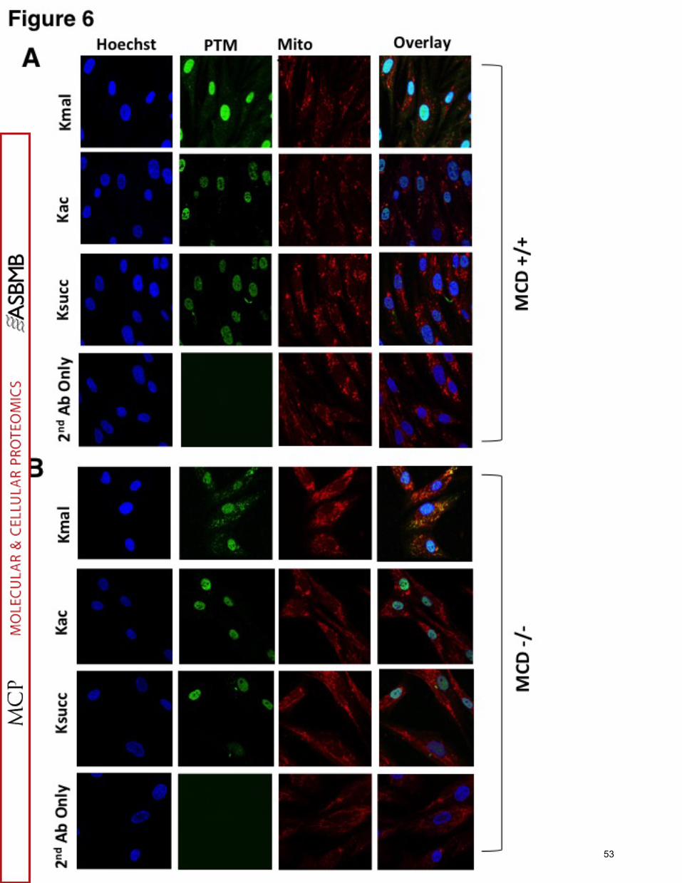
53
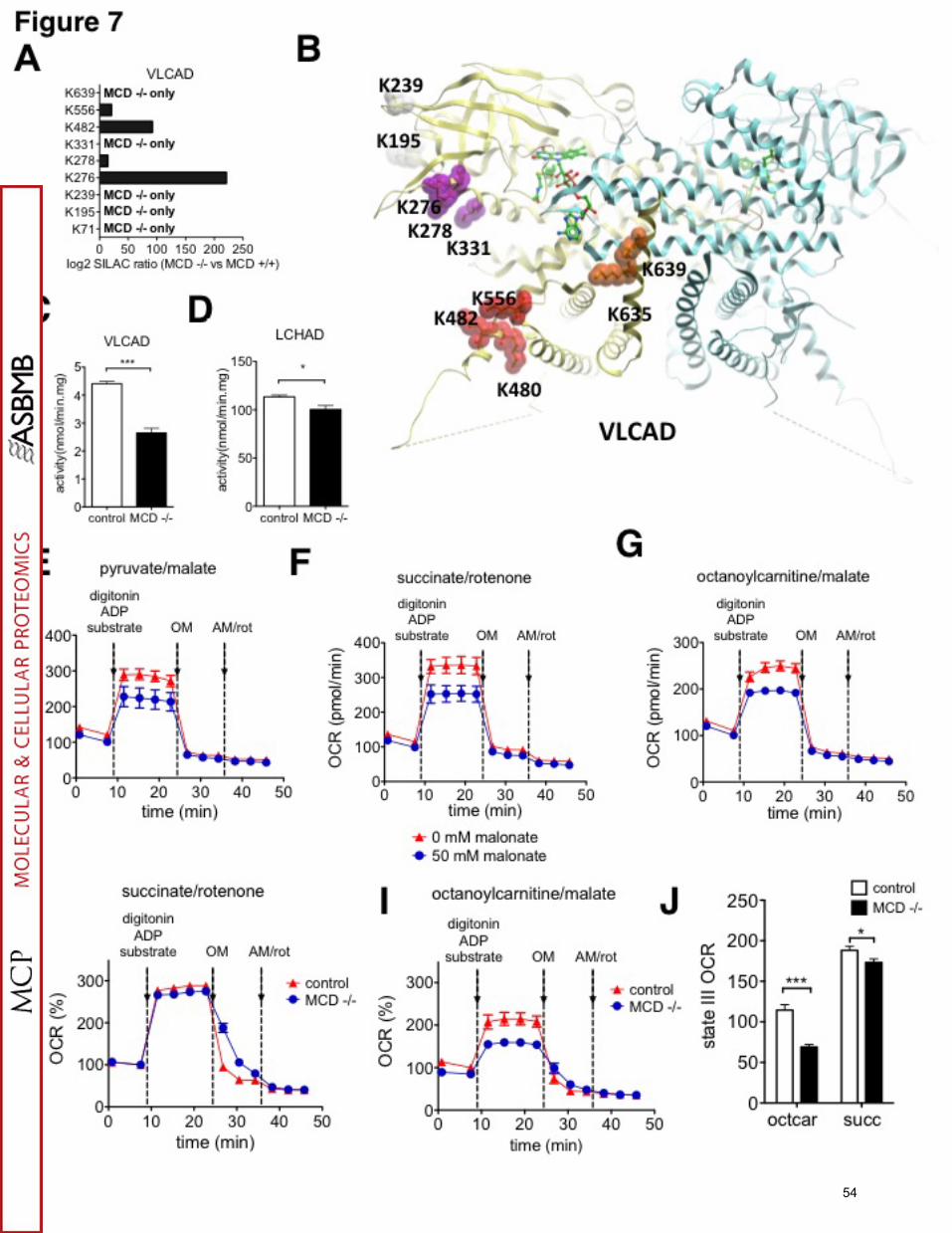
54


