
Individualized pharmacological treatment of
oral mucositis pain in patients with head and
neck cancer receiving radiotherapy
Ingrid Stenström Ling and Britt Larsson
Linköping University Post Print
N.B.: When citing this work, cite the original article.
The original publication is available at www.springerlink.com:
Ingrid Stenström Ling and Britt Larsson, Individualized pharmacological treatment of oral
mucositis pain in patients with head and neck cancer receiving radiotherapy, 2011, Supportive
Care in Cancer, (19), 9, 1343-1350.
http://dx.doi.org/10.1007/s00520-010-0955-1
Copyright: Springer Verlag (Germany)
http://www.springerlink.com/
Postprint available at: Linköping University Electronic Press
http://urn.kb.se/resolve?urn=urn:nbn:se:liu:diva-70215

1
Individualized pharmacological treatment of oral mucositis pain in patients with head and neck cancer
receiving radiotherapy
Authors:
Ingrid Stenstrom Ling, RN1, Britt Larsson MD, PhD
1, 2
1 Pain and Rehabilitation Centre, University Hospital, Linkoping, Sweden.
2 Rehabilitation Medicine, Department of Clinical and Experimental Medicine, Faculty of Health Sciences,
Linkoping University, Linkoping, Sweden.
Address correspondence to: Ingrid Stenstrom Ling, Pain and Rehabilitation Centre, University Hospital,
SE 581 85 Linkoping, Sweden. Phone number: 00 46 10 33684. Fax number: 00 46 10 33682.
E-mail: [email protected].
Number of words: 4109
Number of references: 32
Number of tables: 5

2
Abstract
Purpose: Pain is a prominent symptom in radiotherapy induced oral mucositis (OM). This study assesses the
effect of pharmacological treatment in head and neck cancer patients with OM-induced pain and swallowing
difficulties.
Methods: This study included 82 patients with head and neck cancer undergoing radiotherapy and referred to
the Pain and Rehabilitation Centre at Linkoping University Hospital in Sweden because of OM-induced pain.
During one week, pain assessment, onset of individually tailored choice of drugs, treatment evaluation, and
adjustments, were undertaken.
Combinations of acetaminophen, non-steroid anti-inflammatory drug (NSAID) and opioids were by steps
applied. To evaluate effects, the patients answered the European Organization for Research and Treatment of
Cancer (EORTC) Quality of Life Questionnaire (QLQ-H&N35) in connection with the initial pain assessment
and one week later.
Results: Worsening of soreness in mouth and overall worsening of swallowing difficulties were seen in the
patients referred within the third week of radiotherapy, showing increased severity of OM during the current
week (n=59). Pain and swallowing difficulties were unchanged in patients referred later than the third week,
showing unchanged severity of OM during the current week (n=23).
Conclusion: The answers to the questionnaire showed that the individualized pain treatment with systemic
analgesics exploited to the highest degree was insufficient. Future development of pharmacological possibilities
for treatment of OM related pain is urgent. In addition, development of structured nursing care and patient self-
care can contribute to improved pain relief.
Key words:
Head and neck cancer, radiotherapy, mucositis, pain, pain treatment

3
Introduction
Head and neck cancer is diagnosed in about 650 000 patients worldwide each year (about 6% of all cancer in the
global population) [1]. Current treatments are mainly surgery, radiotherapy (RT), combination of surgery and
RT, or combination of RT and chemotherapy [2]. In patients undergoing RT for head and neck cancer, oral
mucositis (OM) is the most common acute adverse effect [3]. More aggressive tumor treatment methods in
recent years, above all increased use of concomitant chemotherapy, have lead to increased severity of OM [4-6]
and increased mean incidence of OM (80%) [4].
OM, a complex biological process, has been divided into five phases [7]. Several pro-inflammatory and
cytotoxic factors are involved [6-8]. In the average temporal pattern, OM starts with erythema one week after RT
start, coinciding with accumulated radiation dose of 10 Gray (Gy) [6-9]. Confluent erythema and ulcers are seen
during the third week, coinciding with accumulated dose of 30 Gy [6, 8, 9]. Ulceration proceeds during the rest
of the RT period. Fibrin coated ulcers [8] and increased risk of infection [7] are to be expected. A peak of OM
severity is seen in the fifth week of RT [10]. The healing process starts two to three weeks after completed RT
[8], but OM may remain severe five to seven weeks after the RT period [6].
Pain is the most prominent OM symptom [3]. Mild pain arises during the first week of RT [8]. Escalation of pain
is seen from about three weeks of RT [3, 6] in connection with appearance of ulcers [9]. Pain persists until two to
six weeks after completed RT [9]. Sometimes pain remains for six month or more [11]. Another frequent OM
symptom is swallowing difficulties [3-5, 12], mainly due to pain [5], often leading to severe weight loss [5, 12]
and requiring a feeding tube [4, 13].
Thus, OM development with pain and swallowing difficulties requires pain treatment in patients with head and
neck cancer receiving RT. Topical anaesthetics against OM pain are recommended in the literature. Use of
systemic analgesics is also evident, but is too briefly described for clinical use in publications focused on other
aspects of OM [5, 6, 8, 9, 14-16]. Somewhat more elucidating concerning the use of systemic analgesics is the
National Comprehensive Cancer network (NCCN) guidelines for mucositis prevention and management [17].
Non-opioid use for mild pain due to early erythema and opioid use for more severe pain due to ulceration are
recommended [6, 8, 17]. Adjuvant medication with antidepressants and anticonvulsants is even more briefly
mentioned.
To our knowledge, comprehensive and detailed clinical guidelines concerning pharmacological treatment of
OM-induced pain – including pain assessment, choice of drugs, administration routes, pharmaceutical forms, and
evaluation of effect – are lacking. This study was designed as a clinical series of patients treated according to
clinical routine for mucositis-related pain. By the clinical series it was possible to assess the effect of a one-week
step-based individualized pharmacologic regimen in head and neck cancer patients with OM-induced pain and
swallowing difficulties. The hypothesis was that pain relief would be sufficiently improved in the patients by
using a stepwise application of acetaminophen, NSAID, and opioids.

4
Subjects
Ninety-nine patients with head and neck cancer intended for RT were referred for consultation from oncologists
and oto-rhino-laryngologists to the Pain and Rehabilitation Centre at Linkoping University Hospital between
2006 and 2008 due to OM-induced pain. These 99 patients were asked to participate in the study. Eight of these
patients were referred due to tumour related pain before RT. Eighty-two patients participated, as 17 patients were
excluded because of communication difficulties, cognition disturbances, or denial to participate.
The study was conducted as a part of the daily medical work at the Pain Department. It was done in accordance
with Swedish legislation and the patients gave their informed consent.

5
Methods
Procedure
The patients were referred because of pain intensity in mouth, throat, or other locations in head and neck region
of more than 30 mm out of 100 mm according to a visual analogue scale (VAS). The Department’s work in this
study was performed by three experienced nurses in close consultation with physicians specialized in
anaesthesiology. These nurses were well informed about and trained for the procedure of assessment,
intervention and evaluation. The Pain Department’s physicians were responsible for the pain treatment, and there
was a frequent dialogue between the physicians and the nurses administrating the interventions. In the clinical
situation there was strived for pain intensity less than 30 mm out of 100 mm according to VAS for facilitated
oral nutrition, minimized sleeping disturbances due to pain, and ability of patient to endure RT treatment.
The outcome of an initial pain assessment was crucial when the step-based pharmacological intervention, with
systemic analgesics and topical anaesthetics as cornerstones, was tailored and started the same day. The
intervention was based on the World Health Organization (WHO) analgesic ladder for cancer pain relief [18],
excluding the second step in this ladder, i.e. weak opioids. A move from one step to another was taken when
pain intensity was assessed more than 30 mm out of 100 mm according to VAS and/or when moderate or severe
swallowing difficulties persisted according to verbal assessment. Systemic analgesics implied acetaminophen,
NSAID, and strong opioids. Mainly due to tumour induced pain, antidepressants, anticonvulsants, or
betametason were regularly considered. The OM severity and its expected development were considered in the
treatment design. Drug administration routes and pharmaceutical forms were carefully chosen, considering
swallowing ability and patient convenience. Detailed oral information and an easily comprehensible written drug
schedule, revised when necessary, were given to the patient. Furthermore, by frequent dialogue between the
nurses at the Pain Department and the patient, compliance to the treatment was optimized. Evaluation of effect
and possible side effects was conducted every day, except weekends, of the first week. Adjustments were
undertaken in dialogue with the patient.
In summary, for individualized therapy, every point in the initial pain assessment and patient’s different
individual conditions in the pharmacological intervention design was systematically considered, as well as
repeated assessments for evaluation and possible adjustments.
Alterations in pain and swallowing difficulties are closely related to OM development. Therefore, the OM grade
in patients with an irradiated mouth was noted in connection to the initial pain assessment and one week later.
The patients answered a quality of life questionnaire in connection to the initial pain assessment and one week
later. These time points were denoted TQ1 and TQ2, respectively. TQ2 was an appropriate time point for
reassessment, as analgesics with short half-life were consistently used and a steady state was attained at this time
point, making it possible to evaluate the effect of the intervention.
Initial pain assessment
The Department’s treatment of every referred patient started with a structured interview to assess the following:

6
1. Location of pain
2. Duration of pain
3. Continuous or intermittent pain
4. Quality of pain according to the patient’s verbal description of pain experience:
a) descriptors preferably associated with nociceptive pain: e.g., sore, dull, hurting, tender, throbbing
b) descriptors preferably associated with neuropathic pain: e.g., burning, stabbing, tingling, shooting,
radiating
5. Alteration of sensibility
6. Intensity of pain, both continuous and intermittent, according to VAS.
7. Variations in pain, e.g., due to swallowing, chewing, talking, changing in body position, and variations
over day and night
8. Disturbances in daily function
9. Other symptoms induced by RT, such as altered taste, xerostomia, swallowing difficulties, altered
salivation, nausea, weight loss, fatigue, sleeping disturbances, speech difficulties, hoarseness, anxiety
10. Effect of current analgesics
11. Alternative methods to relieve pain, e.g., warmth, cold
12. Smoking, abuse of alcohol and illegal drugs
13. Other diseases and current drug use
In differentiating nociceptive or neuropathic pain, outcomes in especially points 3-7 and 10 were important.
Notwithstanding the difficulties in evaluating the verbal descriptors of the patients, assessment was done in
compliance with screening tools frequently used in clinical practice [19].
Visual analogue scale
The Visual Analogue Scale (VAS) consisted of a 100 mm horizontal line, anchored with “No pain” and “Worst
pain imaginable”. The patient was asked to mark the point on the line corresponding to the intensity of pain
perceived. Recently it has been reported by Fainsinger et al (2009) [20] that pain assessed less than 3 according
to the 11-point (0-10) Numeric Rating Scale (NRS), which correlates well with 30 mm according to VAS, is
categorized as mild pain.
Assessment of mucositis grade
Assessment of OM grade according to a modified version of the WHO scale [21] was done by one trained dentist
at the dental services at the hospital.
Four OM grades were used:
0: No reaction
1: Hyperaemia, impressions, soreness, oedema
2: Erythema, occasional ulcers, soreness
3: Painful erythema, larger fibrin coated ulcers
4: Widespread ulcerated areas, easily bleeding, very painful
Individualized pharmacological intervention

7
Acetaminophen and NSAID
A majority of patients was regularly on treatment with acetaminophen, prescribed by the oncologists, at the
initial pain assessment. As a first step, continued acetaminophen was recommended. As a second step, NSAID,
preferably with short half-life, was additionally prescribed.
Opioids
As a third step, opioids were added if pain relief was insufficient with acetaminophen and NSAID. In case of
severe pain, both NSAID and opioids were prescribed immediately after pain assessment. If a weak opioid was
previously prescribed, a strong opioid instead was prescribed. Oral or enteral morphine and transdermal fentanyl
were preferred. Expected opioid adverse effects always were prevented, preferably prophylaxis against
nausea/vomiting and constipation. Optimal individual opioid dosage was strived for, by regular administration of
a short acting opioid and by adding a rescue dose, for one or two days. The aim was best possible pain relief
according to stipulated goals (section Procedure above) and negligible or acceptable side effects. Daily opioid
doses ranged from 40 mg to 160 mg equivalent to morphine between TQ1 and TQ2. When optimal dosage was
attained, exchange to a slow release opioid was undertaken. Additionally, a short acting opioid was prescribed as
rescue dose against breakthrough pain and as pain prevention, primarily administered before meals.
Adjuvant medication
As a fourth step, amitryptilin, gabapentin, or pregabalin were considered due to neuropathic pain, mainly tumour
related. Betametason was considered for optimized anti-inflammatory effect, impaired general condition, or
antiemetic effect.
Topically acting drugs
Lidocain and benzydamine were prescribed at RT-start by the dental services at the hospital to all patients with
an irradiated mouth at the RT start. Continued use was requested and supported by the dental services and by the
nurses from the Pain Department.
Assessment of pain and swallowing difficulties according to the Quality of Life Questionnaire Head and
Neck 35 (EORTC QLQ-H&N35)
A diagnosis specific module of quality of life questionnaire for head and neck cancer patients (QLQ-H&N35)
designed by The European Organization for Research and Treatment of Cancer (EORTC) was used for clinical
evaluation [22, 23]. The module is a self-administrated questionnaire comprising 35 questions concerning pain
(nos. 31-34), swallowing (nos. 35-38, 49), other physical symptoms, and psychological and social function.
Thirty questions assess symptoms or claims using one of four grades: “not at all”, “a little”, “quite a bit”, or
“very much”. Five questions require “yes” or “no” answers. Data concerning pain and swallowing are presented
in this study.
Outcomes
The primary outcome of the study was pain relief according to the EORTC QLQ-H&N35. The secondary
outcome was decreased swallowing difficulties according to the EORTC QLQ-H&N35.

8
Statistics
Wilcoxon Signed Ranks Test was used for pair-wise calculations. P-values < 0.05 were considered to indicate
statistical significance. The Statistical Package for the Social Sciences version 17.0 was used for all analyses.

9
Results
Dichotomization in early and late intervention groups
Pharmacological pain treatment intervention was started at different time points during RT as the patients were
referred as soon as pain intensity exceeded 30 mm on VAS. The 82 subjects were divided in an early
intervention group (EI) and a late intervention group (LI). Intervention for the EI group (59 patients, mean age
63 years (SD 11), 20 (34%) women) started before RT (eight patients with tumour-related pain) or during week
1-3 of RT, while the intervention for the LI group (23 patients, mean age 66 years (SD 11), 7 (30%) women)
started after week three of RT.
Tumour location and staging, tumour treatment and mucositis grade
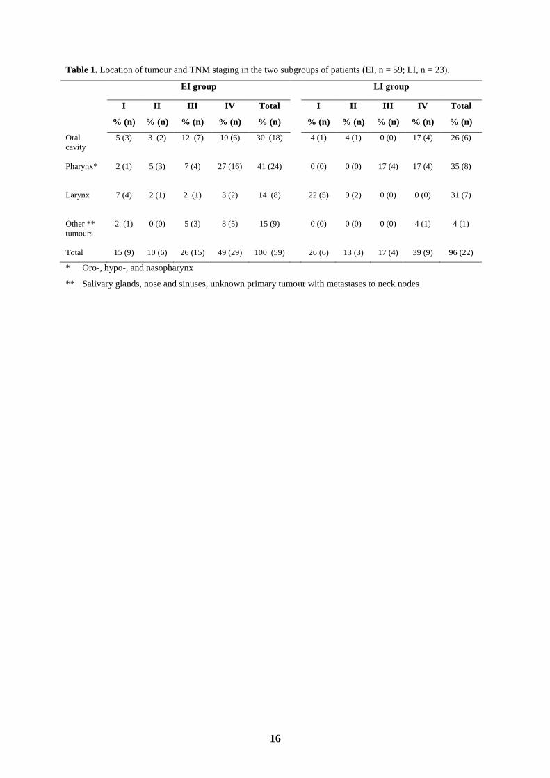
Tumour location and classification according tumour, node, and metastasis (TNM) staging system are shown in
Table 1. Data for TNM staging was missing for one patient (4%) in LI group. Proportion of larynx tumours was
14% in EI group and 31% in LI group. Five (63%) of the larynx tumours in EI group were in stage I and II
according to TNM classification, whereas the seven larynx tumours in LI group were all (100%) in stage I and II.
In the EI group, other tumours comprised 15%; in the LI group, other tumours comprised 4%.
Tumour treatment, chosen considering tumour type, location, size, spreading, and possible metastases, is shown
in Table 2. RT given as standard fractionation was preferably chosen. The distribution of all types of RT
treatments was rather similar in both groups.
In the EI group, the OM grade increased between TQ1 and TQ2 (1.6, standard deviation (SD) 1.3, to 2.2, SD 1.1,
p-value <0.001). In the LI group, the OM grade was unchanged between TQ1 and TQ2 (2.6, SD 0.9, and 2.9, SD
0.9, respectively, p-value 0.059). Thus, at both TQ1 and TQ2 the OM grade was higher in the LI group than in
the EI group (p-values 0.002 and 0.033, respectively).
Pharmacological interventions
Pharmacological interventions during the week between TQ1 and TQ2 are summarized in Table 3. The
pharmacological interventions were rather similar in both groups. Because of likely tumour-related neuropathic
pain, amitryptilin was added for one patient in the EI group and pregabalin was added for one patient in the same
group.
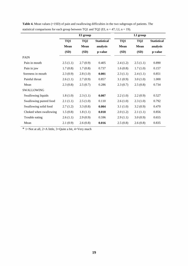
Pain and swallowing difficulties reported in EORTC QLQ-H&N35
Forty-seven patients in the EI group and 19 patients in the LI group answered the questionnaire at both TQ1 and
TQ2. The answers on pain and swallowing questions are presented in Table 4. In the EI group, the pain question
about soreness in the mouth showed unexpectedly significant worsening between TQ1 to TQ2. Significant
worsening was also the case for the three swallowing questions about liquids and solid food, choking when
swallowing, and for the mean score for the five swallowing questions. For the remaining three questions about
pain and the remaining two questions about swallowing difficulties, no significant differences were seen between

10
TQ1 and TQ2. In the LI group, there were no significant differences between TQ1 and TQ2, neither concerning
pain questions nor concerning swallowing questions.
Proportions of patients who reported improvement or worsening in pain and swallowing difficulties for at least
one of four grades (“not at all”, “a little”, “quite a bit”, or “very much”), or who reported no change between
TQ1 and TQ2 are shown in Table 5. The majority of patients in both groups reported unchanged pain and
swallowing difficulties. Missing answers to single questions, either at TQ1 or TQ2 or at both tQ1 and TQ2, in
the questionnaire, are presented in “Missing data”, mean proportion 22 % in the EI group and 23% in the LI
group.

11
Discussion
The individualized pharmacological treatment did not result in sufficient relief of pain or swallowing difficulties
despite exploiting systemic analgesics and topical anaesthetics to the utmost during the first week of advanced
OM pain treatment in patients undergoing RT. We even noticed worsening of complaints in the EI group,
concerning soreness in mouth as well as swallowing difficulties overall, probably due to parallel OM
development.
The hypothesis for the study was that pain relief would be sufficiently improved by using a stepwise application
of acetaminophen, NSAID, and opioids. This hypothesis, tested by comparing pain and swallowing difficulties
according to EORTC QLQ-H&N35 at start of pain treatment and one week later, Table 4, has to be rejected.
In two recent reviews, not mainly addressed to pain treatment, it is incidentally proposed that systemic
analgesics may insufficiently provide pain relief [3, 9]. Also in three recent clinical studies on different aspects
of OM insufficient pain relief with systemic analgesics is mentioned [24-26]. However, the advantage of this
study – a structured individualized intervention with acetaminophen, NSAID, opioids, and topical anaesthetics –
is that it clearly, for the first time to our knowledge, illustrates extensive difficulties achieving improved pain
relief for the patient group studied (Tables 4 and 5).
The referral criterion; pain intensity exceeding 30 mm on VAS, entailed a heterogeneous sample according to the
time for intervention. To handle the sample, we retrospectively dichotomized the patients in early and late
intervention groups. The dichotomization is supported by the literature, suggesting that need of treatment with
systemic analgesics mainly arises within the third week of RT [6]. A peak, 27 of 59 patients in the EI group, of
patients in need of advanced pain intervention during exactly that third week of RT was observed in this study.
By the dichotomizing in early and late referred subjects, it was also possible to compare the effect of early pain
intervention and late pain intervention.
In the EI group, the pain intervention was done in parallel with significantly increased OM severity between
TQ1 and TQ2, the latter possibly contributing to the worsened or unchanged pain and swallowing difficulties
(Table 5). The OM grade in the LI group was unchanged between TQ1 and TQ2 and higher than in the EI group
at both times. However, the pain treatment in the LI group was not as insufficient as in the EI group, inasmuch as
at least no worsening of soreness in the mouth and no worsening of mean score for swallowing questions was
seen.
On the whole, successful outcome of pain treatment was not attained in any of the groups. On the other hand,
without the apparently insufficient pain treatment in this study, the extent of problems related to severe pain
probably would have been still worse during RT. Consequences of severe pain and swallowing difficulties
probably would have resulted in even more undesired breaks in RT and more hospitalizations, both frequent
results reported in the literature [3, 5, 9, 16]. The relief in pain and swallowing difficulties achieved in a minority
of patients between TQ1 and TQ2 (Table 5) would probably not have been realized without our interventions.

12
Retrospectively, we noted that due to supposed neuropathic OM pain, gabapentin or pregabalin were added after
TQ2 for some of the patients. It is well known that nociceptive and inflammatory mechanisms are involved in
OM pain [3, 11, 25]. Treatment of nociceptive pain mostly can be performed with a straightforward design.
Recently, it has been established that neuropathic pain due to OM also is common [3, 11]. Neuropathic pain is
not sensible for analgesics, and topical anaesthetics have too short duration. Instead, treatment with
antidepressants or anticonvulsants is necessary [18]. Onset and dose escalation until acceptable pain relief
require at least as much as a couple of weeks. Thus, an extended time period had been necessary for evaluation
of the intervention with adjuvant medication, which in this clinical series was primary intended for tumour
related neuropathic pain and not for OM pain, as mentioned in section Methods, Procedure. However, in this
study the intervention between TQ1 and TQ2 in fact appeared to be treatment with systemic analgesics due to
nociceptive pain. Enough attention was possibly not directed to different current and expected pain mechanisms
[3, 11, 25]. We agree with the recent publications [3, 11, 25] suggesting use of adjuvant medication more
frequently and earlier in OM development in order to attain improved pain relief. In a recent study Bar Ad et al
(2009) [27] stated, that gabapentin appeared to be promising in reducing the need of opioids for patients with
OM pain, which indicates that the role of gabapentin in OM pain therapy ought to be studied.
Swallowing difficulties remained unchanged or increased for a majority of the patients (Tables 4 and 5). More
severe pain associated with increased swallowing difficulties is reported in a recent study [24]. For optimal
nutrition, continuous nursing support and education concerning adjusted diet and nutritional supplements are
essential [13]. Prophylactic placement of percutaneous endoscopic gastrostomy (PEG) feeding tube has been
recommended to ensure sufficient nutrition and thereby prevent unwanted RT-breaks and hospitalizations due to
severe weight loss [5, 16, 28]. Using PEG feeding tube may also imply pain relief. Patients with PEG can avoid
frequent painful and pain triggering swallowing. Furthermore, the PEG offers an alternative administration route
for almost all oral medication.
Frequent basic oral care is considered to reduce duration and severity of OM [12, 14, 29, 30]. The use of current
clinical guidelines facilitates structured care [14, 29]. Nursing support and frequent patient self-care contribute to
reduced duration and severity of OM pain [30].
Nursing care is important for overall improved management in this patient group [31]. By improving nurse-
based patient education, support, and dialogue, the ability of the patients to cope with pain and swallowing
difficulties may be improved [26, 32].
Methodological considerations
Without any comparison group, a number of unidentified confounding factors are likely to be present, which is a
major concern in the interpretation of the results. Furthermore, a majority of subjects not eligible at RT-start, a
heterogeneous clinical picture at onset of pain treatment and the short observation time are limitations which
have to be considered when the results are interpreted.
13
Mean proportion for missing answers, and thus, also a missing possibility to make the Wilcoxon Signed Ranks
Test, reached 22 % in the EI group and 23% in the LI group. This considerable proportion of missing data might
be caused by limited willingness of patients with less pain or swallowing difficulties to answer the EORTC
QLQ-H&N35 completely. However, it is difficult to assess the significance of the missing data in the
interpretation of the results.
Patients with larynx tumour differed from other tumours (Table 1) regarding target area of RT. Without an
irradiated mouth, they had no mucosal injury located in the mouth, but they did have it located in the larynx area.
It is reasonable to suspect that the answers from larynx patients in both groups decreased mean score for pain
and soreness in mouth, but increased mean score for painful throat. The main results, however, were probably
not influenced.
Conclusion and Clinical Implications
Despite transparent individualized interventions with systemic analgesics, it was shown in this clinical series of
patients with head and neck cancer undergoing RT, that OM pain treatment was insufficient. The clinical
implications of the results are that careful and repeated attention has to be directed to current and expected pain
mechanisms. When systematic analgesics do not provide sufficient OM pain relief, we suggest, according to our
very limited clinical experience and to the recent publication of Bar Ad et al (2009) [27], and in absence of other
possibilities at present, that additional treatment with gabapentin or pregabalin might be tried. However, the
effect of gabapentin and pregabalin on OM pain has to be evaluated in carefully designed studies. Parallel to
development of pharmacological pain treatment it is valuable to further investigate how structured nursing
support and patient self-care can contribute to improved pain relief.

14
References
1. Argiris A, Karamouzis MV, Raben D, Ferris RL (2008) Head and neck cancer. Lancet 371:1695-1709
2. Scully C, Bagan J (2009) Oral squamous cell carcinoma overview. Oral Oncol 45:301-308.
doi:10.1016/j.oraloncology.2009.01.004
3. Epstein JB, Elad S, Eliav E, Jurevic R, Benoliel R (2007) Orofacial pain in cancer: Part II - Clinical
perspectives and management. J Dent Res 86(6):506-518. doi: 10.1177/154405910708600605
4. Trotti A, Bellm LA, Epstein JB, Frame D, Fuchs HJ, Gwede CK, Komaroff E, Nalysnyk L, Zilberberg
M (2003) Mucositis incidence, severity and associated outcomes in patients with head and neck cancer
receiving radiotherapy with or without chemotherapy: a systematic literature review. Radiother Oncol
66(3):253-262. doi:10.1016/S0167-8140(02)00404-8
5. Lalla RV, Sonis ST, Peterson DE (2008) Management of oral mucositis in patients with cancer. Dent
Clin North Am 52(1):61-77, viii
6. Scully C, Sonis S, Diz PD (2006) Oral mucositis. Oral Dis 12(3):229–241. doi:10.1111/j.1601-
0825.2006.01258.x
7. Sonis ST, Elting LS, Keefe D et al (2004) Perspectives on cancer therapy-induced
mucosal injury, pathogenesis, measurement, epidemiology, and consequences for patients. Cancer
100(9 Suppl):1995-2025. doi: 10.1002/cncr.20162
8. Treister N, Sonis S (2007) Mucositis: biology and management. Curr Opin Otolaryngol Head Neck
Surg 15:123-129
9. Napenas JJ, Shetty KV, Streckfus CF (2007) Oral mucositis: Review of pathogenesis, diagnosis,
prevention and management. Gen Dent 55(4):335-344
10. Elting LS, Cooksley CD, Chambers MS, Garden AS (2007) Risk, outcomes and costs of radiation-
induced oral mucositis among patients with head-and-neck malignancies. Int J Radiat Oncol Biol Phys
68(4):1110-1120. doi:10.1016/j.ijrobp.2007.01.053
11. Benoliel R, Epstein J, Eliav E, Jurevic R, Elad S (2007) Orofacial pain in cancer: Part I - Mechanisms. J
Dent Res 86(6):491-505. doi: 10.1177/154405910708600604
12. Rosenthal DI, Trotti A (2009) Strategies for managing radiation-induced mucositis in head and neck
cancer. Semin Radiat Oncol (19):29-34. doi:10.1016/j.semradonc.2008.09.006
13. Raykher A, Russo L, Schattner M et al (2007) Enteral nutrition support of head and neck cancer
patients. Nutr Clin Pract 22(1):68-73. doi: 10.1177/011542650702200168
14. Rubenstein EB, Peterson DE, Schubert et al (2004) Clinical practice guidelines for the prevention and
treatment of cancer therapy–induced oral and gastrointestinal mucositis. Cancer 100(9):2026-2046. doi:
10.1002/cncr.20163
15. Silverman S (2007) Diagnosis and management of oral mucositis. J Support Oncol 5(2, Suppl 1):13-21.
16. Russo G, Haddad R, Posner M, Machtay M (2008) Radiation treatment breaks and ulcerative mucositis
in head and neck cancer. Oncologist 13(8):886–898. doi: 10.1634/theoncologist.2008-0024
17. Bensinger W, Schubert M, Ang K-K, Brizel D, Brown E, Eilers J, Elting L, Mittal B, Schattner M,
Spielberger R, Treister N, Trotti A (2008) NCCN Task Force Report. Prevention and management of
mucositis in cancer care. J Natl Compr Canc Netw. Jan;6 Suppl 1:S1-21; quiz S22-4
18. Laird B, Colvin L, Fallon M (2008) Management of cancer pain: basic principles and neuropathic
cancer pain. Eur J Cancer. 44(8):1078-82. doi:10.1016/j.ejca.2008.03.022
19. Bennett MI, Attal N, Backonja M, Baron R, Bouhassira D, Freynhagen R, Scholz J, Tölle TR, Wittchen
H-U, Staehelin Jensen T (2007) Using screening tools to identify neuropathic pain. Pain 127(3):199-
203. doi:10.1016/j.pain.2006.10.034
20. Fainsinger RL, Fairchild A, Nekolaichuk C, Lawlor P, Lowe S, Hanson J (2009) Is pain intensity a
predictor of the complexity of cancer pain management? J Clin Oncol 27(4):585-90.
doi:10.1200/JCO.2008.17.1660
21. World Health Organization (1979) Handbook for reporting results of cancer treatment. Geneva,
Switzerland, pp15-22
22. Bjordal K, Hammerlid E, Ahlner-Elmqvist M et al (1999) Quality of life in head and neck cancer
patients: validation of the European organization for Research and Treatment of Cancer Quality of Life
Questionnaire-H&N35. J Clin Oncol 17(3):1008-1019
23. Sherman AC, Simonton S, Adams DC et al (2000) Assessing quality of life in patients with head and
neck cancer: cross-validation of the European Organization for Research and Treatment of Cancer
(EORTC) Quality of Life Head and Neck module (QLQ-H&N35). Arch Otolaryngol Head Neck Surg
126(4):459-67
24. Murphy B, MD, Beaumont J, Isitt J, Garden AS, Gwede CK, Trotti AM, Meridith RF, Epstein JB, Le
Q-T, Brizel DM, Bellm LA, Wells N, Cella D (2009) Mucositis-related morbidity and resource
utilization in head and neck cancer patients receiving radiation therapy with or without chemotherapy. J
Pain Symptom Manage 38(4):522-532. doi:10.1016/j.jpainsymman.2008.12.004

15
25. Epstein JB, Wilkie DJ, Fischer DJ, Kim Y, Villines D (2009) Neuropathic and nociceptive pain in head
and neck cancer patients receiving radiation therapy. Head Neck Oncol 1(1):26:1-12. doi:
10.1186/1758-3284-1-26
26. Wong PC, Dodd MJ, Miaskowski C, Paul SM, Bank KA, Shiba GH, Facione N (2006) Mucositis pain
induced by radiation therapy: prevalence, severity, and use of self-care behaviours. J Pain Symptom
Manage 32(1):27-37. doi: 10.1016/j.jpainsymman.2005.12.020
27. Bar Ad V, Weinstein G, Dutta PR, Chalian A, Both S, Quon H (2009) Gabapentin for the treatment of
pain related to radiation-induced mucositis in patients with head and neck tumors treated with intensity-
modulated radiation therapy. Head Neck 32(2):173-177. doi:10:1002/hed.21165
28. Raykher A, Correa L, Russo L et al (2009) The role of pretreatment percutaneous endoscopic
gastrostomy in facilitating therapy of head and neck cancer and optimizing the body mass index of the
obese patient. J Parenter Enteral Nutr 33(4):404-410. doi: 10.1177/0148607108327525
29. McGuire DB, Correa ME, Johnson J, Wienandts P (2006) The role of basic oral care and good clinical
practice principles in the management of oral mucositis. Support Care Cancer 14:541–547. doi:
10.1007/s00520-006-0051-8
30. Harris D, Eilers J, Harriman A et al (2008) Putting evidence into practice: Evidence-Based
Interventions for the Management of Oral Mucositis. Clin J Oncol Nurs 12(1):141-52. doi:
10.1188/08.CJON.141-152
31. Armstrong JA, McCaffrey R (2005) The effects of mucositis on quality of life in patients with head and
neck cancer. Clin J Oncol Nurs 10(1):53-56. doi: 10.1188/06.CJON.53-56
32. Wells M, Donnan PT, Sharp L et al (2008) A study to evaluate nurse-led on-treatment review for
patients undergoing radiotherapy for head and neck cancer. J Clin Nurs 17(11):1428-39. doi:
10.1111/j.1365-2702.2007.01976.x
16
Table 1. Location of tumour and TNM staging in the two subgroups of patients (EI, n = 59; LI, n = 23).
EI group LI group
I
% (n)
II
% (n)
III
% (n)
IV
% (n)
Total
% (n)
I
% (n)
II
% (n)
III
% (n)
IV
% (n)
Total
% (n)
Oral
cavity
5 (3) 3 (2) 12 (7) 10 (6) 30 (18)
4 (1) 4 (1) 0 (0) 17 (4) 26 (6)
Pharynx* 2 (1)
5 (3) 7 (4) 27 (16) 41 (24) 0 (0) 0 (0) 17 (4) 17 (4) 35 (8)
Larynx 7 (4)
2 (1) 2 (1) 3 (2) 14 (8) 22 (5) 9 (2) 0 (0) 0 (0) 31 (7)
Other **
tumours
2 (1)
0 (0) 5 (3) 8 (5) 15 (9) 0 (0) 0 (0) 0 (0) 4 (1) 4 (1)
Total 15 (9) 10 (6) 26 (15) 49 (29) 100 (59) 26 (6) 13 (3) 17 (4) 39 (9) 96 (22)
* Oro-, hypo-, and nasopharynx
** Salivary glands, nose and sinuses, unknown primary tumour with metastases to neck nodes

17
Table 2. Type of radiotherapy in the two subgroups of patients
(EI, n = 59; LI, n = 23).
EI group
% (n)
LI group
% (n)
Primary RT, standard fractionation 62 (37) 74 (17)
Primary RT, altered fractionation 9 (5) 4 (1)
Preoperative RT 17 (10) 9 (2)
Postoperative RT 9 (5) 13 (3)
RT and concomitant chemotherapy 3 (2) 0 (0)

18
Table 3. Pharmacological interventions during the week between TQ1
and TQ2 in the two subgroups of patients (EI, n = 59; LI, n = 23).
EI group
% (n)
LI group
% (n)
NSAID added 5 (3) 0 (0)
Concomitant NSAID, opioid added* 20 (12) 17 (4)
NSAID and opioid added* 41 (24) 35 (8)
Opioid added* 34 (20) 48 (11)
* Includes onset or increased dose of strong opioid and weak opioid
changed to strong opioid
19
Table 4. Mean values (+1SD) of pain and swallowing difficulties in the two subgroups of patients. The
statistical comparisons for each group between TQ1 and TQ2 (EI, n = 47; LI, n = 19).
EI group LI group
TQ1
Mean
(SD)
TQ2
Mean
(SD)
Statistical
analysis
p-value
TQ1
Mean
(SD)
TQ2
Mean
(SD)
Statistical
analysis
p-value
PAIN
Pain in mouth 2.5 (1.1) 2.7 (0.9) 0.405 2.4 (1.2) 2.5 (1.1) 0.890
Pain in jaw 1.7 (0.8) 1.7 (0.8) 0.737 1.6 (0.8) 1.7 (1.0) 0.157
Soreness in mouth 2.3 (0.9) 2.8 (1.0) 0.001 2.3 (1.1) 2.4 (1.1) 0.851
Painful throat 2.6 (1.1) 2.7 (0.9) 0.857 3.1 (0.9) 3.0 (1.0) 1.000
Mean 2.3 (0.8) 2.5 (0.7) 0.286 2.3 (0.7) 2.5 (0.8) 0.734
SWALLOWING
Swallowing liquids 1.8 (1.0) 2.3 (1.1) 0.007 2.2 (1.0) 2.2 (0.9) 0.527
Swallowing pureed food 2.1 (1.1) 2.5 (1.0) 0.110 2.6 (1.0) 2.3 (1.0) 0.792
Swallowing solid food 2.7 (1.2) 3.3 (0.8) 0.004 3.1 (1.0) 3.2 (0.9) 0.470
Choked when swallowing 1.5 (0.8) 1.8 (1.1) 0.018 2.0 (1.2) 2.1 (1.1) 0.856
Trouble eating 2.6 (1.1) 2.9 (0.9) 0.596 2.9 (1.1) 3.0 (0.9) 0.655
Mean 2.1 (0.9) 2.6 (0.8) 0.016 2.5 (0.8) 2.6 (0.8) 0.835
* 1=Not at all, 2=A little, 3=Quite a bit, 4=Very much

20
Table 5. Distributions of alterations (three classes: Improvement, No change, or Worsening) in pain and
swallowing difficulties at least one grade or no change in QLQ-H&N35 between TQ1 and TQ2 in the two
subgroups of patients (EI group n=47; LI group n=19). The variable Missing represents question not answered
at TQ1 or TQ2.
EI group LI group
Improve-
ment
% (n)
No
change
% (n)
Worse-
ning
% (n)
Missing
% (n)
Improve-
ment
% (n)
No
change
% (n)
Worse-
ning
% (n)
Missing
% (n)
PAIN
Pain in mouth
17 (10) 42 (25) 22 (13) 19 (11) 13 (3) 52 (12) 9 (2) 26 (6)
Pain in jaw
22 (13) 39 (23) 17 (10) 22 (13) 4 (1) 57 (13) 17 (4) 22 (5)
Soreness in mouth
9 (5) 32 (19) 37 (22) 22 (13) 26 (6) 39 (9) 13 (3) 22 (5)
Painful throat
20 (12) 39 (23) 20 (12) 21 (12) 26 (6) 30 (7) 26 (6) 18 (4)
SWALLOWING
Swallow liquids
7 (4) 44 (26) 27 (16) 22 (13) 18 (4) 30 (7) 26 (6) 26 (6)
Swallow pureed
food
10 (6)
39 (23) 25 (15) 26 (15) 17 (4) 44 (10) 13 (3) 26 (6)
Swallow solid
food
10 (6)
37 (22) 29 (17) 24 (14) 13 (3) 43 (10) 22 (5) 22 (5)
Choked when
swallowing
5 (3)
53 (31) 22 (13) 20 (12) 26 (6) 30 (7) 22 (5) 22 (5)
Trouble eating 27 (16) 29 (17) 22 (13) 22 (13) 22 (5) 26 (6) 30 (7) 22 (5)


