does this patient need a the clinical problem lumbar … clinical problem ... patient needs an lp...
TRANSCRIPT

11/8/2011
1
Improving Clinical Care: Clinical Decisions Rules You Care AboutDeborah B. Diercks
Disclosure� None
The clinical problem� 37 yo F presents with the complaint of the worse headache her life– Looks good– Complains of 10/10 pain
Does this patient need a lumbar puncture?
N o Y es
O nl y i f
h e r p a .
. . O n
l y i f h e r
d a .. .
51%
19%
8%
21%
1. No2. Yes3. Only if her pain does not
go away4. Only if her dad is a lawyer

11/8/2011
2
Subarachnoid Hemorrhage� Rule 1
– Age >40– Complaint of neck pain or stiffness– Witnessed loss of consciousness – Onset with exertion
� Rule 2 – Arrival by ambulance– Age >45– Vomiting at least once– Diastolic blood pressure >100 mm Hg
� Rule 3 – Arrival by ambulance– Systolic blood pressure >160 mm Hg– Complaint of neck pain or stiffness – Age 45-55
BMJ 2010;341:5204c BMJ 2010;341:5204c
Clinical Implications� Raises the question that not every patient needs an LP� Not prospectively tested so apply with caution� May aid your decision making
Clinical Problem� 55 yo F presents with syncope
– Witnessed event that occurred at church

11/8/2011
3
What percent of syncope patients do you admit?
0 -2 5
%
2 6- 5 0
%
5 1- 7 5
%
> 75 %
43%
4%
11%
41%1. 0-25%2. 26-50%3. 51-75%4. >75%
Syncope Rules� San Francisco
– ECG– dyspnea,– systolic BP 90– Hct 30%– Hx of CHF
� OESIL– ECG– age 65 y– Hx of cardiac dz– No prodrome
� ROSE– ECG– BNP 300,– bradycardia 50, – Hgb 9,– chest pain, – O sat 94%,– stool + for occult blood test
Annals of Emerg Med 2010; 56:, 362-373.e1
LR + >10
LR -<0.1
Annals of Emerg Med 2010; 56:, 362-373.e1
LR + >10
LR -<0.1
Annals of Emerg Med 2010; 56:, 362-373.e1

11/8/2011
4
� Variability in the rules– ECG criteria� Who interpreted the ECG
Annals of Emerg Med 2010; 56:, 362-373.e1
Clinical Implications� Negative LR relatively high� Positive LR relatively low� Challenge with implementation of these rules
Abdominal CT� Adult patients, 18 years of age or older, with blunt torso trauma� Outcome of interest
– patients with intra-abdominal injury who were undergoing acute intervention – Patients with any intra-abdominal injury.
� Patients were considered to have an intra-abdominal injury – spleen, liver, gallbladder, pancreas, kidney, ureter, urinary bladder, gastrointestinal tract, or an intra-abdominal vascular structure
Ann Emerg Med 2009
Clinical Challenge� 25 yo M presents after a MVA at 60 miles per hour– Not intoxicated– No pain
– Does this patient require an Abdominal CT

11/8/2011
5
Decision rule for any intervention Ann Emerg Med. 2009 Oct;54(4):575-84.
Decision Rule for IAI
Validation phase for any injury� Sensitivity
– 95.8% (91.1, 98.4%)� Specificity
– 29.9% (27.5, 32.3%)� PPV
– 11.9% (10.1, 13.9%)� NPV
– 98.6% (97.1, 99.5%)Ann Emerg Med. 2009 Oct;54(4):575-84.
Implications for Practice� Potential change to practice
– Gross hematuria?– Need for observation with tenderness?– Does no image=discharge home

11/8/2011
6
Clinical Challenge� 72 yo male falls off his grandchild’s scooter and hits his head
Does he need a Head CT?
Do you routinely use and FOLLOW CT imaging rules?
N ev e r
: I ba s e
. . .
O cc a s
i o na l l y
A l wa y s
31%
19%
50%
1. Never: I base my decision on my clinical decision making
2. Occasionally3. Always
Head CT - adults� The different decision rules are similar in most respects and classify risk according to the following features– initial GCS on admission and GCS 2 hours post injury
– duration of loss of consciousness or amnesia– presence or absence of specified risk factors
� Some of the commonly used decision rules for adults include the Canadian CT head rule NEXUS II and the New Orleans criteria
Canadian Head CT� CT Head Rule is only required for patients with minor head injuries with any one of the following:
– High risk (for neurological intervention)� GCS score <15 at 2 h after injury� Suspected open or depressed skull fracture� Any sign of basal skull fracture (haemotympanum, ‘racoon’eyes, cerebrospinal fluid otorrhoea/rhinorrhoea, Battle’s sign)� Vomiting two episodes� Age > 65 years
– Medium risk (for brain injury on CT)� Amnesia before impact >30 min� Dangerous mechanism (pedestrian struck by motor vehicle,occupant ejected from motor vehicle, fall from height >3 feet or five stairs)

11/8/2011
7
Canadian Rule-High risk� Sensitivity 100% (95% CI 92–100%)� Specificity 68.7% (95% CI 67–70%)� CT ordering proportion 32.2%
Nexus� Enrolled blunt trauma patients where the decision had been made to perform imaging were assessed for the presence or absence of specified criteria� The decision rule has a sensitivity 98.3% (CI 97.2-99.0%)� 12.8% were classified as low risk and would not have required a scan� It will miss about 1.7% of clinically important injuries
NEXUS Criteria1. Evidence of significant skull fracture2. Scalp hematoma3. Neurologic deficit4. Altered level of alertness5. Abnormal behavior6. Coagulopathy7. Persistent vomiting8. Age greater than 65 years
CDC/ACEP clinical policy1. Which patients with mild TBI should have a noncontrast head CT scan in the ED?Level A: Loss of consciousness or posttraumatic amnesia and one or more of the following:
• Headache� • Vomiting� • Age > 60 years old� • Drug or alcohol intoxication� • Deficits in short-term memory� • Physical evidence of trauma above the clavicle� • Posttraumatic seizure� • GCS score < 15� • Focal neurologic deficit� • Coagulopathy
Level B: Head trauma patients with no loss of consciousness or posttraumatic amnesia and one or more of the following:� • Focal neurologic deficit� • Vomiting� • Severe headache� • Age ≥ 65 years old� • Physical signs of a basilar skull fracture� • GCS score < 15� • Coagulopathy� • Dangerous mechanism of injury.*
Combination of the 2 rules

11/8/2011
8
Head CT - children� For age < 2 years:
– normal mental status (including GCS = 15) – no scalp hematoma or only a frontal scalp hematoma – no loss of consciousness or loss of consciousness for < 5 seconds – non-severe injury mechanism – no palpable skull fracture – acting normally (per parental description).
� For age ≥ 2 years:– normal mental status (including GCS = 15) – no loss of consciousness – no vomiting – non-severe injury mechanism – no signs of basilar skull fracture – no severe headache.
Lancet 2009;374(9696):1160-1170.
Head CT in kids� The prediction rule for age < 2 years
– NPV for ciTBI of 100% [95% confidence interval (CI): 99.7% - 100%],
– sensitivity for ciTBI of 100% (95% CI: 86.3% - 100%) � The prediction rule for age ≥ 2 years
– NPV for ciTBI of 99.95% (95% CI: 99.81% - 99.99%), – sensitivity for ciTBI of 96.8% (95% CI: 89.0% - 99.6%)
� Neither decision rule missed any children who required neurosurgery.
Lancet 2009;374(9696):1160-1170.
Implications on Clinical Practice� May reduce imaging
– Helps MD explain why no imaging needed– Helps parents understand why it may be needed
– About 25% of children age < 2 years and 20% of children age ≥ 2 years met the criteria
– Document need for testing
Clinical Challenge: ACS risk
11/8/2011
9
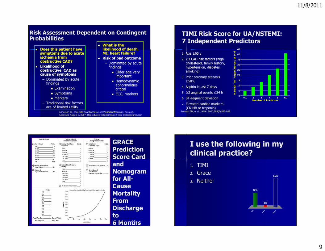
Risk Assessment Dependent on Contingent Probabilities� Does this patient have symptoms due to acute ischemia from obstructive CAD?
� Likelihood of obstructive CAD as cause of symptoms– Dominated by acute findings� Examination� Symptoms� Markers
– Traditional risk factors are of limited utility
� What is the likelihood of death, MI, heart failure?
� Risk of bad outcome– Dominated by acute findings� Older age very important� Hemodynamic abnormalities critical� ECG, markers
Anderson JL, et al. http://cardiosource.com/guidelinefocus/gfc_acs.asp. Accessed August 8, 2007. Reproduced with permission from Cardiosource.com.
TIMI Risk Score for UA/NSTEMI:7 Independent Predictors1. Age ≥65 y2. ≥3 CAD risk factors (high
cholesterol, family history, hypertension, diabetes, smoking)
3. Prior coronary stenosis ≥50%
4. Aspirin in last 7 days5. ≥2 anginal events ≤24 h6. ST-segment deviation7. Elevated cardiac markers
(CK-MB or troponin)Antman EM, et al. JAMA. 2000;284(7):835-842.
Number of Predictors
0
5
10
15
20
25
30
35
40
45
0/1 2 3 4 5 6/7
% D
eath
/ M
I / U
rgen
t Rev
asc
at 1
4 d
GRACE Prediction Score Card and Nomogramfor All-Cause Mortality From Discharge to6 MonthsEagle KA, et al. JAMA. 2004;291(22):2727-2733.
I use the following in my clinical practice?
T I MI
G ra c e
N ei t h
e r
32%
65%
3%
1. TIMI2. Grace3. Neither

11/8/2011
10
To Guide Initial TreatmentInvasive Strategy Preferred ● Recurrent angina or ischemia at rest or with low-level activities● Elevated cardiac biomarkers (TnT or TnI)● New or presumably new ST-segment depression● Signs or symptoms of HF or new or worsening mitral regurgitation● High-risk findings from noninvasive testing● Hemodynamic instability● Sustained ventricular tachycardia● PCI within 6 months● Prior CABG● High risk score (eg, TIMI, GRACE)● Reduced LV function (LVEF <40%)Conservative (Selectively Invasive) Strategy Preferred● Low risk score● Patient or physician preference in absence of high-risk features
Anderson JL, et al. J Am Coll Cardiol. 2007;50(7):e1-e157.
TIMACS: Study Design
Mehta SR, et al. Presented at the American Heart Association 2008 Scientific Sessions, November 8-12, 2008, New Orleans, Louisiana.
� 3,031 patients with UA/NSTEMI and two of:– Age >60– Ischemic ECG changes– Increased biomarkers
� Randomized to: – Early invasive strategy (≤24 h after presentation,
n=1,593) – Delayed strategy (>36 h after presentation,
n=1438).
� Primary end point: death/MI/stroke within 6 months
� Secondary end point: death/MI/refractory ischemia
TIMACS: Other End Points
Mehta SR, et al. Presented at the American Heart Association 2008 Scientific Sessions, November 8-12, 2008, New Orleans, Louisiana.
� Significant reduction in refractory ischemia with early strategy (1.0% vs 3.3%, P<.00001)
� Other efficacy end points similar between strategies– Death (4.9% early vs 6.0% delayed)– MI (4.8% vs 5.8%)– Stroke (1.3 % vs 1.4%)– Repeat revascularization (8.8% vs 8.6%)
� Bleeding events similar– Major bleeding (3.1% vs 3.5%)– Retroperitoneal bleeding (0.1% vs 0.2%)– ≥3 g/dL drop in hemoglobin (2.3% vs 2.6%)
TIMACS: Primary End PointWith Risk Stratification
Mehta SR, et al. Presented at the American Heart Association 2008 Scientific Sessions, November 8-12, 2008, New Orleans, Louisiana.
Dea
th /
MI /
Str
oke
(%)
7.7 6.7
21.6
14.1
30
20
10
0
HR 1.14, P=.43
HR 0.72, P=.005
GRACE score <140,n=2070
GRACE score ≥140,n=961
EarlyIntervention
DelayedIntervention

11/8/2011
11
Implications on Clinical Practice� Can’t rule out disease based on TIMI score� Use scores to identify risks of adverse events– Level of care– Therapy– Management strategy– ? transfer
Clinical Challenge� 76 yo Male presents with chest pain. Has a hx of CAD and positive troponins.– Can I give an antiplatelet agent now?– Will I delay revascularization?� The Clinton paradox
Variables independently associated with CABG – TACTICS TIMI 18
Variable Odds Ratio 95% CI P-value Risk score
Hx prior CABG 0.35 0.2-0.5 <0.0001 -2(+) Troponins 3.9 2.7 – 5.5 <0.0001 3Prior Angina 1.8 1.3 – 2.6 0.001 1ST deviation 1.7 1.3 – 2.2 <0.0001 1Male Gender 1.6 1.2 – 2.2 0.001 1Hx PAD 1.6 1.1 – 2.6 0.038 1
Sadanandan S. et al. JACC 2004;44:799-803
Validation of association between CABG and Increasing Risk Score – TIMI 11B Trial
4.57.7
11.5
0
5
10
15
20
25
<3 3-5 >5
CA
BG
(%
)
N=3,722
P<0.0001C-statistic 0.61
Risk score
(76 / 1700) (130 / 1692) (38 / 330)(n)Sadanandan S. et al. JACC 2004;44:799-803

11/8/2011
12
Validation of CABG Risk Score –TIMI III Registry
8
19.7
25.9
0
5
10
15
20
25
30
<3 3.5 >5
CA
BG
(%
)
N=1,139
P<0.0001C-statistic 0.66
Risk score
(48 / 1078) (121 / 1628) (57 / 523)(n)Sadanandan S. et al. JACC 2004;44:799-803
Implications on Care� Use to stratify risk of CABG
– Not perfect� Prior CABG decreases risk
– Higher in men– PAD– NSTEMI
Clinical Challenge� 85 yo F presents with chest pain. Cardiac markers are negative but ECG has T wave inversions– Presumptive diagnosis Unstable Angina– What treatments should I give� Risk vs. Benefit
Bleeding Risk� Nearly 30% of all patients with ACS will suffer from a bleeding complication.� Thirty day mortality increase > 6 fold � Major bleeding is a predictor of 30-day mortality than� 3 fold increase in 30 day ischemic events

11/8/2011
13
CRUSADE BLEEDING SCORE– Baseline hematocrit
<36% – Creatinine clearance – Heart rate – Systolic blood pressure
≤110 mm Hg or ≥180 mm Hg
– Female sex – Congestive heart disease – Prior vascular disease – Diabetes
Circulation. 2009 Apr 14;119(14):1873-82
http://www.crusadebleedingscore.org/
Treatment bVery Low(n = 16,947)
Low(n =
10,067)
Moderate(n = 8,142)
High(n = 6,105)
Very High(n = 5,404)
Conservative 2.5% 3.2% 6.4% 6.4% 13.9%
Invasive 3.1% 5.6% 8.6% 13.4% 22.6%
Predictors of In-Hospital Bleeding Within 30 Days: GRACE Registry, Patient Characteristics Variable Total Cohort,† HR (95% CI)
History of prior bleeding 2.70 (1.84-3.96)
Glomerular filtration rate <30mL/min‡Early bleeding (0-2d)/Late bleeding (3-30 d) 2.40 (1.76-3.29)/1.83 (1.25-2.68)
Pulmonary artery catheterEarly bleeding (0-1 d)/ Late bleeding (2-30 d)
1.65 (1.18-2.29)/2.04 (1.53-2.72)
Female sexEarly bleeding (0-2 d)/Late bleeding (3-30 d)
1.36 (1.12-1.65)/1.84 (1.44-2.35)
Age per 10-y increaseEarly bleeding (0-2 d)/Late bleeding (3-30 d) 1.35 (1.25-1.47)/1.53 (1.36-1.72)
ST deviation on initial ECG 1.47 (1.21-1.80)
Glomerular filtration rate 30-59 mL/minEarly bleeding (0-2 d)/Late bleeding (3-30 d) 1.44 (1.16-1.79)/1.12 (0.85-1.46)
History of atrial fibrillationEarly bleeding (0-2 d)/Late bleeding (3-30 d) 0.95 (0.69-1.31)/1.41 (1.01-1.96)
†Data for n=28,327 patients with complete covariate information, 766 with major bleeding; ‡Reference category: glomerular filtration rate ≥60 mL/min
Spencer FA, et al. Circulation. 2007;116:2793-2801.
Summary of factors associated with increased risk of bleeding� Age >75� Women� Diabetics� Anemic patients� Prior CVD

11/8/2011
14
Implications on Clinical Practice� Strict adherence to weight adjusted dosing of all medications� Elderly patients think about weight and renal function� Use of a lower dose heparin infusion to a target APTT of 50-70s in patients with UA/NSTEMI� Avoid LMWH and Glycoprotein inhibitors in patients with impaired renal� Consider options that are associated with less bleeding
– Fondaparinux– Bivalirudin
How do we integrate this data into clinical decision making?� Current guidelines have multiple options for anticoagulation and antiplatelet therapy– This knowledge impacts choice of agent– Dose of agent– Time of initiation of agent
Clinical Challenge� 56 yo M presents with the complaint of R arm numbness. Noticed when making coffee in the am. Now all symptoms are gone.– Diagnosis– Disposition
Risk stratification for TIA/Stroke� ABCD2 Score for Transient Ischemic Attack (TIA)
– A simple score (ABCD2) to identify individuals at high early risk of stroke after transient ischemic attack.
� A (Age); 1 point for age >60 years, � B (Blood pressure > 140/90 mmHg); 1 point for hypertension at the acute evaluation, � C (Clinical features); 2 points for unilateral weakness, 1 for speech disturbance without weakness, and � D (symptom Duration); 1 point for 10–59 minutes, 2 points for >60 minutes. � D (Diabetes); 1 point � Total scores ranged from 0 (lowest risk) to 7 (highest risk). �� Stroke risk at 2 days, 7 days, and 90 days:� Scores 0-3: low risk � Scores 4-5: moderate risk � Scores 6-7: high risk

11/8/2011
15
MDcalc.com Clinical Implications� Stratify level of care� Stratifiy follow-up� ACEP Clinical Policy
Bottom Line� Regulatory groups know about these rules– CMC– NQF
Document� IEP-013-10 Use of Computed Tomography (CT) in the Emergency Department (ED) for Atraumatic Headache– CT scan orders and final diagnosis of
nonspecific headache will be labeled ““““inefficient.””””
Dec 2010 NQF did not support the measure. CMS does not have to follow NQF recommendations

11/8/2011
16
Solution� Document threshold of risk and perceived risk based on prediction rules– May not work– Best option