dna the molecular basis of inheritance bea5-c588-4a4e-ab35-0296ed8be7db
TRANSCRIPT

DNA
The Molecular Basis of Inheritance
http://player.discoveryeducation.com/index.cfm?guidAssetID=3692BEA5-C588-4A4E-AB35-0296ED8BE7DB

Identifying the Genetic Material 1928 Fredrick Griffith (English Bacteriologist)
Trying to find a vaccine for pneumonia Vaccine: prepared from killed/weakened microorganisms
introduced into the body to produce immunity Griffith worked with 2 strains of Streptococcus pneumoniae
bacteria S strain
Polysaccharide Capsule “Smooth” edged colonies Virulent – able to cause disease
R strain No Capsule “Rough” edged colonies Nonvirulent - does not cause disease

Griffith’s Experiment
Griffith’s Conclusion: Something had passed from heat killed bacteria to the nonvirulent R strain making them virulent… he called this the “transforming principal”
Griffith did not know what it was, but many scientists thought it was proteins

Today we know…
Transformation – cells take up foreign genetic material, changing their own genes (used for genetic engineering)
Heat killed S bacteria – enzymes were denatured therefore the DNA could not be copied
Proteins are denatured at 600C and DNA is denatured at 900C
DNA of heat killed S bacteria survived and transformed DNA of R bacteria

The Search for what caused the Transformation…
1944 – Oswald Avery, MacLeod, & McCarty (American Bacteriologists)
Experiment:1. Added protease to “R and heat-killed S” mixture
Result Mice died
2. Added DNAase to “R and heat-killed S” mixture Result Mice Lived
Conclusion: DNA, not protein, is the transforming factor in
Griffith’s experiment

More Evidence that DNA is the Genetic Material…
1952 – Alfred Hershey & Martha Chase (NY) Used T2 bacteriophages (phage) – virus
that infects bacteria Composed of nucleic acid surrounded by a
protein coat Viruses infect specific host Viruses are not living
Not composed of cells Cannot reproduce on their own Do not grow and develop

Background Info on Viruses

Hershey & Chase Experiment Experiment:
1. Grew T2 w/radioactive Sulfur 35S (protein coat takes in 35S)2. Grew another group of T2 w/ radioactive Phosphorus 32P (DNA
takes in the 32P)3. 35S-labeled and 32P–labeled phages were used to infect E.Coli
bacteria4. Separated phages from bacteria using a blender and a
centrifuge… the bacterial cells at bottom and viral parts at the top
Results: 35S-labels still in viral parts 32P-labels mostly in the bacterial cells, and new phages also
contained 32P DNA Conclusion:
Viral DNA (not protein) enters bacteria and carries instructions on how to make more phages
Without a doubt, DNA is the hereditary material!

Hershey & Chase Experiment

Structure of DNA By 1950’s most scientists were convinced that
Chromosomes carry genetic material Genes are on chromosomes Genes are made of DNA
Basic Structure of DNA Composed of nucleotides Nucleotides made of 3 parts deoxyribose, phosphate, N base 2 types nitrogen bases:
Purines – double ring of C and N Adenine Guanine
Pyrimidines – single ring of C and N Cytosine Thymine

Discovering DNA’s Structure Erwin Chargaff (NYC)
1947 – DNA composition varies among different species
1949 -Chargaff’s Rules- Discovered regularity of ratios: # Adenines = # Thymines
(ie. Humans A =30%, T=30%) # Guanines = # Cytosines
(ie. Humans G = 20%, C = 20%)
1952 Rosalind Franklin & Maurice Wilkins (England) Developed X-ray crystallography photographs of
DNA Suggested “helix” shape of 2-3 chains of
nucleotides

April 25th, 1953 James Watson & Francis
Crick (England) Built the 1st accurate 3D (tin
and wire) model of DNA “Double Helix” – spiral
staircase Purine is always linked by
h-bond to a pyrimidine 2 strands of DNA are
complimentary to each other
2 strands are anti-parallel 1962 Awarded the Nobel
Prize

More on DNA
Ex. If the sequence of bases on one strand is AATGCGCAT, than the complimentary strand will be: ________________
Human DNA has 3 billion base pairs.. Less than 1% of our DNA makes us different from one another!
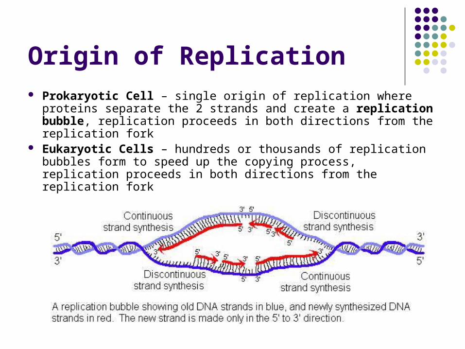
Origin of Replication Prokaryotic Cell – single origin of replication where proteins
separate the 2 strands and create a replication bubble, replication proceeds in both directions from the replication fork
Eukaryotic Cells – hundreds or thousands of replication bubbles form to speed up the copying process, replication proceeds in both directions from the replication fork

DNA Replication Watson and Crick proposed that the complimentary strand of DNA serves
as a template for which the other strand is built…experiments confirmed this 5 years later
DNA Replication: Process of Synthesizing new molecules of DNA1. Helicase breaks H-bonds and opens up the double helix forming
replication forks (point at which DNA separates)2. At the replication fork, DNA Polymerases continuously adds
complimentary nucleotides to exposed bases 3. Process continues until all DNA has been copied, end result is 2 new
molecules of DNA each identical to the original and composed of one new and one old strand
http://www.youtube.com/watch?v=L9RjNNfgaEQ

DNA Synthesis

Proofreading
DNA polymerase only moves to the next nucleotide if the previous nucleotide was a correct match
If mismatched, DNA Polymerase backs up, removes the mismatched nucleotides and replaces it with the correct one(s).
Only 1 error per 1 billion nucleotides! http://www.youtube.com/watch?v
=oJZH4lV3h6I&mode=related&search=

DNA Replication & Aging Every time DNA is copied,
DNA polymerase cannot complete replication on the ends
Eukaryotic DNA has a non-coding, repeating nucleotide sequence on the ends called telomeres that protects genes from being eroded over successive replications
It is believed that telomeres are directly related to the aging process